Review

Applications of the Amniotic Membrane in Vitreoretinal Surgery

1
Department of NEUROFARBA, Ophthalmology, University of Florence, Careggi, 50134 Florence, Italy
2
Department of Ophthalmology, Catholic University of Sacred Hearth-Foundation “Policlinico Universitario A. Gemelli”-IRCCS, 00168 Rome, Italy
*
Author to whom correspondence should be addressed.
J. Clin. Med. 2020, 9(8), 2675; https://doi.org/10.3390/jcm9082675
Submission received: 10 July 2020 / Revised: 14 August 2020 / Accepted: 15 August 2020 / Published: 18 August 2020
(This article belongs to the Special Issue Updates in Ocular Surgery)

Abstract

:
Recently, the use of the human amniotic membrane (hAM) has been extended to treat retinal disorders, such as macular holes that failed to close and retinal tears. The hAM has demonstrated the induction of a recovery process of the external retinal layers involving the external limiting membrane (ELM) and the ellipsoid zone (EZ). After that, the application of the hAM for retinal pathologies was extended to large macular tears, high myopic retinal detachment associated with MH, paravascular tears, serous macular detachment associated with optic pit, complicated retinal detachment and advanced age-related macular degeneration (AMD). The hAM has shown a potential in repairing retinal tissue through a regeneration process. This review aims to highlight the use of the hAM in various vitreo-retinal surgical fields, and to confront it with other cutting-edge surgical techniques used to treat challenging vitreo-retinal pathologies.

1. Introduction

The human amniotic membrane (hAM), also known as amnion, envelopes the human fetus. It is a semitransparent sheet and its thickness is about 0.02–0.05 mm. It is characterized by a epithelium, a basement membrane, a compact layer, a fibroblast layer and a spongy layer [1,2,3,4]. The amniotic membrane has been used in several fields of the eye surgery, such as the covering of corneal ulcers and the reconstruction of the conjunctiva. The amniotic membrane showed excellent anti-angiogenic and anti-microbial properties and poor immunogenicity [5,6,7,8,9,10,11,12]. The hAM can secrete a variety of cytokines including: transforming growth factor (TGFα, TGFβ-1, β-2, and β-3), basic fibroblast growth factor, epithelial growth factor, the hepatocyte growth factor and its receptor and the keratinocyte growth factor and its receptor [13,14,15,16,17,18].
In addition, the amniotic membrane can reconstruct surgically sectioned nerves in the animal model [19,20,21].
The hAM epithelium produces brain natriuretic peptides and hormones that release corticotropin, a cell proliferation promoter [22,23]. The hAM wraps, furthermore, if surgically implanted are a reservoir of neurotrophic factors, such as the nerve growth factor (NGF), brain-derived neurotrophic factor (BDNF), neurotrophin 3 (NT-3), glial cell-derived neurotrophic factor (GDNF) and the ciliary neurotrophic factor (CNTF) [24,25,26].
Some authors have also grown human retinal pigmented epithelium (RPE) cells, in vitro, on a layer of hAM. Here, the cells have proliferated effectively forming an organized mono layer [27]. Furthermore, Ohno-Matsui et al. [28] have shown that RPE cells grown on a hAM sheet do not only organize in a single layer, but secrete numerous growth factors. In addition, a hAM sheet was implanted into the sub-retinal space in pigs with a iatrogenic damage to the retinal pigmented epithelium. The hAM, after the implant, promoted a reduction of the iatrogenic damage and was covered by a mono-layer of epithelial cells [29].
The application of the hAM in vivo for vitreo-retinal pathologies is more recent, and began with macular holes that failed to close and retinal tears [30]. The hAM has demonstrated the induction of a recovery process involving the external retinal layers such as the external limiting membrane (ELM) and the ellipsoid zone (EZ).
The hAM application for retinal pathologies was extended to treat large macular tears [31], high myopic retinal detachment associated with MH [32], paravascular tears [33], serous macular detachment associated with optic pits [34], complicated retinal detachment [30] and advanced age related macular degeneration (AMD) [35].
This chapter resumes the application of the hAM in vitreoretinal pathologies.

2. Materials and Methods

This chapter highlights the use of the hAM in various vitreo-retinal surgical fields, and confronts it with other cutting-edge surgical techniques used to treat challenging vitreo-retinal pathologies.

Amniotic Membrane to Treat Macular Holes, Surgical Technique

We used a cryopreserved hAM delivered from the eye bank in Lucca, Italy or the eye bank in Mestre, Italy.
When we started performing the hAM transplant, the technique involved the intraocular trimming of the hAM plug to adjust its final dimensions. Then we found more accurate and less traumatic the use of a disposable cutaneous punch (Disposable Biopsy Punch, Kai Medical, Solingen, Germany) to regulate the size of the hAM patch. The structural pre-operative optical coherence tomography (OCT) was used to determine the MH diameter and therefore the hAM patch dimension.
We chose a 1 mm, 1.5 mm and 2 mm for macular holes, 3 and 5 mm for exudative and atrophic age-related macular degeneration and 1.5 mm optic pit cases. The plug should have the right diameter because an hAM disc that is larger than the basal diameter of the macular hole may corrugate and halt the anatomical and functional recovery. On the other side, a too short diameter of the AM plug will not induce the MH resolution.
The macular hole should be prepared to contain the hAM plug. This is performed using an illuminated PIK (Alcon PIK Endoilluminator, Alcon Fort Worth, TX, US), or a silicone tip backflush, to gradually detach the MH edges from the RPE. However, particular care must be taken not to damage the RPE.
At the beginning of our experience, we introduced the hAM plug through a 23-gauge trocar, inside the vitreous chamber, using 27-gauge forceps. Plugs of more than 3 mm disc diameter needed a 20-gauge sclerotomy for their intraocular insertion. Using this technique, we observed a hAM wrinkling in some cases, therefore we introduced the DSAEK-inspired insertion technique [36]. The hAM plug was contained in a plastic support and maintained at the entrance of the nasal trocar. Vitreoretinal forceps are passed from the temporal trocar to the nasal trocar and the hAM disc was dragged inside the vitreous chamber. This technique avoids the hAM excessive manipulation and therefore deterioration, which may result in a lower adhesion of the retina to the RPE.
The chorion layer of the hAM, once inside the vitreous chamber, should be recognized because it needs to face the RPE in order to obtain a firm adhesion. It is useful to know that the chorion tends to remain adherent to the vitreo-retinal forceps. Hence by re-grabbing the plug we can have the chorion layer in contact with the forceps. The hAM is inserted inside the MH using a PIK or a flexible laser tip and if the chorion layer will face the RPE the plug will remain adherent during the fluid-air exchange.
Rizzo et al. treated the first eight cases, affected by a MH that failed to close, using a hAM plug, and obtained a 100% closure rate and an average BCVA improvement from 20/800 to 20/50 [30] (Figure 1c,d).

3. Outcomes of the Human Amniotic Membrane Retinal Transplant and Comparison with Other Cutting Edge Vitreoretinal Surgical Techniques

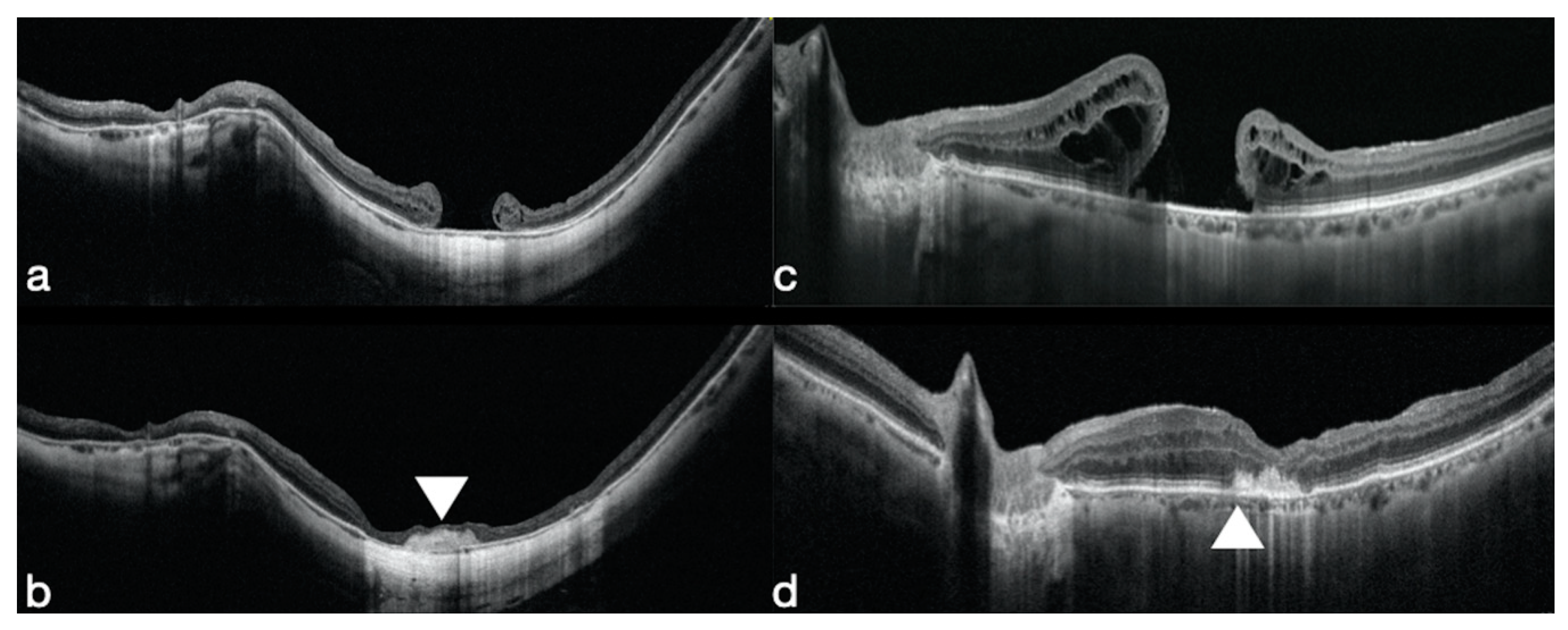
3.1. Macular Holes that Failed to Close

Macular holes that failed to close are still a challenge for vitreoretinal surgeons. The incidence varies from 0 to 39% [37,38], without internal limiting membrane (ILM) peeling and from 0% to 8.6% in eyes in which the ILM has been peeled off [39]. Many modern surgical options have been proposed to treat this condition such as the introduction of autologous fragments from the anterior lens capsule, autologous neuroretina or autologous ILM plugs into the macular hole [40,41,42,43,44]. These techniques aim to produce a sealing of the hole with variable visual acuity (VA) recovery.
The application of the human amniotic membrane (hAM) plug transplanted into the sub-retinal space to treat macular holes that failed to close, has shown an improvement of the anatomical and functional results.
Rizzo et al. treated the first eight cases, affected by a MH that failed to close, using a hAM plug, and obtained a 100% closure rate and a average BCVA improvement from 20/800 to 20/50 [30] (Figure 1c,d).
Morizane et al. treated 10 recurrent MHs using an autologous ILM transplant and obtained a 90% closure rate [40]. Grewal and Mahmoud in 2015 described the autologous neurosensory retinal transplantations for recurrent HMMH [42]. The same authors, in a multicenter study, treated 41 eyes of 41 patients affected by a recurrent MH, using autologous retinal transplant, and they reported a MH closure in 36 eyes out of 41 (87.8%) [45].
In high myopic eyes affected by an unresolved macular hole, which already underwent a PPV with ILM peeling, several techniques have been described, such as the use of anterior and posterior lens capsular flap transplantation [43], an autologous ILM transplantation [40], autologous neurosensory retinal free flap transplantation (ANRFF) with gas or silicone oil tamponade [42] and ILM or ANRFF transplantation with the addition of autologous blood [46,47,48].
Caporossi et al. treated 16 eyes of 16 patients affected by high myopic MH without retinal detachment, using an hAM plug. Fifteen out of 16 eyes closed with one surgery (93.75%) and 100% with two surgeries. The best corrected visual acuity improved from 20/200 to 20/100. Eleven patients (68.75%) had a BCVA improvement; four patients (25%) had no BCVA improvement and one patient had a VA worsening. Ten patients had 20% SF6, as post-operative tamponade, and six had air, no differences in the final BCVA were observed. (Figure 1a,b)
Morizane et al. [40] proposed to harvest extra-foveal ILM remnants and transplant them into the macular hole area. They reported, in a case series of 10 patients, two cases of recurrent HMMH treated with autologous ILM transplantation. They showed a 100% MH closure rate [40].
Chen published six recurrent HMMH of 20 patients treated with capsular lens fragments transplantation with a 100% closure rate and a final mean BCVA of 0.8 logMAR (20/125) [43]. Peng et al. treated 10 cases of HMMH with lens capsular flap transplantation (LCFT) and autologous whole blood application with only one unsuccessful case (10%), and a final BCVA of 1.34 logMAR (20/450) [44].
We may argue that the hAM plug transplant is an easier technique than the retina or the ILM free flap and Rizzo, Caporossi and colleagues did not find any sign of rejection. They reported an extremely high closure rate, and also reported interesting findings in terms of retinal regeneration.

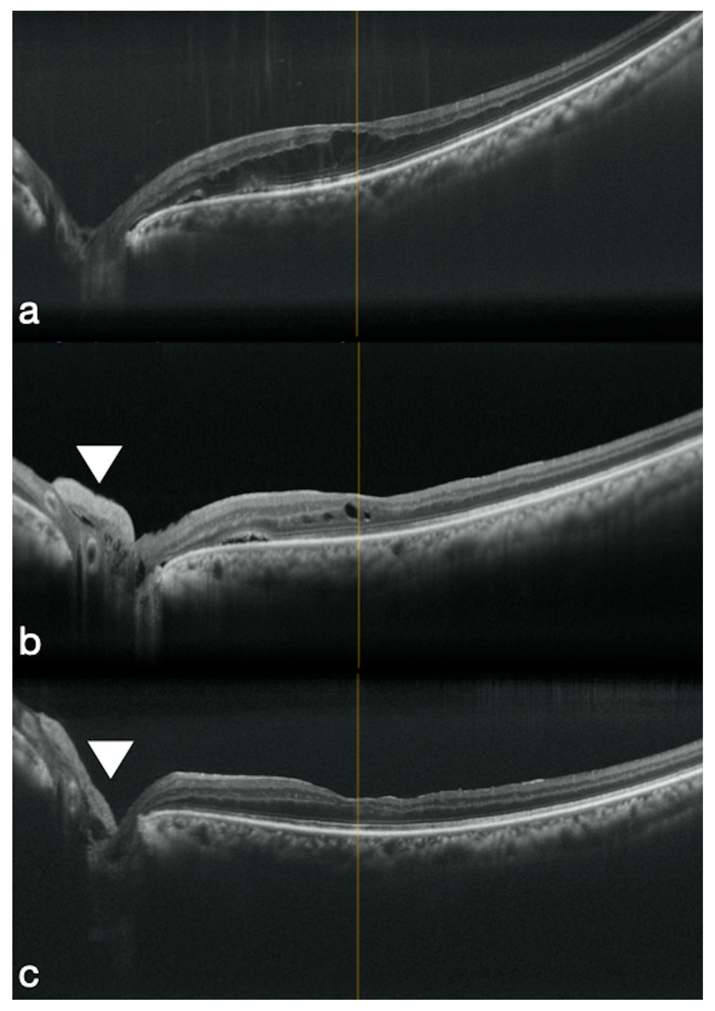
3.2. High Myopic Macular Hole Associated with Retinal Detachment

High myopic macular hole-induced retinal detachment (HMMH RD) often causes severe visual impairment; it occurs mainly in people with highly myopic eyes who have a posterior staphyloma [49,50].
In 2019 Caporossi et al. treated 10 patients affected by HMMH RD, using the hAM, with a 100% macular hole closure and retina reattachment rate. The BCVA improved from 20/1000 to 20/160. Five patients were tamponed with standard silicone oil and five with 16% C3F8. No statistically significant difference was reported between the two groups.
Lai et al. in their case series with autologous blood for HMMH RD enrolled 27 patients and obtained a final closure rate of 96% after one surgery [46].
In 2018 Chen et al. proposed the free ILM flap, and achieved a 100% closure rate and a retinal re-attachment in 13 patients [51].
The hAM transplant is easier than the ILM free flap technique, because there is no need to harvest ILM flaps in a myopic eye. This is relatively challenging, and there is also a risk of iatrogenic retinal tears. Conversely, the hAM, after the intraocular insertion, can be directly positioned into the macular hole. The results, presented by Prof Rizzo and Caporossi, are very encouraging and showed a 100% reattachment rate with also a promising integration of the hAM, that generated an external retinal layer reconstruction.

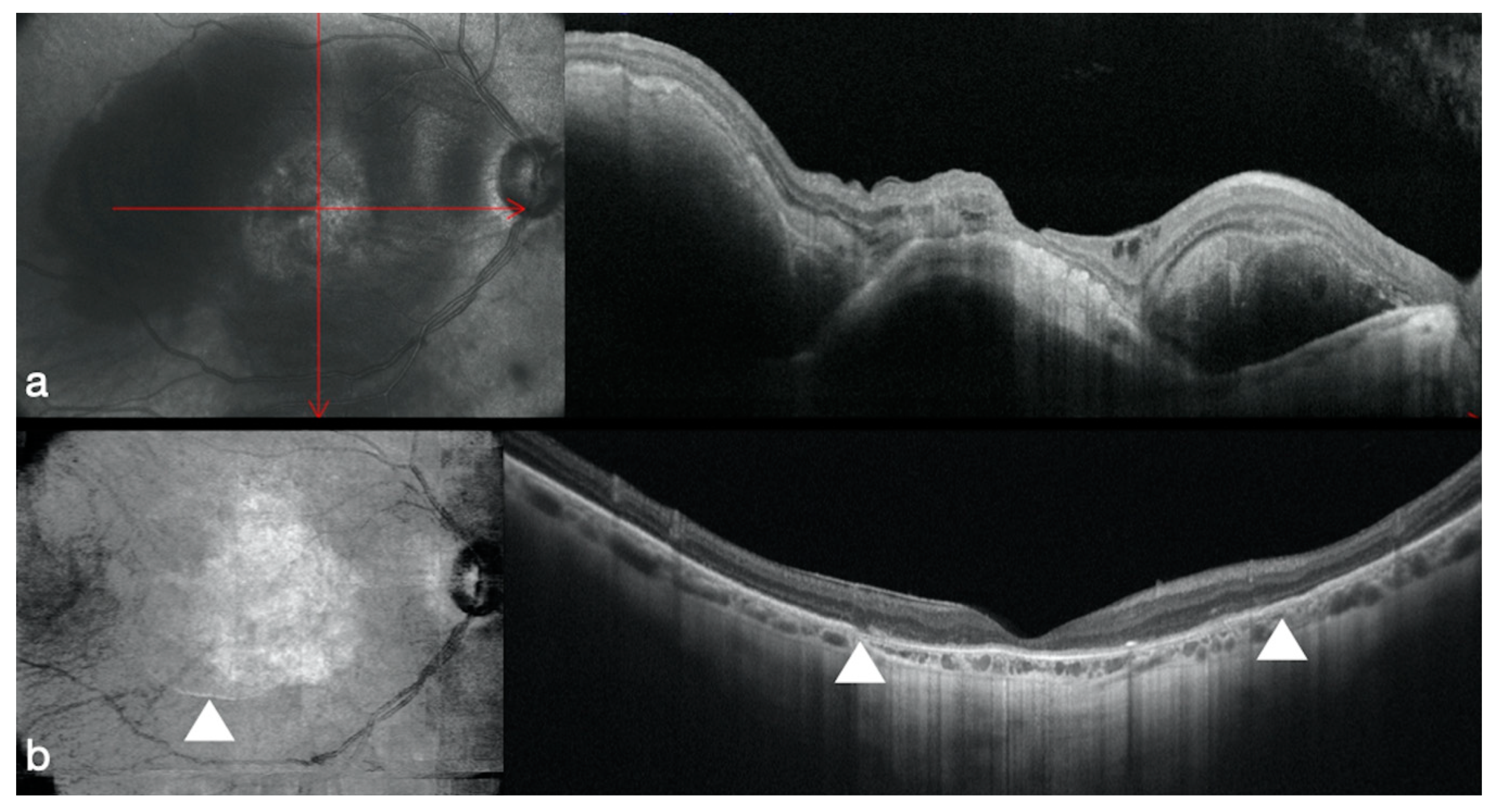
3.3. Complicated Retinal Detachment

In 2019, Rizzo et al. [30] treated six cases of recurrent retinal detachment complicated with PVR, using a hAM implant inside the retinal breaks. All the cases were tamponed using silicone oil, that was removed after 3 months. All the cases achieved a retinal reattachment with a BCVA improvement from 20/2000 to 20/125. In all the cases, the post-operative OCT showed the integration of the hAM patch inside the retinal breaks, and a thin layer of tissue growing over the patch. In all the cases no laser retinopexy was applied.
More recently Caporossi et al. [31] treated two cases of retinal detachment with a large macular tear using a hAM patch positioned under the retina, no laser and silicone oil as endotamponade were applied. In both cases the BCVA improved from light perception to 20/400 and a 3-month follow-up OCT showed a partial regrowth of the retinal layers in the macular area that covered the site of the previous retinal tear.
Retinal detachment after PPV for myopic foveoschisis is a rare complication, and is often associated with macular holes or posterior pole breaks. These breaks are often localized close to vessels, over areas of patchy chorioretinal atrophy. The endophotocoagulation is not possible because these are atrophic areas, and the trans-scleral cryotherapy is not indicated because these lesions are at the posterior pole [52,53,54]. Differently, an ILM plug transplantation can stimulate the posterior-pole retinal breaks closure even without a laser retinopexy [55]. In the case of extended ILM peeling during the first surgery, the ILM remnants are difficult to harvest, Caporossi et al. proposed a hAM patch implant inside the retinal breaks. They reported two cases out of two of retinal reattachment. The BCVA improved from 20/2000 to 20/250 [33].

3.4. Optic Disk Pit Associated Macular Detachment

Serous retinal detachment (ODP-M) is a frequent complication of the optic pit (25–75%) and it is associated with a bad visual acuity prognosis [56,57,58].
Rizzo et al. proposed three cases of ODP-M treated using an hAM plug positioned inside the optic disc pit. The sub-retinal fluid reduced during the first 6 months after surgery and the average visual acuity improved from 20/40 to 25/20 at the 6th month after surgery [34] (Figure 2).
García-Arumí et al. [59] treated 11 patients with PPV, laser and gas as endotamponade. They achieved the anatomical resolution and a significant visual improvement in all but 2 cases. Hirakata et al. [60] achieved the complete fluid resolution in 11 ODP-M patients treated with PPV, PVD induction and gas tamponade without laser. In 2012, Hirakata reported a series of seven patients treated with PPV and PVD induction without laser nor gas tamponade, the BCVA improved but in these cases the sub-retinal fluid did not resolve completely.
Recently, Mohammad et al. [61] proposed the use of the ILM autologous transplant into the optic pit and showed a faster sub-retinal fluid reabsorption than the standard technique.
The autologous scleral transplantation into the optic pit is an alternative technique, although the complete subretinal and intraretinal fluid reabsorption were achieved in 12 months after surgery in all the three cases treated using this technique [62].
Parag et al. [63] reported two cases operated on using a scleral autograft; they reported a complete sub-retinal fluid reabsorption after 1 year.
Rizzo and Caporossi et al. showed the usefulness and safeness of the hAM to treat ODP-M. The hAM, after the intraocular insertion, can be directly positioned into the optic disc pit. The results, presented by Prof Rizzo and Caporossi, are very encouraging, and showed a 100% resolution of the sub-retinal fluid.

3.5. Age-Related Macular Degeneration

End-stage age-related macular degeneration is the leading cause of blindness in the industrialized world and Prof Rizzo’s group for the first time introduced the hAM in the treatment of this degenerative chronic disease [35].
A 41-gauge needle was mounted on a 10-cc syringe containing balancing saline solution (BSS) which was injected in the sub-retinal space and three to four air-fluid exchanges were carried out to provoke a retinal detachment.
In the case of choroidal neovascular lesions (CNV), a 180° retinectomy was created in the temporal retinal periphery, in order to remove the neovascular tissue, which was aspirated using the vitrectomy probe. In the cases of geographic atrophy, the detachment was created at the posterior pole and the retina was delicately divided from the atrophic scar using a cannula or an endoilluminated PIC, introduced through a retinectomy, created near the vascular arcades.
A hAM sheet was positioned in the macular area with the chorion facing the RPE. The retina was re-attached over the hAM patch with the aid of perfluorocarbon, and an endolaser retinopexy was carried out at the edge of the retinectomy.
A perfluorocarbon-silicon oil direct exchange was performed at the end of the surgery. In all the cases a standard silicon oil 1000 cSt (Oxane 1300 Bausch + Lomb Incorporated, Rochester, NY, USA) was used as endotamponade. The patients maintained a face-down position for 7 days. The silicone oil was removed 4 months after the operation in all the cases without any complication.
In the hemorrhagic group (six patients), the initial mean BCVA was 2 logMAR (20/2000 Snellen), and the final mean BCVA was 1.1 logMAR (20/250 Snellen) p = 0.00042.
In the geographic atrophy group (five patients), mean BCVA improved from 1.84 logMAR (20/2000 Snellen), range 20/2000–20/800 (2–1.6 logMAR), to 1.26 logMAR (20/320 Snellen), range 20/2000–20/100 (2–0.7 logMAR) p = 0.0084.
Adaptive optics (AO) analyses were carried out over the retinal areas where the highest functionality was identified, using the microperimetry analysis. The images showed a photoreceptors presence at the hAM edges, which was confirmed by the built-in software.
The better post-operative sensibility responses were registered, using microperimetry, at the edge of the amniotic membrane patch in 10 out of 11 patients, the ones with the better visual recovery (>1.3 logMAR) (Figure 3).
In recent times, interesting new surgical treatments have been suggested for end-stage AMD. Autologous retinal and chorioretinal transplants have been proposed by Dr Parolini [64]. However, the autologous choroidal and retinal transplants are technically challenging procedures with a high risk of intra-operative and post-operative complications, such as retinal detachment with proliferative vitreoretinopathy. The hAM, on the other hand, is easier to insert into the subretinal space than a fragile autologous retinal or chorioretinal transplant. The excessive manipulation of retinal and chorioretinal flaps can lead to tissue disorganization with a tangible impact on the visual funtion. Prof. Da Cruz [65] created an embryonic stem cell-derived retinal pigment epithelium patch. The main problem is that the embryonic stem cell-derived retinal pigment epithelium patch was associated with systemic corticosteroid therapy because of the risk of immunogenic graft rejection. Conversely, the hAM sheet, implanted in the sub retinal space, did not allow any immunologic reaction. Prof Rizzo and Dr Caporossi think that the hAM may be a step forward in the management of the AMD.

4. Conclusions

In the recent years, the application of the hAM for retinal pathologies has been widely investigated.
The ability to be integrated in the retinal tissue without immunologic reactions or major postoperative complications, make it a useful basal membrane, and represent an interesting approach to different retinal pathologies.
Further prospective, larger, randomized controlled studies are needed to confirm our results.

Author Contributions

Conceptualization, S.R. and T.C.; methodology, D.B.; validation, S.R., T.C. and D.B.; data curation, R.T.; writing—original draft preparation, T.C. and D.B.; writing—review and editing, R.T., L.G. and L.O.; visualization, L.D.A. and L.D.L.; supervision, R.T. and B.P.; project administration, S.R. All authors have read and agreed to the published version of the manuscript.

Funding

This research received no external funding.

Conflicts of Interest

The authors declare no conflict of interest.

References

  1. Tosi, G.M.; Massaro-Giordano, M.; Caporossi, A.; Toti, P. Amniotic membrane transplantation in ocular surface disorders. J. Cell Physiol. 2005, 202, 849–851. [Google Scholar] [CrossRef] [PubMed]
  2. Tosi, G.M.; Traversi, C.; Schuerfeld, K.; Mittica, V.; Massaro-Giordano, M.; Tilanus, M.A.; Caporossi, A.; Toti, P. Amniotic membrane graft: Histopathological findings in five cases. J. Cell Physiol. 2005, 202, 852–857. [Google Scholar] [CrossRef] [PubMed]
  3. Kheirkhah, A.; Blanco, G.; Casas, V.; Hayashida, Y.; Raju, V.K.; Tseng, S.C. Surgical strategies for fornix reconstruction based on symblepharon severity. Am. J. Ophthalmol. 2008, 146, 266–275. [Google Scholar] [CrossRef] [PubMed]
  4. Li, W.; He, H.; Chen, Y.T.; Hayashida, Y.; Tseng, S.C. Reversal of myofibroblasts by amniotic membrane stromal extract. J. Cell Physiol. 2008, 215, 657–664. [Google Scholar] [CrossRef] [Green Version]
  5. Deihim, T.; Yazdanpanah, G.; Niknejad, H. Different Light Transmittance of Placental and Reflected Regions of Human Amniotic Membrane That Could Be Crucial for Corneal Tissue Engineering. Cornea 2016, 35, 997–1003. [Google Scholar] [CrossRef]
  6. Niknejad, H.; Yazdanpanah, G.; Kakavand, M. Extract of fetal membrane would inhibit thrombosis and hemolysis. Med. Hypotheses 2015, 85, 197–202. [Google Scholar] [CrossRef]
  7. Niknejad, H.; Yazdanpanah, G.; Ahmadiani, A. Induction of apoptosis, stimulation of cell-cycle arrest and inhibition of angiogenesis make human amnion-derived cells promising sources for cell therapy of cancer. Cell Tissue Res. 2016, 363, 599–608. [Google Scholar] [CrossRef]
  8. Tehrani, F.A.; Ahmadiani, A.; Niknejad, H. The effects of preservation procedures on antibacterial property of amniotic membrane. Cryobiology 2013, 67, 293–298. [Google Scholar] [CrossRef]
  9. Tseng, S.C. Amniotic membrane transplantation for ocular surface reconstruction. Biosci. Rep. 2001, 21, 481–489. [Google Scholar] [CrossRef]
  10. Tseng, S.C.; Espana, E.M.; Kawakita, T.; Di Pascuale, M.A.; Li, W.; He, H.; Liu, T.S.; Cho, T.H.; Gao, Y.Y.; Yeh, L.K.; et al. How does amniotic membrane work? Ocul. Surf. 2004, 2, 177–187. [Google Scholar] [CrossRef]
  11. Waked, N.; El-Kazzi, V. Amniotic membrane utilization in ophthalmological surgical procedures. J. Med. Liban. 2005, 53, 39–44. [Google Scholar]
  12. Cheng, A.M.; Yin, H.Y.; Chen, R.; Tighe, S.; Sheha, H.; Zhao, D.; Casas, V.; Tseng, S.C. Restoration of Fornix Tear Reservoir in Conjunctivochalasis with Fornix Reconstruction. Cornea 2016, 35, 736–740. [Google Scholar] [CrossRef] [PubMed] [Green Version]
  13. Olaya, C.M.; Michael, F.; Fabian, G.; Silva, J.L.; Bernal, J.E. Role of VEGF in the differential growth between the fetal and placental ends of the umbilical cord. J. Neonatal Perinat. Med. 2018. [Google Scholar] [CrossRef]
  14. Heerema-McKenney, A. Defense and infection of the human placenta. APMIS 2018, 126, 570–588. [Google Scholar] [CrossRef] [Green Version]
  15. Sane, M.S.; Misra, N.; Quintanar, N.M.; Jones, C.D.; Mustafi, S.B. Biochemical characterization of pure dehydrated binate amniotic membrane: Role of cytokines in the spotlight. Regen. Med. 2018, 13, 689–703. [Google Scholar] [CrossRef] [PubMed] [Green Version]
  16. Otani, T.; Ochiai, D.; Masuda, H.; Abe, Y.; Fukutake, M.; Matsumoto, T.; Miyakoshi, K.; Tanaka, M. The neurorestorative effect of human amniotic fluid stem cells on the chronic phase of neonatal hypoxic-ischemic encephalopathy in mice. Pediatr. Res. 2018. [Google Scholar] [CrossRef] [PubMed]
  17. Kaur, J.; Bathla, S.C. Regenerative potential of autologous platelet-rich fibrin with and without amnion membrane in the treatment of Grade-II furcation defects: A clinicoradiographic study. J. Indian Soc. Periodontol. 2018, 22, 235–242. [Google Scholar] [CrossRef]
  18. Murri, M.S.; Moshirfar, M.; Birdsong, O.C.; Ronquillo, Y.C.; Ding, Y.; Hoopes, P.C. Amniotic membrane extract and eye drops: A review of literature and clinical application. Clin. Ophthalmol. 2018, 12, 1105–1112. [Google Scholar] [CrossRef] [Green Version]
  19. Meng, H.; Li, M.; You, F.; Du, J.; Luo, Z. Assessment of processed human amniotic membrane as a protective barrier in rat model of sciatic nerve injury. Neurosci. Lett. 2011, 496, 48–53. [Google Scholar] [CrossRef]
  20. Sadraie, S.H.; Parivar, K.; Arabi, F.; Moattari, M.; Kaka, G.; Mansouri, K. Study of Transected Sciatic Nerve Repair by Amniotic Membrane with Betamethasone in Adult Albino Wistar Rats. Arch. Iran. Med. 2016, 19, 612–617. [Google Scholar]
  21. Riccio, M.; Marchesini, A.; Pugliese, P.; De Francesco, F. Nerve repair and regeneration: Biological tubulization limits and future perspectives. J. Cell Physiol. 2019, 234, 3362–3375. [Google Scholar] [CrossRef] [PubMed]
  22. Gupta, A.; Kedige, S.D.; Jain, K. Amnion and Chorion Membranes: Potential Stem Cell Reservoir with Wide Applications in Periodontics. Int. J. Biomater. 2015, 2015, 274082. [Google Scholar] [CrossRef] [PubMed] [Green Version]
  23. Gupta, K.; Venkatesan, B.; Chandra, T.; Rajeswari, K.; Devi, T.K. Amniotic band syndrome with sacral agenesis and umbilical cord entrapment: A case report emphasizing the value of evaluation of umbilical cord. J. Radiol. Case Rep. 2015, 9, 12–19. [Google Scholar] [CrossRef] [PubMed]
  24. Marcus, A.J.; Woodbury, D. Fetal stem cells from extra-embryonic tissues: Do not discard. J. Cell Mol. Med. 2008, 12, 730–742. [Google Scholar] [CrossRef] [Green Version]
  25. Marcus, A.J.; Coyne, T.M.; Black, I.B.; Woodbury, D. Fate of amnion-derived stem cells transplanted to the fetal rat brain: Migration, survival and differentiation. J. Cell Mol. Med. 2008, 12, 1256–1264. [Google Scholar] [CrossRef] [Green Version]
  26. Marcus, A.J.; Coyne, T.M.; Rauch, J.; Woodbury, D.; Black, I.B. Isolation, characterization, and differentiation of stem cells derived from the rat amniotic membrane. Differentiation 2008, 76, 130–144. [Google Scholar] [CrossRef]
  27. Capeans, C.; Pineiro, A.; Pardo, M.; Sueiro-Lopez, C.; Blanco, M.J.; Dominguez, F.; Sanchez-Salorio, M. Amniotic membrane as support for human retinal pigment epithelium (RPE) cell growth. Acta Ophthalmol. Scand. 2003, 81, 271–277. [Google Scholar] [CrossRef]
  28. Ohno-Matsui, K.; Ichinose, S.; Nakahama, K.; Yoshida, T.; Kojima, A.; Mochizuki, M.; Morita, I. The effects of amniotic membrane on retinal pigment epithelial cell differentiation. Mol. Vis. 2005, 11, 1–10. [Google Scholar]
  29. Kiilgaard, J.F.; Scherfig, E.; Prause, J.U.; la Cour, M. Transplantation of amniotic membrane to the subretinal space in pigs. Stem Cells Int. 2012, 2012, 716968. [Google Scholar] [CrossRef]
  30. Rizzo, S.; Caporossi, T.; Tartaro, R.; Finocchio, L.; Franco, F.; Barca, F.; Giansanti, F. A Human Amniotic Membrane Plug to Promote Retinal Breaks Repair and Recurrent Macular Hole Closure. Retina 2019, 39 (Suppl. 1), S95–S103. [Google Scholar] [CrossRef]
  31. Caporossi, T.; Tartaro, R.; De Angelis, L.; Pacini, B.; Rizzo, S. A human amniotic membrane plug to repair retinal detachment associated with large macular tear. Acta Ophthalmol. 2019, 97, 821–823. [Google Scholar] [CrossRef] [PubMed]
  32. Caporossi, T.; De Angelis, L.; Pacini, B.; Tartaro, R.; Finocchio, L.; Barca, F.; Rizzo, S. A human Amniotic Membrane plug to manage high myopic macular hole associated with retinal detachment. Acta Ophthalmol. 2020, 98, e252–e256. [Google Scholar] [CrossRef] [PubMed]
  33. Caporossi, T.; De Angelis, L.; Pacini, B.; Rizzo, S. Amniotic membrane for retinal detachment due to paravascular retinal breaks over patchy chorioretinal atrophy in pathologic myopia. Eur. J. Ophthalmol. 2020, 30, 392–395. [Google Scholar] [CrossRef] [PubMed]
  34. Rizzo, S.; Caporossi, T.; Pacini, B.; De Angelis, L.; De Vitto, M.L.; Gainsanti, F. Management of Optic Disk Pit-associated Macular Detachment with Human Amniotic Membrane Patch. Retina 2020. [Google Scholar] [CrossRef] [PubMed]
  35. Rizzo, S.; Caporossi, T.; Tartaro, R.; Finocchio, L.; Pacini, B.; Bacherini, D.; Virgili, G. Human Amniotic Membrane plug to restore Age related Macular Degeneration photoreceptors’ damage. Ophthalmol. Retin. 2020. [Google Scholar] [CrossRef]
  36. Caporossi, T.; Tartaro, R.; Pacini, B.; De Angelis, L.; Rizzo, S. DSAEK-derived glider technique to introduce human amniotic membrane patch through small-gauge trocar for retinal pathologies. Acta Ophthalmol. 2020, 98, e526–e527. [Google Scholar] [CrossRef]
  37. Imai, M.; Iijima, H.; Gotoh, T.; Tsukahara, S. Optical coherence tomography of successfully repaired idiopathic macular holes. Am. J. Ophthalmol. 1999, 128, 621–627. [Google Scholar] [CrossRef]
  38. Kang, S.W.; Ahn, K.; Ham, D.I. Types of macular hole closure and their clinical implications. Br. J. Ophthalmol. 2003, 87, 1015–1019. [Google Scholar] [CrossRef]
  39. Kumagai, K.; Furukawa, M.; Ogino, N.; Larson, E. Incidence and factors related to macular hole reopening. Am. J. Ophthalmol. 2010, 149, 127–132. [Google Scholar] [CrossRef]
  40. Morizane, Y.; Shiraga, F.; Kimura, S.; Hosokawa, M.; Shiode, Y.; Kawata, T.; Hosogi, M.; Shirakata, Y.; Okanouchi, T. Autologous transplantation of the internal limiting membrane for refractory macular holes. Am. J. Ophthalmol. 2014, 157, 861–869. [Google Scholar] [CrossRef]
  41. Lee, S.M.; Kwon, H.J.; Park, S.W.; Lee, J.E.; Byon, I.S. Microstructural changes in the fovea following autologous internal limiting membrane transplantation surgery for large macular holes. Acta Ophthalmol. 2018, 96, e406–e408. [Google Scholar] [CrossRef] [PubMed] [Green Version]
  42. Grewal, D.S.; Mahmoud, T.H. Autologous Neurosensory Retinal Free Flap for Closure of Refractory Myopic Macular Holes. JAMA Ophthalmol. 2016, 134, 229–230. [Google Scholar] [CrossRef] [PubMed] [Green Version]
  43. Chen, S.N.; Yang, C.M. Lens Capsular Flap Transplantation in the Management of Refractory Macular Hole from Multiple Etiologies. Retina 2016, 36, 163–170. [Google Scholar] [CrossRef] [PubMed]
  44. Peng, J.; Chen, C.; Jin, H.; Zhang, H.; Zhao, P. Autologous Lens Capsular Flap Transplantation Combined with Autologous Blood Application in the Management of Refractory Macular Hole. Retina 2017. [Google Scholar] [CrossRef]
  45. Grewal, D.S.; Charles, S.; Parolini, B.; Kadonosono, K.; Mahmoud, T.H. Autologous Retinal Transplant for Refractory Macular Holes: Multicenter International Collaborative Study Group. Ophthalmology 2019, 126, 1399–1408. [Google Scholar] [CrossRef]
  46. Lai, C.C.; Chen, Y.P.; Wang, N.K.; Chuang, L.H.; Liu, L.; Chen, K.J.; Hwang, Y.S.; Wu, W.C.; Chen, T.L. Vitrectomy with Internal Limiting Membrane Repositioning and Autologous Blood for Macular Hole Retinal Detachment in Highly Myopic Eyes. Ophthalmology 2015, 122, 1889–1898. [Google Scholar] [CrossRef]
  47. Wu, A.L.; Chuang, L.H.; Wang, N.K.; Chen, K.J.; Liu, L.; Yeung, L.; Chen, T.L.; Hwang, Y.S.; Wu, W.C.; Lai, C.C. Refractory macular hole repaired by autologous retinal graft and blood clot. BMC Ophthalmol. 2018, 18, 213. [Google Scholar] [CrossRef] [Green Version]
  48. Liu, P.K.; Chang, Y.C.; Wu, W.C. Management of refractory macular hole with blood and gas-assisted autologous neurosensory retinal free flap transplantation: A case report. BMC Ophthalmol. 2018, 18, 230. [Google Scholar] [CrossRef] [Green Version]
  49. Lim, L.S.; Tsai, A.; Wong, D.; Wong, E.; Yeo, I.; Loh, B.K.; Ang, C.L.; Ong, S.G.; Lee, S.Y. Prognostic factor analysis of vitrectomy for retinal detachment associated with myopic macular holes. Ophthalmology 2014, 121, 305–310. [Google Scholar] [CrossRef]
  50. Coppola, M.; Rabiolo, A.; Cicinelli, M.V.; Querques, G.; Bandello, F. Vitrectomy in high myopia: A narrative review. Int. J. Retin. Vitr. 2017, 3, 37. [Google Scholar] [CrossRef] [Green Version]
  51. Chen, S.N.; Hsieh, Y.T.; Yang, C.M. Multiple Free Internal Limiting Membrane Flap Insertion in the Treatment of Macular Hole-Associated Retinal Detachment in High Myopia. Ophthalmologica 2018, 240, 143–149. [Google Scholar] [CrossRef] [PubMed]
  52. Kuriyama, S.; Matsumura, M.; Harada, T.; Ishigooka, H.; Ogino, N. Surgical techniques and reattachment rates in retinal detachment due to macular hole. Arch Ophthalmol. 1990, 108, 1559–1561. [Google Scholar] [CrossRef] [PubMed]
  53. Matsumura, M.; Kuriyama, S.; Harada, T.; Ishigooka, H.; Ogino, N. Surgical techniques and visual prognosis in retinal detachment due to macular hole. Ophthalmologica 1992, 204, 122–133. [Google Scholar] [CrossRef] [PubMed]
  54. Bovey, E.H.; Gonvers, M. Transscleral diathermy: An additional tool in the management of retinal detachment due to posterior breaks in highly myopic eyes. Retina 1999, 19, 489–494. [Google Scholar] [CrossRef] [PubMed]
  55. Rizzo, S.; Tartaro, R.; Barca, F.; Bacherini, D.; Franco, F.; Caporossi, T. Autologous Internal Limiting Membrane Fragment Transplantation for Rhegmatogenous Retinal Detachment Due to Paravascular or Juxtapapillary Retinal Breaks Over Patchy Chorioretinal Atrophy in Pathologic Myopia. Retina 2018, 38, 198–202. [Google Scholar] [CrossRef]
  56. Postel, E.A.; Pulido, J.S.; McNamara, J.A.; Johnson, M.W. The etiology and treatment of macular detachment associated with optic nerve pits and related anomalies. Trans. Am. Ophthalmol. Soc. 1998, 96, 73–88; discussion 88–93. [Google Scholar]
  57. Brown, G.C.; Augsburger, J.J. Congenital pits of the optic nerve head and retinochoroidal colobomas. J. Can. Ophthalmol. 1980, 15, 144–146. [Google Scholar]
  58. Bonnet, M. Serous macular detachment associated with optic nerve pits. Graefes. Arch. Clin. Exp. Ophthalmol. 1991, 229, 526–532. [Google Scholar] [CrossRef]
  59. Garcia-Arumi, J.; Guraya, B.C.; Espax, A.B.; Castillo, V.M.; Ramsay, L.S.; Motta, R.M. Optical coherence tomography in optic pit maculopathy managed with vitrectomy-laser-gas. Graefes. Arch. Clin. Exp. Ophthalmol. 2004, 242, 819–826. [Google Scholar] [CrossRef]
  60. Hirakata, A.; Okada, A.A.; Hida, T. Long-term results of vitrectomy without laser treatment for macular detachment associated with an optic disc pit. Ophthalmology 2005, 112, 1430–1435. [Google Scholar] [CrossRef]
  61. Mohammed, O.A.; Pai, A. Inverted autologous internal limiting membrane for management of optic disc pit with macular detachment. Middle East Afr. J. Ophthalmol. 2013, 20, 357–359. [Google Scholar] [CrossRef] [PubMed]
  62. Travassos, A.S.; Regadas, I.; Alfaiate, M.; Silva, E.D.; Proenca, R.; Travassos, A. Optic pit: Novel surgical management of complicated cases. Retina 2013, 33, 1708–1714. [Google Scholar] [CrossRef]
  63. Shah, P.K.; Karandikar, S.S.; Narendran, V. Scleral Autograft for Optic Nerve Head Pit Associated Chronic Maculopathy. Ophthalmic. Surg. Lasers Imaging Retin. 2017, 48, 251–254. [Google Scholar] [CrossRef]
  64. Parolini, B.; Grewal, D.S.; Pinackatt, S.J.; Baldi, A.; Di Salvatore, A.; Besozzi, G.; Finzi, A.; Cardillo, D.; Mahmoud, T.H. Combined Autologous Transplantation of Neurosensory Retina, Retinal Pigment Epithelium, and Choroid Free Grafts. Retina 2018, 38 (Suppl. 1), S12–S22. [Google Scholar] [CrossRef] [PubMed]
  65. Da Cruz, L.; Fynes, K.; Georgiadis, O.; Kerby, J.; Luo, Y.H.; Ahmado, A.; Vernon, A.; Daniels, J.T.; Nommiste, B.; Hasan, S.M.; et al. Phase 1 clinical study of an embryonic stem cell-derived retinal pigment epithelium patch in age-related macular degeneration. Nat. Biotechnol. 2018, 36, 328–337. [Google Scholar] [CrossRef] [PubMed] [Green Version]
Figure 1. (a–c) preoperative SD-OCT (spectral domain optical coherence tomography) showing a failed high myopic macular hole and a failed non-high myopic macular hole. (b–d) 6-months OCT (optical coherence tomography) shows in both cases the human amniotic membrane (hAM) patch well positioned under the retina (white arrows) and the macular hole closed. A partial differentiation of the external retinal layers such as the external limiting membrane and the ellipsoid zone is evident (d).
Jcm 09 02675 g001
Figure 2. Preoperative OCT shows an optic disc pit maculopathy (a). 1 and 3 months postoperative OCT shows the hAM patch positioned inside the optic disc area (white arrows) and the macular schisis completely solved (b,c).
Jcm 09 02675 g002
Figure 3. Preoperative OCT showing a sub macular hemorrhage involving the posterior pole (a) 6 months postoperative OCT shows a sub-retinal hAM patch positioned at the posterior pole (white arrows). We can recognize the outer retinal layers such as the external limiting membrane and ellipsoid zone (b).
Jcm 09 02675 g003

