Maternal and Neonatal Hair Cortisol Levels Are Associated with Infant Neurodevelopment at Six Months of Age

 , , , ,
Abstract
:1. Introduction
2. Methods
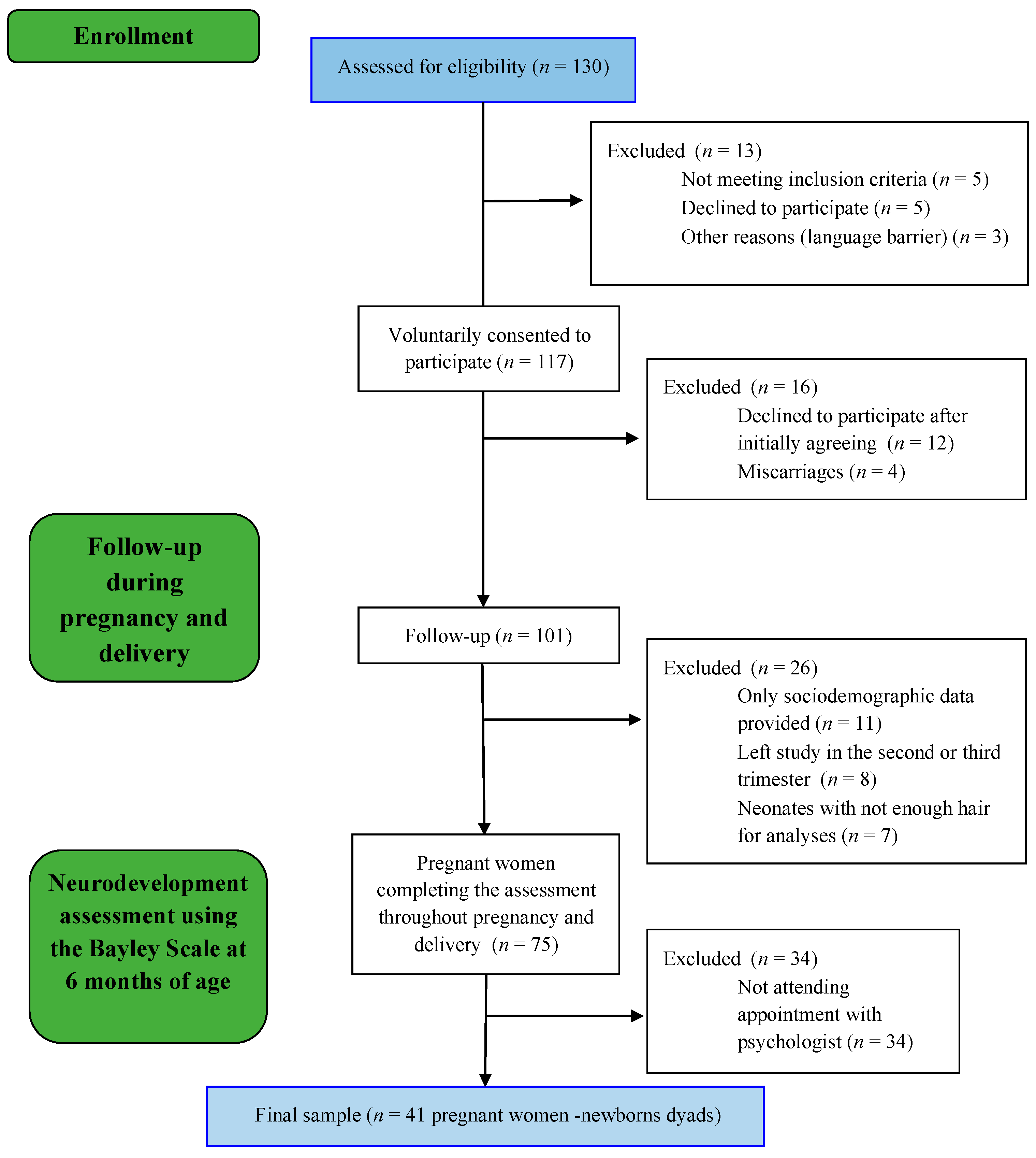
2.1. Participants
2.2. Procedure
2.3. Predictor Variables
Maternal, Newborn, and Infant Hair Cortisol Assay
2.4. Outcome Variables
2.4.1. Infant Neurodevelopment
2.4.2. Medical, Obstetric, and Sociodemographic Information
2.5. Statistical Analyses
3. Results
3.1. Descriptive Sample Characteristics
3.2. Pearson’s Bivariate Correlations between the Predictors
3.3. Maternal and Infant Hair Cortisol Levels
3.4. Cognitive, Language, and Motor Deveopment among Infants
3.5. Hierarchical Linear Regression Analyses for Maternal Hair Cortisol Levels
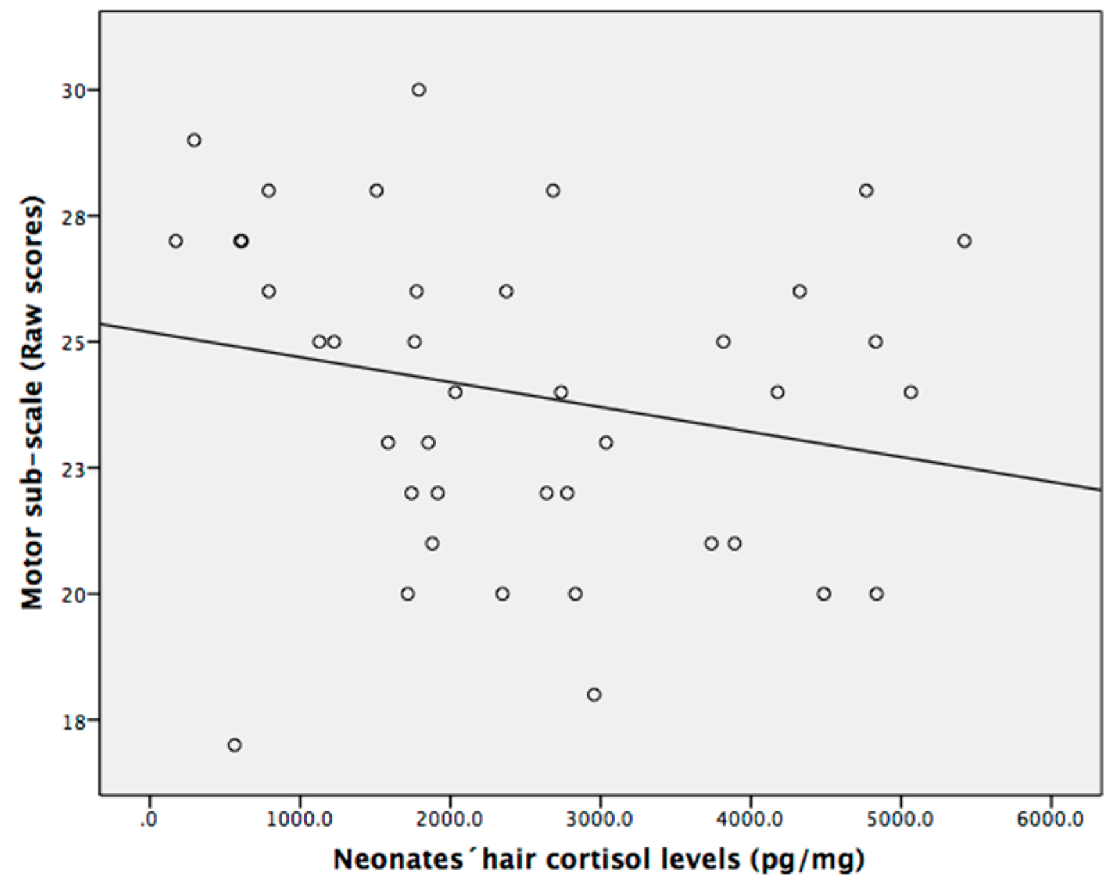
3.6. Hierarchical Linear Regression Analyses for Neonatal Hair Cortisol Levels
4. Discussion
5. Conclusions
Supplementary Materials
Author Contributions
Funding
Acknowledgments
Conflicts of Interest
References
- Herba, C.M.; Glover, V.; Ramchandani, P.G.; Rondon, M.B. Maternal depression and mental health in early childhood: An examination of underlying mechanisms in low-income and middle-income countries. Lancet Psychiatry 2016, 3, 983–992. [Google Scholar] [CrossRef]
- Fox, M.; Thayer, Z.M.; Ramos, I.F.; Meskal, S.J.; Wadhwa, P.D. Prenatal and Postnatal Mother-to-Child Transmission of Acculturation’s Health Effects in Hispanic Americans. J. Women’s Health 2018, 27, 1054–1063. [Google Scholar] [CrossRef] [PubMed]
- Rakers, F.; Rupprecht, S.; Dreiling, M.; Bergmeier, C.; Witte, O.W.; Schwab, M. Transfer of maternal psychosocial stress to the fetus. Neurosci. Biobehav. Rev. 2017. [Google Scholar] [CrossRef] [PubMed]
- La Marca-Ghaemmaghami, P.; Ehlert, U. Stress during pregnancy. Eur. Psychol. 2015, 20, 102–119. [Google Scholar] [CrossRef]
- Karlén, J.; Frostell, A.; Theodorsson, E.; Faresjö, T.; Ludvigsson, J. Maternal influence on child HPA axis: A prospective study of cortisol levels in hair. Pediatrics 2013, 132, e1333–e1340. [Google Scholar] [CrossRef]
- Caparros-Gonzalez, R.A.; Romero-Gonzalez, B.; Strivens-Vilchez, H.; Gonzalez-Perez, R.; Martinez-Augustin, O.; Peralta-Ramirez, M.I. Hair cortisol levels, psychological stress and psychopathological symptoms as predictors of postpartum depression. PLoS ONE 2017, 12, e0182817. [Google Scholar] [CrossRef]
- Matvienko-Sikar, K.; Dockray, S. Effects of a novel positive psychological intervention on prenatal stress and well-being: A pilot randomised controlled trial. Women Birth 2017, 30, e111–e118. [Google Scholar] [CrossRef]
- Sandman, C.A.; Glynn, L.M.; Davis, E.P. Neurobehavioral consequences of fetal exposure to gestational stress. In Fetal Development; Reissland, N., Kisilevsky, B.S., Eds.; Springer: Cham, Switzerland, 2016; pp. 229–265. [Google Scholar]
- Bergman, K.; Sarkar, P.; Glover, V.; O’Connor, T.G. Maternal prenatal cortisol and infant cognitive development: Moderation by infant-mother attachment. Biol. Psychiatry 2010, 67, 1026–1032. [Google Scholar] [CrossRef]
- De Weerth, C.; Buitelaar, J.K.; Beijers, R. Infant cortisol and behavioral habituation to weekly maternal separations: Links with maternal prenatal cortisol and psychosocial stress. Psychoneuroendocrinology 2013, 38, 2863–2874. [Google Scholar] [CrossRef]
- Jung, C.; Ho, J.T.; Torpy, D.J.; Doogue, M.; Lewis, J.G.; Czajko, R.J.; Inder, W.J. A longitudinal study of plasma and urinary cortisol in pregnancy and postpartum. J. Clin. Endocr. Metab. 2011, 96, 1533–1540. [Google Scholar] [CrossRef]
- Stalder, T.; Kirschbaum, C. Analysis of cortisol in hair—State of the art and future directions. Brain Behav. Immun. 2012, 26, 1019–1029. [Google Scholar] [CrossRef] [PubMed]
- Caparros-Gonzalez, R.A.; García-García, I.; Mariñas-Lirola, J.C.; Peralta-Ramírez, M.I. GESTASTRES cohort study protocol on the effects of stress during pregnancy by measuring the cortisol in women’s and newborn’s hair. Rev. Esp. Salud Publica 2018, 92, e201804027. [Google Scholar] [PubMed]
- Jahangard, L.; Mikoteit, T.; Bahiraei, S.; Zamanibonab, M.; Haghighi, M.; Sadeghi Bahmani, D.; Brand, S. Prenatal and Postnatal Hair Steroid Levels Predict Post-Partum Depression 12 Weeks after Delivery. J. Clin. Med. 2019, 8, 1290. [Google Scholar] [CrossRef] [PubMed]
- Wosu, A.C.; Valdimarsdóttir, U.; Shields, A.E.; Williams, D.R.; Williams, M.A. Correlates of cortisol in human hair: Implications for epidemiologic studies on health effects of chronic stress. Ann. Epidemiol. 2013, 23, 797–811. [Google Scholar] [CrossRef]
- Glover, V. Prenatal stress and its effects on the neurodevelopment of the fetus and the child: The mediating role of the placenta. Placenta 2017, 57, 232. [Google Scholar] [CrossRef]
- Huizink, A.C.; Robles de Medina, P.G.; Mulder, E.J.H.; Visser, G.H.A.; Buitelaar, J.K. Stress during pregnancy is associated with developmental outcome in infancy. J. Psychol. Psyc. 2003, 44, 810–818. [Google Scholar] [CrossRef]
- Zhu, P.; Sun, M.S.; Hao, J.H.; Chen, Y.J.; Jiang, X.M.; Tao, R.X.; Huang, K.; Tao, F.B. Does prenatal maternal stress impair cognitive development and alter temperament characteristics in toddlers with healthy birth outcomes? Dev. Med. Child Neurol. 2014, 56, 283–289. [Google Scholar] [CrossRef]
- Buitelaar, J.K.; Huizink, A.C.; Mulder, E.J.; de Medina, P.G.R.; Visser, G.H.A. Prenatal stress and cognitive development and temperament in infants. Neurobiol. Aging 2003, 24, S53–S60. [Google Scholar] [CrossRef]
- Bolten, M.I.; Wurmser, H.; Buske-Kirschbaum, A.; Papoušek, M.; Pirke, K.M.; Hellhammer, D. Cortisol levels in pregnancy as a psychobiological predictor for birth weight. Arch. Women’s Ment. Health 2010, 14, 33–41. [Google Scholar] [CrossRef]
- Davis, E.P.; Sandman, C.A. The timing of prenatal exposure to maternal cortisol and psychosocial stress is associated with human infant cognitive development. Child Dev. 2010, 81, 131–148. [Google Scholar] [CrossRef]
- Castiglioni, V.; Faedo, A.; Onorati, M.; Bocchi, V.D.; Li, Z.; Iennaco, R.; Vuono, R.; Bulfamante, G.P.; Muzio, L.; Martino, G.; et al. Dynamic and cell-specific DACH1 expression in human neocortical and striatal development. Cereb. Cortex 2018, 29, 2115–2124. [Google Scholar] [CrossRef] [PubMed]
- Glover, V.; O’Donnell, K.J.; O’Connor, T.G.; Fisher, J. Prenatal maternal stress, fetal programming, and mechanisms underlying later psychopathology—A global perspective. Dev. Psychopathol. 2018, 30, 843–854. [Google Scholar] [CrossRef] [PubMed]
- Ghaemmaghami, P.; Dainese, S.M.; La Marca, R.; Zimmermann, R.; Ehlert, U. The association between the acute psychobiological stress response in second trimester pregnant women, amniotic fluid glucocorticoids, and neonatal birth outcome. Dev. Psychobiol. 2014, 56, 734–747. [Google Scholar] [CrossRef] [PubMed]
- Sauvé, B.; Koren, G.; Walsh, G.; Tokmakejian, S.; Van Uum, S.H.M. Measurement of cortisol in human hair as a biomarker of systemic exposure. Clin. Investig. Med. 2007, 30, 183–191. [Google Scholar] [CrossRef]
- Chen, Z.; Li, J.; Zhang, J.; Xing, X.; Gao, W.; Lu, Z.; Deng, H. Simultaneous determination of hair cortisol, cortisone and DHEAS with liquid chromatography–electrospray ionization-tandem mass spectrometry in negative mode. J. Chromatogr. B 2013, 929, 187–194. [Google Scholar] [CrossRef]
- Meyer, J.; Novak, M.; Hamel, A.; Rosenberg, K. Extraction and analysis of cortisol from human and monkey hair. J. Vis. Exp. 2014, 83, e50882. [Google Scholar] [CrossRef]
- Russell, E.; Kirschbaum, C.; Laudenslager, M.L.; Stalder, T.; de Rijke, Y.; van Rossum, E.F.; van Uum, S.; Koren, G. Toward standardization of hair cortisol measurement: Results of the first international interlaboratory round robin. Ther. Drug Monit. 2015, 37, 71–75. [Google Scholar] [CrossRef]
- Moss, K.M.; Simcock, G.; Cobham, V.; Kildea, S.; Eigbeili, G.; Laplante, D.P.; King, S. A potential psychological mechanism linking disaster-related prenatal maternal stress with child cognitive and motor development at 16 months: The QF2011 Queensland Flood Study. Dev. Psychol. 2017, 53, 629. [Google Scholar] [CrossRef]
- Tamayo-Ortiz, M.T.; Téllez-Rojo, M.M.; Trejo-Valdivia, B.; Schanaas, L.; Osorio-Valencia, E.; Coull, B.; Bellinger, D.; Wrigth, R.J.; Wright, R.O. Maternal stress modifies the effect of exposure to lead during pregnancy and 24-month old children’s neurodevelopment. Environ. Int. 2017, 98, 191–197. [Google Scholar] [CrossRef]
- Muñoz-Rocha, T.V.; Tamayo, Y.; Ortiz, M.; Romero, M.; Pantic, I.; Schanaas, L.; Bellinger, D.; Claus-henn, B.; Wright, R.; Wright, R.O.; et al. Prenatal co-exposure to manganese and depression and 24-months neurodevelopment. Neurotoxicology 2018, 64, 134–141. [Google Scholar] [CrossRef]
- González-Alzaga, B.; Hernandez, A.F.; Rodriguez-Barranco, M.; Gomez, I.; Aguilar-Garduño, C.; Lopez-Flores, I.; Parron, T.; Lacasaña, M. Pre- and postnatal exposures to pesticides and neurodevelopmental effects in children living in agricultural communities from South-Eastern Spain. Environ. Int. 2015, 85, 229–237. [Google Scholar] [CrossRef]
- Caparros-Gonzalez, R.A.; Gimenez-Asensio, M.J.; González-Alzaga, B.; Aguilar-Garduño, C.; Lorca-Marín, J.A.; Alguacil, J.; Gomez-Becerra, I.; Gomez-Ariza, J.L.; Garcia-Barrera, T.; Hernandez, A.F.; et al. Childhood chromium exposure and neuropsychological development in children living in two polluted areas in southern Spain. Environ. Pollut. 2019, 252, 1550–1560. [Google Scholar] [CrossRef] [PubMed]
- Field, T.; Hernandez-Reif, M.; Diego, M.; Figueiredo, B.; Schanberg, S.; Kuhn, C. Prenatal cortisol, prematurity and low birthweight. Infant Behav. Dev. 2006, 29, 268–275. [Google Scholar] [CrossRef] [PubMed] [Green Version]
- Field, A. Discovering Statistics Using SPSS; Sage Publications: Thousand Oaks, CA, USA, 2009. [Google Scholar]
- D’Anna-Hernandez, K.L.; Ross, R.G.; Natvig, C.L.; Laudenslager, M.L. Hair cortisol levels as a retrospective marker of hypothalamic–pituitary axis activity throughout pregnancy: Comparison to salivary cortisol. Physiol. Behav. 2011, 104, 348–353. [Google Scholar] [CrossRef] [PubMed] [Green Version]
- Braig, S.; Grabher, F.; Ntomchukwu, C.; Reister, F.; Stalder, T.; Kirschbaum, C.; Genuneit, J.; Rothenbacher, D. Determinants of maternal hair cortisol concentrations at delivery reflecting the last trimester of pregnancy. Psychoneuroendocrinology 2015, 52, 289–296. [Google Scholar] [CrossRef]
- Kapoor, A.; Lubach, G.R.; Ziegler, T.E.; Coe, C.L. Hormone levels in neonatal hair reflect prior maternal stress exposure during pregnancy. Psychoneuroendocrinology 2016, 66, 111–117. [Google Scholar] [CrossRef] [Green Version]
- Romero-Gonzalez, B.; Caparros-Gonzalez, R.A.; Gonzalez-Perez, R.; Delgado-Puertas, P.; Peralta-Ramirez, M.I. Newborn infants’ hair cortisol levels reflect chronic maternal stress during pregnancy. PLoS ONE 2018, 13, e0200279. [Google Scholar] [CrossRef]
- Karam, F.; Sheehy, O.; Huneau, M.C.; Chambers, C.; Fraser, W.D.; Johnson, D.; Kao, K.; Martin, B.; Riordan, S.H.; Roth, M.; et al. Impact of maternal prenatal and parental postnatal stress on 1-year-old child development: Results from the OTIS antidepressants in pregnancy study. Arch. Women’s Ment. Health 2016, 19, 835–843. [Google Scholar] [CrossRef]
- Kirschbaum, C.; Tietze, A.; Skoluda, N.; Dettenborn, L. Hair as a retrospective calendar of cortisol production—Increased cortisol incorporation into hair in the third trimester of pregnancy. Psychoneuroendocrinology 2009, 34, 32–37. [Google Scholar] [CrossRef]
- Witt, W.P.; Litzelman, K.; Cheng, E.R.; Wakeel, F.; Barker, E.S. Measuring stress before and during pregnancy: A review of population-based studies of obstetric outcomes. Matern. Child Health J. 2014, 18, 52–63. [Google Scholar] [CrossRef] [Green Version]
- Ord, J.; Fazeli, A.; Watt, P.J. Long-term effects of the periconception period on embryo epigenetic profile and phenotype: The role of stress and how this effect is mediated. In Periconception in Physiology and Medicine; Fazeli, A., Holt, W., Eds.; Springer: Cham, Switzerland, 2017; pp. 117–135. [Google Scholar]
- Murphy, V.E.; Smith, R.; Giles, W.B.; Clifton, V.L. Endocrine regulation of human fetal growth: The role of the mother, placenta, and fetus. Endocr. Rev. 2006, 27, 141–169. [Google Scholar] [CrossRef] [PubMed]
- Howerton, C.L.; Bale, T.L. Prenatal programing: At the intersection of maternal stress and immune activation. Horm. Behav. 2012, 62, 237–242. [Google Scholar] [CrossRef] [PubMed] [Green Version]
- Hoffman, M.C.; D’Anna-Hernandez, K.; Benitez, P.; Ross, R.G.; Laudenslager, M.L. Cortisol during human fetal life: Characterization of a method for processing small quantities of newborn hair from 26 to 42 weeks gestation. Dev. Psychobiol. 2017, 59, 123–127. [Google Scholar] [CrossRef] [PubMed]
- Peltoniemi, O.M.; Lano, A.; Yliherva, A.; Kari, M.A.; Hallman, M. Neonatal Hydrocortisone Working Group. Randomised trial of early neonatal hydrocortisone demonstrates potential undesired effects on neurodevelopment at preschool age. Acta Paediatr. 2016, 105, 159–164. [Google Scholar] [CrossRef] [PubMed]
- Voltas, N.; Arija, V.; Hernández-Martínez, C.; Jiménez-Feijoo, R.; Ferré, N.; Canals, J. Are there early inflammatory biomarkers that affect neurodevelopment in infancy? J. Neuroimmunol. 2017, 305, 42–50. [Google Scholar] [CrossRef]
- Laplante, D.P.; Barr, R.G.; Brunet, A.; Galdabaud du Fort, G.; Meaney, M.L.; Saucier, J.F.; Zelazo, P.R.; King, S. Stress during pregnancy affects general intellectual and language functioning in human toddlers. Pediatric Res. 2004, 56, 400–410. [Google Scholar] [CrossRef] [Green Version]
- Price, A.; Bryson, H.; Mensah, F.; Kemp, L.; Smith, C.; Orsini, F.; Hiscock, H.; Gold, L.; Smith, A.; Bishop, L.; et al. A brief survey to identify pregnant women experiencing increased psychosocial and socioeconomic risk. Women Birth 2018, 32, e351–e358. [Google Scholar] [CrossRef]
- Yamada, J.; Stevens, B.; de Silva, N.; Klein, J.; Koren, G. Hair cortisol as a biologic marker of chronic stress in neonates: A pilot study. Pediatric Res. 2003, 53, 454A. [Google Scholar]
- Graignic-Philippe, R.; Dayan, J.; Chokron, S.; Jacquet, A.Y.; Tordjman, S. Effects of prenatal stress on fetal and child development: A critical literature review. Neurosci. Biobehav. Rev. 2014, 43, 137–162. [Google Scholar] [CrossRef]
- Perra, O.; Phillips, R.; Fyfield, R.; Waters, C.; Hay, D.F. Does mothers’ postnatal depression influence the development of imitation? J. Child Psychol. Psyc. 2015, 56, 1231–1238. [Google Scholar] [CrossRef] [Green Version]
- Langfitt, J.T.; McDermott, M.P.; Brim, R.; Mboma, S.; Potchen, M.J.; Kampondeni, S.D.; Seydel, K.B.; Semrud-Clikeman, M.; Taylor, T.E. Neurodevelopmental Impairments 1 Year After Cerebral Malaria. Pediatrics 2019, 143, e20181026. [Google Scholar] [CrossRef] [Green Version]
- Caparros-Gonzalez, R.A.; Perra, O.; Alderdice, F.; Lynn, F.; Lobel, M.; Garcia-Garcia, I.; Peralta-Ramirez, M.I. Psychometric validation of the Prenatal Distress Questionnaire (PDQ) in pregnant women in Spain. Women Health 2019, 59, 937–952. [Google Scholar] [CrossRef] [Green Version]
- Ibrahim, S.M.; Lobel, M. Conceptualization, measurement, and effects of pregnancy-specific stress: Review of research using the original and revised Prenatal Distress Questionnaire. J. Behav. Med. 2019, 10, 1–8. [Google Scholar] [CrossRef]
- Caparros-Gonzalez, R.A.; Romero-Gonzalez, B.; Quesada-Soto, J.M.; Gonzalez-Perez, R.; Marinas-Lirola, J.C.; Peralta-Ramírez, M.I. Maternal hair cortisol levels affect neonatal development among women conceiving with assisted reproductive technology. J. Reprod. Infant Psychol. 2019, 28, 480–498. [Google Scholar] [CrossRef]
- DiPietro, J.A.; Novak, M.F.; Costigan, K.A.; Atella, L.D.; Reusing, S.P. Maternal psychological distress during pregnancy in relation to child development at age two. Child Dev. 2006, 77, 573–587. [Google Scholar] [CrossRef]
- Dunkel-Schetter, C.; Tanner, L. Anxiety, depression and stress in pregnancy: Implications for mothers, children, research, and practice. Curr. Opin. Psychiatry 2012, 25, 141–148. [Google Scholar] [CrossRef] [Green Version]
- Gutvirtz, G.; Wainstock, T.; Landau, D.; Sheiner, E. Maternal obesity and offspring long-term infectious morbidity. J. Clin. Med. 2019, 8, 1466. [Google Scholar] [CrossRef] [Green Version]


| Group 1 * (n = 41) X (SD)/N (%) | Group 2 * (n = 34) X (SD)/N (%) | Test ** | p-Value | ||
|---|---|---|---|---|---|
| Sociodemographic Variables | |||||
| Age (years) | 32.90 (4.15) | 33.06 (3.72) | −1.12 | 0.21 | |
| Nationality | Spanish | 35 (85.40%) | 29 (85.30%) | 0.01 | 0.99 |
| Immigrant | 6 (14.60%) | 5 (14.7%) | |||
| Marital status | Single/divorced/widow | 4 (9.80%) | 6 (17.60%) | 1.01 | 0.31 |
| Married/cohabitant | 37 (90.20%) | 28 (82.40%) | |||
| Employment situation | Working | 33 (80.50%) | 28 (82.40%) | 0.04 | 0.83 |
| Unemployed | 8 (19.50%) | 6 (17.60%) | |||
| Salary | <1000 € | 15 (36.60%) | 12 (35.30%) | 0.12 | 0.93 |
| 1000–2000 € | 6 (14.60%) | 6 (17.6%) | |||
| >2000 € | 20 (48.8%) | 16 (47.1%) | |||
| Level of education (years) | 13.90 (2.09) | 13.29 (2.55) | 1.13 | 0.26 | |
| Sport | Yes | 22 (53.7%) | 13 (38.2%) | 1.77 | 0.18 |
| No | 19 (46.30%) | 13 (61.80%) | |||
| Hair aspect | Nature | 19 (46.30%) | 14 (41.20%) | 0.75 | 0.38 |
| Dyed | 22 (53.70%) | 20 (58.80%) | |||
| Obstetric Information | |||||
| Primiparous | Yes | 19 (46.3%) | 16 (47.1%) | 0.01 | 0.95 |
| No | 22 (53.70%) | 18 (52.90%) | |||
| Wanted pregnancy | Yes | 36 (87.8%) | 28 (82.4%) | 0.44 | 0.51 |
| No | 5 (12.2%) | 6 (17.60%) | 0.74 | 0.86 | |
| Previous miscarriages | Yes | 16 (39.0%) | 18 (52.90%) | 1.45 | 0.23 |
| No | 25 (61.0%) | 16 (47.10%) | |||
| Labor and birth | Eutocic | 34 (82.90%) | 30 (88.20%) | 1.72 | 0.42 |
| Dystocic | 5 (12.20%) | 4 (11.8%) | |||
| C-section | 2 (4.90%) | 0 (0%) | |||
| Pregnancy method | Spontaneous | 36 (87.80%) | 31 (91.20%) | 0.22 | 0.63 |
| Assisted reproductive Technique | 5 (12.2%) | 3 (8.8%) | |||
| Sex of the fetus | Female | 22 (53.70%) | 19 (55.90%) | 0.04 | 0.84 |
| Male | 19 (46.3%) | 15 (44.1%) | |||
| Infant birth weight (g) | 3200 (377.05) | 3299 (379.46) | −1.12 | 0.26 | |
| Hair Cortisol Levels | |||||
| Maternal hair cortisol levels (pg/mg) | 1st trimester | 303.52 (392.35) | 394.68 (497.01) | −0.88 | 0.38 |
| 2nd trimester | 422.64 (712.78) | 373.88 (514.97) | 0.33 | 0.74 | |
| 3rd trimester | 386.46 (338.93) | 375.38 (569.65) | 0.11 | 0.92 | |
| Postpartum (1 month) | 919.27 (1536.71) | 629.28 (1088.28) | 0.92 | 0.36 | |
| Infant hair cortisol levels (pg/mg) | Postpartum (1 month) | 2747.45 (2209.54) | 2359.72 (1400.36) | 0.87 | 0.38 |
| Maternal Hair Cortisol Levels | Neonatal Hair Cortisol Levels | VIF | |||||
|---|---|---|---|---|---|---|---|
| T1 | T2 | T3 | T4 | T5 | |||
| Maternal hair cortisol levels | T1 | 0.57 * | 0.13 | 0.23 | 0.03 | 1.22 | |
| T2 | 0.55 * | 0.24 | −0.11 | 1.34 | |||
| T3 | 0.14 | −0.01 | 1.25 | ||||
| T4 | −0.09 | 1.07 | |||||
| VIF | 1.13 | 1.19 | 1.09 | 1.05 | |||
| BSID Scales | Cognitive | Receptive Language | Expressive Language | Fine Motor | Gross Motor | ||
|---|---|---|---|---|---|---|---|
| Maternal hair cortisol levels | T1 | R2 | 0.23 | 0.12 | 0.19 | 0.05 | 0.24 |
| β | 0.08 | 0.02 | 0.18 | 0.06 | −0.18 | ||
| F | 1.94 | 0.91 | 1.66 | 0.38 | 2.5 | ||
| p | 0.57 | 0.89 | 0.17 | 0.85 | 0.05 * | ||
| T2 | R2 | 0.23 | 0.12 | 0.15 | 0.05 | 0.23 | |
| β | 0.07 | −0.04 | 0.03 | −0.06 | −0.15 | ||
| F | 2.19 | 1.01 | 1.32 | 0.38 | 2.11 | ||
| p | 0.62 | 0.43 | 0.27 | 0.85 | 0.05 * | ||
| T3 | R2 | 0.19 | 0.14 | 0.17 | 0.06 | 0.24 | |
| β | −0.13 | −0.15 | −0.12 | −0.11 | −0.19 | ||
| F | 2.12 | 1.17 | 1.44 | 0.45 | 2.25 | ||
| p | 0.09 | 0.34 | 0.23 | 0.81 | 0.07 * | ||
| T4 | R2 | 0.31 | 0.12 | 0.20 | 0.04 | 0.25 | |
| β | 0.30 | 0.01 | −0.21 | −0.01 | 0.21 | ||
| F | 3.26 | 0.98 | 1.74 | 0.35 | 2.33 | ||
| p | 0.04 * | 0.43 | 0.15 | 0.87 | 0.05 * | ||
| Cognitive | Receptive Language | Expressive Language | Fine Motor | Gross Motor | ||
|---|---|---|---|---|---|---|
| Neonatal hair cortisol levels | R2 | 0.26 | 0.21 | 0.16 | 0.04 | 0.28 |
| β | 0.13 | −0.32 | −0.04 | −0.04 | −0.31 | |
| F | 2.31 | 1.91 | 1.33 | 0.35 | 2.81 | |
| p | 0.41 | 0.07 | 0.27 | 0.87 | 0.03 ** |
| BSID Scales | ||||||
|---|---|---|---|---|---|---|
| Cognitive | Receptive Language | Expressive Language | Fine Motor | Gross Motor | ||
| Maternal hair cortisol levels | T1 | Not significant | Not significant | Not significant | Not significant | ↓ |
| T2 | Not significant | Not significant | Not significant | Not significant | ↓ | |
| T3 | Not significant | Not significant | Not significant | Not significant | Not significant | |
| T4 | ↑ | Not significant | Not significant | Not significant | ↑ | |
| Neonatal hair cortisol levels | Not significant | Not significant | Not significant | Not significant | ↑ | |
© 2019 by the authors. Licensee MDPI, Basel, Switzerland. This article is an open access article distributed under the terms and conditions of the Creative Commons Attribution (CC BY) license (http://creativecommons.org/licenses/by/4.0/).
Share and Cite
Caparros-Gonzalez, R.A.; Romero-Gonzalez, B.; Gonzalez-Perez, R.; Lucena-Prieto, L.; Perez-Garcia, M.; Cruz-Quintana, F.; Peralta-Ramirez, M.I. Maternal and Neonatal Hair Cortisol Levels Are Associated with Infant Neurodevelopment at Six Months of Age. J. Clin. Med. 2019, 8, 2015. https://doi.org/10.3390/jcm8112015
Caparros-Gonzalez RA, Romero-Gonzalez B, Gonzalez-Perez R, Lucena-Prieto L, Perez-Garcia M, Cruz-Quintana F, Peralta-Ramirez MI. Maternal and Neonatal Hair Cortisol Levels Are Associated with Infant Neurodevelopment at Six Months of Age. Journal of Clinical Medicine. 2019; 8(11):2015. https://doi.org/10.3390/jcm8112015
Chicago/Turabian StyleCaparros-Gonzalez, Rafael A., Borja Romero-Gonzalez, Raquel Gonzalez-Perez, Lidia Lucena-Prieto, Miguel Perez-Garcia, Francisco Cruz-Quintana, and Maria Isabel Peralta-Ramirez. 2019. "Maternal and Neonatal Hair Cortisol Levels Are Associated with Infant Neurodevelopment at Six Months of Age" Journal of Clinical Medicine 8, no. 11: 2015. https://doi.org/10.3390/jcm8112015