Article

Exploring the Genetic Causes for Postnatal Growth Failure in Children Born Non-Small for Gestational Age

1
Department of Pediatrics, Chungnam National University Sejong Hospital, Sejong 30099, Republic of Korea
2
Department of Pediatrics, School of Medicine, Chungnam National University, Daejeon 34134, Republic of Korea
3
Department of Pediatrics, Chungnam National University Hospital, Daejeon 35015, Republic of Korea
4
Department of Radiology, Chungnam National University Sejong Hospital, Sejong 30099, Republic of Korea
*
Author to whom correspondence should be addressed.
J. Clin. Med. 2023, 12(20), 6508; https://doi.org/10.3390/jcm12206508
Submission received: 17 September 2023 / Revised: 6 October 2023 / Accepted: 10 October 2023 / Published: 13 October 2023

Abstract

:
The most common causes of short stature (SS) in children are familial short stature (FSS) and idiopathic short stature (ISS). Recently, growth plate dysfunction has been recognized as the genetic cause of FSS or ISS. The aim of this study was to investigate monogenic growth failure in patients with ISS and FSS. Targeted exome sequencing was performed in patients categorized as ISS or FSS and the subsequent response to growth hormone (GH) therapy was analyzed. We found 17 genetic causes involving 12 genes (NPR2, IHH, BBS1, COL1A1, COL2A1, TRPS1, MASP1, SPRED1, PTPTN11, ADNP, NADSYN1, and CERT1) and 2 copy number variants. A genetic cause was found in 45.5% and 35.7% of patients with FSS and ISS, respectively. The genetic yield in patients with syndromic and non-syndromic SS was 90% and 23.1%, respectively. In the 11 genetically confirmed patients, a gain in height from −2.6 to −1.3 standard deviations after 2 years of GH treatment was found. The overall diagnostic yield in this study was 41.7%. We identified several genetic causes involving paracrine signaling, the extracellular matrix, and basic intracellular processes. Identification of the causative gene may provide prognostic evidence for the use of GH therapy in non-SGA children.

1. Introduction

Short stature (SS) is defined as height below the third percentile for age, sex, and race, or two standard deviations (SD) below the mean [1]. Linear growth during childhood results from normal growth plate chondrogenesis regulated by nutrition, inflammatory cytokines, endocrine signals, extracellular fluid, primary properties, and molecular regulators of chondrocytes [1,2]. SS is caused by decreased growth plate chondrogenesis, which is affected by multiple molecular defects involving hormones, paracrine factors, extracellular matrix (ECM) molecules, and intracellular processes [1,2,3].
The genetic causes of growth plate disorders have been recently investigated using next-generation sequencing (NGS) [3,4,5]. Growth plate disorders caused by defects in RAS-MAPK signaling and cartilage ECM disorders, which are difficult to diagnose due to genetic polymorphisms, are potential mechanisms of idiopathic or familial short stature (FSS) characterized by postnatal growth failure [6,7,8,9,10].
Traditionally, the most common cause of SS is idiopathic short stature (ISS), including FSS, while the constitutional delay of puberty and growth (CDGP) is considered a benign condition. FSS may be an autosomal dominant genetic SS, such as skeletal dysplasia, or other conditions affecting the growth plate [11]. NGS has provided new insights into the pathogenesis and characteristics of SS [12,13], and recent studies on the application of genetic testing in ISS have been published [14,15,16]. The American College of Medical Genetics (ACMG) has also updated its algorithm for genetic testing in SS based on evidence-based practice guidelines [17].
Children born small for gestational age (SGA) without catch-up growth provide a strong indication for genetic evaluation; recombinant human growth hormone (rhGH) therapy was approved in 2001 for their treatment [18,19]. However, the cause of SS in non-SGA infants with no abnormal endocrine function is currently unclear, since they are categorized as ISS or FSS. In the case of a child with ISS where the cause of the disease is unidentified, it is difficult to further evaluate the cause of growth failure and explain the proper prognosis or milestone. Moreover, since the effect of GH treatment has not been elucidated owing to the heterogeneous characteristics of ISS or FSS, it is easy to depart from treatment. Therefore, this study aimed to investigate the genetic causes of ISS and FSS in children using NGS.

2. Materials and Methods
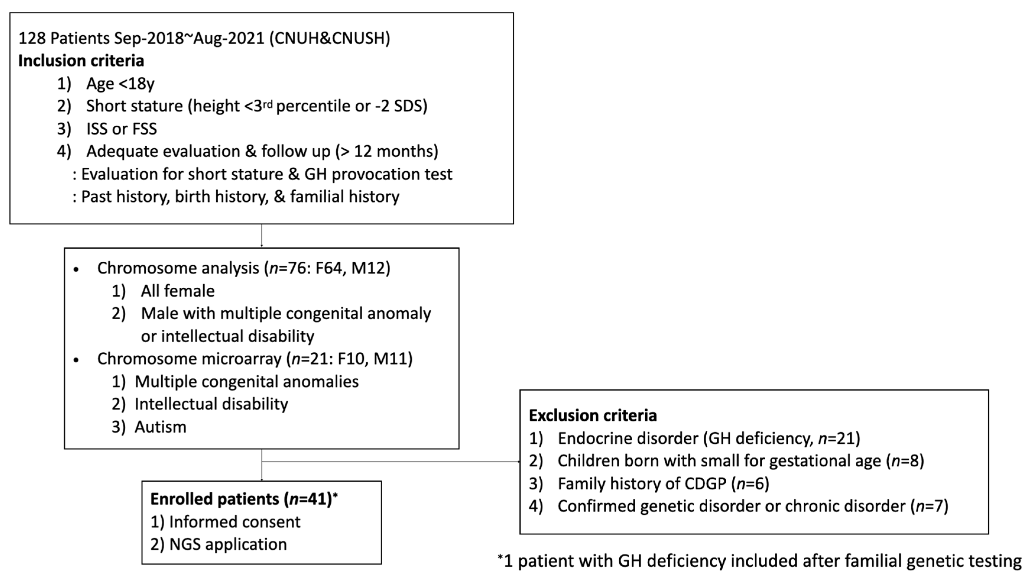
2.1. Patients

This study was approved by the Institutional Review Boards (IRB) of Chungnam National University (CNU) Hospital (IRB No. 2018-12-054) and CNU Sejong Hospital (IRB No. 2020-07-007). The patients were enrolled between September 2018 and August 2021 at CNU Hospital and CNU Sejong Hospital. Enrollment for the study was based on the following inclusion criteria (Figure 1): (1) age below 18 years; (2) SS defined as height less than the third percentile or −2 SD in individuals of the same sex and age by substituting the 2017 growth curve for Korean children and adolescents [20]; (3) ISS or FSS defining by maternal height <151.6 cm (−2 SD) or paternal height <163.8 cm (−2 SD) [20]; and (4) adequate evaluation and follow up (for at least 12 months). The exclusion criteria were: (1) GH deficiency or other pituitary hormonal deficiencies, such as hypothyroidism, adrenal disease, hypogonadism, and diabetes insipidus; (2) SGA without catch-up growth; (3) a parental history of CDGP; (4) children with organic brain lesions, abnormal karyotyping, other thyroid and renal diseases, congenital metabolic disease, diabetes mellitus, or a history of growth-related drug therapy; or (5) inadequate follow up. Birth weight was verified from the birth certificate from the birth institution, and patients were excluded if birth weights were below the third percentile for gestational age (GA) and sex in the Korean reference compatible with SGA [21]. Puberty was assessed according to the method described by Marshall and Tanner, and bone age was assessed according to the method described by Greulich and Pyle. Chromosome study was performed for all girls presenting with SS to exclude Turner syndrome. Chromosome microarray (CMA) was selectively performed for patients with unknown causes of developmental delay or intellectual disability, autism, or multiple congenital anomalies. All girls with Turner’s syndrome were excluded based on chromosomal studies. GH deficiency was confirmed when two different GH provocation tests showed peak GH levels <10 ng/dL. Combination with arginine and levodopa or insulin and levodopa was used for two pharmacologic stimuli and blood samples were drawn every 30 min for 2 hours. At least six hours of fasting was implemented for the GH provocation test. All patients were examined by pediatric endocrinologist. All patients were considered to exhibit ISS or FSS. Patients initiating GH treatment were assessed over a follow-up period of at least one year. Written informed consent was obtained from all participants or their parents.

2.2. Materials

DNA was isolated from blood samples for target exome sequencing. Analysis was performed using the Illumina MiSeq platform and TruSight One Panel (Illumina Inc., San Diego, CA, USA). Segregation analysis was performed for all cases where the variant was detected in the proband. The detected variants were classified by five variant classification categories (pathogenic, likely pathogenic, uncertain significance, likely benign, and benign) according to the ACMG guideline [22]. Each pathogenic criteria was scored as very strong (PVS1), strong (PS1–4); moderate (PM1–6), or supporting (PP1–5) [22].
Non-synonymous variants and nucleotide mutations located within 13 bases of each exon were collected, and mutations were substituted through the Genome Aggregation Database (gnomAD) and the Korean Reference Genome Database (KRGDB); allele frequencies above 1% of the general population were considered normal variants and excluded. The clinical relevance of the symptoms and detected mutations was investigated. The clinical significance of the mutations was evaluated by conducting parent and sibling tests simultaneously.
If the patients and their parents decided to receive GH therapy after the diagnosis of genetic SS, we prospectively followed the patients’ growth profile, laboratory results, and radiological findings during GH treatment. The predicted adult height (PAH) was assessed by the Greulich and Pyle method at the latest evaluation.

2.3. Statistical Analysis

The study population was characterized using the mean ± standard deviation (SD) and/or median for continuous descriptive variables, and categorical variables were expressed as frequencies. Descriptive and comparative statistical analyses were performed using SPSS Statistics for Windows, Version 26.0 (IBM SPSS Statistics for Windows, Version 26.0. IBM Corp, Armonk, NY, USA). Differences between groups were tested using t-test or Kruskal–Wallis and Fisher’s exact tests, as appropriate. Wilcoxon signed rank was used to assess the difference of height SDS in patients before and after GH treatment. Statistical significance was set at p < 0.05.

3. Results

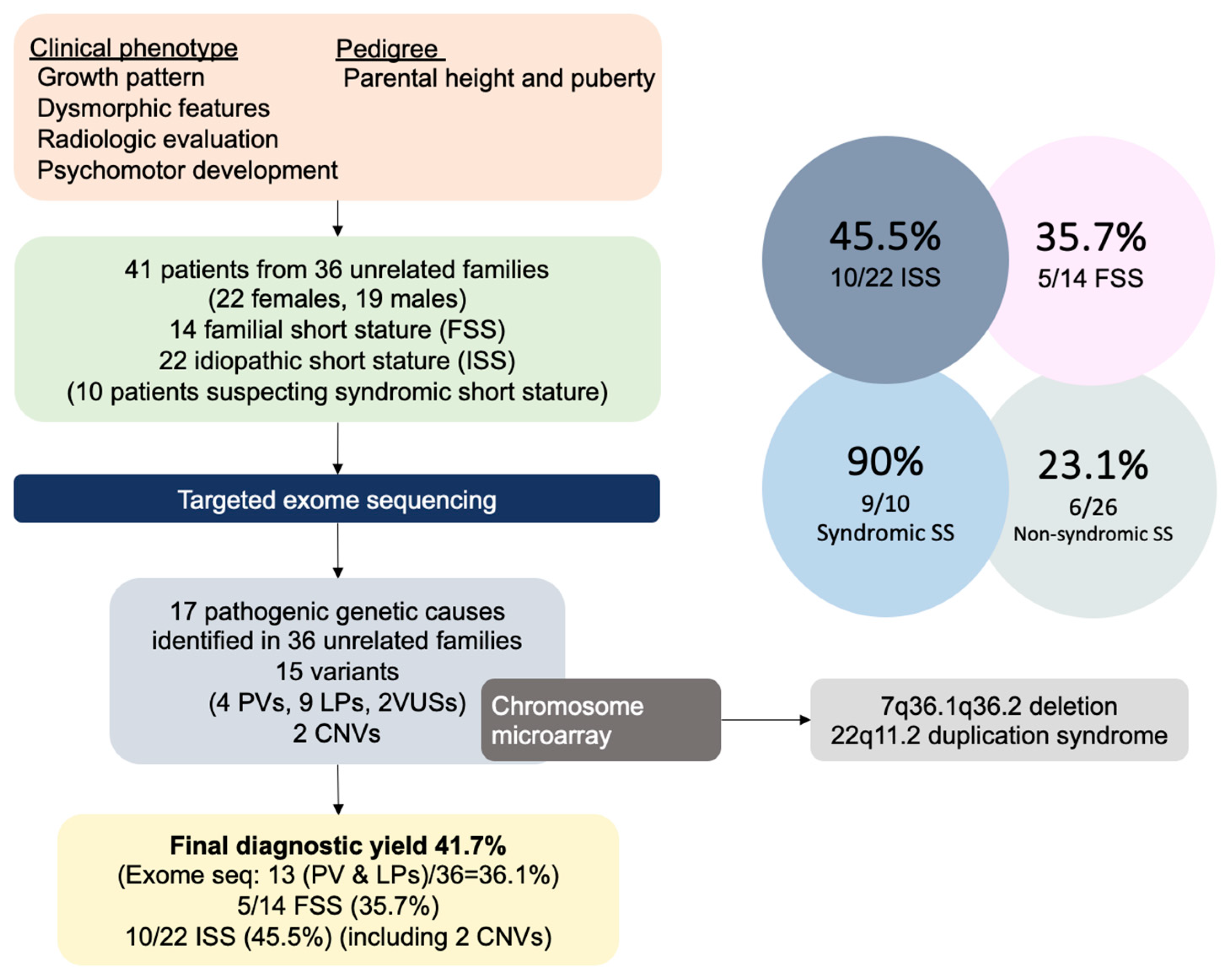
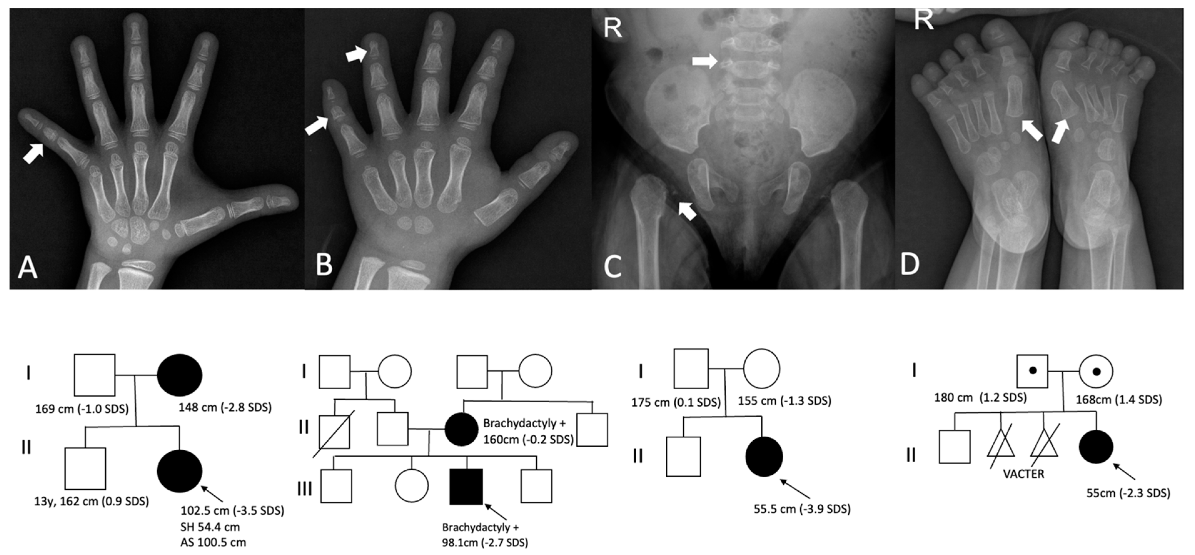
3.1. Clinical Characteristics

A total of 41 patients (22 females and 19 males) from 36 unrelated families were enrolled in this study. Patients’ age ranged from 5 months to 17 years (median 6.7 years, mean 6.5 ± 4.0 years). The mean height SD score (SDS) of the patients was 2.7 ± 1.6 SDS. Their midparental height was −0.5 ± 0.9 SDS. There were 22 unrelated patients with ISS and 19 patients from 14 unrelated families had FSS. Ten patients were suspected to have syndromic SS due to cognitive issues or dysmorphic features.

3.2. Genetic Results

Among the 41 patients with SS, 18 patients from 15 families had causative gene mutations, and 2 unrelated ISS patients had pathogenic copy number variants (CNVs). The total diagnostic yield for molecular changes (13 pathogenic causes in 36 families) was 36.1%. The combined positivity rate for pathogenic molecular changes and CNVs was 41.7% (Figure 2).
In 19 of the 14 unrelated patients with FSS, genetic causes of SS were found in 7 families, involving NPR2, COL1A1, ADNP, TRPS1, SPRED1, and PTPTN11, of which 4 variants were novel. Of the 22 patients with ISS, 10 were found to have genetic causes. The causative genes were BBS1, IHH, COL1A1, COL2A1, MASP1, NADSYN1, and CERT1. Seven novel variants were identified in this study. Interestingly, pathogenic CNVs were detected in two patients with ISS following a preliminary indication using exome sequencing.
A 13-month-old boy (participant 3) with suspected Noonan syndrome (NS) was diagnosed with 22q11.2 microduplication syndrome (Table 1). Initial karyotyping revealed a balanced translocation of 46, XY, t(12;14) (q15;q22). Exome sequencing revealed no pathogenic mutations, but a suspected duplication at 22q11.21. CMA confirmed a 22q11.21 duplication of 2.5 Mb [GRCh37/hg19 22q11.21(chr22:8,916,842-21,465,659) × 3]. In an 11-year-old girl (participant 30) with normal karyotype, a deletion at 7q36 was suspected by exome sequencing (Table 1). Additional CMA revealed a 4 Mb deletion at 7q36.1q36.2 [GRCh37/hg19 7q36.1q36.2 (chr7:150,796,099-155,074,825) × 1].
In this study, a genetic cause was identified in nine out of ten patients suspected with syndromic SS (90%) (Table 1, Figure 2).

3.3. Paracrine Signaling Defects

Four causative genes, NPR2, IHH, BBS1, and TRPS1, were identified and found to be related to paracrine signaling (Table 2).
The NPR2 variant was found in participant 16 (height −3.5 SDS) and her affected mother (height −2.8 SDS) (Table 2). The NPR2 (NM_003995.4) variant was c.1099A>G (p.Lys367Glu). Both the patient and mother showed fifth clinodactyly, and radiological findings revealed a shortened and dysplastic middle phalanx in the fifth finger (Figure 3A).
IHH (NM_002181.4) variant of c.683G>T (p.Arg228Leu) was detected in participant 12 having a proportionate SS, brachydactyly, and fifth clinodactyly. Radiography revealed shortened and dysplastic middle and distal phalanxes (Figure 3B). This variant was inherited from the mother, who also had brachydactyly despite having a normal height (−0.2 SDS).
Participant 36 with syndromic obesity (BMI 34.5 kg/m2) and SS had compound heterozygous BBS1 (NM_024649.4) mutations (Table 2). The patient presented with tapered fingers and brachydactyly. After diagnosis, retinal dystrophy, including foveal depigmentation and macular thinning, renal cysts, multiple hepatic hemangiomas, and a functional ovarian cyst sized 2.3 cm were identified from systematic surveillance.
Participant 31 displayed SS, sparse hair and eyebrows, and a pear-like nose. In addition, cone-shaped phalanxes were identified on hand radiography. Exome sequencing identified the TRPS1 (NM_014112.5) mutation c.2686dup (p.Gln896Profs*8) in this participant and the mutation was inherited from the father (height −2.7 SDS).

3.4. Extracellular Matrix Defects

COL1A1, COL2A1, and MASP1 were identified in this study (Table 2). Two unrelated patients manifested COL1A1 mutations. Participant 18 inherited the COL1A1 (NM_000088.4) variant of c.3766G>A (p.Ala1256Thr) from her mother (height 149 cm, −2.6 SDS). This variant was previously reported in a 41-year-old male with type 1 osteogenesis imperfecta [23]. Participant 8 experienced long bone fractures twice during infancy prior to trauma. Exome sequencing revealed a novel COL1A1 (NM_000088.4) mutation, c.757C>T (p.Arg253*). His father and grandfather carried this mutation and displayed different heights, of 176 cm (0.3 SDS) and 160 cm (−2.7 SDS), respectively. All patients displayed elbow dislocation, blue sclera, hearing defects, and recurrent fracture history during childhood.
Two patients (participants 2 and 20) exhibited a COL2A1-related disorder (Table 2). The radiological findings of participant 20 were compatible with those of spondyloepiphyseal dysplasia (Figure 3C).
Participant 13 with profound SS (height −3.1 SDS) had compound heterozygous mutations with c.851C>T (p.Ser284Leu) and c.744 + 7C>T (splicing) in MASP1 (NM_139125.3). She experienced feeding intolerance and recurrent upper respiratory tract infections. No definite developmental delays were observed.

3.5. Fundamental Cellular Process

SPRED1, PTPN11, ADNP, NADSYN1, and CERT1 variants were identified in this study (Table 2).
Participant 7 had a c.950C>G (p.Ser317*) in the SPRED1 (NM_152594.2) gene, which was inherited from the father. The father displayed a thick nasolabial fold, macrocephaly, and a triangular face. This participant showed macrocephaly (2.8 SDS) and dysmorphic features (Table 1). Fine motor skills only were delayed until 33 months of age for all milestones.
Two families with FSS were diagnosed with NS. Participant 24-1 exhibited FSS; his brother (participant 24-2) and younger sister (participant 24-3) also showed postnatal growth failure. Exome sequencing revealed a known mutation in PTPN11 (NM_002834.5), c.209A>G (p.Lys70Arg), inherited from the mother (−1.5 SDS, Table 1). These siblings showed clinical variability in intellectual ability and height within their families (Table 1). No abnormal echocardiographic findings were observed in these siblings.
Participant 4 had typical facial features and feeding intolerance, but no developmental delay (Table 1). A novel PTPN11 variant, c.1511T>C (p.Met504Thr) inherited from the father (height −0.1 SDS) was identified. Her father was regularly followed up for arrhythmia and pectus carinatum, but no structural anomalies were observed on echocardiography.
Participant 32-1 displayed an ADNP (NM_015339.5) variant for c.2992G>T (p.Asp998Tyr), which was inherited from the non-SS mother. Further familial testing was performed on two older sisters; one older sister (participant 32-2) with the ADNP variant had idiopathic GH deficiency (GH peak 0.9 ng/mL and 6.7 ng/mL) and early puberty. The sisters with the ADNP variant had a prominent forehead with a high anterior hairline, a down-slanting palpebral fissure, a low nasal bridge, a bulbous nose, and a pointed chin similar to their mother.
Participant 1 was born extremely prematurely owing to the premature rupture of membranes at labor. The participant also had a shortened limb, a prominent forehead, bulbous nose, and a low nasal bridge. At 5 months of age, her growth velocity was stunted (Table 1). The CMA was normal, but two compound heterozygous mutations, c.164A>T (p.Asp55Val) and c. 1048-1G>T (splicing) in NADSYN1 (NM_018161.5) were identified (Table 2). This participant showed only gross motor delay at 12 months of age and speech delay at 2 years of age. A skeletal survey revealed coxa valgum and varus deformity of the first metatarsal bone (Figure 3D). The patient had severe feeding intolerance and sleep disturbances, and hypercalcemia due to vitamin D intoxification was noted at 15 months of age. Her calcium level increased to 12.9 mg/dL with a suppressed parathyroid hormone level (0.2 pg/mL). The 25-OH vitamin D2 concentration and the urinary calcium-to-creatinine ratio increased to 105.9 ng/mL and 2.5, respectively. Renal ultrasonography revealed nephrocalcinosis. All medications, including a high dose of cholecalciferol (800 IU/d) from diverse self-nutrients, were discontinued, and the Locasol® formula (Nutricia Corp., Hoofddorp, The Netherlands) was started for hypercalcemia. Since then, the calcium level has been maintained at 9.0–10.1 mg/dL. This participant started GH treatment at 30 months of age and growth velocity was accelerated to 3.4 cm per 3 months of treatment.
Participant 35 exhibited profound SS and severe intellectual disabilities. The CMA and genetic testing for MECP2 and Angelman syndrome returned normal results. Exome sequencing revealed a de novo CERT1 (NM:001130105.1) variant for c.779C>T (p.Ser260Leu) resulting in intellectual developmental disorder, autosomal dominant 34 (MIM# 616351).

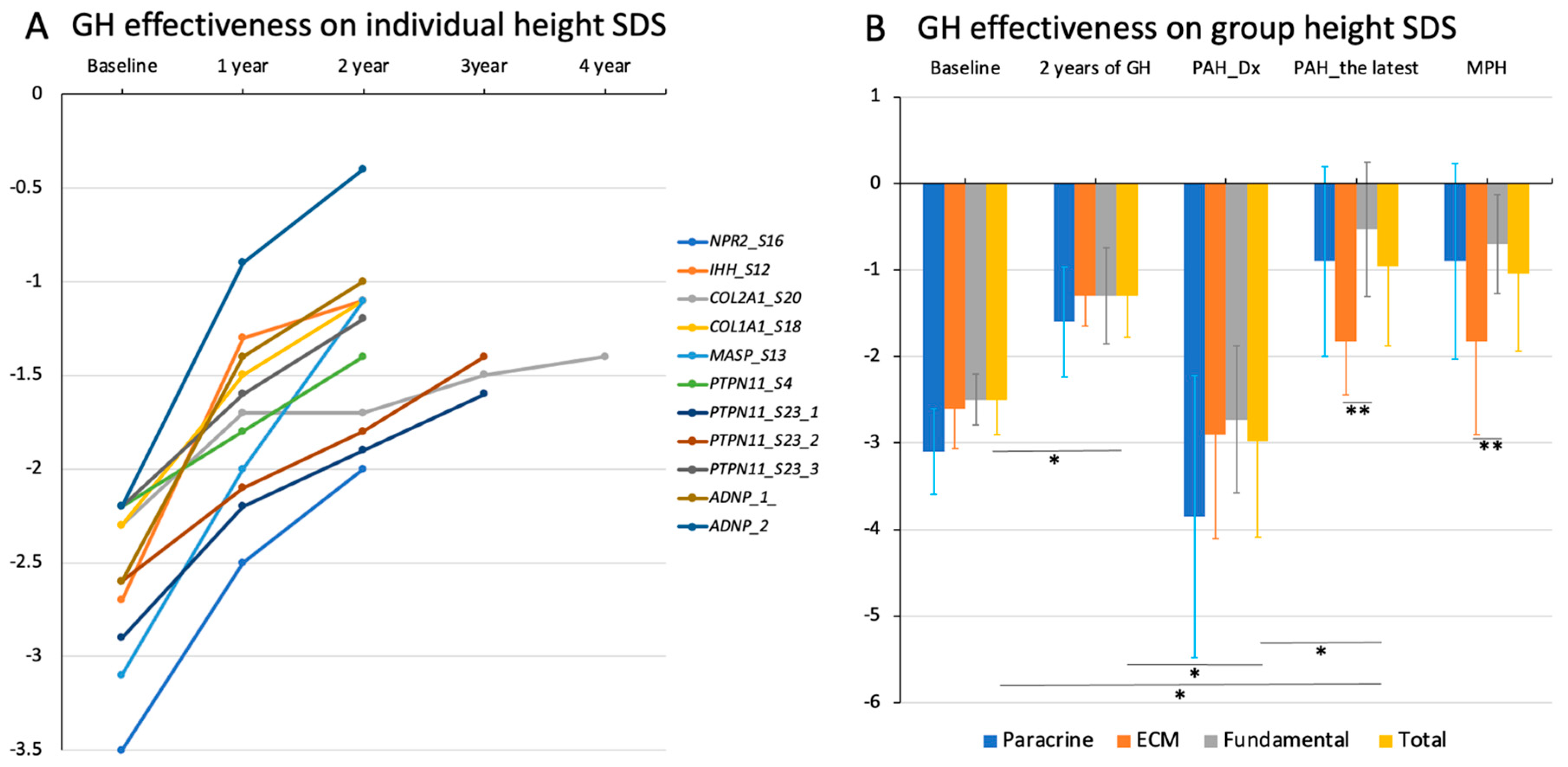
3.6. Growth Response to rhGH Treatment

Of the 15 patients with a genetic variant, 11 patients received rhGH for 2.8 ± 0.6 years (range, 2–3 years 10 months) (Figure 4A). Participant 1 with NADSYN1 variants was excluded from the analysis due to the short period of GH treatment. The median age at GH treatment was 7 years of age (range, 2–11 years), and all patients height significantly increased to −1.3 ± 0.5 SDS at 2 years after GH treatment compared to baseline height −2.6 ± 0.4 SDS (Figure 4B, p = 0.003). When we calculated their PAH at diagnosis and the latest evaluation, their PAH at the latest evaluation was also significantly increased to −1.0 ± 0.9 SDS compared to the PAH SDS at diagnosis (−3.0 ± 1.1) (p = 0.003) (Figure 4B).
Regarding paracrine signaling defect, two patients (NPR2 and IHH variant) were treated with GH therapy. Participant 16 with the NPR2 variant showed a height gain from −3.5 SDS to −1.6 SDS after three years of GH treatment (Figure 4A). As advanced bone age (10 years) and breast budding were noted at the age of 8 years and 6 months, a gonadotropin-releasing hormone (GnRH) stimulation test was performed. The results showed a prepubertal pattern (LH peak 2.7 mIU/mL). The subject showed transient precocious puberty, and pubertal progression was followed up regularly. The height SDS of participant 12 improved by −1.1 SDS after two years of GH treatment (Figure 4A).
In the ECM defect group, three patients (COL1A1, COL2A1, and MASP1) received rhGH. Their height and PAH SDS at baseline, height SDS after 2 years of rhGH, and PAH at the latest evaluation were −2.6 ± 0.5 SDS, −2.9 ± 1.5 SDS, −1.3 ± 0.3, and −1.8 ± 0.6 SDS, respectively. As participant 18 was diagnosed with central precocious puberty (CPP) at the age of 7 years, combined treatment with a GnRH analog and GH therapy was subsequently started. During treatment, her height increased from −2.3 SDS to −1.1 SDS over two years. Participant 13 with MASP variants showed a height gain to −1.5 SDS after two years of GH treatment, but combined treatment with GnRH analog was required due to the diagnosis of CPP at the age of 7 years and 8 months. Participant 20 with the COL2A1 variant received GH treatment for three years and ten months. Although his height increased from −2.3 SDS to −1.4 SDS after GH treatment for about four years, the PAH was reduced to −2.5 SDS for his advanced BA.
In the fundamental cellular defect group, a total of six patients (PTPN11 and ADNP variant) were treated with the rhGH (Figure 4A). Their initial height SDS was increased from −2.5 ± 0.3 SDS to −1.3 ± 0.6 SDS, and the initial PAH SDS (−2.7 ± 0.9) was also significantly improved by −0.5 ± 0.8 PAH SDS at the latest evaluation (Figure 4B).
In addition, there were no significant differences in height SDS between the group at baseline and 2 years after rhGH. However, MPH and PAH SDS at the latest evaluation was significantly higher in the fundamental cellular defect group than in the ECM group (Figure 4B, p < 0.05).

4. Discussion

The results of this study demonstrated the importance of genetic analysis in patients with ISS and FSS. The genetic defects identified in this study involved various molecular pathways, including paracrine signaling, ECM molecules, and fundamental intracellular processes, highlighting the complex nature of growth regulation.
Furthermore, genetic yields differed between patients with syndromic and non-syndromic SS. Ten patients (four FSS, six ISS) were suspected of syndromic SS, among which nine were attributed to a genetic cause (90%); however, the genetic yield of non-syndromic SS patients was 23.1% (6/26). Patients with syndromic SS exhibited a higher rate of genetic identification than patients with non-syndromic SS, emphasizing the need for genetic evaluation in these cases. In contrast, the diagnostic yield in patients with non-syndromic SS was relatively low, suggesting that additional factors beyond genetic causes may contribute to growth impairment.
Interestingly, families with the same gene mutations (IHH, COL1A1, SPRED1, PTPN11, and ADNP) exhibited height variability. The penetrance of ADNP mutations was less than 100%; patients in two families have been reported with ADNP-related disorders wherein the pathogenic variant has been inherited from an unaffected parent [24].
Natriuretic peptide receptor 2 (NPR2) is a receptor for the C-type natriuretic peptide, which stimulates growth plate chondrogenesis [25,26]. In particular, NPR2 heterozygous mutations reportedly have genetic causes in 2–6% of patients with ISS [27,28,29]. In addition, these patients tend to have typical findings, including brachydactyly, shortened metacarpal bone, and clinodactyly. GH treatment significantly improved the height SDS of patients with NPR2 heterozygous mutations [30,31]. Despite the small sample size in our study, NPR2 mutations accounted for 2.9% of the ISS or FSS cases, and this patient exhibited a height gain of 15 cm during the two years of GH treatment.
We further identified a male child with ISS harboring an IHH variant and demonstrating brachydactyly, shortening, and a dysplastic middle phalanx of the fifth finger. In addition to ACAN mutation, IHH is a common mutation in ISS with mild skeletal anomalies [32]. A high prevalence (81%) of IHH and ACAN mutations has been reported among 21 SS patients identified as having genetic causes of ISS [33]. These patients exhibited typical radiological findings, including middle phalanx shortening of the fifth finger. The response to rhGH has been encouraging, with a mean height gain of 0.6 SDS after one year of GH treatment [33]. Our patient was also a good responder, demonstrating +1.7 SDS height change in two years of GH treatment.
Transcriptional repressor GATA binding 1 (TRPS1) has multiple functions for chondrocyte differentiation and mineralization by interacting with IHH/GLI3 [34,35].
MASP1 on chromosome 3q24 encodes for three isoforms, mannan-binding lectin serine peptidase 1 (MASP1), MASP3, and MASP4, and is a disease-causing a gremlin mutation, resulting in 3MC syndrome (MIM#600521) [36]. The 3MC syndrome is a rare autosomal recessive disorder characterized by craniofacial dysmorphisms, including highly arched eyebrows, ptosis, blepharophimosis, cleft lip/palate, postnatal growth failure, hearing loss, and varying degrees of cognitive impairment [36]. To date, 30 patients from 22 families presenting with 3MC syndrome 1 caused by MASP1 mutations have been reported in the literature [36,37,38,39]. Biallelic MASP1 mutations have been identified in girls with ISS exhibiting normal intelligence, mild facial features, and arched eyebrows. Two novel mutations (c.744 + 7C>T and c.851C>T) in MASP1 (NM_139125.3) in the donor splice site affecting exons 5 and 6 and were shared by three isoforms. They are predicted to affect the CUB2 domain, the second most common domain, followed by the serine protease domains, in which mutations causing 3MC syndrome have been frequently reported [37,38,39]. Although the mechanism of postnatal growth failure in 3MC syndrome is unclear, MASP1 has been indicated to play a role in extracellular biological processes, and its molecular function is related to calcium ion binding and ECM components [40].
ADNP-related disorders are genetic causes of autism spectrum disorder, presenting with mild to moderate intellectual disability; developmental delay; multiple anomalies including heart, urogenital, and skeletal defects; and facial features including prominent and high forehead, low nasal bridge, down-slanting palpebral fissures, small chin, and prominent eyelashes [24]. Interestingly, all reported cases have truncated mutations, including nonsense or frameshift mutations, and almost all patients (97.1%) harbor de novo ADNP mutations [37]. Approximately 25% of individuals exhibit SS or GH deficiencies [37]. Since C-terminal ANDP mutations in the two patients were inherited from unaffected parents, the penetrance of ADNP was not considered to be 100% [24]. Our patient was familial and showed no developmental issues, exhibiting only facial features and SS. Notably, the patient carried missense variants located at the C-terminus, which, to our knowledge, have not previously been reported. Considering the limited information on missense ADNP variants, further investigations are needed to establish genotype–phenotype correlations.
The CERT1 gene, termed as COL4A3BP, was identified in three patients with neurodevelopmental disorders, possessing a p.Ser132Leu mutation in CERT1 [41]. Our patient had p.Ser260Leu, and a serine-to-leucine substitution in the serine-repeat motif was considered to impair protein function [41,42].
In this study, we identified a patient with NADSYN1-associated nicotinamide adenine dinucleotide (NAD) deficiency disorders demonstrating severe postnatal growth failure, and mild skeletal anomalies without global development. The first report of NADSYN1-associated NAD deficiency disorder specifies multiple severe anomalies and no living patients [43]. However, high clinical variability in NADSYN1-associated NAD deficiency disorders has recently been reported [44,45]. The clinical phenotype of NADSYN1-associated NAD deficiency is similar to that of the VATER syndrome. Our patient had a family history of abortions due to the VATER syndrome; however, genetic testing for NADSYN1 was not available.
Two patients had CNVs involving chromosome 22q11.2 or 7q36, as determined using NGS. The duplicated region at 22q11.21 included genes, such as LZTR1, TBX1, SERPIND1, and TUBA8, among which LZTR1 is a known causative gene of NS 10 (MIM#616564). Gain of function for LZTR1 is responsible for NS 10 [46]. Considering previous reports regarding the duplication of PTPN11 and RAF1 [47,48,49,50]; further evaluation is required to correlate 22q11.2 microduplication syndrome encompassing LZTR1 with NS 10. A 7q36.1q36.2 deletion containing several pathogenic genes, including DPP6, KMT2C, ASB10, PRKAG2, KCNH2, among which KMT2C is a causative gene for Kleefstra syndrome 2 (MIM#617768). This syndrome is characterized by variable intellectual disability, mild dysmorphic features, SS, and brachycephaly [51].
Our study demonstrated the high diagnostic yield (47.2%) in non-SGA SS patients compared to the general detection rate of 27.1% (95% CI, 18.1–37.2%), reported in a meta-analysis of 20 studies of patients with SS [52].
In comparison to the SGA SS group, defect of GH-IGF axis or imprinting disorder is main cause for SGA without catch-up growth in addition to the growth plate dysfunction [53,54]. The genes for growth plate dysfunction have been identified in ISS or FSS group (Table 3). Collagen gene mutations were detected in FSS patients (10/87, 11.5%) and of unknown origin in SS patients (18/232, 7.8%) [55,56].
Importantly, our study highlighted the effectiveness of GH therapy in patients with growth failure. Although the identified genetic defects involving the ECM may limit the response to GH treatment, most patients in this study displayed height gain after GH treatment. There have been several efforts to assess the efficacy of GH treatment on the genotype of growth plate dysfunction, despite the small numbers of patients [30,33,55,56,57]. One study of nine patients with a collagen gene defect showed a change in height SDS of 1.0 for 2.8 years of GH treatment [55]. Among them, two patients were diagnosed with CPP, and their PAH was not shown. Another study [56] showed the growth response to the rhGH in 10 FSS patients with collagen gene defects. They showed the height SDS change 0.9 after 3 years of rhGH and four patients who reached adult height showed variable height gains, ranging from –0.67 to +1.0 SD and –0.45 to +0.5 SD compared to their baseline height and the height of their affected untreated parent, respectively [56]. Our study also showed an improvement in height SDS from −2.6 to −1.3 (height SDS change 1.3) for 2.8 years of GH treatment in the ECM group. However, since all patients in the ECM group showed advanced BA during the follow-up period, the PAH at the latest evaluation was decrease to the −1.8 SDS.
These findings support the use of GH therapy in non-SGA children with SS, particularly when the causative gene is identified. Further, categorizing patients based on specific molecular defects is essential for customized treatment strategies. Understanding the underlying genetic causes can provide valuable prognostic information and aid in decision-making regarding the administration of GH therapy in non-SGA children with SS. As our study has some limitations, including a small number of patients, a short follow-up period for the efficacy of GH treatment, and the use of targeted panels instead of whole exome sequencing or whole genome sequencing, further study with a larger cohort, long-term follow-up, and comprehensive genetic study is required to evaluate the genetic aspects and rhGH effects of growth plate dysfunction in patients with ISS or FSS.
The efficiency of NGS was evaluated in these patients by identifying the cause of SS, which was initially classified as ISS or FSS. This may be helpful to implement early intervention and predict the efficacy of GH treatment using factor analysis.

5. Conclusions

In conclusion, our study demonstrated a diagnostic yield of 41.7% using targeted exome sequencing and identified multiple genetic causes of ISS or FSS, further elucidating the molecular mechanisms involved in growth regulation. The GH treatment was generally effective at increasing height SDS in this study, and more in the fundamental cellular defect group than the ECM defect group, despite the limited number of patients and treatment period. These findings have implications for the management and treatment of patients with ISS or FSS. Further research is warranted to explore additional genetic factors and their impact on growth outcomes.

Author Contributions

Y.-M.K. initiated the study, received funding and ethical permission, was responsible for the study design, and recruited patients. Y.-M.K., H.-H.L., E.K., G.K., M.K., B.K.L., H.S., Y.K., J.M. and Y.S.L. interpreted the findings, revised the manuscript, agreed on the final version of the manuscript, and accept responsibility for the entire content of the manuscript and approve its submission. All authors have read and agreed to the published version of the manuscript.

Funding

This study was supported by the National Research Foundation of Korea grant funded by the Korean government (Ministry of Science and ICT No.2018R1C1B5086428).

Institutional Review Board Statement

This study was approved by the Institutional Review Boards (IRB) of Chungnam National University (CNU) Hospital (IRB No. 2018-12-054) and CNU Sejong Hospital (IRB No.2020-07-007).

Informed Consent Statement

Informed consent was obtained from all subjects involved in the study.

Data Availability Statement

All data generated or analyzed during this study are included in this article. Further enquiries can be directed to the corresponding author.

Acknowledgments

We thank all the patients and their families for participating in this study.

Conflicts of Interest

The authors declare no conflict of interest.

References

  1. Baron, J.; Sävendahl, L.; De Luca, F.; Dauber, A.; Phillip, M.; Wit, J.M.; Nilsson, O. Short and tall stature: A new paradigm emerges. Nat. Rev. Endocrinol. 2015, 11, 735–746. [Google Scholar] [CrossRef]
  2. Hallett, S.A.; Ono, W.; Ono, N. Growth Plate Chondrocytes: Skeletal Development, Growth and Beyond. Int. J. Mol. Sci. 2019, 20, 6009. [Google Scholar] [CrossRef]
  3. Romero, C.J.; Mehta, L.; Rapaport, R. Genetic techniques in the evaluation of short stature. Endocrinol. Metab. Clin. N. Am. 2016, 45, 345–358. [Google Scholar] [CrossRef]
  4. Guo, M.H.; Shen, Y.; Walvoord, E.C.; Miller, T.C.; Moon, J.E.; Hirschhorn, J.N.; Dauber, A. Whole exome sequencing to identify genetic causes of short stature. Horm. Res. Paediatr. 2014, 82, 44–52. [Google Scholar] [CrossRef]
  5. Wang, S.R.; Carmichael, H.; Andrew, S.F.; Miller, T.C.; Moon, J.E.; Derr, M.A.; Hwa, V.; Hirschhorn, J.N.; Dauber, A. Large-scale pooled next generation sequencing of 1077 genes to identify genetic causes of short stature. J. Clin. Endocrinol. Metab. 2013, 98, E1428–E1437. [Google Scholar] [CrossRef] [PubMed]
  6. Flechtner, I.; Lambot-Juhan, K.; Teissier, R.; Colmenares, A.; Baujat, G.; Beltrand, J.; Ajaltouni, Z.; Pauwels, C.; Pinto, G.; Samara-Boustani, D.; et al. Unexpected high frequency of skeletal dysplasia in idiopathic short stature and small for gestational age patients. Eur. J. Endocrinol. 2014, 170, 677–684. [Google Scholar] [CrossRef] [PubMed]
  7. Hattori, A.; Katoh-Fukui, Y.; Nakamura, A.; Matsubara, K.; Kamimaki, T.; Tanaka, H.; Dateki, S.; Adachi, M.; Muroya, K.; Yoshida, S.; et al. Next generation sequencing-based mutation screening of 86 patients with idiopathic short stature. Endocr. J. 2017, 64, 947–954. [Google Scholar] [CrossRef] [PubMed]
  8. Saltarelli, M.A.; Quarta, A.; Chiarelli, F. Growth plate extracellular matrix defects and short stature in children. Ann. Pediatr. Endocrinol. Metab. 2022, 27, 247–255. [Google Scholar] [CrossRef] [PubMed]
  9. Kim, Y.M.; Lee, Y.J.; Park, J.H.; Lee, H.D.; Cheon, C.K.; Kim, S.Y.; Hwang, J.Y.; Jang, J.H.; Yoo, H.W. High diagnostic yield of clinically unidentifiable syndromic growth disorders by targeted exome sequencing. Clin. Genet. 2017, 92, 594–605. [Google Scholar] [CrossRef] [PubMed]
  10. Andrade, A.C.; Jee, Y.H.; Nilsson, O. New Genetic Diagnoses of Short Stature Provide Insights into Local Regulation of Childhood Growth. Horm. Res. Paediatr. 2017, 88, 22–37. [Google Scholar] [CrossRef]
  11. Plachy, L.; Strakova, V.; Elblova, L.; Obermannova, B.; Kolouskova, S.; Snajderova, M.; Zemkova, D.; Dusatkova, P.; Sumnik, Z.; Lebl, J.; et al. High Prevalence of Growth Plate Gene Variants in Children With Familial Short Stature Treated With GH. J. Clin. Endocrinol. Metab. 2019, 104, 4273–4281. [Google Scholar] [CrossRef] [PubMed]
  12. Murray, P.G.; Clayton, P.E.; Chernausek, S.D. A genetic approach to evaluation of short stature of undetermined cause. Lancet Diabetes Endocrinol. 2018, 6, 564–574. [Google Scholar] [CrossRef]
  13. Faienza, M.F.; Chiarito, M.; Brunetti, G.; D’Amato, G. Growth plate gene involvement and isolated short stature. Endocrine 2021, 71, 28–34. [Google Scholar] [CrossRef] [PubMed]
  14. Vasques, G.A.; Andrade, N.L.M.; Jorge, A.A.L. Genetic causes of isolated short stature. Arch. Endocrinol. Metab. 2019, 63, 70–78. [Google Scholar] [CrossRef]
  15. Andrade, N.L.M.; Funari, M.F.A.; Malaquias, A.C.; Collett-Solberg, P.F.; Gomes, N.L.R.A.; Scalco, R.; Dantas, N.C.B.; Rezende, R.C.; Tiburcio, A.M.F.P.; Souza, M.A.R.; et al. Diagnostic yield of a multigene sequencing approach in children classified as idiopathic short stature. Endocr. Connect. 2022, 11, e220214. [Google Scholar] [CrossRef] [PubMed]
  16. Plachy, L.; Amaratunga, S.A.; Dusatkova, P.; Maratova, K.; Neuman, V.; Petruzelkova, L.; Zemkova, D.; Obermannova, B.; Snajderova, M.; Kolouskova, S.; et al. Isolated growth hormone deficiency in children with vertically transmitted short stature: What do the genes tell us? Front. Endocrinol. 2023, 13, 1102968. [Google Scholar] [CrossRef]
  17. Mintz, C.S.; Seaver, L.H.; Irons, M.; Grimberg, A.; Lozano, R.; ACMG Professional Practice and Guidelines Committee. Focused Revision: ACMG practice resource: Genetic evaluation of short stature. Genet. Med. 2021, 23, 813–815. [Google Scholar] [CrossRef]
  18. Clayton, P.E.; Cianfarani, S.; Czernichow, P.; Johannsson, G.; Rapaport, R.; Rogol, A. Management of the child born small for gestational age through to adulthood: A consensus statement of the International Societies of Pediatric Endocrinology and the Growth Hormone Research Society. J. Clin. Endocrinol. Metab. 2007, 92, 804–810. [Google Scholar] [CrossRef]
  19. Hokken-Koelega, A.C.S.; van der Steen, M.; Boguszewski, M.C.S.; Cianfarani, S.; Dahlgren, J.; Horikawa, R.; Mericq, V.; Rapaport, R.; Alherbish, A.; Braslavsky, D.; et al. International Consensus Guideline on Small for Gestational Age (SGA): Etiology and Management from Infancy to Early Adulthood. Endocr. Rev. 2023, 44, 539–565. [Google Scholar] [CrossRef]
  20. Kim, J.H.; Yun, S.; Hwang, S.S.; Shim, J.O.; Chae, H.W.; Lee, Y.J.; Lee, J.H.; Kim, S.C.; Lim, D.; Yang, S.W.; et al. The 2017 Korean National Growth Charts for children and adolescents: Development, improvement, and prospects. Korean J. Pediatr. 2018, 61, 135–149. [Google Scholar] [CrossRef]
  21. Lim, J.S.; Lim, S.W.; Ahn, J.H.; Song, B.S.; Shim, K.S.; Hwang, I.T. New Korean reference for birth weight by gestational age and sex: Data from the Korean Statistical Information Service (2008–2012). Ann. Pediatr. Endocrinol. Metab. 2014, 19, 146–153. [Google Scholar] [CrossRef]
  22. Richards, S.; Aziz, N.; Bale, S.; Bick, D.; Das, S.; Gastier-Foster, J.; Grody, W.W.; Hegde, M.; Lyon, E.; Spector, E.; et al. Standards and guidelines for the interpretation of sequence variants: A joint consensus recommendation of the American College of Medical Genetics and Genomics and the Association for Molecular Pathology. Genet. Med. 2015, 17, 405–424. [Google Scholar] [CrossRef]
  23. Zhang, Z.L.; Zhang, H.; Ke, Y.H.; Yue, H.; Xiao, W.J.; Yu, J.B.; Gu, J.M.; Hu, W.W.; Wang, C.; He, J.W.; et al. The identification of novel mutations in COL1A1, COL1A2, and LEPRE1 genes in Chinese patients with osteogenesis imperfecta. J. Bone Miner. Metab. 2012, 30, 69–77. [Google Scholar] [CrossRef]
  24. Van Dijck, A.; Vulto-van Silfhout, A.T.; Cappuyns, E.; van der Werf, I.M.; Mancini, G.M.; Tzschach, A.; Bernier, R.; Gozes, I.; Eichler, E.E.; Romano, C.; et al. Clinical Presentation of a Complex Neurodevelopmental Disorder Caused by Mutations in ADNP. Biol. Psychiatry 2019, 85, 287–297. [Google Scholar] [CrossRef]
  25. Rintz, E.; Węgrzyn, G.; Fujii, T.; Tomatsu, S. Molecular mechanism of induction of bone growth by the C-type natriuretic peptide. Int. J. Mol. Sci. 2022, 23, 5916. [Google Scholar] [CrossRef] [PubMed]
  26. Kim, Y.M. A new era of genetic diagnosis for short stature children: A review. Precis. Future Med. 2022, 6, 218–225. [Google Scholar] [CrossRef]
  27. Amano, N.; Mukai, T.; Ito, Y.; Narumi, S.; Tanaka, T.; Yokoya, S.; Ogata, T.; Hasegawa, T. Identification and functional characterization of two novel NPR2 mutations in Japanese patients with short stature. J. Clin. Endocrinol. Metab. 2014, 99, E713–E718. [Google Scholar] [CrossRef] [PubMed]
  28. Vasques, G.A.; Amano, N.; Docko, A.J.; Funari, M.F.; Quedas, E.P.; Nishi, M.Y.; Arnhold, I.J.; Hasegawa, T.; Jorge, A.A. Heterozygous mutations in natriuretic peptide receptor-B (NPR2) gene as a cause of short stature in patients initially classified as idiopathic short stature. J. Clin. Endocrinol. Metab. 2013, 98, E1636–E1644. [Google Scholar] [CrossRef]
  29. Wang, S.R.; Jacobsen, C.M.; Carmichael, H.; Edmund, A.B.; Robinson, J.W.; Olney, R.C.; Miller, T.C.; Moon, J.E.; Mericq, V.; Potter, L.R.; et al. Heterozygous mutations in natriuretic peptide receptor-B (NPR2) gene as a cause of short stature. Hum. Mutat. 2015, 36, 474–481. [Google Scholar] [CrossRef]
  30. Ke, X.; Liang, H.; Miao, H.; Yang, H.; Wang, L.; Gong, F.; Pan, H.; Zhu, H. Clinical Characteristics of Short-Stature Patients With an NPR2 Mutation and the Therapeutic Response to rhGH. J. Clin. Endocrinol. Metab. 2021, 106, 431–441. [Google Scholar] [CrossRef]
  31. Chen, H.; Zhang, S.; Sun, Y.; Chen, J.; Yuan, K.; Zhang, Y.; Yang, X.; Lin, X.; Chen, R. Novel pathogenic NPR2 variants in short stature patients and the therapeutic response to rhGH. Orphanet. J. Rare Dis. 2023, 18, 221. [Google Scholar] [CrossRef] [PubMed]
  32. Sentchordi-Montané, L.; Benito-Sanz, S.; Aza-Carmona, M.; Díaz-González, F.; Modamio-Høybjør, S.; de la Torre, C.; Nevado, J.; Ruiz-Ocaña, P.; Bezanilla-López, C.; Prieto, P.; et al. High prevalence of variants in skeletal dysplasia associated genes in individuals with short stature and minor skeletal anomalies. Eur. J. Endocrinol. 2021, 185, 691–705. [Google Scholar] [CrossRef] [PubMed]
  33. Vasques, G.A.; Funari, M.F.A.; Ferreira, F.M.; Aza-Carmona, M.; Sentchordi-Montané, L.; Barraza-García, J.; Lerario, A.M.; Yamamoto, G.L.; Naslavsky, M.S.; Duarte, Y.A.O.; et al. IHH gene mutations causing short stature with nonspecific skeletal abnormalities and response to growth hormone therapy. J. Clin. Endocrinol. Metab. 2018, 103, 604–614. [Google Scholar] [CrossRef] [PubMed]
  34. Yang, L.; Gong, X.; Wang, J.; Fan, Q.; Yuan, J.; Yang, X.; Sun, X.; Li, Y.; Wang, Y. Functional mechanisms of TRPS1 in disease progression and its potential role in personalized medicine. Pathol. Res. Pract. 2022, 237, 154022. [Google Scholar] [CrossRef]
  35. Wuelling, M.; Kaiser, F.J.; Buelens, L.A.; Braunholz, D.; Shivdasani, R.A.; Depping, R.; Vortkamp, A. Trps1, a regulator of chondrocyte proliferation and differentiation, interacts with the activator form of Gli3. Dev. Biol. 2009, 328, 40–53. [Google Scholar] [CrossRef]
  36. Sirmaci, A.; Walsh, T.; Akay, H.; Spiliopoulos, M.; Sakalar, Y.B.; Hasanefendioğlu-Bayrak, A.; Duman, D.; Farooq, A.; King, M.C.; Tekin, M. MASP1 mutations in patients with facial, umbilical, coccygeal, and auditory findings of Carnevale, Malpuech, OSA, and Michels syndromes. Am. J. Hum. Genet. 2010, 87, 679–686. [Google Scholar] [CrossRef]
  37. Urquhart, J.; Roberts, R.; de Silva, D.; Shalev, S.; Chervinsky, E.; Nampoothiri, S.; Sznajer, Y.; Revencu, N.; Gunasekera, R.; Suri, M.; et al. Exploring the genetic basis of 3MC syndrome: Findings in 12 further families. Am. J. Med. Genet. A 2016, 170A, 1216–1224. [Google Scholar] [CrossRef]
  38. Agaoglu, N.B.; Akgun Dogan, O. Further Expansion of the Mutational Spectrum of 3MC Syndrome: A Novel MASP1 Pathogenic Variant in a Male Patient. Mol. Syndromol. 2021, 12, 379–385. [Google Scholar] [CrossRef]
  39. Durmaz, C.D.; Altıner, Ş. MASP1-related 3MC syndrome in a patient from Turkey. Am. J. Med. Genet. A 2021, 185, 2267–2270. [Google Scholar] [CrossRef]
  40. Ibelli, A.M.G.; Peixoto, J.O.; Zanella, R.; Gouveia, J.J.S.; Cantão, M.E.; Coutinho, L.L.; Marchesi, J.A.P.; Pizzol, M.S.D.; Marcelino, D.E.P.; Ledur, M.C. Downregulation of growth plate genes involved with the onset of femoral head separation in young broilers. Front. Physiol. 2022, 13, 941134. [Google Scholar] [CrossRef]
  41. Fitzgerald, T.W.; Gerety, S.S.; Jones, W.D.; Van Kogelenberg, M.; King, D.A.; McRae, J.; Morley, K.I.; Parthiban, V.; Al-Turki, S.; Ambridge, K.; et al. The Deciphering Developmental Disorders Study. Large-scale discovery of novel genetic causes of developmental disorders. Nature 2015, 519, 223–228. [Google Scholar] [CrossRef]
  42. Tamura, N.; Sakai, S.; Martorell, L.; Colomé, R.; Mizuike, A.; Goto, A.; Ortigoza-Escobar, J.D.; Hanada, K. Intellectual-disability-associated mutations in the ceramide transport protein gene CERT1 lead to aberrant function and subcellular distribution. J. Biol. Chem. 2021, 297, 101338. [Google Scholar] [CrossRef] [PubMed]
  43. Szot, J.O.; Slavotinek, A.; Chong, K.; Brandau, O.; Nezarati, M.; Cueto-González, A.M.; Patel, M.S.; Devine, W.P.; Rego, S.; Acyinena, A.P.; et al. New cases that expand the genotypic and phenotypic spectrum of Congenital NAD Deficiency Disorder. Hum. Mutat. 2021, 42, 862–876. [Google Scholar] [CrossRef] [PubMed]
  44. Kortbawi, H.; Ames, E.; Pritchard, A.; Devine, P.; van Ziffle, J.; Slavotinek, A. Further description of two patients with biallelic variants in NADSYN1 in association with cardiac and vertebral anomalies. Am. J. Med. Genet. A 2022, 188, 2479–2484. [Google Scholar] [CrossRef] [PubMed]
  45. Aubert-Mucca, M.; Janel, C.; Porquet-Bordes, V.; Patat, O.; Touraine, R.; Edouard, T.; Michot, C.; Tessier, A.; Cormier-Daire, V.; Attie-Bitach, T.; et al. Clinical heterogeneity of NADSYN1-associated VCRL syndrome. Clin. Genet. 2023, 104, 114–120. [Google Scholar] [CrossRef] [PubMed]
  46. Yamamoto, G.L.; Aguena, M.; Gos, M.; Hung, C.; Pilch, J.; Fahiminiya, S.; Abramowicz, A.; Cristian, I.; Buscarilli, M.; Naslavsky, M.S.; et al. Rare variants in SOS2 and LZTR1 are associated with Noonan syndrome. J. Med. Genet. 2015, 52, 413–421. [Google Scholar] [CrossRef]
  47. Shchelochkov, O.A.; Patel, A.; Weissenberger, G.M.; Chinault, A.C.; Wiszniewska, J.; Fernandes, P.H.; Eng, C.; Kukolich, M.K.; Sutton, V.R. Duplication of chromosome band 12q24.11q24.23 results in apparent Noonan syndrome. Am. J. Med. Genet. A 2008, 146A, 1042–1048. [Google Scholar] [CrossRef]
  48. Chen, J.L.; Zhu, X.; Zhao, T.L.; Wang, J.; Yang, Y.F.; Tan, Z.P. Rare copy number variations containing genes involved in RASopathies: Deletion of SHOC2 and duplication of PTPN11. Mol. Cytogenet 2014, 7, 28. [Google Scholar] [CrossRef]
  49. Graham, J.M., Jr.; Kramer, N.; Bejjani, B.A.; Thiel, C.T.; Carta, C.; Neri, G.; Tartaglia, M.; Zenker, M. Genomic duplication of PTPN11 is an uncommon cause of Noonan syndrome. Am. J. Med. Genet. 2009, 149A, 2122–2128. [Google Scholar] [CrossRef]
  50. Luo, C.; Yang, Y.F.; Yin, B.L.; Chen, J.L.; Huang, C.; Zhang, W.Z.; Wang, J.; Zhang, H.; Yang, J.F.; Tan, Z.P. Microduplication of 3p25.2 encompassing RAF1 associated with congenital heart disease suggestive of Noonan syndrome. Am. J. Med. Genet. A 2012, 158A, 1918–1923. [Google Scholar] [CrossRef]
  51. Siano, M.A.; De Maggio, I.; Petillo, R.; Cocciadiferro, D.; Agolini, E.; Majolo, M.; Novelli, A.; Della Monica, M.; Piscopo, C. De Novo Mutation in KMT2C Manifesting as Kleefstra Syndrome 2: Case Report and Literature Review. Pediatr. Rep. 2022, 14, 131–139. [Google Scholar] [CrossRef] [PubMed]
  52. Li, Q.; Chen, Z.; Wang, J.; Xu, K.; Fan, X.; Gong, C.; Wu, Z.; Zhang, T.J.; Wu, N. Molecular Diagnostic Yield of Exome Sequencing and Chromosomal Microarray in Short Stature: A Systematic Review and Meta-Analysis. JAMA Pediatr. 2023, e233566. [Google Scholar] [CrossRef] [PubMed]
  53. Toni, L.; Plachy, L.; Dusatkova, P.; Amaratunga, S.A.; Elblova, L.; Sumnik, Z.; Kolouskova, S.; Snajderova, M.; Obermannova, B.; Pruhova, S.; et al. The genetic landscape of children born small for gestational age with persistent short stature (SGA-SS). Horm. Res. Paediatr. 2023. [Google Scholar] [CrossRef] [PubMed]
  54. Hara-Isono, K.; Nakamura, A.; Fuke, T.; Inoue, T.; Kawashima, S.; Matsubara, K.; Sano, S.; Yamazawa, K.; Fukami, M.; Ogata, T.; et al. Pathogenic Copy Number and Sequence Variants in Children Born SGA With Short Stature Without Imprinting Disorders. J. Clin. Endocrinol. Metab. 2022, 107, e3121–e3133. [Google Scholar] [CrossRef]
  55. Chen, M.; Miao, H.; Liang, H.; Ke, X.; Yang, H.; Gong, F.; Wang, L.; Duan, L.; Chen, S.; Pan, H.; et al. Clinical Characteristics of Short-Stature Patients With Collagen Gene Mutation and the Therapeutic Response to rhGH. Front. Endocrinol. 2022, 13, 820001. [Google Scholar] [CrossRef]
  56. Plachy, L.; Dusatkova, P.; Maratova, K.; Petruzelkova, L.; Elblova, L.; Kolouskova, S.; Snajderova, M.; Obermannova, B.; Zemkova, D.; Sumnik, Z.; et al. Familial Short Stature-A Novel Phenotype of Growth Plate Collagenopathies. J. Clin. Endocrinol. Metab. 2021, 106, 1742–1749. [Google Scholar] [CrossRef]
  57. Zhao, Q.; Li, Y.; Shao, Q.; Zhang, C.; Kou, S.; Yang, W.; Zhang, M.; Ban, B. Clinical and genetic evaluation of children with short stature of unknown origin. BMC. Med. Genom. 2023, 16, 194. [Google Scholar] [CrossRef]
Figure 1. Flow chart of patient enrollment.
Jcm 12 06508 g001
Figure 2. Schematic study representation of patients with short stature born non-small for gestational age.
Jcm 12 06508 g002
Figure 3. Radiological findings and pedigree in patients with NPR2 (participant 16), IHH (participant 12), COL2A1 (participant 2), and NADSYN1 gene (participant 1) variants (AD). Simple X-ray of the hand showing a shortened and dysplastic middle phalanx in participant 16 with the NPR2 variant (A) and brachydactyly and shortening of phalanxes in participant 12 with the IHH variant (B). Pelvic X-ray in participant 2 showing the small iliac bone and small irregular epiphyses of both proximal femurs with widening and metaphysis irregularities of both femoral heads and short neck. The white arrow shows the relatively smaller size of the L5 body compared to the L4 body (C). Varus deformity of first metatarsal bone in participant 1 (D).
Jcm 12 06508 g003
Figure 4. Height gains after recombinant human growth hormone treatment by (A) individual and (B) group categories (paracrine signal defect, extracellular matrix (ECM) defect, fundamental cellular defect, and total group) * height SDS was significantly increased at 2 years of GH treatment, and predicted adult height (PAH) at the latest evaluation in fundamental group and total group (* p <0.05); ** the MPH and PAH at latest evaluation between ECM defect and fundamental cellular defect group showed significant difference (** p < 0.05).
Jcm 12 06508 g004
Table 1. Clinical findings for short stature children born non-small for gestational age.
NoSexAgeGA
Week
BW
kg
Mode of DeliveryHt
cm
(SDS)
Wt
kg
(SDS)
Pat.
Ht cm (SDS)
Mat. Ht cm
(SDS)
MPH
cm (SDS)
Psychomotor
Issue
DysmorphismCategory
1F5 m
CA 3 m
29 + 21.15C/sec55
(−2.3)
4.9
(−1.4)
180
(1.2)
168
(1.4)
167.5
(1.3)
Gross motor delayProminent forehead, bulbous nose, low nasal bridge, short limbISS,
R/O SD
2F5 m37 + 53.16NSVD55.5
(−3.9)
6.0
(−1.1)
175
(0.1)
155
(−1.3)
156
(−1.0)
Gross motor delayProminent forehead, brachydactyly, short limbISS,
R/O SD
3M13 m403.51C/sec72.2
(−2.0)
9.1
(−0.7)
170
(−0.8)
155
(−1.3)
169
(−1.0)
None (4.6 y: IQ 120)Macrocephaly, s/p VSD closure, resemble facial features with NSISS,
R/O SD
4F15 m35 + 52.57C/sec (twin)70.0
(−2.7)
7.9
(−1.6)
174
(−0.1)
158
(−0.6)
159.5
(−0.3)
NoneFTT, sparse hair & eyebrows, long philtrum, down-slanting palpebral fissure, high foreheadISS,
R/O SD
5M31 m382.9C/sec82.5
(−2.9)
10.3
(−2.3)
167
(−1.4)
161
(0.0)
170.5
(−0.7)
None-ISS
6M3 y 10 m383.1C/sec91.6
(−2.6)
13.1
(−1.7)
178
(0.6)
153
(−1.7)
172
(−0.4)
None-ISS
7F3 y 11 m40 + 63.5C/sec92.6
(−2.1)
12.8
(−2.0)
166
(−1.6)
148
(−2.8)
150.5
(−2.2)
NoneTriangular face, prominent forehead, macrocephalyFSS, R/O SD
8M4 y 4 m403.6NSVD96.5
(−2.0)
14.6
(−1.7)
176
(0.3)
153
(−1.7)
171
(−0.6)
NoneBlue scleraeISS
9M4 y 10 m403NSVD96.7
(−2.7)
14.3
(−2.4)
169
(−1.0)
153
(−1.7)
167.5
(−1.3)
None-ISS
10F4 y 11 m38 + 52.9NSVD98.2
(−2.3)
13.2
(−2.9)
160
(−2.7)
165
(0.8)
156
(−1.0)
None-FSS
11F4 y 11 m40 + 64NSVD97.9
(−2.4)
15.4
(−1.4)
168
(−1.2)
155
(−1.3)
155
(−1.3)
None-ISS
12M5 y403.1NSVD98.1
(−2.7)
18.1
(−0.4)
176
(0.3)
160
(−0.2)
175
(0.1)
NoneBrachydactylyISS
13F5 y 2 m382.9C/sec96.9
(−3.1)
14.2
(−3.7)
164
(−1.9)
158
(−0.6)
155
(−1.3)
None-ISS
14M6 y 4 m35 + 42.1NSVD104.6
(−2.8)
18.2
(−1.5)
170
(−0.8)
160
(−0.2)
171.5
(−0.5)
None-ISS
15M6 y 5 m39 + 53.9C/sec107.7
(−2.2)
18.5
(−1.4)
167
(−1.4)
153
(−1.7)
166.5
(−1.5)
None-ISS
16F6 y 6 m37 + 22.6C/sec102.5
(−3.5)
16.7
(−2.1)
169
(−1.0)
148
(−2.8)
152
(−1.9)
NoneTriangular face, down-slanting palpebral fissure, prominent philtrum, relative macrocephalyFSS,
R/O SD
17-1M6 y 8 m403C/sec109.8
(−2.1)
17.7
(−2.1)
161
(−2.5)
148
(−2.8)
161
(−2.5)
None-FSS
17-2F7 y 7 m403.03C/sec108.9
(−3.3)
20.8
(−1.2)
161
(−2.5)
148
(−2.8)
149
(−2.6)
None-FSS
18F6 y 11 m382.7C/sec107.8
(−2.7)
20.2
(−0.9)
165
(−1.8)
149
(−2.6)
150.5
(−2.2)
NoneBlue sclerae, triangular face, delayed eruptionFSS
19M7 y392.8NSVD (IVF)105.5
(−3.6)
18.0
(−2.3)
170
(−0.8)
158
(−0.6)
170.5
(−0.7)
None-ISS
20M8 y 6 m39 + 53.2NSVD118.1
(−2.5)
25.8
(−0.8)
153
(−4.1)
153
(−1.7)
159.5
(−2.8)
None-FSS
21F8 y 2 m402.8NSVD111.1
(−3.5)
17.7
(−2.9)
170
(−0.8)
148
(−2.8)
152.5
(−1.8)
None-FSS
22M8 y 10 m39 + 12.8NSVD113.0
(−3.8)
22.0
(−2.0)
170
(−0.8)
160
(−0.2)
171.5
(−0.5)
None-ISS
23F8 y 9 m372.32C/sec (Twin)118.0
(−2.5)
21.0
(−2.1)
176
(0.3)
150
(−2.4)
156.5
(−0.9)
None -FSS
24-1M8 y 8 m37 + 32.6NSVD119.2
(−2.3)
19.7
(−2.7)
176
(0.3)
154
(−1.5)
171.5
(−0.5)
Mild ID (IQ 64)-FSS
24-2M10 y 8 m36 + 62.9NSVD126.8
(−2.7)
25.8
(−2.0)
176
(0.3)
154
(−1.5)
171.5
(−0.5)
None
(IQ 94)
-FSS
24-3F7 y 10 m39 + 53.1NSVD117.3
(−1.6)*
16.6
(−3.2)
176
(0.3)
154
(−1.5)
158.5
(−0.5)
None
(IQ 82)
-FSS
25F10 y403NSVD126
(−2.3)
32.8
(−0.3)
173
(−0.3)
160
(−0.2)
160
(−0.2)
None-ISS
26M10 y 10 m403.2NSVD128.4
(−2.5)
23.0
(−2.7)
165
(−1.8)
149
(−2.6)
163.5
(−2.0)
Speech delayTriangular face, narrow palpebral fissure, low nasal bridgeFSS, R/O SD
27M10 y 11 m403.2NSVD126.7
(−2.9)
25.0
(−2.3)
164
(−1.9)
154
(−1.5)
165.5
(−1.7)
None-ISS
28M10 y 11 m37 + 62.86NSVD130.1
(−2.3)
23.4
(−2.7)
164
(−1.9)
154
(−1.5)
165.5
(−1.7)
None-ISS
29M11 y 8 m413NSVD126.5
(−3.4)
23.8
(−2.9)
170
(−0.8)
160
(−0.2)
171.5
(−0.5)
None-ISS
30F11 y402.9NSVD122.2
(−3.7)
22.5
(−3.0)
170
(−0.8)
160
(−0.2)
158.5
(−0.5)
ADHDs/p PDA ligation, ASD closureISS
31M11 y 11 m38 + 23.3C/sec135.8
(−2.1)
37.3
(−0.9)
160
(−2.7)
164
(0.6)
168.5
(−1.1)
ADHD, mild ID (IQ 69)Sparse hair and eyebrows, pear like noseFSS, R/O SD
32-1F6 y403.6C/sec103.8
(−2.6)
15.4
(−2.5)
161
(−2.5)
162
(0.2)
155
(−1.3)
None FSS
32-2F10 y 1 m402.8C/sec122.9
(−2.9)
26.8
(−1.5)
161
(−2.5)
162
(0.2)
155
(−1.3)
None FSS
(GHD)
32-3F11 y 10 m383.3C/sec135.4
(−2.3)
36.6
(−0.8)
161
(−2.5)
162
(0.2)
155
(−1.3)
None FSS
33F12 y403.1NSVD134.2
(−2.6)
49.6
(0.7)
170
(−0.8)
150
(−2.4)
156
(−1.0)
None-FSS
34F12 y 6 m372.64NSVD134
(−3.1)
27.3
(−2.9)
171
(−0.6)
151
(−2.1)
154.5
(−1.4)
None-FSS
35F14 y362.2C/sec129.3
(−4.9)
21.2
(−6.1)
173
(−0.3)
163
(0.4)
161.5 (0.1)Severe ID, wide gait, stereotypic behaviorMicrocephaly, widely spaced teeth, hypertelorismISS,
R/O SD
36F17 y413.99C/sec151.5
(−2.0)
79.1
(2.7)
166
(−1.5)
161
(0.0)
158
(−0.6)
Moderate IDs/p TOF, polydactyly, and ectopic anus, obesityISS,
R/O SD
GA: gestational age, BW: birth weight, HT: height, SD: standard deviation, HT: paternal height, mat. HT, maternal height; MPH, mid-parental height; CA, corrected age; NSVD; normal spontaneous vaginal delivery, C/sec; cesarean section; ADHD, attention-deficit hyperactivity disorder; ID, intellectual disability; VSD, ventricular septal defect; NS, Noonan syndrome; PDA; patent ductus arteriosus; ASD; atrial septal defect; TOF; tetralogy of Fallot, ISS, idiopathic short stature; FSS, familial short stature; GHD, growth hormone deficiency; SD, syndromic disorder. * The height SDS of participant 24-3 was −2.1 SDS at 9 years of age.
Table 2. Genetic results for children born non-small for gestational age.
Pt. No.CategoryDiseaseGenecDNA
Variant
Protein
Variant
MAF aIn Silico Analysis bInheritanceACMG
Classification [22]
Paracrine signaling
16FSSShort stature with nonspecific skeletal abnormalitiesNPR2c.1099A>Gp.Lys367Glu0/0D/T/D/MatLP (PM1, PM2, PP1, PP4)
12ISSBrachydactyly, type A1IHHc.683G>Tp.Arg228Leu0/0T/D/DMatVUS (PM2, PP3, PP4)
36ISSBardet–Biedl syndrome, type 1BBS1c.1285C>T
c.1339G>A
p.Arg429 *
p.Ala447Thr
0.0008/0
0.00037/0
-
D/T/D
Pat
Mat
PV (PVS1, PM2, PM3, PP3, PP4)
LP (PM1, PM2, PM3, PP3, PP4)
31FSSTrichorhinophalangeal syndrome 1TRPS1c.2686dupp.Gln896Profs *80/0-PatPV (PVS1, PM2, PP1, PP4)
Extracellular matrix (ECM) defects
18FSSOsteogenesis imperfectaCOL1A1c.3766G>Ap.Ala1256Thr0.00134
/0.00045
D/D/DMatVUS (PM2, PP1, PP3, PP4)
8ISSOsteogenesis imperfectaCOL1A1c.757C>Tp.Arg253 *0/0-PatPV (PVS1, PM2, PP1, PP3, PP4, PP5)
20ISSType II collagen disorderCOL2A1c.889G>Ap.Gly297Ser0/0D/T/ DDe novoLP (PM1, PM2, PM6, PP3, PP4)
2ISSType II collagen disorderCOL2A1c.3310G>Cp.Gly1104Arg0/0D/T/DDe novoLP (PS2, PM1, PM2, PP3, PP5)
13ISS3MC syndrome 1MASP1c.851C>T
c.744 + 7C>T
p.Ser284Leu
splicing
0/0
0.0000041/0.00045
D/D/D
D/D/D
Pat
Mat
LP (PM3, PM2, PP2, PP3, PP4)
Fundamental cellular process
24-1
24-2
24-3
FSSNoonan syndromePTPN11c.209A>Gp.Lys70Arg0/0D/D/DMatLP (PM1, PM2 PP1 PP3 PP4 PP5)
4FSSNoonan syndromePTPN11c.1511T>Cp.Met504Thr0/0D/D/DPatLP (PM1, PM2, PP1, PP3, PP4)
7FSSLegius syndromeSPRED1c.950C>Gp.Ser317 *0/0-PatPV (PVS1 PM2 PP1 PP3 PP4)
32-1
32-2
FSSHelsmoortel-van der Aa syndromeADNPc.2992G>Tp.Asp998Tyr0/0D/D/DMatLP (PM1, PM2, PP1, PP4)
35ISSIntellectual developmental disorder, autosomal dominant 34CERT1
(COL4A3BP)
c.779C>Tp.Ser260Leu0/0D/D/DDe novoPV (PS1 PS2 PM1 PM2 PP4)
1ISSNAD synthetase deficiency disorderNADSYN1c.164A>T
c.1048-1G>T
p.Asp55Val
splicing
0.00081/0
0/0
D/D/DPat
Mat
LP (PM3, PM2, PP2, PP3, PP4)
3ISS22q11.2 microduplication syndrome
(Noonan syndrome 10)
LZTR1duplication De novo
30ISS7q36 microdeletion syndrome
(Kleefstra syndrome 2)
KMT2Cdeletion De novo
Pt. No., participant number; MAF a, minor allele frequency (gnomAD database/Korean Reference Genome Database); in silico analysis b, SIFT/Polyphen2/MutationTaster; FSS, familial short stature; ISS, idiopathic short stature; NAD: nicotinamide adenine dinucleotide; D, deleterious; T, tolerant; PV, pathogenic variant; LP, likely pathogenic; VUS, variant of uncertain significance. Bold indicates a novel mutation.
Table 3. Comparison of diagnostic yield using exome sequencing and the genetic results of growth plate dysfunction between SGA SS and non-SGA SS patients.
Patients
[Reference] (Number)
SGA [54]
(n = 176)
SGA Without Imprinting Disorder [55]
(n = 86)
Non-SGA (ISS) [15]
(n = 102)
Non-SGA (ISS and FSS) [This Study]
(n = 41, 36 Families)
Genetic testingWhole exome sequencing or targeted exome sequencing Copy number analysis, targeted exome sequencingTargeted exome sequencingTargeted exome sequencing
Genetic etiology
(CNVs + PV or LP)
74/176 (42%) 13/86 (15.1%)17/102 (16.7%)15/36 (41.7%)
Growth plate dysfunction43/176 (24.4%)5/86 (5.8%)17/102 (16.7%)13/36 (36.1%)
Paracrine signaling7/176 (4%)
FGFR3, FGFR2, NPR2
1/86 (1.1%)
NPR2
8/102 (7.8%)
IHH (4), NPR2 (2), FGFR3 (2)
3/36 (8%)
NPR2, IHH(VUS), BBS1, TRPS1
Extracellular matrix24/176 (13.6%)
ACAN, various collagens, FLNB, MATN3
SHOX deficiency (7)
COL2A1(VUS), SHOX (VUS), ACAN (VUS), FGFR3 (VUS)4/102 (3.9%)
SHOX, ACAN, ACAN/SHOX *, COL2A1/NF1 *
4/36 (11%)
COL1A1(VUS, LP), COL2A1(2), MASP1
Fundamental intracellular/intranuclear processes12/176 (6.8%)
CDC42, KMT2D, LMNA, NSD1, PTPN11, SRCAP, SON, SOS1, SOX9, TLK2
4/86 (4.7%)
SMAD4, PTPN11, CBL, MYCN
4/102 (3.9%)
NF1, PTPN11, BRAF, CBL/SHOX *
6/36 (17%)
PTPN11(2), SPRED1, ADNP, CERT1, NADSYN1
Miscellaneous chromosomal aberrations5/176 (2.8%)
del17q24.2
del1p31.1p31.3
del22q11.2
del6q24.3q25.1
dupXq
8/86 (9.3%)
del1q21.1q21.2
del22q11.2
dupXp22.33
del19p13.12
dup5q35.2q35.3 (2)
del10q26.1q26.3
delXp22.3p11.2/dupXq11.1q28
-2/36 (6%)
dup22q11.2 (LZTR1)
del7q36 (KMT2C)
Pituitary development, GH-IGF-1 or IGF-2 axis, and thyroid axis14/176 (8%)
LHX4, OTX2, PROKR2, PTCH1, POU1F1, GHSR, IGFALS, IGF1R, STAT3, HMGA2, TRHR, THRA
GHR(VUS), IGF1R(VUS)1/102 (0.9%)
GHSR
-
Silver–Russell syndrome12/176 (6.8%)
: 11p15, UPD7
Excluded--
SGA, small for gestational age; ISS, idiopathic short stature; FSS, familial short stature; CNVs, copy number variants; PV, pathogenic variant; LP, likely pathogenic variant; VUS, variant of unknown significance; GH, growth hormone; IGF, insulin-like growth factor; UPD, uniparental disomy. * 3 patients had two different gene mutations.
Disclaimer/Publisher’s Note: The statements, opinions and data contained in all publications are solely those of the individual author(s) and contributor(s) and not of MDPI and/or the editor(s). MDPI and/or the editor(s) disclaim responsibility for any injury to people or property resulting from any ideas, methods, instructions or products referred to in the content.

