
Early Experience with the New XEN63 Implant in Primary Open-Angle Glaucoma Patients: Clinical Outcomes

 , , ,
, , ,
Abstract
:1. Introduction
2. Materials and Methods
2.1. Design
2.2. Patients
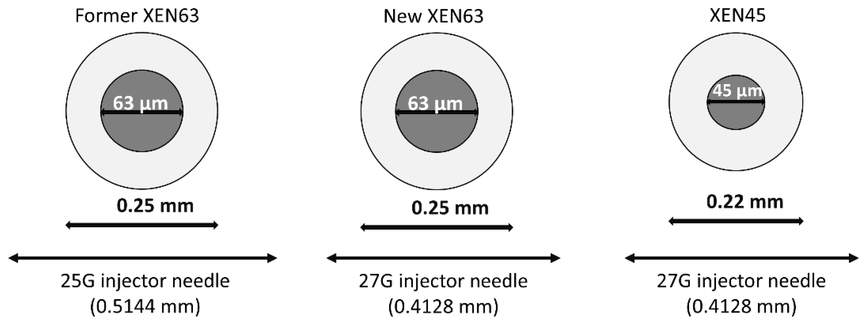
2.3. Device
2.4. Surgical Technique
2.5. Outcomes
2.6. Statistical Analysis
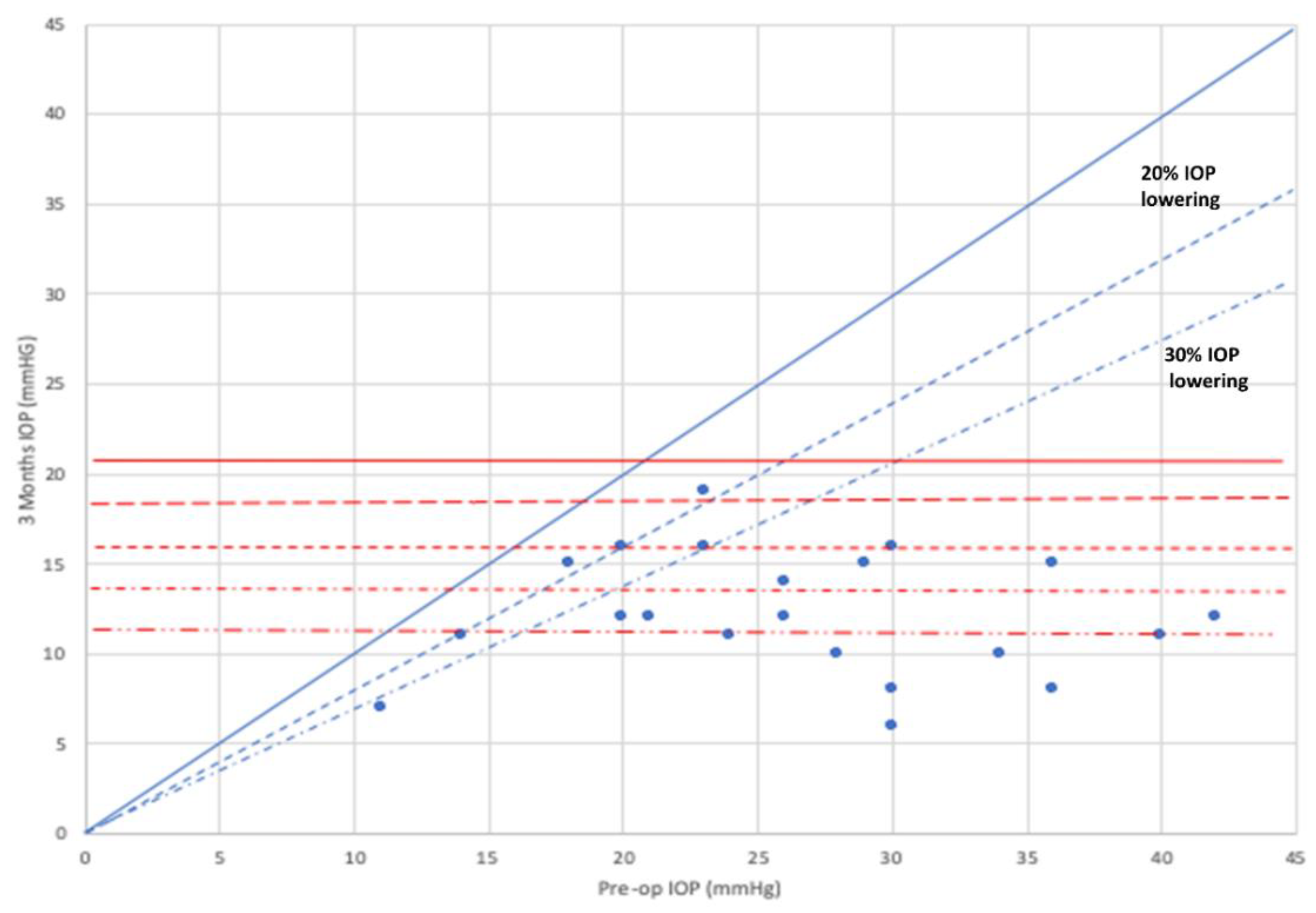
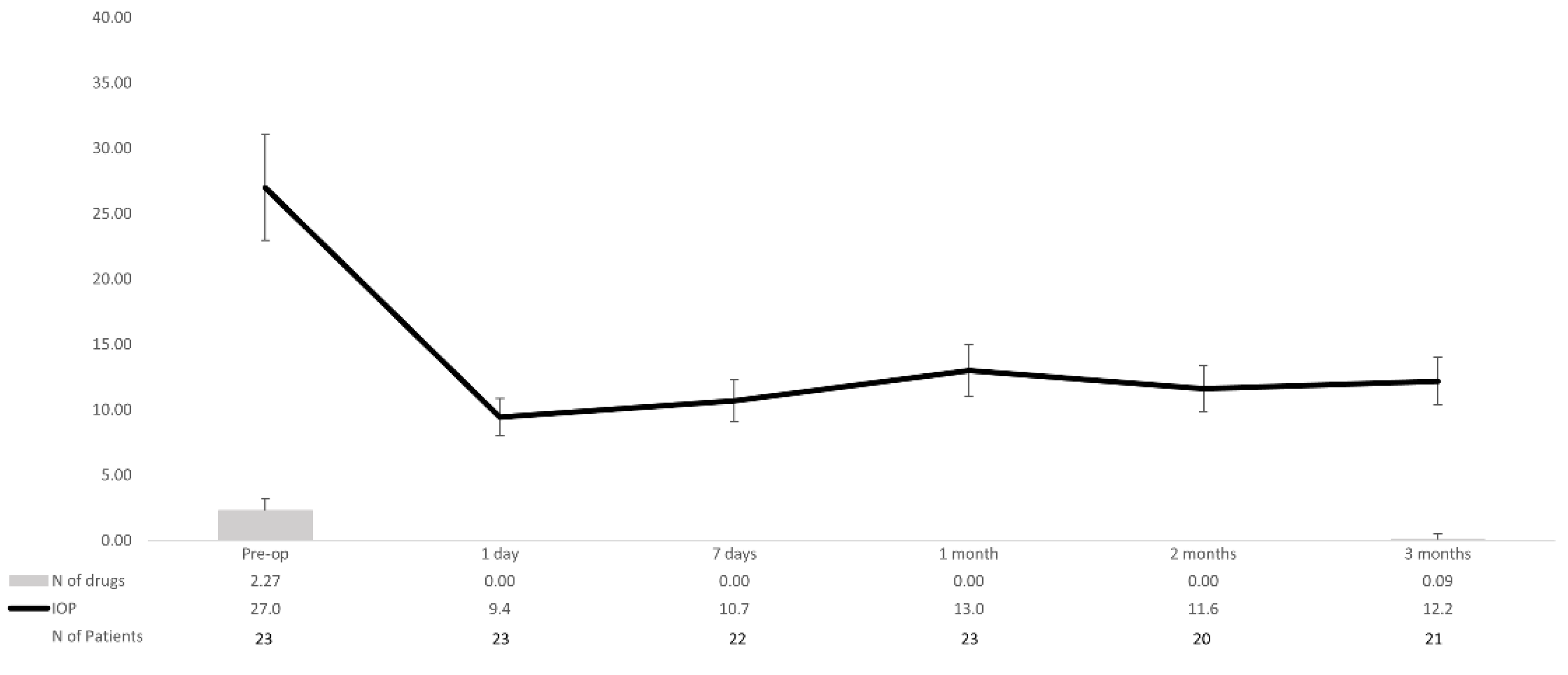
3. Results
4. Discussion
5. Conclusions
Author Contributions
Funding
Institutional Review Board Statement
Informed Consent Statement
Data Availability Statement
Acknowledgments
Conflicts of Interest
References
- Weinreb, R.N.; Khaw, P.T. Primary open-angle glaucoma. Lancet 2004, 363, 1711–1720. [Google Scholar] [CrossRef]
- Tham, Y.C.; Li, X.; Wong, T.Y.; Quigley, H.A.; Aung, T.; Cheng, C.Y. Global prevalence of glaucoma and projections of glaucoma burden through 2040: A systematic review and meta-analysis. Ophthalmology 2014, 121, 2081–2090. [Google Scholar] [CrossRef]
- Boland, M.V.; Ervin, A.M.; Friedman, D.S.; Jampel, H.D.; Hawkins, B.S.; Vollenweider, D.; Chelladurai, Y.; Ward, D.; Suarez-Cuervo, C.; Robinson, K.A. Comparative effectiveness of treatments for open-angle glaucoma: A systematic review for the U.S. Preventive Services Task Force. Ann. Intern. Med. 2013, 158, 271–279. [Google Scholar] [CrossRef]
- Newman-Casey, P.A.; Robin, A.L.; Blachley, T.; Farris, K.; Heisler, M.; Resnicow, K.; Lee, P.P. The Most Common Barriers to Glaucoma Medication Adherence: A Cross-Sectional Survey. Ophthalmology 2015, 122, 1308–1316. [Google Scholar] [CrossRef] [PubMed] [Green Version]
- Lichter, P.R.; Musch, D.C.; Gillespie, B.W.; Guire, K.E.; Janz, N.K.; Wren, P.A.; Richard, M.P.H.; CIGTS Study Group. Interim clinical outcomes in the Collaborative Initial Glaucoma Treatment Study comparing initial treatment randomized to medications or surgery. Ophthalmology 2001, 108, 1943–1953. [Google Scholar] [CrossRef]
- Landers, J.; Martin, K.; Sarkies, N.; Bourne, R.; Watson, P. A twenty-year follow-up study of trabeculectomy: Risk factors and outcomes. Ophthalmology 2012, 119, 694–702. [Google Scholar] [CrossRef]
- Jampel, H.D.; Musch, D.C.; Gillespie, B.W.; Lichter, P.R.; Wright, M.M.; Guire, K.E.; Collaborative Initial Glaucoma Treatment Study Group. Perioperative complications of trabeculectomy in the collaborative initial glaucoma treatment study (CIGTS). Am. J. Ophthalmol. 2005, 140, 16–22. [Google Scholar] [CrossRef] [PubMed]
- Lavia, C.; Dallorto, L.; Maule, M.; Ceccarelli, M.; Fea, A.M. Minimally-invasive glaucoma surgeries (MIGS) for open angle glaucoma: A systematic review and meta-analysis. PLoS ONE 2017, 12, e0183142. [Google Scholar] [CrossRef] [PubMed]
- Ansari, E. An Update on Implants for Minimally Invasive Glaucoma Surgery (MIGS). Ophthalmol. Ther. 2017, 6, 233–241. [Google Scholar] [CrossRef]
- Pillunat, L.E.; Erb, C.; Jünemann, A.G.; Kimmich, F. Micro-invasive glaucoma surgery (MIGS): A review of surgical procedures using stents. Clin. Ophthalmol. 2018, 12, 287. [Google Scholar] [CrossRef] [PubMed] [Green Version]
- Chaudhary, A.; Salinas, L.; Guidotti, J.; Mermoud, A.; Mansouri, K. XEN Gel Implant: A new surgical approach in glaucoma. Expert Rev. Med. Devices 2018, 15, 47–59. [Google Scholar] [CrossRef]
- De Gregorio, A.; Pedrotti, E.; Russo, L.; Morselli, S. Minimally invasive combined glaucoma and cataract surgery: Clinical results of the smallest ab interno gel stent. Int. Ophthalmol. 2018, 38, 1129–1134. [Google Scholar] [CrossRef]
- Reitsamer, H.; Sng, C.; Vera, V.; Lenzhofer, M.; Barton, K.; Stalmans, I.; Apex Study Group. Two-year results of a multicenter study of the ab interno gelatin implant in medically uncontrolled primary open-angle glaucoma. Graefes Arch. Clin. Exp. Ophthalmol. 2019, 257, 983–996. [Google Scholar] [CrossRef] [Green Version]
- Chatzara, A.; Chronopoulou, I.; Theodossiadis, G.; Theodossiadis, P.; Chatziralli, I. XEN Implant for Glaucoma Treatment: A Review of the Literature. Semin. Ophthalmol. 2019, 34, 93–97. [Google Scholar] [CrossRef] [PubMed]
- Marcos-Parra, M.T.; Salinas-López, J.A.; López-Grau, N.S.; Ceausescu, A.M.; Pérez-Santonja, J.J. XEN implant device versus trabeculectomy, either alone or in combination with phacoemulsification, in open-angle glaucoma patients. Graefes Arch. Clin. Exp. Ophthalmol. 2019, 257, 1741–1750. [Google Scholar] [CrossRef] [PubMed]
- Fea, A.M.; Bron, A.M.; Economou, M.A.; Laffi, G.; Martini, E.; Figus, M.; Oddone, F. European study of the efficacy of a cross-linked gel stent for the treatment of glaucoma. J. Cataract Refract. Surg. 2020, 46, 441–450. [Google Scholar] [CrossRef] [PubMed]
- Fea, A.M.; Durr, G.M.; Marolo, P.; Malinverni, L.; Economou, M.A.; Ahmed, I. XEN® Gel Stent: A Comprehensive Review on Its Use as a Treatment Option for Refractory Glaucoma. Clin. Ophthalmol. 2020, 14, 1805–1832. [Google Scholar] [CrossRef] [PubMed]
- Sheybani, A.; Lenzhofer, M.; Hohensinn, M.; Reitsamer, H.; Ahmed, I.I. Phacoemulsification combined with a new ab interno gel stent to treat open-angle glaucoma: Pilot study. J. Cataract Refract. Surg. 2015, 41, 1905–1909. [Google Scholar] [CrossRef] [PubMed]
- Lenzhofer, M.; Kersten-Gomez, I.; Sheybani, A.; Gulamhusein, H.; Strohmaier, C.; Hohensinn, M.; Dick, H.B.; Hitzl, W.; Eisenkopf, L.; Sedarous, F.; et al. Four-year results of a minimally invasive transscleral glaucoma gel stent implantation in a prospective multi-centre study. Clin. Exp. Ophthalmol. 2019, 47, 581–587. [Google Scholar] [CrossRef] [PubMed] [Green Version]
- Fernández-García, A.; Zhou, Y.; García-Alonso, M.; Andrango, H.D.; Poyales, F.; Garzón, N. Comparing Medium-Term Clinical Outcomes following XEN® 45 and XEN® 63 Device Implantation. J. Ophthalmol. 2020, 2020, 4796548. [Google Scholar] [CrossRef]
- Lavin-Dapena, C.; Cordero-Ros, R.; D’Anna, O.; Mogollón, I. XEN 63 gel stent device in glaucoma surgery: A 5-years follow-up prospective study. Eur. J. Ophthalmol. 2020, 1120672120952033. [Google Scholar] [CrossRef]
- Yuan, K.-H.; Maxwell, S. On the Post Hoc Power in Testing Mean Differences. J. Educ. Behav. Stat. 2005, 30, 141–167. [Google Scholar] [CrossRef]
- Gedde, S.J.; Herndon, L.W.; Brandt, J.D.; Budenz, D.L.; Feuer, W.J.; Schiffman, J.C.; Tube Versus Trabeculectomy Study Group. Postoperative complications in the Tube Versus Trabeculectomy (TVT) study during five years of follow-up. Am. J. Ophthalmol. 2012, 153, 804–814.e1. [Google Scholar] [CrossRef] [Green Version]
- Grover, D.S.; Flynn, W.J.; Bashford, K.P.; Lewis, R.A.; Duh, Y.J.; Nangia, R.S.; Niksch, B. Performance and Safety of a New Ab Interno Gelatin Stent in Refractory Glaucoma at 12 Months. Am. J. Ophthalmol. 2017, 183, 25–36. [Google Scholar] [CrossRef] [PubMed] [Green Version]
- Ibáñez-Muñoz, A.; Soto-Biforcos, V.S.; Chacón-González, M.; Rúa-Galisteo, O.; Santos, A.A.-L.; Lizuain-Abadía, M.E.; Mayor, J.L.D.R. One-year follow-up of the XEN® implant with mitomycin-C in pseudoexfoliative glaucoma patients. Eur. J. Ophthalmol. 2019, 29, 309–314. [Google Scholar] [CrossRef]
- Laborda-Guirao, T.; Cubero-Parra, J.M.; Hidalgo-Torres, A. Efficacy and safety of XEN 45 gel stent alone or in combination with phacoemulsification in advanced open angle glaucoma patients: 1-year retrospective study. Int. J. Ophthalmol. 2020, 13, 1250–1256. [Google Scholar] [CrossRef] [PubMed]
- Theilig, T.; Rehak, M.; Busch, C.; Bormann, C.; Schargus, M.; Unterlauft, J.D. Comparing the efficacy of trabeculectomy and XEN gel microstent implantation for the treatment of primary open-angle glaucoma: A retrospective monocentric comparative cohort study. Sci. Rep. 2020, 10, 19337. [Google Scholar] [CrossRef] [PubMed]
- Hengerer, F.H.; Kohnen, T.; Mueller, M.; Conrad-Hengerer, I. Ab Interno Gel Implant for the Treatment of Glaucoma Patients With or Without Prior Glaucoma Surgery: 1-Year Results. J. Glaucoma 2017, 26, 1130–1136. [Google Scholar] [CrossRef] [PubMed]
- Okimoto, S.; Kiuchi, Y.; Akita, T.; Tanaka, J. Using the early postoperative intraocular pressure to predict pressure control after a trabeculectomy. J. Glaucoma 2014, 23, 410–414. [Google Scholar] [CrossRef] [PubMed]
- Esfandiari, H.; Pakravan, M.; Loewen, N.A.; Yaseri, M. Predictive value of early postoperative IOP and bleb morphology in Mitomycin-C augmented trabeculectomy. F1000Research 2017, 6, 1898. [Google Scholar] [CrossRef]
- Karimi, A.; Lindfield, D. Is a Day 1 postoperative review following ab interno Xen gel stent surgery for glaucoma needed? Clin. Ophthalmol. 2018, 12, 2331–2335. [Google Scholar] [CrossRef] [PubMed] [Green Version]
- Popovic, V. Early hypotony after trabeculectomy. Acta Ophthalmol. Scand. 1995, 73, 255–260. [Google Scholar] [CrossRef] [PubMed]
- Saeedi, O.J.; Jefferys, J.L.; Solus, J.F.; Jampel, H.D.; Quigley, H.A. Risk factors for adverse consequences of low intraocular pressure after trabeculectomy. J. Glaucoma 2014, 23, e60–e68. [Google Scholar] [CrossRef] [PubMed]
- Lin, S. Building a safer trabeculectomy. Br. J. Ophthalmol. 2006, 90, 4–5. [Google Scholar] [CrossRef] [PubMed]
- Burr, J.; Azuara-Blanco, A.; Avenell, A.; Tuulonen, A. Medical versus surgical interventions for open angle glaucoma. Cochrane Database Syst. Rev. 2012, 9, CD004399. [Google Scholar] [CrossRef] [PubMed]


| Variable | Overall (n = 23) | XEN 63 (n = 20) | Phaco + XEN63 (n = 3) |
|---|---|---|---|
| Age, years | |||
| Mean ± SD | 67.8 ± 15.3 | 67.3 ± 15.9 | 71.3 ± 12.4 |
| Sex, n (%) | |||
| Women Men | 15 (65.2) 8 (34.8) | 14 (70.0) 6 (30.0) | 1 (33.3) 2 (66.7) |
| Type of glaucoma | |||
| POAG Uveitic PXG PACG Traumatic Missing information | 14 (60.9) 4 (17.4) 1 (4.3) 1 (4.3) 1 (4.3) 2 (8.7) | 12 (60.0) 3 (15.0) 1 (5.0) 1 (5.0) 1 (5.0) 2 (10.0) | 2 (66.7) 1 (33.3) 0 (0.0) 0 (0.0) 0 (0.0) 0 (0.0) |
| Previous laser, n (%) | |||
| No SLT Nd:YAG Iridotomy | 21 (91.3) 1 (4.3) 1 (4.3) | 18 (90.0) 1 (5.0) 1 (5.0) | 3 (100.0) 0 (0.0) 0 (0.0) |
| Previous surgery *, n (%) | |||
| None Cataract Refractive (laser) Trabecular MIG Subconjunctival MIG | 2 (8.7) 14 (60.9) 3 (13.0) 2 (8.7) 2 (8.7) | 0 (0.0) 14 (70.0) 3 (15.0) 2 (10.0) 2 (10.0) | 3 (100.0) 0 (0.0) 0 (0.0) 0 (0.0) 0 (0.0) |
| BCVA, ETDRS | |||
| Mean ± SD | 0.49 ± 0.26 | 0.54 ± 0.24 | 0.23 ± 0.15 |
| ECC | |||
| Mean ± SD | 2217.9 ± 343.1 | 2223.4 ± 297.0 | 2161.0 ± 563.8 |
| MD, dB | |||
| Mean ± SD | −17.03 ± 9.96 | −16.71 ± 10.51 | −18.23 ± 9.44 |
| PSD, dB | |||
| Mean ± SD | 7.00 ± 2.28 | 7.10 ± 3.08 | 6.66 ± 3.2 |
| NTOHM | |||
| Mean ± SD | 2.27 ± 0.94 | 2.26 ± 0.99 | 2.33 ± 0.58 |
| IOP, mm Hg | |||
| Mean ± SD | 27.0 ± 7.8 | 26.5 ± 8.2 | 30.3 ± 3.2 |
| Day 1 | Day 7 | Month 1 | Month 3 | |
|---|---|---|---|---|
| Worse ≥2 lines, n (%) | 4 (17.4) | 4 (17.4) | 2 (8.7) | 2 (8.7) |
| Worse ≥1 line, n (%) | 6 (26.1) | 7 (30.4) | 3 (13.0) | 3 (13.0) |
| Unchanged, n (%) | 9 (39.1) | 6 (26.1) | 8 (34.8) | 6 (26.1) |
| Improvement ≥1 line, n (%) | 0 (0.0) | 2 (8.7) | 3 (13.0) | 2 (8.7) |
| Improvement ≥2 lines, n (%) | 4 (17.4) | 4 (17.4) | 7 (30.4) | 8 (34.8) |
| Study | MMC | Type of Glaucoma | Baseline IOP, mm Hg | M3 IOP, mm Hg | IOP Lowering (%) | IOP Lowering, mm Hg | Mean Preoperative Medications | Mean Postoperative Medications at M3 | Needling Rates at the End of the Study, n (%) |
|---|---|---|---|---|---|---|---|---|---|
| Reitsamer et al. [13] | 10–80 µg/mL 1 | POAG | 21.4 (3.6) * | 15.7 † | −25.0 † | N.A. | 2.7 (0.9) | 0.5 (0.9) | 83 (41.1) |
| Marcos-Parra et al. [15] | 10 µg/mL | OAG 3 | 19.1 (5.4) * | N.A. | N.A. | −6.1 (−9.9 to −0.1) ** | 2.5 (0.8) | N.A. | 13 (20.0) |
| Fea et al. [16] | 20 µg/mL | OAG 3 | 23.9 (7.6) * | 15.1 † | N.A. | N.A. | 3.0 (1.0) | 0.4 † | 79 (46.2) |
| Grover et al. [24] | 20 µg/mL 2 | Refractory OAG 3 | 25.1 (3.7) * | 16.6 (5.5) * | −32.7 † | −8.5 † | 3.5 (1.0) | 0.5 † | 21 (32.3) |
| Ibáñez-Muñoz et al. [25] | 10 µg/mL | OAG 3 | 22.8 (20.8 to 24.7) ** | 16.4 (14.3 to 18.5) ** | N.A. | N.A. | 3.4 (0.8) | N.A. | 19 (26.0) |
| Laborda-Guirao et al. [26] | 20 µg/mL | OAG 3 | 21.0 (5.2) * | 14.5 (13.6 to 15.4) ** | N.A. | −6.7 (−8.8 to −4.6) | 2.8 (2.7 to 3.0) ** | N.A. | 7 (8.8) |
| Theilig et al. [27] | 10 µg/mL | POAG | 24.5 (6.7) * | 16.8 (6.3) | N.A. | N.A. | 3.0 (1.1) * | 1.1 (1.4) * | 42 (42.0) |
| Hengerer et al. [28] | 10 µg/mL | OAG 4 | 32.2 (9.1) * | 14.6 † | N.A. | N.A. | 3.1 (1.0) * | −2.7 (1.2) ‡ | 67 (27.7) *** |
| Current study | 20–30 µg/mL 1 | OAG 3 | 27.0 (7.8) * | 12.2 (3.4) * | −40.8 (23.5) * | −14.8 (−20.1 to −9.5) ** | 2.3 (0.9) * | 0.1 (0.4) * | 3 (13.0) |
Publisher’s Note: MDPI stays neutral with regard to jurisdictional claims in published maps and institutional affiliations. |
© 2021 by the authors. Licensee MDPI, Basel, Switzerland. This article is an open access article distributed under the terms and conditions of the Creative Commons Attribution (CC BY) license (https://creativecommons.org/licenses/by/4.0/).
Share and Cite
Fea, A.M.; Menchini, M.; Rossi, A.; Posarelli, C.; Malinverni, L.; Figus, M. Early Experience with the New XEN63 Implant in Primary Open-Angle Glaucoma Patients: Clinical Outcomes. J. Clin. Med. 2021, 10, 1628. https://doi.org/10.3390/jcm10081628
Fea AM, Menchini M, Rossi A, Posarelli C, Malinverni L, Figus M. Early Experience with the New XEN63 Implant in Primary Open-Angle Glaucoma Patients: Clinical Outcomes. Journal of Clinical Medicine. 2021; 10(8):1628. https://doi.org/10.3390/jcm10081628
Chicago/Turabian StyleFea, Antonio Maria, Martina Menchini, Alessandro Rossi, Chiara Posarelli, Lorenza Malinverni, and Michele Figus. 2021. "Early Experience with the New XEN63 Implant in Primary Open-Angle Glaucoma Patients: Clinical Outcomes" Journal of Clinical Medicine 10, no. 8: 1628. https://doi.org/10.3390/jcm10081628