Article

Identification and Expression of Inward-Rectifying Potassium Channel Subunits in Plutella xylostella

1
Long Ping Branch, Graduate School of Hunan University, Changsha 410125, China
2
Institute of Agricultural Biotechnology, Hunan Academy of Agricultural Sciences, Changsha 410125, China
*
Authors to whom correspondence should be addressed.
Insects 2020, 11(8), 461; https://doi.org/10.3390/insects11080461
Submission received: 10 July 2020 / Revised: 20 July 2020 / Accepted: 21 July 2020 / Published: 22 July 2020
(This article belongs to the Special Issue Insect Molecular Biology)

Abstract

:
In insects, inward-rectifying potassium (Kir) channels regulate vital physiological functions, such as feeding behavior, silk secretion, renal excretion, and immune function. Therefore, they offer promising potential as targets for insecticides. Three types of Kir subunits have been identified in Diptera and Hemiptera, but the Kir subunits of Lepidoptera still remain unclear. This study identified five Kir subunit genes (pxkir1, pxkir2, pxkir3A, pxkir3B, and pxkir4) in the transcriptome of Plutella xylostella. Phylogenetic analysis identified pxkir1, pxkir2, pxkir3A, and pxkir3B as orthologous genes of kir13 in other insects. Interestingly, pxkir4 may be encoding a new class of Kir subunit in Lepidoptera that has not been reported to date. To identify further Kir channel subunits of P. xylostella, the gene expression profiles of five pxkir genes were studied by quantitative real-time PCR. These pxkir genes are expressed throughout the development of P. xylostella. pxkir1 and pxkir2 were highly expressed in thoraxes and legs, while pxkir3 (3A and 3B) and pxkir4 had high expression levels in the midgut and Malpighian tubules. This study identified the composition and distribution of Kir subunits in P. xylostella for the first time, and provides useful information for the further study of Kir channel subunits in Lepidoptera.

Graphical Abstract

1. Introduction

Inwardly rectifying potassium (Kir) channels were first discovered in 1949 [1]. They facilitate the flow of potassium ions into a cell in response to the hyperpolarization of the cell membrane; however, when the membrane is depolarized, there is little to no extracellular flow of potassium ions [2,3,4]. In mammals, Kir channels play important roles in the regulation of the heartbeat, renal function, cell excitability, and insulin release [5,6]. Kir abnormalities can cause a series of diseases of the heart, kidney failure, and diseases of the nervous system [7,8,9]. Kir channels share the same basic topology: (1) Four subunits combine and form a canonical K+ pore-forming transmembrane domain (TMD) and a large cytoplasmic domain [10,11]. (2) An ion channel may be regulated by two gates: one formed by the transmembrane (TM) helices and one formed by the G-LOOP at the apex of the cytoplasmic domain [12,13].
Kir channels can be divided into three subtypes: the traditional Kir subtype, which does not require auxiliary subunits, the G protein-dependent GIRK subtype, and the KATP subtype that can be regulated by both intracellular ATP and sulfonylurea receptors (SURs) [11,13,14]. It has been reported that in mammals, Kir channels form a superfamily consisting of seven subfamilies (Kir1–7) [3]. However, few subunits have been identified in insects, and only three subclasses of Kir channel subunits (Kir1–3) were reported for Hemiptera and Diptera [15,16,17,18,19]. For example, three subunit genes (kir1, kir2, and kir3) were reported for Drosophila melanogaster [16,20,21] and Nilaparvata lugens [19]. Interestingly, kir2 and kir3 are duplicated in both Aedes aegypti and Anopheles gambiae [17,18,22], and kir3 is absent in aphids [23]. Kir channel subunits in Lepidoptera have not been reported to date.
The life-stage and tissue-specific expressions of Kir channel subunits in D. melanogaster, A. aegypti, and N. lugens have been investigated. kir1 is highly expressed during the pupal stage and in Malpighian tubules in adults, and the kir1 is highest expressed in the ovarian tubule of A. gambiae [19,21,24,25]. kir2 and kir3 are also highly expressed in Malpighian tubules [15,19,20,22,26]. In A. gambiae, kir2 has three subclasses and kir3 has two subclasses [17,18,22]. Because of the lack of comprehensive and systematic research and reports, further work is needed to identify the life-stages and tissue-specific expressions of these genes.
In insects, Kir channels exert important physiological functions. RNA interference of the agkir1 gene in A. gambiae has been shown to result in decreased egg production [24]. The small molecules VU590 and VU625 can inhibit Kir1 and Kir2 in mosquitoes, which can lead to renal failure [27,28]. Knockdown of kir1 in D. melanogaster can deteriorate the function of the salivary gland and increase the time spent feeding [29]. The DmKir2 channel regulates the release of the bone morphogenetic protein Dpp in the developing wing of Drosophila, which is necessary for developmental signaling [30]. A recent report indicated that the insecticide flonicamid can act on the Kir channel of soybean aphids and N. lugens. Therefore, Kir channels have been identified as ideal insecticide targets [19].
Plutella xylostella is a destructive pest that causes enormous economic loss every year all over the world (4–5 billion US dollars per year) [31,32,33,34]. Studying the Kir channel will help to develop new strategies for the control of P. xylostella. This study analyzed the transcriptome of P. xylostella, screened the candidate Kir genes, and investigated their expression patterns, which will provide useful information for studying the Kir channels in P. xylostella.

2. Materials and Methods

2.1. Insects and Tissue Isolation

P. xylostella were cultured as previously described [35]. Tissue samples of P. xylostella at different life stages were collected as follows: each first instar sample consisted of 20 larvae, each second instar sample consisted of 10 larvae, each third instar sample consisted of five larvae, and each fourth instar sample consisted of three larvae. Moreover, each of the pre-pupal, female pupal, male pupal, female adult, and male adult samples contained three individuals, and each egg sample contained approximately 100 eggs. Larval tissue: The fourth instar larvae of P. xylostella were dissected under a stereomicroscope (ST70, SDPTOP, Shanghai, China) and different tissue samples were taken, including the head, epidermis, midgut, and Malpighian tubules. Each head sample contained tissues from 10 insects, each epidermal sample contained tissues from three insects, each midgut sample contained tissues from five insects, and each Malpighian tubule sample contained tissues from 10 insects. Adult tissues: female and male adults were dissected under a stereomicroscope and their heads, thoraxes, legs, wings, Malpighian tubules, midguts, ovarian tubules of female adults, and testes of male adults were extracted. Each tissue sample contained tissues from 15 adults (adult thorax tissue did not include legs or wings). Prior to tissue collection, each insect was first rinsed with 75% alcohol for 10 s, and then with sterile water, followed by sterile phosphate buffer saline (PBS, Solarbio, Beijing, China) for 15 s to remove surface residues. Three samples from each growth stage and tissue group were taken, to which 1 mL total RNA extraction reagent (Vazyme, Nanjing, China) was added. The samples were stored at −80 °C until later use.

2.2. RNA Isolation, cDNA Preparation, and TA Cloning

Kir subunits from A. aegypti (AaKir1:EAT39259, AaKir2A:EAT39257, AaKir2B:EAT39255, AaKir2B’:EAT34380, and AaKir3:EAT47212), A. gambiae (AgKir1:EAA01722, AgKir2A:EAA43222, AgKir2A’:EAA01723, AgKir2B:EAA43220, AgKir3A:EAL39730, and AgKir3B:AGAP028144), D. melanogaster (DmKir1:NP_001262861, DmKir2:ACL85253, and DmKir3:NP_001137835), and N. lugens (NlKir1:ASN63883, NlKir2:ASN63884, and NlKir3:ASN63885) were aligned. Conserved domains (protein sequences between TM1 helixes and G-LOOP) of each protein were used as markers to screen for kir genes in the two P. xylostella transcriptomes using Lepbase BLAST (http://blast.lepbase.org/, Accessed on 20 February 2017) [36]. Redundant sequences were deleted and five pxkir candidate genes (pxkir1, pxkir2, pxkir3A, pxkir3B, and pxkir4) were obtained. Specific primers were designed to amplify the full-length coding sequence (CDS) of pxkir genes (Table 1). Five pxkir genes were cloned and sequenced, and four distinct sequences were submitted to the GenBank database (pxkir1:MT274448, pxkir2:MN894569, pxkir3A:MN894570, and pxkir3B:MN894571). The kir genes of Bombyx mori, Manduca sexta, and Danaus plexippus were also investigated using the same method and the accession numbers of each gene are listed in Table 2.
Total RNA extraction and reverse transcription to cDNA were performed following previously described procedures [34]. The specific primers for PCR amplification of kir genes are listed in Table 1. To decrease the base mutations generated during PCR, the reaction was performed using Phanta Super-Fidelity DNA Polymerase. The reaction conditions are listed in Table A1. The PCR products were first separated and recovered via 1.0% agarose gel electrophoresis, followed by ligation to one of the following vectors using ligase. pxkir was ligated to the vector pTOPO (Aidlab Biotechnologies, Beijing, China). The ligated plasmid was then transformed into competent Escherichia coli DH5α cells and positive clones were screened on an LB agar plate (with 100 μg/mL Ampicillin) for plasmid extraction and sequence verification.

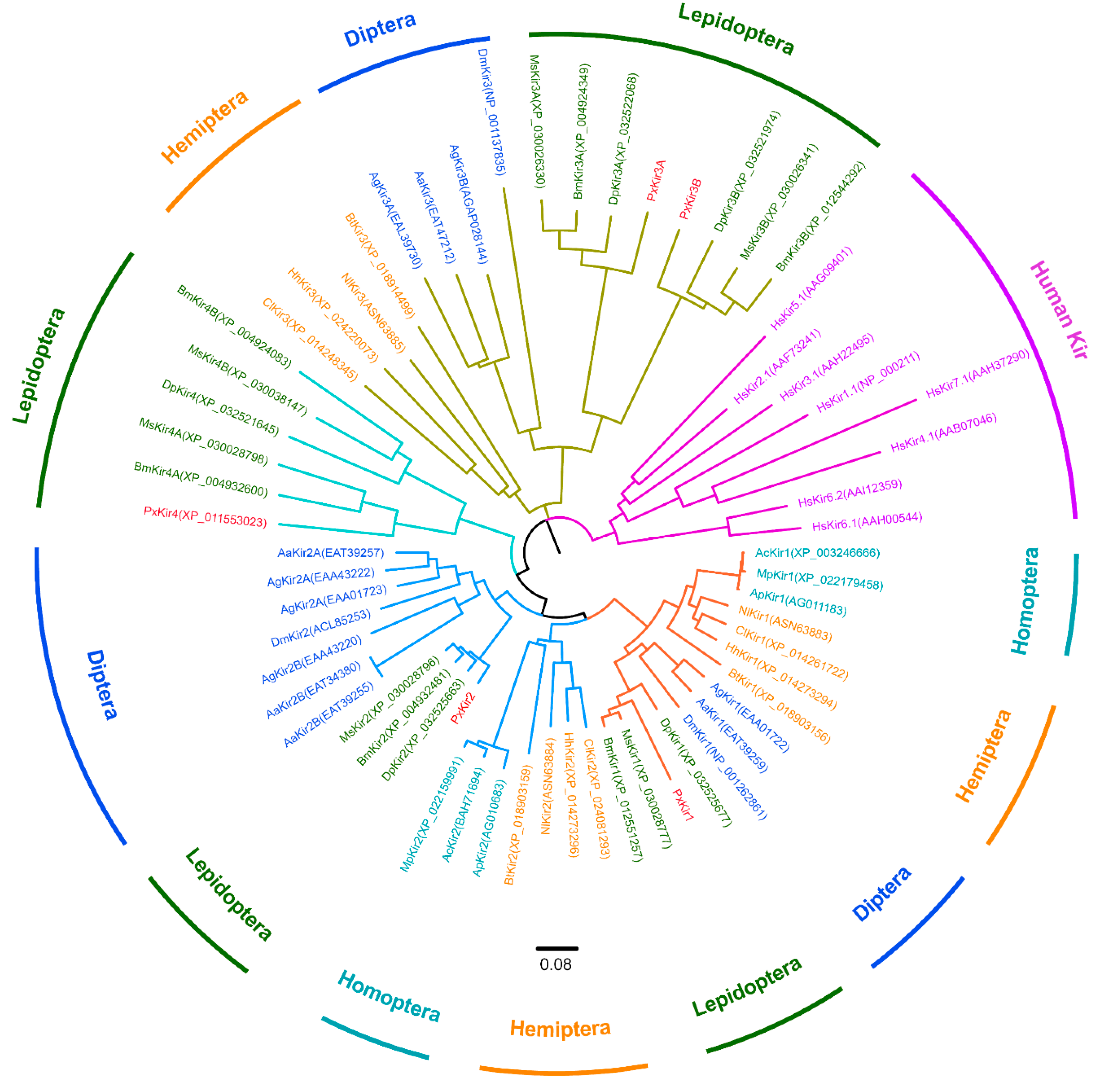
2.3. Sequences Alignment and Phylogenetic Analysis

The Kir channel subunit sequences of other insects were downloaded from GenBank and Clustal X was used for sequence alignment and to analyze the sequence similarity between PxKir subunits and Kir subunits of other species. The transmembrane regions of the protein were analyzed using the protein sequence analysis tool of ExPASy (TMHMM 2.0, http://www.cbs.dtu.dk/services/TMHMM-2.0/, accessed on 05 January 2017). The conserved protein domains were analyzed using the CD-search service (using the NCBI Conserved Domain Database CDD v3.18–55570 PSSMs, http://www.ncbi.nlm.nih.gov/Structure/cdd/wrpsb.cgi, accessed on 4 January 2017). Kir channel amino acid sequences from different species were collected and their evolutionary tree was analyzed using the neighbor-joining method based on the Poisson modified model in MEGA7 software [37].

2.4. Quantitative Real-Time PCR

Head and epidermal samples contained the majority of the central nervous system (CNS) of the organism. Both the midgut and Malpighian tubules are important for digestion and absorption of nutrients as well as for waste discharge. Thorax samples in adults are enriched with muscle tissue, and the ovarian tubules and testes are important for reproduction. Four tissues of larvae and eight tissues of adults were selected for tissue-specific expression analysis. Total RNA was extracted from different samples following the instruction manual of the manufacturer (Vazyme, Nanjing, China). A NanoDrop™ 1000 (Thermo, Wilmington, DE, USA) was used to quantify the extracted total RNA. A 1-μg RNA sample was used in the HiScript Q RT SuperMix for the qPCR (+gDNA wiper) reverse transcription kit. For cDNA synthesis, the total reaction volume was 20 μL. Synthesized cDNA samples were stored at −80 °C until further use. Quantitative analyses of P. xylostella cDNA samples at different life stages were performed using the FastStart Essential DNA Green Master kit (Roche, Basel, Switzerland). A 15-ng cDNA sample, which was reverse transcribed from total RNA, was used as a template for PCR, and Kir subunit genes were amplified using specific primers (see Table 1). P. xylostella ribosomal protein S4 (rpS4, XM_011555372) was amplified and used as internal reference for the quantitative PCR analysis of different insect growth stages. Ribosomal protein L8 (rpL8, NM_001305488) was amplified and used as internal reference for quantitative PCR analyses of different tissues [38,39]. Elongation factor 1 (ef1, EF417849) was also amplified and used to verify the quantitative results (primers shown in Table 1). Three replicates were used per sample. The LightCycler® 96 PCR instrument (Roche Molecular Systems, Inc., Basel, Switzerland) was used for real-time quantitative analysis. The two-step amplification program was conducted as follows: pre-denaturation at 95 °C for 30 s, followed by sample denaturation at 95 °C for 10 s and annealing at 59 °C for 30 s for 40 cycles. The fluorescence value was read at the end of each cycle. The results of three independent samples were analyzed and the expression differences between different samples were compared using the 2−ΔΔCT method [40,41].

2.5. Data Analysis and Statistics

Because of the skewed and wide distribution of the data, gene expression values were log2 transformed, which enabled the application of parametric statistics. As the log of zero is undefined, one was added to all measurements to preserve the zeros in the derived data set [42]. All studies were replicated at least three times and the results were plotted as the means ± standard deviations (SD). One-way analyses of variance (ANOVA) were used, followed by the Tukey–Kramer post-hoc test using GraphPad Prism version 6.01 (GraphPad Software Inc., San Diego, CA, USA).

3. Results

3.1. Kir Channel Subunits in Lepidoptera

The genes of Kir channel subunits of four lepidopteran insects (P. xylostella, B. mori, M. sexta, and D. plexippus) were investigated and 12 partial or complete CDS of kir were identified in the transcriptome of P. xylostella. Furthermore, 19 sequences of kir were identified in the transcriptome of B. mori, as well as 13 kir sequences in both M. sexta and D. plexippus. After removal of redundant sequences, five kir genes were identified in both P. xylostella and D. plexippus, as well as six kir genes in both B. mori and M. sexta (Table 2).
To identify the subclasses of Lepidoptera candidate proteins, 40 previously reported Kir subunit sequences were downloaded from the GenBank database and a neighbor-joining method was used to construct a phylogenetic tree. These sequences included eight human-derived Kir proteins, 14 Kir sequences originating from Diptera (A. aegypti, A. gambiae, and D. melanogaster), six Kir sequences from Homoptera (Acyrthosiphon pisum, Aphis glycines, and Myzus persicae), and 12 Kir sequences from Hemiptera (N. lugens, C. lectularius, B. tabaci, and H. hahalys). The results indicated that insect Kir proteins and human Kir proteins form two independent evolutionary branches. The Kir proteins of Lepidoptera and other insects (i.e., the investigated Diptera and Hemiptera) were located on the same large branch of the phylogenetic tree (Figure 1).
The Kir1 subunits of Lepidoptera and Diptera are located on the same sub branch, while the Kir1 of Hemiptera and Homoptera are located on a different sub branch. Similarly, Kir2 and Kir3 in Lepidoptera, Diptera, Homoptera, and Hemiptera have the same evolutionary relationships with Kir1. Surprisingly, Kir3 from Lepidoptera have two subtypes (Kir3A and Kir3B) and compared with those of Diptera, both Kir3 subunits of Lepidoptera are located on the same sub branch. Moreover, two Kir4 subunits were identified in Lepidoptera. B. mori and M. sexta have Kir4A and Kir4B, but P. xylostella and D. plexippus only have one Kir4. In Lepidoptera, Kir4 was not located on the same branch as Kir1–3, suggesting that it may be a new subclass of the Kir channel in insects.

3.2. The pxkirs Clone

P. xylostella is an important vegetable pest and five Kir-subunit genes from P. xylostella were cloned and sequenced. The results showed that most pxkirs harbored alternative exons, and the pxkirs with the highest splicing frequency were selected for further research in this study. pxkir1 has a full-length open reading frame (ORF) of 1665 bp, and the putative PxKir1 protein has 554 amino acids and a molecular weight (MW) of 63.5 kDa. The CDS of pxkir2 has a length of 1566 bp, and the putative PxKir2 protein has 521 amino acids with a MW of 59.3 kDa. pxkir3A and pxkir3B are relatively similar in size. pxkir3A has a length of 1785 bp, and encodes 594 amino acids (MW: 67.2 kDa). pxkir3B has a sequence length of 1728 bp and encodes 575 amino acids (MW: 65.8 kDa). pxkir4 is the shortest, with a full-length ORF of 1230 bp, and putative PxKir4 has 409 amino acids with an MW of 47.7 kDa.
Sequence homology analysis between Kir subunits in P. xylostella showed that the five putative PxKir subunits have less than 52% sequence similarity and the conservative domains (i.e., the sequences between TM1 and TM2) have less than 73% sequence similarity (Table 3). Specifically, the PxKir1 subunit had 50% similarity with PxKir2, PxKir2 had 51.9% similarity with PxKir4, PxKir3A had 40.8% similarity with PxKir3B, and other sequence similarities remained below 38%. This indicated that Kir subfamilies are significantly different from each other.

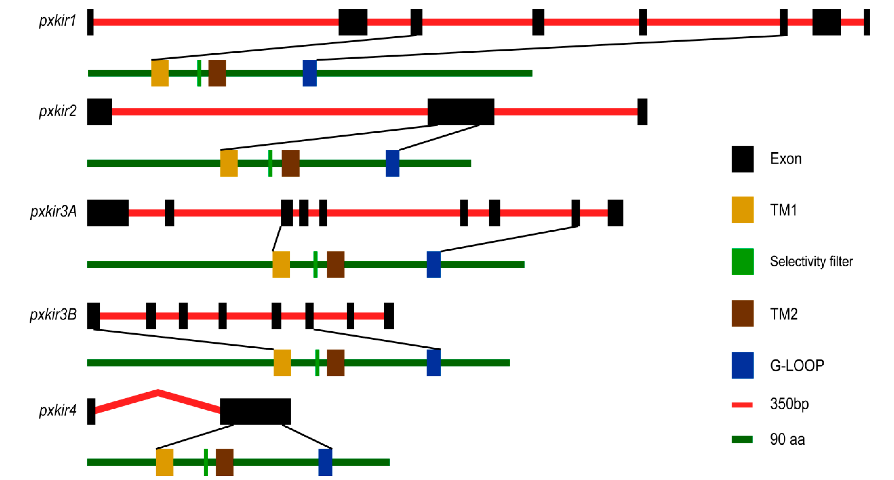
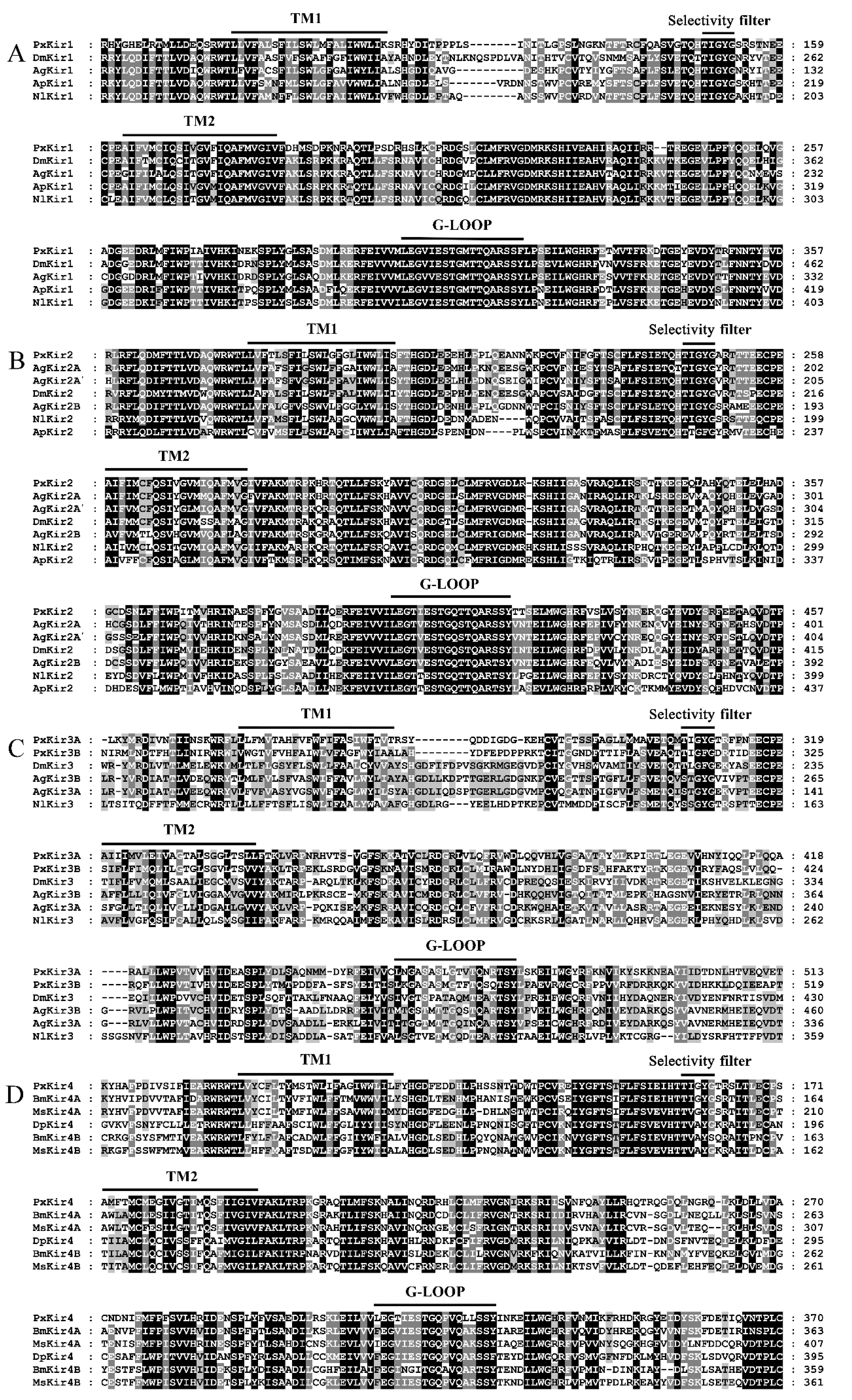
3.3. Sequence Analysis

Genomic sequence analysis indicated that the pxkir1 genomic sequence has a length of 12.1 kb and encodes eight exons. The pxkir2 genomic sequence is 8.9 kb in length and contains three exons. The pxkir3A genomic sequence is 8.5 kb and contains nine exons. The pxkir3B genomic sequence is 4.8 kb and contains eight exons. The pxkir4 is encoded by two exons and has an intron of unknown length (Figure 2). Transmembrane region prediction and CD-search analysis showed that the five deduced proteins all have the conservative domains of the Kir subunit, including the TM1, TM2, and G-LOOP domains. However, the position of conservative regions in the sequence is not consistent. The upstream sequence of TM1 in both PxKir1 and PxKir4 is shorter than other PxKir subunits, while the downstream sequence of the G-LOOP in PxKir1 is longer than that of other PxKir subunits (Figure 2).
Sequence alignments were performed between the PxKir subunits and orthologous in D. melanogaster, A. aegypti, and N. lugens. The results showed that: PxKir1 has a 52–67% sequence similarity and PxKir2 has a 61–77% sequence similarity with orthologous. PxKir3A and PxKir3B have only 32–39% sequence similarity with Kir3 in D. melanogaster, A. aegypti, and N. lugens, indicating that Kir3 showed significant polymorphism between different species. For PxKir4, 59–61% sequence similarity was found for Kir4A and 43–51% for Kir4B in Lepidoptera (Table A2).
Sequence alignments also showed that all PxKir subunits contained two TM helices (TM1 and TM2) and a K+ selectivity filter (containing a “TIGYG” motif between both TM helices). These five candidate PxKir subunits all presented the typical conservative domain features of Kir subunits and should be able to form functional potassium channels (Figure 3). Interestingly, the selectivity filters of Kir4A and Kir4B were significantly different; the sequence was “TI(V)GVG” for Kir4A and “TVAYG(S)” for Kir4B.

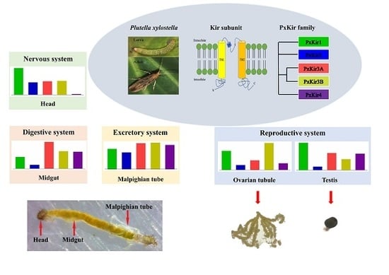
3.4. Developmental and Tissue Distribution of kirs in P. xylostella

The five pxkirs expression profiles were investigated by qRT-PCR. The expression levels of pxkir1 were relatively high throughout all life stages, and the highest mRNA level was found in male adults (the expression was about 1000-fold of the expression level of the pxkir1 in eggs, 95% confidence interval (CI): 953 to 1207-fold, Figure 4A). During the adult stage in different tissues, the highest expression level was found in the thorax, followed by legs and testis, and the pxkir1 mRNA levels were lower in the midguts of larva and the ovarian tubules of female adult. The expression in the thorax was significantly higher than in other tissues, which was more than 2500 times that of the lowest pxkir1 expression in the ovarian tubule (95% CI: 1770 to 4096-fold, Figure 4F).
The pxkir2 mRNA levels were low at the larval and pupae stages, and the highest expression level was found in male adults (approximately 500-fold of the level found in eggs, 95% CI: 425 to 603-fold, Figure 4B). Similar to pxkir1, the highest expression of pxkir2 at the adult stage was in locomotor tissues, and in the thorax, it was approximately 700 times that of the ovarian tubule (95% CI: 619 to 1531-fold, Figure 4G). The expression levels of pxkir1 and pxkir2 differed in reproductive organs. pxkir1 was highly expressed in testes and ovarian tubes (Figure 4F), but pxkir2 showed low expression in both tissues (Figure 4G).
The pxkir3A was also highly expressed in the larval stage, like pxkir1, but it showed significantly lower expression at the pupae and adult stages (Figure 4C). The expression of the pxkir3B was lower during the pupal stage than that of pxkir3A (Figure 4D). pxkir3A was highly expressed in midguts and Malpighian tubules (more than 6000 times in midgut of the larvae stage than that in the ovarian tube, 95% CI: 6019 to 6931-fold, nearly 1500 times in Malpighian tubules of the adult stage than that in the ovarian tube, 95% CI: 1181 to 2093-fold, Figure 4H). pxkir3B was also highly expressed in the midgut and in Malpighian tubules (Figure 4I). pxkir3A was highly expressed in testes and pxkir3B was highly expressed in ovarian tubules.
The expression of pxkir4 was similar to that of pxkir3B, and pxkir4 was almost not expressed in eggs and had low expression in female adults (Figure 4E). Tissue-specific expressions showed that the pxkir4 expression profile was similar to that of pxkir3, and the highest expression was found in Malpighian tubules, followed by the midgut (Figure 4J). Unlike pxkir3, pxkir4 was not expressed in the head of larvae and showed only low expression in the head of adults.

4. Discussion

Kir channels regulate important physiological functions in insects, such as egg production [24], renal function [27,28,43], salivary gland function and feeding [29], developmental signaling [30], and the antiviral immune response [44]. Kir channels have been proposed to be a target of the insecticide flonicamid [19]. Via patch clamp technology, the electrophysiological characteristics of several insect Kir channels have been measured, which provided an efficient tool for the functional research of genes and for Kir-targeting insecticide development [17,22,24,25,43]. At present, most research of the Kir channel focused on Diptera and Hemiptera, and the reports on Lepidoptera are few. Since many important economic insects (e.g., morpho butterfly and silkworm) and agricultural pests (e.g., bollworm, cotton leafworm, cabbage worm, and the diamondback moth) belong to Lepidoptera, it is necessary to investigate the classification and function of Kir subunits in Lepidoptera.
In this study, five kirs were identified in both P. xylostella and D. plexippus, and six kirs were identified in both B. mori and D. plexippus. The kir3 and kir4 in Lepidoptera are diverse, leading to A and B subtypes. The same diversity of Kir2 was reported in mosquitoes [22,45]. Only one kir4 (kir4A) was identified in P. xylostella. The bmkir4B and mskir4B sequences were used for BLAST analysis of the transcriptome of P. xylostella, which did not yield meaningful results. Maybe, the Kir4B was absent because of functional redundancy in P. xylostella. Similarly, the Kir3 subunit was absent from the transcriptome and genome of A. glycines [23] and two Kir channels (Irk1 and Irk2) play redundant roles in the function of the renal tubules of Drosophila [25]. Two Kir subunits were reported in A. glycines [23], three Kir subunits were reported in D. melanogaster and N. lugens [19,25], and four to five Kir subunits were reported in mosquitoes [18,24]. The composition of Kir subunits in different species is quite different; whether the same Kir subtypes that have been identified by phylogenetic analysis have the same function requires further study.
The expression and function of Kir subunits in D. melanogaster, A. aegypti, and N. lugens have been reported: Kir subunits are expressed in the insects’ Malpighian tubules [17,22,25,26], CNS [46], muscle [30], salivary glands [19,29], and reproductive organs [24], where they regulate the insects’ renal function, heart function, patterning, salivary gland function, and virus resistance. This study also investigated the expression of Kir subunits in the CNS (head), digestion and metabolite excretion organs (midgut and Malpighian tubules), locomotive organs (thorax, legs, wings), and reproductive organs (ovarian tubule and testis) of P. xylostella. The pxkirs (except pxkir2) were highly expressed in the Malpighian tubules, pxkir1–3 was enriched in the head, pxkir1 and pxkir2 were highly expressed in the thorax and legs, pxkir3 and pxkir4 were highly expressed in the midgut, pxkir2 was highly expressed in the wings, pxkir3B was highly expressed in the ovarian tubule, and pxkir1, pxkir3A, and pxkir4 were highly expressed in the testes (Figure 4). These data suggested that Kirs may play important roles in the regulation of renal function, gland secretion, locomotion and reproduction of P. xylostella. In particular, Kir3 and Kir4 were highly expressed in Malpighian tubules, which may be potential insecticide targets like AeKir1–2 [15,24,43]. In any case, these data provide useful information for further study of the physiological functions of different Kir subunits in P. xylostella.
Unexpectedly, significant differences of expression were found between pxkirs and homologous genes. For example, comparing the expression levels of kir1 in different development stages showed that kir1 was highly expressed in the pupa and adult stages of D. melanogaster [20] and A. aegypti [18], but was only highly expressed in the pupa stage in A. gambiae [24]. This study showed that pxkir1 was highly expressed throughout all growth stages, and the highest level of pxkir1 mRNA was found in male adults (Figure 4A). Comparing the tissue expression differences of kir1 showed that kir1 was highly expressed in the ovarian tubule of A. aegypti and A. gambiae, and was less expressed in the midgut and fat body [18,24]. However, the expression levels of kir1 in the midgut of C. lectularius [47] and the fat body of N. lugens [19] were all higher than those in the Malpighian tubules. In this study, pxkir1 showed low expression in the ovarian tubule and high expression in the testes (Figure 4F). Moreover, it was difficult to confirm the common expression pattern of Kir2 and Kir3 in different insects because of the gene diversity and alternative splicing of Kir2 [48,49]. In this study, three kinds of alternative splicing were identified for pxkir1 (data not shown). It remains unclear whether the diversity and alternative splicing of kir affect the function of Kir subunits in P. xylostella.
The PxKir4 subunit showed high sequence similarity with PxKir2 (59.2%) and both genes showed a similar extron–intron structure. pxkir4 had a higher expression in the larval midgut and Malpighian tubules (Figure 4J), and a lower expression in the head and eggs (Figure 4E), which is similar to kir2 in other insects [15,20,49]. This suggests that pxkir4 is more closely related to kir2. This study showed that the expression profile of pxkir4 was closer to that of pxkir3, and because of a lack of support by evolutionary analysis and functional analysis, Kir4 was identified as a new type of Kir subunit in Lepidoptera.

5. Conclusions

In this study, five candidate Kir genes (pxkir1, pxkir2, pxkir3A, pxkir3B, and pxkir4) from P. xylostella were cloned for the first time. Sequence analysis showed that all cloned pxkir genes could encode the common conservative domains of the Kir subunit. Phylogenetic analysis suggested that Kir4 in Lepidoptera may be a new subtype of the Kir subunit. The temporal and spatial expressions of the kir genes in P. xylostella were also characterized. These results elucidate the composition and distribution of Kir subunits in P. xylostella and provide useful information for the study of Kir channels in P. xylostella.

Author Contributions

Conceptualization, X.Z.; investigation, X.L. and H.M.; resources, J.X. and W.Z.; data curation, H.M. and X.L.; writing—original draft preparation, X.L.; writing—review and editing, Z.L. and J.L.; supervision, H.Z. and Y.Z.; project administration, X.Z. All authors have read and agreed to the published version of the manuscript.

Funding

This work was financed by The National Science Foundation of China (Grant Nos. 31972305 and 31701811), and the Scientific-Innovative of Hunan Agricultural Sciences and Technology (2017-JC-76).

Acknowledgments

We thank Zezhong Yang from the Department of Plant Protection, Institute of Vegetables and Flowers, Chinese Academy of Agricultural Sciences, Beijing, China, for his help with transcriptome analysis.

Conflicts of Interest

The authors declare no conflicts of interest.

Appendix A

Table A1. Gene amplification procedures of kir in Plutella xylostella.
Cycle Stepspxkir1pxkir2pxkir3apxkir3bpxkir4Cycles
Initial denaturation95 °C 3 min95 °C 3 min95 °C 3 min95 °C 3 min95 °C 3 min1
Denaturation95 °C 15 s95 °C 15 s95 °C 15 s95 °C 15 s95 °C 15 s32
Annealing60 °C 15 s50 °C 15 s62 °C 15 s58 °C 15 s60 °C 15 s
Extension72 °C 120 s72 °C 90 s72 °C 100 s72 °C 100 s72 °C 90 s
Final elongation72 °C 5 min72 °C 5 min72 °C 5 min72 °C 5 min72 °C 5 min1
Table A2. Amino acid sequence similarity of Kirs between Plutella xylostella and other insects.
Kir of P. xylostellaKir of Other InsectsSimilarity (%)
PxKir1AaKir152
AgKir154
DmKir152
NlKir167
ApKir161
PxKir2AaKir2A77
AaKir2B64
AaKir2B’64
AgKir2A74
AgKir2A’67
AgKir2B61
DmKir269
NlKir267
ApKir255
PxKir3AAaKir333
AgKir3A39
DmKir334
NlKir332
PxKir3BAaKir339
AgKir3A38
DmKir338
NlKir336
PxKir4BmKir4A61
BmKir4B47
MsKir4A59
MsKir4B51
DpKir447

References

  1. Minor, D.L., Jr.; Masseling, S.J.; Jan, Y.N.; Jan, L.Y. Transmembrane structure of an inwardly rectifying potassium channel. Cell 1999, 96, 879–891. [Google Scholar] [CrossRef] [Green Version]
  2. Lu, Z.; Klem, A.M.; Ramu, Y. Ion conduction pore is conserved among potassium channels. Nature 2001, 413, 809–813. [Google Scholar] [CrossRef] [PubMed]
  3. Hibino, H.; Inanobe, A.; Furutani, K.; Murakami, S.; Findlay, I.; Kurachi, Y. Inwardly rectifying potassium channels: Their structure, function, and physiological roles. Physiol. Rev. 2010, 90, 291–366. [Google Scholar] [CrossRef] [PubMed] [Green Version]
  4. Gonzalez, C.; Baez-Nieto, D.; Valencia, I.; Oyarzun, I.; Rojas, P.; Naranjo, D.; Latorre, R. K+ channels: Function-structural overview. Compr. Physiol. 2012, 2, 2087–2149. [Google Scholar] [CrossRef]
  5. Abraham, M.R.; Jahangir, A.; Alekseev, A.E.; Terzic, A. Channelopathies of inwardly rectifying potassium channels. FASEB J. 1999, 13, 1901–1910. [Google Scholar] [CrossRef] [PubMed]
  6. Pattnaik, B.R.; Asuma, M.P.; Spott, R.; Pillers, D.A. Genetic defects in the hotspot of inwardly rectifying K+ (Kir) channels and their metabolic consequences: A review. Mol. Genet. Metab. 2012, 105, 64–72. [Google Scholar] [CrossRef] [Green Version]
  7. Mayfield, J.; Blednov, Y.A.; Harris, R.A. Behavioral and Genetic Evidence for GIRK Channels in the CNS: Role in Physiology, Pathophysiology, and Drug Addiction. Int. Rev. Neurobiol. 2015, 123, 279–313. [Google Scholar] [CrossRef] [Green Version]
  8. Sacco, S.; Giuliano, S.; Sacconi, S.; Desnuelle, C.; Barhanin, J.; Amri, E.Z.; Bendahhou, S. The inward rectifier potassium channel Kir2.1 is required for osteoblastogenesis. Hum. Mol. Genet. 2015, 24, 471–479. [Google Scholar] [CrossRef] [Green Version]
  9. Tinker, A.; Aziz, Q.; Li, Y.W.; Specterman, M. ATP-Sensitive Potassium Channels and Their Physiological and Pathophysiological Roles. Compr. Physiol. 2018, 8, 1463–1511. [Google Scholar] [CrossRef]
  10. Clarke, O.B.; Caputo, A.T.; Hill, A.P.; Vandenberg, J.I.; Smith, B.J.; Gulbis, J.M. Domain reorientation and rotation of an intracellular assembly regulate conduction in Kir potassium channels. Cell 2010, 141, 1018–1029. [Google Scholar] [CrossRef] [Green Version]
  11. Li, N.N.; Wu, J.X.; Ding, D.; Cheng, J.X.; Gao, N.; Chen, L. Structure of a Pancreatic ATP-Sensitive Potassium Channel. Cell 2017, 168, 101–110.e110. [Google Scholar] [CrossRef] [PubMed] [Green Version]
  12. Tao, X.; Avalos, J.L.; Chen, J.Y.; MacKinnon, R. Crystal structure of the eukaryotic strong inward-rectifier K+ channel Kir2.2 at 3.1 A resolution. Science 2009, 326, 1668–1674. [Google Scholar] [CrossRef] [PubMed] [Green Version]
  13. Hansen, S.B.; Tao, X.; MacKinnon, R. Structural basis of PIP2 activation of the classical inward rectifier K+ channel Kir2.2. Nature 2011, 477, 495–498. [Google Scholar] [CrossRef] [PubMed] [Green Version]
  14. Nishida, M.; MacKinnon, R. Structural basis of inward rectification: Cytoplasmic pore of the G protein-gated inward rectifier GIRK1 at 1.8 A resolution. Cell 2002, 111, 957–965. [Google Scholar] [CrossRef] [Green Version]
  15. Rouhier, M.F.; Raphemot, R.; Denton, J.S.; Piermarini, P.M. Pharmacological validation of an inward-rectifier potassium (Kir) channel as an insecticide target in the yellow fever mosquito Aedes aegypti. PLoS ONE 2014, 9, e100700. [Google Scholar] [CrossRef]
  16. Reale, V.; Chatwin, H.M.; Evans, P.D. The activation of G-protein gated inwardly rectifying K+ channels by a cloned Drosophila melanogaster neuropeptide F-like receptor. Eur. J. Neurosci. 2004, 19, 570–576. [Google Scholar] [CrossRef]
  17. Piermarini, P.M.; Dunemann, S.M.; Rouhier, M.F.; Calkins, T.L.; Raphemot, R.; Denton, J.S.; Hine, R.M.; Beyenbach, K.W. Localization and role of inward rectifier K+ channels in Malpighian tubules of the yellow fever mosquito Aedes aegypti. Insect Biochem. Mol. Biol. 2015, 67, 59–73. [Google Scholar] [CrossRef] [Green Version]
  18. Yang, Z.X.; Statler, B.M.; Calkins, T.L.; Alfaro, E.; Esquivel, C.J.; Rouhier, M.F.; Denton, J.S.; Piermarini, P.M. Dynamic expression of genes encoding subunits of inward rectifier potassium (Kir) channels in the yellow fever mosquito Aedes aegypti. Comp. Biochem. Physiol. B Biochem. Mol. Biol. 2017, 204, 35–44. [Google Scholar] [CrossRef]
  19. Ren, M.M.; Niu, J.G.; Hu, B.; Wei, Q.; Zheng, C.; Tian, X.R.; Gao, C.F.; He, B.J.; Dong, K.; Su, J.Y. Block of Kir channels by flonicamid disrupts salivary and renal excretion of insect pests. Insect Biochem. Mol. Biol. 2018, 99, 17–26. [Google Scholar] [CrossRef]
  20. Doring, F.; Wischmeyer, E.; Kuhnlein, R.P.; Jackle, H.; Karschin, A. Inwardly rectifying K+ (Kir) channels in Drosophila. A crucial role of cellular milieu factors Kir channel function. J. Biol. Chem. 2002, 277, 25554–25561. [Google Scholar] [CrossRef] [Green Version]
  21. MacLean, S.J.; Andrews, B.C.; Verheyen, E.M. Characterization of Dir: A putative potassium inward rectifying channel in Drosophila. Mech. Dev. 2002, 116, 193–197. [Google Scholar] [CrossRef]
  22. Piermarini, P.M.; Rouhier, M.F.; Schepel, M.; Kosse, C.; Beyenbach, K.W. Cloning and functional characterization of inward-rectifying potassium (Kir) channels from Malpighian tubules of the mosquito Aedes aegypti. Insect Biochem. Mol. Biol. 2013, 43, 75–90. [Google Scholar] [CrossRef] [PubMed] [Green Version]
  23. Piermarini, P.M.; Inocente, E.A.; Acosta, N.; Hopkins, C.R.; Denton, J.S.; Michel, A.P. Inward rectifier potassium (Kir) channels in the soybean aphid Aphis glycines: Functional characterization, pharmacology, and toxicology. J. Insect Physiol. 2018, 110, 57–65. [Google Scholar] [CrossRef] [PubMed]
  24. Raphemot, R.; Estevez-Lao, T.Y.; Rouhier, M.F.; Piermarini, P.M.; Denton, J.S.; Hillyer, J.F. Molecular and functional characterization of Anopheles gambiae inward rectifier potassium (Kir1) channels: A novel role in egg production. Insect Biochem. Mol. Biol. 2014, 51, 10–19. [Google Scholar] [CrossRef] [Green Version]
  25. Wu, Y.P.; Baum, M.; Huang, C.L.; Rodan, A.R. Two inwardly rectifying potassium channels, Irk1 and Irk2, play redundant roles in Drosophila renal tubule function. Am. J. Physiol. Regul. Integr. Comp. Physiol. 2015, 309, R747–R756. [Google Scholar] [CrossRef] [PubMed] [Green Version]
  26. Piermarini, P.M.; Esquivel, C.J.; Denton, J.S. Malpighian Tubules as Novel Targets for Mosquito Control. Int. J. Environ. Res. Public Health 2017, 14, 111. [Google Scholar] [CrossRef] [Green Version]
  27. Lewis, L.M.; Bhave, G.; Chauder, B.A.; Banerjee, S.; Lornsen, K.A.; Redha, R.; Fallen, K.; Lindsley, C.W.; Weaver, C.D.; Denton, J.S. High-throughput screening reveals a small-molecule inhibitor of the renal outer medullary potassium channel and Kir7.1. Mol. Pharmacol. 2009, 76, 1094–1103. [Google Scholar] [CrossRef] [Green Version]
  28. Kharade, S.V.; Sheehan, J.H.; Figueroa, E.E.; Meiler, J.; Denton, J.S. Pore Polarity and Charge Determine Differential Block of Kir1.1 and Kir7.1 Potassium Channels by Small-Molecule Inhibitor VU590. Mol. Pharmacol. 2017, 92, 338–346. [Google Scholar] [CrossRef]
  29. Swale, D.R.; Li, Z.L.; Guerrero, F.; Perez De Leon, A.A.; Foil, L.D. Role of inward rectifier potassium channels in salivary gland function and sugar feeding of the fruit fly, Drosophila melanogaster. Pestic. Biochem. Physiol. 2017, 141, 41–49. [Google Scholar] [CrossRef] [Green Version]
  30. Dahal, G.R.; Pradhan, S.J.; Bates, E.A. Inwardly rectifying potassium channels influence Drosophila wing morphogenesis by regulating Dpp release. Development 2017, 144, 2771–2783. [Google Scholar] [CrossRef] [Green Version]
  31. Fenner, K.; Canonica, S.; Wackett, L.P.; Elsner, M. Evaluating pesticide degradation in the environment: Blind spots and emerging opportunities. Science 2013, 341, 752–758. [Google Scholar] [CrossRef] [PubMed] [Green Version]
  32. Zhang, S.Z.; Zhang, X.L.; Shen, J.; Li, D.Y.; Wan, H.; You, H.; Li, J.H. Cross-resistance and biochemical mechanisms of resistance to indoxacarb in the diamondback moth, Plutella xylostella. Pestic. Biochem. Physiol. 2017, 140, 85–89. [Google Scholar] [CrossRef]
  33. Yin, C.Y.; Wang, R.; Luo, C.; Zhao, K.; Wu, Q.Y.; Wang, Z.Y.; Yang, G.F. Monitoring, Cross-Resistance, Inheritance, and Synergism of Plutella xylostella (Lepidoptera: Plutellidae) Resistance to Pyridalyl in China. J. Econ. Entomol. 2019, 112, 329–334. [Google Scholar] [CrossRef]
  34. Xu, J.; Wang, Z.Y.; Wang, Y.F.; Ma, H.H.; Zhu, H.; Liu, J.; Zhou, Y.; Deng, X.L.; Zhou, X.M. ABCC2 participates in the resistance of Plutella xylostella to chemical insecticides. Pestic. Biochem. Physiol. 2020, 162, 52–59. [Google Scholar] [CrossRef]
  35. Ma, H.H.; Huang, Q.T.; Lai, X.Y.; Liu, J.; Zhu, H.; Zhou, Y.; Deng, X.L.; Zhou, X.M. Pharmacological Properties of the Type 1 Tyramine Receptor in the Diamondback Moth, Plutella xylostella. Int. J. Mol. Sci. 2019, 20, 2953. [Google Scholar] [CrossRef] [PubMed] [Green Version]
  36. Zhou, J.L.; Guo, Z.J.; Kang, S.; Qin, J.Y.; Gong, L.J.; Sun, D.; Guo, L.; Zhu, L.H.; Bai, Y.; Zhang, Z.Z.; et al. Reduced expression of the P-glycoprotein gene PxABCB1 is linked to resistance to Bacillus thuringiensis Cry1Ac toxin in Plutella xylostella (L.). Pest Manag. Sci. 2020, 76, 712–720. [Google Scholar] [CrossRef] [PubMed]
  37. Huang, Q.T.; Ma, H.H.; Deng, X.L.; Zhu, H.; Liu, J.; Zhou, Y.; Zhou, X.M. Pharmacological characterization of a beta-adrenergic-like octopamine receptor in Plutella xylostella. Arch. Insect Biochem. Physiol. 2018, 98, e21466. [Google Scholar] [CrossRef]
  38. Fu, W.; Xie, W.; Zhang, Z.; Wang, S.L.; Wu, Q.J.; Liu, Y.; Zhou, X.; Zhou, X.M.; Zhang, Y.J. Exploring valid reference genes for quantitative real-time PCR analysis in Plutella xylostella (Lepidoptera: Plutellidae). Int. J. Biol. Sci. 2013, 9, 792–802. [Google Scholar] [CrossRef] [Green Version]
  39. You, Y.C.; Xie, M.; Vasseur, L.; You, M.S. Selecting and validating reference genes for quantitative real-time PCR in Plutella xylostella (L.). Genome 2018, 61, 349–358. [Google Scholar] [CrossRef] [Green Version]
  40. Hua, J.F.; Zhang, S.; Cui, J.J.; Wang, D.J.; Wang, C.Y.; Luo, J.Y.; Lv, L.M.; Ma, Y. Functional characterizations of one odorant binding protein and three chemosensory proteins from Apolygus lucorum (Meyer-Dur) (Hemiptera: Miridae) legs. J. Insect Physiol. 2013, 59, 690–696. [Google Scholar] [CrossRef]
  41. Livak, K.J.; Schmittgen, T.D. Analysis of relative gene expression data using real-time quantitative PCR and the 2(-Delta Delta C(T)) Method. J. Methods 2001, 25, 402–408. [Google Scholar] [CrossRef] [PubMed]
  42. Wilkinson, R.; Wang, X.J.; Kassianos, A.J.; Zuryn, S.; Roper, K.E.; Osborne, A.; Sampangi, S.; Francis, L.; Raghunath, V.; Healy, H. Laser capture microdissection and multiplex-tandem PCR analysis of proximal tubular epithelial cell signaling in human kidney disease. PLoS ONE 2014, 9, e87345. [Google Scholar] [CrossRef] [PubMed] [Green Version]
  43. Raphemot, R.; Rouhier, M.F.; Hopkins, C.R.; Gogliotti, R.D.; Lovell, K.M.; Hine, R.M.; Ghosalkar, D.; Longo, A.; Beyenbach, K.W.; Denton, J.S.; et al. Eliciting renal failure in mosquitoes with a small-molecule inhibitor of inward-rectifying potassium channels. PLoS ONE 2013, 8, e64905. [Google Scholar] [CrossRef] [PubMed] [Green Version]
  44. O’Neal, S.T.; Swale, D.R.; Anderson, T.D. ATP-sensitive inwardly rectifying potassium channel regulation of viral infections in honeybees. Sci. Rep. 2017, 7, 8668. [Google Scholar] [CrossRef] [Green Version]
  45. Raphemot, R.; Rouhier, M.F.; Swale, D.R.; Days, E.; Weaver, C.D.; Lovell, K.M.; Konkel, L.C.; Engers, D.W.; Bollinger, S.R.; Hopkins, C.; et al. Discovery and characterization of a potent and selective inhibitor of Aedes aegypti inward rectifier potassium channels. PLoS ONE 2014, 9, e110772. [Google Scholar] [CrossRef]
  46. Chen, R.; Swale, D.R. Inwardly rectifying potassium (Kir) channels represent acritical ion conductance pathway in the nervous systems of insects. Sci. Rep. 2018, 8, 1617. [Google Scholar] [CrossRef]
  47. Mamidala, P.; Mittapelly, P.; Jones, S.C.; Piermarini, P.M.; Mittapalli, O. Molecular characterization of genes encoding inward rectifier potassium (Kir) channels in the bed bug (Cimex lectularius). Comp. Biochem. Physiol. Part B 2013, 164, 275–279. [Google Scholar] [CrossRef]
  48. Rouhier, M.F.; Piermarini, P.M. Identification of life-stage and tissue-specific splice variants of an inward rectifying potassium (Kir) channel in the yellow fever mosquito Aedes aegypti. Insect Biochem. Mol. Biol. 2014, 48, 91–99. [Google Scholar] [CrossRef]
  49. Evans, J.M.; Allan, A.K.; Davies, S.A.; Dow, J.A. Sulphonylurea sensitivity and enriched expression implicate inward rectifier K+ channels in Drosophila melanogaster renal function. J. Exp. Biol. 2005, 208, 3771–3783. [Google Scholar] [CrossRef] [Green Version]
Figure 1. Phylogenetic analysis of PxKirs and various Kir subunits. Neighbor-joining tree showing the phylogenetic relationships of insect and human Kir channels (according to their amino-acid sequences). MEGA 7 was used to construct the tree (the neighbor-joining method was used; bootstrap test used 1000 replicates; model; amino: Poisson correction; gaps and treatment by pairwise deletion). The PxKir subunit is indicated with red color. GenBank accession numbers of Kir proteins are shown in parentheses. Abbreviations of species in alphabetical order: Aa: Aedes aegypti; Ac: Acyrthosiphon pisum; Ap: Aphis glycines; Ag: Anopheles gambiae; Bm: Bombyx mori; Bt: Bemisia tabaci; Cl: Cimex lectularius; Dm: Drosophila melanogaster; Dp: Danaus plexippus; Hh: Halyomorpha halys; Hs: Homo sapiens; Mp: Myzus persicae; Ms: Manduca sexta; Nl: Nilaparvata lugens; Px: Plutella xylostella.
Insects 11 00461 g001
Figure 2. Genomic structures of pxkir and conservative domains of the deduced proteins. The exon−intron organization of pxkir was determined by sequence comparison between genomic sequence and cDNA sequence. The red line of every gene represents the original genomic scaffold sequences and the exons are indicated as black boxes. The green line indicates the deduced protein sequence. The locations encode the two transmembrane helices (TM1 and TM2), selectivity filters, and G-LOOP, which are marked by colored rectangles.
Insects 11 00461 g002
Figure 3. Multiple sequence alignment of putative PxKirs and other orthologous proteins in conservative domains. The respective positions are indicated on the right. Identical residues between orthologous sequences are shown as white characters against a black background, and conservative substitutions are shaded. The transmembrane helices (TM1 and TM2), the selectivity filter (SF), and the G-LOOP are highlighted with black lines. The conservative domains of Kir1 (A), Kir2 (B), Kir3 (C), and Kir4 (D) were aligned. GenBank accession numbers: A. gambiae (AgKir1, EAA01722; AgKir2A, EAA43222; AgKir2A’, EAA01723; AgKir2B, EAA43220; AgKir3A, EAL39730; and AgKir3B, AGAP028144); A. glycines (ApKir1, AG011183; and ApKir2, AG010683); D. melanogaster (DmKir1, NP_001262861; DmKir2, ACL85253; and DmKir3, NP_001137835); N. lugens (NlKir1, ASN63883; NlKir2, ASN63884; and NlKir3, ASN63885); P. xylostella (PxKir1–3, in this study; and PxKir4, XP_011553023); B. mori (BmKir4A, XP_004932600; and BmKir4B, XP_012544061); M. sexta (MsKir4A, XP_030028798; and MsKir4B, XP_030038147); and D. plexippus (DpKir4, XP_032521645).
Insects 11 00461 g003
Figure 4. Log2 relative mRNA expression levels of pxkirs at different developmental stages (AE) and in different tissues (FJ). Different developmental stages of P. xylostella, including eggs, 1st to 4th instar larvae, prepupae, pupae (female and male), and adults (female and male). The expression level of pxkir4 during the egg period was defined as a benchmark for statistical analysis, using rpS4 as the reference gene. Different tissues come from larvae (fourth instar) and adults. Tissue abbreviations: head (Hd), epidermal samples (Ep), Malpighian tubules (Mt), and midgut (Mg). The expression level of the pxkir4 during the egg period was defined as a benchmark for statistical analysis, by using rpL8 as a reference gene. Different lower-case letters above the bars indicate significant differences. The data are presented as means ± SD (N > 3).
Insects 11 00461 g004
Table 1. Primers used for clone and expression analysis of genes in Plutella xylostella.
NamePrimer Sequences (5′-3′)GeneSitePurpose
PxKir1_FATGAAGAGATGTCGGCCATGAAGAGATpxkir1−8DNA cloning
PxKir1_RTACACTTCTGCGTCTGGTGCTGACG1861
PxKir2_FCTTTCGTATTTAGTATCTATGGTTpxkir2−92
PxKir2_RAATGAGATAGTTCGTAAAGTTGCT1830
PxKir3A_FTAAGTTGGACACATCATTGAGCTTpxkir3A−135
PxKir3A_RGAAAATCAGTTTCCTACGTTGGATAGTT1997
PxKir3B_FCTGCTGTAGGCTGATGATGGTGATpxkir3B−123
PxKir3B_RAAGTCAAGAATCGTTTTGTTATGTG1890
PxKir4_FTTTACCGGCATGGCATTCTATTACGpxkir4−9
PxKir4_RGAAAATGAAGGTAGCAACTTTACAAATACA1375
PxKir1-qFGAGGTGCTGCCGTTCTACpxkir1703Real-time Quantitative PCR
PxKir1-qRTCGACTCGATCACTCCCT895
PxKir2-qFATTGGACAGTTCACGATACATAGGAApxkir2336
PxKir2-qRCCGAGCCAGGAGAGGATGAAG575
PxKir3A-qFGGGGGTACCGCTTCAAGAATGTTATpxkir3A1441
PxKir3A-qRGGAGGAACCCTGTGTGACTGAG1626
PxKir3B-qFACAAGGCTGGCTATGGCGACTApxkir3B610
PxKir3B-qRGATGTACCAGAAACCAGCGAAC822
PxKir4-qFTATCATCCAGGAAGTGGCTAAGGCTpxkir462
PxKir4-qRGTCGGGAAACGCATGGTACTTGTGT234
RPL8-qFCGGTCGTGCCTACCACAAATACArpL8559
RPL8-qRCGTGAGGATGCTCCACAGGGT648
RPS4-qFATGGATGTTGTGTCGATTGAAAAGArpS4335
RPS4-qRGAGTGATGCGGTGGATGGTGA423
EF1-qFGCCTCCCTACAGCGAATCef1374
EF1-qRCCTTGAACCAGGGCATCT535
Table 2. The kir genes of P. xylostella, B. mori, M. sexta, and D. plexippus in GenBank.
GeneP. xylostellaB. moriM. sextaD. plexippus
kir1g814.t1 * (MT274448)XM_012695801.2XM_030172912XM_032669786
kir2XM_011554745 (MN894569)XM_004932424.3XM_030172936XM_032669772
kir3AXM_011568587 (MN894570)XM_004924292.3XM_030170470XM_032666177
kir3BXM_011564409 (MN894571)XM_021346768.1XM_030170480AGBW02010886
kir4AXM_011554721XM_004932543.3XM_030172938-
kir4B-XM_012688607.2XM_030182287XM_032665754
* kir1 in Lepbase (http://ensembl.lepbase.org/Plutella_xylostella_pacbiov1/Info/Index accessend on 20 February 2017). The accession numbers of the pxkir gene in this study are shown in parentheses.
Table 3. Amino acid sequence similarity between Plutella xylostella Kir subunits.
ChannelPxKir1 (%)PxKir2 (%)PxKir3A (%)PxKir3B (%)PxKir4 (%)
PxKir110050.0 (72.0)27.0 (41.7)37.7 (45.8)36.0 (47.3)
PxKir2 10032.1 (72.7)31.9 (62.1)51.9 (59.2)
PxKir3A 10040.8 (52.4)35.3 (38.0)
PxKir3B 10034.4 (43.1)
PxKir4 100
Note: The percentages of conservatively substituted amino acids (in parenthesis) between all five PxKir subunits were calculated via pairwise alignments of the complete amino acid sequences.

