Article

Prioritizing Endangered Species in Genome Sequencing: Conservation Genomics in Action with the First Platinum-Standard Reference-Quality Genome of the Critically Endangered European Mink Mustela lutreola L., 1761

1
Institute of Marine and Environmental Sciences, University of Szczecin, Wąska 13 St., 71-415 Szczecin, Poland
2
Polish Society for Conservation Genetics LUTREOLA, Maciejkowa 21 St., 71-784 Szczecin, Poland
3
Wildtier- und Artenschutzstation e.V., Hohe Warte 1, 31553 Sachsenhagen, Germany
4
EuroNerz e.V., Kleine Gildewart 3, 49074 Osnabrück, Germany
5
Vertebrate Genome Laboratory, The Rockefeller University, 1230 York Avenue, Box 366, New York, NY 10065, USA
*
Author to whom correspondence should be addressed.
Int. J. Mol. Sci. 2023, 24(19), 14816; https://doi.org/10.3390/ijms241914816
Submission received: 1 September 2023 / Revised: 23 September 2023 / Accepted: 26 September 2023 / Published: 1 October 2023
(This article belongs to the Special Issue State-of-the-Art Molecular Genetics and Genomics in Poland 2.0)

Abstract

:
The European mink Mustela lutreola (Mustelidae) ranks among the most endangered mammalian species globally, experiencing a rapid and severe decline in population size, density, and distribution. Given the critical need for effective conservation strategies, understanding its genomic characteristics becomes paramount. To address this challenge, the platinum-quality, chromosome-level reference genome assembly for the European mink was successfully generated under the project of the European Mink Centre consortium. Leveraging PacBio HiFi long reads, we obtained a 2586.3 Mbp genome comprising 25 scaffolds, with an N50 length of 154.1 Mbp. Through Hi-C data, we clustered and ordered the majority of the assembly (>99.9%) into 20 chromosomal pseudomolecules, including heterosomes, ranging from 6.8 to 290.1 Mbp. The newly sequenced genome displays a GC base content of 41.9%. Additionally, we successfully assembled the complete mitochondrial genome, spanning 16.6 kbp in length. The assembly achieved a BUSCO (Benchmarking Universal Single-Copy Orthologs) completeness score of 98.2%. This high-quality reference genome serves as a valuable genomic resource for future population genomics studies concerning the European mink and related taxa. Furthermore, the newly assembled genome holds significant potential in addressing key conservation challenges faced by M. lutreola. Its applications encompass potential revision of management units, assessment of captive breeding impacts, resolution of phylogeographic questions, and facilitation of monitoring and evaluating the efficiency and effectiveness of dedicated conservation strategies for the European mink. This species serves as an example that highlights the paramount importance of prioritizing endangered species in genome sequencing projects due to the race against time, which necessitates the comprehensive exploration and characterization of their genomic resources before their populations face extinction.

1. Introduction

The alarming decline of biodiversity worldwide necessitates urgent conservation measures, particularly for wild, endangered, and understudied species. According to the International Union for Conservation of Nature’s (IUCN) Red List of Threatened Species, of the 5973 mammal species assessed, 1340 were classified as threatened with extinction, including 233 critically endangered taxa [
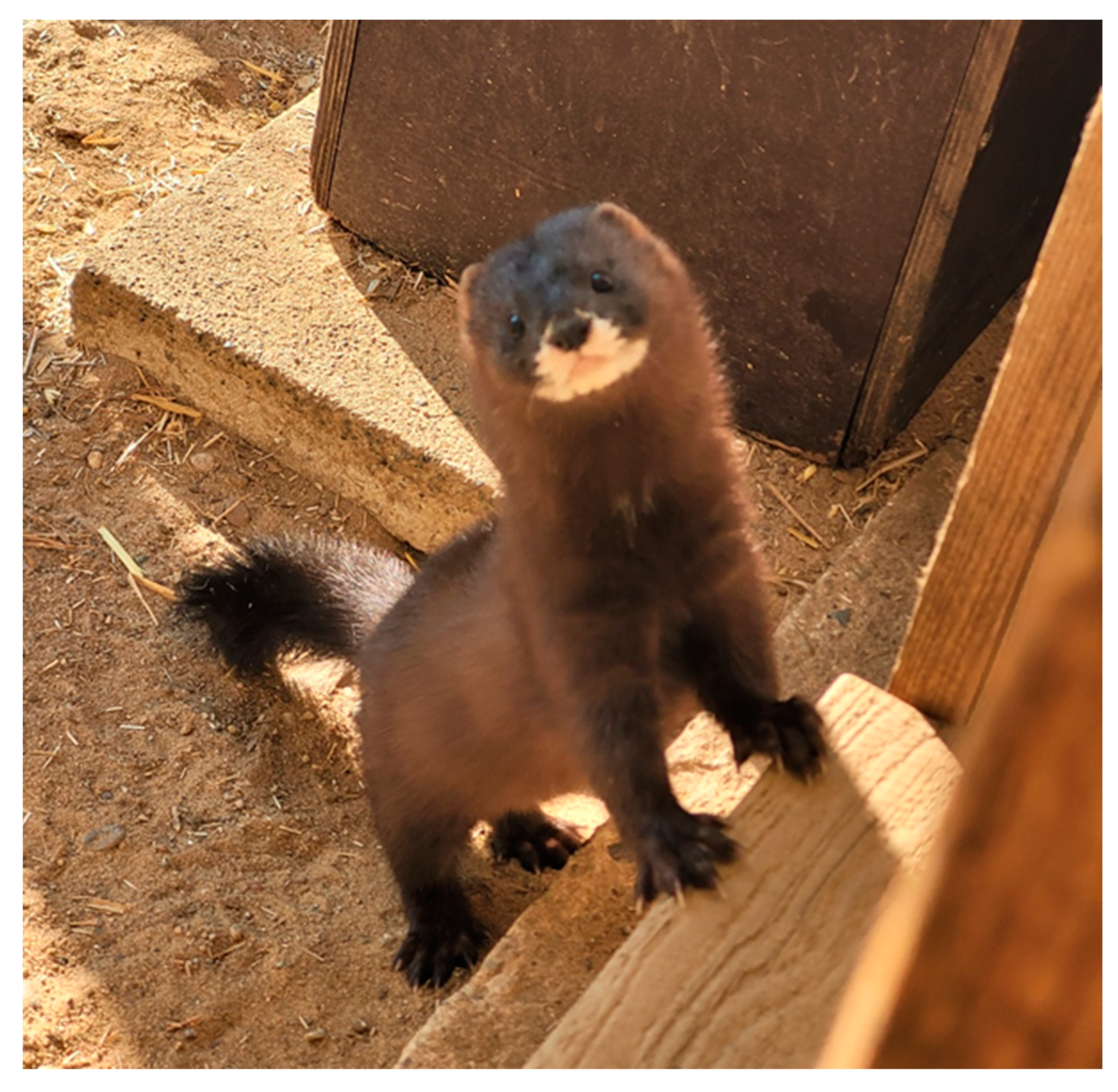
1]. This number includes wild, non-domesticated, non-model species, whose rapidly vanishing genetic resources may never be adequately explored and described. This concerns the European mink Mustela lutreola L., 1761, a critically endangered, semiaquatic, secretive, and solitary representative of the mustelid family (Eukaryota; Metazoa; Chordata; Craniata; Vertebrata; Euteleostomi; Mammalia; Eutheria; Laurasiatheria; Carnivora; Caniformia; Mustelidae; Mustelinae; Mustela) [2,3] (Figure 1). The species is listed in the Annex II to the Bern Convention on the Conservation of European Wildlife and Natural Habitats, the Annexes II and IV (priority species) of the Council Directive 92/43/EEC on the conservation of natural habitats and of wild fauna and flora, and in the Carpathian List of Endangered Species (critically endangered species), and considered one of the most endangered mammalian species in the world [4,5].
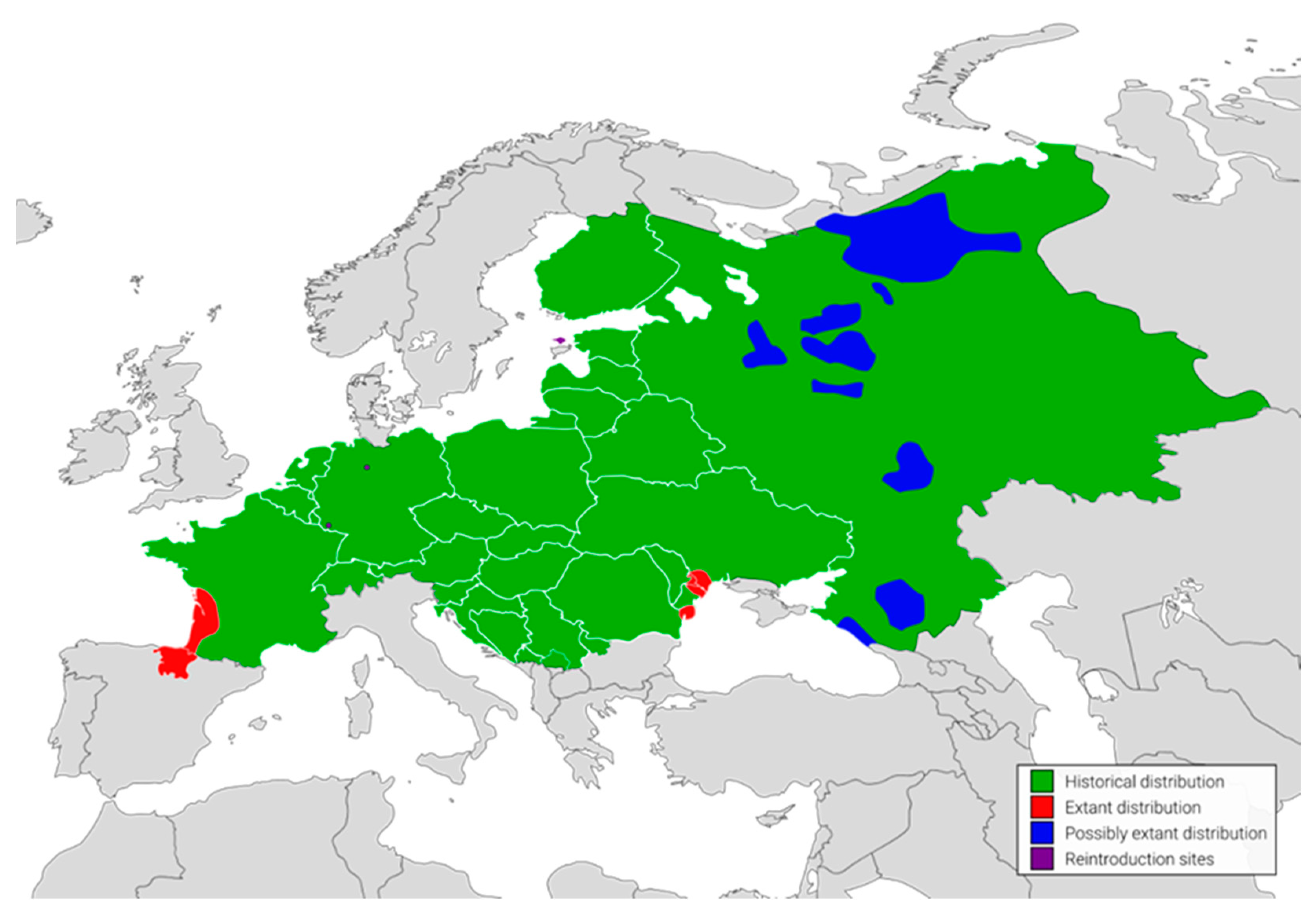
By the 19th century, European mink populations were relatively abundant and distributed across freshwater habitats throughout a large part of continental Europe [5] (Figure 2). Following a combination of habitat loss and fragmentation, commercial over-hunting for fur and the effects of introduced, invasive American mink Neogale vison Schreber, 1777 lead to a dramatic depletion of the species’ populations, in terms of both shrinkage of a geographical range by 97%, and a reduction in the number of individuals persisting in the wild to about 5000 [5,6,7]. What is more, an expected decline rate in number over the next three generations exceeds 80% [5]. These days, only three isolated, declining populations, restricted to the European part of Russia, the Danube Delta, south-western France and north-eastern Spain survived [5]. Reintroduction efforts successfully established populations in Estonia on Hiiumaa Island, as well as in Germany at two locations in Saarland and Lower Saxony [5].
In response to the critical status of M. lutreola, conservation efforts have been initiated in the late 20th century, focusing on captive breeding programmes (i.e., the European Association of Zoos and Aquaria (EAZA) Ex-situ Programme (EEP) for European mink, established in 1992, regional captive breeding programme initiated in Spain in 2004, and the European Mink Breeding Centre of the Ilmen Nature Reserve in Russia, operating since 2010), habitat restoration, local reintroduction initiatives (launched in Estonia, France, Germany, Russia and Spain), reintroduced populations monitoring, and public awareness campaigns [8]. Of key importance for the effectiveness and efficiency of these measures are genomic research, informing targeted and evidence-based conservation strategies [9,10]. By studying the genomic makeup of the European mink, valuable insights into its population structure, genetic diversity, and evolutionary history can be gained. This knowledge is essential for understanding the unique adaptations and vulnerabilities of this species, as well as identifying and quantifying inbreeding, hybridization, and introgression [11,12,13,14,15]. Such information is vital for developing targeted conservation efforts, including captive breeding programmes, reintroduction and translocation strategies, and genetic management plans, to halt ongoing depletion of genetic diversity of M. lutreola sparse, local populations, and preserve their genetic integrity and long-term viability, ultimately aiding in their survival and recovery in the wild [8].
Comprehensive research of the genetic resources of a critically endangered species is of paramount importance from both cognitive and practical perspectives. Not only do we gain new knowledge, enable a deeper understanding of its biology and conservation needs, but we can also provide critical information for informed conservation decision-making. This knowledge is invaluable for collecting and preserving irreplaceable genetic heritage that is on the verge of being lost forever and unnoticed, particularly in the case of a non-charismatic, rare, and elusive species. Meanwhile, despite the alarming situation of the European mink, the number of studies on its genetics and genomics is limited and urgently need to be completed [8]. To date, there has not been a reference genome for the species, its mitochondrial genome was only sequenced in 2022, and no data were stored in the Sequence Read Archive (SRA) in the GenBank [8,16].
To address this problem, we present for the first time a platinum-standard reference-quality genome, i.e., high confidence, contiguous, de novo assembly of the haplotype-resolved diploid genome with full chromosome scaffold, for the European mink. By sharing this valuable resource, we aim to shed light on the species genomics, facilitate future research on its evolutionary history and present population structure, dynamics and adaptation, and thus catalyze global efforts towards the conservation and management of M. lutreola and other threatened mustelids, by offering novel perspectives on conservation genomics of this taxa.

2. Results

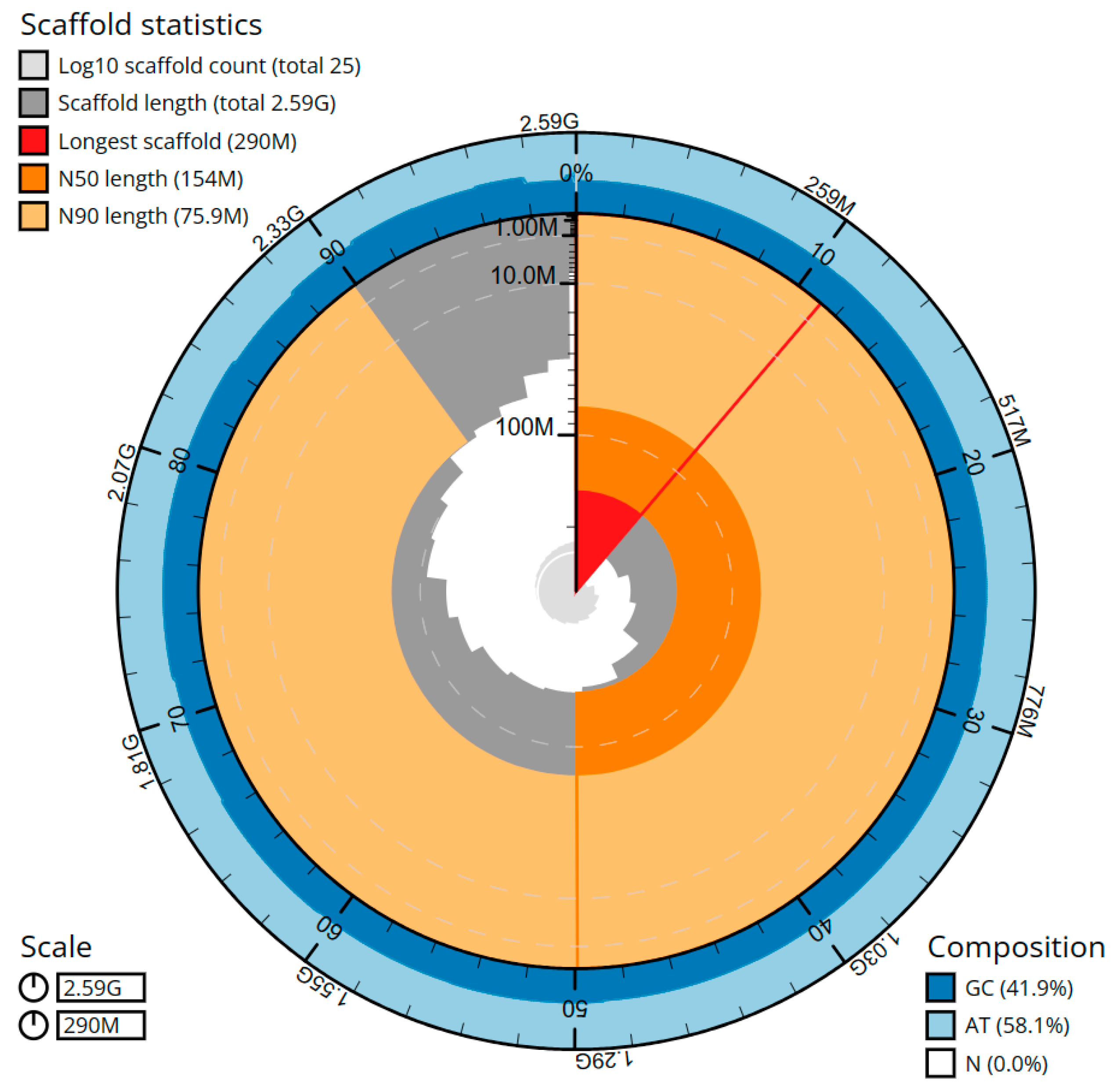
The cumulative length of the final assembly amounts to 2586.27 Mbp, including 39 gaps and encompassing 64 sequence contigs and 25 scaffolds, with a contig N50 (LG = 11) and a scaffold N50 (LG = 7) of 83.36 Mbp and 154.08 Mbp, respectively (Figure 3, Table 1). The presence of the 11 largest contigs containing 50% of the genomic sequences demonstrates significant contiguity within the assembly. On average, 36.15× coverage for the PacBio sequencing and 66.85× for the Hi-C sequencing was achieved. The genome assembly exhibited an average contig length of 40.41 Mbp, representing the mean size of the individual DNA fragments prior to scaffolding. The average scaffold length was 103.45 Mbp, indicating the typical size of the contiguous DNA sequences generated. The total gap length in the scaffolds amounted to 7.8 kbp, with an average gap length of 200 bp. Through manual assembly curation, a total of 27 missing or missed joins were rectified, and no sequence removed as haplotypic duplication, resulting in a 26.5% drop in the scaffold number. The scaffold N50 value remained unaltered, while a minor increase of 0.25% in the N90 length was observed as a result of the curation process.
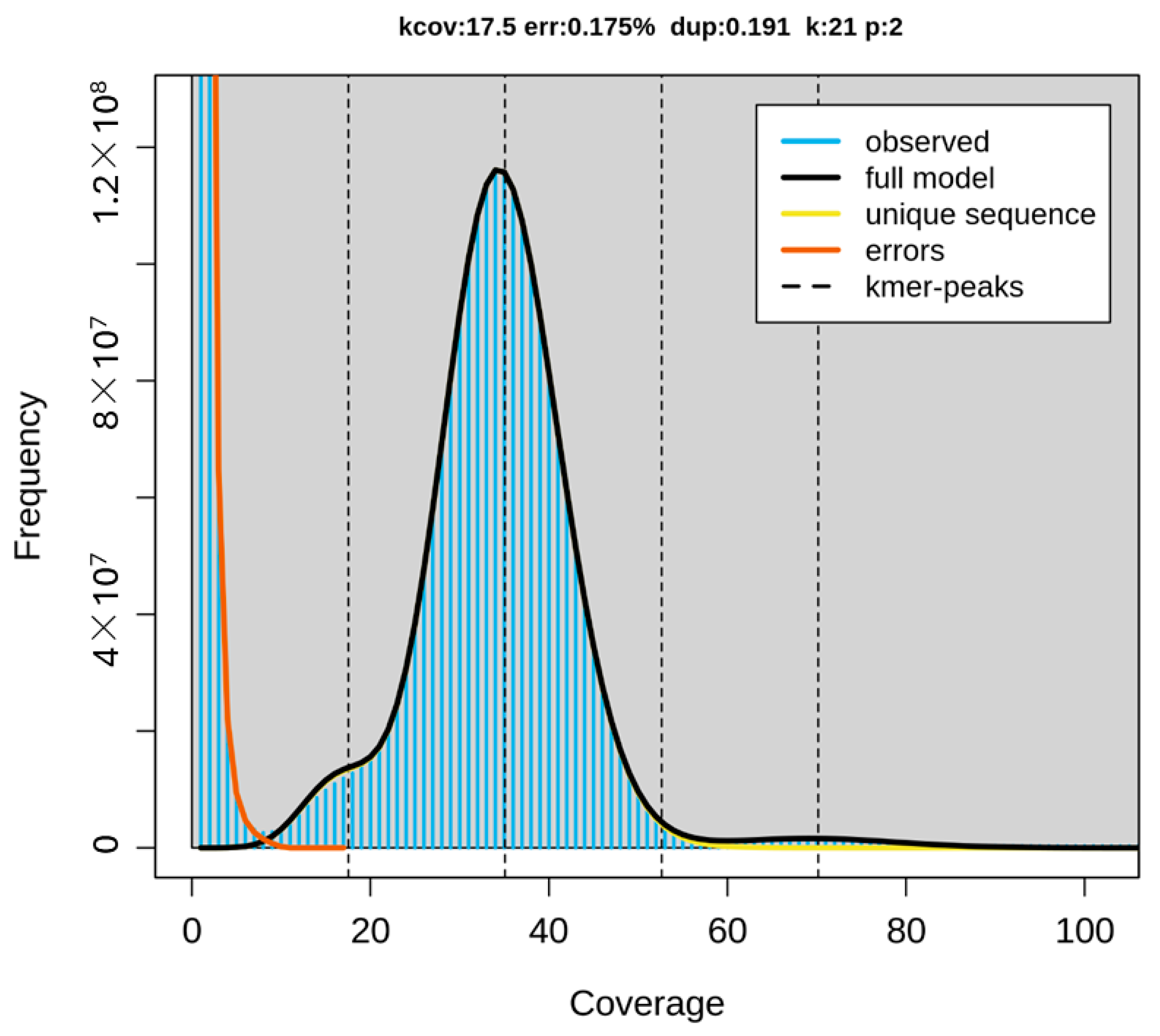
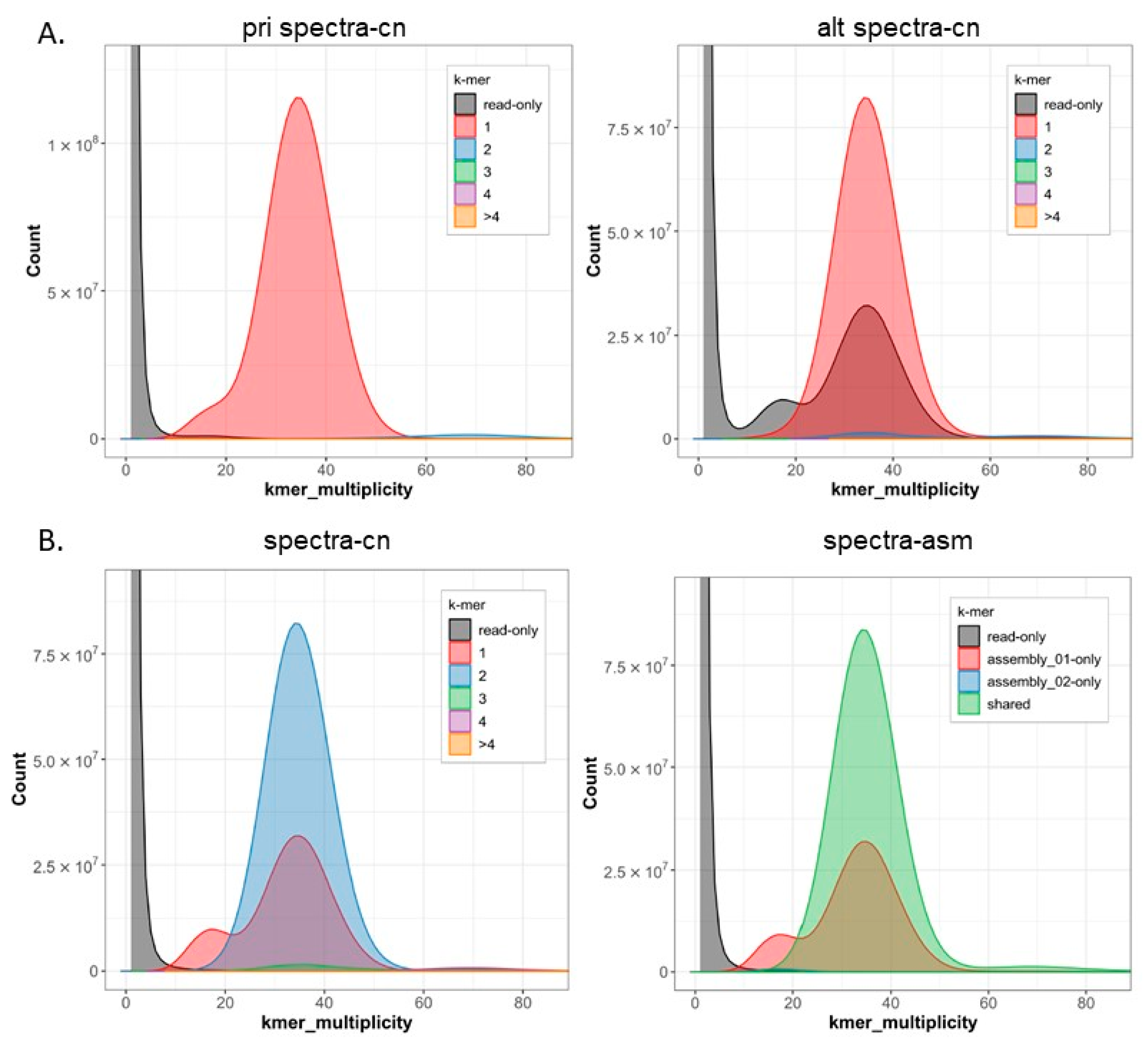
The newly sequenced genome displayed a GC base content of 41.85%, with adenine, cytosine, guanine, and thymine nucleotides accounting for 29.08%, 20.94%, 20.91%, and 29.07%, respectively. Repeat sequences constitute approximately 24.37% of the genome. The genome assembly analysis revealed that 99.83% of the regions were homozygous, while the remaining 0.17% were heterozygous (Figure 4 and Figure 5).
The estimated Quality Value (QV) of the Hifiasm assembly was 64.34, with k-mer completeness of 99.17%, and error rate of 3.6826 × 10⁻⁷. The final, purged assembly had a BUSCO completeness score (C) of 98.15%. The score breakdown indicates that 95.92% of the expected complete single-copy genes (S) were identified as complete sequences, while 2.24% of the single-copy genes were found as duplicates or fragmented sequences (D). The fragmented fraction (F) score was 0.86%, denoting a proportion of expected complete duplicated genes found in the assembly. The missing (M) score, indicating a rate of missing expected complete genes, was 0.98%. Regarding false duplications, the BUSCO results showed promising outcomes, and the primary-only spectra plot from Merqury displayed a clean pattern. The purged assembly demonstrated completeness of 99.13%, a QV of 64.44, and an error rate of 3.6005 × 10⁻⁷. Detailed characteristics of pre- and curated genome assemblies of a M. lutreola are presented in Table 2.
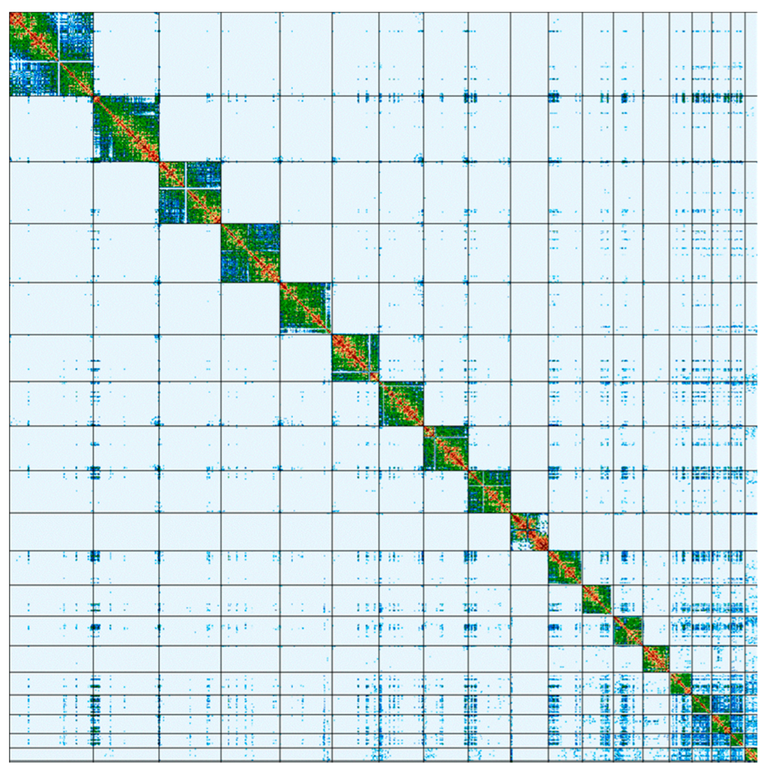
The vast majority (99.9985%) of the genome assembly was assigned to 20 C-scaffolds (pseudomolecules) [17], comprising 18 autosomes, and the X and Y sex chromosomes (Figure 6). Additionally, one unplaced (without a chromosome assignment) and four unlocalized (not localized to a specific position in the chromosome) scaffolds were identified in the assembly. Chromosome-scale scaffolds, confirmed by the Hi-C data, are named in order of size and characterized in Table 3.
The complete mitochondrial genome assembly is 16,552 bp in length, and displayed a level of identity ranging from 99.62% to 99.94% with previously sequenced mitochondrial genomes of M. lutreola, deposited in the GenBank [16].
Metadata for spectral estimates, sequencing runs, contaminants, and pre-curation, and curated assembly statistics are available at https://genomeark.s3.amazonaws.com/index.html?prefix=species/Mustela_lutreola (accesed on 1 July 2023).

3. Discussion

The genome sequencing of various mustelids has provided valuable insights into their genetic composition and evolutionary history. Out of the 67 species in the Mustelidae family, 17 have reference genome assemblies ready and deposited in GenBank, while only in the case of seven species has a chromosome-level assembly been achieved (Table 4) [18,19]. European mink stands as the only critically endangered species within this group that has now attained a reference genome of a platinum quality. A platinum genome is defined as a high-quality, near error-free and gapless, chromosome-level, haplotype-phased, reference genome assembly [20,21]. Additionally, the following standards were drawn for the Vertebrate Genomes Project of the Genome 10K consortium: N50 size of at least 1 Mbp for contigs and 10 Mbp for scaffolds, sequence error frequency of up to 1 in 10,000 bp, structural variants confirmed by multiple technologies, at least 90% of the sequence assigned to chromosomes and haplotype-phased [21,22]. Furthermore, a standard VGP reference genome involves an automated workflow that combines long-read sequencing, linked-read sequencing, optical mapping, and Hi-C data, with a final manual curation step to enhance the genome assembly and minimize errors [20,23]. The genome sequence reported in this article was assembled following the VGP quality requirements and meets the abovementioned conditions.
The size of the reported genome of a M. lutreola fits well with previously sequenced genomes of other representatives of the Mustela genus, being the biggest among the so-called ferret group, clustering the European polecat, the steppe polecat, the black-footed ferret, and the European mink [16,24,25]. The European mink genome assembly was found to be noticeably close in size and the GC-content to the earlier estimates, which predicted the genome size of the species to be around 2.411–2.474 Gbp and the GC content to be approximately 42%, based on the sequenced genomes of the ferret (MusPutFur1.0, GenBank assembly accession: GCF_000215625.1) and the European polecat (polecat_10x_lmp_bionano, GenBank assembly accession: GCA_902207235.1) [16].
The number of whole-chromosome pseudomolecules assembled in the European mink’s reference genome (18 autosomes and two heterosomes) is consistent with a diploid chromosome number reported for this species (2n = 38) [26]. Such a diploid number of chromosomes is typical of the family Mustelidae, occurring in over 60% of its representatives [27].
The high-quality reference genome of European mink represents the fulfilment of earlier calls for whole-genome sequencing of this critically endangered species [8,28], marking a significant step forward in advancing our understanding of the species’ genetic composition and promising to enhance conservation efforts. A platinum quality reference genome serves as a highly accurate and reliable resource for various research applications, including evolutionary studies, population genomics, comparative genomics and functional genomics [13,29]. Such reference genomes are particularly valuable for species of conservation concern, where accurate genomic information is crucial for effective conservation efforts and understanding the species’ biology [10,13]. The significant application potential of genomics in addressing conservation problems is well-documented for various carnivorans, e.g., African wild dog Lycaon pictus, Eastern wolf Canis lupus lycaon, puma Puma concolor, Iberian lynx Lynx pardinus, and wolverine Gulo gulo [11,30,31,32].
The reference genome of the European mink can play a pivotal role in addressing the conservation problems faced by this species, enabling the revision of management units to effectively manage genetic diversity within populations and minimize the outbreeding risk associated with inter-population translocations, providing comprehensive insights into the impacts of captive breeding, resolving phylogeographic questions, and facilitating the evaluation of conservation program efficiency and effectiveness [8,33]. It is important to recognize that the application of genomics to conservation often encounters a significant challenge—the high costs associated with molecular analyses. However, this is where advanced genomic tools come into play. Through techniques like whole-genome re-sequencing and reduced-representation approaches, conservation genomics offers a potential solution to mitigate these cost constraints, as these methodologies can reduce the number of markers required for analysis and monitoring projects in the field of conservation activities [11,12,13,14,15,33].
Comprehensive population genomics studies are crucial, as the limited data on interpopulation genetic diversity could significantly impede the effectiveness of captive breeding, reintroduction programs, and potential translocations for persisting wild populations of M. lutreola [28,34,35,36,37]. By analysing the genetic variation, relatedness between populations, and identifying adaptive traits loci, it aids in the establishment of appropriate management units for targeted conservation. One prominent example of the application of population genomics is the ongoing debate surrounding the potential inclusion of the Spanish conservation breeding initiative in the European Endangered Species Programme (EEP) for European mink [28]. Additionally, plans to obtain new founders from the wild Romanian population emphasize the importance of understanding historical population dynamics and connectivity between existing European mink populations [14,28].
Compared to traditional genetic approaches, the reference genome provides advanced genomic tools to investigate evolutionary relationships among different European mink populations. It helps in determining historical patterns of migration and divergence, shedding light on the species’ phylogenomic and phylogeographic history [38,39]. Such understanding is essential for re-evaluating Evolutionarily Significant Units (ESUs) and Management Units (MUs) for M. lutreola [34,40,41,42]. These units guide conservation actions based on distinct evolutionary lineages and aim to preserve the genetic diversity of the species [40,41]. Moreover, the reference genome of the European mink serves as a valuable tool in resolving uncertainties about its past distribution over continental Europe [43,44]. It helps identify regions of high genetic diversity, indicating historical refugia or areas of long-term stability for the species, as well as regions of genetic variation associated with adaptation to specific environments, habitat use, disease resistance, responses to changing conditions, or other crucial ecological factors [10,29]. Furthermore, it can provide insights into the evolutionary history of the species and its relationship with other mustelids.
Another pressing concern in the European mink conservation revolves around the assessment of the impact of the conservation breeding process on the development of traits essential for survival in the wild, specifically focusing on the adaptation to captivity [8]. Farquharson et al. [45] reported strong effects of inbreeding on the European mink offspring fitness in the EEP captive breeding program, highlighting the importance of addressing genetic management. One of the factors that reduces reproductive success in captivity and reconstituted (reintroduced) populations is aggressive behaviour exhibited by males toward females, which can lead to their exclusion from mating [46,47]. However, without a clear understanding of the heritability of these personality traits, assessing the risk of reducing genetic variation in reintroduced populations due to the release of individuals with specific personality types becomes challenging [46,47,48]. Furthermore, the reference genome can help to identify regions of the genome that are associated with local adaptation or specific traits relevant to survival in the wild [10,29]. This information can guide breeding strategies to ensure that valuable adaptive and survival traits are retained in captive populations, even when introducing new individuals to counteract inbreeding. By avoiding excessive inbreeding, the fitness and resilience of an offspring can be improved, increasing their chances of survival in both captive and future reintroduction settings.
Genomic information is essential to examine the impact of reintroduced individuals on shaping the gene pools of wild populations to address potential issues of outbreeding and the risk of losing unique adaptations [40,43]. In this regard, genomic-scale analyses serve as a valuable tool in evaluating potential fitness losses, thus facilitating more informed decisions and enhancing the success of reintroduction and translocation efforts [10,13]. The reference genome can support conservation breeding strategies by assisting in the selection of founders for captive breeding programs. It also helps optimize breeding pairs and prevents over-representation of certain lineages, ensuring that individuals chosen for reproduction possess optimal genetic diversity and reduce the risk of inbreeding depression [10,13].
With the reference genome, it becomes feasible to conduct genome-wide monitoring of the European mink local populations. This allows researchers to track changes in genetic diversity, detect potential threats to specific populations, and evaluate the effectiveness of conservation interventions over time, across multiple generations. Understanding the genetic variation within and between populations can help identify populations with low genetic diversity, which may be at greater risk of inbreeding depression and reduced adaptive potential, as well as genetically unique and vulnerable populations. Conservation efforts can then prioritize these populations for targeted management and genetic supplementation if necessary (genetic rescue) [49]. Genome-scale monitoring provides real-time information on the genetic health and status of endangered species populations, enabling adaptive management strategies to ensure their long-term survival, recovery, and genetic health [50]. Adaptive management is a dynamic and flexible approach characterized by continuous revisions of conservation strategies based on genetic data and observed fitness outcomes, allowing for timely adjustments and improvements to enhance conservation effectiveness.
In conclusion, embracing reference genome offers a powerful and comprehensive approach to address the conservation issues faced by M. lutreola, enhancing the prospects for its preservation and sustainable recovery. Future research directions in the European mink conservation genomics, based on the reported genome assembly, may involve building a reference pangenome to enable detailed population genomic studies, monitoring interpopulation genetic diversity patterns using restriction-site associated DNA sequencing (RADseq, ddRAD), and conducting a genome-wide scan for runs of homozygosity (ROH) to detect signatures of selection and estimate inbreeding [15,29,51]. Many of the conservation issues observed in this species could have been prevented if decisions regarding the necessary actions were not made before obtaining knowledge about its genome, but resulted from it.
The reported genome assembly also provides perspectives for planning, implementing, monitoring, and evaluating conservation interventions for other closely related taxa. The reference genome of the European mink can serve as a valuable reference for conducting reference-based assembly or designing primers for targeted sequencing of specific genomic locations in mustelids, whose genomes are yet to be fully revealed (e.g., Mustela eversmanii, Mustela sibirica, Mustela itatsi).
In the context of large genome sequencing programs, the prioritization of species sequencing order becomes crucial, as it enables the optimization of funding allocation, research interest, and workload alignment, ensuring that endangered species receive the necessary resources and attention commensurate with their conservation urgency. By prioritizing the comprehensive research of genomic resources in critically endangered species like the European mink, we not only gain valuable knowledge for conservation decision-making, but also preserve the irreplaceable genetic heritage that is on the brink of being lost forever.
The relatively low research interest in the European mink is incongruent with its critical situation. This highlights the significance of promotional and informational campaigns aimed at drawing attention and raising awareness in society about its alarming threat of extinction, ultimately ensuring adequate focus and attention of the scientific community towards understanding and conserving this fascinating species, deserving effective protection.

4. Materials and Methods

4.1. Sample Collection and DNA Extraction

The European Mink Genome Project (https://www.ncbi.nlm.nih.gov/bioproject/986837 (accesed on 1 July 2023)) was initiated in October 2020 by the consortium of the European Mink Centre (Szczecin, Poland, http://europeanminkcentre.com/ (accesed on 1 July 2023)), the Vertebrate Genome Laboratory of the Rockefeller University (New York, USA, https://www.vertebrategenomelab.org/ (accesed on 1 July 2023)), the Wildtier- und Artenschutzstation e.V. association (Sachsenhagen, Germany, https://wildtierstation.de/ (accesed on 1 July 2023)), and the EuroNerz e.V. association (Osnabrück, Germany, https://www.euronerz.de/ (accesed on 1 July 2023)), led by the University of Szczecin (Szczecin, Poland). The assembly was also contributed to by the Vertebrate Genomes Project (VGP), whose assembly pipeline (v. 2.0) and style quality metrics were implemented and followed [21,52,53,54].
Samples were taken from two (mMusLut2 and mMusLut3) captive-born, adult, clinically healthy, closely related M. lutreola males (heterogametic sex), during a routine veterinary examination, in accordance with the principles of animal welfare. Sampled individuals are kept by the European mink conservation breeding facility of the Wildtier- und Artenschutzstation e.V. (Sachsenhagen, Lower Saxony, Germany; 52°23′51″ N 9°12′58″ E), participating in the EAZA EEP for European mink, and are characterized in the Table 5. Both sampled individuals presented phenotypic features typical for the species [2]. The samples were collected by Florian Brandes (veterinarian, the Wildtier- und Artenschutzstation e.V.), assisted by Jakub Skorupski and Przemysław Śmietana (both University of Szczecin), on 14 March 2022 in the Wildtier- und Artenschutzstation e.V. headquarters. Individual mMusLut2 (specimen M1207) was used for acquisition of the genome sequence and HiFi data generation, while individual mMustLut3 (specimen M1287) was used for scaffolding using Hi-C.
Whole blood samples were sterile collected by a cephalic vein venipuncture under inhalant anaesthesia [55,56]. Approximately 0.75 mL of blood per individual was drawn into the BD Vacutainer® tube with K2 EDTA (Becton, Dickinson and Company, New Jersey, NJ, USA). The samples were immediately gently mixed by inverting the tubes 8–10 times to ensure proper anticoagulation and prevent clot formation prior to further laboratory analysis.
After collection, the samples were snap-frozen at −80 °C to preserve the integrity of the biological material. The frozen tubes were packed in insulated containers with at least 10 kg of dry ice and shipped overnight to the Vertebrate Genomes Laboratory (VGL) at the Rockefeller University (New York, NY, USA), for further analyses.
Ultra-high molecular weight (UHMW) DNA was extracted from frozen samples using the Nanobind® Bionano Prep SP Dna isolation kit (Bionano Genomics Inc., San Diego, CA, USA) method, according to the manufacturer’s protocol. The Nanobind® magnetic disk applied, enabled automatic lysis, binding, washing and elution, and efficiently minimized fragmentation and preserved long DNA molecules required for accurate long-read sequencing.

4.2. Sequencing

To achieve an optimal insert size for the PacBio sequencing, the Megaruptor® 3 system (Diagenode Inc., Denville, NY, USA) with a standard hydropore and speed setting 28 was used to shear the UHMW DNA. The fragmented DNA was sheared to an average size of 17,000 bp, suitable for highly accurate long-read sequencing. Sheared DNA was concentrated and cleaned using 0.45× Ampure PB (Pacific Biosciences of California Inc., Menlo Park, CA, USA). The concentration and length of the purified sheared DNA were evaluated with the QubitTM 3.0 fluorometer (Thermo Fisher Scientific Inc., Waltham, MA, USA) and the Femto Pulse system (Agilent, Santa Clara, CA, USA).
The PacBio HiFi (high-fidelity) circular consensus sequencing library was prepared using PacBio’s 3.0 template preparation kit (Pacific Biosciences of California Inc., Menlo Park, CA, USA), following the manufacturer’s protocol. With a single-molecule read accuracy surpassing 99.9%, the PacBio platform produces HiFi reads using circular consensus sequencing (CCS) mode on PacBio long-read systems, spanning an optimal range between 15 kb and 20 kb. An input mass of 5 µg of sheared DNA was used for library construction. The library was size-selected using the Pippin HTTM (Sage Science Inc., Beverly, MA, USA) automated, high throughput gel-based method to obtain the desired insert size. HiFi data were generated for the mMusLut2 (paternal) individual. The Cutadapt v. 4.4 algorithm was used to remove the reads found to have an adapter inside of it [57].
The prepared library was sequenced on the Sequel IIe system using PacBio’s binding kit 3.2 and sequencing plate 2.0 (Pacific Biosciences of California Inc., Menlo Park, CA, USA). A total of six 8M SMRT (single-molecule real-time) cells were used to generate long-read sequencing data [58].
The high-throughput chromosome conformation capture (Hi-C) sequencing data were generated from the blood sample of the mMustLut3 (filial) individual applying the Dovetail™ Hi-C kit (Dovetail Genomics LLC, Scotts Valley, CA, USA) and sequenced on NovaSeq 6000 instrument (Illumina Inc., San Diego, CA, USA), following the manufacturer’s instructions.

4.3. De Novo Genome Assembly, Curation, and Quality Control

The long-read sequencing data were processed using PacBio’s SMRT Analysis software v. 11.1 pipeline (Pacific Biosciences of California Inc., Menlo Park, CA, USA) to obtain high-quality long reads. The Hifiasm (version 0.18.8+galaxy1) assembler, specifically developed for PacBio HiFi reads, was used to generate the continuous and complete de novo assembly [59]. Pseudohaplotype assemblies (primary/principal and alternate) of mMusLut2 were scaffolded using the haplotype-specific Hi-C reads of mMusLut3, using both Hifiasm and YaHS version 1.2a.2+galaxy0 [60] tools. The abovementioned primary assembly, encompassing homozygous and one set of loci for heterozygous regions, represents a more complete representation of an individual’s genome and is preferred for downstream analyses as it provides both homozygous and heterozygous regions. The alternate assembly, or haplotigs, includes the alternate loci from the other haplotype’s heterozygous regions, and being less complete than the primary assembly, it lacks representation of homozygous regions [54].
The Hi-C analysis was performed using the Arima-HiC kit 2 (Arima Genomics Inc., Carlsbad, CA, USA). Utilizing chromosome conformation Hi-C data, the primary assembly contigs were organized and linked into larger scaffolds [61]. The Hi-C reads were aligned to the genome assembly to generate a contact map, visualized using the PretextView v. 0.2.5 (https://github.com/wtsi-hpag/PretextView (accessed on 1 June 2023)).
By decomposing the sequencing data into k-length substrings, counting the occurrence of each k-mer, and determining its frequency, Meryl v. 1.4 enabled the generation of the k-mer profile [62]. Genome properties, including genome size, repetitiveness, and heterozygosity rates, were inferred from the k-mer histogram generated by Meryl using GenomeScope v. 2.0, a tool that utilizes sequencing reads using a kmer-based statistical approach [63]. The expected genome size was computed from the k-mer genome coverage. K-mers were also used for initial, reference-free genome profiling.
The mitochondrial genome was assembled from PacBio High Fidelity reads, with the MitoHiFi v. 3.2 pipeline [64]. The NCBI Reference Sequence NC_056132.1 was used as a reference. The comparison with previously sequenced complete mitogenome sequences of M. lutreola, deposited in the GenBank (Accession No. NC_056132.1, MW197426.1, MW197425.1, MW197424.1, MT304869.1, MW197423.1), was conducted using BLASTN v. 2.14.1+ programme (Nucleotide collection, megablast option) [65].
The gfastats v. 1.3.6 [66] was applied to check for contamination and correct the assemblies at each assembly stage. The Merqury v. 1.3 platform [62] was used to perform quality control at the contiging and purging stages (estimation of the consensus Quality Value (QV) scores of the final assembly and k-mer completeness). The gfastats v. 1.3.6 tool [66] was also used to compute the number of scaffolds, average of scaffold lengths, number of gaps, L50, N50, and total assembled bases.
The BUSCO (Benchmarking Universal Single-Copy Orthologs) scores, assessing the completeness and quality of genome assemblies by evaluating the presence and integrity of a set of highly conserved genes that are expected to be present as single copies in a given genome (vertebrata_odb10 dataset, n = 3354), were granted within the BUSCO v. 5.3.2 software, applying the Metaeuk 6.a5d39d9 and HMMsearch v. 3.1 gene predictor algorithms [67,68]. To improve overall assembly quality, gap filling, removing false duplications, collapsed repeats and very low coverage regions, as well as haplotigs purging was performed using the purge_dups v. 1.2.5+galaxy4 package [69], based on read depth. Parameters derived from the GenomeScope output were used to define cutoffs.
Manual curation of final assembly was performed using HiGlass v. 1.11 [70] and PretextMap v. 0.1.9 [71] tools, to resolve potential mis-assemblies, missed joins, duplications and collapses, and remove any contaminants.

4.4. Genome Annotation and Data Availability

This Whole Genome Shotgun project has been deposited at DDBJ/ENA/GenBank under the accession JAUCGO000000000 (JAUCGO010000001–JAUCGO010000026) and JAUCGP000000000 (JAUCGP010000001–JAUCGP010012043) and will be subjected to comprehensive annotation by the National Centre for Biotechnology Information (NCBI) refseq Eukaryotic Genome Annotation Pipeline [72]. Thus, the first platinum-standard reference-quality genome sequence is released openly for reuse—accession information of raw sequence data and the final assembled genome are given in Table 6.

Author Contributions

Conceptualization—J.S., methodology—J.S., F.B., C.S., W.F., J.B., N.J., T.T., L.A., J.W., Y.S., G.F., O.F. and E.D.J., software—J.S., J.B., N.J., T.T., L.A., J.W., Y.S., G.F., O.F. and E.D.J., validation—J.S., J.B., N.J., T.T., L.A., J.W., Y.S., G.F., O.F. and E.D.J., results analysis and investigation—J.S., J.B., N.J., T.T., L.A., J.W., Y.S., G.F., O.F. and E.D.J., data curation—J.S., J.B., N.J., T.T., L.A., J.W., Y.S., G.F., O.F. and E.D.J., writing—J.S., review and editing—J.S., J.S., F.B., C.S., W.F., P.Ś., J.B., N.J., T.T., L.A., J.W., Y.S., G.F., O.F. and E.D.J., visualization—J.S. and J.W., funding acquisition—J.S. and P.Ś. All authors have read and agreed to the published version of the manuscript.

Funding

The research reported in this manuscript was conducted within the European Mink Whole Genome Sequencing Project, financed by the European Mink Centre consortium (http://europeanminkcentre.com/).

Institutional Review Board Statement

Not applicable.

Informed Consent Statement

Not applicable.

Data Availability Statement

The metadata for spectral estimates, sequencing runs, contaminants, and pre-curation and curated assembly statistics that support the findings of this study are available in the GenomeArk repository with the identifier “Mustela lutreola” (https://genomeark.github.io/genomeark-all/Mustela_lutreola.html (accesed on 1 July 2023) and https://genomeark.s3.amazonaws.com/index.html?prefix=species/Mustela_lutreola (accesed on 1 July 2023)).

Acknowledgments

The research presented herein was enabled by the financial support of the European Mink Centre (http://europeanminkcentre.com/) consortium, under the European Mink Whole Genome Sequencing Project (https://www.ncbi.nlm.nih.gov/bioproject/986837 (accesed on 1 July 2023)). The presented genome assembly was also contributed to by the Vertebrate Genomes Project (VGP, https://vertebrategenomesproject.org/ (accesed on 1 July 2023)).

Conflicts of Interest

The authors declare no conflict of interest. The funder had no role in the design of the study; in the collection, analyses, or interpretation of data; in the writing of the manuscript, or in the decision to publish the results.

References

  1. IUCN. The IUCN Red List of Threatened Species—Summary Statistics. 2023. Available online: https://www.iucnredlist.org/resources/summary-statistics (accessed on 14 July 2023).
  2. Youngman, P.M. Mustela lutreola. Mamm. Species 1990, 362, 1–3. [Google Scholar]
  3. Heptner, V.G.; Naumov, N.P.; Yurgenson, P.B.; Sludskii, A.A.; Chirkova, A.F.; Bannikov, A.G. Part 1b: Carnivora. Mammals of the Soviet Union, 1st ed.; Smithsonian Institution Libraries: Washington, DC, USA; The National Science Foundation Press: New Delhi, India, 2001; Volume 2, pp. 1078–1106.
  4. Witkowski, Z.; Król, W.; Solarz, W. Carpathian List of Endangered Species; WWF: Vienna, Austria; Institute of Nature Conservation, Polish Academy of Sciences: Kraków, Poland, 2003; 64p. [Google Scholar]
  5. Maran, T.; Skumatov, D.; Gomez, A.; Põdra, M.; Abramov, A.V.; Dinets, V. Mustela lutreola. IUCN Red List Threat. Species 2016, 13, e.T14018A45199861. [Google Scholar] [CrossRef]
  6. Maran, T. European mink: Setting of goal for conservation and Estonian case study. Galemys 2003, 15, 1–11. [Google Scholar]
  7. Harrington, L.A.; Põdra, M.; Gómez, A.; Maran, T. Raising awareness of the plight of the critically endangered European mink in Spain is not miscommunication: A response to Melero. Biodivers. Conserv. 2018, 27, 269–271. [Google Scholar] [CrossRef]
  8. Skorupski, J. Fifty Years of Research on European Mink Mustela lutreola L., 1761 Genetics: Where Are We Now in Studies on One of the Most Endangered Mammals? Genes 2020, 11, 1332. [Google Scholar] [CrossRef]
  9. Fuentes-Pardo, A.P.; Ruzzante, D.E. Whole-genome sequencing approaches for conservation biology: Advantages, limitations and practical recommendations. Mol. Ecol. 2017, 26, 5369–5406. [Google Scholar] [CrossRef]
  10. Brandies, P.; Peel, E.; Hogg, C.J.; Belov, K. The Value of Reference Genomes in the Conservation of Threatened Species. Genes 2019, 10, 846. [Google Scholar] [CrossRef]
  11. Steiner, C.C.; Putnam, A.S.; Hoeck, P.E.; Ryder, O.A. Conservation Genomics of Threatened Animal Species. Annu. Rev. Anim. Biosci. 2013, 1, 261–281. [Google Scholar] [CrossRef]
  12. Khan, S.; Nabi, G.; Ullah, M.W.; Yousaf, M.; Manan, S.; Siddique, R.; Hou, H. Overview on the Role of Advance Genomics in Conservation Biology of Endangered Species. Int. J. Genom. 2016, 2016, 1–8. [Google Scholar] [CrossRef]
  13. Supple, M.A.; Shapiro, B. Conservation of biodiversity in the genomics era. Genome Biol. 2018, 19, 131. [Google Scholar] [CrossRef]
  14. Stronen, A.V.; Iacolina, L.; Ruiz-Gonzalez, A. Rewilding and conservation genomics: How developments in (re)colonization ecology and genomics can offer mutual benefits for understanding contemporary evolution. Glob. Ecol. Conserv. 2019, 17, e00502. [Google Scholar] [CrossRef]
  15. Wright, B.; Farquharson, K.A.; McLennan, E.A.; Belov, K.; Hogg, C.J.; Grueber, C.E. From reference genomes to population genomics: Comparing three reference-aligned reduced-representation sequencing pipelines in two wildlife species. BMC Genom. 2019, 20, 453. [Google Scholar] [CrossRef] [PubMed]
  16. Skorupski, J. Characterisation of the Complete Mitochondrial Genome of Critically Endangered Mustela lutreola (Carnivora: Mustelidae) and Its Phylogenetic and Conservation Implications. Genes 2022, 13, 125. [Google Scholar] [CrossRef] [PubMed]
  17. Lewin, H.A.; Graves, J.A.M.; Ryder, O.A.; Graphodatsky, A.S.; O’Brien, S.J. Precision nomenclature for the new genomics. Gigascience 2019, 8, giz086. [Google Scholar] [CrossRef] [PubMed]
  18. Kollias, G.V.; Fernandez-Moran, J. Mustelidae. In Fowler’s Zoo and Wild Animal Medicine; Miller, R.E., Fowler, M.E., Eds.; Elsevier Inc.: St. Louis, MI, USA, 2015; Volume 8, pp. 476–491. [Google Scholar] [CrossRef]
  19. NCBI. Genome List. 2023. Available online: https://www.ncbi.nlm.nih.gov/datasets/genome/?taxon=9655 (accessed on 14 July 2023).
  20. Morin, P.A.; Alexander, A.; Blaxter, M.; Caballero, S.; Fedrigo, O.; Fontaine, M.C.; Foote, A.D.; Kuraku, S.; Maloney, B.; McCarthy, M.L.; et al. Building genomic infrastructure: Sequencing platinum-standard reference-quality genomes of all cetacean species. Mar. Mammal Sci. 2020, 36, 1356–1366. [Google Scholar] [CrossRef]
  21. Rhie, A.; McCarthy, S.A.; Fedrigo, O.; Damas, J.; Formenti, G.; Koren, S.; Uliano-Silva, M.; Chow, W.; Fungtammasan, A.; Kim, J.; et al. Towards complete and error-free genome assemblies of all vertebrate species. Nature 2021, 592, 737–746. [Google Scholar] [CrossRef]
  22. Koepfli, K.-P.; Paten, B.; the Genome 10K Community of Scientists; O’brien, S.J. The Genome 10K Project: A Way Forward. Annu. Rev. Anim. Biosci. 2015, 3, 57–111. [Google Scholar] [CrossRef]
  23. Anonymous. A reference standard for genome biology. Nat. Biotechnol. 2018, 36, 1121. [Google Scholar] [CrossRef]
  24. Davison, A.; Griffiths, H.I.; Brookes, R.C.; Maran, T.; Macdonald, D.W.; Sidorovich, V.E.; Kitchener, A.C.; Irizar, I.; Villate, I.; Gonzalez-Esteban, J.; et al. Mitochondrial DNA and palaeontological evidence for the origins of endangered European mink, Mustela lutreola. Anim. Conserv. 2000, 3, 345–355. [Google Scholar] [CrossRef]
  25. Lodé, T.; Guiral, G.; Peltier, D. European Mink–Polecat Hybridization Events: Hazards From Natural Process? J. Hered. 2005, 96, 89–96. [Google Scholar] [CrossRef]
  26. Graphodatsky, A.S. Mustela lutreola (European Mink). In Atlas of Mammalian Chromosomes; O’Brien, S.J., Menninger, J.C., Nash, W.G., Eds.; John Wiley & Sons: Hoboken, NJ, USA, 2006; p. 487. [Google Scholar]
  27. Franco-De-Sá, J.F.O.; Rosas, F.C.W.; Feldberg, E. Cytogenetic study of the giant otter Pteronura brasiliensis Zimmermann 1780 (Carnivora, Mustelidae, Lutrinae). Genet. Mol. Biol. 2007, 30, 1093–1096. [Google Scholar] [CrossRef]
  28. Maran, T.; Fienieg, E.; Schad, K. Long-Term Management Plan for European Mink (Mustela lutreola) European Endangered Species Programme (EEP); Tallinn Zoo: Tallinn, Estonia; European Association of Zoos: Amsterdam, The Netherlands, 2017; pp. 1–46. [Google Scholar]
  29. Formenti, G.; Theissinger, K.; Fernandes, C.; Bista, I.; Bombarely, A.; Bleidorn, C.; Ciofi, C.; Crottini, A.; Godoy, J.A.; Malukiewicz, J.; et al. The era of reference genomes in conservation genomics. Trends Ecol. Evol. 2022, 37, 197–202. [Google Scholar] [CrossRef]
  30. Campana, M.G.; Parker, L.D.; Hawkins, M.T.; Young, H.S.; Helgen, K.M.; Gunther, M.S.; Woodroffe, R.; Maldonado, J.E.; Fleischer, R.C. Genome sequence, population history, and pelage genetics of the endangered African wild dog (Lycaon pictus). BMC Genom. 2016, 17, 1013. [Google Scholar] [CrossRef]
  31. Ekblom, R.; Brechlin, B.; Persson, J.; Smeds, L.; Johansson, M.; Magnusson, J.; Flagstad, Ø.; Ellegren, H. Genome sequencing and conservation genomics in the Scandinavian wolverine population. Conserv. Biol. 2018, 32, 1301–1312. [Google Scholar] [CrossRef] [PubMed]
  32. Saremi, N.F.; Supple, M.A.; Byrne, A.; Cahill, J.A.; Coutinho, L.L.; Dalén, L.; Figueiró, H.V.; Johnson, W.E.; Milne, H.J.; O’brien, S.J.; et al. Puma genomes from North and South America provide insights into the genomic consequences of inbreeding. Nat. Commun. 2019, 10, 227–245. [Google Scholar] [CrossRef]
  33. Hohenlohe, P.A.; Funk, W.C.; Rajora, O.P. Population genomics for wildlife conservation and management. Mol. Ecol. 2020, 30, 62–82. [Google Scholar] [CrossRef]
  34. Michaux, J.; Libois, R.; Davison, A.; Chevret, P.; Rosoux, R. Is the western population of the European mink, (Mustela lutreola), a distinct Management Unit for conservation? Biol. Conserv. 2004, 115, 357–367. [Google Scholar] [CrossRef]
  35. Arthur, C.; Aulagnier, S.; des Neiges de Bellefroid, M.; Delas, G.; Fournier, P.; Gourreau, J.-M.; Lodé, T.; Michaux, J.; Rosoux, R.; Ruette, S. Deuxième Plan National de Restauration du Vison d’Europe (Mustela lutreola) 2007–2011; Ministère de l’Ecologie, du Développement et de l’Aménagement Durables: Paris, France, 2007; pp. 1–102.
  36. Cabria, M.T.; González, E.G.; Gómez-Moliner, B.J.; Zardoya, R. Microsatellite markers for the endangered European mink (Mustela lutreola) and closely related mustelids. Mol. Ecol. Notes 2007, 7, 1185–1188. [Google Scholar] [CrossRef]
  37. Zuberogoitia, I.; Põdra, M.; Palazón, S.; Gómez, A.; Zabala, N.; Zabala, J. Misleading interpretation of shifting baseline syndrome in the conservation of European mink. Biodivers. Conserv. 2016, 25, 1795–1800. [Google Scholar] [CrossRef]
  38. Emerson, K.J.; Merz, C.R.; Catchen, J.M.; Hohenlohe, P.A.; Cresko, W.A.; Bradshaw, W.E.; Holzapfel, C.M. Resolving postglacial phylogeography using high-throughput sequencing. Proc. Natl. Acad. Sci. USA 2010, 107, 16196–16200. [Google Scholar] [CrossRef]
  39. McGaughran, A.; Liggins, L.; Marske, K.A.; Dawson, M.N.; Schiebelhut, L.M.; Lavery, S.D.; Knowles, L.L.; Moritz, C.; Riginos, C. Comparative phylogeography in the genomic age: Opportunities and challenges. J. Biogeogr. 2022, 49, 2130–2144. [Google Scholar] [CrossRef]
  40. Lynch, M. The genetic interpretation of inbreeding depression and outbreeding depression. Evolution 1991, 45, 622–629. [Google Scholar] [CrossRef] [PubMed]
  41. Fraser, D.J.; Bernatchez, L. Adaptive evolutionary conservation: Towards a unified concept for defining conservation units. Mol. Ecol. 2001, 10, 2741–2752. [Google Scholar] [CrossRef]
  42. Carbonell, R. Managing Spanish European mink populations: Moving from a precautionary approach towards knowledge-based management. J. Nat. Conserv. 2015, 25, 58–61. [Google Scholar] [CrossRef]
  43. Cabria, M.T.; Gonzalez, E.G.; Gomez-Moliner, B.J.; Michaux, J.R.; Skumatov, D.; Kranz, A.; Fournier, P.; Palazon, S.; Zardoya, R. Patterns of genetic variation in the endangered European mink (Mustela lutreola L., 1761). BMC Evol. Biol. 2015, 15, 141. [Google Scholar] [CrossRef]
  44. Lodé, T. The European Mink’s Paradox: Near Extinction but Colonizing New Habitats. JSM Biol. 2017, 2, 1011. [Google Scholar] [CrossRef]
  45. Farquharson, K.A.; Hogg, C.J.; Grueber, C.E. Offspring survival changes over generations of captive breeding. Nat. Commun. 2021, 12, 1–9. [Google Scholar] [CrossRef]
  46. Haage, M.; Maran, T.; Bergvall, U.A.; Elmhagen, B.; Angerbjörn, A. The influence of spatiotemporal conditions and personality on survival in reintroductions–evolutionary implications. Oecologia 2016, 183, 45–56. [Google Scholar] [CrossRef]
  47. Kneidinger, N.; Nagl, A.; Kiik, K.; Schwarzenberger, F.; Maran, T. The individual courtship behaviour of male European mink (Mustela lutreola) is a good indicator for their breeding success. Appl. Anim. Behav. Sci. 2018, 205, 98–106. [Google Scholar] [CrossRef]
  48. van Oers, K.; de Jong, G.; van Noordwijk, A.J.; Drent, P.J.; Kempenaers, B. Contribution of genetics to the study of animal personalities: A review of case studies. Behaviour 2005, 142, 1185–1206. [Google Scholar] [CrossRef]
  49. Whiteley, A.R.; Fitzpatrick, S.W.; Funk, W.C.; Tallmon, D.A. Genetic rescue to the rescue. Trends Ecol. Evol. 2015, 30, 42–49. [Google Scholar] [CrossRef] [PubMed]
  50. Serrouya, R.; Seip, D.R.; Hervieux, D.; McLellan, B.N.; McNay, R.S.; Steenweg, R.; Heard, D.C.; Hebblewhite, M.; Gillingham, M.; Boutin, S. Saving endangered species using adaptive management. Proc. Natl. Acad. Sci. USA 2019, 116, 6181–6186. [Google Scholar] [CrossRef] [PubMed]
  51. Xue, Y.; Prado-Martinez, J.; Sudmant, P.H.; Narasimhan, V.; Ayub, Q.; Szpak, M.; Frandsen, P.; Chen, Y.; Yngvadottir, B.; Cooper, D.N.; et al. Mountain gorilla genomes reveal the impact of long-term population decline and inbreeding. Science 2015, 348, 242–245. [Google Scholar] [CrossRef] [PubMed]
  52. Batut, B.; Hiltemann, S.; Bagnacani, A.; Baker, D.; Bhardwaj, V.; Blank, C.; Bretaudeau, A.; Brillet-Guéguen, L.; Čech, M.; Chilton, J.; et al. Community-Driven Data Analysis Training for Biology. Cell Syst. 2018, 6, 752–758.e1. [Google Scholar] [CrossRef] [PubMed]
  53. Hiltemann, S.; Rasche, H.; Gladman, S.; Hotz, H.-R.; Larivière, D.; Blankenberg, D.; Jagtap, P.D.; Wollmann, T.; Bretaudeau, A.; Goué, N.; et al. Galaxy Training: A powerful framework for teaching! PLoS Comput. Biol. 2023, 19, e1010752. [Google Scholar] [CrossRef]
  54. Lariviere, D.; Ostrovsky, A.; Gallardo, C.; Syme, A.; Abueg, L.; Pickett, B.; Formenti, G.; Sozzoni, M. VGP Assembly Pipeline (Galaxy Training Materials). 2023. Available online: https://training.galaxyproject.org/training-material/topics/assembly/tutorials/vgp_genome_assembly/tutorial.html (accessed on 14 July 2023).
  55. Bixler, H.; Ellis, C. Ferret care and husbandry. Veter Clin. N. Am. Exot. Anim. Pract. 2004, 7, 227–255. [Google Scholar] [CrossRef]
  56. Wolf, T.M. Ferrets. In Manual of Exotic Pet Practice; Mitchell, M.A., Tully, T.N., Jr., Eds.; Saunders: St. Louis, MI, USA, 2009; pp. 345–374. [Google Scholar] [CrossRef]
  57. Martin, M. Cutadapt removes adapter sequences from high-throughput sequencing reads. EMBnet J. 2011, 17, 10–12. [Google Scholar] [CrossRef]
  58. Levene, M.J.; Korlach, J.; Turner, S.W.; Foquet, M.; Craighead, H.G.; Webb, W.W. Zero-Mode Waveguides for Single-Molecule Analysis at High Concentrations. Science 2003, 299, 682–686. [Google Scholar] [CrossRef]
  59. Cheng, H.; Concepcion, G.T.; Feng, X.; Zhang, H.; Li, H. Haplotype-resolved de novo assembly using phased assembly graphs with hifiasm. Nat. Methods 2021, 18, 170–175. [Google Scholar] [CrossRef]
  60. Zhou, C.; McCarthy, S.A.; Durbin, R. YaHS: Yet another Hi-C scaffolding tool. Bioinformatics 2023, 39, btac808. [Google Scholar] [CrossRef]
  61. Rao, S.S.P.; Huntley, M.H.; Durand, N.C.; Stamenova, E.K.; Bochkov, I.D.; Robinson, J.T.; Sanborn, A.L.; Machol, I.; Omer, A.D.; Lander, E.S.; et al. A 3D Map of the Human Genome at Kilobase Resolution Reveals Principles of Chromatin Looping. Cell 2014, 159, 1665–1680. [Google Scholar] [CrossRef] [PubMed]
  62. Rhie, A.; Walenz, B.P.; Koren, S.; Phillippy, A.M. Merqury: Reference-free quality, completeness, and phasing assessment for genome assemblies. Genome Biol. 2020, 21, 245. [Google Scholar] [CrossRef] [PubMed]
  63. Ranallo-Benavidez, T.R.; Jaron, K.S.; Schatz, M.C. GenomeScope 2.0 and Smudgeplot for reference-free profiling of polyploid genomes. Nat. Commun. 2020, 11, 1–10. [Google Scholar] [CrossRef]
  64. Uliano-Silva, M.; Ferreira, J.G.R.N.; Krasheninnikova, K.; Blaxter, M.; Mieszkowska, N.; Hall, N.; Holland, P.; Durbin, R.; Richards, T.; Kersey, P.; et al. MitoHiFi: A python pipeline for mitochondrial genome assembly from PacBio high fidelity reads. bioRxiv 2023, 24, 521667. [Google Scholar] [CrossRef] [PubMed]
  65. Zhang, Z.; Schwartz, S.; Wagner, L.; Miller, W.; Karami, A.; Movaghar, A.F.; Mercier, S.; Ferre, L.; Seligmann, H.; Proença, D.; et al. A Greedy Algorithm for Aligning DNA Sequences. J. Comput. Biol. 2000, 7, 203–214. [Google Scholar] [CrossRef] [PubMed]
  66. Formenti, G.; Abueg, L.; Brajuka, A.; Brajuka, N.; Gallardo-Alba, C.; Giani, A.; Fedrigo, O.; Jarvis, E.D. Gfastats: Conversion, evaluation and manipulation of genome sequences using assembly graphs. Bioinformatics 2022, 38, 4214–4216. [Google Scholar] [CrossRef]
  67. Simão, F.A.; Waterhouse, R.M.; Ioannidis, P.; Kriventseva, E.V.; Zdobnov, E.M. BUSCO: Assessing genome assembly and annotation completeness with single-copy orthologs. Bioinformatics 2015, 31, 3210–3212. [Google Scholar] [CrossRef]
  68. Seppey, M.; Manni, M.; Zdobnov, E.M. BUSCO: Assessing Genome Assembly and Annotation Completeness. Methods Mol. Biol. 2019, 1962, 227–245. [Google Scholar] [CrossRef]
  69. Guan, D.; McCarthy, S.A.; Wood, J.; Howe, K.; Wang, Y.; Durbin, R. Identifying and removing haplotypic duplication in primary genome assemblies. Bioinformatics 2020, 36, 2896–2898. [Google Scholar] [CrossRef]
  70. Kerpedjiev, P.; Abdennur, N.; Lekschas, F.; McCallum, C.; Dinkla, K.; Strobelt, H.; Luber, J.M.; Ouellette, S.B.; Azhir, A.; Kumar, N.; et al. HiGlass: Web-based visual exploration and analysis of genome interaction maps. Genome Biol. 2018, 19, 125. [Google Scholar] [CrossRef]
  71. Harry, E. PretextView (Paired REad TEXTure Viewer): A Desktop Application for Viewing Pretext Contact Maps. 2022. Available online: https://github.com/wtsi-hpag/PretextView (accessed on 15 July 2020).
  72. NCBI. The NCBI Eukaryotic Genome Annotation Pipeline. 2023. Available online: https://www.ncbi.nlm.nih.gov/genome/annotation_euk/process/ (accessed on 15 July 2023).
Figure 1. European mink male in the captive breeding facility of the Wildtier- und Artenschutzstation e.V. in Sachsenhagen, Germany (Author: Jakub Skorupski).
Ijms 24 14816 g001
Figure 2. Historical and current European mink range in Europe (Author: Zygmunt Horodyski).
Ijms 24 14816 g002
Figure 3. Snail plot summary (the BlobToolKit Snailplot) of the Mustela lutreola (mMusLut2) genome assembly (the primary plot is partitioned into 1000 size-ordered bins distributed along the circumference; the scaffold length distribution is presented in dark grey, with the plot’s radius adjusted to the longest scaffold (shown in red); additionally, two arcs in orange and pale-orange represent scaffold N50 and N90, respectively; the cumulative scaffold count is illustrated on a logarithmic scale, depicted by the pale grey spiral, and white scale lines indicate successive orders of magnitude; surrounding the inner plot, the blue and pale-blue areas indicate the GC, AT, and N content in the same bins).
Ijms 24 14816 g003
Figure 4. GenomeScope k-mer profile plot of the European mink dataset (mMusLut2), showing model fit (black) and observed k-mer frequencies (blue), with a distinct peak of very high frequency k-mers of highly enriched organelle sequences (Kcov—mean k-mer coverage, err—estimated error rate of the reads, dup—average rate of read duplications, k—k-mer size used, p—ploidy level).
Ijms 24 14816 g004
Figure 5. Merqury spectrum plots for haploid assemblies of a Mustela lutreola (mMusLut2) genome (pri/01—primary assembly, alt/02—alternate assembly): (A). Copy number spectra (spectra-cn) of the k-mers collected from Illumina reads, (B). Contigs (spectra-cn) and assembly (spectra-asm) spectrum plot for evaluating k-mer completeness (k-mers colored by their presence in the reads and primary/alternate assemblies).
Ijms 24 14816 g005
Figure 6. Hi-C contact map of the mMusLut2 assembly, visualized in PretextMap. Scaffolds representing chromosomes are ordered by size from top left (largest) to bottom right (smallest). The color block demonstrates the intensity of the interaction from blue (low) to red (high).
Ijms 24 14816 g006
Table 1. Overall contigs and scaffolds characteristics for the primary and alternate assemblies of mMusLut2.
NGPrimary AssemblyAlternate Assembly
ContigsScaffoldsContigsScaffolds
LGLengthLGLengthLGLengthLGLength
102161.66 Mbp1290.10 Mbp1370.79 Mbp1370.79 Mbp
204146.10 Mbp3211.29 Mbp4170.53 Mbp4170.53 Mbp
306117.74 Mbp4202.57 Mbp811398.20 Kbp811398.20 Kbp
40894.97 Mbp5177.47 Mbp1321311.74 Kbp1321311.74 Kbp
501183.36 Mbp7154.08 Mbp1968245.42 Kbp1968245.42 Kbp
601472.93 Mbp8151.51 Mbp2788193.83 Kbp2788193.83 Kbp
701863.47 Mbp10133.08 Mbp3841148.62 Kbp3841148.62 Kbp
802342.37 Mbp12104.55 Mbp5262105.12 Kbp5262105.12 Kbp
903125.02 Mbp1575.87 Mbp742464.08 Kbp742464.08 Kbp
1006438.84 Kbp2538.84 Kbp12,04310.04 Kbp12,04310.04 Kbp
1.000x642.59 Gbp252.59 Gbp1370.79 Mbp1370.79 Mbp
NG(X) is the length for which the collection of all contigs/scaffolds of that length or longer covers at least X% of the assembled genome, while LG(X) is the number of contigs/scaffolds equal to or longer than NG(X). NG based on genome size 2.59 Gbp.
Table 2. Characteristics of the Mustela lutreola genome assembly (mMusLut2).
FeaturePre-Curation AssemblyCurated Assembly
Expected genome size2,572,597,6962,586,268,927
Number of scaffolds3425
Total scaffolds length2,586,267,1272,586,268,927
Average scaffold length76,066,680.21103,450,757.08
Scaffold N50154,078,643154,078,643
Scaffold auN163,788,738.52165,291,595.19
Scaffold L5077
Largest Scaffold289,168,877290,104,894
Smallest scaffold38,84438,844
Number of contigs6464
Total contig length2,586,261,1272,586,261,127
Average contig length40,410,330.1140,410,330.11
Contig N5083,356,67283,356,672
Contig auN90,760,552.3990,760,552.39
Contig L501111
Largest contig171,611,272171,611,272
Smallest contig38,84438,844
Number of gaps in scaffolds3039
Total gap length in scaffolds60007800
Average gap length in scaffolds200200
Gap N50 in scaffolds200200
Gap auN in scaffolds200200
Gap L50 in scaffolds1520
Largest gap in scaffolds200200
Smallest gap in scaffolds200200
Base composition (A:C:G:T)752,087,122:541,204,141:541,131,424:751,838,440752,018,420:541,573,618:540,761,947:751,907,142
GC content (%)41.8541.85
Table 3. Chromosomal pseudomolecules in the genome assembly of Mustela lutreola, mMusLut2 (autosomes numbered in descending order of size).
Molecule NameGenBank SequenceSize (bp)GC-Content (%)Unlocalized CountMerqury’s Quality ValueMerqury’s Error Rate
Chromosome 1CM059626.1290,104,89440.0065.93882.5475 × 10−7
Chromosome 2CM059627.1225,180,31140.5064.21173.7917 × 10−7
Chromosome 3CM059628.1211,291,00739.5064.73043.3648 × 10−7
Chromosome 4CM059629.1202,574,36841.5164.74073.3568 × 10−7
Chromosome 5CM059630.1177,472,72141.0163.7854.1831 × 10−7
Chromosome 6CM059631.1160,010,56640.5065.68282.7022 × 10−7
Chromosome 7CM059632.1154,078,64342.0063.14344.8491 × 10−7
Chromosome 8CM059633.1151,511,18741.5063.75564.2116 × 10−7
Chromosome 9CM059634.1146,271,53042.5065.05563.1221 × 10−7
Chromosome 10CM059635.1123,813,67443.0064.55473.5038 × 10−7
Chromosome 11CM059636.1104,548,97643.5063.87294.0993 × 10−7
Chromosome 12CM059637.1100,611,55442.5064.29443.7201 × 10−7
Chromosome 13CM059638.192,103,19039.5063.48474.4826 × 10−7
Chromosome 14CM059639.175,872,63343.0062.76995.2846 × 10−7
Chromosome 15CM059640.170,922,37147.0063.31644.6598 × 10−7
Chromosome 16CM059641.164,033,84646.0163.64454.3207 × 10−7
Chromosome 17CM059642.149,398,02048.5065.35922.9113 × 10−7
Chromosome 18CM059643.142,373,27240.5064.48253.5625 × 10−7
Chromosome XCM059644.1133,082,75640.0066.11912.4439 × 10−7
Chromosome YCM059645.16,758,56147.5165.61012.7479 × 10−7
Mitochondrion MTCM059646.116,55239.50--
Unplaced38,844----
Table 4. Mustelids’ genome assembly information from assemblies in the NCBI Genome Assembly database (https://www.ncbi.nlm.nih.gov/datasets/genome/?taxon=9655 (accessed on 24 July 2023) as of July 2023.
SpeciesAssembly Size (Gbp) 1Assembly Level 2Number of Assembled ChromosomesNumber of AssembliesAssembly Release Date 3Reference Genome
Eira barbara2.4676–2.4697S-205.10.2021
Enhydra lutris kenyoni2.4553S-111.09.2017
Enhydra lutris nereid2.4256S-124.06.2019-
Gulo gulo2.4232S-106.12.2018-
Gulo gulo luscus2.2495–2.3882S-304.08.2022
Lontra canadensis2.4057S-127.01.2020
Lutra lutra1.0815–2.4384C, Ch20209.12.2019
Martes flavigula2.4487Ch21127.03.2023
Martes zibellina2.4207S-120.04.2020
Meles meles2.6927–2.7387S, Ch23218.12.2021
Mellivora capensis3.0912S-115.01.2019
Mustela erminea1.6402–2.4452C, Ch23203.01.2020
Mustela lutreola1.7870–2.5862Ch21211.07.2023
Mustela nigripes2.4985Ch19124.02.2022
Mustela nivalis2.5010–2.5012S-206.07.2021
Mustela putorius2.4566–2.9416S-1007.08.2019-
Mustela putorius furo1.0954–2.5771C, S-1502.06.2011
Neogale vison2.4472–2.6812S, Ch15202.01.2018
Pteronura brasiliensis2.6023S-115.01.2019
Taxidea taxus jeffersonii2.4160S-130.10.2018
1 for multiple assemblies, the size of the smallest and largest assembly is indicated; 2 C—contig, S—scaffold, Ch—chromosome; 3 release date of earliest assembly; newly sequenced genome of the European mink is in bold.
Table 5. Sampled individual characteristics.
FeatureIndividual
Assembly identifiermMusLut2mMustLut3
Ex-situ Programme (EEP) studbook number35873708
SpecimenM1207M1287
Sex
Age at sampling (months)229
Site of birththe Zoological Garden in Osnabrück (Lower Saxony, Germany)the Zoological Garden in Mönchengladbach (North Rhine-Westphalia, Germany)
Degree of kinshipfather (P)son (F1)
Research use of sampleHiFi (high-fidelity) reads, genome assemblyHi-C (all-versus-all chromatin conformation capture) data
Table 6. European mink genome accessions.
InformationIndividual
General identifiers
Isolate (assembly identifier)mMusLut2mMustLut3
Assembly typeprincipal pseudohaplotypealternate pseudohaplotype-
Whole genome sequencing project accession data
BioProjectPRJNA984926PRJNA984927-
BioSample IDSAMN35784236-
WGS projectJAUCGO01JAUCGP01-
Raw data accessions
PacBio Sequel IIe HiFi datahttps://genomeark.s3.amazonaws.com/index.html?prefix=species/Mustela_lutreola/mMusLut2/ (accesed on 1 July 2023) -
Hi-C Dovetail Genomics data--https://genomeark.s3.amazonaws.com/index.html?prefix=species/Mustela_lutreola/mMusLut3/ (accesed on 1 July 2023)
Genome assembly
GenBank AccessionGCA_030435805.1GCA_030435785.1-
Mitochondrial Assembly (GenBank)CM059646.1-
Disclaimer/Publisher’s Note: The statements, opinions and data contained in all publications are solely those of the individual author(s) and contributor(s) and not of MDPI and/or the editor(s). MDPI and/or the editor(s) disclaim responsibility for any injury to people or property resulting from any ideas, methods, instructions or products referred to in the content.

