Article

The Chemical Composition and Transcriptome Analysis Reveal the Mechanism of Color Formation in Tea (Camellia sinensis) Pericarp

College of Horticulture, South China Agricultural University, Guangzhou 510640, China
*
Author to whom correspondence should be addressed.
These authors contributed equally to this work.
Int. J. Mol. Sci. 2023, 24(17), 13198; https://doi.org/10.3390/ijms241713198
Submission received: 16 July 2023 / Revised: 21 August 2023 / Accepted: 22 August 2023 / Published: 25 August 2023
(This article belongs to the Special Issue Molecular and Metabolic Regulation of Plant Secondary Metabolism)

Abstract

:
To elucidate the molecular mechanisms underlying the differential metabolism of albino (white), green, and purple pericarp coloration, biochemical profiling and transcriptome sequencing analyses were performed on three different tea pericarps, Zhongbaiyihao (Camellia sinensis L. var. Zhongbai), Jinxuan (Camellia sinensis L. var. Jinxuan), and Baitangziya (Camellia sinensis L. var. Baitang). Results of biochemical analysis revealed that low chlorophyll content and low chlorophyll/carotene ratio may be the biochemical basis for albino characteristics in the ‘Zhongbaiyihao’ pericarp. The differentially expressed genes (DEGs) involved in anthocyanin biosynthesis, including DFR, F3′5′H, CCoAOMT, and 4-coumaroyl-CoA, were highly expressed in the purple ‘Baitangziya’ pericarp. In the chlorophyll synthesis of white pericarp, GUN5 (Genome Uncoupled 5) and 8-vinyl-reductase both showed high expression levels compared to the green one, which indicated that albino ‘Zhongbaiyihao’ pericarp had a higher chlorophyll synthesis capacity than ‘Jinxuan’. Meanwhile, chlorophyllase (CLH, CSS0004684) was lower in ‘Baitang’ than in ‘Jinxuan’ and ‘Zhongbaiyihao’ pericarp. Among the differentially expressed transcription factors, MYB59, WRKY41-like2 (CS ng17509), bHLH62 like1 (CS ng6804), and bHLH62-like3 (CSS0039948) were downregulated in Jinxuan pericarp, suggesting that transcription factors played a role in regulating tea pericarp coloration. These findings provide a better understanding of the molecular mechanisms and theoretical basis for utilizing functional components of tea pericarp.

1. Introduction

Tea (Camellia sinensis (L.) O. Kuntze) is one of the most important economic crops in South China. In recent years, tea production has become a powerful driver of rural revitalization in less-developed mountainous regions in Southwest China. In efforts to enhance tea production, an enormous amount of research has been conducted on the primary metabolic processes involved in bud and leaf development in different tea varieties [1]. Fruits of tea plant, on the other hand, are often dismissed, with the exception of small amounts used in propagation and oil extraction [2]. Due to the lesser economic value of pericarp compared with young shoots, significantly less research has focused on secondary metabolism during pericarp development. Specifically, albino pericarp is a rare mutation in tea plants, which has not been reported yet.
The fruit of the tea is a capsule that can be divided into three parts based on resource utility: the pericarp, the seed husk, and the seed kernel [2]. Chlorophyll is an important pigment in the chloroplast involved in photosynthesis, and significant changes in its contents can lead to changes in plant color [3]. The albino phenotype is a leaf color mutation that has been identified in a variety of plants, including Arabidopsis [4], maize [5] and rice [6]. Normally, the biosynthesis and degradation of chlorophylls in plants are in dynamic equilibrium. Once the expression levels of genes involved in the chlorophyll degradation pathway change, this balance can be disrupted, leading to abnormal pericarp color [7]. Numerous studies have shown that carotenoids, flavonoids, and alkaloids play critical roles in the formation of plant color [8]. In particular, carotenoids and flavonoids have received particular attention due to their ability to produce bright colors on flower petals and leaves and because of their wide distribution across plants. Carotenoids provide flowers with a range of colors from orange to yellow [9], while flavonoids have been considered the most important secondary metabolites in plants [10]. Anthocyanins are important water-soluble compounds extensively distributed throughout the plant kingdom that provide fruits and flower tissue a red to blue color [11]. In research studies on ornamental plants of the genus Camellia or the family Camelliaceae, anthocyanin chemical composition and color have been widely studied [12]. Moreover, anthocyanins are among the main taste mediators of bitterness in tea [13]. Exploring the similarities between the coloring mechanism of pericarp and leaves of tea and investigating the accumulation mechanism of anthocyanin and other pigments in pericarp may be beneficial to improving the quality of tea production.
Transcriptome high-throughput sequencing (RNA-Seq) technology has been widely used to study the secondary metabolic mechanism of crops [14]. Analysis of gene expression profile by high-throughput sequencing technology is an effective method to study the overall gene expression difference from the transcriptional level. Its advantage is that it can analyze the transcriptional expression profile of new species without a genomics background or cDNA cloned by bacteria and effectively discover and identify target genes [15]. Transcriptomics has opened up new approaches for tea genetic research and can also be used to create markers to assist in breeding, for reducing the cost of marker selection and shortening the breeding process [16,17,18]. In recent years, anthocyanin-rich tea varieties, such as Zijuan (Camellia sinensis var. assamica) have been extensively studied for their anthocyanin accumulation mechanism in leaf tissue [19,20]; however, the coloration mechanisms of tea pericarp of different varieties remains unclear. Studies have suggested that albino mutants in plants may be formed by a variety of factors, including reductions in chlorophyll anabolism [10,21]. Reduced chlorophyll content subsequently inhibits the development of the chloroplast, causing leaf whitening or yellowing [22].
In the present study, the chemical compositions of albino (Zhongbaiyihao), green (Jinxuan), and purple (Baitang) pericarps were analyzed, and the differentially expressed genes (DEGs) related to pericarp color formation were identified by RNA-seq. The aim of this study was to evaluate key compounds responsible for the pericarp color formation of different tea varieties, and to draw connections between well-studied mechanisms of vegetative growth and the lesser-known mechanisms of reproductive growth in tea.

2. Results

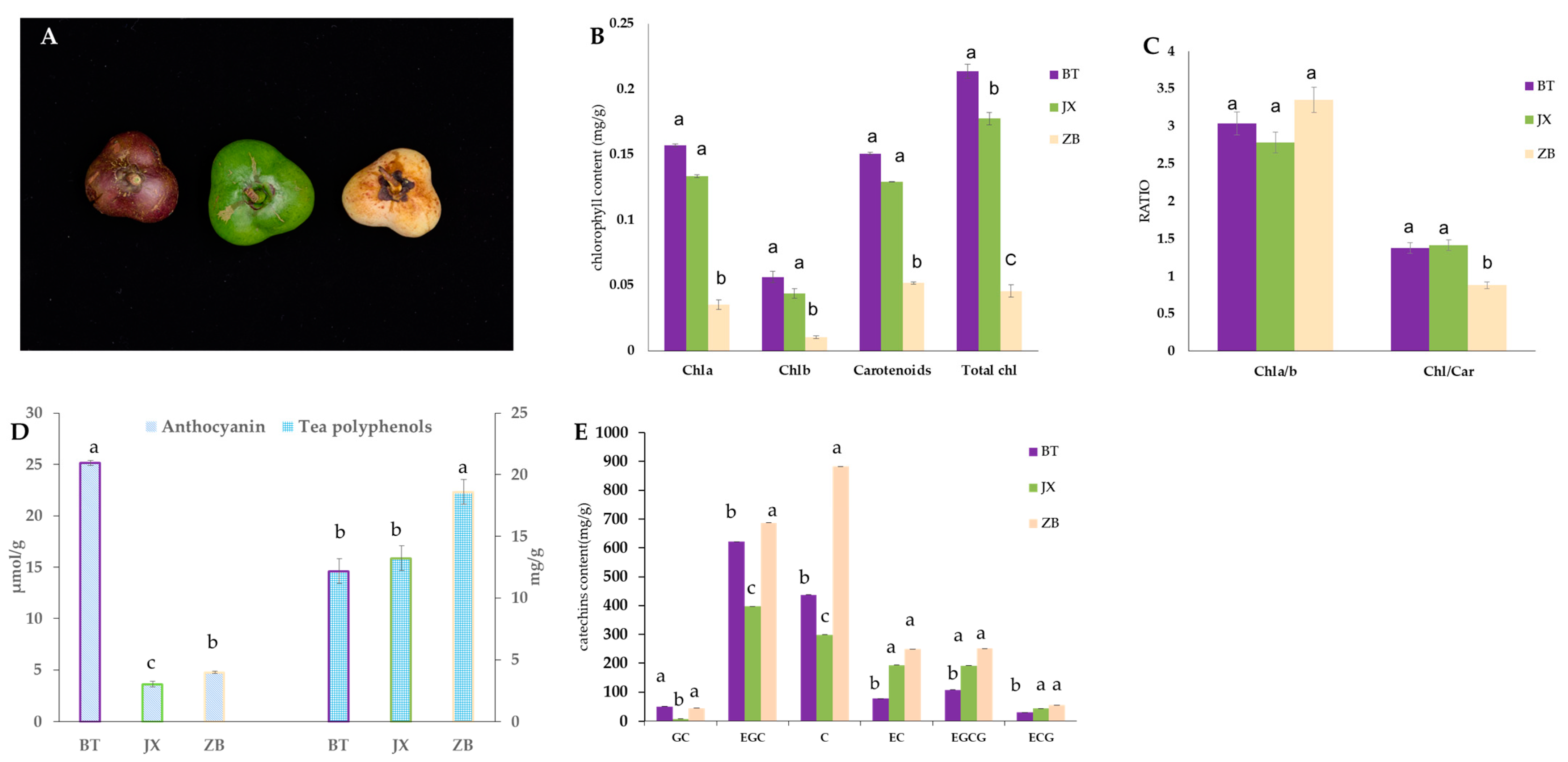
2.1. Pigment Content in Different Tea Pericarps

Phenotypic characteristics of different tea pericarp colors revealed that the pericarp of JX was a normal green color, while ZB was white and BT was purple (Figure 1A). Consistent with phenotypic characteristics, the analysis of tea pericarp pigment contents showed that the levels of total chlorophyll in BT were higher than that of JX. The carotenoid content in the pericarp of ZB was significantly lower than that of the normal green variety JX (Figure 1B,C). The concentrations of tea polyphenols in the ZB pericarp were higher than BT and JX (Figure 1D), while the concentrations of anthocyanin were higher in BT than in JX and ZB (Figure 1E). Catechin analysis revealed that total catechins were higher in white pericarp (ZB), while EGC, GC, and C showed relatively higher levels in BT than in ZJ. The content of C (catechin) in ZB was significantly higher than JX and BT.

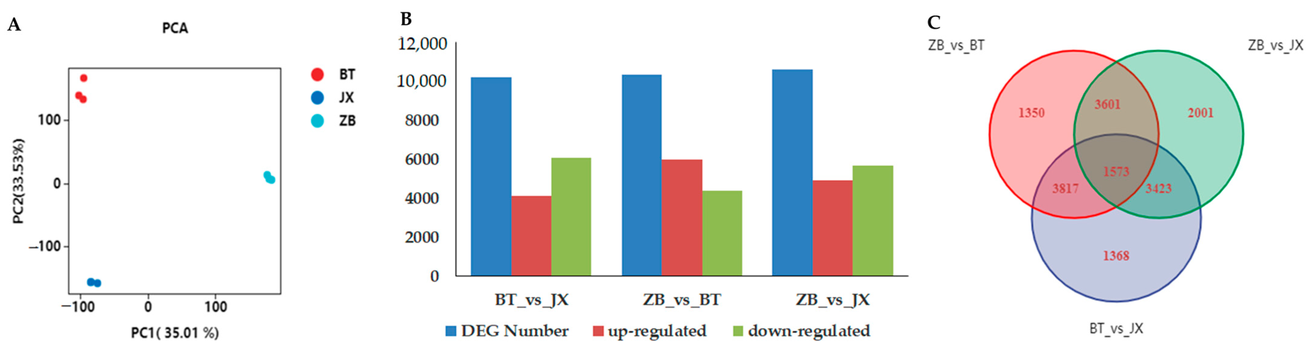
2.2. Transcriptomic Profiling of Different Tea Pericarps

To reveal the molecular mechanism of coloration in different pericarps, transcriptome analysis was performed using the different colored pericarps of ZB, JX, and BT. After data cleaning and raw read quality control, 60,293,150, 65,004,398 and 66,416,772 clean reads were obtained from the ZB, JX, and BT libraries, respectively, with clean data of 5.73 Gb for each sample, Q30 base percentages of 87.22% and above, and an average GC content of 44.73% (Supplementary Table S1). All clean data were mapped to the ‘Shuchazao2’ reference genome of Camellia sinensis var. sinensis (CSS, http://tpia.teaplants.cn/, accessed on 3 August 2022). Clean reads ranging from 72.90 to 89.49% were mapped (Supplementary Table S1). The PCA score plot of sequencing data is shown in Figure 2A and suggests that all pericarp samples were clustered and well separated into three groups. These results implied that the RNA sequencing data were reliable for subsequent analysis.

2.3. Identification of Differentially Expressed Genes between ZB, JX, and BT

To identify differentially expressed genes (DEGs) involved in color formation in different pericarps, fragmentation mapping in millions (FPKM) values for each gene in pericarps of ZB, JX, and BT were analyzed using a false discovery rate (FDR) ≤0.01 and fold change (FC) ≥2 as screening criteria. A total of 10,181, 10,341 and 10,598 DEGs were identified in pairwise comparisons of BT with JX, ZB with BT, and ZB with JX, respectively (Figure 2B). Both BT vs. JX and ZB vs. JX included more down-regulated DEGs than up-regulated genes. ZB vs. BT had significantly more up-regulated DEGs than down-regulated. The Venn analysis results showed that there were a total of 1573 common differentially expressed genes (FC ≥ 2, Q-value ≤ 0.05) among the three types of pericarp (Figure 2C). Among the common DEGs, HYC85, dehydrin DHN1-like, and oleosin 1-like showed the highest expression level in BT pericarp (Supplementary Table S2).

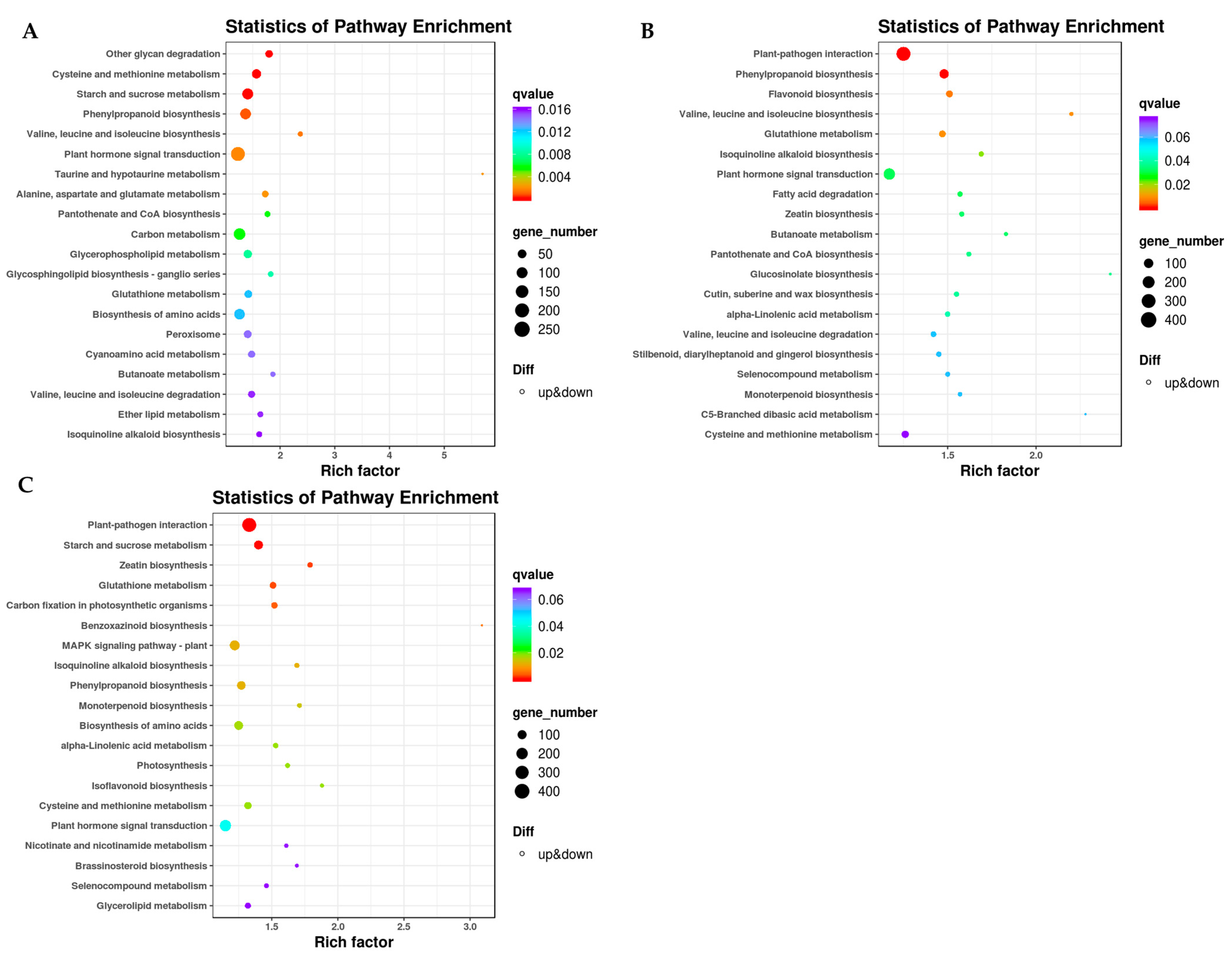
2.4. KEGG Enrichment Analysis

In order to determine the mechanisms of differential color formation in the three varieties of pericarp samples, DEGs involved in the flavonoid pathway, photosynthetic pathway, and the phenylpropane biosynthetic pathway were enriched and classified. In a pairwise comparison of BT and JX pericarp, DEGs in 117 biosynthetic and metabolic pathways were enriched, including phenylpropane biosynthesis (ko00940, 138 genes), flavonoid and flavonol biosynthesis (ko00944, 13 genes), flavonoid biosynthesis (ko00941, 56 genes), and anthocyanin biosynthesis (ko00942, 5 genes) (Figure 3A, Supplementary Table S3). In the paired comparison of ZB and BT pericarp DEGs, the genes were enriched to 117 KEGG pathways. DEGs involved in flavonoid biosynthesis included phenylalanine metabolism (ko00360, 31 genes), phenylpropanoid biosynthesis (k000940, 152 genes), flavonoid biosynthesis (ko00941, 69 genes), flavonol biosynthesis (ko0944, 15 genes), and anthocyanin biosynthesis (k00942, 3 genes) (Figure 3B, Supplementary Table S3). In comparison with JX, 119 DEGs in ZB were enriched in biosynthetic and metabolic pathways, including anthocyanin biosynthesis (ko00942, 6 genes), flavonoid biosynthesis (ko00941, 46 genes), phenylpropane biosynthesis (ko00940, 139 genes), photosynthesis (ko00195, 33 genes), and others (Figure 3C, Supplementary Table S3).
KEGG enrichment analysis indicated that 82 genes were up-regulated and 56 were down-regulated in the benzopropane biosynthetic pathway in BT and JX pairwise comparisons. A total of 38 genes were down-regulated in the flavonoid biosynthetic pathway in the ZB and BT pairwise comparison, and the remaining 45 genes were up-regulated. In the flavonoid and flavonol biosynthetic pathways, 15 DEGs were screened, 10 of which were up-regulated in white pericarps (ZB). In addition, three DEGs involved in the phenylpropane biosynthetic pathway were screened, namely, CCoAOMT (caffeoyl coenzyme A O-methyltransferase, CSS0043057), CCR (cinnamoyl coenzyme A reductase, CSS0026943), and the CsnewGene_15860 (Supplementary Table S3). The expression levels of these genes were significantly lower in white pericarp than in purple pericarp.
In the photosynthetic pathway, photosystem II10 kDa polypeptide (PSII10 kDa polypeptide, CSS0001445), chlorophyll a-b binding protein 13 (CSS0015941), photosystem I subunit O (photosystem I subunit O (CSS0045008), photosystem I reaction center subunit psaK (CSS0016265), photosystem I reaction center subunit N (CSS0016265), photosystem I reaction center subunit N (CSS0010064), HY5 (HY5, CSS00048476) in the plant circadian rhythm pathway, and chlorophyllase (CLH, CSS00004684) in the porphyrin and chlorophyll metabolism pathway were also major DEGs. Excluding CLH, the expression levels of all the above DEGs in white pericarp (Zhongbaiyihao) were significantly higher than those in the conventional green pericarp (Jinxuan) (Supplementary Table S3). These data indicate a key role of DEGs in photosynthesis in photosystem I and photosystem II, as well as in response to light signals and regulation of participation in life activities on a circadian basis.

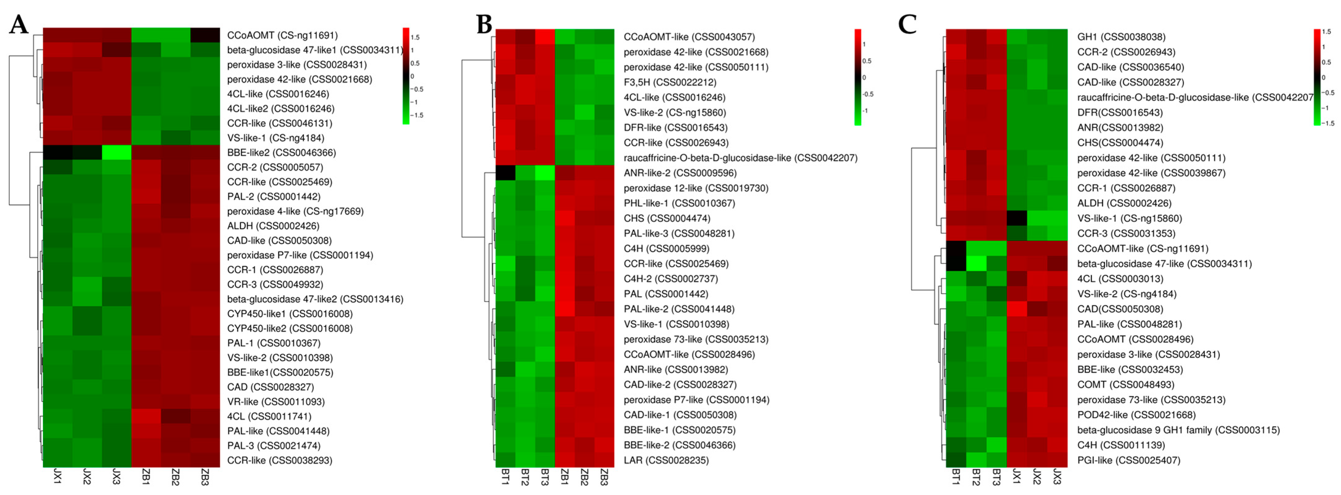
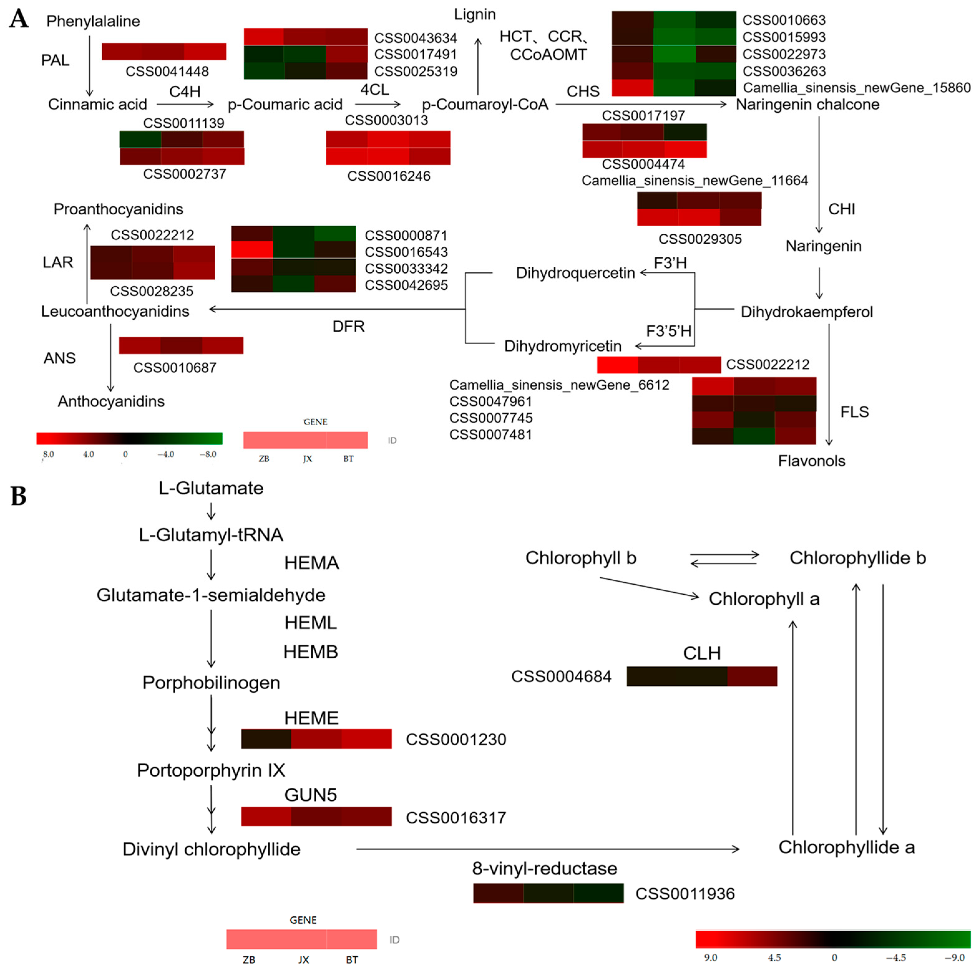
2.5. Identification of DEGs Involved in Flavonoid Biosynthesis

In order to distinguish the critical genes involved in flavonoid and phenylpropanoid pathways in different pericarps, a clustering heat map was created to investigate the expression characteristics of relevant genes using KEGG enrichment analysis. Results showed that compared with ZB, the expression levels of CCoAOMT (CS ng11691), beta glucose 47-like 1 (CSS0034311), peroxidase3-like (CSS0028431), peroxidase 42-like (CSS0021668), 4CL-like (CSS0016246), 4CL-like 2 (CSS0016246), CCR-like (CSS0046131), and VS-like 1 (CS ng4184) were higher in JX pericarp (Figure 4A, Supplementary Table S4). Meanwhile, CCoAOMT like (CSS0043057), peroxidase 42-like (CSS0021668), peroxidase 42-like (CSS0050111), F3,5H (CSS0022212), 4CL-like (CSS0016246), VS-like 2 (CS-ng15860), DFR-like (CSS0016543), CCR-like (CSS0026943), and raucaffricine-O-beta-D-glucosidase-like (CSS0042207) showed higher expression levels in BT pericarp than in that of ZB (Figure 4B, Supplementary Table S4). Similarly, GH1 (CSS0038038), CCR-2 (CSS0026943), CAD like (CSS0036540), CAD like (CSS0028327), raucaffricine-O-beta-D-glucosidase like (CSS0042207), DFR (CSS0016543), ANR (CSS0013982), CHS (CSS0004474), peroxidase 42 like (CSS0050111), peroxidase 42 like (CSS0039867), CCR-1 (CSS0026887), ALDH (CSS0002426), VS-like 1 (S-ng15860), and CCR-3 (CSS0031353) were more highly expressed in BT than JX (Figure 4C, Supplementary Table S4). Comprehensive KEGG enrichment analysis showed that the expression level of genes involved in anthocyanidin synthesis in purple pericarp (BT) was high (such as DFR, ANR, etc.), while in white pericarp (ZB), the expression level of genes involved in downstream anthocyanidin synthesis pathway was relatively low.

2.6. Validation of Gene Expression Levels

Based on the RNA-Seq results of three different colored pericarps, eleven DEGs involved in color formation of tea pericarps were selected to confirm the accuracy of the RNA-Seq results using qRT-PCR (Figure 5A). Correlation analysis was performed to establish correlations between RNA-Seq and qRT-PCR results. The results revealed that the qRT-PCR and RNA-Seq data were highly correlated (correlation coefficient of 0.8171, Figure 5B). These results indicated that the transcriptome data were credible.

3. Discussion

3.1. Differences in Pigment Accumulation Lead to Differences in the Pericarp Color

Previous studies revealed that leaf color formation in purple tea may be due to variations in flavonoid content [23,24]. While higher concentrations of chlorophyll are responsible for green leaf color and photosynthesis [3], albino or etiolated leaves are mainly attributed to lower chlorophyll and higher carotenoid concentrations in leaf tissue [10,21,25]. Some research suggested that carotenoids protect chlorophyll from photo-oxidative damage through certain reductive properties as well as the absorption and transfer of light energy in photosynthesis [26]. Therefore, if carotenoid synthesis is blocked, its chlorophyll protection capacity may be lost and eventually cause abnormal chloroplast development [27]. Some research has suggested that chloroplast in etiolated leaves is inhibited, thereby inhibiting the synthesis of chlorophyll [28]. Similar findings have been confirmed in specific photosensitive etiolated leaf cultivars ‘Yujinxang’ [21], ‘Huangjinya’ [29], and ‘Anjibaicha’ [30].
Similar results have been reported in studies on different fruit peels; the formation of the red color was proposed to be a combination of decreased chlorophyll and increased anthocyanin accumulation during litchi maturation [31,32], while the purple color of the mangosteen fruit pericarp was mainly due to anthocyanins [33]. In addition to anthocyanin, flavones and some flavonols also act as major pigments or co-pigments [31]. Flavonoids affect the colors of both fruits and vegetables as well as that of the grain pericarp [34,35,36]. In a study of sweet osmanthus pericarp, Han et al. found that lignans and phenolic acids were higher in green pericarps than in purple-black pericarps, while the opposite was true of anthocyanins and flavonoids [37].
The results of this study show that the total chlorophyll content of the white pericarp of ‘Zhongbaiyihao’ was significantly lower than the normal green pericarp of ‘Jinxuan’ and the purple pericarp of ‘Baitangziya’, with the Chl/Car ratio showing a similar trend (Figure 1C). Meanwhile, although the Chla/b of ZB was relatively high, the absolute content of chlorophyll in ZB was low (Figure 1B,C), which is not conducive to the formation of purple and green pericarps. Nearly all the chloroplast-related genes appeared to be highly expressed in the white pericarp, which may be related to the lower chlorophyll content in white pericarp and the need to synthesize related substances to maintain normal life activities. In addition, while the content of catechins is relatively high in ZB, catechins are coloress in plants, which could partially explain the white appearance of ZB pericarp. The carotenoid content varied in a similar way to the chlorophyll content of the three tea pericarp varieties. For BT with purple pericarp, high levels of anthocyanins, chlorophyll-a, chlorophyll-b, and carotenoids may be the main reasons for the formation of its purple phenotype (Figure 1B–D). Of course, because anthocyanins and catechins compete for substrate in the flavonoid synthesis pathway, the accumulation of anthocyanins in the purple pericarp reduced the substrate for catechin synthesis, resulting in less accumulation of catechins in the purple pericarp (Figure 1E). We also found that the expression of an R2R3-MYB, MYB114, was up-regulated in purple pericarp, suggesting that this gene may be involved in the regulation of anthocyanins during pericarp coloration (Supplementary Table S3).
The present research results are consistent with previous studies – that is, the purple phenotype was positively correlated with total anthocyanin content [33,37,38,39]. Likewise, the low chlorophyll content in the white pericarp of ‘Zhongbaiyihao’ may be due to the abnormal synthesis and enhanced chlorophyll degradation. For Jinxuan, an adequate amount of chlorophyll may explain its green pericarp.

3.2. Expression of Different Structural Genes Affects the Synthesis of Chlorophylls, Carotenoids, and Flavonoids

Presently, the CCoAOMT, DFR, and F3’5’H genes are significantly up-regulated in the BT pericarp relative to Jinxuan, which is consistent with results of Liu et al. that found that the expression pattern of CCoAOMT was highly correlated with flavonoid concentrations in other plants [40,41]. Although CCoAOMT is not essential for anthocyanin accumulation, up-regulation of CCoAOMT may contribute to the production of more flavonoid derivatives in the pericarp. Flavonoid 3’,5’-hydroxylase (F3’5’H, CYP75A) is responsible for the conversion of the substrate dihydrokaempferol to dihydroquercetin [42], and dihydroflavonol reductase (DFR) is responsible for catalyzing the conversion of dihydromyricetin and dihydroquercetin to leucovorin [43]. Studies have shown that competition between the flavonoid/flavonol pathway and the anthocyanin pathway is primarily a substrate competition between the FLS and DFR, with FLS preferentially utilizing dihydroquercetin and dihydrokaempferolin [44]. FLS facilitates a metabolic shift towards flavonols, resulting in lower anthocyanin accumulation [12,45].
In this study, the higher expression levels of DFR indicates that flavonoid metabolism in purple pericarp shunted more substrate than green pericarp into the anthocyanin biosynthetic pathway (Figure 6A). The results of RNA-seq and qRT-PCR showed a relatively high FLS expression in green and white pericarp compared with the purple pericarp, implying that substrates might be shunted from the anthocyanins’ biosynthesis pathway towards the flavonol pathway in the substrate competition between FLS and DFR (Figure 6A).
Flavonoids and isoflavonoids are important secondary metabolites for plant defence that can function as inhibitors of fungal growth [46]. Vestitone reductase (VR) is a key reductase in the isoflavanone biosynthesis pathway. Previous studies found that VR activity corresponded with the synthesis of vestitone, which improved plant disease resistance [47,48]. The results of this study indicated that the VR gene has a high expression level, which may be due to unique isoflavanone metabolism in tea percarps. It is particularly noteworthy that ZB is a mutant variety of small leaf tea (Camellia sinensis var. sinensis) that was found previously to possess strong disease resistance, which may relate to its special accumulation of flavonoids. Moreover, we speculate that low anthocyanin and high tea polyphenol concentrations observed in ZB could relate to its high VR gene expression, because both anthocyanins and isoflavanones are metabolized through chalcone substrates. Such substrate competition could also affect the white appearance of ZB pericarp, because isoflavanones are colorless, unlike anthocyanins [49].
Meanwhile, we identified four DEGs related to the chlorophyll pathway, i.e., GUN5 (CSS0016317), HEME (CSS0012339), 8-vinyl-reductase (CSS0011936), and CLH (CSS0004684) (Figure 6B). Previous studies have reported that Genome Uncoupled 4 (GUN4) could bind ChlH/GUN5 to enhance chlorophyll biosynthesis by activating magnesium-chelatase, while the function of GUN5 is to shift protoporphyrin IX towards chlorophyll biosynthesis metabolism [50]. Meanwhile, HEME can catalyze uroporphyrinogen III to synthesize coproporphyrinogen III. Previous studies have shown that the content of heme and chlorophyll will decrease in transgenic tobacco with HEME-RNAi silenced [51]. Zhu et al. [52] confirmed that the levels of HEME expression in purple leaf tea were markedly different from green leaves, which resulted in variations in chlorophyll content. Similarly, our previous research also indicated that the lower expression levels of GUN5 and HEME resulted in the lower chlorophyll levels in yellow ‘Huangyu’ tea [53].
In this study, the chlorophyll concentration of white pericarp, in addition to higher GUN5 and 8-vinyl-reductase expression levels compared with green pericarp, indicated that ‘Zhongbaiyihao’ pericarp had a higher chlorophyll synthesis capacity than ‘Jinxuan’. Meanwhile, chlorophyllase (CLH, CSS0004684), which is involved in the chlorophyll degradation pathway, was higher in ‘Baitang’ than in ‘Jinxuan’ and ‘Zhongbaiyihao’ pericarps, suggesting that chlorophyll degradation activity in the pericarp of ‘Baitang’ was higher than those of ‘Jinxuan’ and ‘Zhongbaiyihao’ pericarps, resulting in reduced chlorophyll accumulation. The above results indicate that the higher chlorophyll level in ‘Zhongbaiyihao’ pericarp may be attributed to the higher expression of GUN5, HEME, and 8-vinyl-reductase (Figure 6B), while formation of the purple pericarp of ‘Baitang’ tea fruit may be due to the higher expression of DFR, which promotes the flow of substrates to the anthocyanin biosynthesis pathway.

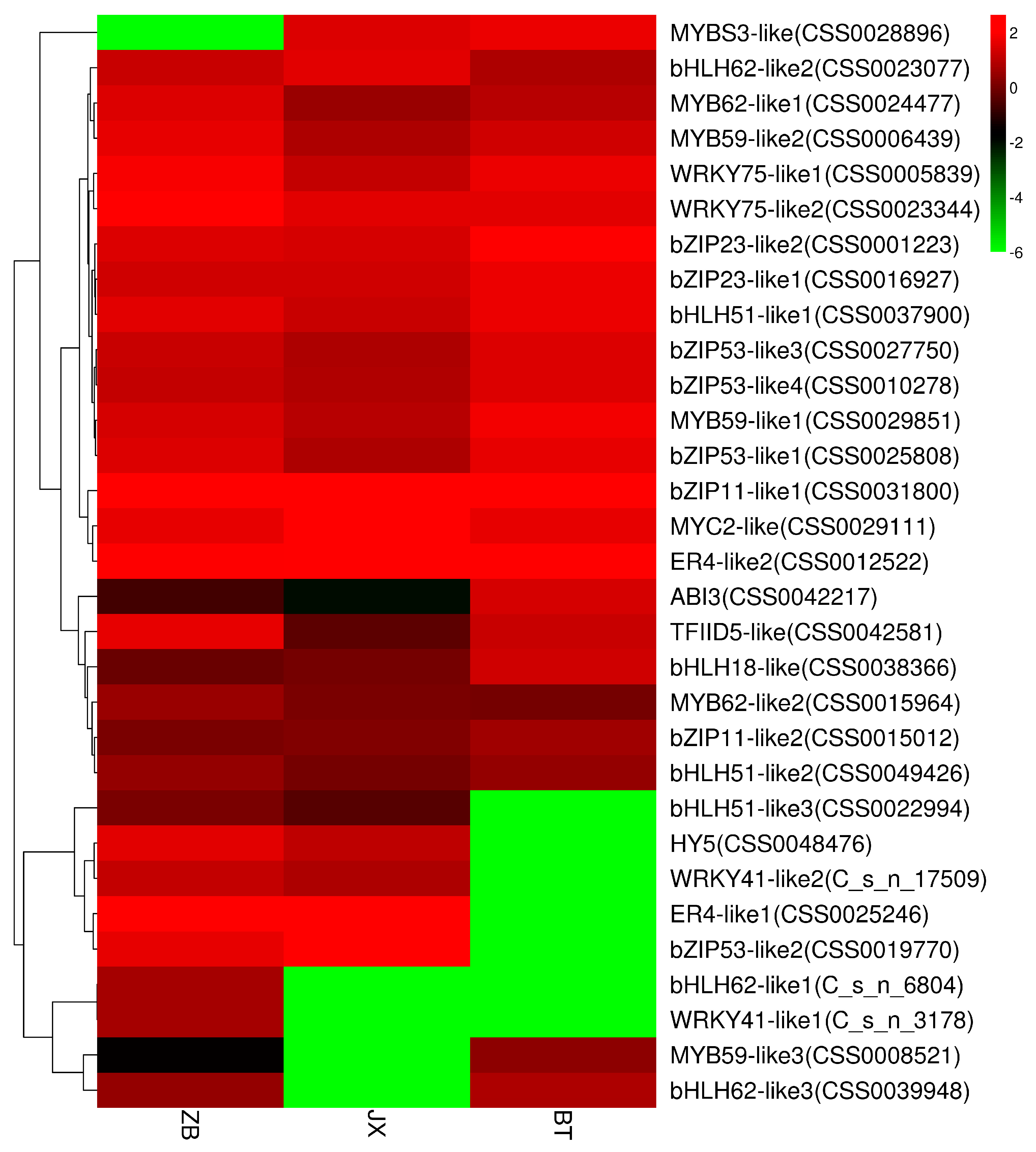
3.3. Transcription Factors Involved in Pigment Accumulation

The content of anthocyanins is determined by structural genes as well as specific transcription factors (TFs) [54]. The structural genes of key enzymes in anthocyanin synthesis are subject to transcriptional regulation by transcription factors, and some of the more widely studied transcription factors include the MYB family, the bHLH family, and WD40 proteins, which regulate anthocyanin biosynthesis by binding to elements acting in the promoter regions of structural genes [55,56,57,58]. This has been well demonstrated in rice [59], cocoa [60], cotton [61], and several other plant species.
In previous research, MYBs3 was found to be a single DNA-binding repeat MYB (R1-MYB) transcription factor that played a key role in cold adaptation in rice, likely by activating relevant genes when plants were subjected to various stressors [62]. Gan et al found that transgenic banana that over-expressed MpMYBS showed significantly higher cold tolerance than wild type, possibly by increasing proline levels in the transgenic banana line [63]. MYBs3 was also found to positively regulate anthocyanin biosynthesis during flower development in Hibiscus syriacus L. var. Shigyoku [64]. Wang et al. found that MYBs3 was involved in regulating resistance-related pathways in Eureka lemon, including phenylpropanoid, flavone/flavonol, and isoflavonoid pathways [65].
Presently, the expression of MYBS3-like (CSS0028896) transcription factor was significantly lower in ZB than in JX and BT (Figure 7). The reason may be that MYBS3-like was involved in regulating flavonoid biosynthetic processes in JX and BT pericarp.
Interestingly, transcription factors involved in suppressing anthocyanin biosynthesis were significantly downregulated in the ZJ pericarp, including bHLH51-like3 (CSS0022994), HY5 (CSS0048476), WRKY41-like2 (CS-ng17509), ERF4-like1 (CSS0025246), bZIP53-like2 (CSS0019770), bHLH62-like1 (CS-ng6804), and WRKY41-like1 (CS-ng3178) (Figure 7). In Arabidopsis thaliana, the WRKY41-1 transcription factor was significantly negatively correlated with anthocyanin levels, acting as a repressor of anthocyanin biosynthesis [66]. Similarly, MYB4-like (R2R3-MYB) and bHLH62 might also repress structural genes involved in anthocyanin synthesis, as it showed a significant negative regulatory relationship in the fruit skin of ‘Red Delicious’ [67]. The expression level of bZIP53 was up-regulated as anthocyanin levels decreased with fading flower color in the chrysanthemum, with a higher expression level of bZIP53 found to occur in response to high temperature [68]. Recent research found that ERF4 affected fruit firmness through TPL4 by reducing ethylene levels [69].
These findings discussed above indicate that the low expression of anthocyanidin transcriptional inhibitors may be related to the accumulation of anthocyanidin in BT pericarp. Correspondingly, due to the high expression of such transcriptional inhibitors, there was a reduction in the accumulation of flavonoids and anthocyanidins in white ZB pericarp. Among the transcription factors mentioned above, MYB59-like3 (CSS0008521), WRKY41-like2 (CS ng3178), bHLH62-like1 (CS ng6804), and bHLH62-like3 (CSS0039948) were down-regulated in JX purple pericarp.
Lai et al. reported that the R2R3-MYB TF, MYB59, played a key role in some biological processes during development in Arabidopsis, negatively regulating leaf senescence when induced by jasmonic acid and salicylic acid in [70]. MYB59 can participate in circadian rhythm regulation by directly targeting CIRCADIAN CLOCK-ASSOCIATED 1 [71]. Other research showed that MYB59 could resist potassium deficiency stress by the positive regulation of Nitrate Transporter1.5 [72], represses calcium homeostasis, and regulates plant growth and stress response [73]. Wiśniewska et al. [74] reported that AtMYB59 may play an extensive role from metabolism modulation to the responses to abiotic and biotic stresses. Considering that the flavonoid content in the Jinxuan pericarp was in between ZB and ZJ, the down-regulation of the transcription factors mentioned above may be related to the metabolic balance of flavonoids, indicating that transcription factors play a role in regulating tea pericarp coloration.

3.4. Accumulation of Pericarp Pigments May Be Related to Light Induction

As a basic leucine zipper (bZIP) transcription factor, HY5 (ELONGATED HYPOCOTYL5) plays an important role in regulating plant growth and development by acquiring light signals through different light responsive cis-elements and transmitting them to downstream action elements [75]. It is the first transcription factor found to be involved in photomorphogenesis and plays a key role in regulating plant anthocyanidin biosynthesis. It was reported that HY5 might regulate GUN5 and HSP90 involved in chlorophyll biosynthesis [76]. Abbas et al. revealed that the HY5 transcription factor was involved in regulating nitrogen uptake and photogenesis, as well as assimilation in plants, and that the above regulatory mechanisms are completed through light response processes [77]. The function of the HY5 transcription factor is also related to phenotypes associated with photomorphogenesis in plants, such as hypocotyl elongation in seedlings and yellowing and de-yellowing of plants [78,79]. In addition, HY5 initiates downstream photomorphogenesis in photosensitive pigments, cryptochromes, and UV-B photoreceptors [80]. Furthermore, HY5 plays a critical role in chlorophyll accumulation and chloroplast development in plants, and can act as a central repressor in light signaling to enhance photomorphogenesis [81]. A recent study found that HY5 and CLH were up-regulated while POR and HemA genes were down-regulated in ‘Xiangfei Huangye’ etiolated tea, suggesting that chlorophyll synthesis was inhibited through increased expression of HY5, causing an increased rate of catabolism. [82].
In the present study, the expression profile of HY5 corroborated previous research results, in that the expression level of HY5 (CSS0048476) was higher in white pericarp than in the normal green pericarp. Interestingly, the expression level of HY5 was not significantly different between white ZB pericarp and purple ZJ pericarp. However, the expression level of CLH (CSS0004684) was significantly higher in the purple BT pericarp than the white and green pericarps. Considering that CLH can have a degrading function in the chlorophyll pathway, it appeared to show a negative effect on chlorophyll synthesis and accumulation. Therefore, in the purple pericarp, we speculate that the high expression level of CLH led to enhanced catabolism of chlorophyll, while the high levels of flavonoids promoted the shunting of upstream substances towards the anthocyanin metabolic pathway, resulting in the purple color of the pericarp.

4. Materials and Methods

4.1. Plant Material

Tea (Camellia sinensis (L.) O. Kuntze) fruits were collected under natural conditions from tea plant varieties having white pericarp (Camellia sinensis var. Zhongbaiyihao), green pericarp (Camellia sinensis var. Jinxuan), and purple pericarp (Camellia sinensis var. Botangziya) grown in the tea garden of South China Agricultural University, Guangzhou, Guangdong Province, on 30 June 2022. The fruit samples were stripped of their pericarps and immediately frozen in liquid nitrogen, then promptly ground into powder and stored at −80 °C for further analysis. The samples were then subjected to further RNA-seq and chemical composition analysis. Each sample consisted of three biological replicates.

4.2. Extraction and Determination of Pigments

4.2.1. Total Chlorophylls and Carotenoids

The total chlorophyll and carotenoids contents were determined by spectroscopy [83]. An amount of 500mg of sample was extracted in 25 mL of 95% ethanol (v/v) for 24 h at room temperature, and the extract was filtered and then fixed to 25 mL using 95% ethanol (v/v). The absorbance values were determined using an automatic microplate elisa reader (Sunrise, TECAN, Austria) as described by Rothenberg et al. [83]. The content of chlorophyll a, chlorophyll b, and carotenoids was calculated as follows:
Chl a = (12.21 × A663 − 2.81 × A646)/(1000 × W) × L;
Chl b = (20.13 × A646 − 5.03 × A663)/(1000 × W) × L;
Caro = (1000 × A470 − 3.27 × Chl a – 104 × Chl b)/(229 × 1000 × W) × L.
where A646, A663, and A470 indicate absorbance values at 646 nm, 663 nm, and 470 nm respectively; L indicates the total volume of the extract solution (mL), and W indicates the fresh weight (FW) of the sample (g); and the chlorophyll and carotenoid contents were determined in mg/g (FW).

4.2.2. Determination of Total Anthocyanins

The total anthocyanin was determined as reported by Wei et al. [84]. An amount of 100 mg of sample powder was extracted in 3 mL of extraction solution (1% HCl in methanol) for 16 h at 4 ℃. The extract was filtered and then fixed to 10mL. The absorbance was measured at 530 nm and 657 nm by a spectrophotometer, using 1 cm cells. Total anthocyanin contents were calculated by the following formula: total anthocyanin = ((A530 − A620) − 0.1(A650 − A620) × 100)/(4.62 × W).

4.2.3. Determination of Total Contents of Polyphenols

Total tea polyphenol contents were determined by the Folin reagent method, which is a modification of the method previously reported by Yu et al. [85]. Briefly, 200 mg of sample powder was extracted in 5 mL of extraction solution (70% methanol solution) in a 70 °C water bath for 10 min. Then, 1 mL extraction was supplemented with 5 mL Folin reagent to react for 8 min. Then, 4 mL of 7.5% Na2CO3 solution was added, and the reaction system was shaken for 60 min at room temperature. The absorbance of the reaction solution was measured at 765 nm by a spectrophotometer (UV-160 Shimadzu, Tokyo, Japan). The tea polyphenol concentration was quantitatively calculated with gallic acid as a calibration standard.

4.2.4. HPLC Analysis of Catechins

The content of catechins was analyzed as described by Mei et al. [86]. Briefly, 200 mg of tea powders were extracted with 8 mL of 70% methanol in a 75 °C water bath for 30 min. The extraction solution was filtered through a 0.22 mm Millipore filter before conducting HPLC analysis. The chromatographic column was a C18 SB column (4.6 × 250 mm, 5 mm, Waters Technologies, Milford, MA, USA). The chromatographic conditions were as follows: Mobile phase A was decreased linearly from 92% in 5 min to 75% at 14 min and then increased linearly from 75% at 14 min to 92% at 30 min. Mobile phase A contained 0.1% acetic acid and 99.9% ultrapure water, and mobile phase B was 100% acetonitrile. The flow rate was 0.80 mL/min, and the column temperature was 30 °C; the injection volume was 20 µL.

4.2.5. RNA Extraction and Transcriptome Sequencing

Trizol (Invitrogen, Carlsbad, CA, USA) was used to extract total RNA from different pericarps [87]. The construction of a cDNA library and sequencing employed a Novaseq6000 by Biomarker Technologies Corporation (Beijing, China). The raw data were uploaded to Beijing Institute of Genomics, National Genomics Data Center, China (https://bigd.big.ac.cn/gsa, accessed on 3 September 2022), and the accession number is CRA007882. The raw data from the sequencing machines were initially filtered to obtain clean data by using SeqPrep software (Version 4.6.1). The HISAT2 program [88] was used to compare the clean reads of the filtered rRNA to the reference genome sequence (Camellia sinensis var. sinensis (CSS), ShuchazaoV2 genome, http://tpia.teaplants.cn/download.html, accessed on 3 August 2022).

4.3. Gene Function Annotation and Expression Level Analysis

In order to annotate the assembled genes, seven public databases were searched for homology, namely, NR (Non-Redundant Protein Sequence Database), KEGG (Kyoto Encyclopedia of Genes and Genomes), COG (Clusters of Orthologous Group of proteins), Swish-Prot database, KOG (Eukaryotic orthologous groups) database, Pfam (Pfam protein family database), and GO (gene ontology) database. The gene expression level was estimated by FPKM (every thousand base segments in the transcript mapped by every million segments). Based on the value of genes in the three varieties of samples, the differentially expressed genes were screened using DESeq2 software (version 1.38.0), with|log2FC| ≥ 1 and p-adj < 0.05 as the threshold. KEGG enrichment was performed using R-Package.

4.4. Quantitative Real-Time PCR and Expression Verification

In order to verify the RNA-Seq results, β- Actin was selected as an internal reference gene. The DEGs involved in chlorophyll metabolism and anthocyanin metabolism pathways were identified through transcriptome data analysis and the KEGG database, and eleven genes were screened for quantitative real-time PCR (qRT-PCR) verification. Total RNA was extracted using RNA Simple Total RNA Kit (TIANGEN) according to the protocol. The thermal profile for the PCR amplification was 95 °C for 5 min, and then 40 cycles of 10 s at 95 °C and 40 cycles of 30 s at 60 °C. The qRT-PCR primers are listed in Supplementary Table S5. Relative expression levels of the genes were quantified using the 2 −∆∆CT method [89].

4.5. Statistical Analysis

Statistical analysis was performed using SPSS software (version 24.0 for Windows, SPSS Inc., Chicago, IL, USA). Significant differences between different groups were determined using Tukey’s post hoc test. A p value less than 0.05 was considered statistically significant. Excel 2010 (Microsoft, Redmond, WA, USA) was applied to drawbar graphs of the experimental data. Some figures and tables related to transcriptomes were prepared on the BMKcloud platform (https://international.biocloud.net/, accessed on 29 May 2023).

5. Conclusions

In this study, chemical components and RNA-seq data were analyzed to explore the mechanisms underlying tea pericarp coloration. A total of 17,133 DEGs were identified. Some DEGs involved in the anthocyanin biosynthesis pathway, such as DFR, F3’5’H, CCoAOMT, and 4-coumaroyl-CoA, were highly expressed in purple BT pericarps, where they were positively correlated with the anthocyanin accumulation. In addition to the CLH gene, 33 DEGs involved in chlorophyll synthesis and degradation and 64 DEGs involved in photosynthesis-related proteins were identified. The low chlorophyll content of ZB may be due to the low expression level of HEME (heme A synthetase, CSS0016239) involved in chlorophyll synthesis and the high expression level of CLH involved in chlorophyll degradation. The high expression of CLH genes was negatively correlated with chlorophyll content in ZB pericarps. Multiple genes involved in repressing anthocyanin biosynthesis pathways were significantly down-regulated in the purple pericarp, including bHLH51-like3 (CSS0022994), HY5 (CSS0048476), WRKY41-like2 (CS-ng17509), and ERF4-like1 (CSS0025246). The expression level of HY5 (CSS0048476) was higher in the white pericarp than in the normal green pericarp, suggesting that transcription factors play a role in regulating the coloration of tea pericarp. Overall, the different colors in the pericarps of different tea varieties might be attributed to flavonoid and chlorophyll biosynthetic pathways. Our results provide new insights for clarifying the molecular mechanisms underlying pericarp coloration.

Supplementary Materials

The following supporting information can be downloaded at https://www.mdpi.com/article/10.3390/ijms241713198/s1.

Author Contributions

Data curation, Y.L.; Formal analysis, Y.L. and H.Z. (Huan Zhang); Funding acquisition, L.Z.; Investigation, Y.L. and H.Z. (Hui Zhou); Methodology, Y.D., K.Z., and H.Z. (Huan Zhang); Software, H.S.; Validation, K.Z.; Visualization, H.S.; Writing—original draft, Y.D.; Writing—review and editing, L.Z. and D.O.R. All authors have read and agreed to the published version of the manuscript.

Funding

This research was funded by National Natural Science Foundation of China, grant number 32072628; 2021 Guangdong Provincial Science and Technology Special Project: 210729116901180.

Institutional Review Board Statement

Not applicable.

Informed Consent Statement

Informed consent was obtained from all subjects involved in the study.

Data Availability Statement

The raw sequence data reported in this paper have been deposited in the Genome Sequence Archive in National Genomics Data Center, China National Center for Bioinformation/Beijing Institute of Genomics, Chinese Academy of Sciences (GSA: CRA007882) that is publicly accessible at https://ngdc.cncb.ac.cn/gsa, accessed on 21 September 2022.

Conflicts of Interest

The authors declare no conflict of interest.

References

  1. Zhao, J.; Li, P.; Xia, T.; Wan, X. Exploring plant metabolic genomics: Chemical diversity, metabolic complexity in the biosynthesis and transport of specialized metabolites with the tea plant as a model. Crit. Rev. Biotechnol. 2020, 40, 667–688. [Google Scholar] [CrossRef] [PubMed]
  2. Gvasaliya, M.V. Economic estimation of new varieties and mutant forms of tea (Camellia sinensis (L.) Kuntze) in russian subtropics. Pomic. Small Fruits Cult. Russ. 2018, 53, 104–111. [Google Scholar] [CrossRef]
  3. Lu, M.; Li, Y.; Jia, H.; Xi, Z.; Gao, Q.; Zhang, Z.-Z.; Deng, W.-W. Integrated proteomics and transcriptome analysis reveal a decreased catechins metabolism in variegated tea leaves. Sci. Hortic. 2022, 295, 110824. [Google Scholar] [CrossRef]
  4. Tzvetkova-Chevolleau, T.; Franck, F.; Alawady, A.E.; Dall’Osto, L.; Carrière, F.; Bassi, R.; Grimm, B.; Nussaume, L.; Havaux, M. The light stress-induced protein ELIP2 is a regulator of chlorophyll synthesis in Arabidopsis thaliana. Plant J. 2007, 50, 795–809. [Google Scholar] [CrossRef] [PubMed]
  5. Schmitz-Linneweber, C.; Williams-Carrier, R.E.; Williams-Voelker, P.M.; Kroeger, T.S.; Vichas, A.; Barkan, A. A Pentatricopeptide Repeat Protein Facilitates the trans-Splicing of the Maize Chloroplast rps12 Pre-mRNA. Plant Cell 2006, 18, 2650–2663. [Google Scholar] [CrossRef]
  6. Wu, L.; Li, R.; Shu, Q.; Zhao, H.; Wu, D.; Li, J.; Wang, R. Characterization of a New Green-Revertible Albino Mutant in Rice. Crop Sci. 2011, 51, 2706–2715. [Google Scholar] [CrossRef]
  7. Hortensteiner, S. Stay-green regulates chlorophyll and chlorophyll-binding protein degradation during senescence. Trends Plant Sci. 2009, 14, 155–162. [Google Scholar] [CrossRef]
  8. Ma, K.F.; Zhang, Q.X.; Cheng, T.R.; Yan, X.L.; Pan, H.T. Substantial Epigenetic Variation Causing Flower Color Chimerism in the Ornamental Tree Prunus mume Revealed by Single Base Resolution Methylome Detection and Transcriptome Sequencing. Genes 2018, 19, 2315. [Google Scholar] [CrossRef]
  9. Cuttriss, A.J.; Cazzonelli, C.I.; Wurtzel, E.T.; Pogson, B.J. Carotenoids. Adv. Bot. Res. 2011, 58, 1–36. [Google Scholar]
  10. Li, C.F.; Ma, J.Q.; Huang, D.J.; Ma, C.L.; Jin, J.Q.; Yao, M.Z.; Chen, L. Comprehensive Dissection of Metabolic Changes in Albino and Green Tea Cultivars. J. Agric. Food Chem. 2018, 66, 2040–2048. [Google Scholar] [CrossRef]
  11. Li, H.; Yang, Z.; Zeng, Q.W.; Wang, S.B.; Luo, Y.W.; Huang, Y.; Xin, Y.C.; He, N.J. Abnormal expression of bHLH3 disrupts a flavonoid homeostasis network, causing differences in pigment composition among mulberry fruits. Hortic. Res. 2020, 7, 83. [Google Scholar] [CrossRef] [PubMed]
  12. Zhou, C.; Mei, X.; Rothenberg, D.O.; Yang, Z.; Zhang, W.; Wan, S.; Yang, H.; Zhang, L. Metabolome and Transcriptome Analysis Reveals Putative Genes Involved in Anthocyanin Accumulation and Coloration in White and Pink Tea (Camellia sinensis) Flower. Molecules 2020, 25, 190. [Google Scholar] [CrossRef] [PubMed]
  13. Hu, J.G.; Zhang, L.J.; Sheng, Y.Y.; Wang, K.R.; Zheng, X.Q. Screening tea hybrid with abundant anthocyanins and investigating the effect of tea processing on foliar anthocyanins in tea. Folia Hortic. 2020, 32, 279–290. [Google Scholar] [CrossRef]
  14. Gao, Z.; Tian, S.; Hou, J.; Zhang, Z.; Yang, L.; Hu, T.; Li, W.; Liu, Y. RNA-Seq based transcriptome analysis reveals the molecular mechanism of triterpenoid biosynthesis in Glycyrrhiza glabra. Bioorg. Med. Chem. Lett. 2020, 30, 127102. [Google Scholar] [CrossRef] [PubMed]
  15. Garber, M.; Grabherr, M.G.; Guttman, M.; Trapnell, C. Computational methods for transcriptome annotation and quantification using RNA-seq. Nat. Methods 2011, 8, 469–477. [Google Scholar] [CrossRef] [PubMed]
  16. Chen, L.B.; Xia, L.F.; Tian, Y.P.; Mei, L.I.; Song, W.X.; Liang, M.Z.; Jiang, C.J. Exploring Sterility Gene from Tea Plant Flower Based on Digital Gene Expression Profiling. Acta Agron. Sin. 2017, 43, 210–217. [Google Scholar] [CrossRef]
  17. Shi, C.Y.; Yang, H.; Wei, C.L.; Yu, O.; Zhang, Z.Z.; Jiang, C.J.; Sun, J.; Li, Y.Y.; Chen, Q.; Xia, T.; et al. Deep sequencing of the Camellia sinensis transcriptome revealed candidate genes for major metabolic pathways of tea-specific compounds. BMC Genomics 2011, 12, 131. [Google Scholar] [CrossRef]
  18. Alagna, F.; D’Agostino, N.; Torchia, L.; Servili, M.; Rao, R.; Pietrella, M.; Giuliano, G.; Chiusano, M.L.; Baldoni, L.; Perrotta, G. Comparative 454 pyrosequencing of transcripts from two olive genotypes during fruit development. BMC Genomics 2009, 10, 399. [Google Scholar] [CrossRef]
  19. Wang, L.; Pan, D.; Liang, M.; Abubakar, Y.S.; Li, J.; Lin, J.; Chen, S.; Chen, W. Regulation of Anthocyanin Biosynthesis in Purple Leaves of Zijuan Tea (Camellia sinensis var. kitamura). Int. J. Mol. Sci. 2017, 18, 833. [Google Scholar] [CrossRef]
  20. Fang, Z.; Hou, Z.; Wang, S.; Liu, Z.; Wei, S.; Zhang, Y.; Song, J.; Yin, J. Transcriptome Analysis Reveals the Accumulation Mechanism of Anthocyanins in Buckwheat (Fagopyrum esculentum Moench) Cotyledons and Flowers. Int. J. Mol. Sci. 2019, 20, 1493. [Google Scholar] [CrossRef]
  21. Xu, P.; Su, H.; Jin, R.; Mao, Y.; Xu, A.; Cheng, H.; Wang, Y.; Meng, Q. Shading Effects on Leaf Color Conversion and Biosynthesis of the Major Secondary Metabolites in the Albino Tea Cultivar “Yujinxiang”. J. Agric. Food Chem. 2020, 68, 2528–2538. [Google Scholar] [CrossRef] [PubMed]
  22. Slattery, R.A.; VanLoocke, A.; Bernacchi, C.J.; Zhu, X.G.; Ort, D.R. Photosynthesis, Light Use Efficiency, and Yield of Reduced-Chlorophyll Soybean Mutants in Field Conditions. Front. Plant Sci. 2017, 8, 549. [Google Scholar] [CrossRef] [PubMed]
  23. Zhou, Q.; Sun, W.; Lai, Z. Differential expression of genes in purple-shoot tea tender leaves and mature leaves during leaf growth. J. Sci. Food Agric. 2016, 96, 1982–1989. [Google Scholar] [CrossRef] [PubMed]
  24. Shen, J.; Zou, Z.; Zhang, X.; Zhou, L.; Wang, Y.; Fang, W.; Zhu, X. Metabolic analyses reveal different mechanisms of leaf color change in two purple-leaf tea plant (Camellia sinensis L.) cultivars. Hortic. Res. 2018, 5, 7. [Google Scholar] [CrossRef]
  25. Yang, Y.; Chen, X.; Xu, B.; Li, Y.; Ma, Y.; Wang, G. Phenotype and transcriptome analysis reveals chloroplast development and pigment biosynthesis together influenced the leaf color formation in mutants of Anthurium andraeanum ‘Sonate’. Front. Plant Sci. 2015, 6, 139. [Google Scholar] [CrossRef]
  26. Demmig-Adams, B.; Adams, W.W. The role of xanthophyll cycle carotenoids in the protection of photosynthesis. Trends Plant Sci. 1996, 1, 21–26. [Google Scholar] [CrossRef]
  27. Fang, J.; Chai, C.; Qian, Q.; Li, C.; Tang, J. Mutations of genes in synthesis of the carotenoid precursors of ABA lead to pre-harvest sprouting and photo-oxidation in rice. Plant J. 2010, 54, 177–189. [Google Scholar] [CrossRef]
  28. Feng, L.; Gao, M.J.; Hou, R.Y.; Hu, X.Y.; Zhang, L.; Wan, X.C.; Wei, S. Determination of quality constituents in the young leaves of albino tea cultivars. Food Chem. 2014, 155, 98–104. [Google Scholar] [CrossRef]
  29. Song, L.B.; Ma, Q.P.; Zou, Z.W.; Sun, K.; Yao, Y.T.; Tao, J.H.; Kaleri, N.A.; Li, X.H. Molecular Link between Leaf Coloration and Gene Expression of Flavonoid and Carotenoid Biosynthesis in Camellia sinensis Cultivar ‘Huangjinya’. Front. Plant Sci. 2017, 8, 803. [Google Scholar] [CrossRef]
  30. Li, C.F.; Xu, Y.X.; Ma, J.Q.; Jin, J.Q.; Huang, D.J.; Yao, M.Z.; Ma, C.L.; Chen, L. Biochemical and transcriptomic analyses reveal different metabolite biosynthesis profiles among three color and developmental stages in ‘Anji Baicha’ (Camellia sinensis). BMC Plant Biol. 2016, 16, 195. [Google Scholar] [CrossRef]
  31. He, M.Y.; Zhou, Y.J.; Zhu, H.; Jiang, Y.M.; Qu, H.X. Metabolome, transcriptome and physiological analyses provide insight into the color transition of litchi pericarp. Postharvest Biol. Technol. 2022, 192, 112031. [Google Scholar] [CrossRef]
  32. Fang, F.; Zhang, X.L.; Luo, H.H.; Zhou, J.J.; Gong, Y.H.; Li, W.J.; Shi, Z.W.; He, Q.; Wu, Q.; Li, L.; et al. An Intracellular Laccase Is Responsible for Epicatechin-Mediated Anthocyanin Degradation in Litchi Fruit Pericarp. Plant Physiol. 2015, 169, 2391–2408. [Google Scholar] [CrossRef] [PubMed]
  33. Palapol, Y.; Ketsa, S.; Stevenson, D.; Cooney, J.M.; Allan, A.C.; Ferguson, I.B. Colour development and quality of mangosteen (Garcinia mangostana L.) fruit during ripening and after harvest. Postharvest Biol. Technol. 2009, 51, 349–353. [Google Scholar] [CrossRef]
  34. Shen, N.; Wang, T.; Gan, Q.; Liu, S.; Wang, L.; Jin, B. Plant flavonoids: Classification, distribution, biosynthesis, and antioxidant activity. Food Chem. 2022, 383, 132531. [Google Scholar] [CrossRef]
  35. Sun, X.; Zhang, Z.; Chen, C.; Wu, W.; Ren, N.; Jiang, C.; Yu, J.; Zhao, Y.; Zheng, X.; Yang, Q.; et al. The C-S-A gene system regulates hull pigmentation and reveals evolution of anthocyanin biosynthesis pathway in rice. J. Exp. Bot. 2018, 69, 1485–1498. [Google Scholar] [CrossRef]
  36. Zhang, J.; Qiu, X.; Tan, Q.; Xiao, Q.; Mei, S. A Comparative Metabolomics Study of Flavonoids in Radish with Different Skin and Flesh Colors (Raphanus sativus L.). J. Agric. Food Chem. 2020, 68, 14463–14470. [Google Scholar] [CrossRef]
  37. Han, Y.; Lu, M.; Yue, S.; Li, K.; Shang, F. Transcriptome and metabolome profiling revealing anthocyanin and phenolic acid biosynthetic mechanisms in sweet osmanthus pericarp. Sci. Hortic. 2021, 289, 110489. [Google Scholar] [CrossRef]
  38. Gao, Q.; Luo, H.; Li, Y.; Liu, Z.; Kang, C. Genetic modulation of RAP alters fruit coloration in both wild and cultivated strawberry. Plant Biotechnol. J. 2020, 18, 1550–1561. [Google Scholar] [CrossRef]
  39. Zhang, Q.; Hao, R.; Xu, Z.; Yang, W.; Wang, J.; Cheng, T.; Pan, H.; Zhang, Q. Isolation and functional characterization of a R2R3-MYB regulator of Prunus mume anthocyanin biosynthetic pathway. Plant Cell Tissue Organ Cult. PCTOC 2017, 131, 417–429. [Google Scholar] [CrossRef]
  40. Wu, L.; Huang, X.; Liu, S.; Liu, J.; Guo, Y.; Sun, Y.; Lin, J.; Guo, Y.; Wei, S. Understanding the formation mechanism of oolong tea characteristic non-volatile chemical constitutes during manufacturing processes by using integrated widely-targeted metabolome and DIA proteome analysis. Food Chem. 2020, 310, 125941. [Google Scholar] [CrossRef]
  41. Liang, W.; Ni, L.; Carballar-Lejarazu, R.; Zou, X.; Sun, W.; Wu, L.; Yuan, X.; Mao, Y.; Huang, W.; Zou, S. Comparative transcriptome among Euscaphis konishii Hayata tissues and analysis of genes involved in flavonoid biosynthesis and accumulation. BMC Genomics 2019, 20, 24. [Google Scholar] [CrossRef]
  42. Sun, Y.; Huang, H.; Meng, L.; Hu, K.; Dai, S.L. Isolation and functional analysis of a homolog of flavonoid 3’,5’-hydroxylase gene from Pericallis x hybrida. Physiol. Plant. 2013, 149, 151–159. [Google Scholar] [CrossRef] [PubMed]
  43. Mei, X.; Zhou, C.; Zhang, W.; Rothenberg, D.O.; Wan, S.; Zhang, L. Comprehensive analysis of putative dihydroflavonol 4-reductase gene family in tea plant. PLoS ONE 2019, 14, e0227225. [Google Scholar] [CrossRef]
  44. Wu, Q.; Wu, J.; Li, S.S.; Zhang, H.J.; Feng, C.Y.; Yin, D.D.; Wu, R.Y.; Wang, L.S. Transcriptome sequencing and metabolite analysis for revealing the blue flower formation in waterlily. BMC Genomics 2016, 17, 897. [Google Scholar] [CrossRef]
  45. Davies, K.M.; Schwinn, K.E.; Deroles, S.C.; Manson, D.G.; Bradley, J.M. Enhancing anthocyanin production by altering competition for substrate between flavonol synthase and dihydroflavonol 4-reductase. Euphytica 2003, 131, 259–268. [Google Scholar] [CrossRef]
  46. Gaige, A.R.; Ayella, A.; Shuai, B. Methyl jasmonate and ethylene induce partial resistance in Medicago truncatula against the charcoal rot pathogen Macrophomina phaseolina. Physiol. Mol. Plant Pathol. 2010, 74, 412–418. [Google Scholar] [CrossRef]
  47. Dixion, R.A. Natural products and plant disease resistance. Nature 2005, 411, 843–847. [Google Scholar] [CrossRef] [PubMed]
  48. López-Meyer, M.; Paiva, N.L. Immunolocalization of vestitone reductase and isoflavone reductase, two enzymes involved in the biosynthesis of the phytoalexin medicarpin. Physiol. Mol. Plant Pathol. 2002, 61, 15–30. [Google Scholar] [CrossRef]
  49. Zhang, Y.; Zeng, J.; Zhang, Z.; Hao, G.; Yu, L.; Nie, M.; Li, J. Breeding Report of a New Albino Tea Variety Zhongbai 1. China Tea 2016, 38, 22–24. [Google Scholar]
  50. Adhikari, N.D.; Froehlich, J.E.; Strand, D.D.; Buck, S.M.; Kramer, D.M.; Larkin, R.M. GUN4-porphyrin complexes bind the ChlH/GUN5 subunit of Mg-Chelatase and promote chlorophyll biosynthesis in Arabidopsis. Plant Cell 2011, 23, 1449–1467. [Google Scholar] [CrossRef]
  51. Hedtke, B.; Alawady, A.; Chen, S.; Bornke, F.; Grimm, B. HEMA RNAi silencing reveals a control mechanism of ALA biosynthesis on Mg chelatase and Fe chelatase. Plant Mol. Biol. 2007, 64, 733–742. [Google Scholar] [CrossRef] [PubMed]
  52. Zhu, M.Z.; Zhou, F.; Ran, L.S.; Li, Y.L.; Tan, B.; Wang, K.B.; Huang, J.A.; Liu, Z.H. Metabolic Profiling and Gene Expression Analyses of Purple-Leaf Formation in Tea Cultivars (Camellia sinensis var. sinensis and var. assamica). Front. Plant Sci. 2021, 12, 606962. [Google Scholar] [CrossRef] [PubMed]
  53. Mei, X.; Zhang, K.; Lin, Y.; Su, H.; Lin, C.; Chen, B.; Yang, H.; Zhang, L. Metabolic and Transcriptomic Profiling Reveals Etiolated Mechanism in Huangyu Tea (Camellia sinensis) Leaves. Int. J. Mol. Sci. 2022, 23, 15044. [Google Scholar] [CrossRef] [PubMed]
  54. Hichri, I.; Barrieu, F.; Bogs, J.; Kappel, C.; Delrot, S.; Lauvergeat, V. Recent advances in the transcriptional regulation of the flavonoid biosynthetic pathway. J. Exp. Bot. 2011, 62, 2465–2483. [Google Scholar] [CrossRef]
  55. Koes, R.; Verweij, W.; Quattrocchio, F. Flavonoids: A colorful model for the regulation and evolution of biochemical pathways. Trends Plant Sci. 2005, 10, 236–242. [Google Scholar] [CrossRef]
  56. Mathews, H.; Clendennen, S.K.; Caldwell, C.G.; Liu, X.L.; Connors, K.; Matheis, N.; Schuster, D.K.; Menasco, D.J.; Wagoner, W.; Lightner, J.; et al. Activation Tagging in Tomato Identifies a Transcriptional Regulator of Anthocyanin Biosynthesis, Modification, and Transport. Plant Cell 2003, 15, 1689–1703. [Google Scholar] [CrossRef]
  57. Payyavula, R.S.; Singh, R.K.; Navarre, D.A. Transcription factors, sucrose, and sucrose metabolic genes interact to regulate potato phenylpropanoid metabolism. J. Exp. Bot. 2013, 64, 5115–5131. [Google Scholar] [CrossRef]
  58. Chen, Y.; Mao, Y.; Liu, H.; Yu, F.; Li, S.; Yin, T. Transcriptome analysis of differentially expressed genes relevant to variegation in peach flowers. PLoS ONE 2014, 9, e90842. [Google Scholar] [CrossRef]
  59. Meng, L.; Qi, C.; Wang, C.; Wang, S.; Zhou, C.; Ren, Y.; Cheng, Z.; Zhang, X.; Guo, X.; Zhao, Z.; et al. Determinant Factors and Regulatory Systems for Anthocyanin Biosynthesis in Rice Apiculi and Stigmas. Rice 2021, 14, 37. [Google Scholar] [CrossRef]
  60. Li, F.; Wu, B.; Yan, L.; Qin, X.; Lai, J. Metabolome and transcriptome profiling of Theobroma cacao provides insights into the molecular basis of pod color variation. J. Plant Res. 2021, 134, 1323–1334. [Google Scholar] [CrossRef]
  61. Zhu, Y.; Bao, Y. Genome-Wide Mining of MYB Transcription Factors in the Anthocyanin Biosynthesis Pathway of Gossypium Hirsutum. Biochem. Genet. 2021, 59, 678–696. [Google Scholar] [CrossRef]
  62. Su, C.F.; Wang, Y.C.; Hsieh, T.H.; Lu, C.A.; Tseng, T.H.; Yu, S.M. A novel MYBS3-dependent pathway confers cold tolerance in rice. Plant Physiol. 2010, 153, 145–158. [Google Scholar] [CrossRef] [PubMed]
  63. Dou, T.X.; Hu, C.H.; Sun, X.X.; Shao, X.H.; Wu, J.H.; Ding, L.J.; Gao, J.; He, W.D.; Biswas, M.K.; Yang, Q.S.; et al. MpMYBS3 as a crucial transcription factor of cold signaling confers the cold tolerance of banana. Plant Cell Tissue Organ Cult. 2016, 125, 93–106. [Google Scholar] [CrossRef]
  64. Wang, X.; Li, L.; Liu, C.; Zhang, M.; Wen, Y. An integrated metabolome and transcriptome analysis of the Hibiscus syriacus L. petals reveal the molecular mechanisms of anthocyanin accumulation. Front. Genet. 2022, 13, 995748. [Google Scholar] [CrossRef] [PubMed]
  65. Wang, Y.; Liao, P.; Zhao, J.F.; Zhang, X.K.; Liu, C.; Xiao, P.A.; Zhou, C.Y.; Zhou, Y. Comparative transcriptome analysis of the Eureka lemon in response to Citrus yellow vein virus infection at different temperatures. Physiol. Mol. Plant Pathol. 2022, 119, 101832. [Google Scholar] [CrossRef]
  66. Duan, S.; Wang, J.; Gao, C.; Jin, C.; Li, D.; Peng, D.; Du, G.; Li, Y.; Chen, M. Functional characterization of a heterologously expressed Brassica napus WRKY41-1 transcription factor in regulating anthocyanin biosynthesis in Arabidopsis thaliana. Plant Sci. 2018, 268, 47–53. [Google Scholar] [CrossRef] [PubMed]
  67. Li, W.F.; Mao, J.; Yang, S.J.; Guo, Z.G.; Ma, Z.H.; Dawuda, M.M.; Zuo, C.W.; Chu, M.Y.; Chen, B.H. Anthocyanin accumulation correlates with hormones in the fruit skin of ‘Red Delicious’ and its four generation bud sport mutants. BMC Plant Biol. 2018, 18, 363. [Google Scholar] [CrossRef] [PubMed]
  68. Shi, Z.; Han, X.; Wang, G.; Qiu, J.; Zhou, L.J.; Chen, S.; Fang, W.; Chen, F.; Jiang, J. Transcriptome analysis reveals chrysanthemum flower discoloration under high-temperature stress. Front. Plant Sci. 2022, 13, 1003635. [Google Scholar] [CrossRef] [PubMed]
  69. Hu, Y.; Han, Z.; Sun, Y.; Wang, S.; Wang, T.; Wang, Y.; Xu, K.; Zhang, X.; Xu, X.; Han, Z.; et al. ERF4 affects fruit firmness through TPL4 by reducing ethylene production. Plant J. Cell Mol. Biol. 2020, 103, 937–950. [Google Scholar] [CrossRef]
  70. He, S.; Zhi, F.; Min, Y.; Ma, R.; Ge, A.; Wang, S.; Wang, J.; Liu, Z.; Guo, Y.; Chen, M. The MYB59 transcription factor negatively regulates salicylic acid- and jasmonic acid-mediated leaf senescence. Plant Physiol. 2023, 192, 488–503. [Google Scholar] [CrossRef]
  71. Lai, A.G.; Doherty, C.J.; Mueller-Roeber, B.; Kay, S.A.; Schippers, J.H.; Dijkwel, P.P. CIRCADIAN CLOCK-ASSOCIATED 1 regulates ROS homeostasis and oxidative stress responses. Proc. Natl. Acad. Sci. USA 2012, 109, 17129–17134. [Google Scholar] [CrossRef]
  72. Du, X.Q.; Wang, F.L.; Li, H.; Jing, S.; Yu, M.; Li, J.; Wu, W.H.; Kudla, J.; Wang, Y. The Transcription Factor MYB59 Regulates K(+)/NO(3) (-) Translocation in the Arabidopsis Response to Low K(+) Stress. Plant Cell 2019, 31, 699–714. [Google Scholar] [CrossRef]
  73. Fasani, E.; DalCorso, G.; Costa, A.; Zenoni, S.; Furini, A. The Arabidopsis thaliana transcription factor MYB59 regulates calcium signalling during plant growth and stress response. Plant Mol. Biol. 2019, 99, 517–534. [Google Scholar] [CrossRef]
  74. Wisniewska, A.; Wojszko, K.; Rozanska, E.; Lenarczyk, K.; Kuczerski, K.; Sobczak, M. Arabidopsis thaliana Myb59 Gene Is Involved in the Response to Heterodera schachtii Infestation, and Its Overexpression Disturbs Regular Development of Nematode-Induced Syncytia. Int. J. Mol. Sci. 2021, 22, 6450. [Google Scholar] [CrossRef]
  75. Zhang, Y.; Li, C.; Zhang, J.; Wang, J.; Yang, J.; Lv, Y.; Yang, N.; Liu, J.; Wang, X.; Palfalvi, G.; et al. Dissection of HY5/HYH expression in Arabidopsis reveals a root-autonomous HY5-mediated photomorphogenic pathway. PLoS ONE 2017, 12, e0180449. [Google Scholar] [CrossRef]
  76. Kindgren, P.; Noren, L.; Lopez Jde, D.; Shaikhali, J.; Strand, A. Interplay between Heat Shock Protein 90 and HY5 controls PhANG expression in response to the GUN5 plastid signal. Mol. Plant 2012, 5, 901–913. [Google Scholar] [CrossRef]
  77. Abbas, N.; Maurya, J.P.; Senapati, D.; Gangappa, S.N.; Chattopadhyay, S. Arabidopsis CAM7 and HY5 Physically Interact and Directly Bind to the HY5 Promoter to Regulate Its Expression and Thereby Promote Photomorphogenesis. Plant Cell 2014, 26, 1036–1052. [Google Scholar] [CrossRef]
  78. Li, J.; Terzaghi, W.; Gong, Y.; Li, C.; Ling, J.J.; Fan, Y.; Qin, N.; Gong, X.; Zhu, D.; Deng, X.W. Modulation of BIN2 kinase activity by HY5 controls hypocotyl elongation in the light. Nat. Commun. 2020, 11, 1592. [Google Scholar] [CrossRef]
  79. Chang, C.S.; Li, Y.H.; Chen, L.T.; Chen, W.C.; Hsieh, W.P.; Shin, J.; Jane, W.N.; Chou, S.J.; Choi, G.; Hu, J.M.; et al. LZF1, a HY5-regulated transcriptional factor, functions in Arabidopsis de-etiolation. Plant J. Cell Mol. Biol. 2008, 54, 205–219. [Google Scholar] [CrossRef]
  80. Casal, J.J. Photoreceptor signaling networks in plant responses to shade. Annu. Rev. Plant Biol. 2013, 64, 403–427. [Google Scholar] [CrossRef]
  81. Liu, X.Q.; Li, Y.; Zhong, S.W. Interplay between Light and Plant Hormones in the Control of Arabidopsis Seedling Chlorophyll Biosynthesis. Front. Plant Sci. 2017, 8, 1433. [Google Scholar] [CrossRef] [PubMed]
  82. Zhang, C.Y.; Liu, G.Z.; Chen, J.J.; Xie, N.C.; Huang, J.N.; Shen, C.W. Translational landscape and metabolic characteristics of the etiolated tea plant (Camellia sinensis). Sci. Hortic. 2022, 303, 111193. [Google Scholar] [CrossRef]
  83. Tian, Y.; Wang, H.; Sun, P.; Fan, Y.; Qiao, M.; Zhang, L.; Zhang, Z. Response of leaf color and the expression of photoreceptor genes of Camellia sinensis cv. Huangjinya to different light quality conditions. Sci. Hortic. 2019, 251, 225–232. [Google Scholar] [CrossRef]
  84. Wei, K.; Zhang, Y.; Wu, L.; Li, H.; Ruan, L.; Bai, P.; Zhang, C.; Zhang, F.; Xu, L.; Wang, L.; et al. Gene expression analysis of bud and leaf color in tea. Plant Physiol. Biochem. PPB 2016, 107, 310–318. [Google Scholar] [CrossRef]
  85. Yu, K.; Huang, X.; He, W.; Wu, D.; Du, C. Kinetics of polyphenol losses during cooking of dried green tea noodles as influenced by microwave treatment of dough. LWT 2023, 180, 114675. [Google Scholar] [CrossRef]
  86. Mei, S.; Yu, Z.; Chen, J.; Zheng, P.; Sun, B.; Guo, J.; Liu, S. The Physiology of Postharvest Tea (Camellia sinensis) Leaves, According to Metabolic Phenotypes and Gene Expression Analysis. Molecules 2022, 27, 1708. [Google Scholar] [CrossRef]
  87. Rothenberg, D.O.; Yang, H.; Chen, M.; Zhang, W.; Zhang, L. Metabolome and Transcriptome Sequencing Analysis Reveals Anthocyanin Metabolism in Pink Flowers of Anthocyanin-Rich Tea (Camellia sinensis). Molecules 2019, 24, 1064. [Google Scholar] [CrossRef]
  88. Kim, D.; Langmead, B.; Salzberg, S.L. HISAT: A fast spliced aligner with low memory requirements. Nat. Methods 2015, 12, 357–360. [Google Scholar] [CrossRef]
  89. Livak, K.J.; Schmittgen, T.D. Analysis of relative gene expression data using real-time quantitative PCR and the 2(-Delta Delta C(T)) Method. Methods 2001, 25, 402–408. [Google Scholar] [CrossRef]
Figure 1. Phenotypic characteristics of Zhongbaiyihao (white), Jinxuan (green), and Baitangziya (purple) pericarp (A) and pigment contents of Zhongbaiyihao (ZB), Jinxuan (JX), and Baitangziya (BT) (BE). Errors bar represent standard deviation of three independent replicates. Bars showing different lower-case letters indicate significant differences between groups (p < 0.05, one-way ANOVA, Student’s t-test). BT is ‘Baitang’ (purple pericarp), JX is ‘Jinxuan’ (green pericarp), ZB is ‘Zhongbaiyihao’ (white pericarp). C, catechin; EC, epicatechin; ECG, epicatechin gallate; GC, gallocatechin; EGC, epigallocatechin; GCG, gallocatechin gallate; ECGC, epigallocatechin gallate.
Ijms 24 13198 g001
Figure 2. Differentially expressed genes (DEGs) in different tea pericarps. (A) Principle component analysis of pericarp samples indicates a high degree of clustering among intra-group samples. (B) DEGs among BT vs. JX, ZB vs. BT, ZB vs. JX. (C) Venn diagram of DEGs between three groups. BT is ‘Baitang’ (purple pericarp), JX is ‘Jinxuan’ (green pericarp), ZB is ‘Zhongbaiyihao’ (white pericarp).
Ijms 24 13198 g002
Figure 3. KEGG enrichment analysis of DEGs in different tea pericarp. JX vs. BT (A), ZB vs. BT (B), and ZB vs. JX (C). BT is ‘Baitang’ (purple pericarp), JX is ‘Jinxuan’ (green pericarp), ZB is ‘Zhongbaiyihao’ (white pericarp).
Ijms 24 13198 g003
Figure 4. DEGs involved in flavonoid and phenylpropanoid biosynthesis in different tea pericarps. JX vs. ZB (A), ZB vs. BT (B), and BT vs. JX (C). BT is ‘Baitang’ (purple pericarp), JX is ‘Jinxuan’ (green pericarp), ZB is ‘Zhongbaiyihao’ (white pericarp).
Ijms 24 13198 g004
Figure 5. Expression analysis for key genes and transcription factors involved in pigment biosynthesis. (A) Expression level analysis of key genes and transcription factors in different pericarp. Errors bar represent standard deviation of three independent replicates. Bars showing different lower-case letters indicate significant differences between groups (p < 0.05, one-way ANOVA, Student’s t-test). (B) Correlation analysis based on RNA-seq and qRT-PCR data. CLH, Chlorophyllase; HY5, ELONGATED HYPOCOTYL5; VR, Vestitone reductase; CHS, Chalcone synthase; DFR, Dihydroflavonol reductase; F3′5′H, Flavonoid 3′,5′-hydroxylase; CoAOMT, Caffeoyl coenzyme A O-methyltransferase; HEME, Uroporphyrinogen III decarboxylase; GUN5, Genome Uncoupled 5; ANR, Anthocyanidin reductase; LAR, Leucoanthocyanidin reductase. BT is ‘Baitang’ (purple pericarp), JX is ‘Jinxuan’ (green pericarp), ZB is ‘Zhongbaiyihao’ (white pericarp).
Ijms 24 13198 g005
Figure 6. DEGs involved in the flavonoids and chlorophyll biosynthetic pathway. (A) DEGs involved in the flavonoids biosynthetic pathway; (B) DEGs involved in the chlorophyll biosynthetic pathway. Heat maps were created based on average expression levels (FPKM values). The color scale represents the FPKM value. Red indicates high expression, and green indicates low expression. BT is ‘Baitang’ (purple pericarp), JX is ‘Jinxuan’ (green pericarp), ZB is ‘Zhongbaiyihao’ (white pericarp). PAL, Phenylalanine ammonia-lyase; C4H, Cinnamate 4-hydroxylase; 4CL, 4-coumarate-CoAligase; HCT, Hydroxycinnamoyl-CoA shikimate/quinatehydroxy-cinnamoyltransferase; CCR, Cinnamoyl Co-A reductase; CoAOMT, Caffeoyl coenzyme A O-methyltransferase; CHS, Chalcone synthase; CHI, Chalcone isomerase; FLS, Flavonol synthase; F3’H, Flavonoid 3′-hydroxylase; F3’5’H, Flavonoid 3′5′-hydroxylase; DFR, Dihydroflavonol 4-reductase; LAR, Leucoanthocyanidin reductase; ANS, Anthocyanidin synthase; HEMA, Glutamyl-tRNA reductase; HEML, glutamate 1-semialdehyde aminotransferase; HEMB, porphobilinogen synthase; HEME, Uroporphyrinogen III decarboxylase; GUN5, Genome Uncoupled 5.
Ijms 24 13198 g006
Figure 7. Differential expression of transcription factors involved in anthocyanidin and flavonoid pathways in different pericarps. The heatmap was created according to the expression levels of related transcription factors based on the FPKM value. The color scale represents the FPKM value. Red indicates high expression, and green indicates low expression. BT is ‘Baitang’ (purple pericarp), JX is ‘Jinxuan’ (green pericarp), ZB is ‘Zhongbaiyihao’ (white pericarp).
Ijms 24 13198 g007
Disclaimer/Publisher’s Note: The statements, opinions and data contained in all publications are solely those of the individual author(s) and contributor(s) and not of MDPI and/or the editor(s). MDPI and/or the editor(s) disclaim responsibility for any injury to people or property resulting from any ideas, methods, instructions or products referred to in the content.

