Article

Aberrant Dopamine System Function in the Ferrous Amyloid Buthionine (FAB) Rat Model of Alzheimer’s Disease

by
Stephanie M. Perez
1,2,*,
Angela M. Boley
1,2,
Alexandra M. McCoy
1,2 and
Daniel J. Lodge
1,2
1
Department of Pharmacology and Center for Biomedical Neuroscience, UT Health San Antonio, San Antonio, TX 78229, USA
2
South Texas Veterans Health Care System, Audie L. Murphy Division, San Antonio, TX 78229, USA
*
Author to whom correspondence should be addressed.
Int. J. Mol. Sci. 2023, 24(8), 7196; https://doi.org/10.3390/ijms24087196
Submission received: 21 March 2023 / Revised: 6 April 2023 / Accepted: 11 April 2023 / Published: 13 April 2023

Abstract

:
Antipsychotics increase the risk of death in elderly patients with Alzheimer’s disease (AD). Thus, there is an immediate need for novel therapies to treat comorbid psychosis in AD. Psychosis has been attributed to a dysregulation of the dopamine system and is associated with aberrant regulation by the hippocampus. Given that the hippocampus is a key site of pathology in AD, we posit that aberrant regulation of the dopamine system may contribute to comorbid psychosis in AD. A ferrous amyloid buthionine (FAB) rodent model was used to model a sporadic form of AD. FAB rats displayed functional hippocampal alterations, which were accompanied by decreases in spontaneous, low-frequency oscillations and increases in the firing rates of putative pyramidal neurons. Additionally, FAB rats exhibited increases in dopamine neuron population activity and augmented responses to the locomotor-inducing effects of MK-801, as is consistent with rodent models of psychosis-like symptomatology. Further, working memory deficits in the Y-maze, consistent with an AD-like phenotype, were observed in FAB rats. These data suggest that the aberrant hippocampal activity observed in AD may contribute to dopamine-dependent psychosis, and that the FAB model may be useful for the investigation of comorbid psychosis related to AD. Understanding the pathophysiology that leads to comorbid psychosis in AD will ultimately lead to the discovery of novel targets for the treatment of this disease.

1. Introduction

Approximately 6.2 million Americans are living with Alzheimer’s disease (AD), a debilitating and progressive neurodegenerative disorder. One often overlooked and prominent comorbid feature of AD involves psychotic symptoms (hallucinations and delusions). Psychosis is present in approximately 50% of individuals with AD [1,2], and has been linked to more rapid cognitive deterioration as well as an increased incidence of other psychiatric and behavioral disturbances [3,4,5,6,7]. Further, a black box warning was issued by the Food and Drug Administration (FDA) for all first- and second-generation antipsychotic medications, as they can induce cardiovascular and metabolic abnormalities that contribute to the increased risk of death in elderly dementia patients [8,9,10,11,12,13]. Thus, therapies to manage psychosis in AD are far from adequate, and gaining a better understanding of the pathophysiological alterations contributing to psychosis in AD will give rise to novel therapeutic targets for this disorder.
The hippocampus plays a key role in learning and memory, and is consistently implicated in AD, where progressive memory impairment is a key feature [14]. Structural and functional alterations in the hippocampus are central aspects of the pathophysiology of AD [15]. MRI studies in humans have reported that hippocampal alterations can be valuable predictors of the transition from mild cognitive impairments to AD [16,17,18]. Further, we have previously demonstrated that the ventral regions of the hippocampus (corresponding to the anterior in primates) play a fundamental role in the regulation of the dopamine system, and that aberrant regulation likely contributes to psychosis in models that display schizophrenia-like pathophysiology [19,20,21]. Interestingly, the anterior regions of the hippocampus are significantly affected in AD [22]. This is of particular importance as the anterior hippocampus innervates limbic regions, such as the prefrontal cortex, amygdala, and nucleus accumbens [23]. Thus, aberrant hippocampal activity not only leads to memory deficits, but also to aberrant control of the mesolimbic dopamine system, resulting in alterations in mood and executive function.
The neuropathology of AD is characterized by extracellular plaques which contain amyloid β (Aβ) peptide and neurofibrillary tangles with hyperphosphorylated tau. Few studies have demonstrated a relationship between AD pathology and its correlation with neuropsychiatric disorders; however, it has been suggested that AD pathologies may be associated with neuropsychiatric symptoms in the early stages of the disease [24,25,26,27,28]. Aβ is, indeed, produced in healthy brains, and is required for synaptic plasticity [29]; however, Aβ can dose-dependently affect learning and memory [30]. Further, there is significant literature suggesting that Aβ plaque deposits contribute to region-specific increases in hippocampal activity in AD. Specifically, Aβ causes impairments in inhibitory interneurons, resulting in a disinhibition of pyramidal neurons and network hyperexcitability [31,32,33]. While Aβ itself may directly contribute to pockets of hippocampal hyperactivity, it may also indirectly contribute by damaging interneurons [31,32,33,34]. Interneurons that express the calcium binding protein parvalbumin (PV) have been implicated in multiple psychiatric disorders, including AD, in which post mortem studies have confirmed a robust (over 50%) decrease in hippocampal parvalbumin [35,36,37,38]. Progressive hippocampal atrophy underlies the impaired episodic memory that is a hallmark of AD [22], and PV interneurons may be more susceptible to the oxidative damage present in AD because they exhibit fast-firing patterns and have high metabolic demand [39]. It is probable that the loss of PV in AD may also contribute to aberrant hippocampal regulation of the mesolimbic dopamine system, resulting in comorbid psychosis. Previous findings from our laboratory demonstrated a decrease in the expression of PV in rodent models of psychosis, which also displayed diminished oscillatory activity, aberrant hippocampal activity, and subsequent dysregulation of the dopamine system [19,40,41,42,43]. Specifically, ventral hippocampal (vHipp) knockdown of PV expression was sufficient to induce the aberrant dopamine signaling that is associated with psychosis [44].
Given that the hippocampus is a key site of pathology in AD, and that aberrant hippocampal regulation of the dopamine system contributes to psychosis in schizophrenia, we posit that the hippocampus may be a site of pathology contributing to comorbid psychosis in AD. Specifically, hippocampal hyperexcitability, caused by Aβ accumulation, underlies psychosis in AD via an interaction with the mesolimbic dopamine system. Herein, we used the ferrous amyloid buthionine (FAB)-treated rat to model AD-related pathologies that correspond to the sporadic form of the disease, representing approximately 95% of all cases [45]. While age is a contributing factor to AD, this model is experimentally advantageous as it rapidly recapitulates key pathological hallmarks of AD, including amyloid deposits, hyperphosphorylated tau protein, neuronal loss, and gliosis [45]. It also allows for alterations in cognitive function to be attributed specifically to AD-like pathology, rather than natural aging. We examined the basal activity of the vHipp and the dopamine neuron activity of the ventral tegmental area (VTA), as well as behaviors associated with AD and psychosis in both FAB and control rats. Understanding how alterations in basal vHipp activity can modulate dopamine neurons and the role each plays in AD pathology is vital to the development of novel pharmacological therapies to treat these devasting symptoms in the elderly population.

2. Results

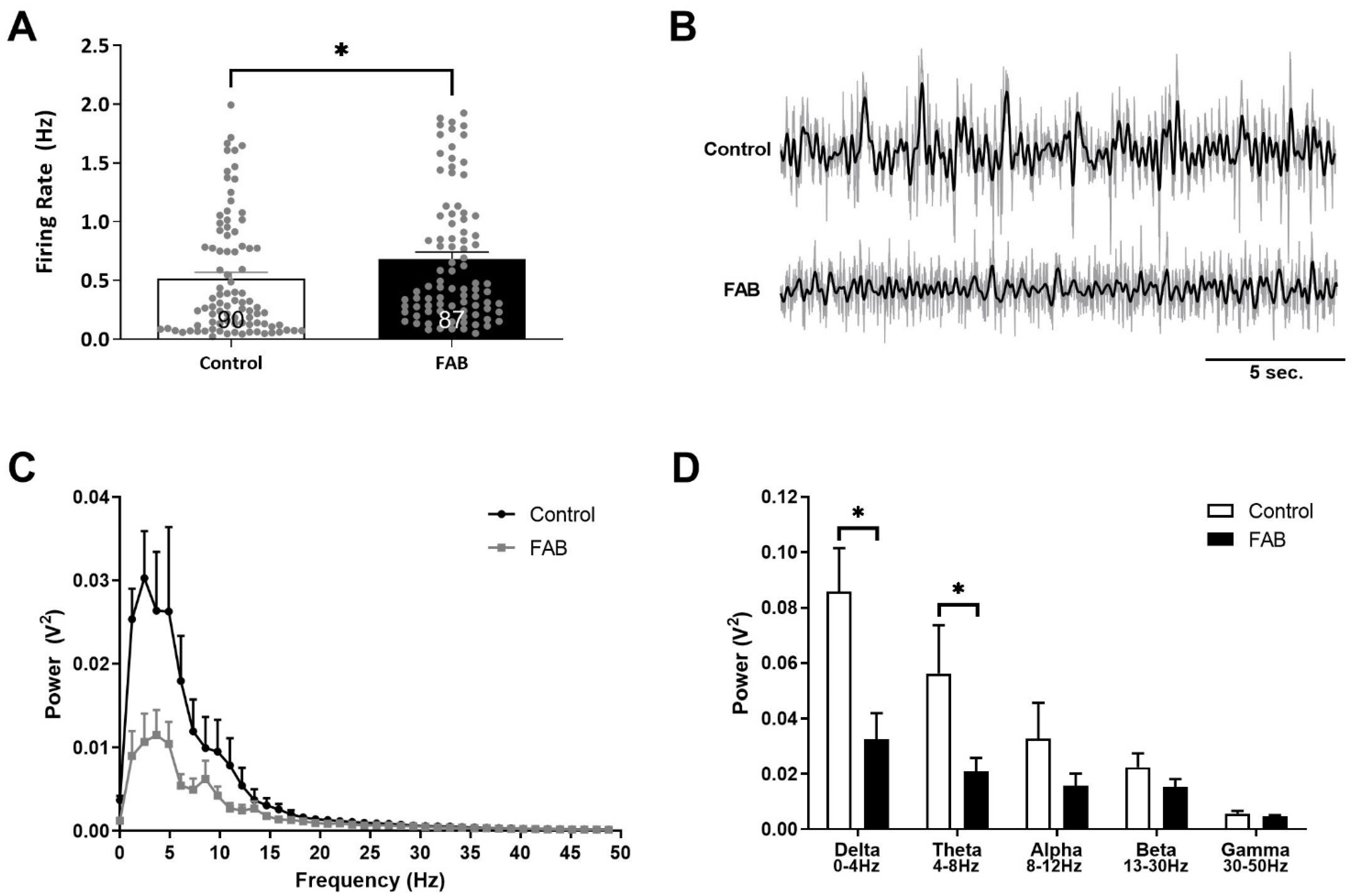
2.1. Firing Rates of Putative Pyramidal Neurons in the vHipp Were Increased, While Coordinated Neuronal Activity Was Significantly Decreased in FAB Rats

Rodent models that display psychosis-related pathologies often exhibit hippocampal hyperactivity [40,41,44,46]. Consistently with these observations, FAB rats displayed a vHipp firing frequency (n = 87 neurons; 0.68 ± 0.06 Hz) that was significantly higher than that in control rats (n = 90 neurons; 0.51 ± 0.05 Hz; Mann–Whitney rank sum test; t = 8733.00; p = 0.004; Figure 1A). Further, recordings of spontaneous oscillatory activity in the vHipp (Figure 1B,C) were significantly reduced in FAB rats (n = 6–7 per group; two-way ANOVA; FStrain(1,64) = 12.972; FFrequecy(4,64) = 8.486; Holm–Sidak; t = 3.602; p < 0.001; Figure 1D). Specifically, FAB rats displayed significant decreases in slow-wave, high-amplitude frequencies in the delta (0–4 Hz; Holm–Sidak; t = 3.773; p < 0.001) and theta (4–8 Hz; Holm–Sidak; t = 2.502; p = 0.015) ranges (Figure 1D). No significant differences were observed in any other frequency ranges.

2.2. FAB Rats Displayed a Significant Increase in Dopamine Neuron Population Activity

Consistently with previous reports, control rats displayed a population activity of 1.03 ± 0.05 cells per track (n = 12 rats) [19,47]. Interestingly, a significant increase in dopamine neuron population activity was observed in FAB rats (n = 12 rats; 1.71 ± 0.14 cells per track; Mann–Whitney rank sum test; t = 93.50; p = 0.001; Figure 2A), as is similar to observations in rodent models used to study psychosis [19,48,49,50]. No differences were observed in the average firing rate (Figure 2B) or percent bursting (Figure 2C) between groups.

2.3. FAB Rats Displayed Augmented Locomotor Activity and Deficits in Working Memory

The acute administration of psychomotor stimulants elicits an exaggerated locomotor response in rodent models used to study psychosis [19,41,46]. Here, we used the N-methyl-D-aspartate (NMDA) receptor antagonist, MK-801, a psychotomimetic compound that produces locomotor-stimulating effects at subanesthetic doses. Interestingly, we demonstrated that both baseline (two-way ANOVA; FStrain(1,101) = 30.085; Holm–Sidak; t = 5.485; p < 0.001) and MK-801-induced locomotor activity were elevated in FAB rats (n = 8 rats) compared to the controls (n = 9 rats; two-way ANOVA; FStrain(1,152) = 32.797; Holm–Sidak t = 5.727; p < 0.001; Figure 3A).
Age-related impairments in sensorimotor gating have been observed with AD progression, as well as in patients with psychosis [51,52,53,54,55]. Further, various rodent models of AD and psychosis have also exhibited deficits in pre-pulse inhibition of startle (PPI) [54,56,57]; however, we observed no PPI deficits in FAB rats when compared to their respective controls (Figure 3B).
To evaluate spatial learning and memory, we performed the Y-maze spontaneous alternation assay (Figure 3C). FAB rats (n = 10 rats; 51.19 ± 3.38%) displayed a significant decrease in percent alternations when compared to control rats (n = 9 rats; 67.02 ± 6.13%). A decrease in performance on this task is indicative of working memory deficits (t-test; t = 2.324 with 17 df; p = 0.033), which is a consistent observation in studies on FAB rats and in other rodent models used to study AD [45,58].
Lastly, while deficits in social behaviors have been reported in rodent models of AD [59], we did not observe any changes in social interaction between the control (n = 7 rats) and FAB-treated rats (n = 10 rats; Figure 3D).

2.4. FAB Rats Exhibited a Significant Decrease in Parvalbumin Protein in the Ventral Hippocampus

Decreases in parvalbumin-expressing interneurons have been reported in rodent models of psychosis [43,48,57,60], as well as in patients with psychosis [61,62] and AD [37,63]. Herein, we quantified parvalbumin protein levels in the vHipp using Western blot. Interestingly, FAB rats (n = 5 rats; 1.94 ± 0.15 uncalibrated optical density) displayed a significant decrease in parvalbumin protein, specifically in the vHipp, when compared to the controls (n = 5 rats; 4.32 ± 0.80 uncalibrated optical density; Mann–Whitney rank sum test; p = 0.008; Figure 4).

3. Discussion

The number of Americans living with AD is climbing rapidly and is projected to continue to increase, barring the discovery of a cure or breakthrough treatment to prevent or slow the progression of the disease. Currently, there is no cure for AD, but certain available FDA-approved drugs may help individuals to cope with symptoms and increase the quality of life of patients and caregivers [64,65]. Psychotic symptoms, including delusions and hallucinations, will occur in approximately 50% of patients over the course of their illness [1,2]. Furthermore, poorer disease outcomes are associated with psychotic symptoms in individuals with AD [66]. Specifically, a rapid decline in cognitive function and greater cognitive impairments have been reported in AD patients with psychosis compared to those without [3,66]. The antipsychotics commonly used to treat psychosis cause severe adverse events in AD patients, including cardiovascular and metabolic abnormalities, stroke, increased risk of parkinsonism, pneumonia, and a two-fold increased risk of death [9,10,67,68,69]. Thus, there is a dire need to develop therapies for AD patients who exhibit symptoms of psychosis.
AD is an age-related progressive disorder which can be categorized into two forms: familial and sporadic. The genetic, or familial, form of AD is a result of mutations in specific genes, such as amyloid precursor protein (APP), presenilin1 (PSEN1), and presenilin 2 (PSEN2). On the other hand, the sporadic form of AD encompasses about 95% of cases, and its etiology has yet to be completely elucidated. Evidence has led to the belief that various genetic (Apolipoprotein E (APOE) gene mutations), epigenetic, and environmental factors contribute to the development of this sporadic form of AD [70,71,72,73,74]. Although AD is an inherently human disorder, animal models that display AD-like phenotypes are essential to further our understanding of the pathologies contributing to AD. Herein, we utilized the FAB rodent model, which models the sporadic form of AD. In this model, an intracerebroventricular cannula is used to deliver a solution containing the human form of Aβ42 (aggregating properties), ferrous sulfate (pro-oxidative agent), and buthionine sulfoximine (facilitator of oxidative stress). This was used over a 3–4-week period to mimic an AD phenotype in Long–Evans rats [45]. The histopathologies present in the brains of FAB rats were consistent with observations in humans, and included hyperphosphorylated Tau protein, amyloid deposits, neuronal loss, and gliosis [45]. Further, previous studies using the FAB model have reported impairments in spatial memory in the Morris water maze task [45], and in this study, we also report deficits in cognition as measured by spontaneous alternation in the Y-maze. Given that progressive cognitive decline is a hallmark of AD and that AD patients with psychosis exhibit more severe cognitive impairments [3,66], we evaluated whether FAB rats displayed any changes in psychosis-related pathologies.
Psychotic symptoms can be observed in multiple mental disorders. They can be managed by antipsychotics, which exert effects by blocking dopamine D2 receptors, suggesting that the contribution of dopamine signaling is important [75,76,77]. Indeed, alterations in dopamine system function are often associated with symptoms of psychosis [76,77,78,79]. One region demonstrated to modulate the activity of VTA dopamine neurons is the vHipp [19]; however, it should be noted that the vHipp does not directly project to VTA dopamine neurons, but, rather, indirectly modulates the activity of these neurons via a multi-synaptic pathway involving the nucleus accumbens and the ventral pallidum (Figure 5). Furthermore, surgical [80,81], pharmacological [82,83], and cell-based therapies specifically targeting the vHipp can restore dopamine system function and reverse behavioral deficits in rodent models used to study psychosis [84]. Aberrant vHipp activity is observed in rodent models used to study schizophrenia, of which psychosis is a major symptom [19,48,49,50]. Indeed, the hippocampus is also a major site of pathology in AD, where alterations in its structure and function can be observed [15]. Specifically, progressive hippocampal atrophy is present in the anterior regions (analogous to ventral regions in the rodent) and underlies impairments in episodic memory [22]. These observations are significant because aberrant activity in the hippocampus could contribute to cognitive deficits, as well as to changes in dopamine system function, which is associated with psychosis.
The functional alterations and aberrant vHipp activity in the FAB rats observed in this study provide evidence that they may be useful for the study of comorbid psychosis-like pathology associated with AD. Herein, we measured spontaneous oscillatory activity by local field potentials (LFPs) in the vHipp of anesthetized rats and found a significant decrease in the delta (0–4 Hz) and theta (4–8 Hz) frequency ranges. Neuronal oscillations serve to coordinate activity into patterns that enhance local information processing and facilitate neurotransmission between regions [85]. It is not surprising that changes in the delta and theta rhythms generated by brain networks were observed, as they have been implicated in memory consolidation and rely heavily on hippocampal network integrity [86,87]. Further, these observations are consistent with other models of AD that display alterations in hippocampal network oscillations [87]. Additionally, FAB rats displayed increased firing rates of putative pyramidal neurons in the vHipp, which is indicative of hyperactivity within this region. Indeed, alterations in hippocampal activity, such as hyperexcitability and alterations in glutamatergic signaling, are present both in individuals with AD and in rodent models used to study AD [34,88,89,90]. The degree of hippocampal overactivation has been measured in AD patients and correlated with a decline in memory [90]. Aberrant hippocampal activity is also a common observation in rodent models that display psychosis-related symptoms [19,41,44], as well as in patients with psychosis [91,92,93].
Alterations in GABAergic markers, specifically in subpopulations of interneurons such as those containing the calcium-binding protein PV, have been observed in post mortem studies of patients who exhibited psychosis [62,94] and those diagnosed with AD [37,95]. It should also be noted that this reduction in PV expression is also a consistent observation in a wide variety of rodent models used to study psychosis [40,48,60,81,96,97,98], as well as those used in the study of AD [99,100]. Here, we demonstrate a decrease in PV expression in the vHipp of FAB-treated rats when compared to age-matched controls. We posit that that the loss of PV interneuron function in the vHipp resulted in aberrant hippocampal activity and was sufficient to produce an increase in dopamine neuron population activity.
Hippocampal hyperactivity and alterations in the hippocampal network oscillations present in the FAB model likely contributed to the dopamine dysfunction reported in this study. Specifically, FAB rats displayed significantly increased dopamine neuron population activity with no changes in the average firing rate or bursting activity. This is not surprising, because previous studies have identified distinct brain regions that can differentially modulate dopamine activity states [19,101,102]. Specifically, these measures are regulated by inputs from the ventral pallidum and tegmental nuclei (the latero-dorsal tegmentum and pedunculopontine tegmentum; for a review, see [103]). Dopamine neuron activity states, whether inactive (hyperpolarized) or active (single-spike or burst fire), can be modulated by various intrinsic and extrinsic mechanisms [103,104,105,106]. Population activity, a third activity state of dopamine neurons, is reliant on the number of spontaneously active dopamine neurons. Under normal conditions, approximately 50% of dopamine neurons are not active in vivo due to GABAergic inputs from the ventral pallidum [102,107]. These silent dopamine neurons are important as they allow for gain of function in the system [108], whereby the magnitude of the dopamine signal in response to a stimulus can be manipulated by regulating the number of spontaneously active neurons. The gain of function is particularly important because it is believed to contribute to psychosis, and several different animal models have displayed sustained increases in the number of spontaneously active dopamine neurons without any observable changes in firing rate or burst firing [19,109]. Of relevance to this study is the fact that aberrant drive from the hippocampus regulates the number of VTA dopamine neurons that fire spontaneously in a variety of rodent models of psychosis [19,40,47]. We demonstrated that FAB rats displayed alterations in dopamine neuron activity that were consistent with those observed in rodent models of psychosis.
Herein, we report various dopamine-dependent behaviors which correlate with psychosis-related pathologies that can be measured in rodent models. Human patients with psychosis exhibit increased sensitivity to psychomotor stimulants, which is suggested to reflect the aberrant mesolimbic dopamine neurotransmission thought to underlie symptoms of psychosis [77]. In rodents, this is modeled by examining the locomotor-inducing effects of the NMDA-antagonist, MK-801. Increases in locomotor activity to levels higher than those of the controls are often observed in rodent models with psychosis-like pathologies [19,110,111,112]. We observed locomotor hyperactivity in response to both the novel environment and MK-801 administration in FAB rats. We have previously demonstrated, in other models, that the stimulant-induced increase in locomotor activity can be attributed to hippocampal function, while changes in baseline locomotion are unaffected [19]. Activation of the vHipp in normal rats can increase the behavioral response to psychomotor stimulants [113], while vHipp inactivation reverses this hyperactivity [19]; therefore, the increased locomotor response to MK-801 observed in FAB rats is likely associated with vHipp-induced enhancement of dopamine neuron population activity [19]. We also evaluated PPI, which is a putative measurement of psychotomimetic effects and sensorimotor gating [57,83,114,115,116], and social interaction. As expected, PPI was dependent on the pre-pulse intensity in control rats. Reduced levels of PPI and social interaction have been observed in other rodent models with psychosis-related pathologies [57,111]; however, FAB rats displayed PPI and social interaction levels similar to the control rats. While these outcomes are, indeed, dopamine-dependent measures, they are complex behaviors modulated by multiple systems [117,118,119,120,121]. It is possible that the duration or severity of pathology induced by FAB was not sufficient to induce deficits in these behaviors.
Given the significant hippocampal pathology in AD combined with comorbid psychosis, we posited that FAB rats would display aberrant hippocampal function and activity, leading to pathological increases in VTA dopamine neuron population activity, and deficits in behaviors related to AD and psychosis. Indeed, in this study, we demonstrated that FAB rats displayed increases in vHipp and dopamine activity, thus introducing a potential novel model for comorbid psychosis in AD. In addition to cognitive deficits, FAB rats also displayed elevated baseline locomotor activity and an augmented response to the systemic administration of MK-801. These data are consistent with observations made in models of psychosis-related pathologies. The FAB model has already been commercialized and used for drug discovery and development, and our study provides evidence that this model can also serve as a useful tool to further investigate AD with comorbid psychosis, allowing for the identification of novel targets in order to develop therapeutic interventions for this devastating disorder. Indeed, drugs targeting α5 GABA-A receptors are of great interest, and are highly expressed in the hippocampus; they have been shown to have pro-cognitive and antipsychotic-like effects in vivo [82,122,123,124]. The results reported herein suggest that such compounds may have therapeutic utility for psychosis present in elderly AD patients.

4. Materials and Methods

All experiments were performed in accordance with the guidelines outlined in the USPH Guide for the Care and Use of Laboratory Animals, and were approved by the Institutional Animal Care and the Use Committee of UT Health San Antonio and the U.S. Department of Veterans Affairs.

4.1. Osmotic Minipump Survival Surgeries

Male Long–Evans rats were obtained from Envigo (Indianapolis, IN, USA). Survival surgeries were performed in a semi-sterile environment under general anesthesia. Rats (~325–350 g) were anesthetized with Fluriso (2–5% isoflurane, USP oxygen flow at 1 L/min) and placed in a stereotaxic apparatus using blunt, atraumatic ear bars. Osmotic minipumps were filled (control: Dulbecco’s phosphate-buffered saline (DPBS), no calcium, no magnesium; FAB: 1 µmol/L Aβ42, 1 mmol/L FeSO4, 12 mmol/L L-buthionine (S,R)-sulfoximine (BSO), and pH 5.1, brought to final volume with DPBS, no calcium, and no magnesium, as previously detailed) [45]. Pumps were primed via intracranial cannula directly into the lateral ventricle for a period of 24 h before delivery (A/P −0.9 mm and M/L +1.4 mm from Bregma; D/V −4.2 mm ventral of the brain surface). The cannula was fixed in place using dental acrylic and stainless-steel anchor screws, while the minipump was implanted subcutaneously into the back (slightly posterior to the scapulae). The FAB mixture was infused at a rate of 2.5 µL/hour for a period of three weeks prior to behavioral testing and electrophysiological recordings. The experimental timeline is depicted in Figure 6A.

4.2. In Vivo Electrophysiology and Local Field Potentials (LFPs)

For non-survival surgeries, rats were anesthetized with chloral hydrate (400 mg/kg; i.p.) prior to placement in a stereotaxic apparatus (Kopf; Tujunga, CA, USA). This anesthetic is required for dopamine neuron physiology as it does not significantly alter dopamine activity when compared to recordings in freely moving animals [115,125]. A core body temperature of 37 °C was maintained using a thermostatically controlled heating pad (PhysioSuite®; Kent Scientific, Torrington, CT, USA), while supplemental anesthesia was administered as required to maintain suppression of the limb withdrawal reflex. Extracellular glass microelectrodes (impedance 6–10 MΩ) were lowered into the vHipp (A/P −5.3 mm and M/L ±5.0 mm from Bregma; D/V −4.0 to −8.5 mm ventral of the brain surface) or VTA (A/P −5.3 mm and M/L ± 0.6 mm from Bregma; D/V −6.5 to −9.0 mm ventral of the brain surface). The firing frequency of spontaneously active putative pyramidal neurons in the vHipp was measured and identified as in previously published studies (neurons with firing frequencies less than 2 Hz [40,41,46]). LFP oscillatory activity was generated from the vHipp using open filter settings (low frequency cutoff: 0.3 Hz; high frequency cutoff: 300 Hz) and recorded for a minimum of ten minutes at a sampling rate of 10 kHz. Power in specific frequency bands was quantified with commercially available computer software (LabChart, Version 8; ADInstruments, Colorado Springs, CO, USA) using a standard Fourier transformation (Hamming window; 8 K). Spontaneously active dopamine neurons were identified using open filter settings (low frequency cutoff: 30 Hz; high frequency cutoff: 30 kHz) and previously established criteria [104,126]: (1) action potential duration >2 ms from the start to the end of the action potential; and (2) frequency between 0.5 and 15 Hz. Five to nine dorsal–ventral passes were made throughout the VTA, in a predetermined pattern (separated by 200 µm), to the medial and lateral regions. Dopamine cells were recorded for 2–3 min and three parameters of dopamine activity were measured: (1) population activity (the number of spontaneously active dopamine neurons encountered per track); (2) basal firing rate; and (3) the proportion of action potentials occurring in bursts (defined as the incidence of spikes with <80 ms between them; termination of a burst is defined by >160 ms between spikes). Electrophysiological recordings were analyzed by the Mann–Whitney rank sum test or two-way ANOVA followed by a Holm–Sidak post hoc test.

4.3. MK-801-Induced Locomotor Response

Rats were placed in an open-field arena (Med Associates, VT, USA), and spontaneous locomotor activity in the x-y plane was then determined by beam breaks and recorded with Open Field Activity software (Version 5, Med Associates). Following a 30-minute baseline recording, all rats were injected with MK-801 (0.75 µg/kg, i.p.) and recorded for an additional 45 min. The dose of MK-801 was specifically chosen to induce a modest locomotor effect in control rats, with the aim to observe an enhanced response in the FAB model. Locomotor data were analyzed by separate two-way ANOVAs, one for each of the relevant periods (baseline; 0.75 µg/kg MK-801), followed by a Holm–Sidak post hoc test.

4.4. Pre-Pulse Inhibition of Startle

Rats were placed in a sound-attenuated chamber (SD Instruments; San Diego, CA, USA) and allowed to acclimate to 65 dB background noise for 5 min prior to exposure to 10 startle-only trials (40 ms, 120 dB, and 15 s average inter-trial intervals (ITI)). Immediately afterward, rats were exposed to 24 trials during which a pre-pulse (20 ms at 69 dB, 73 dB, and 81 dB) was presented 100 ms before the startle pulse. Each pre-pulse + startle pulse was presented 6 times in a pseudo-random order (15 s average ITI). Startle responses were measured from 10 to 80 ms after the onset of the startle pulse and recorded using SR-Lab Analysis software (SD Instrument). PPI data were analyzed by two-way ANOVA, followed by a Holm–Sidak post hoc test.

4.5. Y-Maze Spontaneous Alternation Assay

The Y-maze consisted of three plastic arms separated by 120-degree angles. Each arm was 81 cm long and 20.32 cm wide, with walls 20 cm high. Rats were placed just inside the same arm, facing the center of the maze, and were allowed to move freely through the maze for a period of 10 min. During this time, the number and order of arm entries were recorded. Scoring consisted of recording each arm entry (defined as 80% of the rat’s body entering the arm). The percentage of alternation was then calculated as the number of alternations (triad containing entry to all three arms) divided by the total number of entries minus 2 and multiplied by 100. Y-maze data were analyzed by t-test.

4.6. Social Interaction (SI)

SI was assessed as previously described [127]. In brief, rats were acclimated to the testing arena (100 × 100 × 40 cm) individually for 10 min per day for 2 consecutive days prior to testing on day 3. On the test day, experimental rats were placed in the arena with a weight-matched “stimulus” rat. The 10 min test was scored by two blind experimenters, and the average time spent interacting (i.e., sniffing, climbing on, following, grooming, or wrestling) was reported. SI data were analyzed by t-test.

4.7. Western Blot

The protein expression of PV was measured using Western blot. The vHipp was dissected from a subset of control and FAB rats (n = 5 per group) and homogenized in ice-cold buffer (750 mL) containing a protease inhibitor. Samples were centrifuged (14,000 rpm for 2 min), and the supernatant, containing protein fractions, was collected. Protein concentrations were determined using the Bradford method prior to incubation with Laemmli sample buffer containing 5% dithiothreitol (10 min at 90 °C), then separated (20 µg/lane) at 200 mA on Any kD™ Mini-PROTEAN® TGX™ Precast Protein Gels. Proteins were transferred (100 mA for 1 h) to a nitrocellulose membrane. The membranes were then washed 3 times (10 min each) in TBST and blocked (30 min in 5% BSA or milk in TBST) before incubation in rabbit anti-PV (1:5000; Abcam, Waltham, MA, USA) or mouse anti-GAPDH (1:1000; Abcam) antibody for 1 h. Membranes were washed 3 times (10 min in TBST), followed by either HRP-anti-rabbit 1:10,000 or HRP-anti-mouse 1:5000 (1 h at room temperature). Next, membranes were treated with Pierce™ ECL Western Blotting substrate (1 min) and exposed to high-performance chemiluminescence film (Amersham Hyperfilm™ ECL). Western blot films were scanned, and optical density was measured using ImageJ and analyzed by the Mann–Whitney rank sum test.

4.8. Histology

Upon the cessation of all electrophysiological experiments, rats were transcardially perfused (150 mL saline followed by 150 mL 4% formaldehyde in phosphate buffer). Brains were then extracted, post-fixed for 24 h, and cryoprotected (30% sucrose in 0.1 M phosphate buffer) for 72 h. To verify Fe2+ + Aβ42 + BSO brain infusion, silver staining was performed on a subset of brains that were sectioned coronally (50 µm) on a cryostat and processed using the instructions provided by the FD NeuroSilver Kit II. A separate subset of brains was sectioned (25 µm coronal sections) and processed with a Nissl stain for histochemical verification of cannula tracks into the lateral ventricles (Figure 6B). All brain sections were mounted on gelatin-coated slides and cover-slipped with DPX mountant prior to viewing on a Zeiss Axio Lab.A1 microscope (White Plains, NY, USA). Silver-stained brain slices were imaged with a dark field condenser (Figure 6C). Representative images were captured with a Zeiss AxioCam ICc 1 microscope camera, and all histology was performed with reference to a stereotaxic atlas [128].

4.9. Analysis

Electrophysiological analysis of vHipp and dopamine neuron activity was performed with commercially available computer software (LabChart version 8; ADInstruments; Chalgrove, Oxfordshire, UK). Locomotor activity was collected with Activity Monitor software (MED Associates; St. Albans, VT, USA). PPI data were collected using SR-Lab™ Analysis software (SD Instruments; San Diego, CA, USA). The optical density of the Western blots was measured using ImageJ. All data were analyzed using Prism software (GraphPad Software Inc.; San Diego, CA, USA). Data are represented as mean ± SEM, with n values representing the number of animals per experimental group, unless otherwise stated. Statistics were calculated using SigmaPlot (Systat Software Inc.; Chicago, IL, USA) and plotted with Prism software (GraphPad Software Inc.; San Diego, CA, USA). Significance was determined at p < 0.05.

4.10. Materials

Isoflurane (Fluriso™) was purchased from MWI Animal Health (Boise, ID, USA). Osmotic pumps (Alzet®, Model 2ML4, 0000327), Restore™ Western blot stripper (21059), Pierce™ ECL Western Blotting substrate (32106), and brain infusion kits (Alzet®, 0008663) were purchased from Thermo Fisher Scientific (USA). Chloral hydrate (C8383); DPX mountant (06522); iron (II) sulfate heptahydrate (215422); cOmplete™, Mini, EDTA-free Protease Inhibitor Cocktail (11836170001); anti-mouse IgG (whole molecule) peroxidase antibody produced in goat (A4416); and DPBS, but no calcium and no magnesium (14190144), were sourced from Sigma-Aldrich (St. Louis, MO, USA). Amyloid-β (1-42) peptide (20574) and L-buthionine-(S,R)-sulfoximine (14484) were purchased from Cayman Chemical Company (Ann Arbor, MI, USA). kD™ Mini-PROTEAN® TGX™ Precast Protein Gels (#4569035), 2X Laemmli sample buffer (161-0737), and nitrocellulose/filter paper sandwiches (1620213) were purchased from BioRad (Hercules, CA, USA). Anti-parvalbumin antibody (ab114227), anti-GAPDH antibody (ab9484), and goat anti-rabbit IgG H&L (HRP) (ab6721) were purchased from Abcam (Waltham, MA, USA). An FD NeuroSilver Kit II was purchased from FD NeuroTechnologies, Inc. (Cat. # PK301A; Columbia, MD, USA). All other chemicals or reagents were of either analytical or laboratory grade, and were purchased from standard suppliers.

Author Contributions

Conceptualization, D.J.L. and S.M.P.; formal Analysis, S.M.P.; data curation, S.M.P., A.M.B. and A.M.M.; writing—original draft preparation, S.M.P.; writing—review and editing, D.J.L., S.M.P., A.M.B. and A.M.M.; visualization, S.M.P.; supervision, D.J.L. and S.M.P.; funding acquisition, D.J.L. All authors have read and agreed to the published version of the manuscript.

Funding

This work was supported by Merit Awards #BX004693 (D.J.L.) and #BX004646 (D.J.L.) from the United States Department of Veterans Affairs, Biomedical Laboratory Research and Development Service, and the National Institutes of Health RO1-MH090067 (D.J.L.) and the San Antonio Nathan Shock Center of Excellence Pilot Grant (S.M.P.).

Institutional Review Board Statement

All experiments were performed in accordance with the guidelines outlined in the USPH Guide for the Care and Use of Laboratory Animals, and were approached by the Institutional Animal Care and Use Committees of UT Health San Antonio and the U.S. Department of Veterans Affairs.

Informed Consent Statement

Not applicable.

Data Availability Statement

All data collected for this study are reported herein and can be found associated with this manuscript.

Conflicts of Interest

Lodge has received research funding from Heptares Therapeutics Ltd. and consulting fees from Alkermes, Inc., and is co-inventor of a patent (US 2019/0117637). All of these are unrelated to the work described in this manuscript.

References

  1. Ballard, C.; Walker, M. Neuropsychiatric aspects of Alzheimer’s disease. Curr. Psychiatry Rep. 1999, 1, 49–60. [Google Scholar] [CrossRef] [PubMed]
  2. Haupt, M.; Kurz, A.; Janner, M. A 2-year follow-up of behavioural and psychological symptoms in Alzheimer’s disease. Dement. Geriatr. Cogn. Disord. 2000, 11, 147–152. [Google Scholar] [CrossRef]
  3. Ropacki, S.A.; Jeste, D.V. Epidemiology of and risk factors for psychosis of Alzheimer’s disease: A review of 55 studies published from 1990 to 2003. Am. J. Psychiatry 2005, 162, 2022–2030. [Google Scholar] [CrossRef]
  4. Emanuel, J.E.; Lopez, O.L.; Houck, P.R.; Becker, J.T.; Weamer, E.A.; Demichele-Sweet, M.A.; Kuller, L.; Sweet, R.A. Trajectory of cognitive decline as a predictor of psychosis in early Alzheimer disease in the cardiovascular health study. Am. J. Geriatr. Psychiatry 2011, 19, 160–168. [Google Scholar] [CrossRef] [Green Version]
  5. Ballard, C.; Kales, H.C.; Lyketsos, C.; Aarsland, D.; Creese, B.; Mills, R.; Williams, H.; Sweet, R.A. Psychosis in Alzheimer’s Disease. Curr. Neurol. Neurosci. Rep. 2020, 20, 57. [Google Scholar] [CrossRef] [PubMed]
  6. Creese, B.; Ballard, C.; Jones, E. Cognitive impairment in studies of 5HTTLPR and psychosis in Alzheimer’s disease: A systematic review. Dement. Geriatr. Cogn. Disord. 2013, 35, 155–164. [Google Scholar] [CrossRef]
  7. Wilcock, G.K.; Ballard, C.G.; Cooper, J.A.; Loft, H. Memantine for agitation/aggression and psychosis in moderately severe to severe Alzheimer’s disease: A pooled analysis of 3 studies. J. Clin. Psychiatry 2008, 69, 341–348. [Google Scholar] [CrossRef] [PubMed]
  8. Maust, D.T.; Kim, H.M.; Seyfried, L.S.; Chiang, C.; Kavanagh, J.; Schneider, L.S.; Kales, H.C. Antipsychotics, other psychotropics, and the risk of death in patients with dementia: Number needed to harm. JAMA Psychiatry 2015, 72, 438–445. [Google Scholar] [CrossRef] [Green Version]
  9. De Hert, M.; Detraux, J.; van Winkel, R.; Yu, W.; Correll, C.U. Metabolic and cardiovascular adverse effects associated with antipsychotic drugs. Nat. Rev. Endocrinol. 2011, 8, 114–126. [Google Scholar] [CrossRef]
  10. Coupland, C.A.C.; Hill, T.; Dening, T.; Morriss, R.; Moore, M.; Hippisley-Cox, J. Anticholinergic Drug Exposure and the Risk of Dementia: A Nested Case-Control Study. JAMA Intern. Med. 2019, 179, 1084–1093. [Google Scholar] [CrossRef] [Green Version]
  11. Creese, B.; Da Silva, M.V.; Johar, I.; Ballard, C. The modern role of antipsychotics for the treatment of agitation and psychosis in Alzheimer’s disease. Expert Rev. Neurother. 2018, 18, 461–467. [Google Scholar] [CrossRef]
  12. Vik-Mo, A.O.; Bencze, J.; Ballard, C.; Hortobagyi, T.; Aarsland, D. Advanced cerebral amyloid angiopathy and small vessel disease are associated with psychosis in Alzheimer’s disease. J. Neurol. Neurosurg. Psychiatry 2019, 90, 728–730. [Google Scholar] [CrossRef]
  13. Muhlbauer, V.; Mohler, R.; Dichter, M.N.; Zuidema, S.U.; Kopke, S.; Luijendijk, H.J. Antipsychotics for agitation and psychosis in people with Alzheimer’s disease and vascular dementia. Cochrane Database Syst. Rev. 2021, 12, CD013304. [Google Scholar] [CrossRef]
  14. Blennow, K.; de Leon, M.J.; Zetterberg, H. Alzheimer’s disease. Lancet 2006, 368, 387–403. [Google Scholar] [CrossRef] [PubMed]
  15. Hyman, B.T.; Van Hoesen, G.W.; Damasio, A.R.; Barnes, C.L. Alzheimer’s disease: Cell-specific pathology isolates the hippocampal formation. Science 1984, 225, 1168–1170. [Google Scholar] [CrossRef]
  16. Apostolova, L.G.; Dutton, R.A.; Dinov, I.D.; Hayashi, K.M.; Toga, A.W.; Cummings, J.L.; Thompson, P.M. Conversion of mild cognitive impairment to Alzheimer disease predicted by hippocampal atrophy maps. Arch. Neurol. 2006, 63, 693–699. [Google Scholar] [CrossRef] [PubMed] [Green Version]
  17. Petrella, J.R.; Krishnan, S.; Slavin, M.J.; Tran, T.T.; Murty, L.; Doraiswamy, P.M. Mild cognitive impairment: Evaluation with 4-T functional MR imaging. Radiology 2006, 240, 177–186. [Google Scholar] [CrossRef]
  18. Miller, S.L.; Fenstermacher, E.; Bates, J.; Blacker, D.; Sperling, R.A.; Dickerson, B.C. Hippocampal activation in adults with mild cognitive impairment predicts subsequent cognitive decline. J. Neurol. Neurosurg Psychiatry 2008, 79, 630–635. [Google Scholar] [CrossRef] [PubMed] [Green Version]
  19. Lodge, D.J.; Grace, A.A. Aberrant hippocampal activity underlies the dopamine dysregulation in an animal model of schizophrenia. J. Neurosci. 2007, 27, 11424–11430. [Google Scholar] [CrossRef] [Green Version]
  20. Grace, A.A. Phasic versus tonic dopamine release and the modulation of dopamine system responsivity: A hypothesis for the etiology of schizophrenia. Neuroscience 1991, 41, 1–24. [Google Scholar] [CrossRef]
  21. Grace, A.A. Gating of information flow within the limbic system and the pathophysiology of schizophrenia. Brain Res. Brain Res. Rev. 2000, 31, 330–341. [Google Scholar] [CrossRef] [PubMed]
  22. Maruszak, A.; Thuret, S. Why looking at the whole hippocampus is not enough-a critical role for anteroposterior axis, subfield and activation analyses to enhance predictive value of hippocampal changes for Alzheimer’s disease diagnosis. Front Cell Neurosci. 2014, 8, 95. [Google Scholar] [CrossRef] [Green Version]
  23. Fanselow, M.S.; Dong, H.W. Are the dorsal and ventral hippocampus functionally distinct structures? Neuron 2010, 65, 7–19. [Google Scholar] [CrossRef] [PubMed] [Green Version]
  24. Yasuno, F.; Minami, H.; Hattori, H.; Alzheimer’s Disease Neuroimaging, I. Relationship between neuropsychiatric symptoms and Alzheimer’s disease pathology: An in vivo positron emission tomography study. Int. J. Geriatr. Psychiatry 2021, 36, 598–605. [Google Scholar] [CrossRef] [PubMed]
  25. Geda, Y.E.; Schneider, L.S.; Gitlin, L.N.; Miller, D.S.; Smith, G.S.; Bell, J.; Evans, J.; Lee, M.; Porsteinsson, A.; Lanctot, K.L.; et al. Neuropsychiatric symptoms in Alzheimer’s disease: Past progress and anticipation of the future. Alzheimers Dement 2013, 9, 602–608. [Google Scholar] [CrossRef] [Green Version]
  26. Krell-Roesch, J.; Vassilaki, M.; Mielke, M.M.; Kremers, W.K.; Lowe, V.J.; Vemuri, P.; Machulda, M.M.; Christianson, T.J.; Syrjanen, J.A.; Stokin, G.B.; et al. Cortical beta-amyloid burden, neuropsychiatric symptoms, and cognitive status: The Mayo Clinic Study of Aging. Transl. Psychiatry 2019, 9, 123. [Google Scholar] [CrossRef] [Green Version]
  27. Scaricamazza, E.; Colonna, I.; Sancesario, G.M.; Assogna, F.; Orfei, M.D.; Franchini, F.; Sancesario, G.; Mercuri, N.B.; Liguori, C. Neuropsychiatric symptoms differently affect mild cognitive impairment and Alzheimer’s disease patients: A retrospective observational study. Neurol. Sci. 2019, 40, 1377–1382. [Google Scholar] [CrossRef]
  28. Babulal, G.M.; Ghoshal, N.; Head, D.; Vernon, E.K.; Holtzman, D.M.; Benzinger, T.L.S.; Fagan, A.M.; Morris, J.C.; Roe, C.M. Mood Changes in Cognitively Normal Older Adults are Linked to Alzheimer Disease Biomarker Levels. Am. J. Geriatr. Psychiatry 2016, 24, 1095–1104. [Google Scholar] [CrossRef] [Green Version]
  29. Puzzo, D.; Privitera, L.; Fa, M.; Staniszewski, A.; Hashimoto, G.; Aziz, F.; Sakurai, M.; Ribe, E.M.; Troy, C.M.; Mercken, M.; et al. Endogenous amyloid-beta is necessary for hippocampal synaptic plasticity and memory. Ann. Neurol. 2011, 69, 819–830. [Google Scholar] [CrossRef] [Green Version]
  30. Puzzo, D.; Arancio, O. Amyloid-beta peptide: Dr. Jekyll or Mr. Hyde? J. Alzheimers Dis. 2013, 33 (Suppl. S1), S111-20. [Google Scholar]
  31. Palop, J.J.; Chin, J.; Roberson, E.D.; Wang, J.; Thwin, M.T.; Bien-Ly, N.; Yoo, J.; Ho, K.O.; Yu, G.Q.; Kreitzer, A.; et al. Aberrant excitatory neuronal activity and compensatory remodeling of inhibitory hippocampal circuits in mouse models of Alzheimer’s disease. Neuron 2007, 55, 697–711. [Google Scholar] [CrossRef] [Green Version]
  32. Andrews-Zwilling, Y.; Bien-Ly, N.; Xu, Q.; Li, G.; Bernardo, A.; Yoon, S.Y.; Zwilling, D.; Yan, T.X.; Chen, L.; Huang, Y. Apolipoprotein E4 causes age- and Tau-dependent impairment of GABAergic interneurons, leading to learning and memory deficits in mice. J. Neurosci. 2010, 30, 13707–13717. [Google Scholar] [CrossRef] [Green Version]
  33. Verret, L.; Mann, E.O.; Hang, G.B.; Barth, A.M.; Cobos, I.; Ho, K.; Devidze, N.; Masliah, E.; Kreitzer, A.C.; Mody, I.; et al. Inhibitory interneuron deficit links altered network activity and cognitive dysfunction in Alzheimer model. Cell 2012, 149, 708–721. [Google Scholar] [CrossRef] [Green Version]
  34. Sosulina, L.; Mittag, M.; Geis, H.R.; Hoffmann, K.; Klyubin, I.; Qi, Y.; Steffen, J.; Friedrichs, D.; Henneberg, N.; Fuhrmann, F.; et al. Hippocampal hyperactivity in a rat model of Alzheimer’s disease. J. Neurochem. 2021, 157, 2128–2144. [Google Scholar] [CrossRef]
  35. Marin, O. Interneuron dysfunction in psychiatric disorders. Nat. Rev. Neurosci. 2012, 13, 107–120. [Google Scholar] [CrossRef]
  36. Arai, H.; Emson, P.C.; Mountjoy, C.Q.; Carassco, L.H.; Heizmann, C.W. Loss of parvalbumin-immunoreactive neurones from cortex in Alzheimer-type dementia. Brain Res. 1987, 418, 164–169. [Google Scholar] [CrossRef]
  37. Brady, D.R.; Mufson, E.J. Parvalbumin-immunoreactive neurons in the hippocampal formation of Alzheimer’s diseased brain. Neuroscience 1997, 80, 1113–1125. [Google Scholar] [CrossRef]
  38. Mikkonen, M.; Alafuzoff, I.; Tapiola, T.; Soininen, H.; Miettinen, R. Subfield- and layer-specific changes in parvalbumin, calretinin and calbindin-D28K immunoreactivity in the entorhinal cortex in Alzheimer’s disease. Neuroscience 1999, 92, 515–532. [Google Scholar] [CrossRef]
  39. Kawaguchi, Y. Physiological subgroups of nonpyramidal cells with specific morphological characteristics in layer II/III of rat frontal cortex. J. Neurosci. 1995, 15, 2638–2655. [Google Scholar] [CrossRef] [Green Version]
  40. Perez, S.M.; Lodge, D.J. Hippocampal interneuron transplants reverse aberrant dopamine system function and behavior in a rodent model of schizophrenia. Mol. Psychiatry 2013, 18, 1193–1198. [Google Scholar] [CrossRef] [Green Version]
  41. Boley, A.M.; Perez, S.M.; Lodge, D.J. A fundamental role for hippocampal parvalbumin in the dopamine hyperfunction associated with schizophrenia. Schizophr. Res. 2014, 157, 238–243. [Google Scholar] [CrossRef] [PubMed] [Green Version]
  42. Donegan, J.J.; Tyson, J.A.; Branch, S.Y.; Beckstead, M.J.; Anderson, S.A.; Lodge, D.J. Stem cell-derived interneuron transplants as a treatment for schizophrenia: Preclinical validation in a rodent model. Mol. Psychiatry 2017, 22, 1492–1501. [Google Scholar] [CrossRef] [PubMed] [Green Version]
  43. Lodge, D.J.; Behrens, M.M.; Grace, A.A. A loss of parvalbumin-containing interneurons is associated with diminished oscillatory activity in an animal model of schizophrenia. J. Neurosci. Off. J. Soc. Neurosci. 2009, 29, 2344–2354. [Google Scholar] [CrossRef] [PubMed] [Green Version]
  44. Perez, S.M.; Boley, A.; Lodge, D.J. Region specific knockdown of Parvalbumin or Somatostatin produces neuronal and behavioral deficits consistent with those observed in schizophrenia. Transl. Psychiatry 2019, 9, 264. [Google Scholar] [CrossRef] [Green Version]
  45. Lecanu, L.; Greeson, J.; Papadopoulos, V. Beta-amyloid and oxidative stress jointly induce neuronal death, amyloid deposits, gliosis, and memory impairment in the rat brain. Pharmacology 2006, 76, 19–33. [Google Scholar] [CrossRef]
  46. Shah, A.; Lodge, D.J. A loss of hippocampal perineuronal nets produces deficits in dopamine system function: Relevance to the positive symptoms of schizophrenia. Transl. Psychiatry 2013, 3, e215. [Google Scholar] [CrossRef] [Green Version]
  47. Perez, S.M.; Lodge, D.J. Convergent Inputs from the Hippocampus and Thalamus to the Nucleus Accumbens Regulate Dopamine Neuron Activity. J. NeuroSci. 2018, 38, 10607–10618. [Google Scholar] [CrossRef] [Green Version]
  48. Perez, S.M.; Aguilar, D.D.; Neary, J.L.; Carless, M.A.; Giuffrida, A.; Lodge, D.J. Schizophrenia-Like Phenotype Inherited by the F2 Generation of a Gestational Disruption Model of Schizophrenia. Neuropsychopharmacology 2016, 41, 477–486. [Google Scholar] [CrossRef] [Green Version]
  49. Perez, S.M.; Chen, L.; Lodge, D.J. Alterations in dopamine system function across the estrous cycle of the MAM rodent model of schizophrenia. Psychoneuroendocrinology 2014, 47, 88–97. [Google Scholar] [CrossRef] [Green Version]
  50. Aguilar, D.D.; Chen, L.; Lodge, D.J. Increasing Endocannabinoid Levels in the Ventral Pallidum Restore Aberrant Dopamine Neuron Activity in the Subchronic PCP Rodent Model of Schizophrenia. Int. J. Neuropsychopharmacol. 2014. [Google Scholar] [CrossRef] [Green Version]
  51. Wang, H.; He, J.; Zhang, R.; Zhu, S.; Wang, J.; Kong, L.; Tan, Q.; Li, X.M. Sensorimotor gating and memory deficits in an APP/PS1 double transgenic mouse model of Alzheimer’s disease. Behav. Brain Res. 2012, 233, 237–243. [Google Scholar] [CrossRef]
  52. Jafari, Z.; Kolb, B.E.; Mohajerani, M.H. Prepulse inhibition of the acoustic startle reflex and P50 gating in aging and alzheimer’s disease. Ageing Res. Rev. 2020, 59, 101028. [Google Scholar] [CrossRef]
  53. Braff, D.L. Information processing and attention dysfunctions in schizophrenia. Schizophrenia Bulletin 1993, 19, 233–259. [Google Scholar] [CrossRef] [Green Version]
  54. Braff, D.L.; Geyer, M.A. Sensorimotor gating and schizophrenia. Human and animal model studies. Arch. Gen. Psychiatry 1990, 47, 181–188. [Google Scholar] [CrossRef]
  55. Braff, D.L.; Grillon, C.; Geyer, M.A. Gating and habituation of the startle reflex in schizophrenic patients. Arch. Gen. Psychiatry 1992, 49, 206–215. [Google Scholar] [CrossRef]
  56. Koppel, J.; Jimenez, H.; Azose, M.; D’Abramo, C.; Acker, C.; Buthorn, J.; Greenwald, B.S.; Lewis, J.; Lesser, M.; Liu, Z.; et al. Pathogenic tau species drive a psychosis-like phenotype in a mouse model of Alzheimer’s disease. Behav. Brain Res. 2014, 275, 27–33. [Google Scholar] [CrossRef]
  57. Perez, S.M.; Donegan, J.J.; Lodge, D.J. Effect of estrous cycle on schizophrenia-like behaviors in MAM exposed rats. Behav. Brain Res. 2019, 362, 258–265. [Google Scholar] [CrossRef]
  58. Ohno, M.; Sametsky, E.A.; Younkin, L.H.; Oakley, H.; Younkin, S.G.; Citron, M.; Vassar, R.; Disterhoft, J.F. BACE1 deficiency rescues memory deficits and cholinergic dysfunction in a mouse model of Alzheimer’s disease. Neuron 2004, 41, 27–33. [Google Scholar] [CrossRef] [Green Version]
  59. Kosel, F.; Munoz, P.T.; Yang, J.R.; Wong, A.A.; Franklin, T.B. Age-related changes in social behaviours in the 5xFAD mouse model of Alzheimer’s disease. Behav. Brain Res. 2019, 362, 160–172. [Google Scholar] [CrossRef]
  60. Chen, L.; Perez, S.M.; Lodge, D.J. An augmented dopamine system function is present prior to puberty in the methylazoxymethanol acetate rodent model of schizophrenia. Dev. Neurobiol. 2014, 74, 907–917. [Google Scholar] [CrossRef] [Green Version]
  61. Zhang, Z.J.; Reynolds, G.P. A selective decrease in the relative density of parvalbumin-immunoreactive neurons in the hippocampus in schizophrenia. Schizophr. Res. 2002, 55, 1–10. [Google Scholar] [CrossRef] [PubMed]
  62. Konradi, C.; Yang, C.K.; Zimmerman, E.I.; Lohmann, K.M.; Gresch, P.; Pantazopoulos, H.; Berretta, S.; Heckers, S. Hippocampal interneurons are abnormal in schizophrenia. Schizophr. Res. 2011, 131, 165–173. [Google Scholar] [CrossRef] [PubMed] [Green Version]
  63. Satoh, J.; Tabira, T.; Sano, M.; Nakayama, H.; Tateishi, J. Parvalbumin-immunoreactive neurons in the human central nervous system are decreased in Alzheimer’s disease. Acta Neuropathol. 1991, 81, 388–395. [Google Scholar] [CrossRef] [PubMed]
  64. Cummings, J.L.; Tong, G.; Ballard, C. Treatment Combinations for Alzheimer’s Disease: Current and Future Pharmacotherapy Options. J. Alzheimers Dis. 2019, 67, 779–794. [Google Scholar] [CrossRef] [Green Version]
  65. Breijyeh, Z.; Karaman, R. Comprehensive Review on Alzheimer’s Disease: Causes and Treatment. Molecules 2020, 25, 5789. [Google Scholar] [CrossRef]
  66. Gilley, D.W.; Whalen, M.E.; Wilson, R.S.; Bennett, D.A. Hallucinations and associated factors in Alzheimer’s disease. J. Neuropsychiatry Clin. Neurosci. 1991, 3, 371–376. [Google Scholar]
  67. Ballard, C.; Howard, R. Neuroleptic drugs in dementia: Benefits and harm. Nat. Rev. Neurosci. 2006, 7, 492–500. [Google Scholar] [CrossRef]
  68. Kales, H.C.; Valenstein, M.; Kim, H.M.; McCarthy, J.F.; Ganoczy, D.; Cunningham, F.; Blow, F.C. Mortality risk in patients with dementia treated with antipsychotics versus other psychiatric medications. Am. J. Psychiatry 2007, 164, 1568–1576; quiz 1623. [Google Scholar] [CrossRef]
  69. Kales, H.C.; Kim, H.M.; Zivin, K.; Valenstein, M.; Seyfried, L.S.; Chiang, C.; Cunningham, F.; Schneider, L.S.; Blow, F.C. Risk of mortality among individual antipsychotics in patients with dementia. Am. J. Psychiatry 2012, 169, 71–79. [Google Scholar] [CrossRef] [Green Version]
  70. Abraham, R.; Moskvina, V.; Sims, R.; Hollingworth, P.; Morgan, A.; Georgieva, L.; Dowzell, K.; Cichon, S.; Hillmer, A.M.; O’Donovan, M.C.; et al. A genome-wide association study for late-onset Alzheimer’s disease using DNA pooling. BMC Med. Genom. 2008, 1, 44. [Google Scholar] [CrossRef]
  71. Piaceri, I.; Nacmias, B.; Sorbi, S. Genetics of familial and sporadic Alzheimer’s disease. Front. Biosci. 2013, 5, 167–177. [Google Scholar] [CrossRef] [Green Version]
  72. Grupe, A.; Abraham, R.; Li, Y.; Rowland, C.; Hollingworth, P.; Morgan, A.; Jehu, L.; Segurado, R.; Stone, D.; Schadt, E.; et al. Evidence for novel susceptibility genes for late-onset Alzheimer’s disease from a genome-wide association study of putative functional variants. Hum. Mol. Genet. 2007, 16, 865–873. [Google Scholar] [CrossRef]
  73. Grant, W.B.; Campbell, A.; Itzhaki, R.F.; Savory, J. The significance of environmental factors in the etiology of Alzheimer’s disease. J. Alzheimers Dis. 2002, 4, 179–189. [Google Scholar] [CrossRef]
  74. Li, P.; Marshall, L.; Oh, G.; Jakubowski, J.L.; Groot, D.; He, Y.; Wang, T.; Petronis, A.; Labrie, V. Epigenetic dysregulation of enhancers in neurons is associated with Alzheimer’s disease pathology and cognitive symptoms. Nat. Commun. 2019, 10, 2246. [Google Scholar] [CrossRef] [Green Version]
  75. Seeman, P.; Lee, T. Antipsychotic drugs: Direct correlation between clinical potency and presynaptic action on dopamine neurons. Science 1975, 188, 1217–1219. [Google Scholar] [CrossRef]
  76. Howes, O.D.; Kambeitz, J.; Kim, E.; Stahl, D.; Slifstein, M.; Abi-Dargham, A.; Kapur, S. The nature of dopamine dysfunction in schizophrenia and what this means for treatment. Arch. Gen. Psychiatry 2012, 69, 776–786. [Google Scholar] [CrossRef] [Green Version]
  77. Abi-Dargham, A. Do we still believe in the dopamine hypothesis? New data bring new evidence. Int. J. Neuropsychopharmacol. 2004, 7 (Suppl. S1), S1–S5. [Google Scholar] [CrossRef]
  78. Laruelle, M.; Abi-Dargham, A. Dopamine as the wind of the psychotic fire: New evidence from brain imaging studies. J. Psychopharmacol. 1999, 13, 358–371. [Google Scholar] [CrossRef]
  79. Laruelle, M.; Abi-Dargham, A.; Gil, R.; Kegeles, L.; Innis, R. Increased dopamine transmission in schizophrenia: Relationship to illness phases. Biol. Psychiatry 1999, 46, 56–72. [Google Scholar] [CrossRef]
  80. Perez, S.M.; Carreno, F.R.; Frazer, A.; Lodge, D.J. Vagal nerve stimulation reverses aberrant dopamine system function in the methylazoxymethanol acetate rodent model of schizophrenia. J. Neurosci. 2014, 34, 9261–9267. [Google Scholar] [CrossRef] [Green Version]
  81. Perez, S.M.; Shah, A.; Asher, A.; Lodge, D.J. Hippocampal deep brain stimulation reverses physiological and behavioural deficits in a rodent model of schizophrenia. Int. J. Neuropsychopharmacol. 2013, 16, 1331–1339. [Google Scholar] [CrossRef] [PubMed] [Green Version]
  82. Perez, S.M.; McCoy, A.M.; Prevot, T.D.; Mian, M.Y.; Carreno, F.R.; Frazer, A.; Cook, J.M.; Sibille, E.; Lodge, D.J. Hippocampal α5-GABAA receptors modulate dopamine neuron activity in the rat ventral tegmental area. Biol. Psychiatry Glob. Open Sci. 2022, 3, 78–86. [Google Scholar] [CrossRef] [PubMed]
  83. Gill, K.M.; Lodge, D.J.; Cook, J.M.; Aras, S.; Grace, A.A. A novel alpha5GABA(A)R-positive allosteric modulator reverses hyperactivation of the dopamine system in the MAM model of schizophrenia. Neuropsychopharmacology 2011, 36, 1903–1911. [Google Scholar] [CrossRef] [PubMed]
  84. Perez, S.M.; Lodge, D.J. New approaches to the management of schizophrenia: Focus on aberrant hippocampal drive of dopamine pathways. Drug Des. Dev. Ther. 2014, 8, 887–896. [Google Scholar] [PubMed] [Green Version]
  85. Fries, P. Rhythms for Cognition: Communication through Coherence. Neuron 2015, 88, 220–235. [Google Scholar] [CrossRef] [Green Version]
  86. Buzsaki, G. Theta oscillations in the hippocampus. Neuron 2002, 33, 325–340. [Google Scholar] [CrossRef] [Green Version]
  87. Goutagny, R.; Krantic, S. Hippocampal oscillatory activity in Alzheimer’s disease: Toward the identification of early biomarkers? Aging Dis. 2013, 4, 134–140. [Google Scholar]
  88. Bondi, M.W.; Houston, W.S.; Eyler, L.T.; Brown, G.G. fMRI evidence of compensatory mechanisms in older adults at genetic risk for Alzheimer disease. Neurology 2005, 64, 501–508. [Google Scholar] [CrossRef] [Green Version]
  89. Quiroz, Y.T.; Budson, A.E.; Celone, K.; Ruiz, A.; Newmark, R.; Castrillon, G.; Lopera, F.; Stern, C.E. Hippocampal hyperactivation in presymptomatic familial Alzheimer’s disease. Ann. Neurol. 2010, 68, 865–875. [Google Scholar] [CrossRef] [Green Version]
  90. Bookheimer, S.Y.; Strojwas, M.H.; Cohen, M.S.; Saunders, A.M.; Pericak-Vance, M.A.; Mazziotta, J.C.; Small, G.W. Patterns of brain activation in people at risk for Alzheimer’s disease. N. Engl. J. Med. 2000, 343, 450–456. [Google Scholar] [CrossRef] [Green Version]
  91. Schobel, S.A.; Lewandowski, N.M.; Corcoran, C.M.; Moore, H.; Brown, T.; Malaspina, D.; Small, S.A. Differential targeting of the CA1 subfield of the hippocampal formation by schizophrenia and related psychotic disorders. Arch. Gen. Psychiatry 2009, 66, 938–946. [Google Scholar] [CrossRef] [Green Version]
  92. Heckers, S.; Rauch, S.L.; Goff, D.; Savage, C.R.; Schacter, D.L.; Fischman, A.J.; Alpert, N.M. Impaired recruitment of the hippocampus during conscious recollection in schizophrenia. Nat. Neurosci. 1998, 1, 318–323. [Google Scholar] [CrossRef]
  93. Malaspina, D.; Harkavy-Friedman, J.; Corcoran, C.; Mujica-Parodi, L.; Printz, D.; Gorman, J.M.; Van Heertum, R. Resting neural activity distinguishes subgroups of schizophrenia patients. Biol. Psychiatry 2004, 56, 931–937. [Google Scholar] [CrossRef] [Green Version]
  94. Lewis, D.A.; Hashimoto, T.; Volk, D.W. Cortical inhibitory neurons and schizophrenia. Nat. Rev. Neurosci. 2005, 6, 312–324. [Google Scholar] [CrossRef]
  95. Inaguma, Y.; Shinohara, H.; Inagaki, T.; Kato, K. Immunoreactive parvalbumin concentrations in parahippocampal gyrus decrease in patients with Alzheimer’s disease. J. Neurol. Sci. 1992, 110, 57–61. [Google Scholar] [CrossRef]
  96. Behrens, M.M.; Ali, S.S.; Dao, D.N.; Lucero, J.; Shekhtman, G.; Quick, K.L.; Dugan, L.L. Ketamine-induced loss of phenotype of fast-spiking interneurons is mediated by NADPH-oxidase. Science 2007, 318, 1645–1647. [Google Scholar] [CrossRef] [Green Version]
  97. Francois, J.; Ferrandon, A.; Koning, E.; Angst, M.J.; Sandner, G.; Nehlig, A. Selective reorganization of GABAergic transmission in neonatal ventral hippocampal-lesioned rats. Int. J. Neuropsychopharmacol. Off. Sci. J. Coll. Int. Neuropsychopharmacol. 2009, 12, 1097–1110. [Google Scholar] [CrossRef] [Green Version]
  98. Harte, M.K.; Powell, S.B.; Swerdlow, N.R.; Geyer, M.A.; Reynolds, G.P. Deficits in parvalbumin and calbindin immunoreactive cells in the hippocampus of isolation reared rats. J. Neural Transm. 2007, 114, 893–898. [Google Scholar] [CrossRef]
  99. Ali, F.; Baringer, S.L.; Neal, A.; Choi, E.Y.; Kwan, A.C. Parvalbumin-Positive Neuron Loss and Amyloid-beta Deposits in the Frontal Cortex of Alzheimer’s Disease-Related Mice. J. Alzheimers Dis. 2019, 72, 1323–1339. [Google Scholar] [CrossRef]
  100. Zallo, F.; Gardenal, E.; Verkhratsky, A.; Rodriguez, J.J. Loss of calretinin and parvalbumin positive interneurones in the hippocampal CA1 of aged Alzheimer’s disease mice. Neurosci. Lett. 2018, 681, 19–25. [Google Scholar] [CrossRef]
  101. Floresco, S.B.; Todd, C.L.; Grace, A.A. Glutamatergic afferents from the hippocampus to the nucleus accumbens regulate activity of ventral tegmental area dopamine neurons. J. Neurosci. 2001, 21, 4915–4922. [Google Scholar] [CrossRef] [PubMed] [Green Version]
  102. Floresco, S.B.; West, A.R.; Ash, B.; Moore, H.; Grace, A.A. Afferent modulation of dopamine neuron firing differentially regulates tonic and phasic dopamine transmission. Nat. Neurosci. 2003, 6, 968–973. [Google Scholar] [CrossRef] [PubMed]
  103. Grace, A.A.; Floresco, S.B.; Goto, Y.; Lodge, D.J. Regulation of firing of dopaminergic neurons and control of goal-directed behaviors. Trends Neurosci. 2007, 30, 220–227. [Google Scholar] [CrossRef] [PubMed]
  104. Grace, A.A.; Bunney, B.S. Intracellular and extracellular electrophysiology of nigral dopaminergic neurons--1. Identification and characterization. Neuroscience 1983, 10, 301–315. [Google Scholar] [CrossRef]
  105. Grace, A.A.; Bunney, B.S. The control of firing pattern in nigral dopamine neurons: Burst firing. J. Neurosci. 1984, 4, 2877–2890. [Google Scholar] [CrossRef]
  106. Grace, A.A.; Bunney, B.S. The control of firing pattern in nigral dopamine neurons: Single spike firing. J. Neurosci. 1984, 4, 2866–2876. [Google Scholar] [CrossRef] [Green Version]
  107. Grace, A.A.; Bunney, B.S. Opposing effects of striatonigral feedback pathways on midbrain dopamine cell activity. Brain Res. 1985, 333, 271–284. [Google Scholar] [CrossRef]
  108. Lodge, D.J.; Grace, A.A. The hippocampus modulates dopamine neuron responsivity by regulating the intensity of phasic neuron activation. Neuropsychopharmacology 2006, 31, 1356–1361. [Google Scholar] [CrossRef] [Green Version]
  109. Lodge, D.J.; Grace, A.A. Hippocampal dysfunction and disruption of dopamine system regulation in an animal model of schizophrenia. Neurotox Res. 2008, 14, 97–104. [Google Scholar] [CrossRef] [Green Version]
  110. Le Pen, G.; Gourevitch, R.; Hazane, F.; Hoareau, C.; Jay, T.M.; Krebs, M.O. Peri-pubertal maturation after developmental disturbance: A model for psychosis onset in the rat. Neuroscience 2006, 143, 395–405. [Google Scholar] [CrossRef]
  111. Moore, H.; Jentsch, J.D.; Ghajarnia, M.; Geyer, M.A.; Grace, A.A. A neurobehavioral systems analysis of adult rats exposed to methylazoxymethanol acetate on E17: Implications for the neuropathology of schizophrenia. Biol. Psychiatry 2006, 60, 253–264. [Google Scholar] [CrossRef] [Green Version]
  112. Flagstad, P.; Mork, A.; Glenthoj, B.Y.; van Beek, J.; Michael-Titus, A.T.; Didriksen, M. Disruption of neurogenesis on gestational day 17 in the rat causes behavioral changes relevant to positive and negative schizophrenia symptoms and alters amphetamine-induced dopamine release in nucleus accumbens. Neuropsychopharmacology 2004, 29, 2052–2064. [Google Scholar] [CrossRef]
  113. White, I.M.; Whitaker, C.; White, W. Amphetamine-induced hyperlocomotion in rats: Hippocampal modulation of the nucleus accumbens. Hippocampus 2006, 16, 596–603. [Google Scholar] [CrossRef]
  114. Bjorklund, L.M.; Sanchez-Pernaute, R.; Chung, S.; Andersson, T.; Chen, I.Y.; McNaught, K.S.; Brownell, A.L.; Jenkins, B.G.; Wahlestedt, C.; Kim, K.S.; et al. Embryonic stem cells develop into functional dopaminergic neurons after transplantation in a Parkinson rat model. Proc. Natl. Acad. Sci. USA 2002, 99, 2344–2349. [Google Scholar] [CrossRef] [Green Version]
  115. Hyland, B.I.; Reynolds, J.N.; Hay, J.; Perk, C.G.; Miller, R. Firing modes of midbrain dopamine cells in the freely moving rat. Neuroscience 2002, 114, 475–492. [Google Scholar] [CrossRef]
  116. Carreno, F.R.; Collins, G.T.; Frazer, A.; Lodge, D.J. Selective Pharmacological Augmentation of Hippocampal Activity Produces a Sustained Antidepressant-Like Response without Abuse-Related or Psychotomimetic Effects. Int. J. Neuropsychopharmacol. 2017, 20, 504–509. [Google Scholar] [CrossRef] [Green Version]
  117. Rohleder, C.; Wiedermann, D.; Neumaier, B.; Drzezga, A.; Timmermann, L.; Graf, R.; Leweke, F.M.; Endepols, H. The Functional Networks of Prepulse Inhibition: Neuronal Connectivity Analysis Based on FDG-PET in Awake and Unrestrained Rats. Front. Behav. Neurosci. 2016, 10, 148. [Google Scholar] [CrossRef] [Green Version]
  118. Cano, J.C.; Huang, W.; Fenelon, K. The amygdala modulates prepulse inhibition of the auditory startle reflex through excitatory inputs to the caudal pontine reticular nucleus. BMC Biol. 2021, 19, 116. [Google Scholar] [CrossRef]
  119. Hammer, T.B.; Oranje, B.; Skimminge, A.; Aggernaes, B.; Ebdrup, B.H.; Glenthoj, B.; Baare, W. Structural brain correlates of sensorimotor gating in antipsychotic-naive men with first-episode schizophrenia. J. Psychiatry Neurosci. 2013, 38, 34–42. [Google Scholar] [CrossRef] [Green Version]
  120. Tanimizu, T.; Kenney, J.W.; Okano, E.; Kadoma, K.; Frankland, P.W.; Kida, S. Functional Connectivity of Multiple Brain Regions Required for the Consolidation of Social Recognition Memory. J. Neurosci. 2017, 37, 4103–4116. [Google Scholar] [CrossRef] [Green Version]
  121. Xu, S.; Jiang, M.; Liu, X.; Sun, Y.; Yang, L.; Yang, Q.; Bai, Z. Neural Circuits for Social Interactions: From Microcircuits to Input-Output Circuits. Front. Neural Circuits 2021, 15, 768294. [Google Scholar] [CrossRef] [PubMed]
  122. McCoy, A.M.; Prevot, T.D.; Mian, M.Y.; Cook, J.M.; Frazer, A.; Sibille, E.L.; Carreno, F.R.; Lodge, D.J. Positive allosteric modulation of alpha5-GABAA receptors reverses stress-induced alterations in dopamine system function and prepulse inhibition of startle. Int. J. Neuropsychopharmacol. 2022. [Google Scholar] [CrossRef] [PubMed]
  123. Prevot, T.D.; Li, G.; Vidojevic, A.; Misquitta, K.A.; Fee, C.; Santrac, A.; Knutson, D.E.; Stephen, M.R.; Kodali, R.; Zahn, N.M.; et al. Novel Benzodiazepine-Like Ligands with Various Anxiolytic, Antidepressant, or Pro-Cognitive Profiles. Mol. Neuropsychiatry 2019, 5, 84–97. [Google Scholar] [CrossRef] [PubMed]
  124. Prevot, T.D.; Sumitomo, A.; Tomoda, T.; Knutson, D.E.; Li, G.; Mondal, P.; Banasr, M.; Cook, J.M.; Sibille, E. Reversal of Age-Related Neuronal Atrophy by alpha5-GABAA Receptor Positive Allosteric Modulation. Cereb. Cortex 2021, 31, 1395–1408. [Google Scholar] [CrossRef]
  125. Trulson, M.E.; Preussler, D.W. Dopamine-containing ventral tegmental area neurons in freely moving cats: Activity during the sleep-waking cycle and effects of stress. Exp. Neurol. 1984, 83, 367–377. [Google Scholar] [CrossRef]
  126. Ungless, M.A.; Grace, A.A. Are you or aren’t you? Challenges associated with physiologically identifying dopamine neurons. Trends Neurosci. 2012, 35, 422–430. [Google Scholar] [CrossRef] [Green Version]
  127. Cecchi, C. Emx2: A gene responsible for cortical development, regionalization and area specification. Gene 2002, 291, 1–9. [Google Scholar] [CrossRef]
  128. Paxinos, G.; Watson, C. The Rat Brain in Stereotaxic Coordinates, 2nd ed.; Academic Press Australia: Sydney, Austrilia, 1986. [Google Scholar]
Figure 1. The average firing rate of spontaneously active putative pyramidal neurons in the ventral hippocampus (vHipp) was significantly higher in FAB rats, indicative of regional hyperactivity (A). Representative trace of spontaneous local field potential oscillations (filtered for <4 Hz; dark line) throughout the vHipp of a control (top) and FAB (bottom) rat (B). FAB rats displayed significant decreases in power in lower-frequency oscillations (C), specifically in the delta and theta frequency bands, as summarized in (D). * p < 0.05.
Ijms 24 07196 g001
Figure 2. FAB rats exhibited increased ventral tegmental area (VTA) dopamine neuron population activity (A). No changes were observed in the percent burst firing (B) or the average firing rates (C) of dopamine neurons. Representative dopamine neuron trace and action potential from a control (left) and FAB (right) rat. *** p = 0.001.
Ijms 24 07196 g002
Figure 3. FAB rats displayed a baseline locomotor activity level higher than that of controls, which persisted after systemic MK-801 administration (A). No differences were observed in pre-pulse inhibition of startle (PPI) (B). A significant decrease in the percentage of spontaneous alternations in the Y-maze was observed in FAB rats (C). No differences were observed in the time spent interacting socially (D). * p = 0.033. ** denotes a main effect of strain p < 0.001. The icons used were adapted from BioRender.com (2021), retrieved from https://app.biorender.com/illustrations (accessed on 18 August 2021).
Ijms 24 07196 g003
Figure 4. FAB rats displayed decreased levels of ventral hippocampal (vHipp) parvalbumin (PV). Representative Western blot films demonstrating PV and GAPDH levels in control and FAB rats. * p = 0.008.
Ijms 24 07196 g004
Figure 5. The ventral hippocampus (vHipp) can modulate activity in the ventral tegmental area (VTA) via a multi-synaptic pathway that includes the nucleus accumbens (NAc) and ventral pallidum (VP, left). In the case of psychosis, hyperactivity in hippocampal subfields can drive aberrant dopamine neuron activity by causing a disinhibition of the VTA by the VP, therefore modulating dopamine neurotransmission (right).
Ijms 24 07196 g005
Figure 6. Schematic representation of the experimental timeline (A). Representative coronal brain slice displaying a cannula track into the lateral ventricles, indicated by a black arrow (B). Representative dark field image of a silver-stained brain from a control (left) and FAB (right) rat, with the brighter areas depicting areas of pathological deposits (C). 40× magnification. The icons used were adapted from BioRender.com (2021), retrieved from https://app.biorender.com/illustrations (accessed on 18 August 2021).
Ijms 24 07196 g006
Disclaimer/Publisher’s Note: The statements, opinions and data contained in all publications are solely those of the individual author(s) and contributor(s) and not of MDPI and/or the editor(s). MDPI and/or the editor(s) disclaim responsibility for any injury to people or property resulting from any ideas, methods, instructions or products referred to in the content.

