
The Central Noradrenergic System in Neurodevelopmental Disorders: Merging Experimental and Clinical Evidence
 , , , and
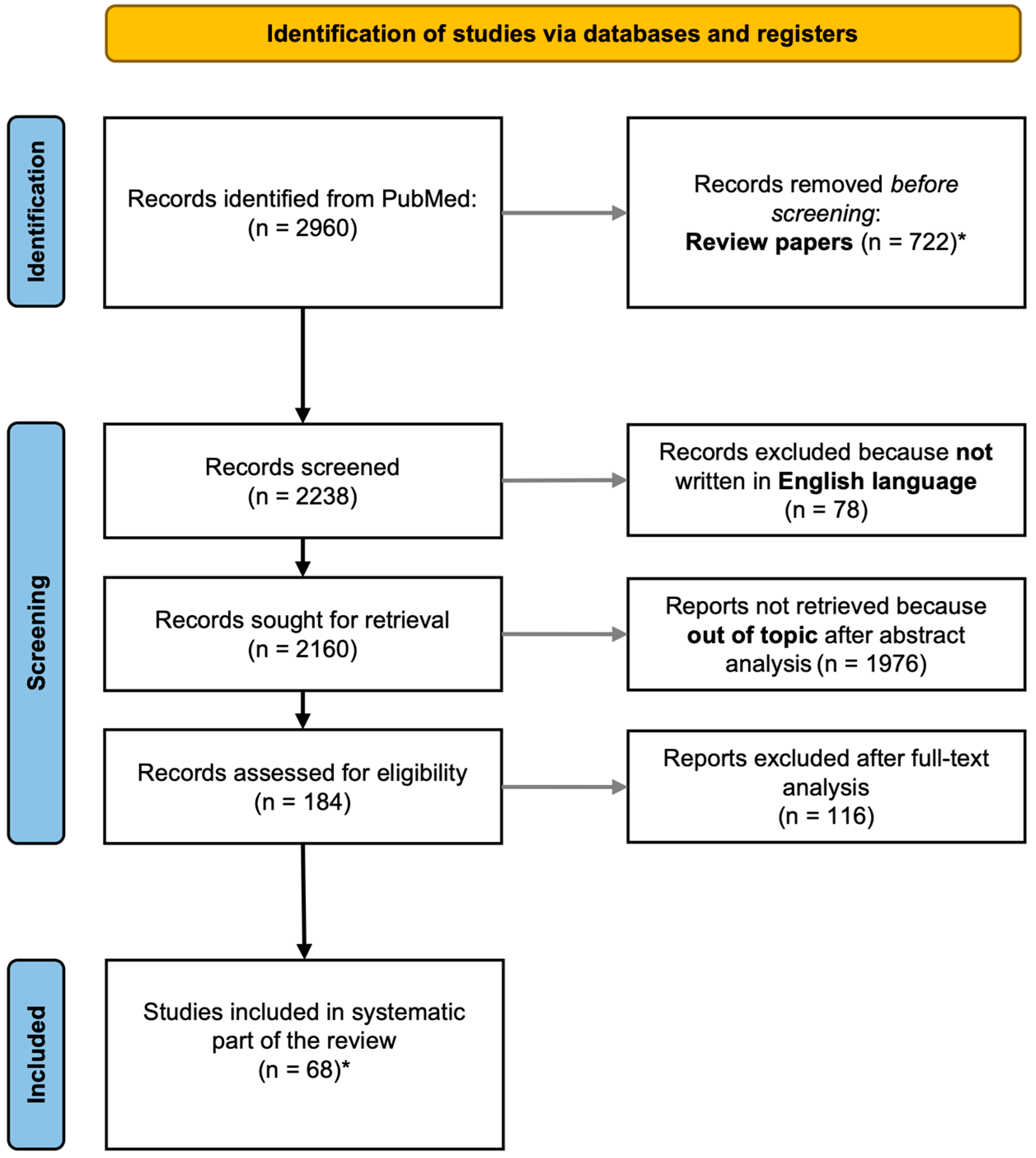
Abstract
:1. Introduction
2. Methods
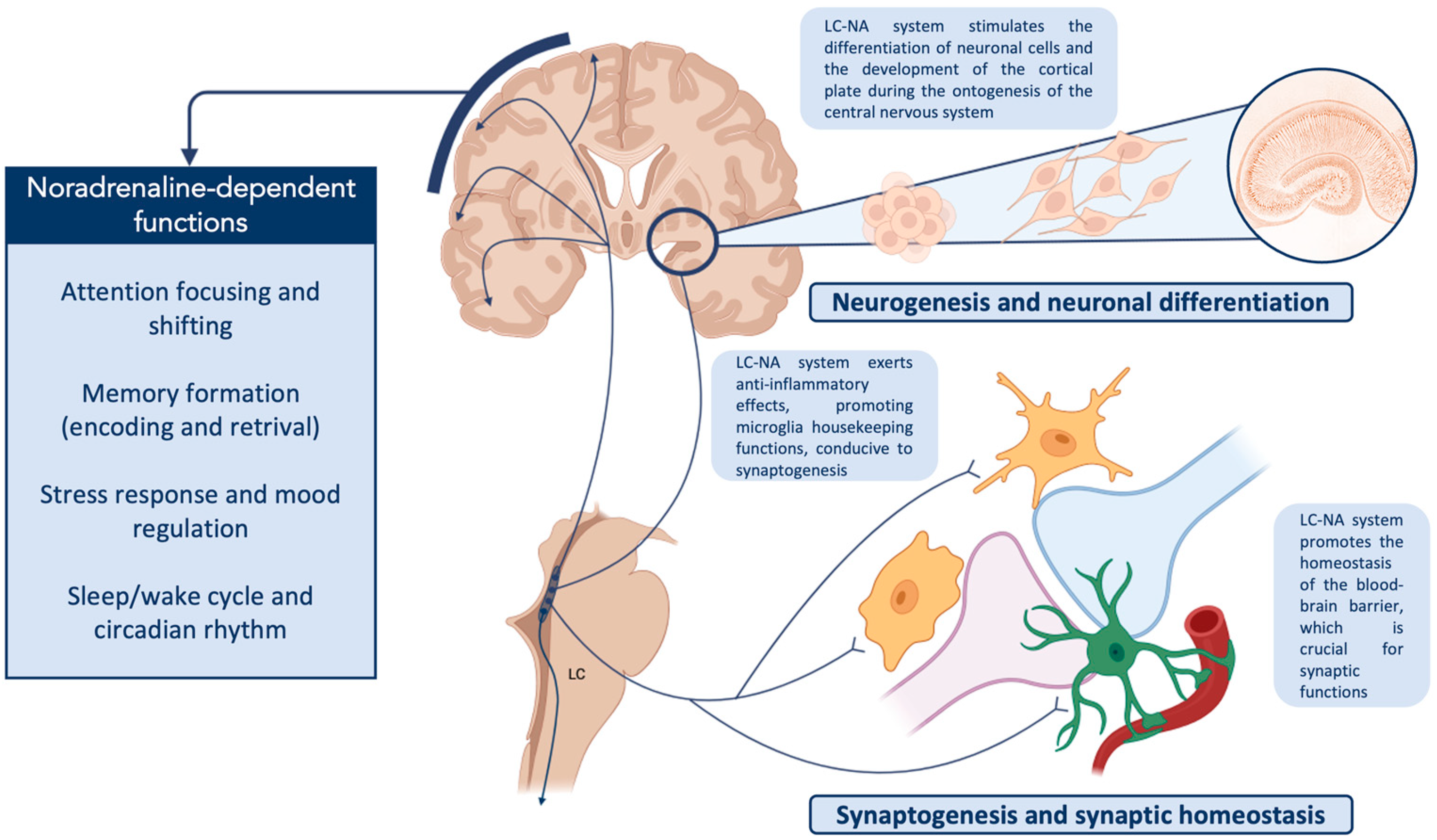
3. LC Anatomy and Basic Description of Its Functions
4. LC Ontogenesis
5. Susceptibility of LC to Different Insults during Ontogenesis and Post-Natal Development
6. The Role of LC-NA Innervation in the Ontogenesis of CNS and Possible Consequences of Its Alteration
7. Preclinical Data Links NdD Symptoms with LC-NA System Dysfunction
8. Clinical Evidence of LC-NA System Involvement in NdD
9. Potential Mechanisms Linking LC Impairment and Pathophysiology of NdD
10. Future Perspectives
11. Conclusions
Author Contributions
Funding
Institutional Review Board Statement
Informed Consent Statement
Data Availability Statement
Conflicts of Interest
References
- Galgani, A.; Lombardo, F.; Della Latta, D.; Martini, N.; Bonuccelli, U.; Fornai, F.; Giorgi, F.S. Locus Coeruleus Magnetic Resonance Imaging in Neurological Diseases. Curr. Neurol. Neurosci. Rep. 2020, 21, 2. [Google Scholar] [CrossRef] [PubMed]
- American Psychiatric Association. Diagnostic and Statistical Manual of Mental Disorders—Text Revision, 5th ed.; American Psychiatric Association: Washington, DC, USA, 2022. [Google Scholar]
- Pearlstein, E. Role of Descending Aminergic Pathways in the Development of Locomotion. Curr. Pharm. Des. 2013, 19, 4332–4340. [Google Scholar] [CrossRef] [PubMed]
- Varghese, M.; Keshav, N.; Jacot-Descombes, S.; Warda, T.; Wicinski, B.; Dickstein, D.L.; Harony-Nicolas, H.; De Rubeis, S.; Drapeau, E.; Buxbaum, J.D.; et al. Autism Spectrum Disorder: Neuropathology and Animal Models. Acta Neuropathol. 2017, 134, 537–566. [Google Scholar] [CrossRef] [PubMed]
- Bast, N.; Poustka, L.; Freitag, C.M. The Locus Coeruleus–Norepinephrine System as Pacemaker of Attention—A Developmental Mechanism of Derailed Attentional Function in Autism Spectrum Disorder. Eur. J. Neurosci. 2018, 47, 115–125. [Google Scholar] [CrossRef]
- London, E.B. Neuromodulation and a Reconceptualization of Autism Spectrum Disorders: Using the Locus Coeruleus Functioning as an Exemplar. Front. Neurol. 2018, 9, 1120. [Google Scholar] [CrossRef]
- Lai, M.C.; Lombardo, M.V.; Baron-Cohen, S. Autism. Lancet 2014, 383, 896–910. [Google Scholar] [CrossRef]
- Drechsler, R.; Brem, S.; Brandeis, D.; Grünblatt, E.; Berger, G.; Walitza, S. ADHD: Current Concepts and Treatments in Children and Adolescents. Neuropediatrics 2020, 51, 315–335. [Google Scholar] [CrossRef]
- Mechler, K.; Banaschewski, T.; Hohmann, S.; Häge, A. Evidence-Based Pharmacological Treatment Options for ADHD in Children and Adolescents. Pharmacol. Ther. 2022, 230, 107940. [Google Scholar] [CrossRef]
- Sharma, A.; Couture, J. A Review of the Pathophysiology, Etiology, and Treatment of Attention-Deficit Hyperactivity Disorder (ADHD). Ann. Pharmacother. 2013, 48, 209–225. [Google Scholar] [CrossRef]
- Counts, S.E.; Mufson, E.J. Locus Coeruleus. In The Human Nervous System; Mai, J.K., Paxinos, G., Eds.; Academic Press: Cambridge, MA, USA, 2012; pp. 427–440. ISBN 9780123742360. [Google Scholar]
- Szabadi, E. Functional Neuroanatomy of the Central Noradrenergic System. J. Psychopharmacol. 2013, 27, 659–693. [Google Scholar] [CrossRef]
- Fuxe, K.; Dahlström, A.B.; Jonsson, G.; Marcellino, D.; Guescini, M.; Dam, M.; Manger, P.; Agnati, L. The Discovery of Central Monoamine Neurons Gave Volume Transmission to the Wired Brain. Prog. Neurobiol. 2010, 90, 82–100. [Google Scholar] [CrossRef] [PubMed]
- Benarroch, E.E. Locus Coeruleus. Cell Tissue Res. 2018, 373, 221–232. [Google Scholar] [CrossRef]
- Sara, S.J. The Locus Coeruleus and Noradrenergic Modulation of Cognition. Nat. Rev. Neurosci. 2009, 10, 211–223. [Google Scholar] [CrossRef] [PubMed]
- Aston-Jones, G.; Waterhouse, B. Locus Coeruleus: From Global Projection System to Adaptive Regulation of Behavior. Brain Res. 2016, 1645, 75–78. [Google Scholar] [CrossRef] [PubMed] [Green Version]
- Aston-Jones, G.; Rajkowski, J.; Cohen, J. Role of Locus Coeruleus in Attention and Behavioral Flexibility. Biol. Psychiatry 1999, 46, 1309–1320. [Google Scholar] [CrossRef]
- Hansen, N. The Longevity of Hippocampus-Dependent Memory Is Orchestrated by the Locus Coeruleus-Noradrenergic System. Neural Plast. 2017, 2017, 2727602. [Google Scholar] [CrossRef]
- Poe, G.R.; Foote, S.; Eschenko, O.; Johansen, J.P.; Bouret, S.; Aston-Jones, G.; Harley, C.W.; Manahan-Vaughan, D.; Weinshenker, D.; Valentino, R.; et al. Locus Coeruleus: A New Look at the Blue Spot. Nat. Rev. Neurosci. 2020, 21, 644–659. [Google Scholar] [CrossRef]
- McCall, J.G.; Siuda, E.R.; Bhatti, D.L.; Lawson, L.A.; McElligott, Z.A.; Stuber, G.D.; Bruchas, M.R. Locus Coeruleus to Basolateral Amygdala Noradrenergic Projections Promote Anxiety-like Behavior. Elife 2017, 6, e18247. [Google Scholar] [CrossRef]
- Giorgi, F.S.; Galgani, A.; Puglisi-Allegra, S.; Limanaqi, F.; Busceti, C.L.; Fornai, F. Locus Coeruleus and Neurovascular Unit: From Its Role in Physiology to Its Potential Role in Alzheimer’s Disease Pathogenesis. J. Neurosci. Res. 2020, 98, 2406–2434. [Google Scholar] [CrossRef]
- Osorio-Forero, A.; Cherrad, N.; Banterle, L.; Fernandez, L.M.J.; Lüthi, A. When the Locus Coeruleus Speaks Up in Sleep: Recent Insights, Emerging Perspectives. Int. J. Mol. Sci. 2022, 23, 5028. [Google Scholar] [CrossRef]
- Van Egroo, M.; Koshmanova, E.; Vandewalle, G.; Jacobs, H.I.L. Importance of the Locus Coeruleus-Norepinephrine System in Sleep-Wake Regulation: Implications for Aging and Alzheimer’s Disease. Sleep Med. Rev. 2022, 62, 101592. [Google Scholar] [CrossRef]
- Moruzzi, G.; Magoun, H.W. Brain Stem Reticular Formation and Activation of the EEG. Electroencephalogr. Clin. Neurophysiol. 1949, 1, 455–473. [Google Scholar] [CrossRef] [PubMed]
- Schwarz, L.A.; Luo, L. Organization of the Locus Coeruleus-Norepinephrine System. Curr. Biol. 2015, 25, R1051–R1056. [Google Scholar] [CrossRef] [PubMed] [Green Version]
- González, M.M.C.; Aston-Jones, G. Circadian Regulation of Arousal: Role of the Noradrenergic Locus Coeruleus System and Light Exposure. Sleep 2006, 29, 1327–1336. [Google Scholar] [CrossRef] [PubMed] [Green Version]
- Giorgi, F.S.; Galgani, A.; Puglisi-Allegra, S.; Busceti, C.L.; Fornai, F. The Connections of Locus Coeruleus with Hypothalamus: Potential Involvement in Alzheimer’s Disease. J. Neural Transm. 2021, 128, 589–613. [Google Scholar] [CrossRef]
- Morris, L.S.; McCall, J.G.; Charney, D.S.; Murrough, J.W. The Role of the Locus Coeruleus in the Generation of Pathological Anxiety. Brain Neurosci. Adv. 2020, 4, 239821282093032. [Google Scholar] [CrossRef]
- Weiss, J.M.; Stout, J.C.; Aaron, M.F.; Quan, N.; Owens, M.J.; Butler, P.D.; Nemeroff, C.B. Depression and Anxiety: Role of the Locus Coeruleus and Corticotropin-Releasing Factor. Brain Res. Bull. 1994, 35, 561–572. [Google Scholar] [CrossRef]
- Borodovitsyna, O.; Flamini, M.D.; Chandler, D.J. Acute Stress Persistently Alters Locus Coeruleus Function and Anxiety-like Behavior in Adolescent Rats. Neuroscience 2018, 373, 7–19. [Google Scholar] [CrossRef] [PubMed]
- Aroca, P.; Lorente-Cánovas, B.; Mateos, F.R.; Puelles, L. Locus Coeruleus Neurons Originate in Alar Rhombomere 1 and Migrate into the Basal Plate: Studies in Chick and Mouse Embryos. J. Comp. Neurol. 2006, 496, 802–818. [Google Scholar] [CrossRef] [PubMed]
- Robertson, S.D.; Plummer, N.W.; De Marchena, J.; Jensen, P. Developmental Origins of Central Norepinephrine Neuron Diversity. Nat. Neurosci. 2013, 16, 1016–1023. [Google Scholar] [CrossRef] [Green Version]
- Verney, C.; Zecevic, N.; Nikolic, B.; Alvarez, C.; Berger, B. Early Evidence of Catecholaminergic Cell Groups in 5- and 6-Week-Old Human Embryos Using Tyrosine Hydroxylase and Dopamine-β-Hydroxylase Immunocytochemistry. Neurosci. Lett. 1991, 131, 121–124. [Google Scholar] [CrossRef]
- Zecevic, N.; Verney, C. Development of the Catecholamine Neurons in Human Embryos and Fetuses, with Special Emphasis on the Innervaton of the Cerebral Cortex. J. Comp. Neurol. 1995, 351, 509–535. [Google Scholar] [CrossRef] [PubMed]
- Ten, A.M.; Belova, T.I.; Korolev, V.V. Ultrastructure of the Locus Coeruleus in the Brain of Rats in Early Ontogenesis. Neurosci. Behav. Physiol. 1984, 14, 173–179. [Google Scholar] [CrossRef] [PubMed]
- Steindler, D.A.; Trosko, B.K. Two Types of Locus Coeruleus Neurons Born on Different Embryonic Days in the Mouse. Anat. Embryol. 1989, 179, 423–434. [Google Scholar] [CrossRef] [PubMed]
- Marshall, K.C.; Christie, M.J.; Finlayson, P.G.; Williams, J.T. Developmental aspects of the locus coeruleus-noradrenaline system. Prog Brain Res 1991, 88, 173–185. [Google Scholar] [CrossRef]
- Bezin, L.; Marcel, D.; Debure, L.I.; Ginovart, N.; Rousset, C.; Pujol, J.F.; Weissmann, D. Postnatal Development of the Tyrosine Hydroxylase-Containing Cell Population within the Rat Locus Coeruleus: Topological Organization Andphenotypic Plasticity. J. Neurosci. 1994, 14, 7486–7501. [Google Scholar] [CrossRef] [PubMed]
- Sanders, J.D.; Happe, H.K.; Bylund, D.B.; Murrin, L.C. Development of the Norepinephrine Transporter in the Rat CNS. Neuroscience 2005, 130, 107–117. [Google Scholar] [CrossRef]
- Dreyfus, C.F.; Markey, K.A.; Goldstein, M.; Black, I.B. Development of Catecholaminergic Phenotypic Characters in the Mouse Locus Coeruleus in Vivo and in Culture. Dev. Biol. 1983, 97, 48–58. [Google Scholar] [CrossRef]
- Holm, P.C.; Rodríguez, F.J.; Kele, J.; Castelo-Branco, G.; Kitajewski, J.; Arenas, E. BMPs, FGF8 and Wnts Regulate the Differentiation of Locus Coeruleus Noradrenergic Neuronal Precursors. J. Neurochem. 2006, 99, 343–352. [Google Scholar] [CrossRef]
- Fan, Y.; Huang, J.; Duffourc, M.; Kao, R.L.; Ordway, G.A.; Huang, R.; Zhu, M.Y. Transcription Factor Phox2 Upregulates Expression of Norepinephrine Transporter and Dopamine β-Hydroxylase in Adult Rat Brains. Neuroscience 2011, 192, 37–53. [Google Scholar] [CrossRef] [Green Version]
- Song, N.N.; Ma, P.; Zhang, Q.; Zhang, L.; Wang, H.; Zhang, L.; Zhu, L.; He, C.H.; Mao, B.; Ding, Y.Q. Rnf220/Zc4h2-Mediated Monoubiquitylation of Phox2 Is Required for Noradrenergic Neuron Development. Development 2020, 147, dev185199. [Google Scholar] [CrossRef]
- Morin, X.; Cremer, H.; Hirsch, M.R.; Kapur, R.P.; Goridis, C.; Brunet, J.F. Defects in Sensory and Autonomic Ganglia and Absence of Locus Coeruleus in Mice Deficient for the Homeobox Gene Phox2a. Neuron 1997, 18, 411–423. [Google Scholar] [CrossRef] [Green Version]
- Sklair, L.; Segal, M. Regulation of the Development of Locus Coeruleus Neurons in Vitro. Prog. Brain Res. 1991, 88, 617–623. [Google Scholar] [CrossRef]
- Robinson, L.J.; Black, I.B.; Dreyfus, C.F. Hippocampal Regulation of the Survival and Morphological Development of Locus Coeruleus Neurons in Dissociated Cell Culture. J. Comp. Neurol. 1993, 333, 567–577. [Google Scholar] [CrossRef]
- Weinshenker, D. Long Road to Ruin: Noradrenergic Dysfunction in Neurodegenerative Disease. Trends Neurosci. 2018, 41, 211–223. [Google Scholar] [CrossRef] [PubMed]
- Landry, J.P.; Hawkins, C.; Wiebe, S.; Balaban, E.; Pompeiano, M. Opposing Effects of Hypoxia on Catecholaminergic Locus Coeruleus and Hypocretin/Orexin Neurons in Chick Embryos. Dev. Neurobiol. 2014, 74, 1030–1037. [Google Scholar] [CrossRef]
- Lavezzi, A.M.; Alfonsi, G.; Matturri, L. Pathophysiology of the Human Locus Coeruleus Complex in Fetal/Neonatal Sudden Unexplained Death. Neurol. Res. 2013, 35, 44–53. [Google Scholar] [CrossRef]
- Lavezzi, A.M.; Ottaviani, G.; Mingrone, R.; Matturri, L. Analysis of the Human Locus Coeruleus in Perinatal and Infant Sudden Unexplained Deaths. Possible Role of the Cigarette Smoking in the Development of This Nucleus. Dev. Brain Res. 2005, 154, 71–80. [Google Scholar] [CrossRef] [PubMed]
- Pagida, M.A.; Konstantinidou, A.E.; Korelidou, A.; Katsika, D.; Tsekoura, E.; Patsouris, E.; Panayotacopoulou, M.T. The Effect of Perinatal Hypoxic/Ischemic Injury on Tyrosine Hydroxylase Expression in the Locus Coeruleus of the Human Neonate. Dev. Neurosci. 2016, 38, 41–53. [Google Scholar] [CrossRef]
- Magalhães, K.S.; Spiller, P.F.; da Silva, M.P.; Kuntze, L.B.; Paton, J.F.R.; Machado, B.H.; Moraes, D.J.A. Locus Coeruleus as a Vigilance Centre for Active Inspiration and Expiration in Rats. Sci. Rep. 2018, 8, 15654. [Google Scholar] [CrossRef] [PubMed] [Green Version]
- Spencer, P.S.; Schaumburg, H.H.; Ludolph, A.C. Experimental and Clinical Neurotoxicology; Oxford University Press: New York, NY, USA, 2000; p. 1310. [Google Scholar]
- Michałowicz, J. Bisphenol A—Sources, Toxicity and Biotransformation. Environ. Toxicol. Pharmacol. 2014, 37, 738–758. [Google Scholar] [CrossRef]
- Tando, S.; Itoh, K.; Yaoi, T.; Ogi, H.; Goto, S.; Mori, M.; Fushiki, S. Bisphenol A Exposure Disrupts the Development of the Locus Coeruleus-Noradrenergic System in Mice. Neuropathology 2014, 34, 527–534. [Google Scholar] [CrossRef] [PubMed]
- Tian, Y.H.; Baek, J.H.; Lee, S.Y.; Jang, C.G. Prenatal and Postnatal Exposure to Bisphenol A Induces Anxiolytic Behaviors and Cognitive Deficits in Mice. Synapse 2010, 64, 432–439. [Google Scholar] [CrossRef] [PubMed]
- Stein, T.P.; Schluter, M.D.; Steer, R.A.; Guo, L.; Ming, X. Bisphenol A Exposure in Children With Autism Spectrum Disorders. Autism Res. 2015, 8, 272–283. [Google Scholar] [CrossRef] [Green Version]
- McGaughy, J.A.; Amaral, A.C.; Rushmore, R.J.; Mokler, D.J.; Morgane, P.J.; Rosene, D.L.; Galler, J.R. Prenatal Malnutrition Leads to Deficits in Attentional Set Shifting and Decreases Metabolic Activity in Prefrontal Subregions That Control Executive Function. Dev. Neurosci. 2014, 36, 532–541. [Google Scholar] [CrossRef]
- Newman, L.A.; Baraiolo, J.; Mokler, D.J.; Rabinowitz, A.G.; Galler, J.R.; McGaughy, J.A. Prenatal Protein Malnutrition Produces Resistance to Distraction Similar to Noradrenergic Deafferentation of the Prelimbic Cortex in a Sustained Attention Task. Front. Neurosci. 2019, 13, 123. [Google Scholar] [CrossRef]
- Rushmore, R.J.; McGaughy, J.A.; Amaral, A.C.; Mokler, D.J.; Morgane, P.J.; Galler, J.R.; Rosene, D.L. The Neural Basis of Attentional Alterations in Prenatally Protein Malnourished Rats. Cereb. Cortex 2021, 31, 497–512. [Google Scholar] [CrossRef] [PubMed]
- Rowe, S.J.; Messenger, N.J.; Warner, A.E. The Role of Noradrenaline in the Differentiation of Amphibian Embryonic Neurons. Development 1993, 119, 1343–1357. [Google Scholar] [CrossRef]
- Messenger, N.J.; Rowe, S.J.; Warner, A.E. The Neurotransmitter Noradrenaline Drivesnoggin-Expressing Ectoderm Cells to ActivateN-Tubulinand Become Neurons. Dev. Biol. 1999, 205, 224–232. [Google Scholar] [CrossRef] [Green Version]
- Sieber-Blum, M.; Ren, Z. Norepinephrine Transporter Expression and Function in Noradrenergic Cell Differentiation. Mol. Cell. Biochem. 2000, 212, 61–70. [Google Scholar] [CrossRef]
- Ren, Z.G.; Pörzgen, P.P.; Youn, Y.H.; Sieber-Blum, M. Ubiquitous Embryonic Expression of the Norepinephrine Transporter. Dev. Neurosci. 2003, 25, 1–13. [Google Scholar] [CrossRef] [PubMed]
- Hu, Y.F.; Caron, M.G.; Sieber-Blum, M. Norepinephrine Transport-Mediated Gene Expression in Noradrenergic Neurogenesis. BMC Genom. 2009, 10, 151. [Google Scholar] [CrossRef] [Green Version]
- Wendlandt, S.; Crow, T.J.; Stirling, R.V. The Involvement of the Noradrenergic System Arising from the Locus Coeruleus in the Postnatal Development of the Cortex in Rat Brain. Brain Res. 1977, 125, 1–9. [Google Scholar] [CrossRef] [PubMed]
- Blue, M.E.; Parnavelas, J.G. The Effect of Neonatal 6-Hydroxydopamine Treatment on Synaptogenesis in the Visual Cortex of the Rat. J. Comp. Neurol. 1982, 205, 199–205. [Google Scholar] [CrossRef] [PubMed]
- Seidler, F.J.; Temple, S.W.; McCook, E.C.; Slotkin, T.A. Cocaine Inhibits Central Noradrenergic and Dopaminergic Activity during the Critical Developmental Period in Which Catecholamines Influence Cell Development. Dev. Brain Res. 1995, 85, 48–53. [Google Scholar] [CrossRef]
- Lidow, M.S. Nonhuman Primate Model of the Effect of Prenatal Cocaine Exposure on Cerebral Cortical Development. Ann. N. Y. Acad. Sci. 1998, 846, 182–193. [Google Scholar] [CrossRef]
- Winzer-Serhan, U.H.; Leslie, F.M. Expression of Alpha2A Adrenoceptors during Rat Neocortical Development. J. Neurobiol. 1999, 38, 259–269. [Google Scholar] [CrossRef]
- Fauser, M.; Weselek, G.; Hauptmann, C.; Markert, F.; Gerlach, M.; Hermann, A.; Storch, A. Catecholaminergic Innervation of Periventricular Neurogenic Regions of the Developing Mouse Brain. Front. Neuroanat. 2020, 14, 64. [Google Scholar] [CrossRef] [PubMed]
- Sakaguchi, T.; Nakamura, S. Some in Vivo Electrophysiological Properties of Locus Coeruleus Neurones in Fetal Rats. Exp. Brain Res. 1987, 68, 122–130. [Google Scholar] [CrossRef]
- Masuda, T.; Nakagawa, S.; Boku, S.; Nishikawa, H.; Takamura, N.; Kato, A.; Inoue, T.; Koyama, T. Noradrenaline Increases Neural Precursor Cells Derived from Adult Rat Dentate Gyrus through Beta2 Receptor. Prog. Neuropsychopharmacol. Biol. Psychiatry 2012, 36, 44–51. [Google Scholar] [CrossRef]
- Coradazzi, M.; Gulino, R.; Fieramosca, F.; Falzacappa, L.V.; Riggi, M.; Leanza, G. Selective Noradrenaline Depletion Impairs Working Memory and Hippocampal Neurogenesis. Neurobiol. Aging 2016, 48, 93–102. [Google Scholar] [CrossRef] [PubMed]
- Thompson, W.A.; Arnold, V.I.; Vijayan, M.M. Venlafaxine in Embryos Stimulates Neurogenesis and Disrupts Larval Behavior in Zebrafish. Environ. Sci. Technol. 2017, 51, 12889–12897. [Google Scholar] [CrossRef] [PubMed]
- Bortolotto, V.; Bondi, H.; Cuccurazzu, B.; Rinaldi, M.; Canonico, P.L.; Grilli, M. Salmeterol, a Β2 Adrenergic Agonist, Promotes Adult Hippocampal Neurogenesis in a Region-Specific Manner. Front. Pharmacol. 2019, 10, 1000. [Google Scholar] [CrossRef] [PubMed] [Green Version]
- Weselek, G.; Keiner, S.; Fauser, M.; Wagenführ, L.; Müller, J.; Kaltschmidt, B.; Brandt, M.D.; Gerlach, M.; Redecker, C.; Hermann, A.; et al. Norepinephrine Is a Negative Regulator of the Adult Periventricular Neural Stem Cell Niche. Stem Cells 2020, 38, 1188–1201. [Google Scholar] [CrossRef]
- de Villiers, A.S.; Russell, V.A.; Sagvolden, T.; Searson, A.; Jaffer, A.; Taljaard, J.J.F. A2 Mediated Inhibition of [3H]Dopamine Release from Nucleus Accumbens Slices and Monoamine Levels in a Rat Model for Attention-Deficit Hyperactivity Disorder. Neurochem. Res. 1995, 20, 427–433. [Google Scholar] [CrossRef]
- Jones, M.D.; Hess, E.J. Norepinephrine Regulates Locomotor Hyperactivity in the Mouse Mutant Coloboma. Pharmacol. Biochem. Behav. 2003, 75, 209–216. [Google Scholar] [CrossRef]
- Yin, X.; Jones, N.; Yang, J.; Asraoui, N.; Mathieu, M.E.; Cai, L.; Chen, S.X. Delayed Motor Learning in a 16p11.2 Deletion Mouse Model of Autism Is Rescued by Locus Coeruleus Activation. Nat. Neurosci. 2021, 24, 646–657. [Google Scholar] [CrossRef]
- Martchek, M.; Thevarkunnel, S.; Bauman, M.; Blatt, G.; Kemper, T. Lack of Evidence of Neuropathology in the Locus Coeruleus in Autism. Acta Neuropathol. 2006, 111, 497–499. [Google Scholar] [CrossRef]
- Pamphlett, R.; Kum Jew, S. Locus Ceruleus Neurons in People with Autism Contain No Histochemically-Detectable Mercury. BioMetals 2016, 29, 171–175. [Google Scholar] [CrossRef] [Green Version]
- Fetit, R.; Hillary, R.F.; Price, D.J.; Lawrie, S.M. The Neuropathology of Autism: A Systematic Review of Post-Mortem Studies of Autism and Related Disorders. Neurosci. Biobehav. Rev. 2021, 129, 35–62. [Google Scholar] [CrossRef]
- Johnston, B.A.; Mwangi, B.; Matthews, K.; Coghill, D.; Konrad, K.; Steele, J.D. Brainstem Abnormalities in Attention Deficit Hyperactivity Disorder Support High Accuracy Individual Diagnostic Classification. Hum. Brain Mapp. 2014, 35, 5179–5189. [Google Scholar] [CrossRef] [Green Version]
- Berridge, C.W.; Waterhouse, B.D. The Locus Coeruleus–Noradrenergic System: Modulation of Behavioral State and State-Dependent Cognitive Processes. Brain Res. Rev. 2003, 42, 33–84. [Google Scholar] [CrossRef] [PubMed]
- Huang, Y.; Yu, S.; Wilson, G.; Park, J.; Cheng, M.; Kong, X.; Lu, T.; Kong, J. Altered Extended Locus Coeruleus and Ventral Tegmental Area Networks in Boys with Autism Spectrum Disorders: A Resting-State Functional Connectivity Study. Neuropsychiatr. Dis. Treat. 2021, 17, 1207–1216. [Google Scholar] [CrossRef]
- Boxhoorn, S.; Bast, N.; Supèr, H.; Polzer, L.; Cholemkery, H.; Freitag, C.M. Pupil Dilation during Visuospatial Orienting Differentiates between Autism Spectrum Disorder and Attention-Deficit/Hyperactivity Disorder. J. Child Psychol. Psychiatry 2020, 61, 614–624. [Google Scholar] [CrossRef]
- Granovetter, M.C.; Burlingham, C.S.; Blauch, N.M.; Minshew, N.J.; Heeger, D.J.; Behrmann, M. Uncharacteristic Task-Evoked Pupillary Responses Implicate Atypical Locus Ceruleus Activity in Autism. J. Neurosci. 2020, 40, 3815–3826. [Google Scholar] [CrossRef] [PubMed]
- Kim, Y.; Kadlaskar, G.; Keehn, R.M.; Keehn, B. Measures of Tonic and Phasic Activity of the Locus Coeruleus—Norepinephrine System in Children with Autism Spectrum Disorder: An Event-Related Potential and Pupillometry Study. Autism Res. 2022, 15, 2250–2264. [Google Scholar] [CrossRef] [PubMed]
- Polzer, L.; Freitag, C.M.; Bast, N. Pupillometric Measures of Altered Stimulus-Evoked Locus Coeruleus-Norepinephrine Activity Explain Attenuated Social Attention in Preschoolers with Autism Spectrum Disorder. Autism Res. 2022, 15, 2167–2180. [Google Scholar] [CrossRef] [PubMed]
- Bast, N.; Boxhoorn, S.; Supér, H.; Helfer, B.; Polzer, L.; Klein, C.; Cholemkery, H.; Freitag, C.M. Atypical Arousal Regulation in Children With Autism but Not With Attention-Deficit/Hyperactivity Disorder as Indicated by Pupillometric Measures of Locus Coeruleus Activity. Biol. Psychiatry Cogn. Neurosci. Neuroimaging 2023, 8, 11–20. [Google Scholar] [CrossRef]
- Blaser, E.; Eglington, L.; Carter, A.S.; Kaldy, Z. Pupillometry Reveals a Mechanism for the Autism Spectrum Disorder (ASD) Advantage in Visual Tasks. Sci. Rep. 2014, 4, 4301. [Google Scholar] [CrossRef] [Green Version]
- Bast, N.; Banaschewski, T.; Dziobek, I.; Brandeis, D.; Poustka, L.; Freitag, C.M. Pupil Dilation Progression Modulates Aberrant Social Cognition in Autism Spectrum Disorder. Autism Res. 2019, 12, 1680–1692. [Google Scholar] [CrossRef] [PubMed] [Green Version]
- Rudling, M.; Nyström, P.; Bölte, S.; Falck-Ytter, T. Larger Pupil Dilation to Nonsocial Sounds in Infants with Subsequent Autism Diagnosis. J. Child Psychol. Psychiatry 2022, 63, 793–801. [Google Scholar] [CrossRef] [PubMed]
- Shirama, A.; Takeda, T.; Ohta, H.; Iwanami, A.; Toda, S.; Kato, N. Atypical Alert State Control in Adult Patients with ADHD: A Pupillometry Study. PLoS ONE 2020, 15, e0244662. [Google Scholar] [CrossRef]
- Kleberg, J.L.; Frick, M.A.; Brocki, K.C. Increased Pupil Dilation to Happy Faces in Children with Hyperactive/Impulsive Symptoms of ADHD. Dev. Psychopathol. 2021, 33, 767–777. [Google Scholar] [CrossRef] [PubMed] [Green Version]
- Kaga, Y.; Ohyama, T.; Goto, Y.; Aoyagi, K.; Ishii, S.; Inukai, T.; Aihara, M. Impairment of Autonomic Emotional Response for Executive Function in Children with ADHD: A Multi-Modal FNIRS and Pupillometric Study during the Wisconsin Card Sorting Test. Brain Dev. 2022, 44, 438–445. [Google Scholar] [CrossRef]
- Vanicek, T.; Spies, M.; Rami-Mark, C.; Savli, M.; Höflich, A.; Kranz, G.S.; Hahn, A.; Kutzelnigg, A.; Traub-Weidinger, T.; Mitterhauser, M.; et al. The Norepinephrine Transporter in Attention-Deficit/Hyperactivity Disorder Investigated With Positron Emission Tomography. JAMA Psychiatry 2014, 71, 1340–1349. [Google Scholar] [CrossRef] [Green Version]
- Sigurdardottir, H.L.; Kranz, G.S.; Rami-Mark, C.; James, G.M.; Vanicek, T.; Gryglewski, G.; Berroterán-Infante, N.; Kautzky, A.; Hienert, M.; Traub-Weidinger, T.; et al. Association of Norepinephrine Transporter Methylation with in Vivo NET Expression and Hyperactivity–Impulsivity Symptoms in ADHD Measured with PET. Mol. Psychiatry 2019, 26, 1009–1018. [Google Scholar] [CrossRef] [PubMed] [Green Version]
- Ghanizadeh, A. Atomoxetine for Treating ADHD Symptoms in Autism. J. Atten. Disord. 2012, 17, 635–640. [Google Scholar] [CrossRef] [PubMed]
- Nagashima, M.; Monden, Y.; Dan, I.; Dan, H.; Tsuzuki, D.; Mizutani, T.; Kyutoku, Y.; Gunji, Y.; Momoi, M.Y.; Eiju Watanabe, M.D.; et al. Neuropharmacological Effect of Methylphenidate on Attention Network in Children with Attention Deficit Hyperactivity Disorder during Oddball Paradigms as Assessed Using Functional Near-Infrared Spectroscopy. Neurophotonics 2014, 1, 015001. [Google Scholar] [CrossRef] [Green Version]
- Aston-Jones Iba, M.; Clayton, E.; Rajkowski, J.; Cohen, J.; Aston-Jones, G. Locus Coeruleus and Regulation of Behavioral Flexibility and Attention: Clinical Implications. In Brain Norepinephrine—Neurobiology and Therapeutics; Ordway, G.A., Schwartz, M.A., Frazer, A., Eds.; Cambridge University Press: Cambridge, UK, 2007; pp. 196–235. [Google Scholar]
- Cortese, S.; Wang, F.; Angriman, M.; Masi, G.; Bruni, O. Sleep Disorders in Children and Adolescents with Autism Spectrum Disorder: Diagnosis, Epidemiology, and Management. CNS Drugs 2020, 34, 415–423. [Google Scholar] [CrossRef]
- Becker, S.P. ADHD and Sleep: Recent Advances and Future Directions. Curr. Opin. Psychol. 2020, 34, 50–56. [Google Scholar] [CrossRef]
- Austerman, J. ADHD and Behavioral Disorders: Assessment, Management, and an Update from DSM-5. Cleve. Clin. J. Med. 2015, 82, S2–S7. [Google Scholar] [CrossRef] [PubMed]
- Oakley, B.; Loth, E.; Murphy, D.G. Autism and Mood Disorders. Int. Rev. Psychiatry 2021, 33, 280–299. [Google Scholar] [CrossRef] [PubMed]
- Hvolby, A.; Christensen, J.; Gasse, C.; Dalsgaard, S.; Dreier, J.W. Cumulative Incidence and Relative Risk of Sleep Problems among Children and Adolescents with Newly Diagnosed Neurodevelopmental Disorders: A Nationwide Register-Based Study. J. Sleep Res. 2021, 30, e13122. [Google Scholar] [CrossRef]
- David, E.; Eva, B.; Christopher, G. Neurodevelopmental Disorders and Comorbidity in Young Adults Attending a Psychiatric Outpatient Clinic. Psychiatry Res. 2022, 313, 114638. [Google Scholar] [CrossRef]
- King, B.H. Psychiatric Comorbidities in Neurodevelopmental Disorders. Curr. Opin. Neurol. 2016, 29, 113–117. [Google Scholar] [CrossRef]
- Osorio-Forero, A.; Cardis, R.; Vantomme, G.; Guillaume-Gentil, A.; Katsioudi, G.; Devenoges, C.; Fernandez, L.M.J.; Lüthi, A. Noradrenergic Circuit Control of Non-REM Sleep Substates. Curr. Biol. 2021, 31, 5009–5023.e7. [Google Scholar] [CrossRef] [PubMed]
- Hohmann, S.; Hohm, E.; Treutlein, J.; Blomeyer, D.; Jennen-Steinmetz, C.; Schmidt, M.H.; Esser, G.; Banaschewski, T.; Brandeis, D.; Laucht, M. Association of Norepinephrine Transporter (NET, SLC6A2) Genotype with ADHD-Related Phenotypes: Findings of a Longitudinal Study from Birth to Adolescence. Psychiatry Res. 2015, 226, 425–433. [Google Scholar] [CrossRef]
- Shang, C.Y.; Chiang, H.L.; Gau, S.S.F. A Haplotype of the Norepinephrine Transporter Gene (SLC6A2) Is Associated with Visual Memory in Attention-Deficit/Hyperactivity Disorder. Prog. Neuropsychopharmacol. Biol. Psychiatry 2015, 58, 89–96. [Google Scholar] [CrossRef]
- Park, S.; Park, J.E.; Cho, S.C.; Kim, B.N.; Shin, M.S.; Kim, J.W.; Cho, I.H.; Kim, S.A.; Park, M.; Park, T.W.; et al. No Association of the Norepinephrine Transporter Gene (SLC6A2) and Cognitive and Behavioural Phenotypes of Patients with Autism Spectrum Disorder. Eur. Arch. Psychiatry Clin. Neurosci. 2014, 264, 507–515. [Google Scholar] [CrossRef]
- Jiang, Y.H.; Ehlers, M.D. Modeling Autism by SHANK Gene Mutations in Mice. Neuron 2013, 78, 8–27. [Google Scholar] [CrossRef] [Green Version]
- Vyas, Y.; Cheyne, J.E.; Lee, K.; Jung, Y.; Cheung, P.Y.; Montgomery, J.M. Shankopathies in the Developing Brain in Autism Spectrum Disorders. Front. Neurosci. 2021, 15, 1701. [Google Scholar] [CrossRef]
- Monteiro, P.; Feng, G. SHANK Proteins: Roles at the Synapse and in Autism Spectrum Disorder. Nat. Rev. Neurosci. 2017, 18, 147–157. [Google Scholar] [CrossRef] [PubMed]
- Sungur, A.Ö.; Redecker, T.M.; Andres, E.; Dürichen, W.; Schwarting, R.K.W.; del Rey, A.; Wöhr, M. Reduced Efficacy of D-Amphetamine and 3,4-Methylenedioxymethamphetamine in Inducing Hyperactivity in Mice Lacking the Postsynaptic Scaffolding Protein SHANK1. Front. Mol. Neurosci. 2018, 11, 419. [Google Scholar] [CrossRef] [PubMed]
- Coe, B.P.; Stessman, H.A.F.; Sulovari, A.; Geisheker, M.R.; Bakken, T.E.; Lake, A.M.; Dougherty, J.D.; Lein, E.S.; Hormozdiari, F.; Bernier, R.A.; et al. Neurodevelopmental Disease Genes Implicated by de Novo Mutation and Copy Number Variation Morbidity. Nat. Genet. 2018, 51, 106–116. [Google Scholar] [CrossRef]
- Morris, G.; Puri, B.K.; Olive, L.; Carvalho, A.; Berk, M.; Walder, K.; Gustad, L.T.; Maes, M. Endothelial Dysfunction in Neuroprogressive Disorders—Causes and Suggested Treatments. BMC Med. 2020, 18, 305. [Google Scholar] [CrossRef]
- Kaye, D.M.; Wiviott, S.D.; Kobzik, L.; Kelly, R.A.; Smith, T.W. S-Nitrosothiols Inhibit Neuronal Norepinephrine Transport. Am. J. Physiol.-Heart Circ. Physiol. 1997, 272, H875–H883. [Google Scholar] [CrossRef]
- Soriano, R.N.; Ravanelli, M.I.; Batalhao, M.E.; Carnio, E.C.; Branco, L.G.S. Propyretic Role of the Locus Coeruleus Nitric Oxide Pathway. Exp. Physiol. 2010, 95, 669–677. [Google Scholar] [CrossRef]
- Yu, T.W.; Chahrour, M.H.; Coulter, M.E.; Jiralerspong, S.; Okamura-Ikeda, K.; Ataman, B.; Schmitz-Abe, K.; Harmin, D.A.; Adli, M.; Malik, A.N.; et al. Using Whole-Exome Sequencing to Identify Inherited Causes of Autism. Neuron 2013, 77, 259–273. [Google Scholar] [CrossRef] [PubMed] [Green Version]
- Kyle, S.M.; Vashi, N.; Justice, M.J. Rett Syndrome: A Neurological Disorder with Metabolic Components. Open Biol. 2018, 8, 170216. [Google Scholar] [CrossRef] [Green Version]
- Liyanage, V.R.B.; Rastegar, M. Rett Syndrome and MeCP2. Neuromol. Med. 2014, 16, 231–264. [Google Scholar] [CrossRef] [Green Version]
- Zhang, X.; Cui, N.; Wu, Z.; Su, J.; Tadepalli, J.S.; Sekizar, S.; Jiang, C. Intrinsic Membrane Properties of Locus Coeruleus Neurons in Mecp2-Null Mice. Am. J. Physiol. Cell Physiol. 2010, 298, C635–C646. [Google Scholar] [CrossRef] [PubMed] [Green Version]
- Bölte, S.; Girdler, S.; Marschik, P.B. The Contribution of Environmental Exposure to the Etiology of Autism Spectrum Disorder. Cell. Mol. Life Sci. 2018, 76, 1275–1297. [Google Scholar] [CrossRef] [PubMed] [Green Version]
- Skogheim, T.S.; Weyde, K.V.F.; Engel, S.M.; Aase, H.; Surén, P.; Øie, M.G.; Biele, G.; Reichborn-Kjennerud, T.; Caspersen, I.H.; Hornig, M.; et al. Metal and Essential Element Concentrations during Pregnancy and Associations with Autism Spectrum Disorder and Attention-Deficit/Hyperactivity Disorder in Children. Environ. Int. 2021, 152, 106468. [Google Scholar] [CrossRef] [PubMed]
- Soto-Moyano, R.; Belmar, J.; Perez, H.; Ruiz, S.; Hernandez, A. Central Noradrenergic Hyperactivity Early in Life: A Hypothesis on the Origin of Morpho-Functional Brain Disorders Induced by Malnutrition. Biol. Res. 1995, 28, 105–111. [Google Scholar]
- Pamphlett, R.; Bishop, D.P.; Jew, S.K.; Doble, P.A. Age-Related Accumulation of Toxic Metals in the Human Locus Ceruleus. PLoS ONE 2018, 13, e0203627. [Google Scholar] [CrossRef]
- Pamphlett, R.; Mak, R.; Lee, J.; Buckland, M.E.; Harding, A.J.; Jew, S.K.; Paterson, D.J.; Jones, M.W.M.; Lay, P.A. Concentrations of Toxic Metals and Essential Trace Elements Vary among Individual Neurons in the Human Locus Ceruleus. PLoS ONE 2020, 15, e0233300. [Google Scholar] [CrossRef] [PubMed]
- Capucciati, A.; Zucca, F.A.; Monzani, E.; Zecca, L.; Casella, L.; Hofer, T. Interaction of Neuromelanin with Xenobiotics and Consequences for Neurodegeneration; Promising Experimental Models. Antioxidants 2021, 10, 824. [Google Scholar] [CrossRef]
- Nakagawa, Y.; Yamada, S. The Relationships Among Metal Homeostasis, Mitochondria, and Locus Coeruleus in Psychiatric and Neurodegenerative Disorders: Potential Pathogenetic Mechanism and Therapeutic Implications. Cell. Mol. Neurobiol. 2022, 43, 963–989. [Google Scholar] [CrossRef]
- Bangasser, D.A.; Wiersielis, K.R.; Khantsis, S. Sex Differences in the Locus Coeruleus-Norepinephrine System and Its Regulation by Stress. Brain Res. 2016, 1641, 177–188. [Google Scholar] [CrossRef] [Green Version]
- Levy, F. Synaptic Gating and ADHD: A Biological Theory of Comorbidity of ADHD and Anxiety. Neuropsychopharmacology 2004, 29, 1589–1596. [Google Scholar] [CrossRef] [Green Version]
- Packer, A. Neocortical Neurogenesis and the Etiology of Autism Spectrum Disorder. Neurosci. Biobehav. Rev. 2016, 64, 185–195. [Google Scholar] [CrossRef] [PubMed]
- Zatkova, M.; Bakos, J.; Hodosy, J.; Ostatnikova, D. Synapse Alterations in Autism: Review of Animal Model Findings. Biomed. Pap. Med. Fac. Palacky Univ. Olomouc 2016, 160, 201–210. [Google Scholar] [CrossRef] [PubMed] [Green Version]
- Washbourne, P. Synapse Assembly and Neurodevelopmental Disorders. Neuropsychopharmacology 2014, 40, 4–15. [Google Scholar] [CrossRef] [PubMed] [Green Version]
- Giorgi, F.S.; Mauceli, G.; Blandini, F.; Ruggieri, S.; Paparelli, A.; Murri, L.; Fornai, F. Locus Coeruleus and Neuronal Plasticity in a Model of Focal Limbic Epilepsy. Epilepsia 2006, 47, 21–25. [Google Scholar] [CrossRef] [PubMed]
- Lemon, N.; Aydin-Abidin, S.; Funke, K.; Manahan-Vaughan, D. Locus Coeruleus Activation Facilitates Memory Encoding and Induces Hippocampal LTD That Depends on β-Adrenergic Receptor Activation. Cereb. Cortex 2009, 19, 2827–2837. [Google Scholar] [CrossRef] [PubMed] [Green Version]
- Hansen, N.; Manahan-Vaughan, D. Locus Coeruleus Stimulation Facilitates Long-Term Depression in the Dentate Gyrus That Requires Activation of β-Adrenergic Receptors. Cereb. Cortex 2015, 25, 1889–1896. [Google Scholar] [CrossRef] [PubMed] [Green Version]
- Bacon, T.J.; Pickering, A.E.; Mellor, J.R. Noradrenaline Release from Locus Coeruleus Terminals in the Hippocampus Enhances Excitation-Spike Coupling in CA1 Pyramidal Neurons Via β-Adrenoceptors. Cereb. Cortex 2020, 30, 6135–6151. [Google Scholar] [CrossRef] [PubMed]
- Feinstein, D.L.; Kalinin, S.; Braun, D. Causes, Consequences, and Cures for Neuroinflammation Mediated via the Locus Coeruleus: Noradrenergic Signaling System. J. Neurochem. 2016, 139, 154–178. [Google Scholar] [CrossRef] [PubMed] [Green Version]
- Giorgi, F.S.; Biagioni, F.; Galgani, A.; Pavese, N.; Lazzeri, G.; Fornai, F. Locus Coeruleus Modulates Neuroinflammation in Parkinsonism and Dementia. Int. J. Mol. Sci. 2020, 21, 8630. [Google Scholar] [CrossRef] [PubMed]
- Takano, T. Role of Microglia in Autism: Recent Advances. Dev. Neurosci. 2015, 37, 195–202. [Google Scholar] [CrossRef] [PubMed]
- Edmonson, C.A.; Ziats, M.N.; Rennert, O.M. A Non-Inflammatory Role for Microglia in Autism Spectrum Disorders. Front. Neurol. 2016, 7, 9. [Google Scholar] [CrossRef] [PubMed] [Green Version]
- Bokobza, C.; Van Steenwinckel, J.; Mani, S.; Mezger, V.; Fleiss, B.; Gressens, P. Neuroinflammation in Preterm Babies and Autism Spectrum Disorders. Pediatr. Res. 2018, 85, 155–165. [Google Scholar] [CrossRef] [PubMed]
- Gzielo, K.; Nikiforuk, A. Astroglia in Autism Spectrum Disorder. Int. J. Mol. Sci. 2021, 22, 11544. [Google Scholar] [CrossRef] [PubMed]
- de Moura, A.B.; Abitante, M.S.; Silva, R.H.; Quevedo, J.; Réus, G.Z. Microglial Activation in the Neurodevelopment: A Narrative Review. Curr Mol Med 2021, 22, 722–734. [Google Scholar] [CrossRef] [PubMed]
- Fiorentino, M.; Sapone, A.; Senger, S.; Camhi, S.S.; Kadzielski, S.M.; Buie, T.M.; Kelly, D.L.; Cascella, N.; Fasano, A. Blood–Brain Barrier and Intestinal Epithelial Barrier Alterations in Autism Spectrum Disorders. Mol. Autism 2016, 7, 179. [Google Scholar] [CrossRef] [PubMed] [Green Version]
- Kalinin, S.; Feinstein, D.L.; Xu, H.; Huesa, G.; Pelligrino, D.A.; Galea, E. Degeneration of Noradrenergic Fibres from the Locus Coeruleus Causes Tight-junction Disorganisation in the Rat Brain. Eur. J. Neurosci. 2006, 24, 3393–3400. [Google Scholar] [CrossRef] [PubMed]
- Koman, L.A.; Smith, B.P.; Shilt, J.S. Cerebral Palsy. Lancet 2004, 363, 1619–1631. [Google Scholar] [CrossRef] [PubMed]
- Giorgi, F.S.; Blandini, F.; Cantafora, E.; Biagioni, F.; Armentero, M.-T.; Pasquali, L.; Orzi, F.; Murri, L.; Paparelli, A.; Fornai, F. Activation of Brain Metabolism and Fos during Limbic Seizures: The Role of Locus Coeruleus. Neurobiol. Dis. 2008, 30, 388–399. [Google Scholar] [CrossRef] [PubMed]
- Giorgi, F.S.; Ferrucci, M.; Lazzeri, G.; Pizzanelli, C.; Lenzi, P.; AlessandrÏ, M.G.; Murri, L.; Fornai, F. A Damage to Locus Coeruleus Neurons Converts Sporadic Seizures into Self-sustaining Limbic Status Epilepticus. Eur. J. Neurosci. 2003, 17, 2593–2601. [Google Scholar] [CrossRef] [PubMed]
- Giorgi, F.S.; Pizzanelli, C.; Biagioni, F.; Murri, L.; Fornai, F. The Role of Norepinephrine in Epilepsy: From the Bench to the Bedside. Neurosci. Biobehav. Rev. 2004, 28, 507–524. [Google Scholar] [CrossRef] [PubMed]
- Heneka, M.T.; Ramanathan, M.; Jacobs, A.H.; Dumitrescu-Ozimek, L.; Bilkei-Gorzo, A.; Debeir, T.; Sastre, M.; Galldiks, N.; Zimmer, A.; Hoehn, M.; et al. Locus Ceruleus Degeneration Promotes Alzheimer Pathogenesis in Amyloid Precursor Protein 23 Transgenic Mice. J. Neurosci. 2006, 26, 1343–1354. [Google Scholar] [CrossRef] [PubMed] [Green Version]
- Heneka, M.T.; Nadrigny, F.; Regen, T.; Martinez-Hernandez, A.; Dumitrescu-Ozimek, L.; Terwel, D.; Jardanhazi-Kurutz, D.; Walter, J.; Kirchhoff, F.; Hanisch, U.K.; et al. Locus Ceruleus Controls Alzheimer’s Disease Pathology by Modulating Microglial Functions through Norepinephrine. Proc. Natl. Acad. Sci. USA 2010, 107, 6058–6063. [Google Scholar] [CrossRef] [PubMed] [Green Version]
- Gesi, M.; Soldani, P.; Giorgi, F.S.; Santinami, A.; Bonaccorsi, I.; Fornai, F. The Role of the Locus Coeruleus in the Development of Parkinson’s Disease. Neurosci. Biobehav. Rev. 2000, 24, 655–668. [Google Scholar] [CrossRef] [PubMed]
- Zucca, F.A.; Bellei, C.; Giannelli, S.; Terreni, M.R.; Gallorini, M.; Rizzio, E.; Pezzoli, G.; Albertini, A.; Zecca, L. Neuromelanin and Iron in Human Locus Coeruleus and Substantia Nigra during Aging: Consequences for Neuronal Vulnerability. J. Neural Transm. 2006, 113, 757–767. [Google Scholar] [CrossRef] [PubMed]
- Liu, K.Y.; Marijatta, F.; Hämmerer, D.; Acosta-Cabronero, J.; Düzel, E.; Howard, R.J. Magnetic Resonance Imaging of the Human Locus Coeruleus: A Systematic Review. Neurosci. Biobehav. Rev. 2017, 83, 325–355. [Google Scholar] [CrossRef] [PubMed] [Green Version]
- Liu, K.Y.; Acosta-Cabronero, J.; Cardenas-Blanco, A.; Loane, C.; Berry, A.J.; Betts, M.J.; Kievit, R.A.; Henson, R.N.; Düzel, E.; Howard, R.; et al. In Vivo Visualization of Age-Related Differences in the Locus Coeruleus. Neurobiol. Aging 2019, 74, 101–111. [Google Scholar] [CrossRef] [PubMed]
- Clewett, D.V.; Lee, T.-H.; Greening, S.; Ponzio, A.; Margalit, E.; Mather, M. Neuromelanin Marks the Spot: Identifying a Locus Coeruleus Biomarker of Cognitive Reserve in Healthy Aging. Neurobiol. Aging 2016, 37, 117–126. [Google Scholar] [CrossRef] [PubMed] [Green Version]
- Mather, M.; Joo Yoo, H.; Clewett, D.V.; Lee, T.H.; Greening, S.G.; Ponzio, A.; Min, J.; Thayer, J.F. Higher Locus Coeruleus MRI Contrast Is Associated with Lower Parasympathetic Influence over Heart Rate Variability. Neuroimage 2017, 150, 329–335. [Google Scholar] [CrossRef] [PubMed] [Green Version]
- Keren, N.I.; Taheri, S.; Vazey, E.M.; Morgan, P.S.; Granholm, A.C.E.; Aston-Jones, G.S.; Eckert, M.A. Histologic Validation of Locus Coeruleus MRI Contrast in Post-Mortem Tissue. Neuroimage 2015, 113, 235–245. [Google Scholar] [CrossRef] [PubMed] [Green Version]
- Trujillo, P.; Petersen, K.J.; Cronin, M.J.; Lin, Y.C.; Kang, H.; Donahue, M.J.; Smith, S.A.; Claassen, D.O. Quantitative Magnetization Transfer Imaging of the Human Locus Coeruleus. Neuroimage 2019, 200, 191–198. [Google Scholar] [CrossRef] [PubMed]
- Priovoulos, N.; Jacobs, H.I.L.; Ivanov, D.; Uludağ, K.; Verhey, F.R.J.; Poser, B.A. High-Resolution in Vivo Imaging of Human Locus Coeruleus by Magnetization Transfer MRI at 3T and 7T. Neuroimage 2018, 168, 427–436. [Google Scholar] [CrossRef] [PubMed]
- Dixon, W.T.; Engels, H.; Castillo, M.; Sardashti, M. Incidental Magnetization Transfer Contrast in Standard Multislice Imaging. Magn. Reason. Imaging 1990, 8, 417–422. [Google Scholar] [CrossRef] [PubMed]
- Sled, J.G. Modelling and Interpretation of Magnetization Transfer Imaging in the Brain. Neuroimage 2018, 182, 128–135. [Google Scholar] [CrossRef] [PubMed]
- Watanabe, T.; Tan, Z.; Wang, X.; Martinez-Hernandez, A.; Frahm, J. Magnetic Resonance Imaging of Noradrenergic Neurons. Brain Struct. Funct. 2019, 224, 1609–1625. [Google Scholar] [CrossRef] [PubMed] [Green Version]
- Galgani, A.; Lombardo, F.; Martini, N.; Vergallo, A.; Bastiani, L.; Hampel, H.; Hlavata, H.; Baldacci, F.; Tognoni, G.; De Marchi, D.; et al. Magnetic Resonance Imaging Locus Coeruleus Abnormality in Amnestic Mild Cognitive Impairment Is Associated with Future Progression to Dementia. Eur. J. Neurol. 2023, 30, 32–46. [Google Scholar] [CrossRef] [PubMed]
- Jacobs, H.I.L.; Becker, J.A.; Kwong, K.; Engels-Domínguez, N.; Prokopiou, P.C.; Papp, K.V.; Properzi, M.; Hampton, O.L.; Uquillas, F.d.; Sanchez, J.S.; et al. In Vivo and Neuropathology Data Support Locus Coeruleus Integrity as Indicator of Alzheimer’s Disease Pathology and Cognitive Decline. Sci. Transl. Med. 2021, 13, eabj2511. [Google Scholar] [CrossRef] [PubMed]
- Dahl, M.J.; Mather, M.; Düzel, S.; Bodammer, N.C.; Lindenberger, U.; Kühn, S.; Werkle-Bergner, M. Rostral Locus Coeruleus Integrity Is Associated with Better Memory Performance in Older Adults. Nat. Hum. Behav. 2019, 3, 1203–1214. [Google Scholar] [CrossRef] [PubMed]
- Liu, K.Y.; Kievit, R.A.; Tsvetanov, K.A.; Betts, M.J.; Düzel, E.; Rowe, J.B.; Howard, R.; Hämmerer, D. Noradrenergic-Dependent Functions Are Associated with Age-Related Locus Coeruleus Signal Intensity Differences. Nat. Commun. 2020, 11, 1712. [Google Scholar] [CrossRef] [PubMed] [Green Version]
- Bachman, S.L.; Cole, S.; Yoo, H.J.; Nashiro, K.; Min, J.; Mercer, N.; Nasseri, P.; Thayer, J.F.; Lehrer, P.; Mather, M. Daily Heart Rate Variability Biofeedback Training Decreases Locus Coeruleus MRI Contrast in Younger Adults. medRxiv 2022. [Google Scholar] [CrossRef]


Disclaimer/Publisher’s Note: The statements, opinions and data contained in all publications are solely those of the individual author(s) and contributor(s) and not of MDPI and/or the editor(s). MDPI and/or the editor(s) disclaim responsibility for any injury to people or property resulting from any ideas, methods, instructions or products referred to in the content. |
© 2023 by the authors. Licensee MDPI, Basel, Switzerland. This article is an open access article distributed under the terms and conditions of the Creative Commons Attribution (CC BY) license (https://creativecommons.org/licenses/by/4.0/).
Share and Cite
Galgani, A.; Bartolini, E.; D’Amora, M.; Faraguna, U.; Giorgi, F.S. The Central Noradrenergic System in Neurodevelopmental Disorders: Merging Experimental and Clinical Evidence. Int. J. Mol. Sci. 2023, 24, 5805. https://doi.org/10.3390/ijms24065805
Galgani A, Bartolini E, D’Amora M, Faraguna U, Giorgi FS. The Central Noradrenergic System in Neurodevelopmental Disorders: Merging Experimental and Clinical Evidence. International Journal of Molecular Sciences. 2023; 24(6):5805. https://doi.org/10.3390/ijms24065805
Chicago/Turabian StyleGalgani, Alessandro, Emanuele Bartolini, Marta D’Amora, Ugo Faraguna, and Filippo Sean Giorgi. 2023. "The Central Noradrenergic System in Neurodevelopmental Disorders: Merging Experimental and Clinical Evidence" International Journal of Molecular Sciences 24, no. 6: 5805. https://doi.org/10.3390/ijms24065805