
Frailty and the Interactions between Skeletal Muscle, Bone, and Adipose Tissue-Impact on Cardiovascular Disease and Possible Therapeutic Measures
 ,
,
Abstract
:1. Introduction
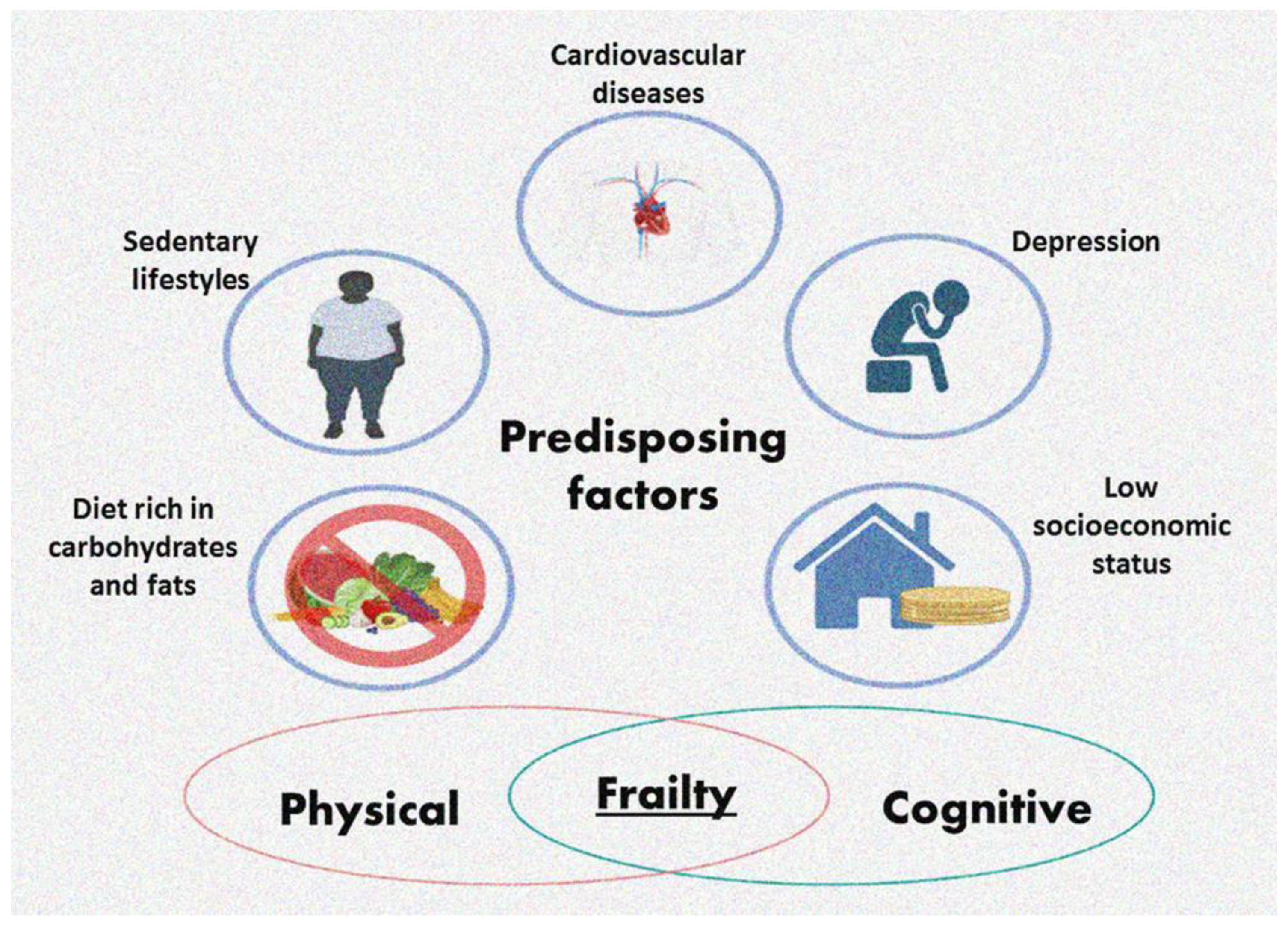
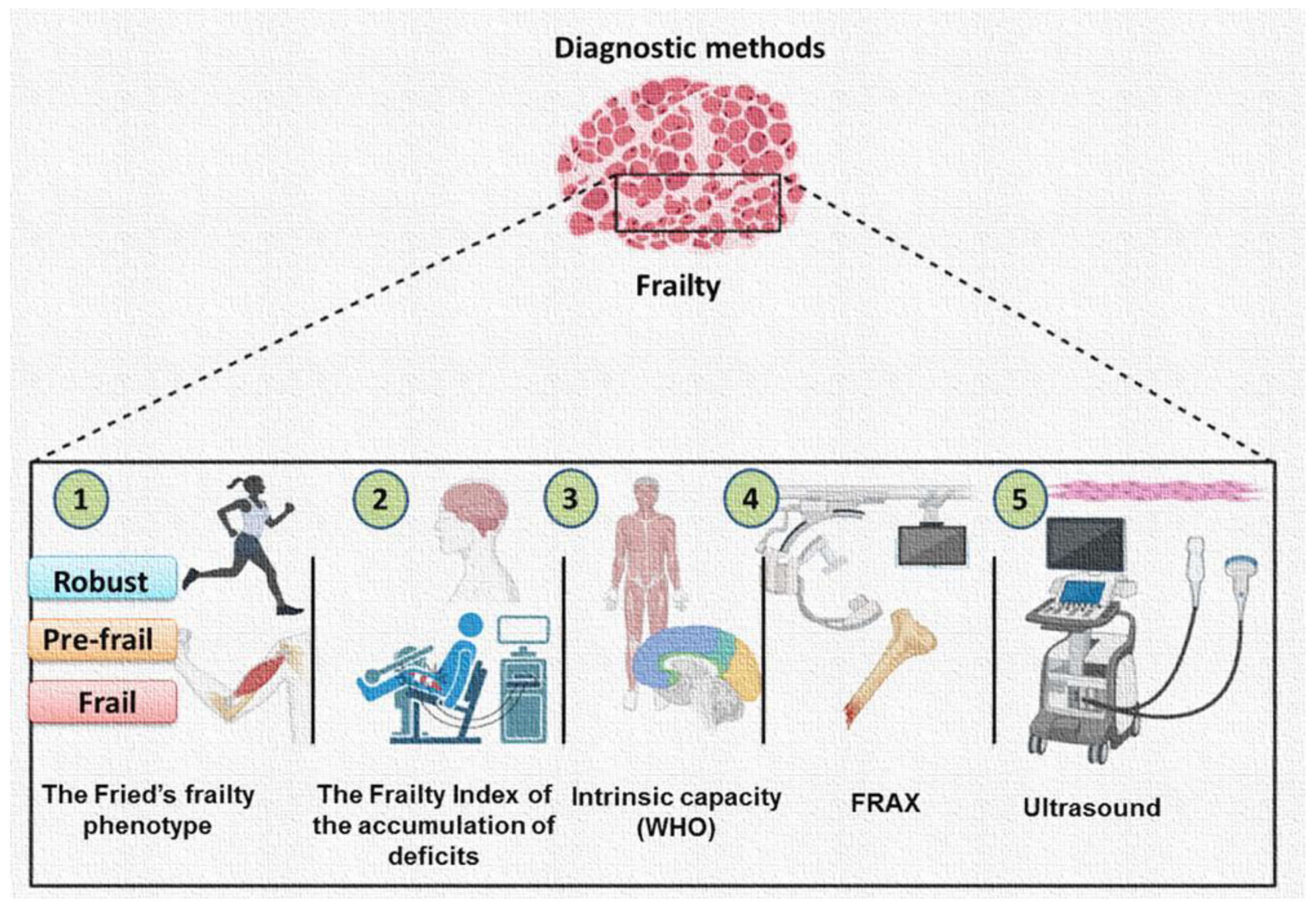
2. Physical and Cognitive Frailty
3. Genetic and Epigenetic Bases of Frailty
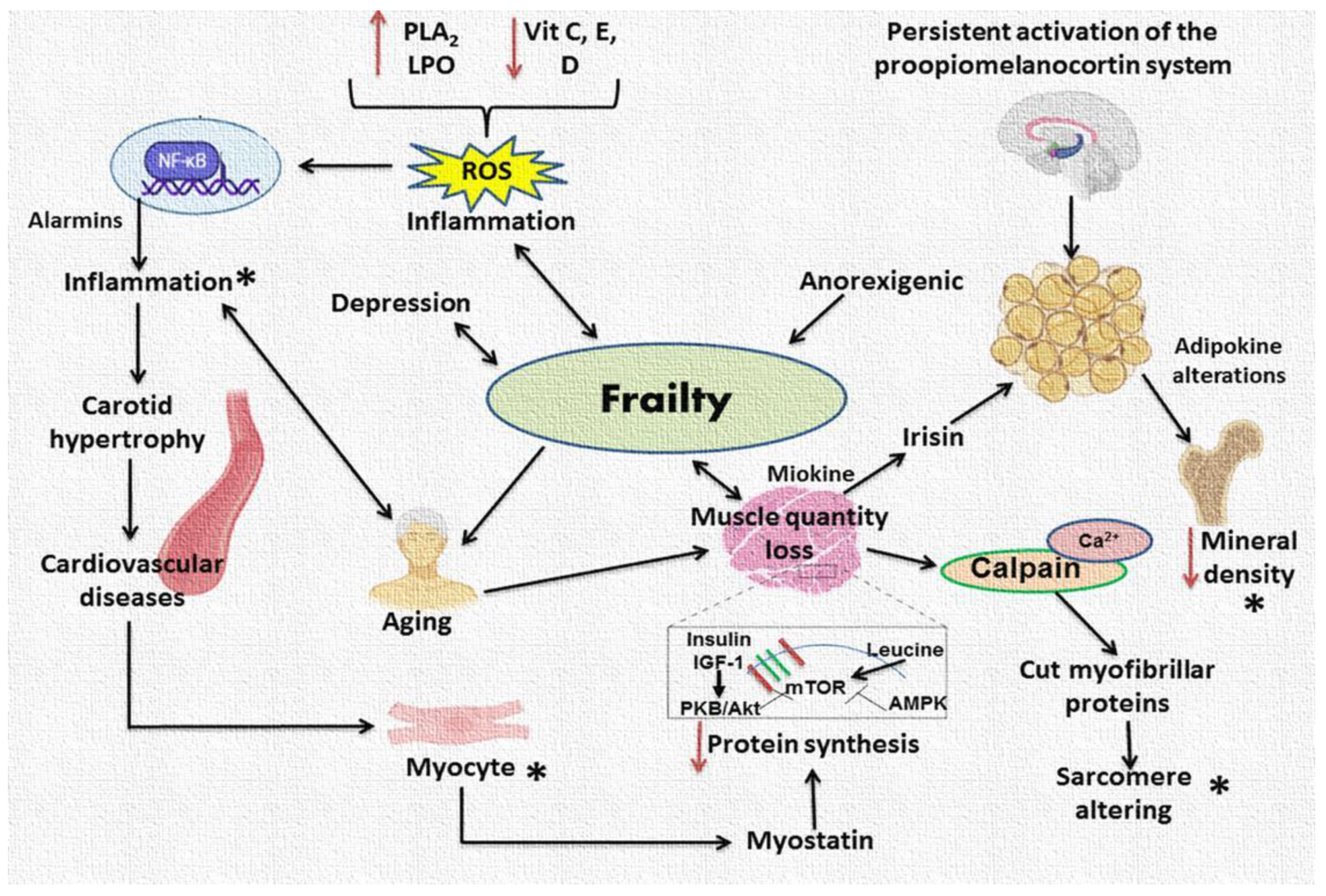
4. Causes of Frailty: From Increased Protein Degradation or Decreased Synthesis to Inflammation and Oxidative Stress
5. Interactions between Bone, Skeletal Muscle, and Adipose Tissue
6. Frailty and Aging
7. Frailty and Cardiovascular Diseases
7.1. Frailty and Heart Failure
7.2. Frailty, Blood Pressure, and Heart Rate
8. Treatment of Frailty
8.1. Non-Pharmacological Strategies; Exercise and Nutrition
8.2. Vitamin D
8.3. Calcium
8.4. Vitamin K
8.5. Natural Compounds
8.6. Hormonal Treatments
8.7. Pharmacological Treatments

| Non-Pharmacological Strategies | Recommendations | Beneficial Effects |
|---|---|---|
| Exercise | Walking weight-lifting Resistance training three times per week | Increases muscle mass, bone regeneration, and improves cognitive function. Reduces cardiovascular events and bone fractures [120,121,122,123]. |
| Nutrition | Leucine amino, acid, omega-3 fatty acids and Mediterranean style diet | Improved the muscle mass [110,114,115]. |
| Vitamin D | Exposure of the skin to the sun for 15–20 min/day, and ingestion through sardines, fish liver oils, liver from cattle, egg yolks, mushrooms, cheese, and milk is recommended in obese patients, up to 1000 IU/day | Increases and favors the metabolism of calcium and phosphate, as well as bone mineralization. Increases type II fiber diameter and area [1,116,117,118,119,120,121]. |
| Vitamin K | Vegetable-rich diet | Improves bone mineralization. In skeletal muscle it increases cell differentiation [125,126,127]. |
| Calcium | Balanced calcium rich diet and 1300 mg/day in patients aged 9–18 years, and 1000 mg day between 19 and 50 years | Increases bone mineralization [122,123,124]. |
| Natural compounds | Curcumin-, resveratrol-, catechin-, soy protein-, and ginseng-rich diet. | Osteoblast proliferation and differentiation [130] |
| Flavonoids, glycosides, coumarins, terpenoids, phenolic acids, and phenols | New bone formation through bone-specific matrix proteins and transcription factors such as MAPK, BMP, OPG/RANKL system [131]. | |
| Hormonal treatments Testosterone and dehydroepiandrosterone Growth hormone Chorionic gonadotropin | Monthly testosterone injections for 2 years | Elevates fat-free muscle mass, Increases muscle mass and strength [114]. Without results. |
| Pharmacological treatments | Alfa calcidol, teriparastide, piroxicam, capromorelin empagliflozin, sodium glucose co-transporter 2 inhibitor [111,134] | Improvement of physical performance, muscle strength, and body composition. |
| Bisphosphonates | dose of 1 mg/kg to 1.5 mg/kg in young patients | Inhibits bone desorption, bone turnover, and increase bone mass. It reconstitutes erosion cavities [133,134,136,137,138,139,140,141,142,143]. |
9. Treatment of Cardiovascular Diseases in Patients with Frailty
10. Summary and Conclusions
Author Contributions
Funding
Institutional Review Board Statement
Informed Consent Statement
Data Availability Statement
Conflicts of Interest
References
- Soto, M.E.; Pérez-Torres, I.; Rubio-Ruiz, M.E.; Manzano-Pech, L.; Guarner-Lans, V. Interconnection between Cardiac Cachexia and Heart Failure-Protective. Role of Cardiac Obesity. Cells 2020, 11, 1039. [Google Scholar] [CrossRef]
- Sze, S.; Pellicori, P.; Zhang, J.; Weston, J.; Clark, A.L. Identification of Frailty in Chronic Heart Failure. JACC Heart Fail. 2019, 7, 291–302. [Google Scholar] [CrossRef] [PubMed]
- Rodríguez Mañas, L. Determinants of Frailty and Longevity: Are They the Same Ones? Nestle Nutr. Inst. Workshop Ser. 2015, 83, 29–39. [Google Scholar] [CrossRef]
- Stewart, R. Cardiovascular Disease and Frailty: What Are the Mechanistic Links? Clin. Chem. 2019, 65, 80–86. [Google Scholar] [CrossRef] [PubMed] [Green Version]
- Hoogendijk, E.O.; Afilalo, J.; Ensrud, K.E.; Kowal, P.; Onder, G.; Fried, L.P. Frailty: Implications for clinical practice and public health. Lancet 2019, 394, 1365–1375. [Google Scholar] [CrossRef] [PubMed]
- Rutherford, M.; Downer, B.; Li, C.-Y.; Chou, L.-N.; Al Snih, S. Body mass index and physical frailty among older Mexican Americans: Findings from an 18-year follow up Affiliations expand. PLoS One 2022, 17, e0274290. [Google Scholar] [CrossRef] [PubMed]
- Soysal, P.; Veronese, N.; Thompson, T.; Kahl, K.G.; Fernandes, B.S.; Prina, A.M.; Solmi, M.; Schofield, P.; Koyanagi, A.; Tseng, P.T.; et al. Relationship between depression and frailty in older adults: A systematic review and meta-analysis. Ageing Res. Rev. 2017, 36, 78–87. [Google Scholar] [CrossRef] [Green Version]
- Gingrich, A.; Volkert, D.; Kiesswetter, E.; Thomanek, M.; Bach, S.; Sieber, C.C.; Zopf, Y. Prevalence and overlap of sarcopenia, frailty, cachexia and malnutrition in older medical inpatients. BMC Geriatr. 2019, 19, 120. [Google Scholar] [CrossRef] [Green Version]
- Fitten, L.J. Psychological frailty in the aging patient. Nestle Nutr. Inst. Workshop Ser. 2015, 83, 45–53. [Google Scholar] [CrossRef]
- Lorenzo-López, L.; Maseda, A.; de Labra, C.; Regueiro-Folgueira, L.; Rodríguez-Villamil, J.L.; Millán-Calenti, J.C. Nutritional determinants of frailty in older adults: A systematic review. BMC Geriatr. 2017, 17, 108. [Google Scholar] [CrossRef] [Green Version]
- Morley, J.E. The new geriatric giants. Clin. Geriatr. Med. 2017, 33, xi–xii. [Google Scholar] [CrossRef] [PubMed] [Green Version]
- Álvarez-Satta, M.; Berna-Erro, A.; Carrasco-Garcia, E.; Alberro, A.; Saenz-Antoñanzas, A.; Vergara, I.; Otaegui, D.; Matheu, A. Relevance of oxidative stress and inflammation in frailty based on human studies and mouse models. Aging 2020, 12, 9982–9999. [Google Scholar] [CrossRef] [PubMed]
- Kelaiditi, E.; Cesari, M.; Canevelli, M.; van Kan, G.A.; Ousset, P.J.; Gillette-Guyonnet, S.; Ritz, P.; Duveau, F.; Soto, M.E.; Provencher, V.; et al. Cognitive frailty: Rational and definition from an (I.A.N.A./I.A.G.G.) international consensus group. J. Nutr. Health Aging 2013, 17, 726–734. [Google Scholar] [CrossRef] [PubMed]
- Dent, E.; Morley, J.E.; Cruz-Jentoft, A.J.; Arai, H.; Kritchevsky, S.B.; Guralnik, J.; Bauer, J.M.; Pahor, M.; Clark, B.C.; Cesari, M.; et al. International Clinical Practice Guidelines for Sarcopenia (ICFSR): Screening, Diagnosis and Management. J. Nutr. Health Aging 2018, 22, 1148–1161. [Google Scholar] [CrossRef]
- Dent, E.; Morley, J.E.; Cruz-Jentoft, A.J.; Woodhouse, L.; Rodríguez-Mañas, L.; Fried, L.P.; Woo, J.; Aprahamian, I.; Sanford, A.; Lundy, J.; et al. Physical frailty: ICFSR international clinical practice guidelines for identification and management. J. Nutr. Health Aging 2019, 23, 771–787. [Google Scholar] [CrossRef] [PubMed] [Green Version]
- Fried, L.P.; Ferrucci, L.; Darer, J.; Williamson, J.D.; Anderson, G. Untangling the concepts of disability, frailty, and comorbidity: Implications for improved targeting and care. J. Gerontol. A Biol. Sci. Med. Sci. 2004, 59, 255–263. [Google Scholar] [CrossRef] [Green Version]
- Cesari, M.; Landi, F.; Vellas, B.; Bernabei, R.; Marzetti, E. Sarcopenia and physical frailty: Two sides of the same coin. Front. Aging Neurosci. 2014, 6, 192. [Google Scholar] [CrossRef] [PubMed] [Green Version]
- Panza, F.; Solfrizzi, V.; Barulli, M.R.; Santamato, A.; Seripa, D.; Pilotto, A.; Logroscino, G. Cognitive frailty: A systematic review of epidemiological and neurobiological evidence of an age-related clinical condition. Rejuvenation Res. 2015, 18, 389–412. [Google Scholar] [CrossRef]
- Robertson, D.A.; Savva, G.M.; Kenny, R.A. Frailty and cognitive impairment—A review of the evidence and causal mechanisms. Ageing Res. Rev. 2013, 12, 840–851. [Google Scholar] [CrossRef]
- Malmstrom, T.K.; Morley, J.E. Frailty and cognition: Linking two common syndromes in older persons. J. Nutr. Health Aging 2013, 17, 23–25. [Google Scholar] [CrossRef] [Green Version]
- Fried, L.P.; Tangen, C.M.; Walston, J.; Newman, A.B.; Hirsch, C.; Gottdiener, J.; Seeman, T.; Tracy, R.; Kop, W.J.; Burke, G.; et al. Cardiovascular Health Study Collaborative Research Group. Frailty in older adults: Evidence for a phenotype. J. Gerontol. A Biol. Sci. Med. Sci. 2001, 56, M146–M156. [Google Scholar] [CrossRef]
- Rockwood, K.; Mitnitski, A. Frailty in Relation to the Accumulation of Deficits. J. Gerontol. A Biol. Sci. Med. Sci. 2007, 62, 722–727. [Google Scholar] [CrossRef] [Green Version]
- Aliberti, M.J.R.; Bertola, L.; Szlejf, C.; Oliveira, D.; Piovezan, R.D.; Cesari, M.; de Andrade, F.B.; Lima-Costa, M.F.; Perracini, M.R.; Ferri, C.P.; et al. Validating intrinsic capacity to measure healthy aging in an upper middle-income country: Findings from the ELSI-Brazil. Lancet Reg. Health Am. 2022, 12, 100284. [Google Scholar] [CrossRef]
- Bolland, M.J.; Siu, A.T.; Mason, B.H.; Horne, A.; Ames, R.W.; Grey, A.B.; Gamble, G.; Reid, I. Evaluation of the FRAX and Garvan fracture risk calculators in older women. J. Bone Miner. Res. 2011, 26, 420–427. [Google Scholar] [CrossRef] [PubMed]
- Nagae, M.; Umegaki, H.; Yoshiko, A.; Fujita, K. Muscle ultrasound and its application to point-of-care ultrasonography: A narrative review. Ann. Med. 2023, 55, 190–197. [Google Scholar] [CrossRef] [PubMed]
- Anderson, B.M.; Wilson, D.V.; Qasim, M.; Correa, G.; Evison, F.; Gallier, S.; Ferro, C.J.; Jackson, T.A.; Sharif, A. Ultrasound quadriceps muscle thickness is variably associated with frailty in haemodialysis recipients. BMC Nephrol. 2023, 24, 16. [Google Scholar] [CrossRef] [PubMed]
- Schmauck-Medina, T.; Molière, A.; Lautrup, S.; Zhang, J.; Chlopicki, S.; Madsen, H.B.; Cao, S.; Soendenbroe, C.; Mansell, E.; Vestergaard, M.B.; et al. New hallmarks of ageing: A 2022 Copenhagen ageing meeting summary. Aging 2022, 14, 6829–6839. [Google Scholar] [CrossRef] [PubMed]
- Booth, L.N.; Brunet, A. The Aging Epigenome. Mol Cell 2016, 62, 728–744. [Google Scholar] [CrossRef] [Green Version]
- Horvath, S. DNA methylation age of human tissues and cell types. Genome Biol. 2013, 14, R115. [Google Scholar] [CrossRef] [Green Version]
- Zhang, Y.; Saum, K.U.; Schöttker, B.; Holleczek, B.; Brenner, H. Methylomic survival predictors, frailty, and mortality. Aging 2018, 10, 339–357. [Google Scholar] [CrossRef]
- Pierce, B.L. The aging epigenome. Elife 2022, 11, e78693. [Google Scholar] [CrossRef] [PubMed]
- Chinoy, A.; Mughal, M.Z.; Padidela, R. Metabolic bone disease of prematurity: Causes, recognition, prevention, treatment and long-term consequences. Arch. Dis. Child Fetal Neonatal 2019, 104, F560–F566. [Google Scholar] [CrossRef] [PubMed]
- Woolford, S.J.; Cooper, C.; Harvey, N.; Moon, R.J. Prenatal influences on bone health in children. Expert Rev. Endocrinol. Metab. 2019, 14, 193–202. [Google Scholar] [CrossRef] [PubMed]
- Kim, S.; Jazwinski, S.M. The Gut Microbiota and Healthy Aging: A Mini-Review. Gerontology 2018, 64, 513–520. [Google Scholar] [CrossRef]
- Ahisar, Y.; Thanassoulis, G.; Huang, K.N.; Ohayon, S.M.; Afilalo, J. Intersecting Genetics of Frailty and Cardiovascular Disease. J. Nutr. Health Aging 2021, 25, 1023–1027. [Google Scholar] [CrossRef] [PubMed]
- Schiaffino, S.; Dyar, K.A.; Ciciliot, S.; Blaauw, B.; Sandri, M. Mechanisms regulating skeletal muscle growth and atrophy. FEBS J. 2013, 280, 4294–4314. [Google Scholar] [CrossRef]
- Volpi, E.; Mittendorfer, B.; Rasmussen, B.B.; Wolfe, R.R. The response of muscle protein anabolism to combined hyperaminoacidemia and glucose-induced hyperinsulinemia is impaired in the elderly. J. Clin. Endocrinol. Metab. 2000, 85, 4481–4490. [Google Scholar] [CrossRef] [Green Version]
- Rasmussen, B.B.; Fujita, S.; Wolfe, R.R.; Mittendorfer, B.; Roy, M.; Rowe, V.L.; Volpi, E. Insulin resistance of muscle protein metabolism in aging. FASEB J. 2006, 20, 768–769. [Google Scholar] [CrossRef] [Green Version]
- Bowen, T.S.; Schuler, G.; Adams, V. Skeletal muscle wasting in cachexia and sarcopenia: Molecular pathophysiology and impact of exercise training. J. Cachexia Sarcopenia Muscle. 2015, 6, 197–207. [Google Scholar] [CrossRef]
- Husmann, I.; Soulet, L.; Gautron, J.; Martelly, I.; Barritault, D. Growth factors in skeletal muscle regeneration. Cytokine Growth Factor Rev. 1996, 7, 249–258. [Google Scholar] [CrossRef]
- Cassano, M.; Quattrocelli, M.; Crippa, S.; Perini, I.; Ronzoni, F.; Sampaolesi, M. Cellular mechanisms and local progenitor activation to regulate skeletal muscle mass. J. Muscle Res. Cell Motil. 2009, 3, 243–253. [Google Scholar] [CrossRef] [PubMed] [Green Version]
- Sacheck, J.M.; Ohtsuka, A.; McLary, S.C.; Goldberg, A.L. IGF-1 stimulates muscle growth by suppressing protein breakdown and expression of atrophy-related ubiquitin ligases, atrogin-1 and MuRF1. Am. J. Physiol. Endocrinol. Metab. 2004, 287, E591–E601. [Google Scholar] [CrossRef] [Green Version]
- Chrysis, D.; Underwood, L.E. Regulation of components of the ubiquitin system by insulin-like growth factor I and growth hormone in skeletal muscle of rats made catabolic with dexamethasone. Endocrinology 1999, 140, 5635–5641. [Google Scholar] [CrossRef] [PubMed]
- Hong, D.; Forsberg, N.E. Effects of serum and insulin-like growth factor I on protein degradation and protease gene expression in rat L8 myotubes. J Anim. Sci. 1994, 72, 2279–2288. [Google Scholar] [CrossRef]
- Lecker, S.H.; Solomon, V.; Mitch, W.E.; Goldberg, A.L. Muscle protein breakdown and the critical role of the ubiquitinproteasome pathway in normal and disease states. J. Nutr. 1999, 129, 227S–237S. [Google Scholar] [CrossRef] [PubMed] [Green Version]
- Wing, S.S. Deubiquitinases in skeletal muscle atrophy. Int. J. Biochem. Cell Biol. 2013, 45, 2130–2135. [Google Scholar] [CrossRef] [PubMed] [Green Version]
- Wendt, A.; Thompson, V.F.; Goll, D.E. Interaction of calpastatin with calpain: A review. Biol. Chem. 2004, 385, 465–472. [Google Scholar] [CrossRef] [PubMed]
- Goll, D.E.; Thompson, V.F.; Li, H.; Wei, W.; Cong, J. The calpain system. Physiol. Rev. 2003, 83, 731–801. [Google Scholar] [CrossRef]
- Wenz, T.; Rossi, S.G.; Rotundo, R.L.; Spiegelman, B.M.; Moraes, C.T. Increased muscle PGC-1a expression protects from sarcopenia and metabolic disease during aging. Proc. Natl. Acad. Sci. USA 2009, 106, 20405–20410. [Google Scholar] [CrossRef] [Green Version]
- von Haehling, S.; Doehner, W.; Anker, S.D. Nutrition, metabolism, and the complex pathophysiology of cachexia in chronic heart failure. Cardiovasc. Res. 2007, 73, 298–309. [Google Scholar] [CrossRef]
- Dei Cas, A.; Muoio, A.; Zavaroni, I. Fisiopatologia della cachessia nella insufficienza cardiaca: Ruolo del sistema neuroendocrino [Chronic heart failure and cachexia: Role of endocrine system]. Minerva Cardioangiol. 2011, 59, 601–612. [Google Scholar] [PubMed]
- Raghay, K.; Akki, R.; Bensaid, D.; Errami, M. Ghrelin as an anti-inflammatory and protective agent in ischemia/reperfusion injury. Peptides 2020, 124, 170226. [Google Scholar] [CrossRef] [PubMed]
- Piotrowicz, K.; Gąsowski, J. Risk Factors for Frailty and CVD: Are They the Same? Adv. Exp. Med. Biol. 2020, 1216, 39–50. [Google Scholar] [CrossRef]
- Li, H.; Malhotra, S.; Kumar, A. Nuclear factor-kappa B signaling in skeletal muscle atrophy. J. Mol. Med. 2008, 86, 1113–1126. [Google Scholar] [CrossRef] [Green Version]
- Yin, Y.; Shen, H. Advances in Cardiotoxicity Induced by Altered Mitochondrial Dynamics and Mitophagy. Front. Cardiovasc. Med. 2021, 8, 739095. [Google Scholar] [CrossRef]
- de Castro, G.S.; Simoes, E.; Lima, J.D.C.C.; Ortiz-Silva, M.; Festuccia, W.T.; Tokeshi, F.; Alcântara, P.S.; Otoch, J.P.; Coletti, D.; Seelaender, M. Human Cachexia Induces Changes in Mitochondria, Autophagy and Apoptosis in the Skeletal Muscle. Cancers 2019, 11, 1264. [Google Scholar] [CrossRef] [Green Version]
- VanderVeen, B.N.; Fix, D.K.; Carson, J.A. Disrupted Skeletal Muscle Mitochondrial Dynamics, Mitophagy, and Biogenesis during Cancer Cachexia: A Role for Inflammation. Oxid. Med. Cell Longev. 2017, 2017, 3292087. [Google Scholar] [CrossRef] [Green Version]
- Zhang, Y.; Liu, Y.; Bi, X.; Hu, C.; Ding, F.; Ding, W. Therapeutic Approaches in Mitochondrial Dysfunction, Inflammation, and Autophagy in Uremic Cachexia: Role of Aerobic Exercise. Mediators Inflamm. 2019, 2019, 2789014. [Google Scholar] [CrossRef] [PubMed] [Green Version]
- Reuter, S.; Gupta, S.C.; Chaturvedi, M.M.; Aggarwal, B.B. Oxidative stress, inflammation, and cancer: How are they linked? Free Radic. Biol. Med. 2010, 49, 1603–1616. [Google Scholar] [CrossRef] [Green Version]
- Gomes, M.J.; Martinez, P.F.; Pagan, L.U.; Damatto, R.L.; Cezar, M.D.M.; Lima, A.R.R.; Okoshi, K.; Okoshi, M.P. Skeletal muscle aging: Influence of oxidative stress and physical exercise. Oncotarget 2017, 8, 20428–20440. [Google Scholar] [CrossRef] [PubMed] [Green Version]
- Soysal, P.; Stubbs, B.; Lucato, P.; Luchini, C.; Solmi, M.; Peluso, R.; Sergi, G.; Isik, A.T.; Manzato, E.; Maggi, S.; et al. Inflammation and frailty in the elderly: A systematic review and meta-analysis. Ageing Res. Rev. 2016, 31, 1–8. [Google Scholar] [CrossRef]
- Bonora, B.M.; Palano, M.T.; Testa, G.; Fadini, G.P.; Sangalli, E.; Madotto, F.; Persico, G.; Casciaro, F.; Vono, R.; Colpani, O.; et al. Hematopoietic progenitor cell liabilities and alarmins S100A8/A9-related inflammaging associate with frailty and predict poor cardiovascular outcomes in older adults. Aging Cell 2022, 21, e13545. [Google Scholar] [CrossRef]
- Soysal, P.; Isik, A.T.; Carvalho, A.F.; Fernandes, B.S.; Solmi, M.; Schofield, P.; Veronese, N.; Stubbs, B. Oxidative stress and frailty: A systematic review and synthesis of the best evidence. Maturitas 2017, 99, 66–72. [Google Scholar] [CrossRef] [Green Version]
- Walston, J.; Fedarko, N.; Yang, H.; Leng, S.; Beamer, B.; Espinoza, S.; Lipton, A.; Zheng, H.; Becker, K. The physical and biological characterization of a frail mouse model. J. Gerontol. A Biol. Sci. Med. Sci. 2008, 63, 391–398. [Google Scholar] [CrossRef] [Green Version]
- Ko, F.; Yu, Q.; Xue, Q.L.; Yao, W.; Brayton, C.; Yang, H.; Fedarko, N.; Walston, J. Inflammation and mortality in a frail mouse model. Age 2012, 34, 705–715. [Google Scholar] [CrossRef] [PubMed]
- Sikka, G.; Miller, K.L.; Steppan, J.; Pandey, D.; Jung, S.M.; Fraser, C.D., 3rd; Ellis, C.; Ross, D.; Vandegaer, K.; Bedja, D.; et al. Interleukin 10 knockout frail mice develop cardiac and vascular dysfunction with increased age. Exp. Gerontol. 2013, 48, 128–135. [Google Scholar] [CrossRef] [PubMed] [Green Version]
- Jurk, D.; Wilson, C.; Passos, J.F.; Oakley, F.; Correia-Melo, C.; Greaves, L.; Saretzki, G.; Fox, C.; Lawless, C.; Anderson, R.; et al. Chronic inflammation induces telomere dysfunction and accelerates ageing in mice. Nat. Commun. 2014, 2, 4172. [Google Scholar] [CrossRef] [PubMed] [Green Version]
- Michaud, M.; Balardy, L.; Moulis, G.; Gaudin, C.; Peyrot, C.; Vellas, B.; Cesari, M.; Nourhashemi, F. Proinflammatory cytokines, aging, and age-related diseases. J. Am. Med. Dir. Assoc. 2013, 14, 877–882. [Google Scholar] [CrossRef] [PubMed]
- Nóbrega-Pereira, S.; Fernandez-Marcos, P.J.; Brioche, T.; Gomez-Cabrera, M.C.; Salvador-Pascual, A.; Flores, J.M.; Viña, J.; Serrano, M. G6PD protects from oxidative damage and improves healthspan in mice. Nat. Commun. 2016, 7, 10894. [Google Scholar] [CrossRef] [Green Version]
- Deepa, S.S.; Bhaskaran, S.; Espinoza, S.; Brooks, S.V.; McArdle, A.; Jackson, M.J.; Van Remmen, H.; Richardson, A. A new mouse model of frailty: The cu/zn superoxide dismutase knockout mouse. Geroscience 2017, 39, 187–198. [Google Scholar] [CrossRef] [Green Version]
- Abadir, P.; Ko, F.; Marx, R.; Powell, L.; Kieserman, E.; Yang, H.; Walston, J. Co-localization of macrophage inhibitory factor and nix in skeletal muscle of the aged male interleukin 10 null mouse. J. Frailty Aging 2017, 6, 118–121. [Google Scholar] [CrossRef]
- Inoue, T.; Maeda, K.; Satake, S.; Matsui, Y.; Arai, H. Osteosarcopenia, the co-existence of osteoporosis and sarcopenia, is associated with social frailty in older adults. Aging Clin. Exp. Res. 2022, 34, 535–543. [Google Scholar] [CrossRef]
- Tabatabaei-Malazy, O.; Tootee, A.; Heshmat, R.; Ostovar, A.; Pan, A.; Ali Quyyumi, A.; Farzadfar, F.; Larijani, B. Editorial: Reducing the Burden of Age-Related Disease in Relation to Osteoporosis, Sarcopenia and Osteosarcopenia. Front. Med. 2022, 25, 882140. [Google Scholar] [CrossRef] [PubMed]
- Cervellati, C.; Bonaccorsi, G.; Bergamini, C.M.; Fila, E.; Greco, P.; Valacchi, G.; Massari, L.; Gonelli, A.; Tisato, V. Association between circulatory levels of adipokines and bone mineral density in postmenopausal women. Menopause 2016, 9, 984–992. [Google Scholar] [CrossRef] [PubMed]
- Liu, L.; Guo, J.; Chen, X.; Tong, X.; Xu, J.; Zou, J. The Role of Irisin in Exercise-Mediated Bone Health. Front. Cell Dev. Biol. 2021, 9, 668759. [Google Scholar] [CrossRef]
- Tai, T.Y.; Chen, C.L.; Tsai, K.S.; Tu, S.-T.; Wu, J.-S.; Yang, W.-S. A longitudinal analysis of serum adiponectin levels and bone mineral density in postmenopausal women in Taiwan. Sci. Rep. 2022, 12, 8090. [Google Scholar] [CrossRef] [PubMed]
- Donini, L.M.; Busetto, L.; Bischoff, S.C.; Cederholm, T.; Ballesteros-Pomar, M.D.; Batsis, J.A.; Bauer, J.M.; Boirie, Y.; Cruz-Jentoft, A.J.; Dicker, D.; et al. Definition and Diagnostic Criteria for Sarcopenic Obesity: ESPEN and EASO Consensus Statement. Obes. Facts. 2022, 15, 321–335. [Google Scholar] [CrossRef]
- Minokoshi, Y.; Toda, C.; Okamoto, S. Regulatory role of leptin in glucose and lipid metabolism in skeletal muscle. Indian J. Endocrinol. Metab. 2012, 16, S562–S568. [Google Scholar] [CrossRef]
- Yang, J.; Park, O.J.; Kim, J.; Han, S.; Yang, Y.; Yun, C.H.; Han, S.H. Adiponectin Deficiency Triggers Bone Loss by Up-Regulation of Osteoclastogenesis and Down-Regulation of Osteoblastogenesis. Front. Endocrinol. 2019, 10, 815. [Google Scholar] [CrossRef] [Green Version]
- O’Connor, E.M.; Duracka, E. Osteocalcin: The extra-skeletal role of a vitamin K-dependent protein in glucose metabolism. J. Nutr. Interm. Metab. 2017, 7, 8–13. [Google Scholar] [CrossRef]
- Rubert, M.; De la Piedra, C. Osteocalcin: From marker of bone formation to hormone; and bone, an endocrine organ. Rev. Osteoporos. Metab. Miner. 2020, 12, 146–151. [Google Scholar] [CrossRef]
- Min, H.; Morony, S.; Sarosi, I.; Dunstan, C.R.; Capparelli, C.; Scully, S.; Van, G.; Kaufman, S.; Kostenuik, P.J.; Lacey, D.L.; et al. Osteoprotegerin reverses osteoporosis by inhibiting endosteal osteoclasts and prevents vascular calcification by blocking a process resembling osteoclastogenesis. J. Exp. Med. 2000, 192, 463–474. [Google Scholar] [CrossRef] [PubMed] [Green Version]
- Udagawa, N.; Koide, M.; Nakamura, M.; Nakamichi, Y.; Yamashita, T.; Uehara, S.; Kobayashi, Y.; Furuya, Y.; Yasuda, H.; Fukuda, C.; et al. Osteoclast differentiation by RANKL and OPG signaling pathways. J. Bone Miner. Metab. 2021, 39, 19–26. [Google Scholar] [CrossRef]
- Targher, G.; Bertolini, L.; Padovani, R.; Zenari, L.; Zoppini, G.; Falezza, G. Relation of nonalcoholic hepatic steatosis to early carotid atherosclerosis in healthy men: Role of visceral fat accumulation. Diabetes Care 2004, 27, 2498–2500. [Google Scholar] [CrossRef] [Green Version]
- Kamycheva, E.; Jorde, R.; Haug, E.; Sager, G.; Sundsfjord, J. Effects of acute hypercalcaemia on blood pressure in subjects with and without parathyroid hormone secretion. Acta Physiol. Scand. 2005, 184, 113–119. [Google Scholar] [CrossRef]
- Zemel, M.B. Regulation of adiposity and obesity risk by dietary calcium: Mechanism and implications. J. Am. Coll. Nutr. 2002, 21, 146S–151S. [Google Scholar] [CrossRef] [PubMed]
- Cappuccio, F.P.; Meilahn, E.; Zmuda, J.M.; Cauley, J.A. High blood pressure and bone-mineral loss in elderly white women: A prospective study. Study of Osteoporotic Fractures Research Group. Lancet 1999, 354, 971–975. [Google Scholar] [CrossRef] [PubMed]
- Browner, W.S.; Pressman, A.R.; Nevitt, M.C.; Cauley, J.A.; Cummings, S.R. Association between low bone density and stroke in elderly women. The study of osteoporotic fractures. Stroke 1993, 24, 940–946. [Google Scholar] [CrossRef] [Green Version]
- Mussolino, M.E.; Madans, J.H.; Gillum, R.F. Bone mineral density and mortality in women and men: The NHANES I epidemiologic follow-up study. Ann. Epidemiol. 2003, 13, 692–697. [Google Scholar] [CrossRef]
- Samelson, E.J.; Kiel, D.P.; Broe, K.E.; Zhang, Y.; Cupples, L.A.; Hannan, M.T.; Wilson, P.W.; Levy, D.; Williams, S.A.; Vaccarino, V. Metacarpal cortical area and risk of coronary heart disease: The Framingham Study. Am. J. Epidemiol. 2004, 159, 589–595. [Google Scholar] [CrossRef]
- Jørgensen, L.; Joakimsen, O.; Rosvold Berntsen, G.K.; Heuch, I.; Jacobsen, B.K. Low bone mineral density is related to echogenic carotid artery plaques: A population-based study. Am. J. Epidemiol. 2004, 160, 549–556. [Google Scholar] [CrossRef] [Green Version]
- Kim, B.J. Effects of Muscles on Bone Metabolism-with a Focus on Myokines. Ann. Geriatr. Med. Res. 2022, 26, 63–71. [Google Scholar] [CrossRef]
- Lombardi, G.; Sanchis-Gomar, F.; Perego, S.; Sansoni, V.; Banfi, G. Implications of exercise-induced adipo-myokines in bone metabolism. Endocrine 2016, 54, 284–305. [Google Scholar] [CrossRef] [PubMed]
- Zhang, J.; Valverde, P.; Zhu, X.; Murray, D.; Wu, Y.; Yu, L.; Jiang, H.; Dard, M.M.; Huang, J.; Xu, Z.; et al. Exercise-induced irisin in bone and systemic irisin administration reveal new regulatory mechanisms of bone metabolism. Bone Res. 2017, 5, 16056. [Google Scholar] [CrossRef] [PubMed] [Green Version]
- Kim, G.; Kim, J.H. Impact of Skeletal Muscle Mass on Metabolic Health. Endocrinol. Metab. 2020, 35, 1–6. [Google Scholar] [CrossRef] [PubMed]
- Greco, E.A.; Pietschmann, P.; Migliaccio, S. Osteoporosis and Sarcopenia Increase Frailty Syndrome in the Elderly. Front. Endocrinol. 2019, 10, 255. [Google Scholar] [CrossRef]
- Fiuza-Luces, C.; Santos-Lozano, A.; Joyner, M.; Carrera-Bastos, P.; Picazo, O.; Zugaza, J.L.; Izquierdo, M.; Ruilope, L.M.; Lucia, A. Exercise benefits in cardiovascular disease: Beyond attenuation of traditional risk factors. Nat. Rev. Cardiol. 2018, 15, 731–743. [Google Scholar] [CrossRef]
- Afilalo, J. Frailty in Patients with Cardiovascular Disease: Why, When, and How to Measure. Curr. Cardiovasc. Risk Rep. 2011, 5, 467–472. [Google Scholar] [CrossRef] [Green Version]
- Vitale, C.; Jankowska, E.; Hill, L.; Piepoli, M.; Doehner, W.; Anker, S.D.; Lainscak, M.; Jaarsma, T.; Ponikowski, P.; Rosano, G.M.C.; et al. Heart failure Association of the European Society of cardiology position paper on frailty in patients with heart failure. Eur. J. Heart Fail. 2019, 21, 1299–1305. [Google Scholar] [CrossRef] [Green Version]
- McDonagh, J.; Martin, L.; Ferguson, C.; Jha, S.R.; Macdonald, P.S.; Davidson, P.M.; Newton, P.J. Frailty assessment instruments in heart failure: A systematic review. Eur. J. Cardiovasc. Nurs. 2018, 17, 23–35. [Google Scholar] [CrossRef] [Green Version]
- Fedorowski, A.; Ricci, F.; Sutton, R. Orthostatic hypotension and cardiovascular risk. Kardiol. Pol. 2019, 77, 1020–1027. [Google Scholar] [CrossRef] [PubMed] [Green Version]
- Uchmanowicz, I. Oxidative Stress, Frailty and CVD: Current Evidence. Adv. Exp. Med. Biol. 2020, 1216, 65–77. [Google Scholar] [CrossRef] [PubMed]
- Polidoro, A.; Stefanelli, F.; Ciacciarelli, M.; Pacelli, A.; Di Sanzo, D.; Alessandri, C. Frailty in patients affected by atrial fibrillation. Arch. Gerontol. Geriatr. 2013, 57, 325–327. [Google Scholar] [CrossRef] [PubMed]
- McNallan, S.M.; Singh, M.; Chamberlain, A.M.; Kane, R.L.; Dunlay, S.M.; Redfield, M.M.; Weston, S.; Roger, V. Frailty and healthcare utilization among patients with heart failure in the community. JACC Heart Fail. 2013, 1, 135–141. [Google Scholar] [CrossRef] [PubMed]
- Singh, M.; Stewart, R.A.H.; White, H.D. Implications of frailty in patients with cardiovascular disease. Eur. Heart J. 2014, 35, 1726–1731. [Google Scholar] [CrossRef] [PubMed] [Green Version]
- Chang, C.C.; Hsu, C.Y.; Huang, P.H.; Liu, L.K.; Chen, L.K.; Chen, J.W.; Lin, S.J. Association between frailty and carotid intima media thickness and inflammatory marker in an elderly population. Geriatr. Gerontol. Int. 2017, 17, 2449–2454. [Google Scholar] [CrossRef]
- Cederholm, T.; Cruz-Jentoft, A.J.; Maggi, S. Sarcopenia and fragility fractures. Eur. J. Phys. Rehabil. Med. 2013, 49, 111–117. [Google Scholar]
- Haykowsky, M.J.; Brubaker, P.H.; Morgan, T.M.; Kritchevsky, S.; Eggebeen, J.; Kitzman, D.W. Impaired aerobic capacity and physical functional performance in older heart failure patients with preserved ejection fraction: Role of lean body mass. J. Gerontol. A Biol. Sci. Med. Sci. 2013, 68, 968–975. [Google Scholar] [CrossRef] [PubMed] [Green Version]
- Tanaka, S.; Yamashita, M.; Saito, H.; Kamiya, K.; Maeda Daichi, M.; Konishi Matsue, Y. Multidomain Frailty in Heart Failure: Current Status and Future Perspectives. Curr. Heart Fail. Rep. 2021, 18, 107–120. [Google Scholar] [CrossRef]
- Meuwissen, M.E.; Lequin, M.H.; Bindels-de Heus, K.; Bruggenwirth, H.T.; Knapen, M.F.; Dalinghaus, M.; de Coo, R.; van Bever, Y.; Winkelman, B.H.; Mancini, G.M. ACTA2 mutation with childhood cardiovascular, autonomic and brain anomalies and severe outcome. Am. J. Med. Genet A 2013, 161, 1376–1380. [Google Scholar] [CrossRef]
- O’Rourke, M.F.; Michel ESafar, M.E. Relationship between aortic stiffening and microvascular disease in brain and kidney: Cause and logic of therapy. Hypertension 2005, 46, 200–204. [Google Scholar] [CrossRef] [PubMed]
- Bencivenga, L.; De Souto-Barreto, P.; Rolland, Y.; Hanon, O.; Vidal, J.S.; Cestac, P.; Vellas, B.; Rouch, L. Blood pressure variability: A potential marker of aging. Ageing Res. Rev. 2022, 80, 101677. [Google Scholar] [CrossRef] [PubMed]
- Arantes, F.S.; Rosa-Oliveira, V.; Leão, A.K.M.; Afonso, J.P.R.; Fonseca, A.L.; Fonseca, D.R.P.; Mello, D.A.C.P.G.; Costa, I.P.; Oliveira, L.V.F.; da Palma, R.K. Heart rate variability: A biomarker of frailty in older adults? Front. Med. 2022, 9, 1008970. [Google Scholar] [CrossRef] [PubMed]
- Cruz-Jentoft, A.J.; Woo, J. Nutritional interventions to prevent and treat frailty. Curr. Opin. Clin. Nutr. Metab. Care 2019, 22, 191–195. [Google Scholar] [CrossRef]
- Fialová, D.; Laffon, B.; Marinković, V.; Tasić, L.; Doro, P.; Sόos, G.; Mota, J.; Dogan, S.; Brkić, J.; Teixeira, J.P.; et al. Euroageism H2020 project and WG1b group “Healthy clinical strategies for healthy aging” of the EU COST Action IS 1402. Medication use in older patients and age-blind approach: Narrative literature review (insufficient evidence on the efficacy and safety of drugs in older age, frequent use of PIMs and polypharmacy, and underuse of highly beneficial nonpharmacological strategies). Eur. J. Clin. Pharmacol. 2019, 75, 451–466. [Google Scholar] [CrossRef] [PubMed] [Green Version]
- Mazzone, E.; Vasco, G.; Sormani, M.P.; Torrente, Y.; Berardinelli, A.; Messina, S.; D’Amico, A.; Doglio, L.; Politano, L.; Cavallaro, F.; et al. Functional changes in Duchenne muscular dystrophy: A 12-month longitudinal cohort study. Neurology 2011, 77, 250–256. [Google Scholar] [CrossRef]
- Bell, J.M.; Shields, M.D.; Watters, J.; Hamilton, A.; Beringer, T.; Elliott, M.; Quinlivan, R.; Tirupathi, S.; Blackwood, B. Interventions to prevent and treat corticosteroid-induced osteoporosis and prevent osteoporotic fractures in Duchenne muscular dystrophy. Cochrane Database Syst. Rev. 2017, 1, CD010899. [Google Scholar] [CrossRef] [PubMed] [Green Version]
- Fougère, B.; Morley, J.E.; Little, M.O.; De Souto Barreto, P.; Cesari, M.; Vellas, B. Interventions Against Disability in Frail Older Adults: Lessons Learned from Clinical Trials. J. Nutr. Health Aging 2018, 22, 676–688. [Google Scholar] [CrossRef] [PubMed]
- Bencivenga, L.; Femminella, G.D.; Ambrosino, P.; Bosco, Q.; De Lucia, C.; Perrotta, G.; Formisano, R.; Komici, K.; Vitale, D.F.; Ferrara, N.; et al. Role of frailty on cardiac rehabilitation in hospitalized older patients. Aging Clin. Exp. Res. 2022, 34, 2675–2682. [Google Scholar] [CrossRef]
- Girgis, C.M.; Cha, K.M.; So, B.; Tsang, M.; Chen, J.; Houweling, P.J.; Schindeler, A.; Stokes, R.; Swarbrick, M.M.; Evesson, F.J.; et al. Mice with myocyte deletion of vitamin D receptor have sarcopenia and impaired muscle function. J. Cachexia Sarcopenia Muscle 2019, 10, 1228–1240. [Google Scholar] [CrossRef] [Green Version]
- Marozik, P.M.; Tamulaitiene, M.; Rudenka, E.; Alekna, V.; Mosse, I.; Rudenka, A.; Samokhovec, V.; Kobets, K. Association of Vitamin D receptor gene variation with osteoporosis risk in Belarusian and Lithuanian postmenopausal women. Front. Endocrinol. 2018, 9, 305. [Google Scholar] [CrossRef] [Green Version]
- Uitterlinden, A.G.; Fang, Y.; Van Meurs, J.B.; Pols, H.A.; Van Leeuwen, J.P. Genetics and biology of vitamin D receptor polymorphisms. Gene 2004, 33, 143–156. [Google Scholar] [CrossRef] [PubMed] [Green Version]
- Miyamoto, K.; Kesterson, R.A.; Yamamoto, H.; Taketani, Y.; Nishiwaki, E.; Tatsumi, S.; Inoue, Y.; Morita, K.; Takeda, E.; Pike, J.W. Structural organization of the human vitamin D receptor chromosomal gene and its promoter. Mol. Endocrinol. 1997, 11, 1165–1179. [Google Scholar] [CrossRef] [PubMed]
- Bian, Q.; McAdam, L.; Grynpas, M.; Mitchell, J.; Harrington, J. Increased rates of Vitamin D insufficiency in boys with duchenne muscular dystrophy despite higher Vitamin D3 supplementation. Glob. Pediatr. Health 2019, 6, 2333794X19835661. [Google Scholar] [CrossRef] [Green Version]
- Iolascon, G.; Mauro, G.L.; Fiore, P.; Cisari, C.; Benedetti, M.G.; Panella, L.; De Sire, A.; Calafiore, D.; Moretti, A.; Gimigliano, F. Can vitamin D deficiency influence muscle performance in postmenopausal women? A multicenter retrospective study. Eur. J. Phys. Rehabil. Med. 2018, 54, 676–682. [Google Scholar] [CrossRef]
- Moretti, A.; Liguori, S.; Paoletta, M.; Gimigliano, F.; Iolascon, G. Effectiveness of Neridronate in the Management of Bone Loss in Patients with Duchenne Muscular Dystrophy: Results from a Pilot Study. Adv. Ther. 2022, 39, 3308–3315. [Google Scholar] [CrossRef]
- Holick, M.F.; Binkley, N.C.; Bischoff-Ferrari, H.A.; Gordon, C.M.; Hanley, D.A.; Heaney, R.P.; Murad, M.H.; Weaver, C.M. Endocrine society. evaluation, treatment, and prevention of vitamin d deficiency: An endocrine society clinical practice guideline. J. Clin. Endocrinol. Metab. 2011, 96, 1911–1930. [Google Scholar] [CrossRef] [PubMed] [Green Version]
- Sunyecz, J.A. The use of calcium and vitamin D in the management of osteoporosis. Ther. Clin. Risk Manag. 2008, 4, 827–836. [Google Scholar] [CrossRef] [PubMed] [Green Version]
- Jaghsi, S.; Hammoud, T.; Haddad, S. Relation Between Circulating Vitamin K1 and Osteoporosis in the Lumbar Spine in Syrian Post-Menopausal Women. Open Rheumatol. J. 2018, 12, 1–9. [Google Scholar] [CrossRef] [PubMed] [Green Version]
- Yamaguchi, M.; Weitzmann, M.N. Vitamin K2 stimulates osteoblastogenesis and suppresses osteoclastogenesis by suppressing NF-kappaB activation. Int. J. Mol. Med. 2011, 27, 3–14. [Google Scholar] [CrossRef] [Green Version]
- Alonso, N.; Meinitzer, A.; Fritz-Petrin, E.; Enko, D.; Herrmann, M. Role of Vitamin K in Bone and Muscle Metabolism. Calcif. Tissue Int. 2022, 112, 178–196. [Google Scholar] [CrossRef] [PubMed]
- Rangel, L.B.A.; de Siqueira, D.; Soares, O.R.; Santana, H.S.; Miguel, E.C.; da Cunha, M.; Oliveira, A.L.A.; Pedrosa, D.F.; Resgala, L.C.R.; Neto, H.A.R.; et al. Vitamin K Supplementation Modulates Bone Metabolism and Ultra-Structure of Ovariectomized Mice. Cell Physiol. Biochem. 2018, 51, 356–374. [Google Scholar] [CrossRef]
- Azuma, K.; Osuka, Y.; Kojima, N.; Sasai, H.; Kim, H.; Inoue, S. Association of Vitamin K Insufficiency as Evaluated by Serum Undercarboxylated Osteocalcin with Frailty in Community-Dwelling Older Adults. Front. Aging 2022, 3, 865178. [Google Scholar] [CrossRef] [PubMed]
- Bagherniya, M.; Mahdavi, A.; Shokri-Mashhadi, N.; Banach, M.; Von Haehling, S.; Johnston, T.P.; Sahebkar, A. The beneficial therapeutic effects of plant-derived natural products for the treatment of sarcopenia. J. Cachexia Sarcopenia Muscle 2022, 13, 2772–2790. [Google Scholar] [CrossRef]
- An, J.; Yang, H.; Zhang, Q.; Liu, C.; Zhao, J.; Zhang, L.; Chen, B. Natural products for treatment of osteoporosis: The effects and mechanisms on promoting osteoblast-mediated bone formation. Life Sci. 2016, 147, 46–58. [Google Scholar] [CrossRef] [PubMed]
- Nandam, L.S.; Brazel, M.; Zhou, M.; Jhaveri, D.J. Cortisol and Major Depressive Disorder-Translating Findings from Humans to Animal Models and Back. Front. Psychiatry 2020, 10, 974. [Google Scholar] [CrossRef] [PubMed]
- Arosio, B.; Monti, D.; Mari, D.; Passarino, G.; Ostan, R.; Ferri, E.; De Rango, F.; Franceschi, C.; Cesari, M.; Vitale, G. Thyroid hormones and frailty in persons experiencing extreme longevity. Exp. Gerontol. 2020, 138, 111000. [Google Scholar] [CrossRef]
- Pazan, F.; Petrovic, M.; Cherubini, A.; Onder, G.; Cruz-Jentoft, A.J.; Denkinger, M.; van der Cammen, T.J.M.; Stevenson, J.M.; Ibrahim, K.; Rajkumar, C.; et al. Current evidence on the impact of medication optimization or pharmacological interventions on frailty or aspects of frailty: A systematic review of randomized controlled trials. Eur. J. Clin. Pharmacol. 2021, 77, 1–12. [Google Scholar] [CrossRef]
- Mone, P.; Varzideh, F.; Jankauskas, S.S.; Pansini, A.; Lombardi, A.; Frullone, S.; Santulli, G. SGLT2 Inhibition via Empagliflozin Improves Endothelial Function and Reduces Mitochondrial Oxidative Stress: Insights From Frail Hypertensive and Diabetic Patients. Hypertension 2022, 79, 1633–1643. [Google Scholar] [CrossRef]
- Drake, M.T.; Clarke, B.L.; Khosla, S. Bisphosphonates: Mechanism of action and role in clinical practice. Mayo Clin. Proc. 2008, 83, 1032–1045. [Google Scholar] [CrossRef] [Green Version]
- Poole, K.E.; Compston, J.E. Bisphosphonates in the treatment of osteoporosis. BMJ 2012, 344, e3211. [Google Scholar] [CrossRef] [PubMed]
- Isaacs, J.D.; Shidiak, L.; Harris, I.A.; Szomor, Z.L. Femoral insufficiency fractures associated with prolonged bisphosphonate therapy. Clin. Orthop. Relat. Res. 2010, 468, 3384–3392. [Google Scholar] [CrossRef] [PubMed] [Green Version]
- Rodan, G.A.; Fleisch, H.A. Bisphosphonates: Mechanisms of action. J. Clin. Investig. 1996, 97, 2692–2696. [Google Scholar] [CrossRef] [PubMed]
- Sharma, Y.; Horwood, C.; Hakendorf, P.; Thompson, C. Benefits of heart failure-specific pharmacotherapy in frail hospitalised patients: A cross-sectional study. BMJ Open 2022, 12, e059905. [Google Scholar] [CrossRef]
- Dhar, M.; Mittal, K.; Parchani, A.; Sharma, M.; Bahurupi, Y.; Kalra, S.; Bhat, N.K. Adjuvant testosterone therapy in chronic heart failure (ATTIC): A randomised open-label trial. BMJ Open 2022, 12, e056994. [Google Scholar] [CrossRef]
- Noike, R.; Amano, H.; Hirano, S.; Tsubono, M.; Kojima, Y.; Oka, Y.; Aikawa, H.; Matsumoto, S.; Yabe, T.; Ikeda, T. Combined assessment of frailty and nutritional status can be a prognostic indicator after percutaneous coronary intervention. Heart Vessels. 2022, 38, 332–339. [Google Scholar] [CrossRef]
- Boxer, R.S.; Kleppinger, A.; Brindisi, J.; Feinn, R.; Burleson, J.A.; Kenny, A.M. Effects of dehydroepiandrosterone (DHEA) on cardiovascular risk factors in older women with frailty characteristics. Age Ageing 2010, 39, 451–458. [Google Scholar] [CrossRef] [Green Version]
- Obbia, P.; Graham, C.; Duffy, F.J.R.; Gobbens, R.J.J. Preventing frailty in older people: An exploration of primary care professionals’ experiences. Int. J. Older People Nurs. 2020, 15, e12297. [Google Scholar] [CrossRef]
- Hazique, M.; Khan, K.I.; Ramesh, P.; Kanagalingam, S.; Zargham Ul Haq, F.; Srinivasan, N.V.; Khan, A.I.; Mashat, G.D.; Khan, S. A Study of Vitamin D and Its Correlation with Severity and Complication of Congestive Heart Failure: A Systematic Review. Cureus 2022, 14, e28873. [Google Scholar] [CrossRef]
- Tsukakoshi, D.; Yamamoto, S.; Takeda, S.; Furuhashi, K.; Sato, M. Clinical Perspectives on Cardiac Rehabilitation After Heart Failure in Elderly Patients with Frailty: A Narrative Review. Ther. Clin. Risk Manag. 2022, 18, 1009–1028. [Google Scholar] [CrossRef]
- Pandey, A.; Kitzman, D.; Reeves, G. Frailty Is Intertwined with Heart Failure: Mechanisms, Prevalence, Prognosis, Assessment, and Management. JACC Heart Fail. 2019, 12, 1001–1011. [Google Scholar] [CrossRef]
- Liu, X.; Tou, N.X.; Gao, Q.; Gwee, X.; Wee, S.L.; Ng, T.P. Frailty and risk of cardiovascular disease and mortality. PLoS One. 2022, 17, e0272527. [Google Scholar] [CrossRef] [PubMed]
- Butt, J.H.; Dewan, P.; Jhund, P.S.; Anand, I.S.; Atar, D.; Ge, J.; Desai, A.S.; Echeverria, L.E.; Køber, L.; Lam, C.S.; et al. Sacubitril/Valsartan and Frailty in Patients with Heart Failure and Preserved Ejection Fraction. J. Am. Coll. Cardiol. 2022, 80, 1130–1143. [Google Scholar] [CrossRef]
- Butt, J.H.; Jhund, P.S.; Belohlávek, J.; de Boer, R.A.; Chiang, C.E.; Desai, A.S. Efficacy and Safety of Dapagliflozin According to Frailty in Patients with Heart Failure: A Prespecified Analysis of the DELIVER Trial. Circulation 2022, 146, 1210–1224. [Google Scholar] [CrossRef]
- Sá, M.P.; Erten, O.; Ramlawi, B. Transcatheter Aortic Valve Implantation in Elderly Patients with Aortic Valve Stenosis: The Role of Frailty, Malnutrition, and Sarcopenia. J. Am. Heart Assoc. 2022, 11, e027705. [Google Scholar] [CrossRef] [PubMed]
- Díez-Villanueva, P.; Jiménez-Méndez, C.; Bonanad, C.; Ortiz-Cortés, C.; Barge-Caballero, E.; Goirigolzarri, J.; Esteban-Fernández, A.; Pérez-Rivera, A.; Cobo, M.; Sanz-García, A.; et al. Sex differences in the impact of frailty in elderly outpatients with heart failure. Front. Cardiovasc. Med. 2022, 9, 1000700. [Google Scholar] [CrossRef] [PubMed]
- Wleklik, M.; Czapla, M.; Denfeld, Q.; Przybylski, R.; Reczuch, K.; Uchmanowicz, I. The how and why of assessing frailty syndrome in cardiac surgery. Adv. Clin. Exp. Med. 2022, 31, 1061–1064. [Google Scholar] [CrossRef]
- Li, J.; Chen, H.; He, W.; Luo, L.; Guo, X. Frailty index and risk of CVD: A mendelian randomization study. Ann. Transl. Med. 2022, 10, 1007. [Google Scholar] [CrossRef]
- Chan, V.; Rheaume, A.R.; Chow, M.M. Impact of frailty on 30-day death, stroke, or myocardial infarction in severe carotid stenosis: Endarterectomy versus stenting. Clin. Neurol. Neurosurg. 2022, 222, 107469. [Google Scholar] [CrossRef]
- Noda, T.; Kamiya, K.; Hamazaki, N.; Nozaki, K.; Ichikawa, T.; Yamashita, M.; Uchida, S.; Maekawa, E.; Terada, T.; Reed, J.L.; et al. Prognostic impact of the coexistence of hepato-renal dysfunction and frailty in patients with heart failure. J. Cardiol. 2022, 81, 215–221. [Google Scholar] [CrossRef]
- Anand, A.; Harley, C.; Visvanathan, A.; Shah, A.S.V.; Cowell, J.; MacLullich, A.; Shenkin, S.; Mills, N.L. The relationship between preoperative frailty and outcomes following transcatheter aortic valve implantation: A systematic review and meta-analysis. Eur. Heart J. Qual. Care Clin. Outcomes 2017, 3, 123–132. [Google Scholar] [CrossRef] [PubMed] [Green Version]
- Veronese, N. Frailty as Cardiovascular Risk Factor (and Vice Versa). Adv. Exp. Med. Biol. 2020, 1216, 51–54. [Google Scholar] [CrossRef] [PubMed]


| Objective | Design | Observation |
|---|---|---|
| To explore specific pharmacotherapy for heart failure in frail hospitalized patients with heart failure. | Transversal Multicentric | Frail patients are less likely to receive HF-specific pharmacotherapy than their non-frail counterparts. There is an association between the use of HF-specific pharmacotherapy and better clinical primary outcomes: days alive and out of hospital DAOH and reduction of mortality at 30 and 180 days in frail patients [144]. |
| To assess differences in functional capacity, frailty, and quality of life at 3 months compared to baseline. To explore the mean change from baseline in 3 months in cardiac remodeling by echocardiography. To measure serum brain natriuretic peptide levels and the incidence of adverse drug reactions. | Randomized Controlled Trial an open-label parallel design, | Testosterone can be detrimental to heart health, due to its ability to prevent myocardial tissue from proliferating and retaining fluid and salt with long-term damage. However, the serum testosterone level may predict the risk of cardiovascular events, with patients with the greatest increase in serum free testosterone level showing a significantly higher risk of such events. However, there are differences in functional capacity, frailty, and quality of life at 3 months compared to baseline. Secondary endpoints included mean change from baseline at 3 months in cardiac remodeling by echocardiography, serum brain natriuretic peptide levels, and the incidence of adverse drug reactions. Logistic regression analysis of this study showed that this effect did not cause significant harm. On the other hand, low testosterone levels can constitute a marker of disease. More studies are required in this context [145]. |
| To investigate the impact of frailty and malnutrition on cardiac prognosis by combining the Clinical Frailty Scale (CFS) and the Geriatric Nutritional Risk Index (GNRI) in patients who underwent percutaneous coronary intervention (PCI). | Prospective | The cardiac prognosis of patients with frailty and malnutrition was poorly investigated by combining the Clinical Frailty Scale and the Geriatric Nutritional Risk Index in patients undergoing percutaneous coronary intervention. This study found that patients with frailty and malnutrition had a higher risk of MACE after percutaneous coronary intervention than patients with only frailty or malnutrition. Post-percutaneous coronary intervention patients should be evaluated if they suffer from of combined frailty and malnutrition. This work suggests a perspective to evaluate and treat patients with frailty while monitoring their heart disease, and if they should undergo PCI [146]. |
| To investigate the effects of dehydroepiandrosterone on cardiovascular risk factors in older women with frailty characteristics | Double-blind, randomized, placebo-controlled trial | There were no consistent effects of dehydroepiandrosterone on cardiovascular risk; however, this study shows that short-term dehydroepiandrosterone therapy is safe for older women with regard to cardiovascular risk factors. This study is novel and recruited women with evidence of physical frailty [147]. |
| To explore the views and experiences of frailty from the perspective of primary care professionals, including nurses, who work directly with elderly people within the community. | Epidemiology | This is an area of opportunity to enhance the early assessment of frailty and to be able to plan preventive multifactorial interventions through effective detection strategies by primary care professionals [148]. |
| To summarize the available evidence on possible cardiovascular risk harms and benefits of vitamin D. | Systematic review | A total of 6509 patients were identified from 13 published journal articles that underwent selection analysis. The role of vitamin D supplementation in CHF was evaluated. CHF patients with sufficient vitamin D had a lower risk of death from all causes, even after accounting for potential confounding variables. There is a need for long-term, fully-enrolled, randomized, placebo-controlled studies of high-dose vitamin D3 supplementation in patients with CHF, caused by left ventricular systolic dysfunction. The possibility of long-term advantages of vitamin D administration is suggested [149]. |
| To examine rehabilitation modalities for patients with heart failure and frailty who require comprehensive intervention. | Review | Frailty comprises multiple domains, where the interventions include exercise, nutrition, and medication; the psychophysiological, cognitive and social state must be evaluated, stratifying these deficiencies. These types of interventions are more necessary than individual intervention in the various symptoms associated with the combination of heart failure and frailty. There is a relationship between frailty and HF prognosis; however, interventions that improve prognosis are not yet well defined. Interventions aimed at improving the prognosis of multidomain frailty are needed [150]. |
| To examine rehabilitation modalities for patients with heart failure and frailty who require comprehensive intervention. | Review | It is relevant to integrate frailty and heart failure through the early detection of both in the outpatient and hospital phase. Classifying and staging the risk of frailty can lead to new management strategies with better results in this vulnerable population [151]. |
| To investigate whether frailty predicts an increased risk of fatal and nonfatal cardiovascular disease incidents among elderly adults living in a community | Cohort | A population cohort of 5015 participants aged 55 years and above, free of CVD at baseline, was followed up for 10 years. Frailty was associated with an increased risk of CVD morbidity, and especially mortality, mediated in part by traditional cardio metabolic and vascular risk factors, comorbid depression and associated cognitive impairment, and chronic inflammation. Given that pre-frailty and frailty are reversible through lifestyle interventions and reduction of the burden of cardiovascular disease and mortality, therapeutic interventions should target the population with pre-frailty and early frailty [152]. |
| To investigate the efficacy of sacubitril/valsartan according to frailty status in 4796 patients with heart failure with preserved ejection fraction. | Randomized clinical trial | Frailty is an increasingly common problem, and frail patients are less likely to receive new pharmacological therapies because the risk–benefit profile is perceived to be less favorable than in non-frail patients. This study investigated the efficacy of sacubitril/valsartan based on frailty status [153]. |
| To investigate the efficacy and tolerability of dapagliflozin according to frailty status in patients with heart failure with preserved or mildly reduced ejection fraction. | Randomized clinical trial | They found that the benefit of dapagliflozin was consistent across the frailty range studied. The improvement in health-related quality of life with dapagliflozin occurred early, and was greater in patients with a higher degree of frailty [154]. |
| To explore the role of frailty, malnutrition, and sarcopenia | Review | Malnutrition is one of the hallmarks of frailty in elderly patients and a predictor of worse outcomes in elderly patients with severe aortic valve stenosis. In this study, the authors comment that it is clear that there are some components that can be intervened on, before or after TAVI, to possibly optimize the results in this population. The process aims to improve the physical and nutritional condition in order to improve the results after TAVI; a response was expected in the study proposed by PERFORM-TAVR (Protein and exercise to reverse frailty in older men and women undergoing aortic valve replacement). A trans catheter [NCT03522454]) is undergoing a randomized, controlled trial that could provide information on whether this intervention could improve TAVI outcomes in elderly patients [155]. |
| To address the role of frailty and sexual differences in the management and prognosis of elderly outpatients with heart failure. | Prospective multicentric | Frailty is common among patients with HF. This study suggests taking the results obtained by some previous studies with caution, since the sample of patients included showed that they had high heterogeneity, and there is evidence that they included patients undergoing an acute episode of HF, in whom frailty was not uniformly evaluated. It has been recommended to evaluate frailty on an outpatient basis, and not in the setting of an acute HF event. In the results of this study, they comment that although the prevalence of frailty was lower, a greater proinflammatory state could explain, at least in part, why there was a greater prognostic impact in older men with HF [156]. |
| To present selected and commonly used tools in the evaluation of shortening fraction in patients with CVD, including those of greatest relevance in cardiac surgery. | Review | The identification of frailty syndrome in cardiac surgery can be performed for different purposes. Screening scales are appropriate for perioperative risk stratification, and these tools may be necessary to define specific and individualized preoperative management strategies, optimize the patient’s condition, and reduce complications. In clinical settings, tools to assess frailty must have the ability to differentiate between potentially reversible and irreversible frailty, so that one objective could be to improve the identification of patients who are candidates for cardiac surgery, and those who survive, to improve their survival and quality of life [157]. |
| To determine the potential causality in association of the frailty index with cardiovascular outcomes in coronary artery disease, myocardial infarction, atrial fibrillation, and heart failure. | Review meta-analysis | They included 184,305 cases and 60,801 controls and reported that there is genetic evidence of a causal association between the frailty index and the risk of CVD. More studies are needed to demonstrate the causality related to the risk of atrial fibrillation [158]. |
| To determine the impact of age and frailty on the pooled outcomes of death, stroke, and myocardial infarction (MI) at 30 days, in patients receiving Carotid endarterectomy or carotid artery stenting, for severe symptomatic carotid stenosis. | Retrospective | Age and frailty have a significant impact on the risk of death, stroke, and myocardial infarction at 30 days in patients with severe symptomatic carotid stenosis who receive carotid endarterectomy, but not carotid artery stenting [159]. |
| To assess the prognostic impact of the coexistence of hepatorenal dysfunction and frailty in patients with heart failure. | Retrospective | Complex multiorgan interactions, such as the coexistence of hepatorenal dysfunction in HF, negatively affect the patient’s prognosis. However, the association between hepatic/renal dysfunction and frailty, and the effects of their coexistence on HF prognosis, remains unclear [160]. |
| To determine the association between frailty and outcomes after TAVI. | Systematic Review 54 studies. | In TAVI patients, frailty is associated with poorer early and late health. The tools that identify frailty have not been evaluated during the selection of patients with aortic stenosis who undergo trans catheter aortic valve implantation (TAVI), therefore, randomized trials are necessary to determine if frailty influences the outcome, or improves or worsens after the intervention, and how treatment should be considered [161]. |
| To analyze frailty as a cardiovascular risk factor (and vice versa). | Meta-analysis | An important association between frailty and CVD is suggested. There is currently more research regarding frailty as a potential risk factor for CVD than that regarding CVD being a risk factor for frailty. It is required to investigate, in both directions, the influence that one has on the other [162]. |
| To discuss the pathophysiology and molecular mechanisms underlying muscle changes during aging, highlighting the role of oxidative stress. To review strategies used to prevent and treat age-associated muscle disorders. To evaluate the role of physical exercise and introduce new agents under development. | Review | CVDs, such as HF, myocardial infarction, atrial fibrillation, severe heart valve disease, and stroke, increase the risk of frailty. CVD comorbidities and frailty are influenced by the same risk factors, including low physical activity, smoking, dietary patterns, obesity, and diabetes. Additionally, many biomarkers, reflecting multisystem dysfunction, are common [60]. |
Disclaimer/Publisher’s Note: The statements, opinions and data contained in all publications are solely those of the individual author(s) and contributor(s) and not of MDPI and/or the editor(s). MDPI and/or the editor(s) disclaim responsibility for any injury to people or property resulting from any ideas, methods, instructions or products referred to in the content. |
© 2023 by the authors. Licensee MDPI, Basel, Switzerland. This article is an open access article distributed under the terms and conditions of the Creative Commons Attribution (CC BY) license (https://creativecommons.org/licenses/by/4.0/).
Share and Cite
Soto, M.E.; Pérez-Torres, I.; Rubio-Ruiz, M.E.; Cano-Martínez, A.; Manzano-Pech, L.; Guarner-Lans, V. Frailty and the Interactions between Skeletal Muscle, Bone, and Adipose Tissue-Impact on Cardiovascular Disease and Possible Therapeutic Measures. Int. J. Mol. Sci. 2023, 24, 4534. https://doi.org/10.3390/ijms24054534
Soto ME, Pérez-Torres I, Rubio-Ruiz ME, Cano-Martínez A, Manzano-Pech L, Guarner-Lans V. Frailty and the Interactions between Skeletal Muscle, Bone, and Adipose Tissue-Impact on Cardiovascular Disease and Possible Therapeutic Measures. International Journal of Molecular Sciences. 2023; 24(5):4534. https://doi.org/10.3390/ijms24054534
Chicago/Turabian StyleSoto, María Elena, Israel Pérez-Torres, María Esther Rubio-Ruiz, Agustina Cano-Martínez, Linaloe Manzano-Pech, and Verónica Guarner-Lans. 2023. "Frailty and the Interactions between Skeletal Muscle, Bone, and Adipose Tissue-Impact on Cardiovascular Disease and Possible Therapeutic Measures" International Journal of Molecular Sciences 24, no. 5: 4534. https://doi.org/10.3390/ijms24054534