Review

Chitosan Nanoparticles as Oral Drug Carriers

by
Omar Rodrigo Guadarrama-Escobar
1,
Pablo Serrano-Castañeda
1,
Ericka Anguiano-Almazán
1,
Alma Vázquez-Durán
2,
Ma. Concepción Peña-Juárez
1,
Ricardo Vera-Graziano
3,
Miriam Isabel Morales-Florido
1,4,
Betsabe Rodriguez-Perez
5,
Isabel Marlen Rodriguez-Cruz
6,
Jorge Esteban Miranda-Calderón
4 and
José Juan Escobar-Chávez
1,*
1
Unidad de Investigación Multidisciplinaria-Lab 12, Facultad de Estudios Superiores Cuautitlán-Universidad Nacional Autónoma de México, Carretera Cuautitlán-Teoloyucan, km 2.5 San Sebastián Xhala, Cuautitlán Izcalli 54714, Mexico
2
Unidad de Investigación Multidisciplinaria L14 (Ciencia y Tecnología de los Materiales), Facultad de Estudios superiores Cuautitlán, Universidad Nacional Autónoma de México, Estado de México 54714, Mexico
3
Instituto de Investigaciones en Materiales, Universidad Nacional Autónoma de México, Apartado Postal 70-360, CU, Coyoacán, Ciudad de México 04510, Mexico
4
Laboratorio de Farmacia Molecular y Liberación Controlada, Departamento de Sistemas Biológicos, Universidad Autónoma Metropolitana, Xochimilco 04960, Mexico
5
Laboratorio de Servicio de Análisis de Propóleos (LASAP), Unidad de Investigación Multidisciplinaria (UIM), Facultad de Estudios Superiores Cuautitlán, Universidad Nacional Autónoma de México, Cuautitlán Izcalli 54714, Mexico
6
Unidad de Enseñanza e Investigación, Hospital Regional de Alta Especialidad de Zumpango, Carretera Zumpango-Jilotzingo #400, Barrio de Santiago, 2ª Sección, Zumpango 55600, Mexico
*
Author to whom correspondence should be addressed.
Int. J. Mol. Sci. 2023, 24(5), 4289; https://doi.org/10.3390/ijms24054289
Submission received: 15 December 2022 / Revised: 9 February 2023 / Accepted: 16 February 2023 / Published: 21 February 2023

Abstract

:
The use of nanoparticles as drug delivery systems has increased in importance in the last decades. Despite the disadvantages of difficulty swallowing, gastric irritation, low solubility, and poor bioavailability, oral administration stands out as the most widely used route for therapeutic treatments, though it may not always be the most effective route. The effect of the first hepatic pass is one of the primary challenges that drugs must overcome to carry out their therapeutic effect. For these reasons, controlled-release systems based on nanoparticles synthesized from biodegradable natural polymers have been reported to be very efficient in enhancing oral delivery in multiple studies. Chitosan has been shown to have an extensive variability of properties and roles in the pharmaceutical and health fields; of its most important properties are the ability to encapsulate and transport drugs within the body and enhance the drug interaction with the target cells, which improves the efficacy of the encapsulated drugs. The physicochemical properties of chitosan give it the ability to form nanoparticles through multiple mechanisms, which will be addressed in this article. The present review article focuses on highlighting the applications of chitosan nanoparticles for oral drug delivery.

Graphical Abstract

1. Introduction

During the last decades, alternative ways of medication administration have gained attention. Several delivery route options are outlined in this review, along with their strengths and weaknesses; fundamental and physicochemical criteria features that would make a drug an appropriate candidate for pharmaceutical formulation; and methods to evaluate delivery viability, toxicity at the place of delivery, and feasibility [1].
There are numerous studies on novel drug delivery approaches, but oral administration remains the most effective and easiest to administer, and it induces minimal side effects [1,2]. However, the main disadvantage of oral administration is poor bioavailability [3]. To overcome these limitations, the use of nanocarriers as drug delivery systems by oral route has become known, thanks to the development of nanotechnology and, more specifically, nanomedicine.
Nanomedicine is a subdivision of nanotechnology, which uses nanometric particles [4]. Nanoparticles (NPs) are capable of functioning as pharmaceutical carriers for a variety of delivery systems. Studies have shown that NPs have been applicable for use in the pharmaceutical and biomedical sectors to treat illnesses including diabetes, cancer, and HIV [5]. Furthermore, NPs can interact with the immune system in many ways, particularly by eliciting inflammation and interacting with dendritic cells. In addition, NPs have been developed to increase therapeutic limitations and membrane crossing, and with the development of personalized therapies, their therapeutic efficacy has been improved [6].
NPs can be comprised of a multitude of substances, but some existing NPs may be toxic to humans [5]. Polymeric NPs, such as the chitosan NP, are commonly 10–1000 nm in dimension and, being formulated from polymers, have a natural bioadaptability, biocompatibility, and biodegradability [4].
Chitosan NPs have the benefit of slowing and controlling the release of drugs, improving their solubility and stability, and decreasing their toxicity [7].
This review focuses on the development, importance, and impact that chitosan-based polymer NPs have acquired in the fields of pharmacology and health, highlighting the novel alternatives for drug delivery, the reduction in adverse effects, and the upgrading of bioavailability, efficacy, and acceptance by patients.

2. Drug Administration

Drug administration depends on the individual’s physiology and the formulation [8].
The absorption mechanism and characteristics of the drug are the essential aspects that define the proper delivery system for optimal bioavailability and efficacy. In contrast to drug prescription, which mostly lies in the hands of health care personnel, drug administration is an everyday practice for almost all humans [9,10].

Drug Delivery Systems

Drug delivery systems are engineered technologies that perform the targeted delivery and/or controlled release of therapeutic agents [11,12].
Many of the pharmacologic properties of free drugs can be improved using drug delivery systems such as nanocarriers, which are made primarily of lipids or polymers and their associated therapeutics [11].
Drug delivery systems are designed to either alter the pharmacokinetics (PK) and biodistribution (BD) of their associated drugs, function as drug reservoirs (i.e., as sustained-release systems), or can sometimes perform both functionalities.
Nanotechnology is a novel pharmaceutical technology pertaining to nanosized particles of a variety of materials, and it opens a new avenue for drug delivery methods. According to the U.S. Environmental Protection Agency (EPA), nanotechnology is defined as “the creation and use of structures, devices, and systems that have novel properties and functions because of their small size” [12,13].

3. Non-Oral Administration Routes

In comparison with the oral route, the intramuscular route avoids the gastrointestinal tract. However, it presents pain and risk of injury, and it requires adequate personnel (Table 1) [9].
Other administration routes, such as ocular, transdermal, subcutaneous, or nasal delivery, have also been developed for localized drug administration with the avoidance of undesirable systemic effects [11].
The aim of the transdermal route is to deliver the medication across the skin layers to the blood tissue. Drug absorption in this case occurs through the intercellular, transcellular, and transappendageal pathways [9,11]. Transdermal drug delivery systems present many inconveniences, including skin irritation or contact dermatitis, risk of allergic reactions, poor permeability of some drugs through the skin, and insufficient skin absorption of drugs with large particle sizes [12]. Possible toxic effects and drug uptake limitation are important in transdermal systems as skin conditions change with age [13].
Subcutaneous drug administration is used due to the simplicity of the injection method, the ability to deposit large volumes of medication, and the freedom of choosing a specific injection site. However, with this administration route, the rate and extent of bioavailability are dependent on a large number of biopharmaceutical and biological factors [14].
An alternative route that presents significant challenges is the ocular drug delivery route. The anatomy, physiology, and biochemistry of the eye render this organ highly impervious to external substances. In ocular drug delivery systems, the primary limitation is the rapid and extensive elimination of conventional eye drops, resulting in extensive loss of the medication. Only 5%–10% of the total administered medication reaches the target tissue, causing an extremely poor intraocular bioavailability [15].
Inhaled medications are one of the cornerstones of pharmacological treatment. Several inhaler devices exist, and each device has particular attributes to get the optimal inhalation of drugs; however, the correct use of the inhaler device is not guaranteed and is prone to patient error. Moreover, eliminating the toxicity of NPs, polymers, and other excipients is critical for the development of safe inhalable formulations [16,17].

4. Oral Administration

Despite some disadvantages, oral administration is the natural route of drug administration, which has advantages (Table 2) such as sustained release, ease of administration, and ease of use. In addition, the GI tract’s large surface area (>300 m2) lined with a mucosal layer paves the way for drug attachment and subsequent absorption [9,18]. Many developments have been made to optimize oral drug absorption, including the use of absorption enhancers, enzyme inhibitors, enteric coating, and microparticles or nanoparticles [19].
The main function of the GI tract is food digestion and protection against microbial agents [20].
The oral administration of drugs is desirable, but there occurs the enzyme degradation of these drugs, and of the drug compounds (Table 3) [21]. Oral ingestion remains the preferred mode of delivery for most drugs, largely due to simplicity [22].
The gastrointestinal tract has an area of 300–400 m2 for drug absorption by enterocytes, and the gastrointestinal tract contains specialized cells such as Peyer cells, M cells, and goblet cells for this function [19]. In the GI tract, any drug will encounter a series of barriers before it reaches the capillaries in the subepithelial tissue [20].

4.1. Gastrointestinal Tract Structure

The function of the GI tract includes digestion, excretion, and protection. The GI tract has many sophisticated and autonomous functions coordinated over a range of length and time scales [23]. The GI tract can be divided into upper and lower portions. The oral cavity, pharynx, esophagus, stomach, and the initial portion of the small intestine, known as the duodenum, are the parts that make up the upper GI; on the other hand, the lower GI tract includes the rest of the small intestine, consisting of the jejunum and ileum, as well as the large intestine, consisting of the cecum, colon, and rectum. The structure of the GI tract is similar throughout all its segments [9].
The GI tract developed to enable the transport of nutrients throughout its length. The small intestine measures approximately 1.5 m in length, with a diameter of 6–7.5 cm. The surface area of the small intestine is significantly enlarged by the existence of villi and microvilli, which increase the intestinal surface area 30-fold and 600-fold, respectively (Table 4). Furthermore, drug molecules trapped within the GI tract mucus are protected against the shearing forces caused by flowing gastric juice [9,24]. Absorption is helped by the increased mucosal surface area provided by elongated villus folds lined by absorptive enterocytes. Each enterocyte has microvilli that comprise a fine apical brush border that further increases surface area [25].
Intestinal mucus is the main barrier through which ingested NPs must pass. Surface charge can play a critical role in absorption. A net neutral or positive surface charge prevents mucoadhesion and allows penetration, while a negative surface charge in hydrophilic and lipophilic compounds hinders penetration. Small NPs penetrate easier than bigger ones [25].
The small intestine is the main site of nutrient absorption. The pH of the duodenum is 6–7 in humans, 4 in mice and 5 in rats. The physicochemical behavior of the intestine is complex.
The intestinal epithelium is the major barrier limiting the absorption of macromolecules [26]. This epithelial cell layer is made of enterocytes, goblets cells, and M cells. The most abundant in the intestine is the enterocyte cells, which specialize in transporting nutrients by active transport or passive diffusion [27].

4.2. Devices and Materials

The oral administration of drugs has limitations, such as low stability in the GI tract, as well as low permeability and solubility. That is the reason for the application of pharmaceutical biotechnology to improve the physicochemical and biopharmaceutical properties of pharmaceuticals [28].
The devices developed for oral drug administration can be classified as intestinal patches, GI microneedles, and particulate carriers, which include microparticles, NPs, micelles, and liposomes [9].
Swelling polymers increase their weight (10–1000 times) in aqueous media. Swelling polymers have been used to develop and generate swellable matrices or devices and super disintegrants [29].
Associated with chemical approaches, physical strategies are more feasible in improving the pharmacodynamic and pharmacokinetic properties of drugs. These approaches are based on covalent and noncovalent interactions of drugs with absorption enhancers, enzyme inhibitors, or colloidal carrier systems such as microparticles, NPs, or nanoemulsions [30].
Surfactants, chelating agents, and fatty acids are regular absorption enhancers that increase drug penetration due to their capacity to alter the membrane fluidity of the intestine’s lipid bilayer [31]. Enzyme inhibitors contribute to protecting peptides from degradation while in the GI tract. To improve the oral bioavailability of peptides and proteins in drugs, enzyme inhibitors are usually used in combination with absorption enhancers [32,33].
Enhanced oral delivery includes the improvement of the physicochemical properties of pharmaceuticals as well as nanocarriers [28].
Erosion of polymers is a complex phenomenon, as it involves swelling, diffusion, and dissolution. Erosion occurs in two ways: homogeneously and heterogeneously. Homogenous erosion occurs at the same rate throughout the matrix, whereas heterogeneous erosion occurs from the polymer’s surface toward its inner core [34].

5. Ingestion of Nanoparticles

The intake of exogenous, engineered nanoparticles primarily results from hand-to-mouth contact in the workplace. Nanoparticles can be ingested directly via food, drinking water, drugs, or drug delivery systems [35].
Both the biological characteristics of the GI tract as well as the properties (particle size, coating, aggregation, among others) of the NPs impact ingestion and bioavailability studies [25]. Another challenge in oral drug administration is short gut residence time and poor mucosal contact; a method to overcome this is the use of adhesion promoters such as linear or tethered polymer chains to promote bio-adhesion, which has been well documented [36].
The translocation of particles through the intestinal barrier is a multistep process that requires diffusion across the mucus layer, contact with enterocytes and M cells, and uptake via cellular entry or paracellular transport. The most common mechanism for the uptake of NPs into intestinal epithelial cells appears to be endocytosis [25].
One way to overcome absorption barriers is the generation of gene NPs. These protect the gene or drug and enhance cellular uptake through endocytosis, of which a promising polymer for these systems is chitosan [21].
One benefit of NPs’ formulation is the potential for providing targeted and localized drug delivery. Several studies have shown that NPs can increase the oral bioavailability of drugs through different mechanisms [22].
The generation of orally targeted NPs is essential to understand the disease as well as the physiological barriers and specific receptors presented by the different regions of the GI tract [22]. The convenience and other advantages of oral delivery also make NP formulation a promising strategy for mass vaccination programs [9].

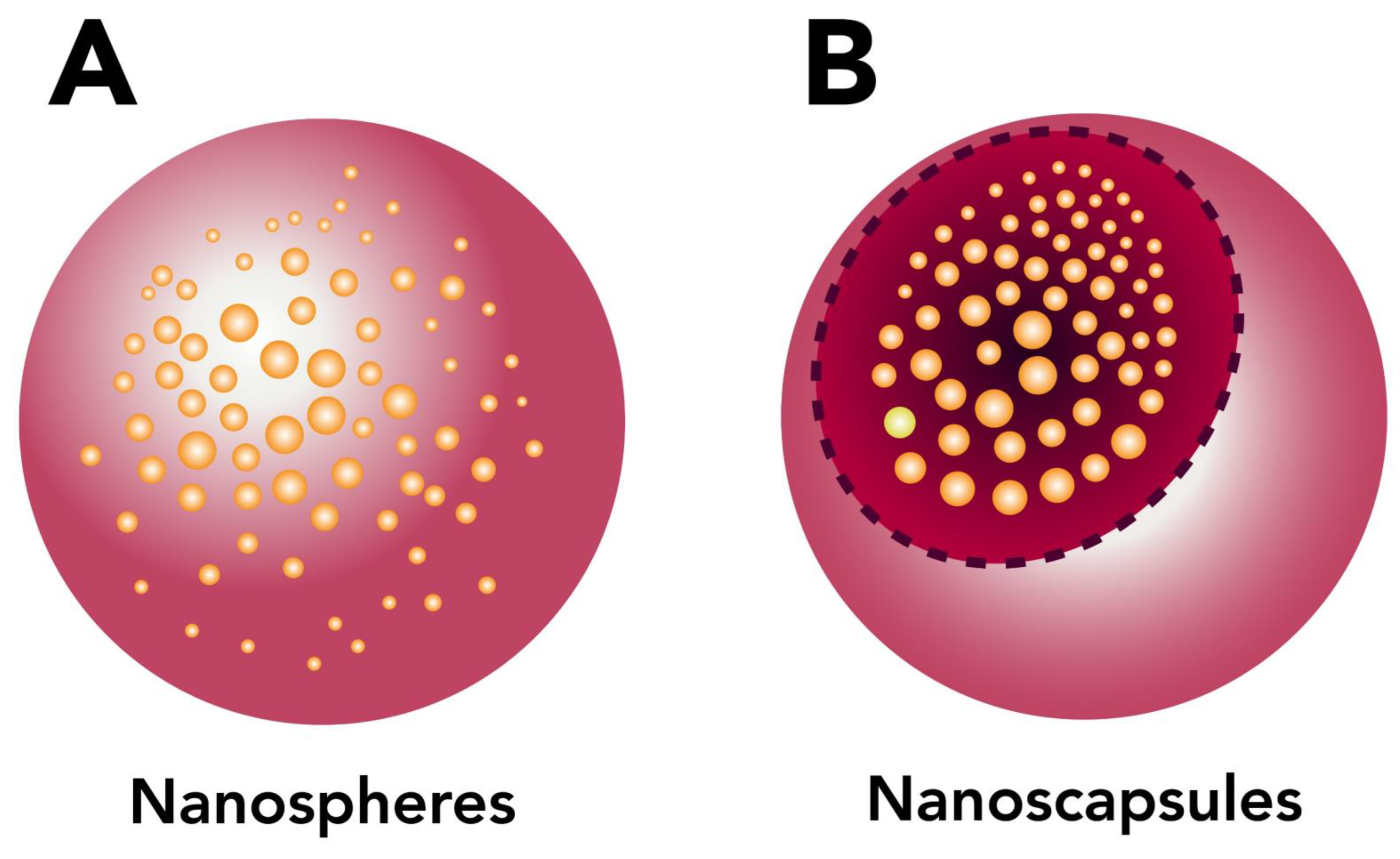
6. Polymeric Nanoparticles

NPs as a delivery system have achieved advantages by overcoming the challenges of typical dosage methods [36]. According to the definition from the National Nanotechnology Initiative (NNI), NPs are structures with sizes ranging from 1 to 100 nm in at least one dimension. However, the prefix “nano” is commonly used for particles that are up to several hundred nanometers in size [37].
Nanotechnology is the science of the nanoscale [38]. NPs are made up of three layers: (a) the surface layer, (b) the outer layer, and (c) the core (Figure 1) [39].
The nanocarriers must be able to integrate into the biological system and must not cause any negative or toxic effects [40].
NPs have also been used in immunotherapy (vaccines), which, by containing foreign substances and being in contact with the immune system, generates a specific immune response. Properties such as size, charge, and rigidity, among others, determine their interaction with the immune system [41].
Polymeric NPs can deliver drugs and overcome biological barriers, as well as target drugs to specific cells [42].
Most polymeric NPs are now biodegradable and biocompatible due to the accomplishments of researchers in the last few decades in developing NPs for drug delivery systems. These biodegradable NPs are coated with controlled-release polymers that can release proteins, peptides, antigens, DNA transporters, and target specific sites [43].
Polymeric systems have become popular due to their ability to provide a sustained release of the associated active compounds [44]. For more than five decades, the pharmaceutical industry has been using procedures such as coating and encapsulation to incorporate polymers with bioactivity [45]. Various polymers have been used for site-specific drug delivery while minimizing side effects [46].
Biodegradable and bioerodible polymers are a crucial group of materials for drug delivery [47]. Biopolymers are of natural origin (vegetable, animal, bacterial, fungi). They include networks of polysaccharides, cellulose, starch, gelatin, and collagen among others, with potential applications in the pharmaceutical industry [48]. In recent years, biodegradable polymers have garnered considerable attention as potential drug delivery devices, given their applications in the controlled release (CR) of drugs, their ability to target particular organs and tissues, their potential as carriers of DNA in gene therapy, and their ability to deliver proteins, peptides, and genes through a peroral route of administration [46].
The objective of a delivery system is to release at the desired site for a specified time to exert the therapeutic effect [45].
The purpose of nanotechnology is the delivery drugs to target sites so that the pharmacologically desired effect of the drug is maximized and the limitations and drawbacks that would hinder the required effectiveness are overcome [49]. NPs consist of macromolecular materials and can be used therapeutically as adjuvants in vaccines or drug carriers [50]. The challenges of biological barriers such as the passage of substances through the blood-brain barrier have been overcome with the use of NPs [49].
Polymeric NPs, which possess better reproducibility profiles than liposomes (Figure 2), have been used as alternative drug carriers to overcome many drug delivery problems. Polymers used to form NPs can be either synthetic or natural polymers [50]. Natural polymers such as chitosan, albumin, and heparin have been used for the delivery of oligonucleotides, DNA, and proteins, as well as drugs. Polymeric NPs are popular due to their ability to deliver drugs, as well as for their biodegradability. Chitosan is one of the most widely used cationic polymers [51].
Environmentally responsible polymers are a class of smart polymers consisting of linear, cross-linked copolymers (Table 5). Their characteristic feature is their ability to undergo physicochemical change in response to external stimuli such as pH, temperature, etc. [45].
Polymeric microparticles and NPs have been applied to gene delivery, and particularly in vaccine design (e.g., DNA vaccine). Synthetic vectors based on polycation enable gene delivery by cell-targeted ligands [51].
There are two varieties of NPs, depending on the preparation process: nanospheres or nanocapsules (Figure 3) [55]. Nanospheres have a matrix structure where the drug is located. The nanocapsules have a membrane and contain the drug inside [50].

6.1. Preparation of Polymeric Nanoparticles

Polymeric NPs have been widely explored in the pharmaceutical fields since their launch [56,57]. Polymeric NPs can be prepared by different methods, such as solvent evaporation, nanoprecipitation, salting out, dialysis, and supercritical fluid technology [43].
Among the available families of nanocarriers, polymeric vectors have been widely researched, owing to several beneficial properties, including biocompatibility, biodegradability, non-immunogenicity, and nontoxicity. Progress has also been made in designing potent, stable NPs for tissue and cell targeting by conjugating ligands in the polymeric NPs. Altogether, these advancements have improved NP performance [56].
The synthesis of metal NPs includes the processes of spray pyrolysis, liquid infiltration, rapid solidification, and others. The synthesis of ceramic nanocomposites includes the powder process, polymeric precursor process, and sol-gel process. Finally, the fabrication of polymeric nanocomposites includes intercalation, in situ intercalative polymerization, melt intercalation, template synthesis, mixing, in situ polymerization, and the sol-gel process [58].

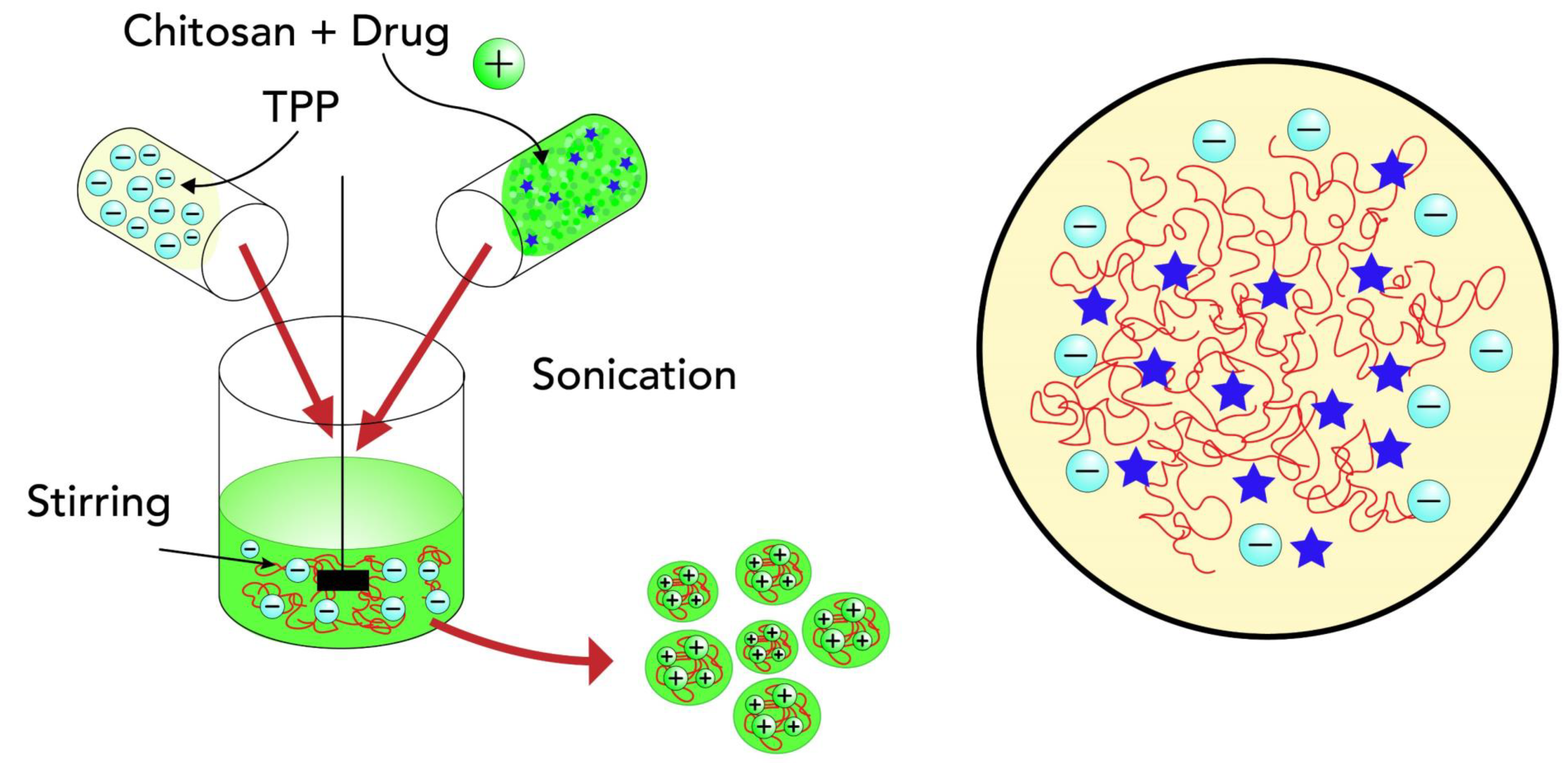
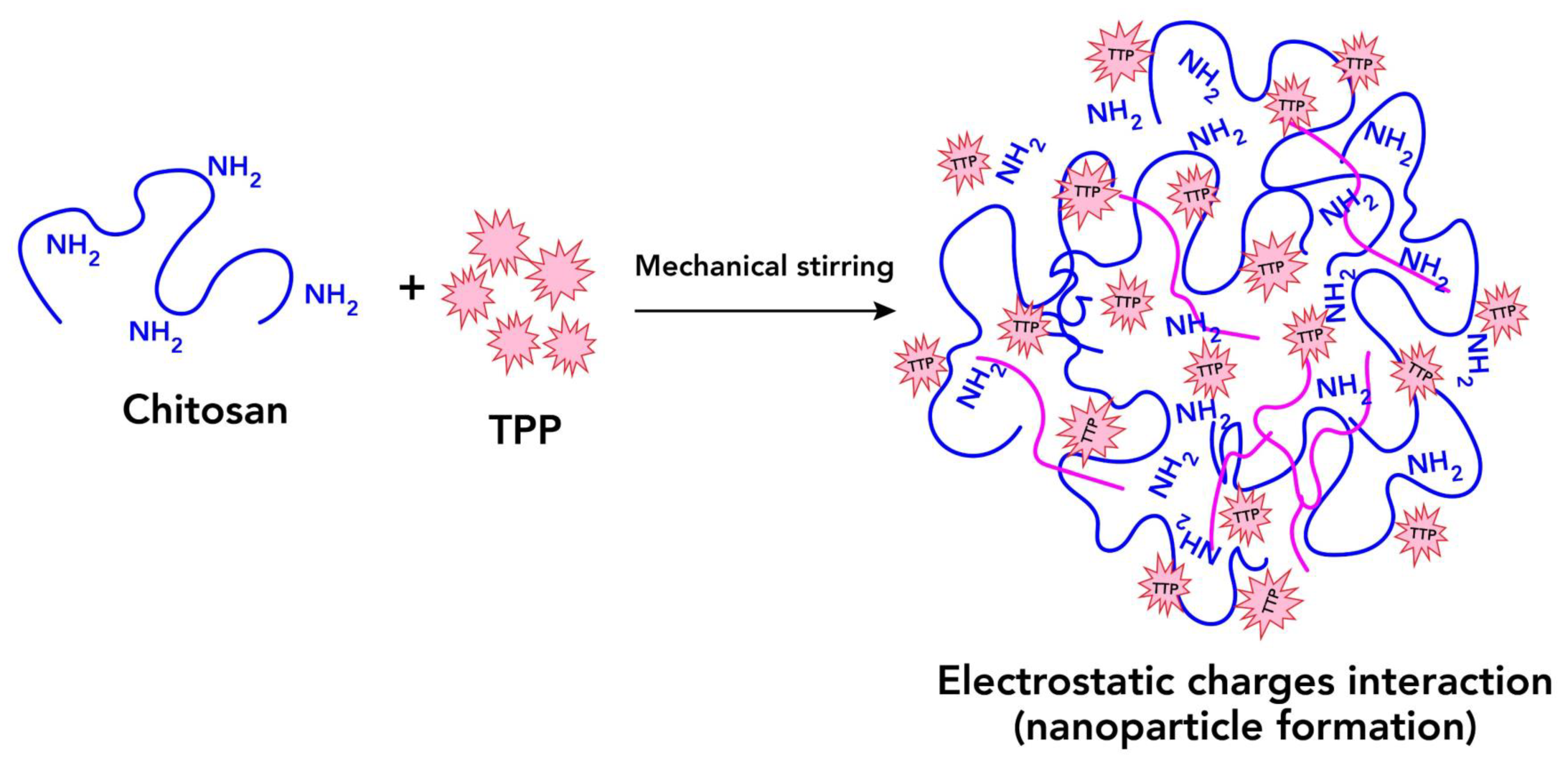
6.1.1. Ionic Cross-Link Method

In this technique, an ionic cross-link is conducted by the aggregation of chitosan or its derivates with oppositely charged macromolecules or in the presence of an ionic cross-link agent (Figure 4) [59].
A cross-link is formed through a chemical reaction, such as van der Waals forces, which link two polymers together [60].
Ionically cross-linked chitosan NPs are based on the formation of complexes with the amino group and a polyanion (tripolyphosphate TPP) [61].

6.1.2. Covalent Cross-Link Method

Covalent cross-linking is more attractive than the ionic gelation method. Bodnar et al. [61] reported that the synthesis of chitosan NPs covalently cross-linked with tartaric acid resulted in particles 60–280 nm in size [62]. Covalent cross-linking enhances the chemical and mechanical properties of the material [63].

6.1.3. Reverse Micellar Method

In the reverse micellar formation of NPs, the surfactant is dissolved in an organic solvent forming micelles and chitosan is added under continuous stirring; to this transparent solution, a crosslinker is added. The limitations are the use of organic solvents as well as the washing steps [61].

6.1.4. Precipitation/Coacervation

NPs are prepared by alkaline precipitation (pH > 6.5). Chitosan is injected into the organic solvent by employing a nozzle. The NPs are obtained by filtration or centrifugation, then rinsed with water, and a crosslinking agent is added to modulate the release of the substances [61].

6.1.5. Emulsion–Droplet Coalescence Method

The NPs are formed by the emulsification of an organic polymeric solution in an aqueous phase, after which the organic solvent is evaporated. The organic solution is poured into the aqueous phase. Emulsification is carried out under high-shear force conditions to reduce the size of the emulsion droplet. The evaporation of the solvent leads to the formation of NPs [61].

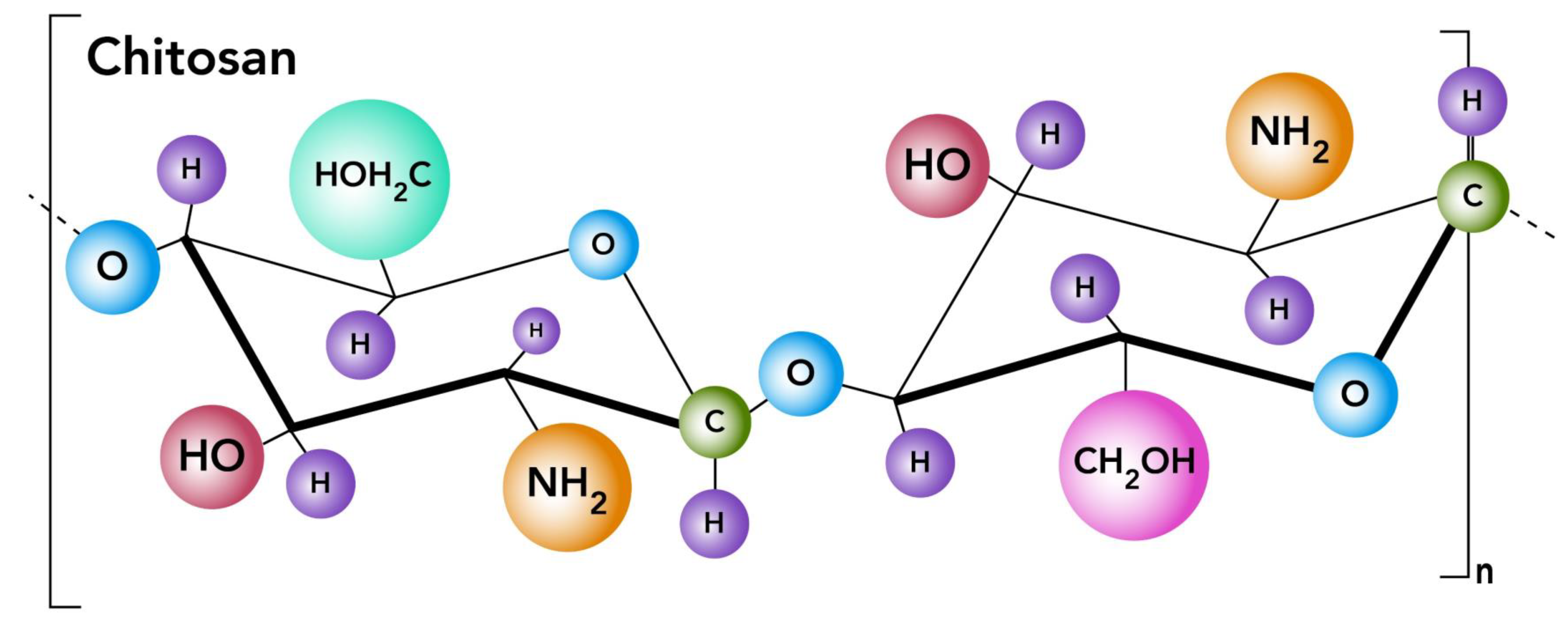
7. Chitosan Nanoparticles

Chitosan is a cationic polysaccharide and has been considered a promising nanomaterial [64]. Chitosan offers outstanding biological properties, including biocompatibility, biodegradability, and nontoxicity, that make it increasingly important in various applications in the pharmaceutical and biomedical fields [65].
The shellfish industry makes very common use of the meat, and the head and shells are discarded as waste (80,000 tons of waste per year) [66]; shell waste is recycled to obtain commercially viable products such as chitin [67].
Chitosan is the N-acetyl derivate of chitin obtained by N-deacetylation. Chitosan is widely used in the encapsulation of active food ingredients, enzyme immobilization, controlled drug delivery, and plant growth promotion in agriculture. Chitosan has properties such as biodegradability, biocompatibility, antimicrobial, bioactivity, nontoxicity, and a polycationic nature [68].
Chitosan (Figure 5) is well-known for its hydrophilic, biocompatible, biodegradable, and nontoxic properties. The use of chitosan NP for oral and nasal drug delivery routes has been reported in previous studies [69]. NP technology is an increasingly accepted formulation technique as it overcomes the limitations of conventional oral drug delivery.
The positively charged chitosan will bind to cell membranes and is reported to decrease the trans-epithelial electrical resistance (TEER) of cell monolayers, as well as to increase paracellular permeability. Chitosan solutions have been shown to increase transcellular and paracellular permeability in a reversible, dose-dependent manner that depends on the molecular weight and degree of deacetylation of the chitosan. Low-molecular-weight chitosan possesses the ability to penetrate cells, where it is suspected of binding to cell DNA, prohibiting mRNA synthesis and causing the termination of cell multiplication [60]. The mechanism of action, which includes interaction with the tight junction proteins and ZO-1 proteins, redistribution of F-actin, and slight destabilization of the plasma membrane, appears to be mediated by the positive charges on the chitosan. Thus, the ability of chitosan to enhance permeation is influenced by the pH of the environment [21].
Chitosan is biodegradable and, due to its low molecular weight, is eliminated by the kidneys, and if it is of higher molecular weight, it can be degraded into smaller fragments for renal elimination.
Mucoadhesive NPs are able to have their surface coated with mucoadhesive polymers such as chitosan or Carbopol [66]. Mucus is a blend of molecules including salts, lysozyme, and mucins, which are highly hydrated glycoproteins primarily responsible for the viscoelastic properties of mucus. Sialic acid residues on mucins have a pH of 2.6, making them negatively charged at physiological pH [21].
In addition, the formation of chitosan into microparticles and NPs also preserves mucoadhesion [21]. The application of biodegradable nanosystems is one of the most successful advancements in the pharmaceutical industry [40].
Chitosan can be used as an oral gene carrier due to its adhesive properties. On the other hand, researchers have found that in vitro, chitosan-mediated transfection depends on the cell type, serum concentration, pH, and chitosan molecular weight [51].
The protection offered by NPs has generated the development of systems with macromolecules and proteins, among others, since these promote the absorption of therapeutic substances [61].
The process of NP formation is based on electrostatic interactions between the amine group of chitosan and a negatively charged group of polyanions such as tripolyphosphate (Figure 6). This method is easy in aqueous media [51].
One study has shown that the intratumoral administration of interleukine-12 co-formulated with the biodegradable polysaccharide chitosan could enhance the anti-tumor activity of interleukine-12 in mice bearing established colorectal (MC32a) and pancreatic (Panc02) tumors [51].
Chitosan nanospheres have applications for drug delivery in the gastrointestinal, ophthalmic, nasal, sublingual, transdermal, and vaginal tract [51].
The absorption-promoting effect of chitosan has been extensively studied by the combination of mucoadhesion and the transient opening of tight junctions in the mucosal cell membrane, which have been experimentally verified both in vitro and in vivo [61].
The mechanism of chitosan NP transport across the GI tract is likely through adsorptive endocytosis. Chitosan NP internalization is higher in the jejunum and ileum than in the duodenum [61].
Chitosan NPs can be applied to mucosal delivery (pulmonary, nasal), where peptides and proteins can be administered [66].
Oral formulations are considered a desirable alternative to intravenous drug administration due to the advantage of offering adaptability to tune the dosing schedule to individual patient responses based on efficacy and toxicity. Oral formulations of NPs can increase the number of patients treated [70].

7.1. Applications of Chitosan Nanoparticles for Oral Drug Delivery

Recently, the use of chitosan in pharmaceutical development has increased due to its compatibility with other components such as surfactants, starches, etc. Chitosan increases cell membrane permeability, both in vivo and in vitro. Chitosan has the potential of serving as an absorption enhancer across intestinal epithelia, prolonging the residence time of delivery systems at absorption sites and relaxing the tight junctions of cell membranes [71]. The cationic nature of chitosan permits it to form complexes with oppositely charged drugs and excipients, thereby altering the physicochemical characteristics of the formulation. Reacting chitosan with controlled amounts of multivalent anions results in cross-links between chitosan molecules (Table 6) [72].

7.1.1. Gene Delivery

In gene therapy, transfection is hindered by the orientation of the system to the target cell as well as the degradation of endolysosomes and intercellular trafficking of plasmid DNA [95].
Gene carriers have the disadvantage of low transfection and toxicity, and they even provoke severe immune responses [7]. Nucleic acids are being developed for gene therapy and vaccination [21]. As a non-virus carrier, chitosan has exceptional compatibility and biodegradability [7,95].
Dastan and Turan [96] improved chitosan microparticles and reported a sustained release profile of DNA with a high potential transfer of DNA tested in different cell lines such as human embryonic kidney, Swiss 3T3, and HeLa [97].

7.1.2. Protein and Peptide Delivery

Proteins and peptides usually have a high molecular weight and low lipophilicity, which is why they are usually administered subcutaneously. However, NPs have been shown to administer peptides and proteins orally. Chitosan NPs are gaining increased attention for their ability to serve as carriers for oral protein and peptide delivery [80,87].

7.1.3. Drugs

Antiviral, antiallergic, and hormone drugs can be loaded in chitosan NPs through an ionic cross-link method [7,98]. For example, in the research performed by Shailender et al. [89], the ionic gelation method was used in the preparation of tenofovir disoproxil fumarate chitosan NPs [50,99].
Cancer has become one of the most lethal and prevalent conditions throughout the world. The success of current therapies is primarily limited by tumor recurrence, metastasis, acquired resistance, and the presence of side effects [100]. The cancer treatment doxorubicin, for example, produces side effects such as cardiotoxicity. To minimize these side effects, the drug has been encapsulated in chitosan NPs. This has caused several advantages, including better delivery, improved cell- or tissue-targeted drug delivery, and enhanced absorption in the entire small intestine [101,102].
Tuberculosis continues to be the leading cause of mortality worldwide, and it is also an occupational disease in health care. Noncompliance is the primary limitation of treatment, largely because treatment involves continuous, frequent, and multidrug dosing. Chitosan NPs could improve a long-duration drug formulation, releasing the antitubercular agents in a slow and sustained manner [92].

7.1.4. Vaccines

The effect of chitosan as an adjuvant for the generation of vaccines makes it a safer therapy [71].
Oral vaccination is a highly promising application of chitosan NPs. Food allergy is a common and often fatal condition with no effective treatment. Orally administering NPs prepared by complexing plasmid DNA with chitosan has been shown to result in a transduced gene expression in the intestinal epithelium of patients with food allergies [103].
Olivera et al. [104] developed a vaccine for the control of schistosomiasis, which is recognized as the most important human helminth infection in terms of morbidity and mortality. They described that chitosan NPs with plasmid DNA encoding the Rho1-GTPase protein of Schistosoma mansoni were able to induce high levels of the modulatory cytokine IL-10. It resulted in a significant reduction in liver pathology. Mice immunized with only chitosan NPs presented with 47% protection against parasitic infection.

8. Discussion

The oral route of administration continues to be the first choice of both patients and doctors; although, as mentioned, the administered medications must cross many barriers and physiological processes that reduce their bioavailability and, consequently, their efficacy. For this reason, nanoparticulate or nanometric drug delivery systems based on biocompatible polymers have acquired great relevance. Their physicochemical characteristics have allowed them to be included within the unconventional forms of administration, and they have enabled the vectorization of active ingredients with low solubility or bioavailability, increasing their interaction with the target organs or cells through different routes of administration. Due to their easy preparation and high capacity to encapsulate peptides, drugs, and genes without interfering with their biological activity, chitosan-based NPs are the first choice when treating diseases through non-conventional methods. Chitosan NPs enable medications to cross physical and biological barriers, increasing bioavailability and leading to a more powerful effect with fewer adverse effects, which can be achieved without requiring invasive or painful routes of administration by using the oral route.

Author Contributions

Conceptualization, O.R.G.-E. and J.J.E.-C.; methodology, O.R.G.-E.; formal analysis, J.J.E.-C., O.R.G.-E., P.S.-C., A.V.-D.; investigation, O.R.G.-E., M.I.M.-F., M.C.P.-J., E.A.-A.; resources, J.J.E.-C., R.V.-G.; writing—original draft preparation, O.R.G.-E., J.J.E.-C., R.V.-G. and J.E.M.-C.; writing—review and editing, O.R.G.-E., J.J.E.-C., I.M.R.-C. and B.R.-P.; visualization, O.R.G.-E.; supervision, J.J.E.-C.; project administration, J.J.E.-C.; funding acquisition, J.J.E.-C. All authors have read and agreed to the published version of the manuscript.

Funding

This research was funded by PAPIIT IG 100220, PAPIIT IG 200423, Cátedra de Investigación 2206, and CONACyT CF 140617. The first author would like to acknowledge the financial support from COMECYT through a postdoctoral fellowship (CAT2022-0053).

Institutional Review Board Statement

Not applicable.

Informed Consent Statement

Not applicable.

Data Availability Statement

Data sharing not applicable.

Conflicts of Interest

The authors declare no conflict of interest.

References

  1. Mathias, N.R.; Hussain, M.A. Non-invasive systemic drug delivery: Developability considerations for alternate routes of administration. J. Pharm. Sci. 2010, 99, 1–20. [Google Scholar] [CrossRef] [PubMed]
  2. Ding, J.; Cheng, Y.; Li, M.; Gref, R.; Chen, X. Smart Polymeric Nanocarriers. J. Nanomater. 2016, 2016, 3710921. [Google Scholar] [CrossRef]
  3. Ramadhani, N.; Shabir, M.; McConville, C. Preparation and characterisation of Kolliphor®P 188 and P 237 solid dispersion oral tablets containing the poorly water soluble drug disulfiram. Int. J. Pharm. 2014, 475, 514–522. [Google Scholar] [CrossRef] [PubMed]
  4. Kasiramar, G. An Overview on Polymeric Nanoparticles used in the treatment of Diabetes Mellitus. Pharmatutor 2019, 5, 40. [Google Scholar]
  5. Ghadi, A.; Mahjoub, S.; Tabandeh, F.; Talebnia, F. Synthesis and optimization of chitosan nanoparticles: Potential applications in nanomedicine and biomedical engineering. Casp. J. Intern. Med. 2014, 5, 156–161. [Google Scholar]
  6. Mitchell, M.J.; Billingsley, M.M.; Haley, R.M.; Wechsler, M.E.; Peppas, N.A.; Langer, R. Engineering precision nanoparticles for drug delivery. Nat. Rev. Drug Discov. 2020, 20, 101–124. [Google Scholar] [CrossRef]
  7. Zeng, Z.W.; Wang, J.J.; Xiao, R.Z.; Xie, T.; Zhou, G.L.; Zhan, X.R.; Wang, S.L. Recent advances of chitosan nanoparticles as drug carriers. Int. J. Nanomed. 2011, 6, 765–774. [Google Scholar] [CrossRef] [Green Version]
  8. Davis, J.L. Pharmacologic Principles. In Equine Internal Medicine, 4th ed.; Elsevier: Amsterdam, The Netherlands, 2018; pp. 79–137. [Google Scholar]
  9. Homayun, B.; Lin, X.; Choi, H.J. Challenges and recent progress in oral drug delivery systems for biopharmaceuticals. Pharmaceutics 2019, 11, 129. [Google Scholar] [CrossRef] [Green Version]
  10. Seidling, H.M.; Lampert, A.; Lohmann, K.; Schiele, J.T.; Send, A.J.F.; Witticke, D.; Haefeli, W.E. Safeguarding the process of drug administration with an emphasis on electronic support tools. Br. J. Clin. Pharmacol. 2013, 76, 25–36. [Google Scholar] [CrossRef] [Green Version]
  11. Peña-Juárez, M.C.; Guadarrama-Escobar, O.R.; Escobar-Chávez, J.J. Transdermal Delivery Systems for Biomolecules. J. Pharm. Innov. 2021, 17, 319–332. [Google Scholar] [CrossRef]
  12. Singla, V.; Saini, S.; Joshi, B.; Rana, A.C. Emulgel: A New Platform for Topical Drug Delivery. Int. J. Pharma Bio Sci. 2012, 3, 485–498. [Google Scholar]
  13. Berlin, J.; May-McCarver, D.G.; Notterman, D.A.; Ward, R.M.; Weismann, D.N.; Wilson, G.S.; Wilson, J.T.; Bennett, D.R.; Hoskins, I.A.; Kaufman, P.; et al. Alternative Routes of Drug Administration-Advantages and Disadvantages (Subject Review). Pediatrics 1997, 100, 143–152. [Google Scholar]
  14. Subcutaneous Drug Administration. In Handbook of Behavioral Neuroscience; Elsevier: Amsterdam, The Netherlands, 1994; pp. 35–45.
  15. Kumari, B. Ocular drug delivery system: Approaches to improve ocular bioavailability. GSC Biol. Pharm. Sci. 2019, 6, 001–010. [Google Scholar] [CrossRef]
  16. Rogliani, P.; Calzetta, L.; Coppola, A.; Cavalli, F.; Ora, J.; Puxeddu, E.; Matera, M.G.; Cazzola, M. Optimizing drug delivery in COPD: The role of inhaler devices. Respir. Med. 2017, 124, 6–14. [Google Scholar] [CrossRef] [Green Version]
  17. Yurdasiper, A.; Arici, M.; Ozyazici, M. Nanopharmaceuticals. In Emerging Nanotechnologies in Immunology: The Design, Applications and Toxicology of Nanopharmaceuticals and Nanovaccines; Elsevier: Amsterdam, The Netherlands, 2018; pp. 165–201. [Google Scholar]
  18. Sastry, S.V.; Nyshadham, J.R.; Fix, J.A. Recent technological advances in oral drug delivery—A review. Pharm. Sci. Technol. Today 2000, 3, 138–145. [Google Scholar] [CrossRef]
  19. Dupeyrón, D.; Rieumont, J.; González, M.; Castaño, V.M. Protein Delivery by Enteric Copolymer Nanoparticles. J. Disper. Sci. Technol. 2009, 30, 1188–1194. [Google Scholar] [CrossRef]
  20. Lundquist, P.; Artursson, P. Oral absorption of peptides and nanoparticles across the human intestine: Opportunities, limitations and studies in human tissues. Adv. Drug Deliv. Rev. 2016, 106, 256–276. [Google Scholar] [CrossRef]
  21. Bowman, K.; Leong, K.W. Chitosan nanoparticles for oral drug and gene delivery. Int. J. Nanomed. 2006, 1, 117–128. [Google Scholar] [CrossRef] [PubMed]
  22. Date, A.A.; Hanes, J.; Ensign, L.M. Nanoparticles for oral delivery: Design, evaluation and state-of-the-art. Physiol. Behav. 2017, 176, 139–148. [Google Scholar] [CrossRef] [PubMed] [Green Version]
  23. Bardakjian, B.L. Gastrointestinal system. Biomed. Imaging 2003, 2, 6-1–6-13. [Google Scholar]
  24. Martínez Bueno, C. Physiology of intestinal absorption. Rev. Esp. Enferm. Apar. Dig. 1966, 25, 933–941. [Google Scholar] [PubMed]
  25. Zia, L.; Johnson, I.; Mansurov, B.; Morgan, J.; Redi, M.; Saez-Trumper, D.; Taraborelli, D. Knowledge Gaps—Wikimedia Research 2030. Int. J. Biomed. Nanosci. Nanotechnol. 2019, 1–8. [Google Scholar]
  26. Salama, N.N.; Eddington, N.D.; Fasano, A. Tight junction modulation and its relationship to drug delivery. Adv. Drug Deliv. Rev. 2006, 58, 15–28. [Google Scholar] [CrossRef]
  27. Johnson, L.R. Physiology of the Gastrointestinal Tract, 4th ed.; Barrett, K., Johnson, L., Ghishan, F., Merchant, J., Said, H., Wood, J., Eds.; Elsevier: Amsterdam, The Netherlands, 2006; Available online: https://www.elsevier.com/books/physiology-of-the-gastrointestinal-tract/johnson/978-0-12-088394-3 (accessed on 4 November 2022).
  28. Magalhães, J.; Vieira, A.; Santos, S.; Pinheiro, M.; Reis, S. Oral Administration of Nanoparticles-Based TB Drugs. In Multifunctional Systems for Combined Delivery, Biosensing and Diagnostics; Elsevier: Amsterdam, The Netherlands, 2017; pp. 307–326. [Google Scholar]
  29. Omidian, H.; Park, K. Swelling agents and devices in oral drug delivery. J. Drug Deliv. Sci. Technol. 2008, 18, 83–93. [Google Scholar] [CrossRef]
  30. Jain, V.; Gupta, A.; Pawar, V.K.; Asthana, S.; Jaiswal, A.K.; Dube, A.; Chourasia, M.K. Chitosan-Assisted Immunotherapy for Intervention of Experimental Leishmaniasis via Amphotericin B-Loaded Solid Lipid Nanoparticles. Biotechnol. Appl. Biochem. 2014, 174, 1309–1330. [Google Scholar] [CrossRef] [PubMed]
  31. Renukuntla, J.; Vadlapudi, A.D.; Patel, A.; Boddu, S.H.; Mitra, A.K. Approaches for enhancing oral bioavailability of peptides and proteins. Int. J. Pharm. 2013, 447, 75–93. [Google Scholar] [CrossRef] [PubMed] [Green Version]
  32. Beneke, C.E.; Viljoen, A.M.; Hamman, J.H. Polymeric plant-derived excipients in drug delivery. Molecules 2009, 14, 2602–2620. [Google Scholar] [CrossRef] [PubMed]
  33. Herrero, E.P.; Alonso, M.J.; Csaba, N. Polymer-based oral peptide nanomedicines. Ther. Deliv. 2012, 3, 657–668. [Google Scholar] [CrossRef]
  34. Mohammed, M.A.; Syeda, J.T.M.; Wasan, K.M.; Wasan, E.K. An overview of chitosan nanoparticles and its application in non-parenteral drug delivery. Pharmaceutics 2017, 9, 53. [Google Scholar] [CrossRef] [Green Version]
  35. Gupta, R.; Xie, H. Nanoparticles in daily life: Applications, toxicity and regulations. J. Env. Pathol. Toxicol. Oncol. 2018, 37, 209–230. [Google Scholar] [CrossRef]
  36. Atyabi, F.; Dinarvand, R. Chitosan-Pluronic nanoparticles as oral delivery of anticancer gemcitabine: Preparation and in vitro study. Int. J. Nanomed. 2012, 7, 1851–1863. [Google Scholar] [CrossRef] [PubMed] [Green Version]
  37. Stewart, C.E.; Torr, E.E.; Mohd Jamili, N.H.; Bosquillon, C.; Sayers, I. Evaluation of Differentiated Human Bronchial Epithelial Cell Culture Systems for Asthma Research. J. Allergy Clin. Immunol. 2019, 6, 975–991. [Google Scholar] [CrossRef] [PubMed] [Green Version]
  38. Booth, T.J.; Baker, M.A.B. Nanotechnology: Building and Observing at the Nanometer Scale. In Pharmacognosy: Fundamentals, Applications and Strategy; Elsevier Inc.: Amsterdam, The Netherlands, 2017; pp. 633–643. [Google Scholar]
  39. Khan, I.; Saeed, K.; Khan, I. Nanoparticles: Properties, applications and toxicities. Arab. J. Chem. 2019, 12, 908–931. [Google Scholar] [CrossRef]
  40. Napier, M.E.; DeSimone, J.M. Nanoparticle drug delivery platform. Polym. Rev. 2007, 47, 321–327. [Google Scholar] [CrossRef]
  41. Liu, Y.; Hardie, J.; Zhang, X.; Rotello, V.M. Effects of engineered nan.noparticles on the innate immune system. Semin. Immunol. 2017, 34, 25–32. [Google Scholar] [CrossRef]
  42. Katas, H.; Raja, M.A.G.; Lam, K.L. Development of Chitosan Nanoparticles as a Stable Drug Delivery System for Protein/siRNA. Int. J. Biomater. 2013, 2013, 146320. [Google Scholar] [CrossRef] [Green Version]
  43. Fide, S.; Karaismailoglu, S.; Derman, S. Synthesis and characterization methods of polymeric nanoparticles 2. Preparation Methods of Polymeric Nanoparticles. Eur. J. Pharm. Sci. 2018, 1, 1–9. [Google Scholar]
  44. Campos, E.; Oliveira, J.L.; Da Silva, C.M.G.; Pascoli, M.; Pasquoto, T.; Lima, R.; Abhilash, P.C.; Fraceto, L.F.U. Polymeric and Solid Lipid Nanoparticles for Sustained Release of Carbendazim and Tebuconazole in Agricultural Applications. Sci. Rep. 2015, 5, srep13809. [Google Scholar] [CrossRef] [Green Version]
  45. Son, G.H.; Lee, B.J.; Cho, C.W. Mechanisms of drug release from advanced drug formulations such as polymeric-based drug-delivery systems and lipid nanoparticles. J. Pharm. Investig. 2017, 47, 287–296. [Google Scholar] [CrossRef]
  46. Soppimath, K.S.; Aminabhavi, T.M.; Kulkarni, A.R.; Rudzinski, W.E. Biodegradable polymeric nanoparticles as drug delivery devices. J. Control. Release 2001, 70, 1–20. [Google Scholar] [CrossRef]
  47. Liechty, W.B.; Kryscio, D.R.; Slaughter, B.V.; Peppas, N.A. Polymers for drug delivery systems. Annu. Rev. Chem. Biomol. Eng. 2010, 1, 149–173. [Google Scholar] [CrossRef] [PubMed] [Green Version]
  48. Singh, A.V. Biopolymers in Drug Delivery: A Review. Pharmacologyonline 2011, 674, 666–674. [Google Scholar]
  49. El-Say, K.M.; El-Sawy, H.S. Polymeric nanoparticles: Promising platform for drug delivery. Int. J. Pharm. 2017, 58, 675–691. [Google Scholar] [CrossRef] [PubMed]
  50. Nagpal, K.; Singh, S.K.; Mishra, D.N. Chitosan Nanoparticles: A Promising System in Novel Drug Delivery. Chem. Pharm. Bull. 2010, 58, 1423–1430. [Google Scholar] [CrossRef] [PubMed] [Green Version]
  51. Bolhassani, A.; Javanzad, S.; Saleh, T.; Hashemi, M.; Aghasadeghi, M.R.; Sadat, S.M. Polymeric nanoparticles Potent vectors for vaccine delivery targeting cancer and infectious diseases. Hum. Vaccines Immunother. 2014, 10, 321–332. [Google Scholar] [CrossRef] [Green Version]
  52. Lu, Y.; Shah, K.W.; Xu, J. Synthesis, morphologies and building applications of nanostructured polymers. Polymers 2017, 9, 506. [Google Scholar] [CrossRef] [PubMed]
  53. Zielińska, A.; Carreiró, F.; Oliveira, A.M.; Neves, A.; Pires, B.; Venkatesh, D.N.; Durazzo, A.; Lucarini, M.; Eder, P.; Silva, A.M.; et al. Polymeric Nanoparticles: Production, Characterization, Toxicology and Ecotoxicology. Molecules 2020, 25, 3731. [Google Scholar] [CrossRef]
  54. Chenthamara, D.; Subramaniam, S.; Ramakrishnan, S.G.; Krishnaswamy, S.; Essa, M.M.; Lin, F.H.; Qoronfleh, M.W. Therapeutic efficacy of nanoparticles and routes of administration. Biomater. Res. 2019, 23, 20. [Google Scholar] [CrossRef]
  55. Allémann, E.; Gurny, R.; Doelker, E. Drug-loaded nanoparticles—Preparation methods and drug targeting issues. Eur. J. Pharm. Biopharm. 1993, 39, 173–191. [Google Scholar]
  56. Kumari, R.M.; Sharma, N.; Gupta, N.; Chandra, R.; Nimesh, S. Synthesis and evolution of polymeric nanoparticles. In Design and Development of New Nanocarriers; Elsevier: Amsterdam, The Netherlands, 2018; pp. 401–438. [Google Scholar]
  57. Pineda-Álvarez, R.A.; Bernad-Bernad, M.J.; Rodríguez-Cruz, I.M.; Escobar-Chávez, J.J. Development and Characterization of Starch/Gelatin Microneedle Arrays Loaded with Lecithin–Gelatin Nanoparticles of Losartan for Transdermal Delivery. J. Pharm. Innov. 2020, 17, 71–84. [Google Scholar] [CrossRef]
  58. Rane, A.V.; Kanny, K.; Abitha, V.K.; Thomas, S. Synthesis of Inorganic Nanomaterials; Elsevier: Amsterdam, The Netherlands, 2018; pp. 121–139. Available online: https://www.sciencedirect.com/science/article/pii/B9780081019757000051 (accessed on 14 October 2022).
  59. Garg, U.; Chauhan, S.; Nagaich, U.; Jain, N. Current advances in chitosan nanoparticles based drug delivery and targeting. Adv. Pharm. Bull. 2019, 9, 195–204. [Google Scholar] [CrossRef] [Green Version]
  60. Ryan, C.; Alcock, E.; Buttimer, F.; Schmidt, M.; Clarke, D.; Pemble, M.; Bardosova, M. Synthesis and characterisation of cross-linked chitosan composites functionalised with silver and gold nanoparticles for antimicrobial applications. Sci. Technol. Adv. Mater. 2017, 18, 528–540. [Google Scholar] [CrossRef] [PubMed] [Green Version]
  61. Wang, Y.; Li, P.; Tran, T.T.-D.; Zhang, J.; Kong, L. Manufacturing techniques and surface engineering of polymer based nanoparticles for targeted drug delivery to cancer. Nanomaterials 2016, 6, 26. [Google Scholar] [CrossRef] [PubMed] [Green Version]
  62. Tallury, P.; Kar, S.; Bamrungsap, S.; Huang, Y.-F.; Tan, W.; Santra, S. Ultra-small water-dispersible fluorescent chitosan nanoparticles: Synthesis, characterization and specific targeting. Chem. Commun. 2009, 17, 2347–2349. [Google Scholar] [CrossRef] [PubMed]
  63. Lorke, S.; Müller, U.; Meissl, R.; Brüggemann, O. Covalent cross-linking of polymers at room temperature. Int. J. Adhes. Adhes. 2019, 91, 150–159. [Google Scholar] [CrossRef]
  64. Li, J.; Cai, C.; Li, J.; Li, J.; Li, J.; Sun, T.; Wang, L.; Wu, H.; Yu, G. Chitosan-based nanomaterials for drug delivery. Molecules 2018, 23, 2661. [Google Scholar] [CrossRef] [Green Version]
  65. Saikia, C.; Gogoi, P. Chitosan: A Promising Biopolymer in Drug Delivery Applications. Mol. Genet. Genomic. Med. 2015, 4, 1–10. [Google Scholar] [CrossRef]
  66. Ahmed, A.B.; Konwar, R.; Sengupta, R. Atorvastatin calcium loaded chitosan nanoparticles: In vitro evaluation and in vivo pharmacokinetic studies in rabbits. Braz. J. Pharm. 2015, 51, 467–477. [Google Scholar] [CrossRef] [Green Version]
  67. Chakraborty, S.P.; Mahapatra, S.K.; Sahu, S.K.; Pramanik, P.; Roy, S. Antioxidative effect of folate-modified chitosan nanoparticles. Asian Pac. J. Trop. Biomed. 2011, 1, 29–38. [Google Scholar] [CrossRef] [Green Version]
  68. Divya, K.; Jisha, M.S. Chitosan nanoparticles preparation and applications. Environ. Chem. Lett. 2018, 16, 101–112. [Google Scholar] [CrossRef]
  69. Bhumkar, D.R.; Joshi, H.M.; Sastry, M.; Pokharkar, V.B. Chitosan reduced gold nanoparticles as novel carriers for transmucosal delivery of insulin. Pharm. Res. 2007, 24, 1415–1426. [Google Scholar] [CrossRef] [PubMed] [Green Version]
  70. Pridgen, E.M.; Alexis, F.; Farokhzad, O.C. Polymeric Nanoparticle Technologies for Oral Drug Delivery. Clin. Gastroenterol. Hepatol. 2014, 12, 1605–1610. [Google Scholar] [CrossRef] [PubMed] [Green Version]
  71. Peniche, H.; Peniche, C. Chitosan nanoparticles: A contribution to nanomedicine. Polym. Int. 2011, 60, 883–889. [Google Scholar] [CrossRef]
  72. Singla, A.K.; Chawla, M. Chitosan: Some pharmaceutical and biological aspects—An update. J. Pharm. Pharmacol. 2001, 53, 1047–1067. [Google Scholar] [CrossRef]
  73. Papadimitriou, S.; Bikiaris, D.; Avgoustakis, K.; Karavas, E.; Georgarakis, M. Chitosan nanoparticles loaded with dorzolamide and pramipexole. Carbohydr. Polym. 2008, 73, 44–54. [Google Scholar] [CrossRef]
  74. Sun, L.; Chen, Y.; Zhou, Y.; Guo, D.; Fan, Y.; Guo, F.; Zheng, Y.; Chen, W. Preparation of 5-fluorouracil-loaded chitosan nanoparticles and study of the sustained release in vitro and in vivo. Asian J. Pharm. Sci. 2017, 12, 418–423. [Google Scholar] [CrossRef]
  75. Rosch, J.G.; Winter, H.; DuRoss, A.N.; Sahay, G.; Sun, C. Inverse-Micelle Synthesis of Doxorubicin-Loaded Alginate/Chitosan Nanoparticles and In Vitro Assessment of Breast Cancer Cytotoxicity. J. Colloid Interface Sci. 2019, 28, 69–74. [Google Scholar] [CrossRef]
  76. Dube, A.; Nicolazzo, J.A.; Larson, I. Chitosan nanoparticles enhance the intestinal absorption of the green tea catechins (+)-catechin and (−)-epigallocatechin gallate. Eur. J. Pharm. Sci. 2010, 41, 219–225. [Google Scholar] [CrossRef]
  77. Barbosa, A.I.; Costa Lima, S.A.; Reis, S. Application of pH-responsive fucoidan/chitosan nanoparticles to improve oral quercetin delivery. Molecules 2019, 24, 346. [Google Scholar] [CrossRef] [Green Version]
  78. El-Shabouri, M.H. Positively charged nanoparticles for improving the oral bioavailability of cyclosporin-A. Int. J. Pharm. 2002, 249, 101–108. [Google Scholar] [CrossRef]
  79. Saeed, R.M.; Dmour, I.; Taha, M.O. Stable Chitosan-Based Nanoparticles Using Polyphosphoric Acid or Hexametaphosphate for Tandem Ionotropic/Covalent Crosslinking and Subsequent Investigation as Novel Vehicles for Drug Delivery. Front. Bioeng. Biotechnol. 2020, 8, 4. [Google Scholar] [CrossRef] [PubMed] [Green Version]
  80. Wong, C.Y.; Al-Salami, H.; Dass, C.R. The role of chitosan on oral delivery of peptide-loaded nanoparticle formulation. J. Drug Target 2018, 26, 551–562. [Google Scholar] [CrossRef] [PubMed]
  81. Cui, F.; Zhang, L.; Zheng, J.; Kawashima, Y. A study of insulin-chitosan complex nanoparticles used for oral administration. Drug Deliv. Sci. Technol. 2004, 14, 435–439. [Google Scholar] [CrossRef]
  82. Mumuni, M.A.; Kenechukwu, F.; Ofokansi, K.C.; Attama, A.A.; Díaz, D.D. Insulin-loaded mucoadhesive nanoparticles based on mucin-chitosan complexes for oral delivery and diabetes treatment. Carbohydr. Polym. 2020, 229, 115506. [Google Scholar] [CrossRef]
  83. Wang, J.; Kong, M.; Zhou, Z.; Yan, D.; Yu, X.; Cheng, X.; Feng, C.; Liu, Y.; Chen, X. Mechanism of surface charge triggered intestinal epithelial tight junction opening upon chitosan nanoparticles for insulin oral delivery. Carbohydr. Polym. 2017, 157, 596–602. [Google Scholar] [CrossRef]
  84. Li, L.; Yang, L.; Li, M.; Zhang, L. A cell-penetrating peptide mediated chitosan nanocarriers for improving intestinal insulin delivery. Carbohydr. Polym. 2017, 174, 182–189. [Google Scholar] [CrossRef]
  85. Lazaridou, M.; Christodoulou, E.; Nerantzaki, M.; Kostoglou, M.; Lambropoulou, D.A.; Katsarou, A.; Pantopoulos, K.; Bikiaris, D.N. Formulation and in-vitro characterization of chitosan-nanoparticles loaded with the iron chelator deferoxamine mesylate (DFO). Pharmaceutics 2020, 12, 238. [Google Scholar] [CrossRef] [Green Version]
  86. Liu, H.; He, J. Simultaneous release of hydrophilic and hydrophobic drugs from modified chitosan nanoparticles. Mater. Lett. 2015, 161, 415–418. [Google Scholar] [CrossRef]
  87. Cánepa, C.; Imperiale, J.C.; Berini, C.A.; Lewicki, M.; Sosnik, A.; Biglione, M.M. Development of a Drug Delivery System Based on Chitosan Nanoparticles for Oral Administration of Interferon-α. Biomacromolecules 2017, 18, 3302–3309. [Google Scholar] [CrossRef]
  88. Russo, E.; Gaglianone, N.; Baldassari, S.; Parodi, B.; Cafaggi, S.; Zibana, C.; Donalisio, M.; Cagno, V.; Lembo, D.; Caviglioli, G. Preparation, characterization and in vitro antiviral activity evaluation of foscarnet-chitosan nanoparticles. Colloids Surf. B Biointerfaces 2014, 118, 117–125. [Google Scholar] [CrossRef]
  89. Shailender, J.; Ravi, P.R.; Sirukuri, M.R.; Dalvi, A.; Priya, O.K. Chitosan nanoparticles for the oral delivery of tenofovir disoproxil fumarate: Formulation optimization, characterization and ex vivo and in vivo evaluation for uptake mechanism in rats. Drug Dev. Ind. Pharm. 2018, 44, 1109–1119. [Google Scholar] [CrossRef] [PubMed]
  90. Dong, W.; Wang, X.; Liu, C.; Zhang, X.; Zhang, X.; Chen, X.; Kou, Y.; Mao, S. Chitosan based polymer-lipid hybrid nanoparticles for oral delivery of enoxaparin. Int. J. Pharm. 2018, 547, 499–505. [Google Scholar] [CrossRef] [PubMed]
  91. Yan, J.; Guan, Z.-Y.; Zhu, W.-F.; Zhong, L.-Y.; Qiu, Z.-Q.; Yue, P.-F.; Wu, W.-T.; Liu, J.; Huang, X. Preparation of puerarin chitosan oral nanoparticles by ionic gelation method and its related kinetics. Pharmaceutics 2020, 12, 216. [Google Scholar] [CrossRef] [PubMed] [Green Version]
  92. Patel, B.K.; Parikh, R.H.; Aboti, P.S. Development of Oral Sustained Release Rifampicin Loaded Chitosan Nanoparticles by Design of Experiment. J. Drug Deliv. 2013, 2013, 370938. [Google Scholar] [CrossRef] [PubMed] [Green Version]
  93. Sorasitthiyanukarn, F.N.; Muangnoi, C.; Na Bhuket, P.R.; Rojsitthisak, P.; Rojsitthisak, P. Chitosan/alginate nanoparticles as a promising approach for oral delivery of curcumin diglutaric acid for cancer treatment. Mater. Sci. Eng. C 2018, 93, 178–190. [Google Scholar] [CrossRef] [PubMed]
  94. Shamekhi, F.; Tamjid, E.; Khajeh, K. Development of chitosan coated calcium-alginate nanocapsules for oral delivery of liraglutide to diabetic patients. Int. J. Biol. Macromol. 2018, 120, 460–467. [Google Scholar] [CrossRef] [PubMed]
  95. Zheng, F.; Shi, X.-W.; Yang, G.-F.; Gong, L.-L.; Yuan, H.-Y.; Cui, Y.-J.; Wang, Y.; Du, Y.-M.; Li, Y. Chitosan nanoparticle as gene therapy vector via gastrointestinal mucosa administration: Results of an in vitro and in vivo study. Life Sci. 2007, 80, 388–396. [Google Scholar] [CrossRef]
  96. Dastan, T.; Turan, K. (17) In Vitro Characterization and Delivery of Chitosan-DNA Microparticle Intomammalian Cells|Request PDF. Available online: https://www.researchgate.net/publication/8348095_In_vitro_characterization_and_delivery_of_chitosan-DNA_microparticle_intomammalian_cells/citations (accessed on 14 October 2022).
  97. Dastan, T.; Turan, K. In Vitro Characterization and Delivery of Chitosan-DNA Microparticles into Mammalian Cells. Available online: https://pubmed.ncbi.nlm.nih.gov/15367377/ (accessed on 30 September 2022).
  98. Donalisio, M.; Leone, F.; Civra, A.; Spagnolo, R.; Ozer, O.; Lembo, D.; Cavalli, R. Acyclovir-loaded chitosan nanospheres from nano-emulsion templating for the topical treatment of herpesviruses infections. Pharmaceutics 2018, 10, 46. [Google Scholar] [CrossRef] [Green Version]
  99. Sakono, N.; Nakamura, K.; Ohshima, T.; Hayakawa, R.; Sakono, M. Tyrosinase-mediated Peptide Conjugation with Chitosan-coated Gold Nanoparticles. Anal. Sci. 2019, 35, 79–83. [Google Scholar] [CrossRef] [Green Version]
  100. Vyas, R.; Gupta, N.; Nimesh, S. Chitosan nanoparticles for efficient and targeted delivery of anticancer drugs. In Nanobiomaterials in Cancer Therapy: Applications of Nanobiomaterials; Elsevier Inc.: Amsterdam, The Netherlands, 2016; pp. 281–306. [Google Scholar]
  101. Zhao, D.; Yu, S.; Sun, B.; Gao, S.; Guo, S.; Zhao, K. Biomedical Applications of Chitosan and Its Derivative Nanoparticles. Polymers 2018, 10, 462. [Google Scholar] [CrossRef] [Green Version]
  102. Mahmood, M.A.; Madni, A.; Rehman, M.; Rahim, M.A.; Jabar, A. Ionically cross-linked chitosan nanoparticles for sustained delivery of docetaxel: Fabrication, post-formulation and acute oral toxicity evaluation. Int. J. Nanomed. 2019, 14, 10035–10046. [Google Scholar] [CrossRef] [PubMed] [Green Version]
  103. Chen, M.-C.; Mi, F.-L.; Liao, Z.-X.; Hsiao, C.-W.; Sonaje, K.; Chung, M.-F.; Hsu, L.-W.; Sung, H.-W. Recent advances in chitosan-based nanoparticles for oral delivery of macromolecules. Adv. Drug Deliv. Rev. 2013, 65, 865–879. [Google Scholar] [CrossRef] [PubMed]
  104. Oliveira, C.R.; Rezende, C.M.F.; Silva, M.R.; Borges, O.M.; Pêgo, A.P.; Goes, A.M. Oral vaccination based on DNA-chitosan nanoparticles against schistosoma mansoni infection. Sci. World J. 2012, 2012, 938457. [Google Scholar] [CrossRef] [PubMed] [Green Version]
Figure 1. Example of mono and bilayer nanoparticles.
Ijms 24 04289 g001
Figure 2. Nanoliposomes.
Ijms 24 04289 g002
Figure 3. (A) Scheme of nanospheres where the active is dispersed throughout the particle. (B) Scheme of nanocapsules where the active is in the core.
Ijms 24 04289 g003
Figure 4. Synthesis of nanoparticles based on the ionic crosslink method.
Ijms 24 04289 g004
Figure 5. Chitosan molecular structure.
Ijms 24 04289 g005
Figure 6. Electrostatic interaction between NH2 group of chitosan and negative charge of TPP.
Ijms 24 04289 g006
Table 1. Advantages and disadvantages of oral administration vs. other routes.
Administration PathwayAdvantages Disadvantages
EnteralOralPresents the greatest comfort for the patient and the most economical.
The effect can be local or systemic
Easy access.
Formulations can be created that protect the active ingredients from degradation.
Impossible to use in uncooperative patients with vomiting or loss of consciousness.
Unpredictable absorption due to the first pass liver effect.
In the case of stomach or bowel surgery, it is not an option.
RectalExcellent absorption through hemorrhoidal veins that connect directly into the vena cava.
Prevents the effect of the first liver past.
It is an unpleasant way for patients.
It cannot be used after surgery.
Parenteral Subcutaneous They are the first choice for active ingredients with low bioavailability in oral forms.
Rapid absorption and effect.
Depending on the formulation, they can have a prolonged effect.
Absorption becomes irregular in poorly perfused areas or organs.
Small volumes.
Irritating substances cannot be administered.
Intramuscular Absorption and immediate effect.
The first choice for drugs with low bioavailability orally or when the GI tract is compromised.
Painful
A bad application can imply paralysis or muscular atrophy.
Intravenous Dependable and reproducible effects.
The full dose enters the systemic circulation, allowing an immediate response.
Requires special equipment or trained personnel.
It is invasive and painful.
The devices as well as the contact area are susceptible to infection.
There is no way to withdraw the drug in case of adverse reactions.
Topic Non-invasive and easy to administer.
Painless.
Local effect.
Patient satisfaction levels are high.
Molecular way and low-fat solubility are determining factors for its absorption.
Low absorption.
Skin irritation.
Inhale Immediate absorption thanks to the large surface area of the respiratory tracts.Bioavailability depends on the patient’s inhalation technique and the size of the molecule.
Table 2. Advantages and disadvantages of oral administration.
Advantages Disadvantages
Simple to use and easy to access; it is convenient for the patient.Patient cooperation is required.
It is a safe and practical way.Absorption cannot be predicted due to irregular release.
In case of adverse reaction, it can be removed by physical means such as vomiting or gastric lavage.Gastric mucosa irritation.
It is an economical and effective method.The taste of some medicines can be unpleasant.
Does not require trained personnel or training.In extremely apprehensive patient it is not useful.
The effect can be prolonged beyond the therapeutic period.The effect can be prolonged beyond the therapeutic period.
Table 3. Challenges/barriers for oral drug delivery.
Physicochemical Barriers Biopharmaceutical Barriers Physiologic Barriers Clinic Barriers
Low solubility. Low permeability.Presence of mucus.Fed- and fasted-state variability in drug absorption.
pH dependent solubility or degradation. Degradation in the GI tract.Differences in the pH of the GI tract (pH 1.2–7.4).Inter and intra individual differences in the oral route.
Extensive ionization at GI pH range.Presence of drug efflux transporters. Rapid gastric emptying. Deficiencies in the permanence and emptying of the GI tract.
High lipophilicity. Variation of pH and mucosal layer thickness variations in the GI tract, depending upon the location.Gastric and intestinal motility.Presence of diseases.
High molecular weight. Effect of the firs live pass.
Presence of digestive enzymes and microbiota of the GI tract.
Table 4. Physicochemical attributes of several sections of the GI tract.
Part pHExtent (cm)Mean Length (cm)Mucus Regular Depth (µm)Mucus Income (hour)Surface Area (m2)
Stomach1.5–520NA245 +/− 20024–480.053
Duodenum 5.0–7.0 17–5668015.5252
Jejunum 6.0–7.4280–1000
Ileum
Colon 5.5–7.080–31393132 +/− 250.35
Total 835
Table 5. Criteria for ideal polymeric carriers for nanoparticle delivery systems [50].
Carriers Nanoparticles
Raw materials for massive production [52]Raw materials for massive production [52]
Low costLimited or no use of organic solvents
Reduction of environmental cost [52]Reproducibility and repeatability
Easy and flexible processing methods [52]Protect drugs and other molecules with biological activity against the environment [53]
Non-toxic and immunogenicFreeze-drying capacity
Soluble in waterStable after administration
Lightweight, chemical stability, and elasticityBiocompatibility, biodegradability and non-toxicity [54]
Protect drugs and molecules against the environment [53]Bioavailability and therapeutic index [53]
Potential use for controlled release [53]
Table 6. Many drugs loaded in chitosan nanoparticles with medical applications.
DrugFunctionResultsAutorReferences
Pramipexole hydrochloride Symptoms of Parkinson’s disease Diameter 243 +/− 12–337 +/− 13 nm
Zeta potential 23 +/− mV
Entrapment efficiency 63%
S. Papadimitriou, D. Bikiaris, K. Avgoustakis, E. Karavas, and M. Georgarakis[73]
5-fluorouracil
Tamoxifen
Doxorubicin hydrochloride
Chemotherapeutic Diameter: 283.9 +/− 5.25 nm
Zeta potential 45.3 +/− 3.23 mV
Encapsulation efficiency 44.28 +/− 1.69%
M. A. Mohammed et al., L. Sun et al., J. G. Rosch et al.[34,74,75]
Chatechin and apigallocatechin
Quercetin
Flavonoids (antioxidants) Diameter 110–335 nm
Zeta potential 30 mV
M. A. Mohammed et al., A. Dube et al., A. I. Barbosa et al., [34,76,77]
Alendronate sodium Osteoporosis treatment Diameter 200 nmM. A. Mohammed, J. T. M. Syeda, K. M. Wasan, and E. K. Wasan[34]
Cyclosporin AImmunosuppression Diameter: 150 nm
Zeta potential: +30 mV
M. H. El-Shabouri[78]
Protein and gene delivery Gene therapy Diameter: 350 nm C. Y. Wong et al., R. M. Saeed et al.[79,80]
Insulin Diabetes mellitus treatmentDiameter: 100–200 nm
Encapsulation efficiency: 85%
F. Cui et al., L. Li et al.[81,82,83,84]
Deferoxamine Iron-chelating drug Diameter: 150–400 nmM. Lazaridou et al.[85]
Aspirin
Probucol
Treatment of restenosis (hypolipemic and antiplatelet agent) H. Liu and J. He[86]
Interferon-αCancer treatment and antiviral activity Diameter 200 nm
Entrapment efficiency 89%
C. C. Cánepa, J. C. Imperiale, C. A. Berini, M. Lewicki, A. Sosnik, and M. M. Biglione[87]
Foscarnet
Tenofovir disoproxil fumarate
Antiviral agent
Antiretroviral therapy (HIV)
Diameter 450 nm
Zeta potential 20/25 mV
Drug loading 55%
E. Russo et al., J. Shailender et al.[88,89]
Heparin Anticoagulant properties (venous thrombosis, pulmonary embolisms) Microemulsion method
Diameter 146 +/− 33 nm
Zeta potential 35 mV
W. Dong et al. W. Dong et al. [71,90]
Puerarin Treatment of coronary heart disease.Diameter 126.28 nm
PDI 0.122
Encapsulation rate 94.49%
J. Yan et al.[91]
Rifampicin Antibiotic Diameter 221.9 nm
Entrapment efficiency 44.17%
Drug loading 42.96%
B. K. Patel, R. H. Parikh, and P. S. Aboti[92]
Curcumin diglutaric acid (CG)Prodrug of curcuminDiameter 345 nm
Zeta potential 22.1 mV
F. N. Sorasitthiyanukarn, C. Muangnoi, P. Ratnatilaka Na Bhuket, P. Rojsitthisak, and P. Rojsitthisak[93]
Liraglutide Diabetes treatment Diameter 100 nm
Loading efficiency 92.5%
Loading capacity 54.16%
F. Shamekhi, E. Tamjid, and K. Khajeh[94]
Disclaimer/Publisher’s Note: The statements, opinions and data contained in all publications are solely those of the individual author(s) and contributor(s) and not of MDPI and/or the editor(s). MDPI and/or the editor(s) disclaim responsibility for any injury to people or property resulting from any ideas, methods, instructions or products referred to in the content.

