Article

Characteristic of the Ascorbate Oxidase Gene Family in Beta vulgaris and Analysis of the Role of AAO in Response to Salinity and Drought in Beet

1
Faculty of Biological and Veterinary Sciences, Nicolaus Copernicus University, 87-100 Torun, Poland
2
Centre for Modern Interdisciplinary Technologies, Nicolaus Copernicus University, 87-100 Torun, Poland
3
Faculty of Engineering and Natural Sciences, Sabanci University, Istanbul 34956, Turkey
*
Author to whom correspondence should be addressed.
Int. J. Mol. Sci. 2022, 23(21), 12773; https://doi.org/10.3390/ijms232112773
Submission received: 6 September 2022 / Revised: 7 October 2022 / Accepted: 20 October 2022 / Published: 23 October 2022
(This article belongs to the Special Issue Advanced Research in Plant Responses to Environmental Stresses 2.0)

Abstract

:
Ascorbate oxidase, which is known to play a key role in regulating the redox state in the apoplast, cell wall metabolism, cell expansion and abiotic stress response in plants, oxidizes apo-plastic ascorbic acid (AA) to dehydroascorbic acid (DHA). However, there is little information about the AAO genes and their functions in beets under abiotic stress. The term salt or drought stress refers to the treatment of plants with slow and gradual salinity/drought. Contrastingly, salt shock consists of exposing plants to high salt levels instantaneously and drought shock occurs under fast drought progression. In the present work, we have subjected plants to salinity or drought treatments to elicit either stress or shock and carried out a genome-wide analysis of ascorbate oxidase (AAO) genes in sugar beet (B. vulgaris cv. Huzar) and its halophytic ancestor (B. maritima). Here, conserved domain analyses showed the existence of twelve BvAAO gene family members in the genome of sugar beet. The BvAAO_1–12 genes are located on chromosomes 4, 5, 6, 8 and 9. The phylogenetic tree exhibited the close relationships between BvAAO_1-12 and AAO genes of Spinacia oleracea and Chenopodium quinoa. In both beet genotypes, downregulation of AAO gene expression with the duration of salt stress or drought treatment was observed. This correlated with a decrease in AAO enzyme activity under defined experimental setup. Under salinity, the key downregulated gene was BvAAO_10 in Beta maritima and under drought the BvAAO_3 gene in both beets. This phenomenon may be involved in determining the high tolerance of beet to salinity and drought.

1. Introduction

Ascorbate oxidase (AAO) is an enzyme belonging to the blue copper oxidase family and regulates the redox state of the ascorbate pool in apoplasts [1,2]. AAOs are localized in the cell wall [3] and participate in the oxidation of apo-plastic ascorbate (AA) to mono-dehydroascorbic acid (MDHA), which is converted to dehydroascorbic acid (DHA), and then AA is generated by DHARs in the cytoplasm. The AA exchange between apoplast and cytoplasm is involved in the maintenance of redox homeostasis [1,4]. Although the AAO may be considered disadvantageous due to its role in the consumption of ascorbate levels [5], this enzyme has been found to play an important role in cell growth [5], cell wall metabolism [6] and stress responses [3,7,8]. Under abiotic stress conditions, AA is quickly oxidized to DHA and the redox state in the apoplast and cytoplasm is impaired, regulating in stress responses through ABA synthesis and H2O2 accumulation [4,9]. Contrary to ascorbate peroxidases (APXs) that use AA to scavenge ROS and decrease oxidative stress, AAOs induce ROS production/synthesis in the apoplast. Thus, overexpression of the AAO gene leads to decreased abiotic stress tolerance through increased ROS levels and disrupts stress signal transduction from the apoplast to the cytoplasm [7,10].
The regulation of signaling pathways by AAOs has been previously found in cotton [11]. Pan et al. (2019) identified eight AAO genes from Gossypium hirsutum (GhAO). Bioinformatic analyses revealed that GhAOs are involved in plant development, light response and leaf senescence [11]. Contrary to G. hirsutum, sugar beet AAO genes have not been previously identified and characterized. There is only one study showing the effects of AAOs on sugar beet metabolism. Singh et al. (2020) reported that the AAO-sprayed sugar beets displayed a systemic resistance against beet cyst nematode, Heterodera schachtii, resulting from the induction of hormonal defense pathways [8]. After infection with this nematode, sugar beet roots increased the levels of some hormones such as jasmonic acid (JA), salicylic acid (SA) and ethylene (ET). Pignocchi et al. (2006) showed that the redox state of AA in the leaf apoplast affects antioxidative enzyme activities, responses of plant growth to hormones and MAP kinases and expression of genes such as catalase, glycolate oxidase and photorespiration [3]. The expression of the AAO gene is upregulated by auxin in tobacco, which is linked with the stimulation of plant growth [2].
As economically important edible crops, sugar beets (Beta vulgaris L. ssp. vulgaris) are used for the production of refined table sugar, food additives, bioethanol and biofertilizers [12,13,14,15]. The beets originated from Beta vulgaris ssp. maritima or Beta maritima (sea beet or wild beet), which is highly salt-tolerant during germination and seedling stages compared to other beet cultivars and is distributed across the Mediterranean Sea and European North Atlantic Ocean [16,17,18]. Sugar beets have lower genetic diversity as compared to B. maritima [18,19] and therefore wild beet is a considerable resource for stress-related studies and crop improvement [20]. Although sugar beet and wild beet varieties have common stress response mechanisms, such as maintenance of leaf turgor, reduction of stomatal conductance and transpiration, accumulation of Na+ and Cl- in leaves and an increment in proline and sucrose concentrations [21,22], they exhibit different tolerance to salt and drought conditions [16,18]. In general, wild species tend to be more tolerant to stressful conditions. This was also confirmed experimentally for the wild beet (Beta maritima) versus cultivated beet species [23]. Moreover, we have confirmed the higher stress tolerance of Beta maritima compared to Beta vulgaris cv. Huzar in our previous experiments [17,24,25]. Wild beet populations survive in salt marshes and seashore cliffs and hence display variations in their phenotypes [18]. They show salt tolerance traits including a higher relative growth rate (RGR), high succulence index, higher volume of the palisade and spongy parenchyma cells, smaller leaf area, more leaves, efficient osmotic adjustment and higher antioxidative enzyme activities as compared to cultivated beets [26,27,28,29,30]. Moreover, the wild beets adapt to low water potential, high levels of Na+ and Cl- and they can regulate leaf temperature in response to stress [31]. In a previous study, some morphological parameters, such as fresh biomass, total seedling length and hypocotyl/root/cotyledon lengths under salinity were found to show ecotype-specific variations in three wild beet ecotypes and a sugar beet variety [16]. Under drought stress, B. maritima displayed a better performance than sugar beets with respect to specific leaf weight, which is associated with leaf thickness [27,32]. In the present work, we have used the wild halophytic ancestor of beet (B. maritima) and sugar beet (B. vulgaris) cv. Huzar, which is a moderately salt-tolerant species and has been reported to show differential responses to salt stress at biochemical and molecular levels compared to B. maritima [17,24].
Salinity and drought stress are major constraints that decrease sugar beet growth and production [23,33]. Salt or drought stress results from treating plants with relatively slow and gradual increments in salinity/drought [34,35]. In contrast, salt shock occurs when, plants are exposed instantaneously to high salt. Consistently, a rapid desiccation imposed to plants results in a drought shock [35]. Although sugar beet is a highly salt- and drought-resistant crop plant [17,36], salt and drought stress cause yield loss and reduction in growth [37,38,39,40]. Hence, understanding the abiotic stress response mechanisms at the molecular level for this crop is required. Recently, it has been demonstrated that epigenetic marks, including DNA methylation and histone modifications, play a crucial role in the regulation of gene expression in response to abiotic stress [41,42]. DNA cytosine methylation is a transcriptional switch, controlling the activity of effector genes [43]. It is correlated with the repression of gene expression. DNA methylation was altered in different regions of stress-responsive genes in Arabidopsis [44], soybean [45], rice [46] and tobacco [47], etc. In a recent study, salt stress brought about the alterations in DNA methylation levels of gene promoters and first exons of salt-inducible genes in sugar beet and sea beet [42]. DNA methylation levels were reduced under salt stress and shock in B. vulgaris, but increased in response to salt stress in B. maritima [42]. These findings suggest the involvement of DNA methylation in salt-tolerance and gene expression in beets under salt stress. However, there is little information about epigenetic regulation of abiotic stress response in sugar beet and Beta maritima [23,42,48]. Hence, in this paper, we have detected DNA methylation patterns along with AAO transcript levels under salt/drought stress and shock treatments.
The aim of the study was to characterize AAO genes in Beta vulgaris and to analyze the probable role of AAO in response to salinity and drought in Beta vulgaris cv. Huzar and Beta maritima. Firstly, AAO genes were identified in B. vulgaris and their chromosomal positions and conserved domain protein family were investigated. A phylogenetic tree and phylogram were constructed using AAO amino acid sequences from B. vulgaris, Spinacia oleracea and Chenopodium quinoa. In addition, the transcript levels of AAO genes BvAAO_3, BvAAO_4 and BvAAO_10 in leaves were determined under stress and shock. Moreover, DNA methylation patterns in CpG islands localized in promoter regions of BvAAO_3, BvAAO_4 and BvAAO_10 genes were compared in B. vulgaris and B. maritima after 3, 7 and 11 days of stress and shock. Furthermore, the concentration of total ascorbate (ASC) and the activity of the AAO enzyme were determined. This is the first study aimed at investigating the characteristics of AAO members in Beta vulgaris. The research results summarized in this study might contribute to better understanding the responses of AAO genes to salt and drought stress in sugar beet and its wild ancestor, Beta maritima.

2. Results

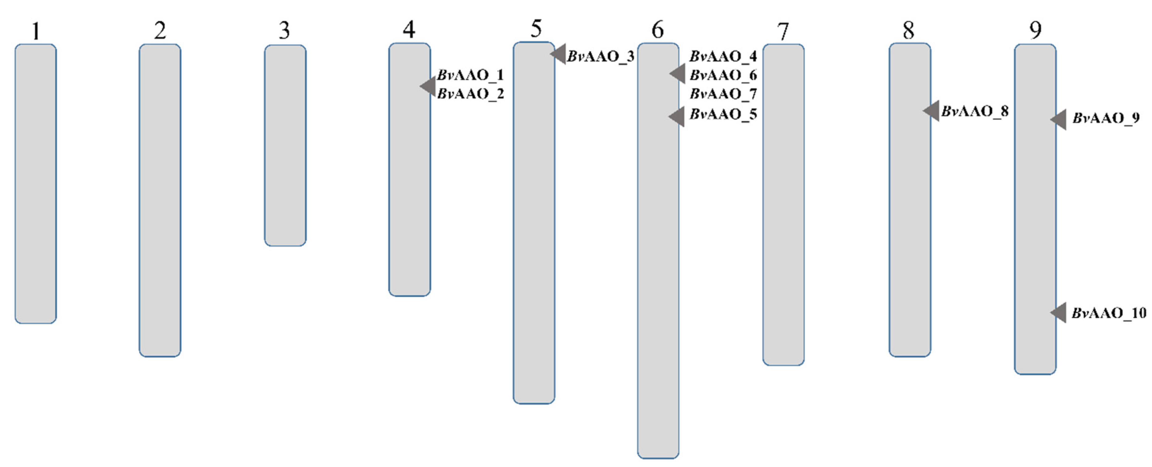
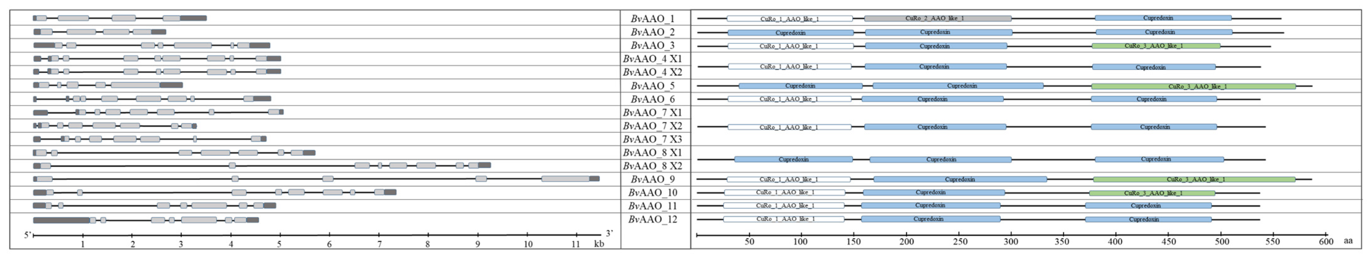
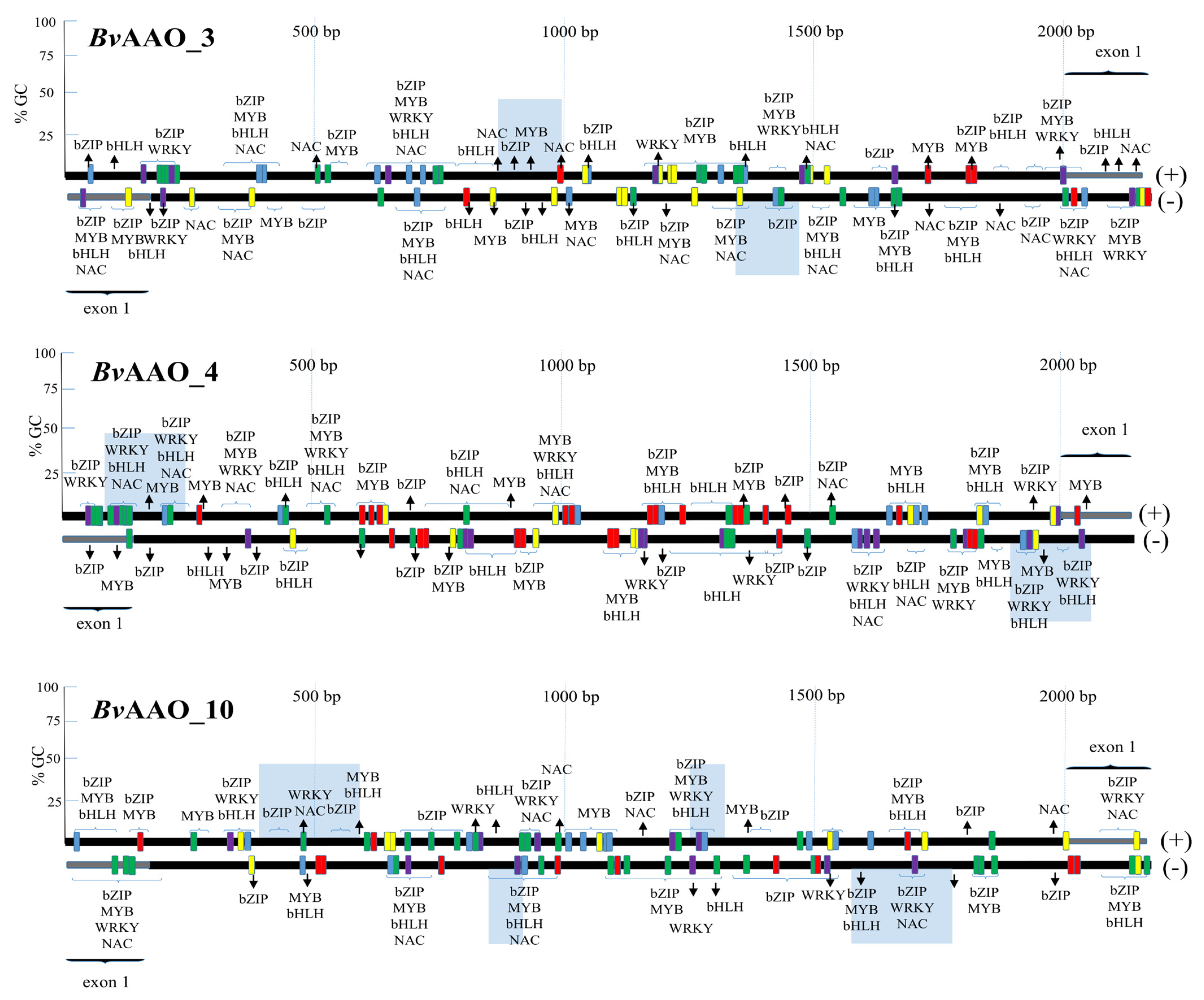
2.1. Characteristics of Genes and Proteins of Beta vulgaris Ascorbate Oxidases

Analysis of the RefBeet-1.2.2 beet reference genome deposited with GenBank allowed the identification of 12 ascorbate oxidase genes located on chromosomes 4, 5, 6, 8 and 9 (Figure 1). The two genes encoding this enzyme have not been assigned a chromosomal location. The exact characteristics of individual genes and the proteins they encode are presented in Table 1. For the purposes of this study, the genes were marked with consecutive numbers that follow the abbreviation of the enzyme name.
Figure 2 shows the structure of individual beet AAO genes. They differ significantly in the number and length of exons and introns. The BvAAO_8 and BvAAO_9 genes are the longest. The shortest were BvAAO_2 and BvAAO_5.
The proteins encoded by the AAO genes are composed of 537–588 amino acids (Table 1). Their molecular weight falls within a range of 59.68–65.9 kDa and the isoelectric point is between 6.68–9.84. According to computer analysis, the signal peptide is detected in all proteins except BvAAO_9. According to the outcome of Plant-mPLoc analysis, all beet AAO proteins are located in the cell membrane. In turn, analyzes using the YLoc program indicated the cytoplasmic localization of the BvAAO_2, BvAAO_4, BvAAO_5, BvAAO_6, BvAAO_8, BvAAO_9 and BvAAO_10 proteins; peroxisome localization in the case of BvAAO_1, BvAAO_3. The BvAAO_7 and BvAAO_11 proteins were identified by the program in the extracellular space and the BvAAO_12 protein was characterized by the secretory pathway (Table 1).
Conserved protein domains of cupredoxins have been detected in beet AAO proteins. These domains contain copper centers and are involved in intermolecular electron transfer reactions. Depending on the copper-binding site, there are types I, II and III of such domains (CuRo_1_AAO_like_1; CuRo_2_AAO_like_1; CuRo_3_AAO_like_1). The domain of the cupredoxin superfamily (Cupredoxin) also has a type 1 copper center. Although the AAO proteins may have different functions, 3 cupredoxin domains were detected in each of them. The presence of the different types of domains has an equal configuration for the individual proteins (Figure 2).

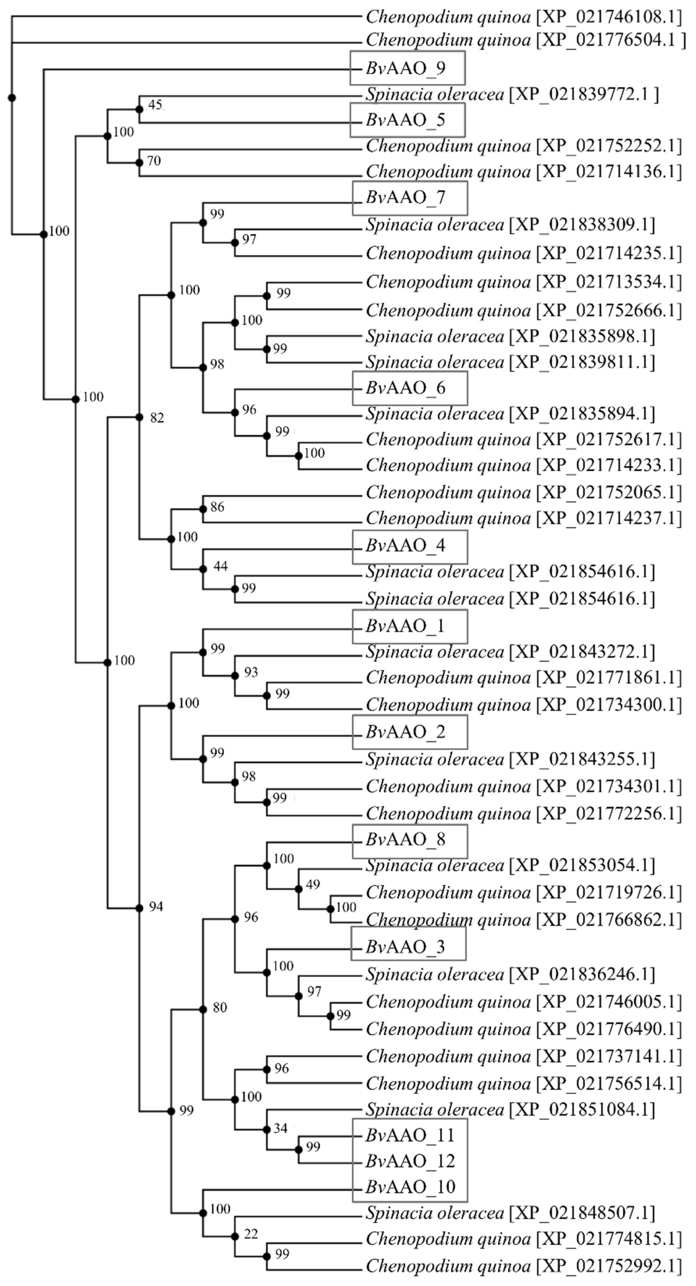
2.2. Phylogenetic and Evolutionary Analyses of AAO Gene Family

A phylogenetic tree was constructed based on the AAO Beta vulgaris protein sequences and according to the highest alignment index of the AAO protein sequences of Spinacia oleracea and Chenopodium quinoa. Based on the analysis of the reference genomes of these species, 13 spinach ascorbate oxidase sequences and 23 Chenopodium quinoa sequences were compared. Analyzing the evolutionary similarity of the beet AAO sequence, it can be noticed that the BvAAO_9 protein is evolutionarily most distant from the rest of the proteins. In addition, no close structural similarity to any of the AAO proteins of spinach and quinoa was demonstrated. Taking into account the next phylogenetic branch, we can distinguish here known groups, i.e., BvAAO_5 and the rest of the beet AAO proteins. Noticeable here also is the closer similarity of BvAAO_5 to the spinach protein sequence XP_021839772.1 and then to the two quinoa sequences XP_021752252.1 and XP_021714136.1. On the subsequent branches of the tree, individual beet AAO proteins are presented, which have their counterparts among the ascorbic oxidase proteins in the two plant species analyzed (Figure 3).

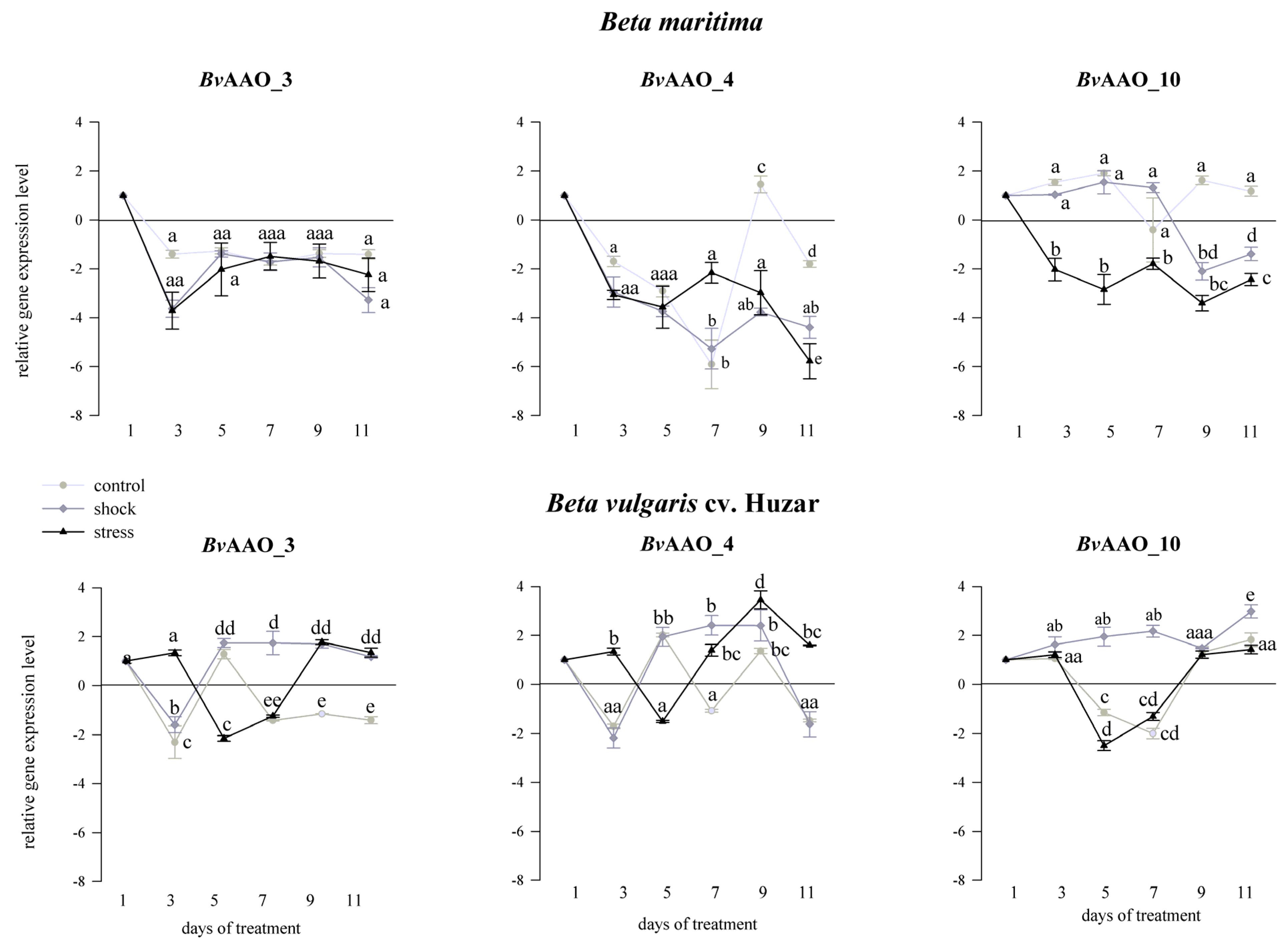
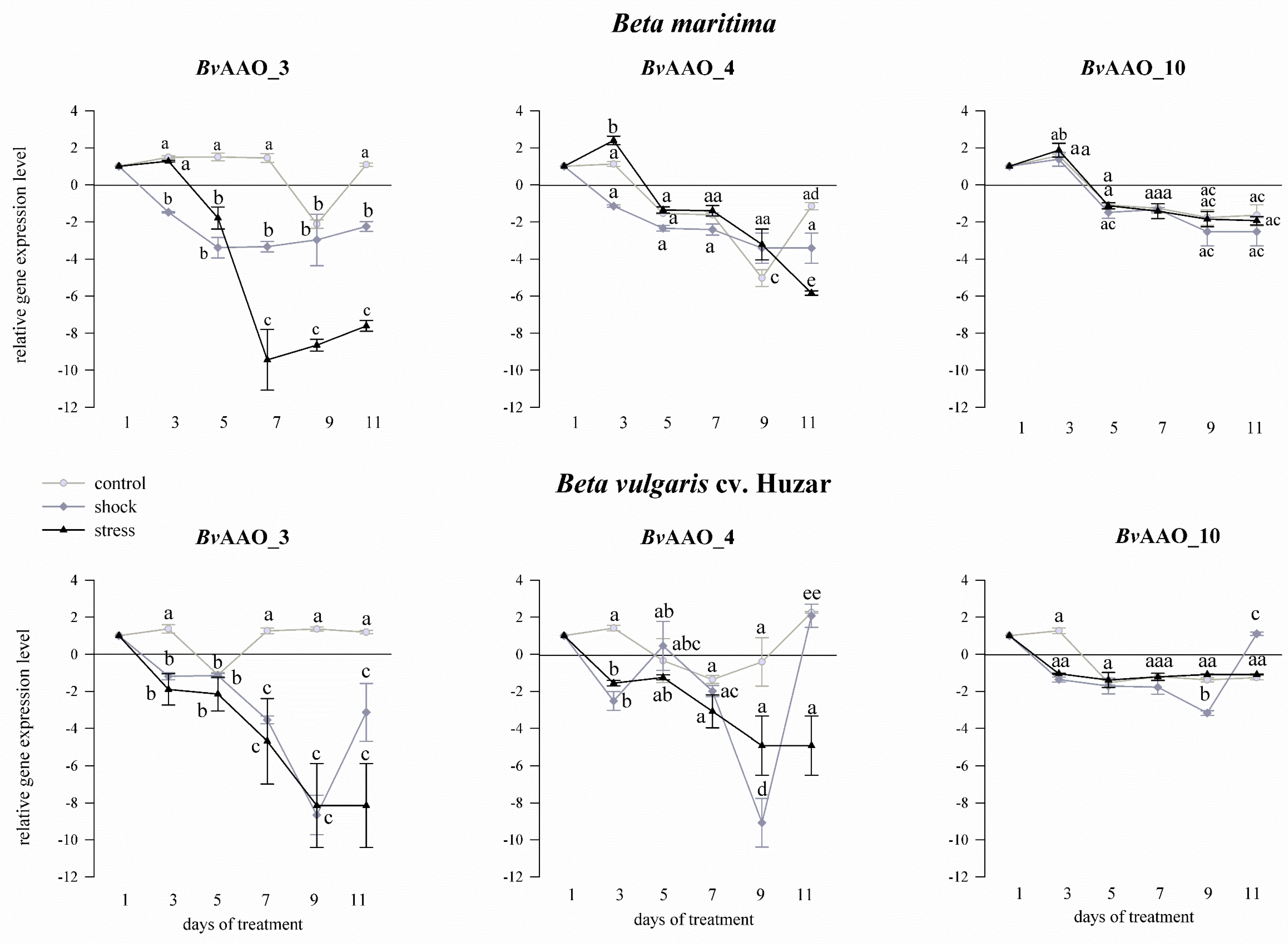
2.3. Analysis of the AAO Beta vulgaris Gene Expression Profile under Drought and Salinity Conditions

The expression level of AAO genes was determined in salt/drought shocked/stressed leaves of Beta vulgaris cv. Huzar and Beta maritima. Untreated controls were analyzed in line with the treated plants. Since only the BvAAO_3, BvAAO_4 and BvAAO_10 ascorbate oxidase genes were transcriptionally active in the analyzed tissue, the results of the expression analyzes are presented only for these genes.
When compared to the treatment starting day (“day 1”) a downregulation of BvAAO_3 expression was observed, in the control, shock or salt stress treated Beta maritima leaves, the reduced expression level maintained over the treatment period. There were no significant differences in BvAAO_3 expression levels between the control samples and those from plants treated with stress and salt shock. On the 3rd day, in the leaves of Beta vulgaris cv. Huzar BvAAO_3 expression was observed to be downregulated in the control and salt shock treated samples relative to day 1. In turn, the expression of BvAAO_3 in the samples treated with salt stress was upregulated (Figure 4). On the 5th day, the opposite effect was observed, i.e., an increase in BvAAO_3 expression in control plants and those representing salt shock and a significant decrease for the salt stress variant. This downregulation persisted until the 7th day of the treatment, after which an increase in the level of expression was observed until the end of the experiment. In the controls on the 7th day, there was a decrease in the expression level of BvAAO_3, which remained at a similar level on the 9th and 11th days. In the case of the samples representing salt shock, the upregulation of expression of this gene on the 5th day of treatment was maintained until the end of the experiment.
When the plants were treated with drought, the expression profile of the BvAAO_3 gene was different. In control samples, the transcript level in the two beet species was similar and did not change much. On the other hand, in the samples representing plants treated with shock and drought stress, a significant downregulation of BvAAO_3 expression was observed, as compared to the controls, at each sampling point (Figure 5).
Analyzing the effect of salinity, the expression of the BvAAO_4 gene in Beta maritima leaves was downregulated, relative to “day 1”, at all time-points of the experiment for the samples representing both the salt shock and salt stress treatment. The lowest transcript level was observed on the 11th day of the treatment. In the samples coming from control plants, the effect was similar except on day 9 when a slight increase in expression was observed. In leaves of Beta vulgaris cv. Huzar BvAAO_4 expression level varied depending on the treatment day and variant of the experiment. Relative to “day 1”, downregulation of this gene expression was observed on the 3rd, 7th and 11th day of the experiment, while on the 5th and 9th day a significant upregulation was observed in the control leaves. A similar profile of changes was observed under the salt shock treatment, except on the 7th day, in which the increase in the level of the BvAAO_4 transcript was maintained. In the case of samples representing salt stress on the 5th day, a decrease in the expression of this gene was observed, while on the remaining days of treatment a significant increase occurred (Figure 4).
Treating Beta maritima plants with drought, except for the stress variant on the 3rd day, brought about a decrease in the expression of the BvAAO_4 gene, observed in all experimental variants and on all treatment days. In leaves of Beta vulgaris cv. Huzar, the decrease in BvAAO_4 expression level was also observed in the stressed plants. The samples representing control plants showed unchanging transcript levels, except on the 11th day, when a slight increase was detected. Under drought shock, BvAAO_4 levels were similar to that observed in the stress variant on the 3rd, 5th and 7th days, but turned on the 11th day to the control level (Figure 5).
Taking drought into account, the expression level of BvAAO_10 in Beta maritima did not change significantly, as compared to “day 1”, in the samples representing control plants and those treated with salt shock up to the 7th day of the experiment. On the 9th and 11th days of treatment, a significant decrease in salt-shocked plants was observed. In contrast to the remaining variants, significant decreases in the level of the BvAAO_10 transcript were observed in plants treated with salt stress on each measurement day. In B. vulgaris cv. Huzar expression level of BvAAO_10 did not change compared to the control in the case of salt shock and on the 3rd, 9th and 11th days in the rest of the experimental variants. Significant downregulation of BvAAO_10 expression occurred on the 5th and 7th day of the experiment in control plants and plants treated with salt stress (Figure 4). In the drought testing experiment, the expression level of BvAAO_10 remained at a similar level compared to the control in all experimental variants in both beet genotypes (Figure 5).

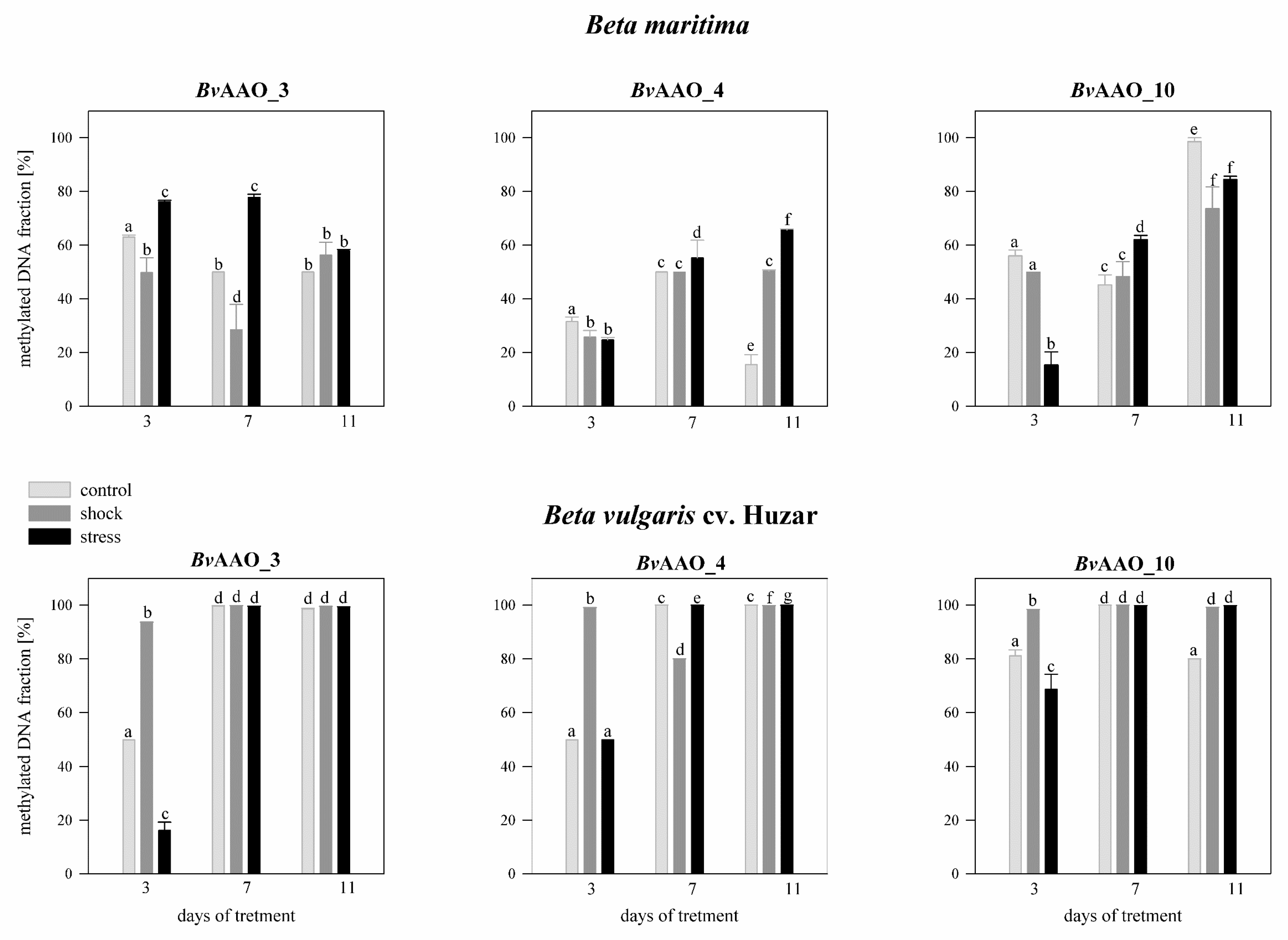
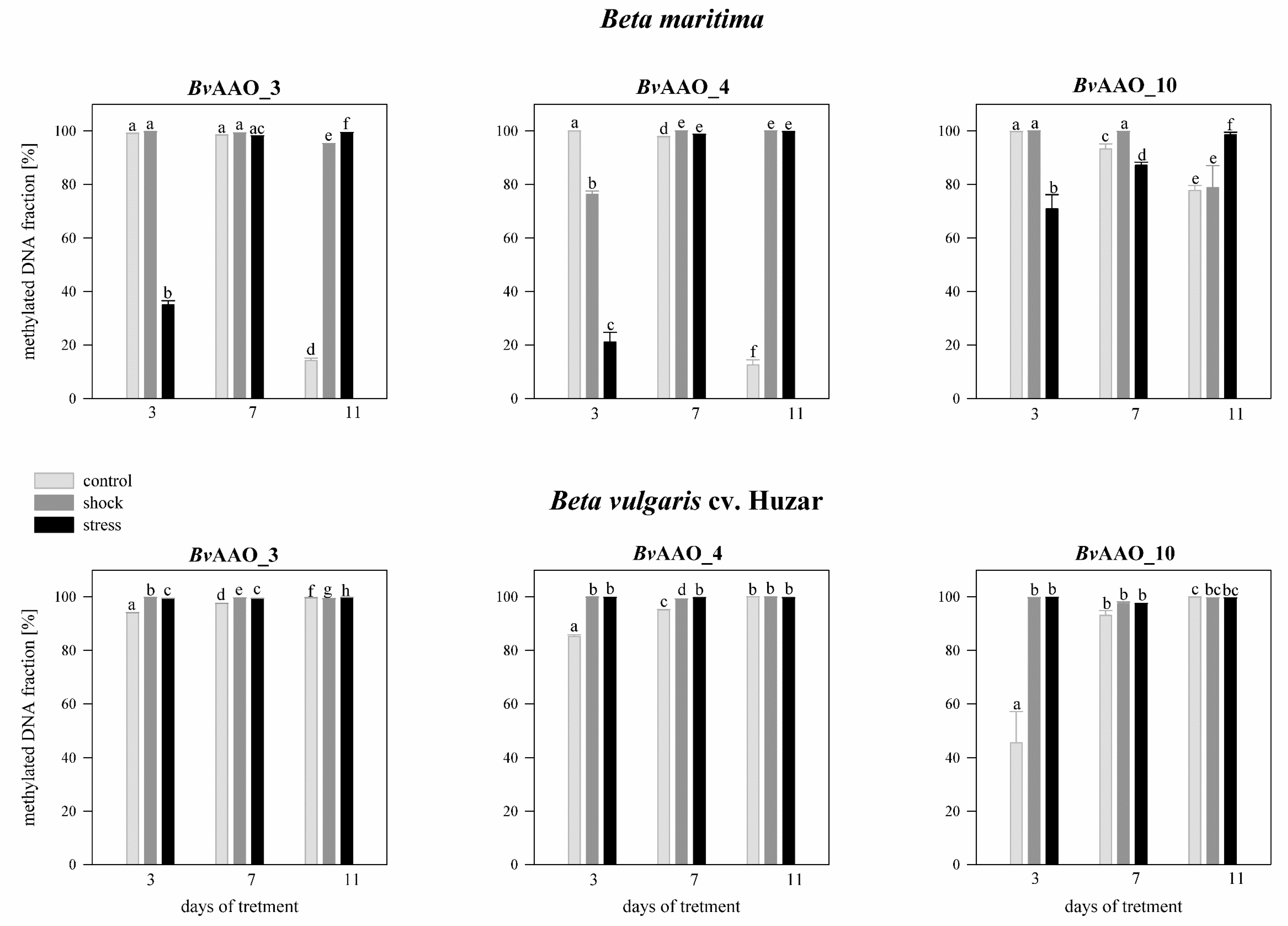
2.4. Analysis of the Methylation Level of the CpG Islands of the Promoter Regions of the AAO Beta vulgaris Genes under Drought and Salinity Conditions

Figure 6 shows the location of the CpG islands of the promoter regions of the BvAAO_3, BvAAO_4 and BvAAO_10 genes. Importantly, numerous sequences containing transcription factors (TFs)-binding motifs were identified in the analyzed regions. Among them, the sites binding TFs involved in the salinity and/or drought response, i.e., representing the bZIP, MYB, WRKY, NAC and bHLH families, were present (Figure 6).
Analyzing the effect of salinity on the methylation level of the BvAAO_3 gene CpG island in Beta maritima on day 3 of treatment, it was revealed that in the control plants methylation level was above 63%, whereas on the 7th and 11th days the methylation level decreased to about 50%. In the case of plants treated with salt shock, the methylation level of the CpG BvAAO_3 island was about 49%, on the 7th treatment day it decreased to 28% and on the 11th day it increased to 56%. In plants treated with salt stress, on the 3rd and 7th day of the experiment the methylation level was at the level of 76–78% and then on the 11th day of treatment it decreased to the level of the other variants. In Beta vulgaris cv. Huzar, on the 3rd day of treatment the level of CpG BvAAO_3 island methylation was 53% in the control plants, about 98% in shock-treated plants and 18% in salt-stressed. In the following days, the levels of methylation reached almost 100% in all variants of the experiment (Figure 7).
In drought-treated Beta maritima plants, the level of methylation of the CpG BvAAO_3 island, measured on the 3rd day of the experiment was close to 100% in the control and drought-shock variants, while in the case of plants subjected to drought stress, it was 35%. On the 7th day of treatment, the methylation level was close to 100% in all experimental variants. It persisted on day 11 in plants treated with drought shock and stress. Contrastingly, in control plants, it decreased to the level of 14%. In Beta vulgaris cv. Huzar the methylation level of the CpG BvAAO_3 island was close to 100% on all measurement days and in all experimental variants (Figure 8).
The methylation levels of the CpG island of BvAAO_4 in Beta maritima on day 3 amounted to about 31% in the control plants and about 24–25% in the plants treated with salt shock or stress. On the 7th day of the experiment, the methylation level increased to about 50–55% in all experimental variants. This level was maintained on day 11 in the salt-shock treated variant. In turn, in control plants, it decreased to the level of 13%, but in those treated with salt stress it increased to the level of 65%. In Beta vulgaris cv. Huzar observed a high percentage of methylation of the CpG BvAAO_3 island on the 7th and 11th day of treatment and on the 3rd day in the salt-shocked variant. In the remaining variants, on the 3rd day, the methylation amounted to 50% (Figure 7).
In an experiment aimed at analyzing the effect of drought on Beta maritima, the methylation level of the CpG island BvAAO_4 in the control plants was close to 100% on the 3rd and 7th days of treatment, but it decreased to about 12% on the 11th day. In plants treated with salt shock, the methylation level on the 3rd day of the experiment was about 75% and increased to nearly 100% on the 7th and 11th days. Similarly, in the drought- stressed variant it was 21% on the 3rd day of the experiment and increased to nearly 100% in the following days of treatment. In Beta vulgaris cv. Huzar, the percentage of methylation of CpG BvAAO_4 was very high in all experiment variants and all measurement times (Figure 8).
The level of CpG island methylation BvAAO_10 in Beta maritima, on day 3 of treatment, was about 50–56% in the control and salt shock-treated plants and about 15% in the salt stress-treated plants. In the case of the first two variants, methylation levels decreased slightly on day 7 of the experiment and increased significantly on day 11. In plants treated with salt stress, the level of CpG island methylation in the BvAAO_10 gene increased with the duration of the experiment, to 62% and 84%, respectively. In the control plants, Beta vulgaris cv. Huzar on the 3rd day showed an 81% methylation level of the CpG island, which increased to almost 100% on day 7 and then, on the 11th day, decreased to the level detected on the 3rd day. In plants treated with salt shock, the methylation level remained close to 100% during the course of the experiment. In the salt stress treatment variant, on the 3rd day of the experiment, the methylation level was 68% and on the 7th and 11th days it increased to almost 100% (Figure 7).
Analyzing the effect of drought revealed high methylation of CpG island in BvAAO_10 gene to levels reaching near 100% in Beta maritima on the 3rd and 7th days of treatment in the control variant and under drought-shock. Then, a decrease was observed on the 11th day of the experiment. In plants treated with drought stress, the methylation level increased with treatment time to 70%, 87% and 98%, respectively. In Beta vulgaris cv. Huzar high levels of near 100% CpG island methylation BvAAO_10 were observed in all variants and time points of the experiment, except for the control plants on the 3rd day (45%, Figure 8).

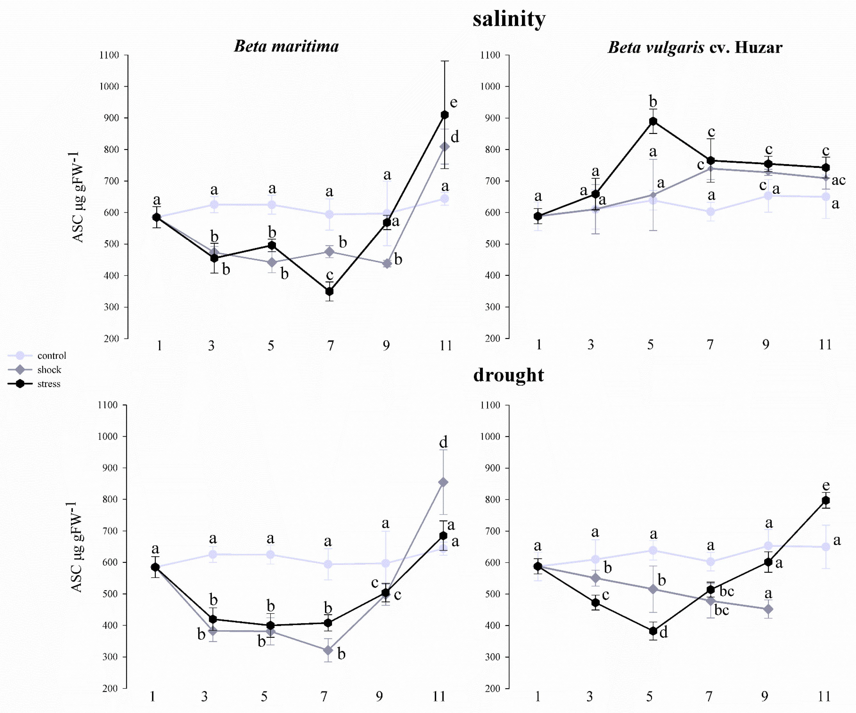
2.5. Analysis of Total Ascorbate Concentration in Beta vulgaris Leaves under Drought and Salinity Conditions

Changes in ascorbate content in leaves of Beta vulgaris ssp. maritima and Beta vulgaris cv. Huzar were tested over a 11-day-long in plants subjected to stress-generating factors or in those maintained under control conditions, which did not involve exposing plants to stress factors. The total ascorbate levels, i.e., the sum of its oxidized and reduced forms, were determined. Leaf total ascorbate concentrations were expressed as µg ASC per gram fresh weight (FW) of leaves and are presented in Figure 9.
A similar trend of changes in ascorbate content was observed in Beta maritima leaves treated with both salinity and drought, irrespective of the intensity of the treatment. From the 3rd to the 9th day of the experiment, a significant decrease in ASC concentration was observed, compared to the control. The exception was the salt -treated Beta maritima leaves on day 9, where the ASC level was similar to that in the control. On the 11th day, in the case of salt shock and salt stress, as well as drought shock, a significant increase in ASC concentration was observed (Figure 9). In Beta vulgaris cv. Huzar a profile of changes was different. The leaves of plants treated with salt stress on day 5 displayed a significant increase in ASC which was maintained until the end of the experiment. In leaves treated with salt shock, similar increase was observed only from the 7th day of the experiment. Changes in ASC concentration under drought conditions were comparable to those observed in Beta maritima. With respect of the untreated to the control, a significant decrease in ASC concentration occurred by the 9th day of treatment, except for the leaves of plants treated with salt stress. On the 11th day of the experiment, a significant increase in ASC concentration was observed in this variant, as compared to the control. The ASC concentration for the drought shock treatment variant on day 11th was not determined due to the death of the plants (Figure 9).

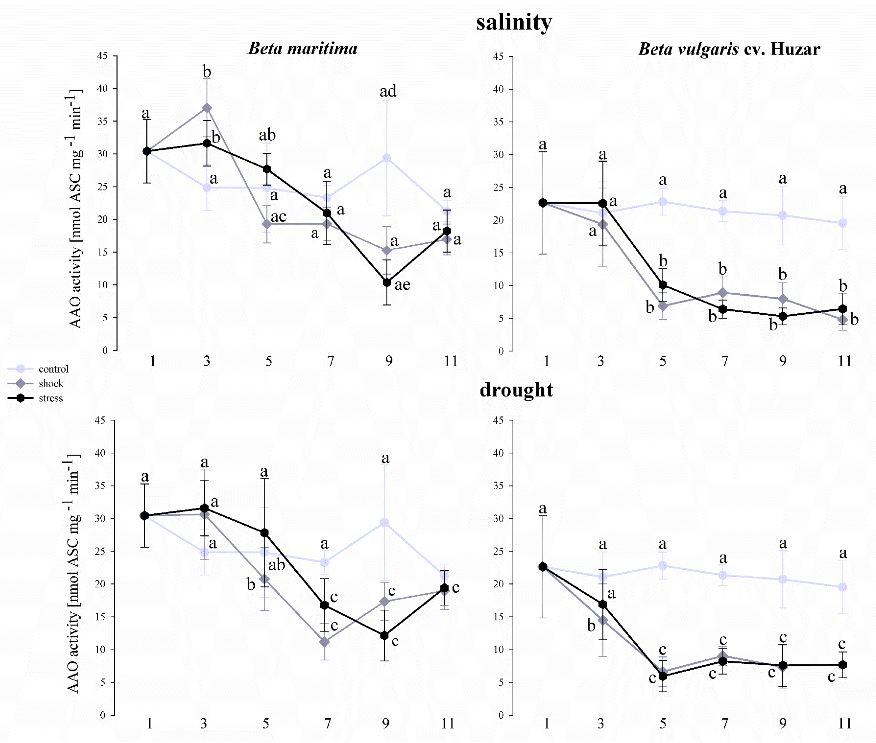
2.6. Analysis of Ascorbate Oxidase Activity in Beta vulgaris Leaves in Drought and Saline Conditions

The experiments also included the determination of AAO activity in two beet subspecies (Figure 10). In Beta maritima, an increase in enzyme activity was observed in leaves treated with both, salt shock and stress, on the 3rd day of the experiment, compared to the control. On day 5, this increase was still visible in the samples representing salt stress treatment, although the difference was statistically insignificant. Similarly, in the leaves representing the salt shock-treated leaves, there was a decrease in activity relative to the control on the 5th day of treatment, although these differences were not statistically significant. On the 7th and 11th days of the experiment, AAO activity did not differ between the variants. On day 9, a significant decrease in AAO activity was observed, relative to control, in leaves of plants treated with salt stress. The leaves treated with salt shock also showed a decrease in AAO activity, although the difference was not statistically significant when compared to untreated control. Under the drought treatment, the AAO activity was similar in all variants on the 3rd and 11th days of the experiment. On the 5th day a significant decrease in AAO activity relative to the control were observed in the samples representing drought shock-treated and on days 7th and 9th by drought shock and drought stress-treated plants. In Beta vulgaris cv. Huzar a significant decrease in AAO activity was observed between the 5th and 11th day of the experiment, in leaves treated with both saline and drought. Furthermore, a decrease in activity occurred in the leaves treated with drought shock on the 3rd day of treatment (Figure 10).

3. Discussion

AAO is a member of the polymorphic oxidase family that catalyzes redox reactions and plays an important role in many plant physiological processes. The enzyme could also have a role during stress responses modifying the ascorbic acid levels and therefore changing the cellular redox balance, which might provide the plant cell with an excellent system to sense the environment and react appropriately by contributing to the stress-related control. The expression of AAO is regulated by complex transcriptional and post-transcriptional controls, with transcript levels induced by growth promoters, e.g., auxin, jasmonates, and reduced by growth suppressors, e.g., salicylic acid [49]. On the other hand, experiments with many plants show that suppression of the AAO gene increases their stress tolerance, while overexpression has the opposite effect [50,51]. Overexpression of the AAO gene in tobacco inhibited germination and seed yield upon salt stress [51]. In another report, upregulation of cucumber AAO gene expression reduced the apo-plastic AA redox state and oxidative stress tolerance in tobacco plants, which is associated with the lower transcription of genes involved in AA recycling under oxidative stress conditions [52]. Transgenic tobacco overexpressing the AAO gene from cucumber displayed an increase in apo-plastic DHA accumulation, lower stomatal conductance and higher water content as compared to WT [4]. In contrast, suppression of the tomato AAO gene resulted in AA accumulation in fruits and enhanced drought stress tolerance by higher photosynthetic capacity [53]. Similarly, Yamamoto et al. (2005) reported that the transgenic tobacco plants that have suppressed AAO gene expression in antisense orientation showed increased germination, photosynthetic activity and seed yield and therefore salt tolerance compared to WT and sense plants under high salt concentrations [51].
This study aimed to characterize the ascorbate oxidase genes in Beta vulgaris, as well as to evaluate selected elements of the salinity and drought response at the epigenetic and transcriptional levels associated with AAO in the leaves of Beta vulgaris and its halophytic ancestor Beta maritima. Literature data show only two studies presenting the characteristics of ascorbate oxidase genes in other plant species [11,49]. Thus, there is no study of this enzyme in Beta vulgaris. The conducted analyses allowed distinguishing of 12 genes of beet ascorbate oxidase. Genomic characterization of other plant species revealed five AAO genes in Oryza sativa, three AAO genes in Arabidopsis thaliana, seven AAO genes in Glycine max, four AAO genes in Zea mays, six AAO genes in Sorghum bicolor and eight AAO genes in Gossypium hirsutum [11,49]. In each of the 12 beet AAO protein sequences, three from the cupredoxin domain were localized. This is consistent with the presented data of other studies characterizing the genes of ascorbate oxidases in other plant species [11,49]. Interestingly, individual proteins contain different configuration combinations of domain types, which can presumably be associated with different functions of individual AAO proteins and/or their different localization. Different computer programs point to different locations of the same beet AAO. The literature data is also inconsistent in this matter. Caputo et al. indicate that immunogold transmission electron microscopy analysis showed labeling of AAO in the cytosol, walls and chloroplasts of Chaetomorpha linum cells, while in angiosperms this enzyme is almost exclusively limited to the extracellular matrix [54]. According to other literature data, AAO activity is mainly related to the cell wall fraction. It has been suggested that it may play an important role in regulating the oxygen balance and controlling the growth of plant cells, perhaps by controlling transport processes through the plasma membrane [55]. Phylogenetic analysis shows that all beet AAO genes, except BvAAO_9, have structural equivalents in the genes of Spinacia oleracea and Chenopodium quinoa. Interestingly, this is the only beet protein sequence in which the program failed to detect a signal peptide.
The conducted experiments showed that the BvAAO_3, BvAAO_4 and BvAAO_10 genes were transcriptionally active in the leaves of two beet species. Sanmartin et al. also showed that of at least four AAO genes in Cucumis melo, only CmAO1 and CmAO4 were transcriptionally active. Moreover, researchers proved that their expression was regulated under the influence of wounding and heat stress [9]. The expression profiles of the BvAAO_3, BvAAO_4 and BvAAO_10 genes are different under salinity and drought treatments. The effects of shock and stress in both treatments also have a different effect on the expression level of AAO genes. Moreover, the transcriptional response of these three genes differs in the leaves of Beta vulgaris and its halophytic ancestor Beta maritima.
When analyzing the effect of salinity on the level of gene expression in Beta maritima, a decrease in the expression of all three genes was observed and, in the case of salt stress, a significant decrease in BvAAO_10 expression was observed compared to the control and salt shock-treated samples up to the 7th day of the experiment. A different situation was observed in Beta vulgaris cv. Huzar. Here, increases and decreases in expression were observed alternately and their directions were mostly opposite for the salt stress treated and non-salt stress treatments. Taking into account the fact that the comparison concerned the halophyte and the beet cultivar, it can be assumed that the important element of higher salinity tolerance is the suppression of the expression of ascorbate oxidase genes during the stress. It is also noticeable that the expression profile of the tested genes in the salt shock-treated samples is similar to that in the control, suggesting that the salt shock treatment of plants does not initiate changes in the regulation of transcription of AAO genes in most cases. Analyzing the impact of drought on the expression level of BvAAO_3, BvAAO_4 and BvAAO_10 genes, we observe its decrease in the treated samples or no effect. The BvAAO_3 gene in both beet species is characterized by a decrease in expression compared to the control, similar to the BvAAO_4 gene in Beta vulgaris cv. Huzar. We can see that, in contrast to salinity, a cultivated beet genotype downregulates the expression of two AAO genes during drought, while Beta maritima only one. The sole literature data on the regulation of the expression level under salt and drought stress were presented by Batth et al. [49]. The researchers also showed different expression profiles depending on the AAO gene and the type of stress. Similar observations were made in the case of Oryza sativa. Here, the downregulation of the OsAAO2 gene under salt stress and the downregulation of the OsAAO2, OsAAO3 and OsAAO5 genes under drought conditions were observed. Conversely, in the case of most AAO genes in Arabidopsis thaliana and Zea Mays, salt stress and drought caused their over-expression [49]. These observations may also be due to differences in salt and drought stress tolerance by these three plant species, which result in different regulations of AAO gene expression.
Analyzing the effect of salinity on the level of methylation of the CpG islands of the promoter regions of the BvAAO_3, BvAAO_4 and BvAAO_10 genes, we also observe differences depending on the intensity and type of stress, as well as the beet genotype. In the case of salinity, the methylation level of the CpG islands of the studied genes in Beta vulgaris cv. Huzar was much higher than in the halophytic ancestor. This observation does not translate into a decrease in the expression level of AAO genes in this genotype. The explanation for this may be the involvement of other epigenetic factors, which may directly or indirectly affect gene expression and/or modify the effects of DNA methylation [42,56,57]. Taking into account the salt stress, in all cases there was an increase in the level of CpG island methylation with the duration of the experiment. When analyzing the treatment of plants with drought, a higher degree of methylation of the CpG islands of the AAO genes is also observed in the beet cultivar. In Beta martima, on the other hand, these values are significantly higher than the levels of methylation in the salinity treatment. As there is no literature data on the regulation of AAO gene methylation levels in beet and other plant species, we cannot compare the obtained results. However, there is no doubt that, during salinity or drought, selected epigenetic elements are regulated to increase the level of DNA methylation and thus silence the expression of the ascorbate oxidase genes of two beet species.
Ascorbate is a very important component of plant cells due to its numerous functions. One of them is ROS detoxification, which is especially important under oxidative stress-generating conditions. ASC scavenges singlet oxygen, superoxide anion and hydroxyl radical. Moreover, it is electron donor in many necessary redox reactions [10]. Studies on transgenic plants with reduced ascorbate production indicate a significant increase in the amount of hydrogen peroxide, which exerts a destructive effect on many areas of cellular functioning [58]. In turn, it has been confirmed that exogenous application of ASC mitigates the effects of salinity and stimulates plant regeneration after stress, [59,60]. In our study, we observed an initial decrease in ASC concentration in leaves of Beta maritima treated with salinity and in leaves of both drought-treated beet genotypes, compared to the control. After this time, in these experimental systems, a significant increase in ASC concentration occurred, compared to the control. In turn, in leaves of Beta vulgaris cv. Huzar treated with salinity, an increase in ASC was observed, lasting from the 5th day of the experiment. The initial decrease in ASC could indicate that the pool of this type of antioxidant was partially used up, as a result of the ROS detoxification. Similar results were observed, by Chen et al., 2021 [61]. The increase in total ASC concentration over time may indicate that mechanisms leading to an increase in its amount under stress conditions are activated. One such mechanism may be the reduction in the expression level of genes encoding AAO, which results in a decrease in the amount and activity of this enzyme. The increase in ASC concentration under abiotic stress conditions was also confirmed in other studies [62,63]. Analyses aimed at the determination of AAO activity in our experimental system showed a decrease in the activity of this enzyme over time of treatments with stress-inducing factors. This correlated with our results, demonstrating the reduction of AAO gene expression under stress conditions. AAO regulates the redox state of the apoplast [10]. It has been proven that reducing the activity of this enzyme is promotes increase in plant resistance to oxidative stress since, under limited AAO activity, larger amounts of reduced ascorbate is maintained and this may be used for ROS scavenging [4,51]. Consequently, increasing the ascorbic acid concentration allows for a reduction in the amount of H2O2 and, consequently, reduces sensitivity to stress [10,64]. It should be kept in mind that the response to salinity differs between the two beet genotypes analyzed. Beta maritima is a halophytic plant and thus is more resistant to salinity than Beta vulgaris cv. Huzar. Therefore, it is possible to observe the activation of other mechanisms of the response to salinity, which involve different rate of ascorbate production and/or consumption.

4. Materials and Methods

4.1. Identification of Ascorbate Oxidase Sequences in Beta vulgaris

The source of the gene sequences analyzed in the study of Beta vulgaris subsp. vulgaris was the reference genome of this plant with the number RefBeet-1.2.2 deposited at GenBank (https://www.ncbi.nlm.nih.gov/gene/?term=ascorbate%20oxidase%20beta%20vulgaris; accessed on 5 January 2021).

4.2. Bioinformatic Analysis

The open reading frames (ORFs) of AAO genes were predicted using the ORF finder (https://www.ncbi.nlm.nih.gov/orffinder/; accessed on 10 January 2021). For a few, the longest ORF was chosen. Parameters such as molecular weight (MW), amino acid (AA) composition and theoretical isoelectric point (pI) were determined using the ProtParam of the Expasy package (https://web.expasy.org/protparam/; accessed on 21 February 2021). The presence of signal peptide was predicted with SignalP-5.0 (http://www.cbs.dtu.dk/services/SignalP/; accessed on 21 February 2021) and subcellular targeting was predicted with Plant mPLoc (http://www.csbio.sjtu.edu.cn/bioinf/plant-multi/; accessed on 21 February 2021) and Yloc (https://bio.tools/yloc; accessed on 21 February 2021). The chromosomal positions and structure of the beet AAO genes were acquired from the NCBI genome browser. Selected cis-regulatory elements were identified, in the sequences, using the Database of Plant Cis-acting Regulatory DNA Elements—New PLACE (https://www.dna.affrc.go.jp/PLACE/?action=newplace; accessed on 30 February 2021), while the potential sites recognized by selected salt-related transcription factors were determined using the Plant Promoter Analysis Navigator PlantPAN 2.0 program (http://plantpan2.itps.ncku.edu.tw/; accessed on 30 February 2021).

4.3. Sequence Alignment and Phylogenetic Analysis

Alignment and phylogenetic trees were constructed in CLC Sequence Viewer 8.0 (Qiagen, CLC bio A/S, Aarhus, Denmark). Phylogenetic and molecular evolutionary analyses were conducted using the neighbor-joining method according to the amino acids sequence with 1000 bootstrap replicates.

4.4. Plant Material

Beta vulgaris ssp. maritima (B. maritima) and sugar beet, Beta vulgaris ssp. vulgaris cv. Huzar were used as plant material. B. maritima seeds were obtained from National Germplasm Resources Laboratory (Beltsville, MD, USA) and the sugar beet seeds from Greater Poland Sugar Beet Breeding–WHBC (Poznań, Poland). The seeds (5 per pot) were sown into pots filled with sand and vermiculite (1/1 v/v) and plants were watered regularly with half-strength Hoagland solution. Plants were cultured for four weeks in a growth chamber with a photoperiod of 16 h of light and 8 h of darkness with standard irradiation of 30 ± 5 µmol m−2 s−1, provided by T8 15 W 6500 K “Daylight” tubes (POLAMP, Ełk, Poland). The temperature regime was 25 °C during the day and 18 °C at the night. Subsequently, treatments with salinity or drought were performed.

4.5. Exposing Plants to Salinity

Salt treatment was started when the first pair of mature leaves were fully developed. Salt shock—Plants were watered, over an 11-day-long treatment period, in two-day-long intervals, with half-strength Hoagland medium supplemented with 150 mM NaCl. Untreated controls were watered with a NaCl-unsupplemented medium. The leaf samples representing intact leaf blades were collected for analysis on first, third, fifth, seventh, ninth and eleventh day of the experiment (Skorupa et al., 2019). Salt stress—Over the first 11 days of treatment, plants were watered in two-day-long intervals, with half-strength Hoagland medium supplemented with increasing concentrations of NaCl, until the final concentration of 150 mM NaCl was reached. (first day of treatment–25 mM NaCl, third day–50 mM NaCl, fifth day–75 mM NaCl, seventh day–100 mM NaCl, ninth day–125 mM NaCl, eleventh day–150 mM NaCl). Untreated controls were watered with a NaCl-unsupplemented medium. Plants were watered with 200 mL of solution per 2 l of sand/vermiculite mixture. Leaves representing the second pair of true leaves were collected for analysis.

4.6. Exposing Plants to Drought

Drought treatment was started when the first pair of mature leaves were fully developed. Drought shock—Over 11 days of treatments, plants were not watered. Untreated controls were watered with a 30 mL half-strength Hoagland medium. The leaf samples representing intact leaf blades were collected for analysis on the first, third, fifth, seventh, ninth and eleventh days of the experiment. Drought stress—Over 11 days of treatment, plants were watered in two-day-long intervals, with half-strength Hoagland medium with decreasing water volume. (1st day of treatment–30 mL/per pot, third day–25 mL/per pot, fifth day–20 mL/per pot, seventh day–15 mL/per pot, ninth day–10 mL/, eleventh day–5 mL/per pot). Untreated controls were watered with a 30 mL NaCl-unsupplemented medium. Plants were watered with 200 mL of solution per 2 l of sand/vermiculite mixture. Leaves representing the second pair of true leaves were collected for analysis.
62 experimental variants were analyzed in the experiments. Two variants were represented by Beta maritima and Beta vulgaris cv. Huzar leaves on the day of starting the experiment, before applying the stresses. Sixty experimental variants were represented by the leaves of two beet species grown under control conditions or treated with drought or salinity on the respective leaf harvest days. In the case of RT qPCR analyses and methylation analyses, the number of analysis samples was: 62 experimental variants × 3 biological replicates × 3 technical repetitions = 558. For the determination of total ascorbate and AAO activity, the number of analyzed samples was: 62 experimental variants × 6 biological replicates × 3 repetitions technical = 1116.

4.7. RT qPCR Analysis

Total RNA was isolated from the same leaf samples using the TriPure reagent (Roche Diagnostics GmbH, Mannheim, Germany). Total RNA preparations were subjected to DNase treatment, which was carried out using DNase I (Thermo Scientific, Vilnius, Lithuania) according to the manufacturer’s instructions. The first-strand cDNA was synthesized from 1 μg of total RNA using random hexamers and Transcriptor High Fidelity cDNA Synthesis Kit (Roche Diagnostics GmbH, Mannheim, Germany), following the manufacturer’s instructions. Gene-specific primers were designed using Primer3Plus software. To ensure the correctness of the quantification, the expression was normalized to the β-actin reference gene. To determine the PCR efficiencies, standard curves for both target and control genes were obtained using a series of cDNA dilutions as a template. RT qPCR was performed in three biological replicates, each of which consisted of three technical replicates, using FastStart SYBR Green Master Mix, following the standard PCR program suggested by the manufacturer (Roche Diagnostics GmbH, Mannheim, Germany). The relative level of gene expression was calculated according to the Pfaffl method. The control against which the calculations were performed was represented by the leaves of the two beet genotypes at the start of the experiment, before the start of the stress treatment. Blank controls without a cDNA template were included in every experimental setup. A list of PCR primers used for experimental is provided in Table S1. The ordinal number of the primer sequences in Table S1 for the BvAAO_3, BvAAO_4, BvAAO_10 0 genes, respectively, are 3, 4 and 10. The qPCR efficiency values for the genes analyzed are summarized in Table S2.

4.8. Analysis of the CpG Island Methylation Level of Selected Beet AAO Genes

The level of methylation of the CpG islands of the promoter regions of the BvAAO_3, BvAAO_4 and BvAAO_10 genes was analyzed. Measurements were taken on the 3rd, 7th and 11th day of treating plants with saline or drought. The location of the islands and the analysis of their methylation level was determined according to Skorupa et al. 2021 [24]. The primer sequences for analysis are listed in the Table S1. The qPCR efficiency values for the genes analyzed are summarized in Table S2.

4.9. Determination of the Concentration of Total Ascorbate

150 mg of leaf tissue was extracted in a mortar with 1 mL of 5% TCA. The samples were then centrifuged at 14,000× g and 4 °C for 10 min. 16.8 μL of 10 mM DTT and 80 mM K2HPO4 were added to 135 μL of supernatant and incubated for 5 min at RT. In the next step, 40 µL of 85% H3PO4, 685 µL of 0.5% α, α′-dipyridyl and 140 µL of 1% FeCl3 were added successively, followed by incubation for 30 min at RT and centrifugation at 14,000× g for 5 min. The absorbance values were measured spectrophotometrically at a wavelength of 525 nm (Analytik Jena, Jena, Germany). The blank was a sample containing water with 5% TCA, instead of a plant extract. Ascorbate concentrations (μg/mL) were read from a standard curve prepared from a series of ascorbic acid concentrations. Leaf ascorbate concentrations are expressed as µg ascorbate/gFW.

4.10. Measurement of the Enzymatic Activity of Ascorbate Oxidase

200 mg of plant tissue was homogenized in 1 mL of extraction buffer (66 mM Na+/K+ phosphate buffer pH 6.1, 0.5 mM EDTA) and centrifuged for 10 min at 10,000× g at 4 °C. The AAO activity was calculated on the basis of the change in absorbance at the wavelength of 265 nm for 60 s, in the reaction mixture with the following composition: 960 μL of the buffer (66 mM Na+/K+ phosphate buffer pH 6.1), 20 μL of the sample and 20 μL of 2.5 mM acid ascorbic acid. The calculations were made using the millimolar absorption coefficient for ascorbic acid at 265 nm, which is 14 mM−1 cm−1. The enzyme activity was expressed in AAO, nmol ASC mg of total protein−1 min−1].

4.11. Statistics

Statistical significance of differences between mean values were determined with two-way ANOVA followed by Tukey’s HSD test in SigmaPlot 2008 (Systat Software, Palo Alto, Santa Clara, CA, USA). Differences at the level of p < 0.01 were considered as significant. Mean and standard deviation were calculated. Error bars shown in all figures represent the standard deviation calculated from all repetitions of each experiment.

5. Conclusions

The response of a sugar beet cultivar Beta vulgaris cv. Huzar and its halophytic ancestor Beta maritima to drought and salinity is manifested by downregulating the expression of ascorbate oxidase genes. This correlated with a decrease in AAO enzyme activity in a given experimental setup. Silencing of the AAO gene expression is most pronounced in the downregulation of AAO transcripts in Beta maritima under salinity, compared to a crop and conversely, under drought, in Beta vulgaris cv. Huzar. Considering the high level of drought and salinity stress tolerance in wild beet, Beta maritima and relatively moderate tolerance to these stresses in a beet crop variety, suppression of AAO gene expression in beet leaves under stress conditions may contribute to the mechanism of increasing salinity and drought tolerance. Moreover, it can be concluded that the downregulation of BvAAO_10 expression plays a key role in response to salinity treatment in Beta maritima, whereas downregulation of the BvAAO_3 gene is predominant under drought in both beet genotypes. This phenomenon may be involved in determining the high tolerance of beet to salinity and drought.

Supplementary Materials

The following supporting information can be downloaded at: https://www.mdpi.com/article/10.3390/ijms232112773/s1.

Author Contributions

Conceptualization, M.S. and J.T.; methodology, M.S. and J.T; software, M.S. and J.S.; validation, M.S. and J.S.; formal analysis, J.S.; investigation, M.S. and J.M.; data curation, M.S.; writing—original draft preparation, M.S., S.Y. and J.T; writing—review and editing, M.S.; S.Y. and J.T. visualization, M.S.; supervision, J.T. and A.T.; project administration, M.S.; funding acquisition, M.S. All authors have read and agreed to the published version of the manuscript.

Funding

This research was funded in part by National Science Center, Poland, UMO-2021/43/D/NZ9/00302.

Institutional Review Board Statement

The study was conducted in accordance with the Declaration of Helsinki.

Conflicts of Interest

The authors declare no conflict of interest.

References

  1. Foyer, C.H.; Kyndt, T.; Hancock, R.D. Vitamin C in Plants: Novel Concepts, New Perspectives, and Outstanding Issues. Antioxid. Redox Signal. 2020, 32, 463–485. [Google Scholar] [CrossRef] [PubMed]
  2. Pignocchi, C.; Foyer, C.H. Apoplastic ascorbate metabolism and its role in the regulation of cell signalling. Curr. Opin. Plant Biol. 2003, 6, 379–389. [Google Scholar] [CrossRef]
  3. Pignocchi, C.; Kiddle, G.; Hernandez, I.; Foster, S.J.; Asensi, A.; Taybi, T.; Barnes, J.; Foyer, C.H. Ascorbate oxidase-dependent changes in the redox state of the apoplast modulate gene transcript accumulation leading to modified hormone signaling and orchestration of defense processes in tobacco. Plant Physiol. 2006, 141, 423–435. [Google Scholar] [CrossRef] [Green Version]
  4. Fotopoulos, V.; De Tullio, M.C.; Barnes, J.; Kanellis, A.K. Altered stomatal dynamics in ascorbate oxidase over-expressing tobacco plants suggest a role for dehydroascorbate signalling. J. Exp. Bot. 2008, 59, 729–737. [Google Scholar] [CrossRef]
  5. De Tullio, M.C.; Guether, M.; Balestrini, R. Ascorbate oxidase is the potential conductor of a symphony of signaling pathways. Plant Signal. Behav. 2013, 8, e23213. [Google Scholar] [CrossRef] [PubMed] [Green Version]
  6. Smirnoff, N. The function and metabolism of ascorbic acid in plants. Ann. Bot. 1996, 78, 661–669. [Google Scholar] [CrossRef] [Green Version]
  7. Garchery, C.; Gest, N.; Do, P.T.; Alhagdow, M.; Baldet, P.; Menard, G.; Rothan, C.; Massot, C.; Gautier, H.; Aarrouf, J.; et al. A diminution in ascorbate oxidase activity affects carbon allocation and improves yield in tomato under water deficit. Plant Cell Environ. 2013, 36, 159–175. [Google Scholar] [CrossRef] [PubMed]
  8. Singh, R.R.; Nobleza, N.; Demeestere, K.; Kyndt, T. Ascorbate Oxidase Induces Systemic Resistance in Sugar Beet Against Cyst Nematode Heterodera schachtii. Front. Plant Sci. 2020, 11, 591715. [Google Scholar] [CrossRef] [PubMed]
  9. Sanmartin, M.; Pateraki, I.; Chatzopoulou, F.; Kanellis, A.K. Differential expression of the ascorbate oxidase multigene family during fruit development and in response to stress. Planta 2007, 225, 873–885. [Google Scholar] [CrossRef]
  10. Xiao, M.; Li, Z.; Zhu, L.; Wang, J.; Zhang, B.; Zheng, F.; Zhao, B.; Zhang, H.; Wang, Y.; Zhang, Z. The Multiple Roles of Ascorbate in the Abiotic Stress Response of Plants: Antioxidant, Cofactor, and Regulator. Front. Plant Sci. 2021, 12, 598173. [Google Scholar] [CrossRef]
  11. Pan, Z.; Chen, L.; Wang, F.; Song, W.; Cao, A.; Xie, S.; Chen, X.; Jin, X.; Li, H. Genome-Wide Identification and Expression Analysis of the Ascorbate Oxidase Gene Family in Gossypium hirsutum Reveals the Critical Role of GhAO1A in Delaying Dark-Induced Leaf Senescence. Int. J. Mol. Sci. 2019, 20, 6167. [Google Scholar] [CrossRef] [PubMed] [Green Version]
  12. Ghaffari, H.; Tadayon, M.R.; Nadeem, M.; Cheema, M.; Razmjoo, J. Proline mediated changes in antioxidant enzymatic activities and physiology of sugar beet under drought stress. Acta Physiol. Plant. 2019, 41, 23. [Google Scholar] [CrossRef]
  13. Liu, H.; Wang, Q.; Yu, M.; Zhang, Y.; Wu, Y.; Zhang, H. Transgenic salt-tolerant sugar beet (Beta vulgaris L.) constitutively expressing an Arabidopsis thaliana vacuolar Na/H antiporter gene, AtNHX3, accumulates more soluble sugar but less salt in storage roots. Plant Cell Environ. 2008, 31, 1325–1334. [Google Scholar] [CrossRef] [PubMed]
  14. Maung, T.A.; Gustafson, C.R. The economic feasibility of sugar beet biofuel production in central North Dakota. Biomass and Bioenergy 2011, 35, 3737–3747. [Google Scholar] [CrossRef] [Green Version]
  15. Zhang, Y.; Nan, J.; Yu, B. OMICS Technologies and Applications in Sugar Beet. Front. Plant Sci. 2016, 7, 900. [Google Scholar] [CrossRef] [Green Version]
  16. Pinheiro, C.; Ribeiro, I.C.; Reisinger, V.; Planchon, S.; Veloso, M.M.; Renaut, J.; Eichacker, L.; Ricardo, C.P. Salinity effect on germination, seedling growth and cotyledon membrane complexes of a Portuguese salt marsh wild beet ecotype. Theor. Exp. Plant Physiol. 2018, 30, 113–127. [Google Scholar] [CrossRef]
  17. Skorupa, M.; Golebiewski, M.; Kurnik, K.; Niedojadlo, J.; Kesy, J.; Klamkowski, K.; Wojcik, K.; Treder, W.; Tretyn, A.; Tyburski, J. Salt stress vs. salt shock-the case of sugar beet and its halophytic ancestor. BMC Plant Biol. 2019, 19, 57. [Google Scholar] [CrossRef] [Green Version]
  18. Ribeiro, I.C.; Pinheiro, C.; Ribeiro, C.M.; Veloso, M.M.; Simoes-Costa, M.C.; Evaristo, I.; Paulo, O.S.; Ricardo, C.P. Genetic Diversity and Physiological Performance of Portuguese Wild Beet (Beta vulgaris spp. maritima) from Three Contrasting Habitats. Front. Plant Sci. 2016, 7, 1293. [Google Scholar] [CrossRef] [Green Version]
  19. Kraft, T.; Fridlund, B.; Hjerdin, A.; Sall, T.; Tuvesson, S.; Hallden, C. Estimating genetic variation in sugar beets and wild beets using pools of individuals. Genome 1997, 40, 527–533. [Google Scholar] [CrossRef]
  20. Taghizadegan, M.; Toorchi, M.; Moghadam Vahed, M.; Khayamim, S. Evaluation of sugar beet breeding populations based morpho-physiological characters under salinity stress. Pak. J. Bot. 2019, 51, 11–17. [Google Scholar] [CrossRef]
  21. Daoud, S.; Harrouni, C.; Huchzermeyer, B.; Koyro, H.W. Comparison of salinity tolerance of two related subspecies of Beta vulgaris: The sea beet (Beta vulgaris ssp. maritima) and the sugar beet (Beta vulgaris ssp. vulgaris). In Biosaline Agriculture and High Salinity Tolerance; Abdelly, C., Öztürk, M., Ashraf, M., Grignon, C., Eds.; Birkhäuser Basel: Basel, Switzerland, 2008. [Google Scholar] [CrossRef]
  22. Koyro, H.-W.; Daoud, S.; Harrouni, C.; Huchzermeyer, B. Strategies of a potential cash crop halophyte (Beta vulgaris ssp. maritima) to avoid salt injury. Trop. Ecol. 2006, 47, 191–200. [Google Scholar]
  23. Yolcu, S.; Alavilli, H.; Ganesh, P.; Panigrahy, M.; Song, K. Salt and Drought Stress Responses in Cultivated Beets (Beta vulgaris L.) and Wild Beet (Beta maritima L.). Plants 2021, 10, 1843. [Google Scholar] [CrossRef] [PubMed]
  24. Skorupa-Kłaput, M.; Szczepanek, J.; Kurnik, K.; Tretyn, A.; Tyburski, J. The expression patterns of plasma membrane aquaporins in leaves of sugar beet and its halophyte relative, Beta vulgaris ssp. maritima, in response to salt stress. Biologia 2015, 70, 467–477. [Google Scholar] [CrossRef]
  25. Dunajska-Ordak, K.; Skorupa-Klaput, M.; Kurnik, K.; Tretyn, A.; Tyburski, J. Cloning and Expression Analysis of a Gene Encoding for Ascorbate Peroxidase and Responsive to Salt Stress in Beet (Beta vulgaris). Plant Mol. Biol. Report 2014, 32, 162–175. [Google Scholar] [CrossRef] [PubMed] [Green Version]
  26. Bor, M.; Özdemir, F.; Türkan, I. The effect of salt stress on lipid peroxidation and antioxidants in leaves of sugar beet Beta vulgaris L. and wild beet Beta maritima L. Plant Sci. 2003, 164, 77–84. [Google Scholar] [CrossRef]
  27. Wisniewska, A.; Andryka-Dudek, P.; Czerwinski, M.; Choluj, D. Fodder beet is a reservoir of drought tolerance alleles for sugar beet breeding. Plant Physiol. Biochem. 2019, 145, 120–131. [Google Scholar] [CrossRef]
  28. Van Geyt, J.P.C.; Lange, W.; Oleo, M.; De Bock, T.S.M. Natural variation within the genus Beta and its possible use for breeding sugar beet: A review. Euphytica 1990, 49, 57–76. [Google Scholar] [CrossRef]
  29. Yu, B.; Chen, M.; Grin, I.; Ma, C. Mechanisms of Sugar Beet Response to Biotic and Abiotic Stresses. In Mechanisms of Genome Protection and Repair; Zharkov, D.O., Ed.; Springer International Publishing: Cham, Switzerland, 2020; pp. 167–194. [Google Scholar] [CrossRef]
  30. Rozema, J.; Cornelisse, D.; Zhang, Y.; Li, H.; Bruning, B.; Katschnig, D.; Broekman, R.; Ji, B.; van Bodegom, P. Comparing salt tolerance of beet cultivars and their halophytic ancestor: Consequences of domestication and breeding programmes. AoB Plants 2015, 7, plu083. [Google Scholar] [CrossRef] [Green Version]
  31. Wang, Y.; Zhan, Y.; Wu, C.; Gong, S.; Zhu, N.; Chen, S.; Li, H. Cloning of a cystatin gene from sugar beet M14 that can enhance plant salt tolerance. Plant Sci. 2012, 191, 93–99. [Google Scholar] [CrossRef]
  32. Chołuj, D.; Wisniewska, A.; Szafranski, K.M.; Cebula, J.; Gozdowski, D.; Podlaski, S. Assessment of the physiological responses to drought in different sugar beet genotypes in connection with their genetic distance. J. Plant Physiol. 2014, 171, 1221–1230. [Google Scholar] [CrossRef]
  33. Munns, R. Plant adaptations to salt and water stress: Differences and commonalities. Adv. Bot. Res. 2011, 57, 1–32. [Google Scholar] [CrossRef]
  34. Shavrukov, Y. Salt stress or salt shock: Which genes are we studying? J. Exp. Bot. 2013, 64, 119–127. [Google Scholar] [CrossRef] [PubMed] [Green Version]
  35. Talamè, V.; Ozturk, N.Z.; Bohnert, H.J.; Tuberosa, R. Barley transcript profiles under dehydration shock and drought stress treatments: A comparative analysis. J. Exp. Bot. 2007, 58, 229–240. [Google Scholar] [CrossRef] [PubMed] [Green Version]
  36. Wedeking, R.; Mahlein, A.K.; Steiner, U.; Oerke, E.C.; Goldbach, H.E.; Wimmer, M.A. Osmotic adjustment of young sugar beets (Beta vulgaris) under progressive drought stress and subsequent rewatering assessed by metabolite analysis and infrared thermography. Funct. Plant Biol. 2017, 44, 119–133. [Google Scholar] [CrossRef] [Green Version]
  37. Khayamim, S.; Afshari, R.T.; Sadeghian, S.Y.; Poustini, K.; Rouzbeh, F.; Abbasi, Z. Seed Germination, Plant Establishment, and Yield of Sugar Beet Genotypes under Salinity Stress. J. Agr. Sci. Technol. 2014, 16, 779–790. [Google Scholar]
  38. Lv, X.; Chen, S.; Wang, Y. Advances in Understanding the Physiological and Molecular Responses of Sugar Beet to Salt Stress. Front. Plant Sci. 2019, 10, 1431. [Google Scholar] [CrossRef] [Green Version]
  39. Romano, A.; Sorgonà, A.; Lupini, A.; Araniti, F.; Stevanato, P.; Cacco, G.; Abenavoli, M.R. Morpho-physiological responses of sugar beet (Beta vulgaris L.) genotypes to drought stress. Acta Physiol. Plant. 2013, 35, 853–865. [Google Scholar] [CrossRef]
  40. Szymanska, S.; Tyburski, J.; Piernik, A.; Sikora, M.; Mazur, J.; Katarzyna, H. Raising Beet Tolerance to Salinity through Bioaugmentation with Halotolerant Endophytes. Agronomy 2020, 10, 1571. [Google Scholar] [CrossRef]
  41. Kim, J.M.; To, T.K.; Nishioka, T.; Seki, M. Chromatin regulation functions in plant abiotic stress responses. Plant Cell Environ. 2010, 33, 604–611. [Google Scholar] [CrossRef]
  42. Skorupa, M.; Szczepanek, J.; Mazur, J.; Domagalski, K.; Tretyn, A.; Tyburski, J. Salt stress and salt shock differently affect DNA methylation in salt-responsive genes in sugar beet and its wild, halophytic ancestor. PLoS ONE 2021, 16, e0251675. [Google Scholar] [CrossRef]
  43. Mirouze, M.; Paszkowski, J. Epigenetic contribution to stress adaptation in plants. Curr. Opin. Plant Biol. 2011, 14, 267–274. [Google Scholar] [CrossRef]
  44. Colaneri, A.C.; Jones, A.M. Genome-Wide Quantitative Identification of DNA Differentially Methylated Sites in Arabidopsis Seedlings Growing at Different Water Potential. PLoS ONE 2013, 8, e59878. [Google Scholar] [CrossRef] [PubMed] [Green Version]
  45. Song, Y.; Ji, D.; Li, S.; Wang, P.; Li, Q.; Xiang, F. The Dynamic Changes of DNA Methylation and Histone Modifications of Salt Responsive Transcription Factor Genes in Soybean. PLoS ONE 2012, 7, e41274. [Google Scholar] [CrossRef] [PubMed]
  46. Paul, A.; Dasgupta, P.; Roy, D.; Chaudhuri, S. Comparative analysis of Histone modifications and DNA methylation at OsBZ8 locus under salinity stress in IR64 and Nonabokra rice varieties. Plant Mol Biol. 2017, 95, 63–88. [Google Scholar] [CrossRef] [PubMed]
  47. Choi, C.S.; Sano, H. Abiotic-stress induces demethylation and transcriptional activation of a gene encoding a glycerophosphodiesterase-like protein in tobacco plants. Mol. Genet. Genom. 2007, 277, 589–600. [Google Scholar] [CrossRef]
  48. Yolcu, S.; Ozdemir, F.; Güler, A.; Bor, M. Histone acetylation influences the transcriptional activation of POX in Beta vulgaris L. and Beta maritima L. under salt stress. Plant Physiol. Biochem. 2016, 100, 37–46. [Google Scholar] [CrossRef]
  49. Batth, R.; Singh, K.; Kumari, S.; Mustafiz, A. Transcript Profiling Reveals the Presence of Abiotic Stress and Developmental Stage Specific Ascorbate Oxidase Genes in Plants. Front. Plant Sci. 2017, 8, 198. [Google Scholar] [CrossRef] [PubMed] [Green Version]
  50. Abdelgawad, K.F.; El-Mogy, M.M.; Mohamed, M.I.A.; Garchery, C.; Stevens, R.G. Increasing ascorbic acid content and salinity tolerance of cherry tomato plants by suppressed expression of the ascorbate oxidase gene. Agronomy 2019, 9, 51. [Google Scholar] [CrossRef] [Green Version]
  51. Yamamoto, A.; Bhuiyan, M.N.; Waditee, R.; Tanaka, Y.; Esaka, M.; Oba, K.; Jagendorf, A.T.; Takabe, T. Suppressed expression of the apoplastic ascorbate oxidase gene increases salt tolerance in tobacco and Arabidopsis plants. J. Exp. Bot. 2005, 56, 1785–1796. [Google Scholar] [CrossRef] [Green Version]
  52. Fotopoulos, V.; Sanmartin, M.; Kanellis, A.K. Effect of ascorbate oxidase over-expression on ascorbate recycling gene expression in response to agents imposing oxidative stress. J. Exp. Bot. 2006, 57, 3933–3943. [Google Scholar] [CrossRef] [Green Version]
  53. Zhang, Y.; Li, H.; Shu, W.; Zhang, C.; Zhang, W.; Ye, Z. Suppressed Expression of Ascorbate Oxidase Gene Promotes Ascorbic Acid Accumulation in Tomato Fruit. Plant Mol. Biol. Report. 2010, 29, 638–645. [Google Scholar] [CrossRef]
  54. Caputo, E.; Ceglie, V.; Lippolis, M.; La Rocca, N.; Tullio, M. Identification of a NaCl-induced ascorbate oxidase activity in Chaetomorpha linum suggests a novel mechanism of adaptation to increased salinity. Environ. Exp. Bot. 2010, 69, 63–67. [Google Scholar] [CrossRef]
  55. He, S.B.; Hu, A.L.; Zhuang, Q.Q.; Peng, H.P.; Deng, H.H.; Chen, W.; Hong, G.L. Ascorbate Oxidase Mimetic Activity of Copper(II) Oxide Nanoparticles. Chembiochem 2020, 21, 978–984. [Google Scholar] [CrossRef]
  56. Garg, R.; Narayana Chevala, V.; Shankar, R.; Jain, M. Divergent DNA methylation patterns associated with gene expression in rice cultivars with contrasting drought and salinity stress response. Sci. Rep. 2015, 5, 14922. [Google Scholar] [CrossRef] [Green Version]
  57. Yaish, M.W.; Al-Lawati, A.; Al-Harrasi, I.; Patankar, H.V. Genome-wide DNA Methylation analysis in response to salinity in the model plant caliph medic (Medicago truncatula). BMC Genomics 2018, 19, 78. [Google Scholar] [CrossRef] [PubMed] [Green Version]
  58. Hoang, M.T.T.; Doan, M.T.A.; Nguyen, T.; Tra, D.-P.; Chu, T.N.; Dang, T.P.T.; Quach, P.N.D. Phenotypic Characterization of Arabidopsis Ascorbate and Glutathione Deficient Mutants under Abiotic Stresses. Agronomy 2021, 11, 764. [Google Scholar] [CrossRef]
  59. Farooq, A.; Bukhari, S.A.; Akram, N.A.; Ashraf, M.; Wijaya, L.; Alyemeni, M.N.; Ahmad, P. Exogenously Applied Ascorbic Acid-Mediated Changes in Osmoprotection and Oxidative Defense System Enhanced Water Stress Tolerance in Different Cultivars of Safflower (Carthamus tinctorious L.). Plants 2020, 9, 104. [Google Scholar] [CrossRef] [Green Version]
  60. Noman, A.; Ali, S.; Naheed, F.; Ali, Q.; Farid, M.; Rizwan, M.; Irshad, M.K. Foliar application of ascorbate enhances the physiological and biochemical attributes of maize (Zea mays L.) cultivars under drought stress. Arch. Agron. Soil Sci. 2015, 61, 1659–1672. [Google Scholar] [CrossRef]
  61. Chen, X.; Zhou, Y.; Cong, Y.; Zhu, P.; Xing, J.; Cui, J.; Xu, W.; Shi, Q.; Diao, M.; Liu, H.-y. Ascorbic Acid-Induced Photosynthetic Adaptability of Processing Tomatoes to Salt Stress Probed by Fast OJIP Fluorescence Rise. Front. Plant Sci. 2021, 12, 594400. [Google Scholar] [CrossRef] [PubMed]
  62. Airaki, M.; Leterrier, M.; Mateos, R.M.; Valderrama, R.; Chaki, M.; Barroso, J.B.; Del RÍO, L.A.; Palma, J.M.; Corpas, F.J. Metabolism of reactive oxygen species and reactive nitrogen species in pepper (Capsicum annuum L.) plants under low temperature stress. Plant Cell Environ. 2012, 35, 281–295. [Google Scholar] [CrossRef] [PubMed]
  63. Kanayama, Y.; Sato, K.; Ikeda, H.; Tamura, T.; Nishiyama, M.; Kanahama, K. Seasonal changes in abiotic stress tolerance and concentrations of tocopherol, sugar, and ascorbic acid in sea buckthorn leaves and stems. Sci. Hortic. 2013, 164, 232–237. [Google Scholar] [CrossRef]
  64. Venkatesh, J.; Park, S.W. Role of L-ascorbate in alleviating abiotic stresses in crop plants. Bot. Stud. 2014, 55, 38. [Google Scholar] [CrossRef] [PubMed]
Figure 1. Chromosomal positions of AAO genes in Beta vulgaris.
Ijms 23 12773 g001
Figure 2. Exon−intron structure (left side) and conserved protein domain family (right side) of AAO genes in Beta vulgaris. UTRs−dark gray rectangles, exons−light gray rectangles, introns−black lines, the first cupredoxin domain of plant ascorbate oxidase homologs−white rectangles, the second cupredoxin domain of plant ascorbate oxidase homologs−gray rectangle, the third cupredoxin domain of plant ascorbate oxidase homologs−green rectangles, cupredoxin superfamily domain−blue rectangles.
Ijms 23 12773 g002
Figure 3. Phylogenetic analysis of AAO genes. Trees were built using amino acid sequences from Beta vulgaris, Spinacia oleracea and Chenopodium quinoa. The linear phylogram was constructed using the neighbor−joining method and bootstrapped 1000 times. Beet AAO genes are marked with gray boxes.
Ijms 23 12773 g003
Figure 4. Expression profiles of AAO genes BvAAO_3, BvAAO_4 and BvAAO_10 determined in leaves of Beta vulgaris ssp. maritima and Beta vulgaris cv. Huzar subjected to salt shock or stress. Control−light gray lines, salt shock−gray lines, salt stress−dark gray lines. Different letters denote significant differences at p < 0.01 (ANOVA followed by Tukey’s test). “Whiskers” indicate standard deviation.
Ijms 23 12773 g004
Figure 5. Expression profiles of AAO genes BvAAO_3, BvAAO_4 and BvAAO_10 determined in leaves of Beta vulgaris ssp. maritima and Beta vulgaris cv. Huzar subjected to drought shock or stress. Control−light gray lines, drought shock−gray lines, drought stress−dark gray lines. Different letters denote significant differences at p < 0.01 (ANOVA followed by Tukey’s test). “Whiskers” indicate standard deviation.
Ijms 23 12773 g005
Figure 6. Structure and motif composition of the promoter regions of AAO genes BvAAO_3, BvAAO_4 and BvAAO_10. The analyses were performed on sugar beet sequences derived from the RefBeet-1.2.2 reference genome. CpG islands−light blue squares, selected cis−regulatory sequence elements (TATABOX−red squares, CAATBOX−green squares, WRKY−purple squares, MYB−yellow squares, GATABOX−blue squares) and potential places recognized by specific stress−related transcription factors (bZIP, MYB, WRKY, bHLH, NAC) are shown. “+”−sense strand; “−” antisense strand.
Ijms 23 12773 g006
Figure 7. Methylated DNA fraction [%] of CpG islands in the promoter region of AAO genes BvAAO_3, BvAAO_4 and BvAAO_10 determined in leaves of Beta vulgaris ssp. maritima and Beta vulgaris cv. Huzar subjected to salt shock or stress. Control−light gray bars, salt shock−gray bars, salt stress−black bars. Different letters denote significant differences at p < 0.01 (ANOVA followed by Tukey’s test). “Whiskers” indicate standard deviation.
Ijms 23 12773 g007
Figure 8. Methylated DNA fraction [%] of CpG islands in the promoter region of AAO genes BvAAO_3, BvAAO_4 and BvAAO_10 determined in leaves of Beta vulgaris ssp. maritima and Beta vulgaris cv. Huzar subjected to drought shock or stress. Control−light gray bars, salt shock−gray bars, salt stress−black bars. Different letters denote significant differences at p < 0.01 (ANOVA followed by Tukey’s test). “whiskers” indicate standard deviation.
Ijms 23 12773 g008
Figure 9. Total ascorbate concentration determined in leaves of Beta vulgaris ssp. maritima and Beta vulgaris cv. Huzar subjected to salt shock or stress and drought shock or stress. Control−light gray bars, salt shock−gray bars, salt stress−black bars. Different letters denote significant differences at p < 0.01 (ANOVA followed by Tukey’s test). “whiskers” indicate standard deviation.
Ijms 23 12773 g009
Figure 10. AAO activity determined in leaves of Beta vulgaris ssp. maritima and Beta vulgaris cv. Huzar subjected to salt shock or salt stress and drought shock or drought stress. Control−light gray bars, salt shock−gray bars, salt stress−black bars. Different letters denote significant differences at p < 0.01 (ANOVA followed by Tukey’s test). “Whiskers” indicate standard deviation.
Ijms 23 12773 g010
Table 1. Characteristics and properties of AAO genes and the proteins they encode in Beta vulgaris.
Name
in This Work
Name/
GeneID
LocationORFExonsAAMW
(kDa)
pISignal PPlant-
mPLoc
Yloc
1BvAAO_1LOC104890152Chromosome 4,
NC_025815.2 (3428816–3432297)
2253455762.948.98Yes, 24–25
(0.96)
Cell
membrane
Peroxisome
(62.51%)
2BvAAO_2LOC104890153Chromosome 4,
NC_025815.2 (3436425–3439102)
2145455863.308.79Yes, 25–26
(0.94)
Cell
membrane
Cytoplasm
(86%)
3BvAAO_3LOC104892300Chromosome 5,
NC_025816.2 (3577518–3582274)
2478754961.008.40Yes, 24–25
(0.93)
Cell
membrane
Peroxisome
(64.50%)
4BvAAO_4LOC104895593Chromosome 6,
NC_025817.2 (8750906–8755896)
variant X12063953859.689.41Yes, 24–25
(0.96)
Cell
membrane
Cytoplasm
(74.22%)
variant X22053953859.689.41Yes, 24–25
(0.96)
Cell
membrane
Cytoplasm
(74.22%)
5BvAAO_5LOC104895712Chromosome 6,
NC_025817.2 (10362403–10365259)
2095558865.906.68Yes, 35–36
(0.57)
Cell
membrane
Cytoplasm
(85.23%)
6BvAAO_6LOC104895596Chromosome 6,
NC_025817.2 (8771987–8776770)
2044853760.786.71Yes, 24–25
(0.78)
Cell
membrane
Cytoplasm
(87.40%)
7BvAAO_7LOC104895595Chromosome 6,
NC_025817.2 (8760298–8765330)
variant X12054854261.259.06Yes, 24–25
(0.93)
Cell
membrane
Extracellular
space
(98.22%)
variant X21798854261.259.06Yes, 24–25
(0.93)
Cell
membrane
Extracellular
space
(98.22%)
variant X31884854261.259.06Yes, 24–25
(0.93)
Cell
membrane
Extracellular
space
(98.22%)
8BvAAO_8LOC104900974Chromosome 8,
NC_025819.2 (8345583–8354836)
variant X11989854862.619.84Yes, 29–30
(0.92)
Cell
membrane
Cytoplasm
(75.26%)
variant X22024854362.299.76Yes, 24–25
(0.91)
Cell
membrane
Cytoplasm
(80.90%)
9BvAAO_9LOC104903081Chromosome 9,
NC_025820.2 (11718104–11729554)
2063558865.608.55NoCell
membrane
Cytoplasm
(91.22%)
10BvAAO_10LOC104903859Chromosome 9,
NC_025820.2 (32099171–32106493)
2117853760.848.96Yes, 22–23
(0.97)
Cell
membrane
Cytoplasm
(64.34%)
11BvAAO_11LOC104884409NW_017567596.1 (164256–159396)2176753959.829.40Yes, 20–21
(0.95)
Cell
membrane
Extracellular
space
(82.94%)
12BvAAO_12LOC104884407NW_017567596.1 (158898–154368)3014753959.899.46Yes, 20–21
(0.93)
Cell
membrane
Secretory
Pathway
(96.60%)
Publisher’s Note: MDPI stays neutral with regard to jurisdictional claims in published maps and institutional affiliations.

