
Alginate: Enhancement Strategies for Advanced Applications
by
 , , and
Alejandro Hurtado
1, 
Alaa A. A. Aljabali
2,
Vijay Mishra
3,
Murtaza M. Tambuwala
4 and
and
Ángel Serrano-Aroca
1,*
1
Biomaterials and Bioengineering Laboratory, Centro de Investigación Traslacional San Alberto Magno, Universidad Católica de Valencia San Vicente Mártir, c/Guillem de Castro 94, 46001 Valencia, Spain
2
Department of Pharmaceutics and Pharmaceutical Technology, Faculty of Pharmacy, Yarmouk University, Irbid 21163, Jordan
3
School of Pharmaceutical Sciences, Lovely Professional University, Phagwara 144411, Punjab, India
4
School of Pharmacy and Pharmaceutical Science, Ulster University, Coleraine BT52 1SA, Northern Ireland, UK
*
Author to whom correspondence should be addressed.
Int. J. Mol. Sci. 2022, 23(9), 4486; https://doi.org/10.3390/ijms23094486
Submission received: 12 March 2022
/
Revised: 14 April 2022
/
Accepted: 17 April 2022
/
Published: 19 April 2022
(This article belongs to the Collection State-of-the-Art Macromolecules in Spain)
Abstract
:Alginate is an excellent biodegradable and renewable material that is already used for a broad range of industrial applications, including advanced fields, such as biomedicine and bioengineering, due to its excellent biodegradable and biocompatible properties. This biopolymer can be produced from brown algae or a microorganism culture. This review presents the principles, chemical structures, gelation properties, chemical interactions, production, sterilization, purification, types, and alginate-based hydrogels developed so far. We present all of the advanced strategies used to remarkably enhance this biopolymer’s physicochemical and biological characteristics in various forms, such as injectable gels, fibers, films, hydrogels, and scaffolds. Thus, we present here all of the material engineering enhancement approaches achieved so far in this biopolymer in terms of mechanical reinforcement, thermal and electrical performance, wettability, water sorption and diffusion, antimicrobial activity, in vivo and in vitro biological behavior, including toxicity, cell adhesion, proliferation, and differentiation, immunological response, biodegradation, porosity, and its use as scaffolds for tissue engineering applications. These improvements to overcome the drawbacks of the alginate biopolymer could exponentially increase the significant number of alginate applications that go from the paper industry to the bioprinting of organs.
1. Introduction
Alginate has established itself as an excellent biodegradable, renewable, and biocompatible material with non-immunogenicity and easy gelation capacity [1,2]. This biopolymer can be produced from brown algae or microorganism culture [3,4]. Alginate is a U.S. FDA-approved product currently used in several medical applications, such as thickening stabilizing agents for dental impression materials and wound healing treatments in gel form [5]. Alginate currently possesses an immense number of industrial applications in a wide range of fields, such as biotechnology, bioengineering, biomedicine, and clinical applications, the pharmaceutical industry, chemical, textile, packaging and construction industry, food and drinks, aquaculture, dentistry, paper industry, arts and crafts, and the leisure industry (Table 1).
Biotechnology, bioengineering, biomedicine, and clinical areas include dressings for wound healing [6,7], heavy metal chelators [8,9], tissue engineering [10,11,12,13], control drug delivery [14,15], 3D bio-printing [16,17], prosthesis, dental molds and impression materials [18,19], and cell immobilization [2,22]. In drug delivery, alginate hydrogels can efficiently encapsulate compounds such as insulin [53] or diverse enzymes [54] to be released because they possess adequate porosity for these purposes. Alginate can be used as a carrier of proteins to protect them from degradation and minimize denaturation [55]. There is an increasing use of alginate hydrogels in the human body, resulting in no adverse effects [56]; these gels have great potential in tissue regeneration, such as bone, muscle, nerve, pancreatic, or hepatic tissue, in combination with cell transplantation and/or delivery of growth factors [57,58,59]. The pharmaceutical industry includes food supplements [23], treatment for gastric reflux [24,25], and cancer therapy [26,27]. Alginate can be combined with drugs (e.g., ketoprofen) in mixtures to exhibit anti-inflammatory effects after oral administration for colon diseases [60].
Alginate has also shown great potential in the chemical, textile, packaging, and construction industry, including cosmetics [28,29], textile inks [30,31], detergents [30,31], adhesives [33,34], welding [35,36,37] and building insulation [38]. Alginate has shown great potential in developing biodegradable packaging using this biopolymer in the form of films [39]. Other fields of application of this biopolymer include food and drinks, such as ice creams and beer, aquaculture, arts and crafts, and the paper and leisure industry.
However, even though there are currently many industrial applications, alginate hydrogels possess very weak mechanical properties, especially when they are hydrated in water. These materials have very low electrical and thermal conductivity, non-antibacterial activity, and very poor cell binding activity, which is essential for different advanced applications, such as tissue engineering. Therefore, if these poor properties are enhanced, the application fields of this biopolymer can be increased even more in biomedicine and other industrial fields where the mechanical performance, electrical and/or thermal behavior, water sorption and diffusion, in vitro and in vivo biological functionality, antimicrobial activity, and porosity are essential. In this sense, much research on alginate combined with other materials has been focused on enhancing its chemical, physical, and biological properties. Therefore, alginate-based materials have been fabricated with a broad range of different hybrid polymeric structures based on interpenetrating polymeric networks (IPN) [61,62,63], semi-interpenetrated networks (semi-IPN) [64,65], or combined with other materials, such as fibers, nanofibers, metallic, ceramic, and polymer nanoparticles, and carbon nanomaterials, including graphene (Gr) and its derivatives [4,66,67,68,69,70,71,72,73,74]. Furthermore, porous alginate-based supports (scaffolds) for regenerative medicine are being developed with an improved structure composed of pores with different shapes, interconnection, and porosity degrees, depending on the application, and generated by advanced methods, including bioprinting [17,75,76,77]. Several reviews on alginate-based materials have recently been published for environmental [78], food and packaging [46,79], heavy metals and radionuclides adsorption [80], and biomedical applications [81], in particular, in wound dressing [82], drug delivery [83], and tissue engineering [84,85,86]. However, in this review, we present a profound journey to learn the principles of the alginate biopolymer, its chemical structure, gelation properties and interactions, production, and purification, and the types of alginate-based hydrogels developed so far. A recompilation of advanced procedures to remarkably enhance the physicochemical characteristics and biological activities of the alginate materials in various forms, such as injectable gels, fibers, microcapsules, films, hydrogels, etc., and scaffolds are shown here for the first time in the literature. Thus, we present here all the enhancement strategies of this biopolymer achieved so far in terms of mechanical reinforcement, thermal and electrical performance, wettability, water sorption and diffusion, antimicrobial capacity, in vivo and in vitro biological behavior, including toxicity, cell adhesion, proliferation and differentiation, immunological response, biodegradation, porosity, and its use as a scaffold for tissue engineering applications. These enhancement strategies could exponentially increase the already immense number of alginate applications in a broad range of industries, from the paper industry to the bioprinting of organs.
2. Alginate: Chemical Structure, Gelation, Properties, Production, Types, and Purification
2.1. Chemical Composition
Sodium alginate (SA) is a compound that belongs to the group of polymeric sugars and is usually called alginic acid [87]. This biopolymer is a carbohydrate polymer [88] in the form of linear polysaccharides formed by a random sequence of two acid blocks, α-L-guluronic acid (G), and the β-D-mannuronic acid (M) presented in the form of blocks with mannuronic and guluronic (M/G ratio) at different proportions [89,90] and linked with an o-glycosidic bond (1-4) [91]. Alginates are composed of these two types of molecules distributed in different dispositions [92]. Figure 1a shows a short alginate chain’s chemical structure and the M and G blocks that make up the alginate structure.
2.2. Alginate Gelation
Alginate polymer chains can generate gels that can be manufactured by acid precipitation or ionic reticulation, called ionotropic gelation [94]. Precipitation of acids to form alginate gels occurs as soon as the pH is reduced to parameters lower than the pKa or a dissociation coefficient of the biomaterial. Alginate has a negative charge over a fairly large pH spectrum [95]. The lowering of the pH rate impacts the alginate mixture in a couple of ways: (1) the precipitation of alginate generating those aggregates is caused when the pH is rapidly reduced; (2) the alginate gel is generated with a gradual and more stable reduction of pH [96]. Another factor is the consistency of the alginate achieved by molecular bonding of hydrogen and having the M-block residue factor in as they act appropriately in the mechanism of gel formation. However, gelation by ionic cross-linking to form ionic gels is caused when carboxylic group residues possessing a negative charge interact with divalent or trivalent cations or cationic materials [97,98]. The formation of alginate gels is mainly due to abundant GG blocks, which are responsible for the specific anionic bond, and the gelling properties of alginate hydrogels come from there [99]. It should be noted that the units of the MM block form a linear polymer, whereas those of the GG block form loop-shaped or rotational groups (see Figure 1a) [100]. The ability of SA to form gels when it is immersed in a solution containing divalent cations such as Ca2+ is the fundamental concept for using this biomaterial in different fields, including biological and technological fields [101]. Alginate has many carboxyl and hydroxyl groups in its polymeric structure, making alginate a suitable precursor, since it acts as a chelating agent [102]. Gelation by ionic crosslinking is generated by applying two external or internal techniques. These two vary in the divalent cations applied and in the gel formation in terms of kinetics. First, the external way is based on introducing the ions with an external base to generate the gels, when the alginate “sol” phase, typically in the droplet structure, penetrates the solution formed by water, the ions in the form of salt. The latter quickly reach the “sol” phase and are dispersed mainly on the surface, generating hydrogels of mixed form, where in the more central parts of the gel, there is less concentration of divalent ions. However, the inner gelling mechanism usually introduces ions with a quiescent mechanism into the “sol” phase. Thus, many examples of this technique include calcium, non-soluble minerals, such as phosphate, calcium oxalate, citrate, and carbonate. The established and controlled changes in the characteristics of the overall complex, such as variations in pH or the solubility capacity of ions, promote their release and generate the onset of crosslinking, generating simple, non-heterogeneous hydrogels with a correct dispersion of these ions [94]. However, the obtained beads are more sensitive to breakage [83]. Thus, alginate, in a medium with different divalent cations, such as calcium, zinc, or magnesium, among many others, forms the so-called “egg-box” structure (Figure 1b) [103].
Thus, hydrophilic alginate gels are generated by applying the characteristic cross-linking of SA using different types of divalent charged cations, such as Ca2+, Zn2+, Mg2+, etc. [104]. Alginate hydrogels can swell with a three-dimensional (3D) shape without being solved when they are in an aqueous medium. The divalent cations induce intercommunication and gel bonding zones [93]. In this cross-linking process, two G-block structures are “linked” to each other by the union of a divalent cation (Figure 1c) [103]. Random GM blocks also contribute to the bonding zone and, thus, form this characteristic structure [105]. The so-called egg structure has also been reported to be formed by chains of four consecutive G-blocks [106].
On the other hand, gelation kinetics and features may vary depending directly on temperature [107], and one of the complications is that the ions must be known since the properties are related to characteristics such as valence or atomic radius [94]. Ion binding in isolation is a requirement for the hydrogel, so the constitution and order of monomers have great value in the characteristics of the formed calcium alginate hydrogels [108]. The alginate structure can be transformed into macroporous epichlorohydrin cross-linked alginate beads, acquiring the property of absorbing proteins [109].
Alginate gelation can occur by other methods, such as photo-crosslinking, thermal gelation, or a synergistic mixture of these methods [110]. This biomaterial allows cross-linking with compounds, such as glutaraldehyde or other mixtures [111]. These cross-linking approaches lack a level of safety comparable to ion cross-linking, especially for use in biomedical techniques, as they may contain some toxicity that precludes their use. The process may be approached with photo-crosslinking, even with the proper chemical starters [42]. For example, the use of argon ions for crosslinking with alginate in combination with methacrylate produces clean and flexible hydrogels, used to seal a corneal perforation in vivo, demonstrating yet another use in surgical techniques without the need for sutures [112]. Alternative methods of photo-crosslinking usually involve the use of a light sensitizer or the release of acid, which can be hazardous to the organism [58].
Problems can be encountered to produce homogeneous alginate-based materials when using external cation crosslinking solutions. For example, to produce homogeneous films of calcium alginate, it is necessary to produce films of sodium alginate by solvent casting, followed by immersion in a calcium chloride aqueous crosslinking solution [67,69,71]. The crosslinking solutions must be prepared with the suitable concentrations of divalent cations and immersion time must be controlled in order to tailor the required mechanical and water sorption properties.
2.3. Alginate Properties
The physicochemical properties of alginate may be modified because they depend on the conformation of the biopolymer [113]. The mannuronic and guluronic building blocks that make up the alginate structure and the conformation and partitioning in the alginate structure must be considered to vary the alginate characteristics [39]. There are up to 200 different types of alginate with different molecular mass and/or chemical compositions of the associated elements [114]. Alginates show different percentages of glycosidic monomers that make up the alginate structure [115]. Therefore, its applications depend on its properties and are directly proportional to its structure type. On the other hand, alginates are non-toxic, biocompatible, and biodegradable materials [116]. The ability to degrade alginate is impossible in mammals, mainly because the enzyme necessary for the process is not present [117]. However, degradation does occur in alginate hydrogels cross-linked with ions as there is an outflow of ions into the environment with exchange reactions. The degradation rate of alginates can be controlled and may be very slow, preventing degradation in the short-term [118]. Other important physicochemical properties of alginate involve selective bonding of divalent cations, the basis of hydrogel formation [119]. The pH is essential for the solubility of alginates [119]. Thus, a very low pH causes the precipitation of alginate chains [120]. Any variation in ionic strength is likely to affect solubility and viscosity; dispersion and solubility will vary with different ionic strengths [121]. Thus, alginate will have another state depending on pH: alginate becomes a gel at neutral pH, alginate swells at pH 3, and alginate becomes more viscous at pH 8 [122]. The principles of the properties of alginate gelling are based on this simple characteristic [123]. The selectivity of alginate blocks is very relevant as G-blocks are more efficiently bound to divalent ions than M-blocks [124]. In addition, G-blocks have a significant value because they have an overall characteristic that they can be presented as regulators or modulators of gelation when dissolved with gelling alginates [125]. The selectivity of this polysaccharide for positive ions with double valence is precise to the number of ions in the natural biomaterial hydrogel [126].
The control mechanism for the union of alginates with ions becomes a procedure of great importance for its final structure [127]. The mediated introduction of binding cations is made possible by two mechanisms for preparing an alginate natural biomaterial hydrogel: the dissemination and the inner fixation methods [119]. The dissemination process differs because it allows the introduction of a crosslinking cation from a large external reservoir into an alginate solution [128]. The diffusion setting is characterized by a much faster gel rate and is applied for immobilization purposes, where each of the polysaccharide dissolution droplets makes a single drop of gel with an active agent included within it [127]. The internal method differs from diffusion by the mediated secretion of the cross-linking ion already inside the hydrogel [129].
Properties of the material, such as stability and ionically cross-linked gels, are essential for biological use [130]. Alginate is sensitive to depolymerization because it is composed of simple chain polymeric material [130]. Thus, the stability of glycosidic bonds is variable but can be broken by techniques that apply pH variation or oxidation with free radicals [131]. Hydrolysis of the gel occurs when complex stability is reduced to a pH below 5 because there is a higher concentration of protons; instead, decomposition is generated at a pH above 5.
Autoclaving should not be used to sterilize SA. γ-radiation leads to irreversible alginate damage with hydroxyl free radical (OH*) formation [132]. Since this polymer can be dissolved in water at ambient temperature, sterile filtration is recommended. Crosslinked alginate in the form of films can be sterilized by cleaning with absolute ethanol and subsequent UV irradiation for 1 h per side [68,71,104,133,134,135].
2.4. Alginate Production
Currently, alginate biosynthesis can arise from brown algae, such as Laminaria Hyperborea, or bacteria, such as Pseudomonas aeruginosa [136].
2.4.1. Alginate Produced by Brown Algae
The generation of brown algae began in California in 1929; soon after, in 1939, it was developed in some European countries and Japan [137]. At present, there is a high regional imbalance in seaweed production [138]. Thus, 35.8 million tons of global seaweed production is contributed to by 49 countries/territories and 97% of the world production comes from Asia. Thus, the major producer countries per area are China, Indonesia, Republic of Korea, and Philippines in Asia. In Americas, Chile is the major producer at the same level of Japan. In Europe, Norway and France are the major producers but with production rates three and eight times lower than that of Japan, respectively. Alginates are derived from brown seaweed and are produced mainly in the USA, Norway, China, Canada, France, and Japan [139]. Among the advantages of obtaining alginate from brown algae is that most alginophytes have large quantities of the product and generate large areas on the rocky shores with little depth [140,141]. Figure 2 shows the areas where some algae used for alginate production are found throughout the world [142].
Alginate is found in algae, mainly in the cell wall and especially in species such as brown algae [143]. Alginate extraction from brown algae is carried out based on various mechanisms, such as applying various centrifuges, variations in pH, and allowing precipitation [99]. The yield of alginate extracted from brown algae is about 44.32% on average [144]. More than 200 types of alginate are commercially available, and most of them are extracted from wild brown seaweed [58]. Among the species of brown algae from which alginate is extracted, Macrocystis pyrifera is one of the most essential [145]. Among the species that produce alginate, from the industrial point of view, the most relevant are Ascophyllum nodosum, Macrocystis pyrifera, and Laminaria hyperborea [142]. A novel method is used to cultivate brown algae in the laboratory using free-living gametophytes, as they have advantages in terms of genetic selection capacity, clone generation, and production of large quantities [142,146]. The relative molecular mass of alginate ranges from 32 to 400 kg/mol [147], and the leading causes of variability are the algae’s species and age [148]. Table 2 shows the main types of alginate extracted from different brown algae.
2.4.2. Alginate Produced from Bacterial Culture
Alginate is a bio-polysaccharide that was first isolated from marine macroalgae, but later, this sugar was observed in differentiated bacteria [161]. Alginate is an essential component of the biofilm of bacteria that produce pulmonary fibrosis disease [162]. Of the bacterial types, the most relevant for alginate production are the species Pseudomonas aeruginosa [163] and Azotobacter vinelandii [164]; they are capable of producing large amounts of this biopolymer as an excretion polysaccharide in the bacterial biofilm [163]. In the first bacterium, alginate has a relevant role in the biofilm structure, and A. vinelandii uses alginates that possess high G-block and low M-block concentrations to be more persistent in cysts [165]. The PMI-GMP isomerase enzyme is fully involved in different early and late alginate production processes in these bacteria [166]. Multiple genes are found in the alginate generation pathway necessary for its production (Table 3), some of which are responsible for the proper functioning of alginate polymerase [163].
Some genes are not directly related to alginate synthesis but produce alternative proteins [180]. Alginate acts as an inducer of increased alginate production in a positive feedback reaction at the gene promoter of the biosynthesis pathway [170]. These genes could be used for molecular modifications in genetic engineering [181]. Alginate matrix is governed by an enzyme called mannuronan-C-5 epimerase that interconnects stereo-specific epimers [182]. Thus, alginate can be chemically modified by acetylation and epimerization, located in the time interval in which they are transferred to the periplasmatic space [183]. Genetic engineering and protein engineering can be used to produce bacterial alginates by modeling bacterial genetics [165]. Finally, it should be noted that only alginates produced from bacterial cultures have a greater capacity to interact with water molecules because the glycosidic product is found with acetylation at carbon positions 2 and 3 [164]. The main alginate types produced from different bacterial cultures, M/G ratios, and bacteria features from bacteria are shown in Table 4.
As mentioned above, there are multiple genes involved in synthesizing these genes; up to 24 stand out in the bacterium P. aeruginosa [167]. Most of these genes are found on the bacterial chromosome, and so far, there are no studies that show that it is located in plasmids [180]. There is a tremendous advantage in alginates produced by bacteria over alginates produced by algae because the quality generated is much better, and their characteristics and activities have been studied in detail [97,189]. However, alginate produced from bacterial culture presents several disadvantages concerning alginate extracted from brown algae. Thus, in the first place, the production of bacterial alginate is much more expensive than that produced from brown algae [190]. Furthermore, extraction is interrupted by the enzyme alginate lyase, and, in addition, the mucoid strains of P. aeruginosa often stop producing alginate when grown in the laboratory [136]. Furthermore, the alginate produced by bacteria has the existence of the enzyme epimerase that hinders the procedure [136,191].
2.5. Alginate Purification Methods
After extraction, the alginate contains residues, such as heavy metals, protein compounds, toxins, and polyphenols [97] that compromise the biocompatibility of this biopolymer [147,165]. Therefore, accurate extraction techniques must be performed, eliminating upstream and unnecessary compounds, and purification must be performed to eliminate downstream compounds, avoiding an immunogenic response in biomedical applications [83]. Alginate used in medicine and introduced into the body without purification leads to cell overgrowth around the capsules of this biopolymer, so purification techniques must be used to reduce contaminants such as immunogenic proteins [192,193]. Unpurified alginate, raw alginate, in a sphere for directed microencapsulation introduced into living organisms, is also known to produce characteristic pathogen-associated molecular patterns (PAMPs) and damage-associated molecular patterns (DAMPS) that stimulate the immune response [53]. The viscosity must also be considered within alginate purification, as this parameter is affected after purification [194]. Alginate can be purified by filtration, extraction, and precipitation [195]. A novel alginate purification method is the chemical purification procedure applied to alginates with variations in the proportions of M-monomers and G-monomers [196]. It improves stability and reduces diffusion, permeability, and increases the practical defense of quiescent cells against the body’s response to the immune process in such a case [197]. Correct elimination of downstream compounds decreases immunogenic factors promoting the non-activation of the immune system [198]. Some of these impurities include endotoxins, certain proteins, and polyphenols [198]. Polyphenols can be dangerous for humans as reported by the World Health Organization, and can possibly accumulate in the body. Endotoxins and proteins have been associated with a reduced biocompatibility of the alginate. Thus, a considerable reduction of proteinic contaminants is obtained using size exclusion chromatography for alginate purification [199]. Another technique for alginate purification is the removal of mitogenic compounds by free-flowing electrophoresis [200]. The purification of alginate for clinical use has increased biocompatibility [201]. Many researchers prefer to buy ultrapure alginate directly from specialized companies, such as NovaMatrix-DuPont [202], to reduce work time. These companies offer well-characterized ultra-purified alginates with different molecular weighs and M/G rations in sterile conditions. Peptide-coupled alginates with enhanced cell adhesion properties can also be purchased.
3. Enhancement of Physicochemical Properties
A broad range of material engineering enhancement approaches have been achieved so far in the alginate biopolymer, in terms of mechanical reinforcement, thermal and electrical properties, wettability, and water sorption and diffusion.
3.1. Mechanical Reinforcement
The mechanical performance of alginate increases at higher concentrations [197] and higher G block contents with respect to the amount of M blocks [203]. Even though alginates possess excellent properties, such as biodegradability, biocompatibility, etc., their mechanical performance is relatively poor, especially in the hydrated state [204]. In this regard, many material engineering approaches have been developed, such as reinforcement combining alginate with other compounds, polymers, by nanofilling advanced materials such as nanofibers, carbon nanomaterials, nanometals, nanocellulose, or nanoclay, among others.
3.1.1. Reinforcement with Other Polymers
Several multicomponent polymer structures using alginate have been developed, such as mixtures [205], graft copolymers [206,207], semi-interpenetrating polymer networks (semi-IPNs), such as alginate/chitosan [65], calcium alginate/polycaprolactone [208], or sodium alginate-g-poly(sodium acrylate) [64], and the full interpenetrated polymer network (IPN), such as alginate/poly(acrylic acid) [62,63] or sterculia gum/calcium alginate [61]. In addition, to achieve mechanical reinforcement, other enhancements of properties can be achieved by these multicomponent systems, such as heat-resistant capacity [209], cell adhesive properties [65], sensibility as drug delivery systems [95], etc. Different polymers such as cellulose acetate phthalate (CAP), polyphosphate (PP), sodium carboxymethylcellulose (CMC), cellulose sulphate (CS), and dextran sulfate (DS) have mixed with alginate to enhance its mechanical properties [210,211]. The microcapsules formed with alginate that are loaded with insulin were reinforced with another polymer, such as chitosan to increase insulin protection [211]. The mucoadhesive properties of chitosan have been combined with alginate [212]. Alginate can also be mixed with polymers of different chemical nature, such as those belonging to or derived from cellulose, acrylic polymers, pectins, and polyvinylpyrrolidone [213]. The mixture of polyethylene glycol acrylate with alginate has successfully promoted chemical stability for Langerhans microencapsulation of the islets for insulin release [214].
In recent reports, benzoyl peroxide has generated a polymerized matrix of itaconic acid with SA as an improvement [206]. Other investigations highlighted that using alginate with other compounds to generate a stable polymeric matrix has a significant value. Polymers such as poly(N-isopropylacrylamide) (PIPAAm) and poly (acrylic acid) (PAA) have been used to generate spheres by processes such as microwave-mediated secretion of indomethacin (IND) [207]. Hydrogels formed by semi-IPN have been developed using alginate and other common polysaccharides, hyaluronic acid [215] or chitosan [65]. Other semi-IPNs able to trap glucose isomerase consist of hydrogels that intertwine polyacrylamide and alginate chains [216]. A very recent study has reported the use of alginate and xanthan gum to produce a heart patch [217].
3.1.2. Reinforcement with Fibers and Nanofibers
The mechanical properties of alginate-based hydrogels can be improved with the incorporation of fibers and nanofibers. Bonding with fibers and nanofibers promotes the improvement of properties, such as tensile strength, elongation, breakage, and compression resistance [218]. Sodium alginate-polyvinyl alcohol hydrogels have been reinforced with cellulose nanofibers [219]. These composites showed enhanced density, viscoelasticity, and mechanical strength: 79.5 kPa in compressive strength, 3.2 times higher than that of the neat hydrogel. Cellulose nanofibers and bio-extracts showed synergistic effects for fabricating high strength sodium alginate-based composite bio-sponges with antibacterial properties [220]. Cellulose fibers have been used to reinforce a bioadhesive formulation based on a combination of gelatin and alginate crosslinked with water-soluble carbodiimide [221]. These composite materials showed a dramatic increase in the viscosity and in the burst strength. Cellulose fibers and cellulose nanowhiskers isolated from mulberry pulp were used with alginate hydrogels to increase mechanical properties and tensile strength [222]. The bonding of alginate hydrogels with cellulose nanofibers (NFC) and microfibrillated cellulose (MFC) reinforced mechanical properties, tensile strength, and reduced water vapor permeability [223,224]. Nanocrystalline nanocellulose were used with alginate hydrogel films to significantly increase tensile strength, water vapor permeability, and molecular interactions [225]. Alginate hydrogels bonded with nanocellulose to provide excellent thermal stability and high water resistance have also been reported [226]. Improved mechanical performance of alginate hydrogels can be achieved with cellulose nanocrystal(s) (CNC) and cellulose nanocrystal oxide (OCNC) [73]. Hydrogel alginate spheres, together with magnetic nanocellulose, were used to improve drug delivery and release, mechanical reinforcement, and specific physicochemical characteristics of the final complex [227].
The mechanical properties of an alginate hydrogel were improved by reinforcement with ethanol-treated polycaprolactone nanofibers [228] and with aligned electrospun gelatin nanofibers [229]. Thus, the alginate/gelatin nanofiber hydrogel increased up to 541% in tensile modulus and 1690% in tensile strength, while keeping good transparency. Fiber reinforcement is also used in additive manufacturing. Thus, a 3D-printed fiber-reinforced hydrogel composite consisting of a combination of alginate/acrylamide gel precursor solution and an epoxy-based UV-curable adhesive can be fabricated [230]. Alginate was mixed with nanocellulose to allow 3D printing as a high-printability ink for fabric scaffolds [75,76] and 3D bioprinting with NFC [17,77].
3.1.3. Reinforcement with Carbon Nanomaterials
Calcium alginate films were synthesized with the incorporation of several amounts of carbon nanomaterials (CNMs), such as carbon nanofibers (CNFs) and graphene oxide (GO) [4,66,67,68,69]. These composite structures showed enhanced mechanical performance and much faster water diffusion through the carbon nanochannels formed in the alginate polymeric matrices. Thus, reinforced calcium alginate/GO composites have been developed (Figure 3) [71].
Compared with calcium alginate without any reinforcing agent, a nine-fold increase in the compression modulus of calcium alginate is seen when dry and up to a six-fold increase when hydrated for 1% w/w of GO. According to Figure 3, the opacity of alginate films with GO increases with the addition of GO to the composite. When little GO loading (0.1% w/w) is introduced into the alginate, the compressive modulus significantly increases. The addition of GO with an increase of calcium ions allows to generate alginate gels with a significant improvement in the mechanical reinforcement characteristic, which can be up to a multiple of four times the increase of this parameter [4]. A similar effect is presented when CNFs are used to reinforce calcium alginate [66]. In addition, the water absorption of the final composite increases with a very low amount of CNFs introduced into the alginate polymeric matrix. However, GO nanomaterial is more expensive than CNFs, so it is a factor that must be considered to produce these materials at a large scale.
Carbon nanofibers are CBNs with a high degree of hydrophobicity [231]. Therefore, the composite structure generated by incorporating CNFs into the hydrophilic alginate polymer is not homogeneous (Figure 4) [68].
Figure 4 shows that in the swollen state, when the alginate gels are immersed in pure water, the pores undergo opening to introduce this water into the interior of the composite. It is of note that swelling at the body temperature (37 ± 0.5 °C) produces a morphology with larger pores than swelling at 26 ± 0.5 °C (Figure 4) [68]. With the addition of more CNFs, higher tensile properties are achieved and high resistance when they are not in the swollen state [66]. The combination of zinc cations with GO presents itself as an exciting mixture for the medical industry and increases properties, such as thermal characteristics, degradation, and exact dielectric properties [104,232]. Single-walled carbon nanotubes (SWCNT) are used to promote breakage reinforcement and mechanical traction of many hydrogels [233,234]. Thus, alginate combined with SWCNTs also showed increased mechanical reinforcement, making these composites ideal candidates for many biomedical applications [235]. Carbon nanotubes (CNTs), in combination with alginate, can be used for drug portability due to greater flexural strength, more excellent stability, less drug leakage, and a much more sustainable release profile [236].
Multi-walled carbon nanotubes (MWCNTs) in alginate hydrogels increase porosity and decrease the degree of the degradability of this hydrogel [237]. However, CNFs have recently shown to be capable of accelerating the biodegradability of the hydrophobic poly(3-hydroxybutyrate-co-3-hydroxyvalerate) (PHBV) biopolymer [238].
CNTs, similar to other CBNS, reinforced the dehydration and swelling of alginate hydrogels [239]. The addition of GO into SA has increased structural, morphological, and thermal stability and improved mechanical and traction properties [240,241]. Moreover, 3D cross-linked networks of GO in the form of irregular tubes incorporated into alginate hydrogels showed mechanical improvements due to microstructural changes [69]. Reduced GO (rGO) increases properties just like normal GO or Gr, making it a good alternative material [242,243]. GO improves the compression performances of alginate hydrogels when applied in a small concentration (0.1% w/w) [4]. The physical properties of alginate can significantly change depending on the green synthesis followed [67]. Thus, novel routes to synthesize alginate using small concentrations of GO have been proposed to generate a measurable increase in water diffusion capacity and different mechanical traction characteristics, among other properties [67].
3.1.4. Reinforcement with Nanoparticles
In addition to CBNs, a broad range of nanoparticles of different chemical nature, such as ceramic, metal/metal oxide, and polymeric nanoparticles can be dispersed in alginate matrix to form blends in the form of hydrogels, films, mats, fibers, or microcapsules with superior properties [244]. These composites with nanofillers have tailored functionality and promising physical, chemical, electrical, and biological properties than pristine ones. Thus, an enhancement strategy consisting of in situ reduction of silver nanoparticles (AgNPs) by sodium alginate to obtain a silver-loaded composite with enhanced mechanical and antimicrobial properties has been reported recently [245]. Alginate biofunctional films modified with melanin and zinc oxide/AgNPs showed enhanced tensile strength [246]. Alginate-based mats reinforced with ZnO nanoparticles were prepared via an electrospinning technique and subjected to a washing-cross-linking process composed of homogeneous nanofibers with a diameter of 100 ± 30 nm [247]. Nanocomposite films based on sodium alginate and polyaniline and TiO2 nanoceramic, synthesized by the solution casting method, showed promising mechanical, electrical, and antimicrobial activity for food packaging applications [248]. Preparation and characterization of polyaniline/sodium alginate-doped TiO2 nanoparticles showed promising mechanical and electrical properties and antimicrobial activity for food packaging applications [248]. ZnO and ZnO/CaO nanoparticles in alginate films were characterized mechanically to be used for food packaging [249]. Hydroxyapatite nanoparticles have also been used to enhance the mechanical performance and provide antibacterial properties to alginate films [250]. The use of titanium in combination with calcium alginate is a procedure that promotes immobilization efficiency, increasing the mechanical properties of the hydrogel up to three times compared to pure calcium alginate [251]. Montmorillonite nanoclay (MMT) and CaCl2-reinforced alginate-based nanocomposite film was prepared by the solvent casting method to increase internal mechanical and physicochemical properties [252,253]. The use of different nanoclays, such as MMT, laponite (LP), and sumecton (SUM), together with alginate, can be used as reinforcing agents for biomedical applications [74]. Biopolymers, such as alginate, are often combined with two types of nanoclays, such as kaolinite sheets and halloysite nanotubes, to enhance mechanical performance [254,255]. Incorporating nano-silica into an IPN produced with poly(acrylic acid) and alginate by UV polymerization increased its compressive strength and fracture resistance [63]. In bioprinting applications, cell-laden alginate–gelatin composite bio-ink with bioactive glass nanoparticles showed enhanced mechanical and biological properties [256].
3.2. Improvement of Thermal Properties
The thermal properties of alginate can be improved by incorporating nanomaterials with excellent thermal properties, such as CBNs or metallic nanoparticles, among others [257,258]. Thus, GO provides enhanced thermal resistance and stability to sodium alginate [240], and this enhancement increases with GO content [259]. Moreover, 3D cross-linked networks of GO showed enhanced thermal properties than single GO nanosheets when incorporated into calcium alginate composite hydrogels [69]. Furthermore, the thermal properties of calcium alginate/GO composites can be tailored following different chemical routes [67]. The introduction of other CBNs, such as rGO into calcium alginate, can efficiently improve its thermal stability [260]. Alginate-CuO nanocomposite showed enhanced thermal stability with respect to neat alginate. In addition, these composites exhibited antifungal activity [261]. Thermogravimetric analysis (TGA) showed that the thermal stability of alginate/AgNPs composite films increased distinctively compared with neat alginate films [262]. The addition of inorganic and organic nanofillers, such as MMT and cellulose nanocrystals, has improved the thermal capacity of the alginate [263,264]. The carboxymethyl konjac glucomannan polymer increased the physical properties, especially the thermal stability of SA hydrogels due to the intermolecular hydrogen bonds between both polymers [265]. The combination of SA, Gr, and polyvinyl alcohol (PVA) increased the thermal stability of the final composite [266,267]. Thermal stability can be improved by combining halloysite and alginate [268]. However, the union of alginate with glycerol decreased the thermal decomposition capacity of the final composite [269]. Zinc cross-linked alginate improved thermal stability compared to that of SA [270]. Molybdenum disulfide nanoleafs incorporated into alginate hydrogels can provide enhanced thermal resistance [271]. The thermomechanical properties of alginate films were improved by incorporating SiO2, PVA, and glycerol [272].
3.3. Enhancement of Electrical Properties
Many materials, such as Gr, CNTs, CNFs, AgNPs, polypyrrole (PPy), among others, are excellent electrical conductive materials [257,273,274]. Therefore, they can provide electrical conductivity to alginate when they are incorporated into its biopolymer matrix. However, in these composites, the percolation threshold; that is, the connectivity between the conductive nanomaterials incorporated into the biopolymer, plays a very important role [275]. Parameters, such as nanoparticle types and forms, synthesis methods, treatments, and dimensionality, as well as polymer types and dispersion methods, have impact on the percolation threshold and, thus, on the maximum conductivity of the composite. The development of conductive composites has great potential in biomedical applications that allow the application of electrostimulation [276,277]. In this context, various CNTs have been successfully applied to increase the electrical conductivity of alginate hydrogels [278]. Nanofibers composed of alginate and CNTs produced by electrospinning showed increased mechanical and electrical properties when a large amount of CNTs were added [279]. Oher graphene-based materials have been proposed to enhance the electrical properties of alginate [280,281,282,283,284,285]. Thus, very recently, rGO, which has an electrical conductivity close to that of graphene, has been used to be produce electroactive calcium–alginate/polycaprolactone/rGO nanohybrid hydrogels for skeletal muscle tissue engineering [208]. Alginate complexes with metals, such as Ba2+, Sr2+, Pb2+, Cd2+, or Zn2+, present electrical properties similar to semiconductor materials [286]. Other strategies followed to increase the electrical properties of alginate consisting of incorporating bentonite (BNT) clay into the alginate matrix to form SA/BNT composites [287]. These composites showed increased conductivity and dielectric constants with an increasing percentage of BNT in the SA matrix [287]. SA, PVA, and graphene nanospheres bonded by the electrospection technique provided improved electrical properties suitable for the fabrication of conductive scaffolds for nervous tissue engineering application [277]. Aligned and random fibers made of Gr, SA, and PVA have been proposed as conductive scaffolds for peripheral nerve engineering [276]. The results of this study revealed that the aligned fibrous scaffolds closely mimicked the anisotropic structure of the native sciatic nerve and electrical stimulation significantly enhanced PC12 cell proliferation. Another approach to increase the electrical properties of alginate consisted of adding PPy, by chemical polymerization [288]. This conductive polymer achieved an improvement of up to more than 10 times the electrical conductance compared to natural alginate.
3.4. Enhancement of Wettability
The wettability of a material surface is usually characterized by contact angle (CA) measurements between solid and liquid interfaces [67,289,290]. Hydrophilicity or wettability of biomaterials is considered a very important parameter for certain applications, such as cell adhesion in tissue engineering [291]. Alginate hydrogels have a high degree of hydrophilicity, so water droplets extend along the alginate film surface, with an average CA of less than 15° [292]. The incorporation of CBNs, such as GO nanosheets into calcium alginate, increases wettability [71]. The hydrophilicity of calcium alginate/GO composites can be tailored following different chemical routes [67]. Hydrophilic alginate-based multidentate biopolymers have been proposed for surface modification of CdS quantum dots [293]. A coating structure formed by alginate/Bioglass® [294] and the addition of the water-soluble PVA polymer [295] showed suitable hydrophilic behavior for dental and orthopedic applications [296]. A study with alginate microcapsules showed that the smaller the size, the greater the wettability [297]. The addition of inulin increased wettability of microcapsules. Hydrophilic membranes consisting of an active alginate layer and supporting the chitosan layer on top of the base porous blended polyvinylidene fluoride (PVDF) membrane were prepared for pervaporation dehydration applications [298]. The blend of PVDF with 1% PMMA was shown useful in giving better surface properties for adhesion of the alginate and chitosan top layers. In addition, the high mechanical strength of PMMA [299] enhanced the mechanical performance of the alginate-based composite.
3.5. Enhancement of Water Sorption and Diffusion Properties
The enhancement of water sorption and diffusion properties is very desirable in certain fields, such as biomedical engineering and industrial bioprocesses that employ immobilized cells or enzymes in calcium alginate hydrogels (biocatalysts) [4]. The improvement of water diffusion in bioprocesses implies the enhancement of mass transport and, thus, the increase of industrial productivity. This enhancement is also very desirable in many biomedical applications, such as tissue engineering, which, due to mass transport, plays an important role in cell survival [300]. One of the strategies to improve water diffusion (up to more than six times) consists of adding a minuscule amount of GO (0.1% w/w) to calcium alginate, simultaneously improving its compression modulus by a multiple of four-fold [4]. It has been reported that the transport of water through graphene-based nanochannels is ultrafast [301,302]. However, the addition of GO produced a reduction in the swelling properties of the calcium alginate/GO hydrogel generated. The incorporation of low amounts of GO are desirable in order to reduce production costs as much as possible [4,71]. Significant improvement in liquid water diffusion is also achieved by adding CNFs to cross-linked calcium alginate [66]. However, the incorporation of CNFs also reduce the water sorption of calcium alginate. Swelling of the alginate hydrogel was considerably inhibited by the union of GO or CNF nanoparticles, the alginate polymer chains and divalent ions of Ca2+ [4,66].
Swelling biomaterials can considerably increase membrane permeability but have an adverse effect and significantly decrease membrane selectivity [303]. Water absorption by SA can be significantly reduced by introducing GO nanosheets [304,305]. This phenomenon is attributed to the formation of hydrogen bonds between oxygenated compounds, which produces improved resistance to swelling. In addition, the interaction between divalent metal ions, such as Ca2+ and oxygenated functional groups in the basal plane and edges of the GO nanosheets in the alginate composite, considerably reduces the water absorption capacity [67]. PVA biopolymer is added to alginate to reduce swelling because it is hydrophilic, compact, and has a high degree of crystallinity [303]. Therefore, a small amount of PVA improves swelling resistance and reduces the entry of water into the alginate structure. Swelling measurements can be used as indicators of the degree of crosslinking of alginate [306].
4. Enhancement of Biological Properties
Even though alginate possesses excellent biological performance, the enhancement of biological properties, such as biodegradation, antimicrobial activity, cell adhesion, proliferation, differentiation, and immunological challenges is very desirable in a wide variety of industrial fields.
4.1. Enhancement of Biodegradation
The biodegradation of alginate can be enhanced because the biopolymer strands may be relatively oxidized after the addition of sodium periodate (Figure 5), breaking the bonds between the carbons of the cis-diol group and converting the chair conformation into an open chain. This reaction helps the degradation of the biopolymer backbone [307].
This oxidation, however, does not affect the alginate’s ability to produce alginate hydrogels by crosslinking with divalent ions, such as calcium [118]. The degradation behavior of the gel highly depends on the degree of oxidation, in addition to pH and media temperature.
Poly(aldehyde guluronate) (PAG) hydrogels can be prepared from alginate by acid hydrolysis and oxidation, followed by covalent cross-linking with adipic acid dihydrazide (AAD) (Figure 6) [308].
These hydrogels were degradable in aqueous media due to the hydrolysis of hydrazone bonds formed between the aldehyde of PAG and the hydrazide of AAD [308]. Furthermore, with the increase of the AAD crosslinker, a slow degradation process of the hydrogel is achieved. An alternative approach to control the degradation of alginate hydrogels consists of adjusting the molecular weight distribution [309]. Another strategy to regulate the degradation rates of hydrogels is by the modulation of the dissociation rates of the polymer chains via a size mismatch in the crosslinking zones [310]. The degradation of the alginate gel can also be regulated using a combination of partial oxidation of polymer chains and a bimodal molecular weight distribution of polymer [311]. The oxidation rate can modify many parameters of alginate hydrogels other than biodegradability, especially the photocrosslinked hydrogels formed by alginate oxidation and methacrylation (OMA) [112]. These OMAs can be used for tissue engineering and other biomedical applications because they are biodegrade and do not show any cytotoxic effects in human cells, such as human bone marrow-derived mesenchymal stem cells (hBMMSCs). Therefore, in the biomedical field, these enhancement strategies are critical. Thus, alginate oxidation increased the cell viability of corneal epithelial cells, which was even more improved with the additional introduction of type IV collagen [307]. When partially oxidized alginate is applied, it promotes the generation of structures that resemble cartilage, unlike gels that are not partially oxidized [118]. Another example is the more rapidly degrading oxidized binary hydrogels facilitating the formation of new bone tissues from transplanted bone marrow stromal cells, as compared with the non-oxidized hydrogels [309]. Encapsulation of fibroblasts has been shown to cause accelerated degradation of the alginate hydrogel [312].
4.2. Antimicrobial Activity
Alginate-based materials possess low or no toxicity and are capable of inactivating a wide variety of viruses affecting different organisms: in humans by the human immunodeficiency virus type 1, the hepatitis A, B, and C viruses, Sindbis virus, herpes simplex virus type 1 and 2, poliovirus type 1, rabies virus, rubella virus, and influenza virus; in mice by murine norovirus; in bacteria by the T4 coliphage, and in plants by the tobacco mosaic virus and the potato virus X [313]. Furthermore, biocompatible calcium alginate films, prepared by solvent casting and subsequent crosslinking with calcium cations, have recently shown antiviral activity against enveloped viruses, such as SARS-CoV-2 Delta variant [314]. The antiviral activity of these calcium alginate films is attributed to its compacted negative charges that may bind to viral envelopes inactivating membrane receptors. However, calcium alginate does not have antibacterial activity [71,133]. The antibacterial capacity of alginate-based materials is very desirable for certain biomedical applications, for example, to heal wounds, such as dressings, and for their introduction into the organism in the form of scaffolds for tissue engineering [315]. Combining alginate with materials with intrinsic antimicrobial properties such as zinc, silver, copper, or carbon nanomaterials, among others, constitutes an advanced strategy to achieve this goal.
4.2.1. Zinc
The combination of zinc-based materials, such as ZnO nanoparticles with alginate, exhibited high antimicrobial capacities, of up to 99% efficiency in Gram-positive pathogens, such as Staphylococcus aureus, or 100% in Gram-negative bacteria, such as Escherichia coli [316]. This antibacterial capacity provides a tremendous advantage for food and clinical use [317]. Nano zinc oxide was impregnated effectively over cellulose fibers through sodium alginate matrix to produce next generation fibers with antibacterial activity [318]. Other preparations consisted of flexible and porous bandages made of alginate hydrogels with ZnO for healing wounds [319]. These bandages showed a directed biodegradable profile, antibacterial capacity, and rapid healing. Novel zinc alginate hydrogels prepared by internal setting method showed intrinsic antibacterial activity [320]. In addition, Zn can be used for alginate encapsulation to avoid infections [321]. However, it should be noted that the use of high concentrations of Zn2+ [104] or ZnO [319] produces remarkable cytotoxicity.
4.2.2. Silver
Silver ions and silver nanoparticles are often used as an antimicrobial agents [322,323]. In fact, AgNPs are currently used in a broad range of industrial applications such as wound healing in biomedicine, food and textile industries, paints, household products, catheters, implants, cosmetics, and in combination with many types of materials to prevent infections [273].
Silver containing alginate shows antimicrobial activity, improves antioxidant capacity active, and reduces pro-inflammatory cytokine concentration [324,325]. In addition, the amount of free radicals and the use of this silver alginate in wound healing increase its effectiveness in infected wounds [326]. This hybrid material considerably reduced the growth of S. aureus [324]. Silver can be released from alginate into ions to kill bacteria [326]. A porous complex made of chitosan, alginate, and AgNP showed antimicrobial and anticancer properties [327]. However, extensive use of this metallic antimicrobial agent has produced bacterial resistance [328,329]. A compound formed by SA hydrogels and PVA and silver showed antibacterial activity [330]. Beads generated from a dual crosslinked PVA/SA/silver nanocomposite present a new structure that is cheap and exploits antimicrobial capability for food preservation [331]. AgNPs in sodium alginate and PVA increased mechanical capacity, and IC50 dose showed an increase in antibacterial and antifungal effects [332].
4.2.3. Copper
Copper nanoparticles (CuNPs) incorporated into alginate provides antibacterial activity to neat alginate [333]. Therefore, recent studies have shown that the union of CuO with alginate increases structural properties and antibacterial activity, especially against S. aureus and E. coli [334]. The incorporation of copper-based materials, such as dendritic copper microparticles in the alginate biopolymer matrix, is low cost compared to other metals and ease of use [335]. Nanocomposites based on polypropylene non-woven fabric, alginate and copper oxides nanoparticles showed maximum reduction of tested microorganisms [336]. Other proposed antimicrobial platforms consisted of several combination of materials, such as bacterial cellulose/alginate/chitosan composites incorporating copper (II) sulfate [337], wool fabric treated with alginate/Cu2+ [31], polylactide/alginate/copper [338], alginate/copper systems on cotton and bamboo fabrics [339], or copper-doped bioglass/alginate [340]. In the field of additive manufacturing, porous materials made of copper–tungsten–silver alloys showed antiviral activity against a viral model of SARS-CoV-2 [341] and 3D-printed alginate/bacterial cellulose composite hydrogels with incorporated copper nanostructures showed antimicrobial capacity [342]. However, it is essential to be aware that several metals, such as Cu, have certain cytotoxicity, producing oxidative stress, so the concentration introduced into alginate hydrogels must be controlled [343].
4.2.4. Carbon-Based Nanomaterials
Carbon-based nanomaterials are next generation materials that have recently been proposed to treat COVID-19 because they have shown antibacterial and antiviral activity against 13 enveloped positive-sense single-stranded RNA viruses, including SARS-CoV-2 [344]. They are broad-spectrum antimicrobial materials, capable of inducing tissue regeneration and characterized by a low risk of microbial resistance. Thus, the incorporation of a low amount (0.1% w/w) of a CBN, such as CNFs, into calcium alginate films showed antibacterial activity against the life-threatening methicillin-resistant Staphylococcus epidermidis, or MRSE (see antimicrobial inhibition zone in Figure 7b), and no cytotoxicity was observed in human keratinocyte HaCaT cells [133].
Other CNBs, such as GO (0.5% or 1% w/w) combined with alginate, exhibited high antibacterial properties against multidrug-resistant bacteria, such as MRSE, and other relevant pathogens, such as S. aureus, ensuring no cytotoxicity in human HaCaT cells [71].
CNFs incorporated into calcium alginate films enhanced its antiviral properties against bacteriophage T4 [345]. Furthermore, GO and CNFs combined with LED irradiation increased the antibacterial activity of these two nanomaterials [346]. Contrary to the above, incorporating GO at 1% concentration into zinc alginate films did not increase the bacteria-killing properties [104]. The antimicrobial activity of CNMs are attributed to different antimicrobial mechanisms, such as membrane stress, oxidative stress, entrapment, electron transfer, and photothermal hypotheses [347].
4.2.5. Other Alternative Materials
Other alternative materials that can provide antimicrobial properties to alginate gels include nanoclays, quaternary ammonium compounds, lactoperoxidase systems, κ-carrageenan, chitosan, and cobalt (II), among others. Thus, alginate combined with two types of nanoclays leaves and halloysite nanotubes may promote the antimicrobial activity of the final complex [348,349]. Another antimicrobial method consists of complex binding of the alginate–quaternary ammonium complex by reaction of SA bound to (trimethoxysilyl)propyl-octadecyl dimethyl ammonium (TSA) with subsequent crosslinking with CaCl2 [350]. The lactoperoxidase system and cross-linked alginate hydrogels generate a characteristic antimicrobial activity [351]. Antimicrobial films have been developed based on alginate crosslinked with calcium and κ-carrageenan [352]. Antimicrobial chitosan-SA polyion complexes [353] and nanoparticles [354] have been developed. Alginate/chitosan particles with diclofenac by dropwise addition to the CaCl2 solution have been developed for biomedical applications [355]. Furthermore, a novel alginate derived cationic surfactant-cobalt(II) complex through the reaction of alginate with cationic surfactant showed good antimicrobial activity against Gram-positive and Gram-negative bacteria and fungi. Although, the antiviral properties of this complex have not been tested, in the field of additive manufacturing, porous materials made of cobalt-based superalloys showed potent antiviral activity against a viral model of SARS-CoV-2 [356].
4.3. Enhancement of Cell Adhesion, Proliferation, and Differentiation
Cell adhesion on alginate structures has been studied, and this characteristic is generally very low [68,135,357,358]. In this regard, several strategies have been developed to overcome this drawback of alginate gels for biomedical applications requiring cell adhesion, such as regenerative medicine and tissue engineering. Thus, peptide-coupled alginates obtained by chemical functionalization of alginates have shown to increase cell adhesion [359,360]. Sulfated alginates that resemble heparin have been developed for biomedical applications [361,362,363]. Heparin is a sulfated compound with a negative charge, formed via uronic acid dimers linked to glucosamine molecules through a bond that engages carbons 1,4 [364]. Heparin can mediate with different proteins and factors relevant to biological development [365]. Hydrogels presenting heparin are applied in an injectable form and can mediate different processes, such as those mentioned above, or deliver factors that aid cell growth in target tissues [365].
Cell adhesion and proliferation can also be increased with the incorporation of CNM into different biomaterials, such as PHBV [290,366]. Examples of these carbon nanomaterials are GO and CNFs, which, added in low concentrations, significantly increase cell adhesion and the proliferation capacity of the biomaterials. Other materials used in biomedicine that exhibited improved cell adhesion with the incorporation of GO are polycaprolactone [367] and gelatin [368]. However, alginate-based films with CNFs [68] or GO [135] showed non-cytotoxic effects in human HaCaT cells used, but did not significantly enhance cell adhesion, unlike the other biomaterials. Alginate supports do not produce an increase in cell adhesion capacity when non-hydrophilic CNFs or hydrophilic GO are introduced. However, the incorporation of these CBNs into alginate is capable of enhancing many other physicochemical and antibacterial properties [4,66,67,71,133]. Alginate-catechol is an adhesive gel capable of remaining adherent to endothelial cells under flow above physiological shear stress [369]. Furthermore, the alginate-catechol matrix exhibits enhanced mechanical stress strengthening and chemical stability that does not change its morphology even if the pH values vary over a large pH spectrum [370]. Recent studies have shown that enhancing properties, such as cell adhesion, cell spreading, and neurite outgrowth, are achieved by binding alginate with up to three laminin-active proteins [371].
Mouse embryonic stem cell culture using alginate hydrogels as 3D scaffolds efficiently supports neural differentiation [372]. However, enhancement strategies combining alginate with other biomaterials has shown successful results in this research area. Thus, human adipose-derived stem cells encapsulated in alginate/gelatin microspheres showed much higher cell proliferation as well as adipogenic differentiation compared with encapsulation in pure alginate [373]. Mineralized alginate matrices have also shown osteogenic differentiation of human mesenchymal stem cells [374].
Alginate–gelatin with mouse planta dermis bio-ink facilitates the proliferation, migration, and sweat gland cell differentiation of mouse mesenchymal stem cells (MSCs) [375]. That study demonstrated that the chemical constituents of a bio-ink play a critical role in promoting the migration and development of MSCs. Furthermore, the physical properties of a bio-ink and spatial conformation can promote the differentiation of MSCs into sweat gland cells [375]. Alginate–gelatin microcapsules have also shown to enhance bone differentiation of mesenchymal stem cells [376]. The combination of alginate with another natural hydrogel, agarose, improved the in vitro differentiation of human dental pulp stem cells in chondrocytes [377]. Therefore, further research must be performed to continue developing new advanced alginate-based materials with enhanced cell adhesion, proliferation, and differentiation.
4.4. Enhancement Immunoengineering Strategies
SA has potential application in enhancing immunity in biomedicine [378,379]. Although cell encapsulation in alginate gels is a very promising therapy for cell transplantation according to the research performed so far in this field, the few clinical trials based on cell encapsulation are still under evaluation [380]. Encapsulation of the transplanted cells can solve the problem of immune rejection, by providing a physical barrier between the transplanted cells and the recipient’s immune cells [381,382]. However, due to the difficulties encountered when trying to prevent the immune responses generated by the various microcapsule components, progress in the area has been slow [380]. In this regard, the immune responses produced by the alginate polymer can be minimized using ultrapure alginates [381]. A strategy consisting of incorporating fucoidan from Fucus vesiculosus in ultrapure alginate for microencapsulation of primary rat islets showed that both viability and glucose responsiveness of rat islets in these gel microcapsules were significantly higher compared to islets encapsulated in alginate alone [383]. Encapsulation and immunoengineering strategies combined with cell therapy have been applied as enhanced strategies to improve cell replacement therapies for management of type 1 diabetes (T1D) [381]. For example, a potential novel strategy to improve long-term survival of pancreatic islet grafts for T1D treatment consists of an immune regulatory 3D-printed alginate–pectin construct for immunoisolation of insulin producing β-cells [384]. The incorporation of immunomodulatory molecules in alginate capsules demonstrated to be for long-term engraftment and functioning of insulin-producing cells [385]. Thus, for example, the combination of alginate with crystalline GW2580, a colony-stimulating factor-1 receptor inhibitor, showed long-term release of the immunomodulator, which reduced fibrosis and facilitated glycemic control for xenogeneic islets transplantation in mice [386].
5. Porous Alginate Scaffolds for Tissue Engineering
The fabrication of polymer supports, usually called scaffolds, with a high degree of interconnected porosity, is required for tissue engineering applications [387,388,389,390,391,392,393,394]. Alginate scaffolds can be produced following several strategies, which include gas foaming and microfluidic gas foaming [395], electrospinning [396], the leaching technique [397], freeze-drying [398], and 3D printing of biomaterials with cells (bioprinting) or without cells [399], among other methods [400,401]. Figure 8 presents a summary illustration of alginate extraction from brown algae or microbial culture, crosslinking, and primary techniques for alginate manipulation, encapsulation, and scaffold fabrication methods.
The enhancement strategies developed so far to improve the physicochemical and biological properties of alginate are very important for the fabrication of next generation alginate scaffolds for tissue engineering applications. In this regard, for example, novel alginate scaffolds composed of porous alginate that incorporate tiny poly(lactic-co-glycolic acid) microspheres capable of controlling the release of angiogenic factors, such as a basic fibroblast growth factor, have been developed [402]. Alginate/hydroxyapatite (HAp) scaffolds have also been developed with 82% porosity to allow the growth of osteoblastic cell lines favoring a promising approach for bone tissue engineering applications [403].
The fabrication of alginate scaffolds by the electrospun technique is inexpensive and easy to process and build [404]. In this technique, it is possible to modify the sizes of the structures and the diameter of strands depending on the properties of the natural biopolymer alginate used [10]. Electrospinning of pure alginate is a well-known technique to produce alginate scaffolds with potential applications in tissue engineering [405]. However, enhancement strategies, such as the introduction of gelatin to reinforce alginate electrospun nanofibers, can be applied to achieve improved scaffolding and transplantation in corneal tissue engineering [406]. The structure formed by electrospun alginate/poly(ethylene) oxide (PEO) and Pluronic F127 as a surfactant generated a scaffold with marked porosity for tissue engineering [407]. The use of alginate has been proposed in conjunction with PEO and peptides to improve cell adhesion, which is essential for tissue engineering applications [408]. However, the problems begin when the alginate concentration increases because the structure becomes viscous and cannot be injected, and the solution needs to then introduce surfactants or cosolvents to mediate in the phase transition [404]. If only alginate-based materials are desired, the water-soluble PEO of the nanofibers formed can be eliminated by incubation in water [409]. Three-dimensional electrospun alginate can be produced in combination with another biodegradable polymer, poly (ε-caprolactone) (PCL). These composite scaffolds can be produced with determined pore sizes to allow enhanced viability and tissue regeneration [410]. Electrospun nanofibrous scaffolds reinforced with magnesium oxide nanoparticles showed improved physicochemical properties, such as resistance to traction and elasticity [411]. Improving the resistance and durability of alginate hydrogels by the application of a method based on the layer-by-layer electrospinning of nanofibers showed very promising results at a structural level for tissue engineering [228]. An innovative 3D nanofiber hydrogel composed of alginate bonded with polyaniline in nanofiber form to promote a more stable and reinforced structure for lithium-ion battery applications have been reported via in situ polymerizations instead of electrospinning [412].
The particle leaching method, or porogen technique, and the freeze-drying method were combined with calcium alginate beads and keratin was used to create a flexible structure for fibroblast proliferation [413]. This process is fast and easy, and porosity and size can be controlled to generate various scaffolds with these biopolymers [414]. Alginate-chitosan/hydroxyapatite polyelectrolyte complex porous scaffolds with mechanical resistance and thermal stability were developed by combining the formation of the polyelectrolyte complex (PEC) with freeze-drying [415]. A porous matrix, a scaffold of calcium alginate/gelatin with enhanced properties, was generated by combining porogen leaching and lyophilization, generating a microenvironment for cell adhesion, generation, and tissue regeneration [13]. The mixture of freeze-drying and leaching can increase the pore size of the final alginate scaffold [416]. Alginate can also be used in the form of microsphere porogens to produce porous scaffolds with another biopolymers, such as collagen [417]. Moreover, 3D printing technologies or additive manufacturing techniques are broadly used to fabricate scaffolds, such as fused deposition modeling (FDM) [418]. Thus, 3D printing was used to fabricate porous alginate/gelatin hydrogel scaffolds for tissue engineering [419]. Moreover, 3D printing can handle materials and cells, such as human chondrocytes with nanocellulose-alginate, while moving in the three axes allowing structures with volume for bioengineering applications [77]. Furthermore, 3D printing has been investigated as a promising technique to build tissues by applying microscopic structure control and macroscopic layer by layer production [420]. The 3D tracing technique is also applied to form cell-loaded porous alginate, and this technique is used to form scaffolds for bone problems or cartilaginous tissues [421]. PCL and alginate were combined to form a three-dimensional structure capable of withstanding cell activity and providing a mechanically stable material [422]. Alginate-based heart valves as scaffolding fabricated by the 3D bioprinting technique showed increased viability, correct dispersion, and cell retention [423].
Bioprinting or 3D bioprinting is a technique that allows the direct manufacturing of an artificial living tissue by combining biomaterials, cells, and growth factors using optical and different computational methods and NMR technology data of the tissue or organ to be copied [17]. The structural matrices are multicellular (bio-links) in a sequential layer-by-layer methodology based on these advanced technologies. Moreover, 3D printing can be used to obtain an automatic and reproducible production of living and functional 3D tissues that are more practical tools when carrying out drug experiments, toxicological studies, and even transplants [16]. However, not any material can be used for this purpose since it must meet specific biocompatibility requirements and basic structural and/or mechanical properties for which the most recommended are hydrogels such as alginate [424]. Thus, alginate-based hydrogels are the main biomaterials applied in generating 3D structures because they are polymer of natural origin, biodegradable, non-cytotoxic, and do not generate a response from the immune system, they are also economical compared to other biopolymers. Furthermore, this polymer is obtained from renewable sources, such as brown algae or microorganism culture [4]. Nevertheless, alginate also has its drawbacks. Thus, alginate degradation is slow and difficult to control [425]. This is a serious problem because, for a suitable tissue regeneration, the material must be degraded and allow the cells to generate extracellular components themselves that promote the surface matrix. In addition, the properties required for the manufacturing of different tissues are different, so it is usually necessary to combine alginate with other biomaterials to achieve optimal mechanical and structural properties in each case [424]. Thus, alginate combined with other biomaterials has a fundamental role in tissue regeneration, highlighting cartilage, bone [426], and vascular tissue [427]. Among the main peculiarities, it is still a challenge for the scientific community to reduce the “bench-to-bedside” gap for the proper functionality of bioprinted tissue. To this end, several research groups have been working in alginate constructions, such as alginate scaffolds with sustained release of the BMP-2 protein for osteogenicity by bioprinting [428], mesoporous bioglass/alginate scaffolds with porosity up to 70% [429], alginate scaffolds with calcium phosphate for osteochondral regeneration [430], and alginate scaffolds with CBNs such as GO [431].
Freeze-drying is an easy method to produce three-dimensional porous materials for regenerative purposes [432]. In-process freeze monitoring is essential for pore formation during scaffold production [433]. Many scaffolds are produced by this technique, such as alginate scaffolds with immobilized Arg-Gly-Asp (RGD) peptide [434,435], curcumin-loaded chitosan nanoparticles impregnated into collagen–alginate scaffolds [436], and alginate/poly (lactic-co-glycolic acid)/calcium phosphate cement scaffolds [437].
Sophisticated methods, such as the four-step process that consists of applying a preparation followed by crosslinking, and a step similar to freeze drying, freezing, and lyophilization, has been used to prepared chitosan–alginate as scaffolding material for cartilage tissue engineering [438]. Chitosan/alginate-based scaffolds have also been produced by thermally-induced phase separation and subsequent sublimation of the solvent [439], by in-situ co-precipitation containing different amounts (0, 10, and 30 wt.%) of HAp [440]. A coaxial structured collagen–alginate scaffolds was designed with an outer collagen and an inner alginate part [441]. These biocompatible scaffolds showed good structural stability and increased mechanical performance compared to pure collagen scaffold under a similar pore structure. Furthermore, they showed good cytotoxicity. Porous bony scaffolds coated with alginate–hydroxyapatite have been recently developed by a combination of several techniques that include coating, 3D printing and freeze-drying for femoral applications [442]. Alginate sulfate-based hydrogel/nanofiber composite scaffolds with controlled Kartogenin delivery have been proposed for tissue engineering [443]. Very recently, chitosan/alginate/hydroxyapatite hybrid scaffolds have been developed using 3D printing and impregnating techniques for potential cartilage regeneration [444]. Table 5 summarizes the most relevant alginate-based scaffolds showing the fabrication method, materials combined with alginate, pore size/shape, porosity, regenerative field, year of publication, and reference.
6. Conclusions and Future Perspectives
In this review, alginate biopolymer’s most relevant enhancement strategies, including the principles, chemical structure, gelation properties, chemical interactions, production, sterilization, purification, types, and alginate-based hydrogels developed so far, and scaffolds for regenerative medicine, have been exposed. Emphasis was placed on the materials and compounds combined with alginate to develop novel composite and nanocomposite materials with improved physicochemical and biological properties that solve the drawbacks of this well-known natural biomaterial. Alginate has been proposed as a natural antiviral material capable of inactivating enveloped viruses, such as SARS-CoV-2 for the current coronavirus pandemic. Therefore, the excellent properties and the increasing amount of enhancement strategies of this biopolymer render alginate-based materials a family with great potential in a broad range of fields, from the food industry to the most sophisticated biomedical technologies.
Author Contributions
Idea: Á.S.-A.; conceptualization: A.H. and Á.S.-A.; methodology: A.H. and Á.S.-A.; software: A.H. and Á.S.-A.; validation: A.H., A.A.A.A., V.M., M.M.T., and Á.S.-A.; formal analysis: A.H., A.A.A.A., V.M., M.M.T., and Á.S.-A.; investigation: A.H. and Á.S.-A.; data curation: A.H. and Á.S.-A.; writing—original draft preparation: A.H. and Á.S.-A.; writing—review and editing: A.H., A.A.A.A., V.M., M.M.T., and Á.S.-A.; visualization: A.H. and Á.S.-A.; supervision: Á.S.-A.; project administration: Á.S.-A.; funding acquisition: Á.S.-A. All authors have read and agreed to the published version of the manuscript.
Funding
This work was founded by the Fundación Universidad Católica de Valencia San Vicente Mártir, grant 2020-231-006UCV, the Spanish Ministry of Science and Innovation (PID2020-119333RB-I00/AEI/10.13039/501100011033) (awarded to Á.S.-A.).
Institutional Review Board Statement
Not applicable.
Informed Consent Statement
Not applicable.
Data Availability Statement
Not applicable.
Acknowledgments
The authors acknowledge the support given by the Fundación Universidad Católica de Valencia San Vicente Mártir and the Spanish Ministry of Science and Innovation.
Conflicts of Interest
The authors declare no conflict of interest.
References
- Augst, A.D.; Kong, H.J.; Mooney, D.J. Alginate hydrogels as biomaterials. Macromol. Biosci. 2006, 6, 623–633. [Google Scholar] [CrossRef] [PubMed]
- Bickerstaff, G.; Fraser, J.E.; Bickerstaff, G.F. Entrapment in Calcium Alginate. Immobil. Enzym. Cells 2003, 1, 61–66. [Google Scholar]
- Selgas, R.; Serrano-Aroca, Á. Green Composites Films with Antibacterial Properties. In Green Composites; Springer: Singapore, 2021; pp. 485–506. [Google Scholar]
- Serrano-Aroca, Á.; Ruiz-Pividal, J.F.; Llorens-Gámez, M. Enhancement of water diffusion and compression performance of crosslinked alginate with a minuscule amount of graphene oxide. Sci. Rep. 2017, 7, 11684. [Google Scholar] [CrossRef] [PubMed]
- Xu, Z.; Lam, M.T. Alginate application for heart and cardiovascular diseases. In Springer Series in Biomaterials Science and Engineering; Springer: Singapore, 2018; Volume 11, pp. 185–212. [Google Scholar]
- Hunt, N.C.; Shelton, R.M.; Grover, L.M. An alginate hydrogel matrix for the localised delivery of a fibroblast/keratinocyte co-culture. Biotechnol. J. 2009, 4, 730–737. [Google Scholar] [CrossRef] [PubMed] [Green Version]
- Balakrishnan, B.; Mohanty, M.; Umashankar, P.R.; Jayakrishnan, A. Evaluation of an in situ forming hydrogel wound dressing based on oxidized alginate and gelatin. Biomaterials 2005, 26, 6335–6342. [Google Scholar] [CrossRef]
- Eliaz, I.; Weil, E.; Wilk, B. Integrative medicine and the role of modified citrus pectin/alginates in heavy metal chelation and detoxification-Five case reports. Forsch. Komplementarmed. 2007, 14, 358–364. [Google Scholar]
- Borgiallo, A.; Rojas, R. Reactivity and Heavy Metal Removal Capacity of Calcium Alginate Beads Loaded with Ca–Al Layered Double Hydroxides. ChemEngineering 2019, 3, 22. [Google Scholar] [CrossRef] [Green Version]
- Sun, J.; Tan, H. Alginate-Based Biomaterials for Regenerative Medicine Applications. Materials 2013, 6, 1285–1309. [Google Scholar] [CrossRef]
- Wang, C.C.; Yang, K.C.; Lin, K.H.; Liu, H.C.; Lin, F.H. A highly organized three-dimensional alginate scaffold for cartilage tissue engineering prepared by microfluidic technology. Biomaterials 2011, 32, 7118–7126. [Google Scholar] [CrossRef]
- Kostenko, A.; Swioklo, S.; Connon, C.J. Alginate in corneal tissue engineering. Biomed. Mater. 2022, 17, 022004. [Google Scholar] [CrossRef]
- Cuadros, T.R.; Erices, A.A.; Aguilera, J.M. Porous matrix of calcium alginate/gelatin with enhanced properties as scaffold for cell culture. J. Mech. Behav. Biomed. Mater. 2015, 46, 331–342. [Google Scholar] [CrossRef] [PubMed]
- Silva, D.; Pinto, L.F.V.; Bozukova, D.; Santos, L.F.; Serro, A.P.; Saramago, B. Chitosan/alginate based multilayers to control drug release from ophthalmic lens. Colloids Surf. B Biointerfaces 2016, 147, 81–89. [Google Scholar] [CrossRef] [PubMed]
- Mandal, S.; Basu, S.K.; Sa, B. Sustained release of a water-soluble drug from alginate matrix tablets prepared by wet granulation method. AAPS PharmSciTech 2009, 10, 1348–1356. [Google Scholar] [CrossRef] [Green Version]
- Kang, H.W.; Lee, S.J.; Ko, I.K.; Kengla, C.; Yoo, J.J.; Atala, A. A 3D bioprinting system to produce human-scale tissue constructs with structural integrity. Nat. Biotechnol. 2016, 34, 312–319. [Google Scholar] [CrossRef] [PubMed]
- Axpe, E.; Oyen, M.L. Applications of alginate-based bioinks in 3D bioprinting. Int. J. Mol. Sci. 2016, 17, 1976. [Google Scholar] [CrossRef] [PubMed] [Green Version]
- Blind, A.; Hulterström, A.; Berggren, D. Treatment of nasal septal perforations with a custom-made prosthesis. Eur. Arch. Oto-Rhino-Laryngol. 2009, 266, 65–69. [Google Scholar] [CrossRef] [Green Version]
- Murata, H.; Kawamura, M.; Hamada, T.; Chimori, H.; Nikawa, H. Physical properties and compatibility with dental stones of current alginate impression materials. J. Oral Rehabil. 2004, 31, 1115–1122. [Google Scholar] [CrossRef]
- Lan, S.F.; Kehinde, T.; Zhang, X.; Khajotia, S.; Schmidtke, D.W.; Starly, B. Controlled release of metronidazole from composite poly-ε- caprolactone/alginate (PCL/alginate) rings for dental implants. Dent. Mater. 2013, 29, 656–665. [Google Scholar] [CrossRef]
- Rommel, D.; Abarca-Quinones, J.; Christian, N.; Peeters, F.; Lonneux, M.; Labar, D.; Bol, A.; Gregoire, V.; Duprez, T. Alginate moulding: An empirical method for magnetic resonance imaging/positron emission tomography co-registration in a tumor rat model. Nucl. Med. Biol. 2008, 35, 571–577. [Google Scholar] [CrossRef]
- Andersen, T.; Auk-emblem, P.; Dornish, M. 3D Cell Culture in Alginate Hydrogels. Microarrays 2015, 4, 133–161. [Google Scholar] [CrossRef]
- Suleria, H.A.; Osborne, S.; Gobe, G. Marine-based Nutraceuticals: An innovative Trend in the Food and Supplement Industries. Mar. Drugs 2015, 13, 6336–6351. [Google Scholar] [CrossRef] [PubMed] [Green Version]
- Baldassarre, M.E.; Di Mauro, A.; Pignatelli, M.C.; Fanelli, M.; Salvatore, S.; Di Nardo, G.; Chiaro, A.; Pensabene, L.; Laforgia, N. Magnesium Alginate in Gastro-Esophageal Reflux: A Randomized Multicenter Cross-Over Study in Infants. Int. J. Environ. Res. Public Health 2020, 17, 83. [Google Scholar] [CrossRef] [PubMed] [Green Version]
- Washington, N. Investigation into the Barrier Action of an Alginate Gastric Reflux Suppressant, Liquid Gaviscon®. Drug Investig. 1990, 2, 23–30. [Google Scholar] [CrossRef]
- Ferreira, N.N.; Ferreira, L.M.B.; Miranda-Gonçalves, V.; Reis, R.M.; Seraphim, T.V.; Borges, J.C.; Baltazar, F.; Gremião, M.P.D. Alginate hydrogel improves anti-angiogenic bevacizumab activity in cancer therapy. Eur. J. Pharm. Biopharm. 2017, 119, 271–282. [Google Scholar] [CrossRef] [PubMed]
- Mirrahimi, M.; Beik, J.; Mirrahimi, M.; Alamzadeh, Z.; Teymouri, S.; Mahabadi, V.P.; Eslahi, N.; Ebrahimi Tazehmahalleh, F.; Ghaznavi, H.; Shakeri-Zadeh, A.; et al. Triple combination of heat, drug and radiation using alginate hydrogel co-loaded with gold nanoparticles and cisplatin for locally synergistic cancer therapy. Int. J. Biol. Macromol. 2020, 158, 617–626. [Google Scholar] [CrossRef] [PubMed]
- Langley, N.; Michniak-kohn, B.; Osborne, D.W. The Role of Microstructure in Topical Drug Product Development; Springer International Publishing: Berlin/Heidelberg, Germany, 2019; Volume 36, ISBN 978-3-030-17354-8. [Google Scholar]
- Song, J.; Chen, H. Preparation of aroma microcapsules with sodium alginate and tetradecylallyldimethylammonium bromide (TADAB) and its potential applications in cosmetics. Flavour Fragr. J. 2018, 33, 160–165. [Google Scholar] [CrossRef]
- Herrera-Alonso, M.; McCarthy, T.J.; Jia, X. Nylon surface modification: 2. Nylon-supported composite films. Langmuir 2006, 22, 1646–1651. [Google Scholar] [CrossRef]
- Heliopoulos, N.S.; Papageorgiou, S.K.; Galeou, A.; Favvas, E.P.; Katsaros, F.K.; Stamatakis, K. Effect of copper and copper alginate treatment on wool fabric. Study of textile and antibacterial properties. Surf. Coat. Technol. 2013, 235, 24–31. [Google Scholar] [CrossRef]
- Khodagholi, F.; Eftekharzadeh, B.; Yazdanparast, R. A new artificial chaperone for protein refolding: Sequential use of detergent and alginate. Protein J. 2008, 27, 123–129. [Google Scholar] [CrossRef]
- Imam, S.H.; Bilbao-Sainz, C.; Chiou, B.-S.; Glenn, G.M.; Orts, W.J. Biobased adhesives, gums, emulsions, and binders: Current trends and future prospects. J. Adhes. Sci. Technol. 2013, 27, 1972–1997. [Google Scholar] [CrossRef]
- Balakrishnan, B.; Joshi, N.; Jayakrishnan, A.; Banerjee, R. Self-crosslinked oxidized alginate/gelatin hydrogel as injectable, adhesive biomimetic scaffolds for cartilage regeneration. Acta Biomater. 2014, 10, 3650–3663. [Google Scholar] [CrossRef] [PubMed]
- Henke, K.D.; Dilger, A.; Franz, W. Buchbesprechungen–Book Reviews. Complement. Med. Res. 1997, 4, 182–183. [Google Scholar]
- Heilig, M.L. United States Patent Office. ACM SIGGRAPH Comput. Graph. 1994, 28, 131–134. [Google Scholar] [CrossRef]
- Mchugh, D.J. Chapter 2-Production, Properties and Uses of Alginates; FAO Food and Agriculture Organization of the United Nations: Rome, Italy, 2013; pp. 1–42. ISBN 9251026122. [Google Scholar]
- Lacoste, C.; El Hage, R.; Bergeret, A.; Corn, S.; Lacroix, P. Sodium alginate adhesives as binders in wood fibers/textile waste fibers biocomposites for building insulation. Carbohydr. Polym. 2018, 184, 1–8. [Google Scholar] [CrossRef]
- Rinaudo, M. Biomaterials based on a natural polysaccharide: Alginate. Tip 2014, 17, 92–96. [Google Scholar] [CrossRef] [Green Version]
- Bahramparvar, M.; Tehrani, M.M. Application and functions of stabilizers in ice cream. Food Rev. Int. 2011, 27, 389–407. [Google Scholar] [CrossRef]
- Sheu, T.Y.; Marshall, R.T. Microentrapment of Lactobacilli in Calcium Alginate Gels. J. Food Sci. 1993, 58, 557–561. [Google Scholar] [CrossRef]
- Qin, Y.; Jiang, J.; Zhao, L.; Zhang, J.; Wang, F. Food Ingredient; Elsevier Inc.: Amsterdam, The Netherlands, 2018; ISBN 9780128114490. [Google Scholar]
- Meyers, S.P.; Butler, D.P.; Hastings, W.H. Alginates as binders for crustacean rations. Progress. Fish-Culturist 1972, 34, 9–12. [Google Scholar] [CrossRef]
- Rehm, B.H.A. (Ed.) Alginates: Biology and Applications; Springer: Berlin/Heidelberg, Germany, 2010. [Google Scholar]
- Jackson, B.G.; Roberts, R.T.; Wainwright, T. Mechanism of beer foam stabilization by propylene glycol alginate. J. Inst. Brew. 1980, 86, 34–37. [Google Scholar] [CrossRef]
- Parreidt, T.S.; Müller, K.; Schmid, M. Alginate-based edible films and coatings for food packaging applications. Foods 2018, 7, 170. [Google Scholar] [CrossRef] [Green Version]
- Petzold, G.; Rodríguez, A.; Valenzuela, R.; Moreno, J.; Mella, K. Alginate as a versatile polymer matrix with biomedical and food applications. Mater. Biomed. Eng. 2019, 323–350. [Google Scholar]
- Storebakken, T. Binders in fish feeds. Aquaculture 1985, 47, 11–26. [Google Scholar] [CrossRef]
- Meyers, S.P.; Zein-Eldin, Z.P. Binders and Pellet Stability in Development of Crustacean Diets1. Proc. Annu. Work.-World Maric. Soc. 2009, 3, 351–364. [Google Scholar] [CrossRef]
- Burhouse, S.; Hartman, T.P.V. Casts of fluid preserved specimens. Methods Mol. Biol. 2019, 1897, 155–162. [Google Scholar] [PubMed]
- Yudhi, A. Facilitating Consumer Involvement in Design for Additive Manufacturing/3D Printing Products; Loughborough University: Loughborough, UK, 2016; pp. 1–245. [Google Scholar]
- Cazon, A.; Aizpurua, J.; Paterson, A.; Bibb, R.; Campbell, R.I. Customised design and manufacture of protective face masks combining a practitioner-friendly modelling approach and low-cost devices for digitising and additive manufacturing: This paper analyses the viability of replacing conventional practice with AM me. Virtual Phys. Prototyp. 2014, 9, 251–261. [Google Scholar] [CrossRef] [Green Version]
- Paredes Juárez, G.A.; Spasojevic, M.; Faas, M.M.; de Vos, P. Immunological and technical considerations in application of alginate-based microencapsulation systems. Front. Bioeng. Biotechnol. 2014, 2, 26. [Google Scholar] [CrossRef] [Green Version]
- Fabra, M.J.; Pérez-Bassart, Z.; Talens-Perales, D.; Martínez-Sanz, M.; López-Rubio, A.; Marín-Navarro, J.; Polaina, J. Matryoshka enzyme encapsulation: Development of zymoactive hydrogel particles with efficient lactose hydrolysis capability. Food Hydrocoll. 2019, 96, 171–177. [Google Scholar] [CrossRef]
- Jain, A.; Gupta, Y.; Jain, S.K. Perspectives of biodegradable natural polysaccharides for site-specific drug delivery to the colon. J. Pharm. Pharm. Sci. 2007, 10, 86–128. [Google Scholar]
- Lee, H.; Ahn, S.; Chun, W.; Kim, G. Enhancement of cell viability by fabrication of macroscopic 3D hydrogel scaffolds using an innovative cell-dispensing technique supplemented by preosteoblast-laden micro-beads. Carbohydr. Polym. 2014, 104, 191–198. [Google Scholar] [CrossRef]
- Liu, G.; Pareta, R.A.; Wu, R.; Shi, Y.; Zhou, X.; Liu, H.; Deng, C.; Sun, X.; Atala, A.; Opara, E.C.; et al. Skeletal myogenic differentiation of urine-derived stem cells and angiogenesis using microbeads loaded with growth factors. Biomaterials 2013, 34, 1311–1326. [Google Scholar] [CrossRef] [Green Version]
- Lee, K.Y.; Mooney, D.J. Alginate: Properties and biomedical applications. Prog. Polym. Sci. 2012, 37, 106–126. [Google Scholar] [CrossRef] [PubMed] [Green Version]
- Legallais, C.; David, B.; Dore, E.; Jaffrin, M.Y. Mass transfers in a fluidized bed bioreactor using alginate beads for a future bioartificial liver. Int. J. Artif. Organs 2004, 27, 284–293. [Google Scholar]
- Agüero, L.; Zaldivar-Silva, D.; Peña, L.; Dias, M. Alginate microparticles as oral colon drug delivery device: A review. Carbohydr. Polym. 2017, 168, 32–43. [Google Scholar] [CrossRef] [PubMed]
- Kulkarni, R.V.; Patel, F.S.; Nanjappaiah, H.M.; Naikawadi, A.A. In vitro and in vivo evaluation of novel interpenetrated polymer network microparticles containing repaglinide. Int. J. Biol. Macromol. 2014, 69, 514–522. [Google Scholar] [CrossRef]
- Bekin, S.; Sarmad, S.; Gürkan, K.; Keçeli, G.; Gürdaǧ, G. Synthesis, characterization and bending behavior of electroresponsive sodium alginate/poly(acrylic acid) interpenetrating network films under an electric field stimulus. Sensors Actuators, B Chem. 2014, 202, 878–892. [Google Scholar] [CrossRef]
- Lin, H.R.; Ling, M.H.; Lin, Y.J. High strength and low friction of a PAA-alginate-silica hydrogel as potential material for artificial soft tissues. J. Biomater. Sci. Polym. Ed. 2009, 20, 637–652. [Google Scholar] [CrossRef]
- Wang, W.; Wang, A. Synthesis and swelling properties of pH-sensitive semi-IPN superabsorbent hydrogels based on sodium alginate-g-poly(sodium acrylate) and polyvinylpyrrolidone. Carbohydr. Polym. 2010, 80, 1028–1036. [Google Scholar] [CrossRef]
- TIǧlI, R.S.; Gumüşderelioǧlu, M. Evaluation of alginate-chitosan semi IPNs as cartilage scaffolds. J. Mater. Sci. Mater. Med. 2009, 20, 699–709. [Google Scholar] [CrossRef]
- Llorens-Gámez, M.; Serrano-Aroca, Á. Low-Cost Advanced Hydrogels of Calcium Alginate/Carbon Nanofibers with Enhanced Water Diffusion and Compression Properties. Polymers 2018, 10, 405. [Google Scholar] [CrossRef] [Green Version]
- Serrano-Aroca, Á.; Iskandar, L.; Deb, S. Green synthetic routes to alginate-graphene oxide composite hydrogels with enhanced physical properties for bioengineering applications. Eur. Polym. J. 2018, 103, 198–206. [Google Scholar] [CrossRef] [Green Version]
- Llorens-Gámez, M.; Salesa, B.; Serrano-Aroca, Á. Physical and biological properties of alginate/carbon nanofibers hydrogel films. Int. J. Biol. Macromol. 2020, 151, 499–507. [Google Scholar] [CrossRef] [PubMed]
- Serrano-Aroca, Á.; Deb, S. Synthesis of irregular graphene oxide tubes using green chemistry and their potential use as reinforcement materials for biomedical applications. PLoS ONE 2017, 12, e0185235. [Google Scholar] [CrossRef] [PubMed] [Green Version]
- Fan, L.; Du, Y.; Huang, R.; Wang, Q.; Wang, X.; Zhang, L. Preparation and characterization of alginate/gelatin blend fibers. J. Appl. Polym. Sci. 2005, 96, 1625–1629. [Google Scholar] [CrossRef]
- Martí, M.; Frígols, B.; Salesa, B.; Serrano-Aroca, Á. Calcium alginate/graphene oxide films: Reinforced composites able to prevent Staphylococcus aureus and methicillin-resistant Staphylococcus epidermidis infections with no cytotoxicity for human keratinocyte HaCaT cells. Eur. Polym. J. 2019, 110, 14–21. [Google Scholar] [CrossRef]
- Callegaro, S.; Minetto, D.; Pojana, G.; Bilanicová, D.; Libralato, G.; Volpi Ghirardini, A.; Hassellöv, M.; Marcomini, A. Effects of alginate on stability and ecotoxicity of nano-TiO2 in artificial seawater. Ecotoxicol. Environ. Saf. 2015, 117, 107–114. [Google Scholar] [CrossRef]
- Liu, X.; Gao, Y.; Guo, M.; Sha, N. Secrecy throughput optimization for the wpcns with non-linear eh model. IEEE Access 2019, 7, 59477–59490. [Google Scholar] [CrossRef]
- Hasany, M.; Thakur, A.; Taebnia, N.; Kadumudi, F.B.; Shahbazi, M.A.; Pierchala, M.K.; Mohanty, S.; Orive, G.; Andresen, T.L.; Foldager, C.B.; et al. Combinatorial Screening of Nanoclay-Reinforced Hydrogels: A Glimpse of the “holy Grail” in Orthopedic Stem Cell Therapy? ACS Appl. Mater. Interfaces 2018, 10, 34924–34941. [Google Scholar] [CrossRef]
- Müller, M.; Öztürk, E.; Arlov, Ø.; Gatenholm, P.; Zenobi-Wong, M. Alginate Sulfate–Nanocellulose Bioinks for Cartilage Bioprinting Applications. Ann. Biomed. Eng. 2017, 45, 210–223. [Google Scholar] [CrossRef]
- Nguyen, D.; Hgg, D.A.; Forsman, A.; Ekholm, J.; Nimkingratana, P.; Brantsing, C.; Kalogeropoulos, T.; Zaunz, S.; Concaro, S.; Brittberg, M.; et al. Cartilage Tissue Engineering by the 3D Bioprinting of iPS Cells in a Nanocellulose/Alginate Bioink. Sci. Rep. 2017, 7, 658. [Google Scholar] [CrossRef]
- Markstedt, K.; Mantas, A.; Tournier, I.; Martínez Ávila, H.; Hägg, D.; Gatenholm, P. 3D bioprinting human chondrocytes with nanocellulose-alginate bioink for cartilage tissue engineering applications. Biomacromolecules 2015, 16, 1489–1496. [Google Scholar] [CrossRef]
- Wang, B.; Wan, Y.; Zheng, Y.; Lee, X.; Liu, T.; Yu, Z.; Huang, J.; Ok, Y.S.; Chen, J.; Gao, B. Alginate-based composites for environmental applications: A critical review. Crit. Rev. Environ. Sci. Technol. 2019, 49, 318–356. [Google Scholar] [CrossRef] [PubMed]
- Puscaselu, R.G.; Lobiuc, A.; Dimian, M.; Covasa, M. Alginate: From food industry to biomedical applications and management of metabolic disorders. Polymers 2020, 12, 2417. [Google Scholar] [CrossRef] [PubMed]
- Sutirman, Z.A.; Sanagi, M.M.; Wan Aini, W.I. Alginate-based adsorbents for removal of metal ions and radionuclides from aqueous solutions: A review. Int. J. Biol. Macromol. 2021, 174, 216–228. [Google Scholar] [CrossRef] [PubMed]
- Ahmad Raus, R.; Wan Nawawi, W.M.F.; Nasaruddin, R.R. Alginate and alginate composites for biomedical applications. Asian J. Pharm. Sci. 2021, 16, 280–306. [Google Scholar] [CrossRef]
- Varaprasad, K.; Jayaramudu, T.; Kanikireddy, V.; Toro, C.; Sadiku, E.R. Alginate-based composite materials for wound dressing application:A mini review. Carbohydr. Polym. 2020, 236, 116025. [Google Scholar] [CrossRef]
- Uyen, N.T.T.; Hamid, Z.A.A.; Tram, N.X.T.; Ahmad, N. Fabrication of alginate microspheres for drug delivery: A review. Int. J. Biol. Macromol. 2020, 153, 1035–1046. [Google Scholar] [CrossRef]
- Hernández-González, A.C.; Téllez-Jurado, L.; Rodríguez-Lorenzo, L.M. Alginate hydrogels for bone tissue engineering, from injectables to bioprinting: A review. Carbohydr. Polym. 2020, 229, 115514. [Google Scholar] [CrossRef]
- Rastogi, P.; Kandasubramanian, B. Review of alginate-based hydrogel bioprinting for application in tissue engineering. Biofabrication 2019, 11, 042001. [Google Scholar] [CrossRef]
- Reakasame, S.; Boccaccini, A.R. Oxidized Alginate-Based Hydrogels for Tissue Engineering Applications: A Review. Biomacromolecules 2018, 19, 3–21. [Google Scholar] [CrossRef]
- Tavassoli-Kafrani, E.; Shekarchizadeh, H.; Masoudpour-Behabadi, M. Development of edible films and coatings from alginates and carrageenans. Carbohydr. Polym. 2016, 137, 360–374. [Google Scholar] [CrossRef]
- Draget, K.I.; Skjåk-Bræk, G.; Stokke, B.T. Similarities and differences between alginic acid gels and ionically crosslinked alginate gels. Food Hydrocoll. 2006, 20, 170–175. [Google Scholar] [CrossRef]
- Kaplan, D.L. (Ed.) Biopolymers from Renewable Resources; Springer: Berlin/Heidelberg, Germany, 1998. [Google Scholar]
- Vauchel, P.; Kaas, R.; Arhaliass, A.; Baron, R.; Legrand, J. A New Process for Extracting Alginates from Laminaria digitata: Reactive Extrusion. Food Bioprocess Technol. 2008, 1, 297–300. [Google Scholar] [CrossRef]
- Davies, D.G.; Chakrabarty, A.M.; Geesey, G.G. Exopolysaccharide production in biofilms: Substratum activation of alginate gene expression by Pseudomonas aeruginosa. Appl. Environ. Microbiol. 1993, 59, 1181–1186. [Google Scholar] [CrossRef] [PubMed] [Green Version]
- Østgaard, K.; Knutsen, S.H.; Dyrset, N.; Aasen, I.M. Production and characterization of guluronate lyase from Klebsiella pneumoniae for applications in seaweed biotechnology. Enzyme Microb. Technol. 1993, 15, 756–763. [Google Scholar] [CrossRef]
- Sikorski, P.; Mo, F.; Skjåk-Braek, G.; Stokke, B.T. Evidence for egg-box-compatible interactions in calcium-alginate gels from fiber X-ray diffraction. Biomacromolecules 2007, 8, 2098–2103. [Google Scholar] [CrossRef]
- Ching, S.H.; Bansal, N.; Bhandari, B. Alginate gel particles–A review of production techniques and physical properties. Crit. Rev. Food Sci. Nutr. 2017, 57, 1133–1152. [Google Scholar] [CrossRef]
- Ju, H.K.; Kim, S.Y.; Kim, S.J.; Lee, Y.M. pH/temperature-responsive semi-IPN hydrogels composed of alginate and poly(N-isopropylacrylamide). J. Appl. Polym. Sci. 2002, 83, 1128–1139. [Google Scholar] [CrossRef]
- Helgerud, T.; Gserd, O.; Fjreide, T.; Andersen, P.O.; Larsen, C.K. Alginates. In Food Stabilisers, Thickeners and Gelling Agents; Wiley-Blackwell: Oxford, UK, 2009; pp. 50–72. [Google Scholar]
- Gonzalez-Pujana, A.; Orive, G.; Pedraz, J.L.; Santos-Vizcaino, E.; Hernandez, R.M. Alginate Microcapsules for Drug Delivery. In Alginates and Their Biomedical Applications; Springer: Berlin/Heidelberg, Germany, 2018; pp. 67–100. [Google Scholar]
- Dorati, R.; Genta, I.; Ferrari, M.; Vigone, G.; Merico, V.; Garagna, S.; Zuccotti, M.; Conti, B. Formulation and stability evaluation of 3D alginate beads potentially useful for cumulus-oocyte complexes culture. J. Microencapsul. 2016, 33, 137–145. [Google Scholar] [CrossRef]
- Andriamanantoanina, H.; Rinaudo, M. Characterization of the alginates from five madagascan brown algae. Carbohydr. Polym. 2010, 82, 555–560. [Google Scholar] [CrossRef]
- Draget, K.I.; Skjåk-Bræek, G.; Smidsrød, O. Alginate based new materials. Int. J. Biol. Macromol. 1997, 21, 47–55. [Google Scholar] [CrossRef]
- Braccini, I.; Pérez, S. Molecular basis of Ca2+-induced gelation in alginates and pectins: The egg-box model revisited. Biomacromolecules 2001, 2, 1089–1096. [Google Scholar] [CrossRef] [PubMed]
- Li, D.; Lv, C.; Liu, L.; Xia, Y.; She, X.; Guo, S.; Yang, D. Egg-box structure in cobalt alginate: A new approach to multifunctional hierarchical mesoporous N-doped carbon nanofibers for efficient catalysis and energy storage. ACS Cent. Sci. 2015, 1, 261–269. [Google Scholar] [CrossRef] [Green Version]
- Grant, G.T.; Morris, E.R.; Rees, D.A.; Smith, P.J.C.; Thom, D. Biological interactions between polysaccharides and divalent cations: The egg-box model. FEBS Lett. 1973, 32, 195–198. [Google Scholar] [CrossRef] [Green Version]
- Frígols, B.; Martí, M.; Salesa, B.; Hernández-Oliver, C.; Aarstad, O.; Ulset, A.-S.T.; Sætrom, G.I.; Aachmann, F.L.; Serrano-Aroca, Á.; Teialeret Ulset, A.S.; et al. Graphene oxide in zinc alginate films: Antibacterial activity, cytotoxicity, zinc release, water sorption/diffusion, wettability and opacity. PLoS ONE 2019, 14, e0212819. [Google Scholar] [CrossRef] [PubMed] [Green Version]
- Li, L.; Fang, Y.; Vreeker, R.; Appelqvist, I.; Mendes, E. Reexamining the egg-box model in calcium-Alginate gels with X-ray diffraction. Biomacromolecules 2007, 8, 464–468. [Google Scholar] [CrossRef]
- Borgogna, M.; Skjåk-Bræk, G.; Paoletti, S.; Donati, I. On the initial binding of alginate by calcium ions the tilted egg-box hypothesis. J. Phys. Chem. B 2013, 117, 7277–7282. [Google Scholar] [CrossRef]
- Smidsrød, O. Molecular basis for some physical properties of alginates in the gel state. Faraday Discuss. Chem. Soc. 1974, 57, 263. [Google Scholar] [CrossRef]
- Pawar, S.N.; Edgar, K.J. Alginate derivatization: A review of chemistry, properties and applications. Biomaterials 2012, 33, 3279–3305. [Google Scholar] [CrossRef]
- Zhang, W.; Ji, X.; Sun, C.; Lu, X. Fabrication and characterization of macroporous epichlorohydrin cross-linked alginate beads as protein adsorbent. Prep. Biochem. Biotechnol. 2013, 43, 431–444. [Google Scholar] [CrossRef]
- Cellesi, F.; Tirelli, N.; Hubbell, J.A. Towards a fully-synthetic substitute of alginate: Development of a new process using thermal gelation and chemical cross-linking. Biomaterials 2004, 25, 5115–5124. [Google Scholar] [CrossRef]
- Hennink, W.E.; van Nostrum, C.F. Novel crosslinking methods to design hydrogels. Adv. Drug Deliv. Rev. 2012, 64, 223–236. [Google Scholar] [CrossRef]
- Jeon, O.; Alt, D.S.; Ahmed, S.M.; Alsberg, E. The effect of oxidation on the degradation of photocrosslinkable alginate hydrogels. Biomaterials 2012, 33, 3503–3514. [Google Scholar] [CrossRef] [PubMed] [Green Version]
- Fertah, M.; Belfkira, A.; Dahmane, E.M.; Taourirte, M.; Brouillette, F. Extraction and characterization of sodium alginate from Moroccan Laminaria digitata brown seaweed. Arab. J. Chem. 2017, 10, S3707–S3714. [Google Scholar] [CrossRef] [Green Version]
- Fu, S.; Thacker, A.; Sperger, D.M.; Boni, R.L.; Velankar, S.; Munson, E.J.; Block, L.H. Rheological evaluation of inter-grade and inter-batch variability of sodium alginate. AAPS PharmSciTech 2010, 11, 1662–1674. [Google Scholar] [CrossRef] [PubMed] [Green Version]
- Martinsen, A.; Skjak Braek, G.; Smidsrod, O. Alginate as Immobilization Material: I. Correlation between Chemical and Physical Properties of Alginate Gel Beads. Biotechnol. Bioeng 1987, 33, 79–89. [Google Scholar] [CrossRef] [PubMed]
- Jeon, O.; Bouhadir, K.H.; Mansour, J.M.; Alsberg, E. Photocrosslinked alginate hydrogels with tunable biodegradation rates and mechanical properties. Biomaterials 2009, 30, 2724–2734. [Google Scholar] [CrossRef]
- Chowhan, A.; Giri, T.K. Polysaccharide as renewable responsive biopolymer for in situ gel in the delivery of drug through ocular route. Int. J. Biol. Macromol. 2020, 150, 559–572. [Google Scholar] [CrossRef]
- Bouhadir, K.H.; Lee, K.Y.; Alsberg, E.; Damm, K.L.; Anderson, K.W.; Mooney, D.J. Degradation of partially oxidized alginate and its potential application for tissue engineering. Biotechnol. Prog. 2001, 17, 945–950. [Google Scholar] [CrossRef]
- Draget, K.I.; Skjåk Bræk, G.; Smidsrød, O. Alginic acid gels: The effect of alginate chemical composition and molecular weight. Carbohydr. Polym. 1994, 25, 31–38. [Google Scholar] [CrossRef]
- Myklestad, S.; Haug, A. Studies on the Solubility of Alginic Acid from Ascophyllum Nodosum at Low pH. In Proceedings of the Fifth International Seaweed Symposium, Halifax, NS, Canada, 25–28 August 1965; Pergamon Press Ltd.: Oxford, UK, 1966. [Google Scholar]
- Haug, A.; Smidsrød, O.; Wachtmeister, C.A.; Kristiansen, L.A.; Jensen, K.A. Fractionation of Alginates by Precipitation with Calcium and Magnesium Ions. Acta Chem. Scand. 1965, 19, 1221–1226. [Google Scholar] [CrossRef]
- Efentakis, M.; Buckton, G. The effect of erosion and swelling on the dissolution of theophylline from low and high viscosity sodium alginate matrices. Pharm. Dev. Technol. 2002, 7, 69–77. [Google Scholar] [CrossRef] [PubMed]
- Haug, A.; Smidsrød, O.; Högdahl, B.; Øye, H.A.; Rasmussen, S.E.; Sunde, E.; Sørensen, N.A. Selectivity of Some Anionic Polymers for Divalent Metal Ions. Acta Chem. Scand. 1970, 24, 843–854. [Google Scholar] [CrossRef] [Green Version]
- Reynolds, A.E.; Enquist, L.W. Biological interactions between polysaccharides. Rev. Med. Virol. 1973, 16, 393–403. [Google Scholar] [CrossRef]
- Tai, C.; Bouissil, S.; Gantumur, E.; Carranza, M.S.; Yoshii, A.; Sakai, S.; Pierre, G.; Michaud, P.; Delattre, C. Use of anionic polysaccharides in the development of 3D bioprinting technology. Appl. Sci. 2019, 9, 2596. [Google Scholar] [CrossRef] [Green Version]
- Skjåk-Bræk, G.; Grasdalen, H.; Smidsrød, O. Inhomogeneous polysaccharide ionic gels. Carbohydr. Polym. 1989, 10, 31–54. [Google Scholar] [CrossRef]
- Smidsrød, O.; Haug, A.; Larsen, B.; Alivaara, A.; Trætteberg, M. Degradation of Alginate in the Presence of Reducing Compounds. Acta Chem. Scand. 1963, 17, 2628–2637. [Google Scholar] [CrossRef]
- Draget, K.I.; Simensen, M.K.; Onsøyen, E.; Smidsrød, O. Gel strength of Ca-limited alginate gels made in situ. Hydrobiologia 1993, 260–261, 563–565. [Google Scholar] [CrossRef]
- Alnaief, M.; Alzaitoun, M.A.; García-González, C.A.; Smirnova, I. Preparation of biodegradable nanoporous microspherical aerogel based on alginate. Carbohydr. Polym. 2011, 84, 1011–1018. [Google Scholar] [CrossRef]
- Draget, K.I.; Smidsrod, O.; Skjåk-bræk, G. Alginates from Algae. In Polysaccharides and Polyamides in the Food Industry: Properties, Production, and Patents; Chapter 1; Steinbüchel, A., Rhee, S.K., Eds.; Wiley-VCH2: Weinheim, Germany, 2005; pp. 2–30. [Google Scholar]
- Holme, H.K.; Davidsen, L.; Kristiansen, A.; Smidsrød, O. Kinetics and mechanisms of depolymerization of alginate and chitosan in aqueous solution. Carbohydr. Polym. 2008, 73, 656–664. [Google Scholar] [CrossRef]
- Leo, W.J.; Mcloughlin, A.J.; Malone, D.M. Effects of Sterilization Treatments on Some Properties of Alginate Solutions and Gels. Biotechnol. Prog. 1990, 6, 51–53. [Google Scholar] [CrossRef]
- Salesa, B.; Martí, M.; Frígols, B.; Serrano-Aroca, Á. Carbon nanofibers in pure form and in calcium alginate composites films: New cost-effective antibacterial biomaterials against the life-threatening multidrug-resistant Staphylococcus epidermidis. Polymers 2019, 11, 453. [Google Scholar] [CrossRef] [PubMed] [Green Version]
- Martí, M.; Frígols, B.; Serrano-Aroca, Á. Antimicrobial Characterization of Advanced Materials for Bioengineering Applications. J. Vis. Exp. 2018, 138, e57710. [Google Scholar] [CrossRef] [PubMed] [Green Version]
- Salesa, B.; Llorens-Gámez, M.; Serrano-Aroca, Á. Study of 1D and 2D carbon nanomaterial in alginate films. Nanomaterials 2020, 10, 206. [Google Scholar] [CrossRef] [Green Version]
- Ertesvåg, H.; Valla, S. Biosynthesis and applications of alginates. Polym. Degrad. Stab. 1998, 59, 85–91. [Google Scholar] [CrossRef]
- McHugh, D.J. Seaweeds uses as Human Foods. In A Guide to the Seaweed Industry; Food and Agriculture Organization of the United Nations: Roma, Italy, 2003; ISBN 92-5-104958-0. [Google Scholar]
- FAO-Food and Agriculture Organization of United States Global status of seaweed production, trade and utilization. In Proceedings of the Seaweed Innovation Forum Belize, Belize City, Belize, 28 May 2021.
- Capuzzo, E.; McKie, T. Seaweed in the UK and abroad–status, products, limitations, gaps and Cefas role. Cefas 2016, 66. [Google Scholar]
- Quinn, G.; Gary, P.J.; Damiano, C.; Teehan, G. Treatment of Resistant Hypertension: An Update in Device Therapy. In Blood Pressure: From Bench to Bed; BoD–Books on Demand: Norderstedt, Germany, 2018. [Google Scholar]
- Bixler, H.J.; Porse, H. A decade of change in the seaweed hydrocolloids industry. J. Appl. Phycol. 2011, 23, 321–335. [Google Scholar] [CrossRef]
- Peteiro, C. Alginate Production from Marine Macroalgae, with Emphasis on Kelp Farming. In Alginates and Their Biomedical Applications; Springer: Berlin/Heidelberg, Germany, 2018; pp. 27–66. [Google Scholar]
- Olatunji, O. Others Aquatic Biopolymers. In Aquatic Biopolymers; Springer: Berlin/Heidelberg, Germany, 2020; pp. 349–355. [Google Scholar]
- Kane, S.N.; Mishra, A.; Dutta, A.K. Preface: International Conference on Recent Trends in Physics (ICRTP 2016). J. Phys. Conf. Ser. 2016, 755, 011001. [Google Scholar]
- Larsen, B.; Haug, A. Biosynthesis of alginate. Part I. Composition and structure of alginate produced by Azotobacter vinelandii (Lipman). Carbohydr. Res. 1971, 17, 287–296. [Google Scholar] [CrossRef]
- Pérez, R.; Kaas, R. Culture expérimentale de l’ algue Undaria pinnatifida sur les côtes de France. Sci. Pêche 1984, 343, 3–16. [Google Scholar]
- Sosnik, A. Alginate Particles as Platform for Drug Delivery by the Oral Route: State-of-the-Art. ISRN Pharm. 2014, 2014, 926157. [Google Scholar] [CrossRef] [Green Version]
- Patil, J.; Marapur, S.; Gurav, P.; Banagar, A. Ionotropic Gelation and Polyelectrolyte Complexation Technique: Novel Approach to Drug Encapsulation. In Handbook of Encapsulation and Controlled Release; CRC Press: Boca Raton, FL, USA, 2015; pp. 273–296. [Google Scholar]
- Vauchel, P.; Arhaliass, A.; Legrand, J.; Kaas, R.; Baron, R. Decrease in dynamic viscosity and average molecular weight of alginate from Laminaria digitata during alkaline extraction. J. Phycol. 2008, 44, 515–517. [Google Scholar] [CrossRef] [PubMed] [Green Version]
- Torres, M.R.; Sousa, A.P.A.; Silva Filho, E.A.T.; Melo, D.F.; Feitosa, J.P.A.; de Paula, R.C.M.; Lima, M.G.S. Extraction and physicochemical characterization of Sargassum vulgare alginate from Brazil. Carbohydr. Res. 2007, 342, 2067–2074. [Google Scholar] [CrossRef] [PubMed] [Green Version]
- King, K. Changes in the functional properties and molecular weight of sodium alginate following γ irradiation. Top. Catal. 1994, 8, 83–96. [Google Scholar] [CrossRef]
- Fujiki, K.; Matsuyama, H.; Yano, T. Protective effect of sodium alginates against bacterial infection in common carp, Cyprinus carpio L. J. Fish Dis. 1994, 17, 349–355. [Google Scholar] [CrossRef]
- Clementi, F.; Mancini, M.; Moresi, M. Rheology of Alginate from Azotobacter vinelandii in Aqueous Dispersions. J. Food Eng. 1998, 36, 51–62. [Google Scholar] [CrossRef]
- Fourest, E.; Volesky, B. Alginate Properties and Heavy Metal Biosorption by Marine Algae. Appl. Biochem. Biotechnol. 1997, 67, 215–226. [Google Scholar] [CrossRef]
- Rioux, L.E.; Turgeon, S.L.; Beaulieu, M. Characterization of polysaccharides extracted from brown seaweeds. Carbohydr. Polym. 2007, 69, 530–537. [Google Scholar] [CrossRef]
- Chee, S.Y.; Wong, P.K.; Wong, C.L. Extraction and characterisation of alginate from brown seaweeds (Fucales, Phaeophyceae) collected from Port Dickson, Peninsular Malaysia. J. Appl. Phycol. 2011, 23, 191–196. [Google Scholar] [CrossRef]
- Sari-Chmayssem, N.; Taha, S.; Mawlawi, H.; Guégan, J.P.; Jeftić, J.; Benvegnu, T. Extracted and depolymerized alginates from brown algae Sargassum vulgare of Lebanese origin: Chemical, rheological, and antioxidant properties. J. Appl. Phycol. 2016, 28, 1915–1929. [Google Scholar] [CrossRef]
- Sousa, A.D.P.A.; Barbosa, P.S.F.; Torres, M.R.; Martins, A.M.C.; Martins, R.D.; Alves, R.D.S.; De Sousa, D.F.; Alves, C.D.; Costa-Lotufo, L.V.; Monteiro, H.S.A. The renal effects of alginates isolated from brown seaweed Sargassum vulgare. J. Appl. Toxicol. 2008, 28, 364–369. [Google Scholar] [CrossRef] [Green Version]
- Hartmann, M.; Dentini, M.; Ingar Draget, K.; Skjåk-Bræk, G. Enzymatic modification of alginates with the mannuronan C-5epimerase AlgE4 enhances their solubility at low pH. Carbohydr. Polym. 2006, 63, 257–262. [Google Scholar] [CrossRef]
- Rehm, B.H.A.; Moradali, M.F. (Eds.) Alginates and Their Biomedical Applications; Springer Series in Biomaterials Science and Engineering; Springer: Berlin/Heidelberg, Germany, 2018; Volume 11, ISBN 978-981-10-6909-3. [Google Scholar]
- Slack, M.P.E.; Nichols, W.W. the Penetration of Antibiotics through Sodium Alginate and Through the Exopolysaccharide of a Mucoid Strain of Pseudomonas Aeruginosa. Lancet 1981, 318, 502–503. [Google Scholar] [CrossRef]
- Darzins, A.; Chakrabarty, A.M. Cloning of genes controlling alginate biosynthesis from a mucoid cystic fibrosis isolate of Pseudomonas aeruginosa. J. Bacteriol. 1984, 159, 9–18. [Google Scholar] [CrossRef] [PubMed] [Green Version]
- Rehm, B.H.A.; Valla, S. Bacterial alginates: Biosynthesis and applications. Appl. Microbiol. Biotechnol. 1997, 48, 281–288. [Google Scholar] [CrossRef]
- Clementi, F. Alginate production by Azotobacter vinelandii. Crit. Rev. Biotechnol. 1997, 17, 327–361. [Google Scholar] [CrossRef]
- Hay, I.D.; Rehman, Z.U.; Ghafoor, A.; Rehm, B.H.A. Bacterial biosynthesis of alginates. J. Chem. Technol. Biotechnol. 2010, 85, 752–759. [Google Scholar]
- Mathews, C.K. Enzyme Organization in DNA Precursor Biosynthesis. Prog. Nucleic Acid Res. Mol. Biol. 1993, 44, 167–203. [Google Scholar]
- Gacesa, P. Bacterial alginate biosynthesis-Recent progress and future prospects. Microbiology 1998, 144, 1133–1143. [Google Scholar] [CrossRef] [Green Version]
- Darzins, A.; Nixon, L.L.; Vanags, R.I.; Chakrabarty, A.M. Cloning of Escherichia coli and Pseudomonas aeruginosa phosphomannose isomerase genes and their expression in alginate-negative mutants of Pseudomonas aeruginosa. J. Bacteriol. 1985, 161, 249–257. [Google Scholar] [CrossRef] [Green Version]
- Goldberg, J.B.; Dahnke, T. Pseudomonas aeruginosa AlgB, which modulates the expression of alginate, is a member of the NtrC subclass of prokaryotic regulators. Mol. Microbiol. 1992, 6, 59–66. [Google Scholar] [CrossRef]
- Wozniak, D.J.; Ohman, D.E. Pseudomonas aeruginosa AlgB, a two-component response regulator of the NtrC family, is required for algD transcription. J. Bacteriol. 1991, 173, 1406–1413. [Google Scholar] [CrossRef] [PubMed] [Green Version]
- Coyne, M.J.; Russell, K.S.; Coyle, C.L.; Goldberg, J.B. The Pseudomonas aeruginosa algC gene encodes phosphoglucomutase, required for the synthesis of a complete lipopolysaccharide core. J. Bacteriol. 1994, 176, 3500–3507. [Google Scholar] [CrossRef] [PubMed] [Green Version]
- Berry, A.; DeVault, J.D.; Chakrabarty, A.M. High osmolarity is a signal for enhanced algD transcription in mucoid and nonmucoid Pseudomonas aeruginosa strains. J. Bacteriol. 1989, 171, 2312–2317. [Google Scholar] [CrossRef] [PubMed] [Green Version]
- Deretic, V.; Gill, J.F.; Chakrabarty, A.M. Gene algD coding for GDPmannose dehydrogenase is transcriptionally activated in mucoid Pseudomonas aeruginosa. J. Bacteriol. 1987, 169, 351–358. [Google Scholar] [CrossRef] [Green Version]
- Franklin, M.J.; Ohman, D.E. Identification of algI and algI in the Pseudomonas aeruginosa alginate biosynthetic gene cluster which are required for alginate O acetylation. J. Bacteriol. 1996, 178, 2186–2195. [Google Scholar] [CrossRef] [Green Version]
- Franklin, M.J.; Chitnis, C.E.; Gacesa, P.; Sonesson, A.; White, D.C.; Ohman, D.E. Pseudomonas aeruginosa AlgG is a polymer level alginate C5-mannuronan epimerase. J. Bacteriol. 1994, 176, 1821–1830. [Google Scholar]
- Schiller, N.L.; Monday, S.R.; Boyd, C.M.; Keen, N.T.; Ohman, D.E. Characterization of the Pseudomonas aeruginosa alginate lyase gene (algL): Cloning, sequencing, and expression in Escherichia coli. J. Bacteriol. 1993, 175, 4780–4789. [Google Scholar] [CrossRef] [Green Version]
- Zielinski, N.A.; Maharaj, R.; Roychoudhury, S.; Danganan, C.E.; Hendrickson, W.; Chakrabarty, A.M. Alginate synthesis in Pseudomonas aeruginosa: Environmental regulation of the algC promoter. J. Bacteriol. 1992, 174, 7680–7688. [Google Scholar] [CrossRef] [Green Version]
- Roychoudhury, S.; Sakai, K.; Schlictman, D.; Chakrabarty, A.M. Signal transduction in exopolysaccharide alginate synthesis: Phosphorylation of the response regulator AlgR1 in Pseudomonas aeruginosa and Escherichia coli. Gene 1992, 112, 45–51. [Google Scholar] [CrossRef]
- Poschet, J.F.; Boucher, J.C.; Firoved, A.M.; Deretic, V. Conversion to mucoidy in Pseudomonas aeruginosa infecting cystic fibrosis patients. Methods Enzymol. 2001, 336, 65–76. [Google Scholar]
- Schweizer, H.P.; Po, C.; Bacic, M.K. Identification of Pseudomonas aeruginosa glpM, whose gene product is required for efficient alginate biosynthesis from various carbon sources. J. Bacteriol. 1995, 177, 4801–4804. [Google Scholar] [CrossRef] [PubMed] [Green Version]
- Ahmed, N. Genetics of Bacterial Alginate: Alginate Genes Distribution, Organization and Biosynthesis in Bacteria. Curr. Genomics 2007, 8, 191–202. [Google Scholar]
- Indergaard, M.; Skjåk-Bræk, G. Characteristics of alginate from Laminaria digitata cultivated in a high-phosphate environment. Hydrobiologia 1987, 151–152, 541–549. [Google Scholar] [CrossRef]
- Hay, I.D.; Rehman, Z.U.; Moradli, M.F.; Wang, Y.; Rehm, B.H.A.; Hay, I.D.; Rehman, Z.U. Microbial alginate production, modification and its applications. Microb. Biotechnol. 2013, 6, 637–650. [Google Scholar] [CrossRef] [PubMed] [Green Version]
- Fata Moradali, M.; Donati, I.; Sims, I.M.; Ghods, S.; Rehm, B.H.A. Alginate polymerization and modification are linked in pseudomonas aeruginosa. MBio 2015, 6, e00453-15. [Google Scholar] [CrossRef] [PubMed] [Green Version]
- Sugawara, E.; Nikaido, H. Alginates: Biology and Applications. Antimicrob. Agents Chemother. 2009, 13, 7250–7257. [Google Scholar]
- Gacesa, P. Alginates. Carbohydr. Polym. 1988, 8, 161–182. [Google Scholar] [CrossRef]
- Conti, E.; Flaibani, A.; O’Regan, M.; Sutherland, I.W. Alginate from Pseudomonas fluorescens and P. putida: Production and properties. Microbiology 1994, 140, 1125–1132. [Google Scholar] [CrossRef] [Green Version]
- Govan, J.R.W.; Fyfe, J.A.M.; Jarman, T.R. Isolation of alginate-producing mutants of Pseudomonas fluorescens, Pseudomonas putida and Pseudomonas mendocina. J. Gen. Microbiol. 1981, 125, 217–220. [Google Scholar] [CrossRef] [Green Version]
- Dhamecha, D.; Movsas, R.; Sano, U.; Menon, J.U. Applications of alginate microspheres in therapeutics delivery and cell culture: Past, present and future. Int. J. Pharm. 2019, 569, 118627. [Google Scholar]
- Adeyeye, O.A.; Sadiku, E.R.; Babu Reddy, A.; Ndamase, A.S.; Makgatho, G.; Sellamuthu, P.S.; Perumal, A.B.; Nambiar, R.B.; Fasiku, V.O.; Ibrahim, I.D.; et al. The Use of Biopolymers in Food Packaging. In Green Biopolymers and their Nanocomposites; Springer: Berlin/Heidelberg, Germany, 2019; pp. 137–158. [Google Scholar]
- Piggott, N.H.; Sutherland, I.W.; Jarman, T.R. Enzymes involved in the biosynthesis of alginate by Pseudomonas aeruginosa. Eur. J. Appl. Microbiol. Biotechnol. 1981, 13, 179–183. [Google Scholar] [CrossRef]
- Dusseault, J.; Tam, S.K.; Ménard, M.; Polizu, S.; Jourdan, G.; Yahia, L.; Hallé, J.P. Evaluation of alginate purification methods: Effect on polyphenol, endotoxin, and protein contamination. J. Biomed. Mater. Res.-Part A 2006, 76, 243–251. [Google Scholar] [CrossRef] [PubMed]
- Langlois, G.; Dusseault, J.; Bilodeau, S.; Tam, S.K.; Magassouba, D.; Hallé, J.P. Direct effect of alginate purification on the survival of islets immobilized in alginate-based microcapsules. Acta Biomater. 2009, 5, 3433–3440. [Google Scholar] [CrossRef] [PubMed]
- Hallé, J.P.; Leblond, F.A.; Pariseau, J.F.; Jutras, P.; Brabant, M.J.; Lepage, Y. Studies on small (<300 μm) microcapsules: II-Parameters governing the production of alginate beads by high voltage electrostatic pulses. Cell Transplant. 1994, 3, 365–372. [Google Scholar]
- De Vos, P.; De Haan, B.J.; Wolters, G.H.J.; Strubbe, J.H.; Van Schilfgaarde, R. Improved biocompatibility but limited graft survival after purification of alginate for microencapsulation of pancreatic islets. Diabetologia 1997, 40, 262–270. [Google Scholar] [CrossRef] [Green Version]
- Klöck, G.; Frank, H.; Houben, R.; Zekorn, T.; Horcher, A.; Siebers, U.; Wöhrle, M.; Federlin, K.; Zimmermann, U. Production of purified alginates suitable for use in immunoisolated transplantation. Appl. Microbiol. Biotechnol. 1994, 40, 638–643. [Google Scholar] [CrossRef]
- Klöck, G.; Pfeffermann, A.; Ryser, C.; Gröhn, P.; Kuttler, B.; Hahn, H.J.; Zimmermann, U. Biocompatibility of mannuronic acid-rich alginates. Biomaterials 1997, 18, 707–713. [Google Scholar] [CrossRef]
- Orive, G.; Tam, S.K.; Pedraz, J.L.; Hallé, J.P. Biocompatibility of alginate-poly-l-lysine microcapsules for cell therapy. Biomaterials 2006, 27, 3691–3700. [Google Scholar] [CrossRef]
- Ménard, M.; Dusseault, J.; Langlois, G.; Baille, W.E.; Tam, S.K.; Yahia, L.; Zhu, X.X.; Hallé, J.P. Role of protein contaminants in the immunogenicity of alginates. J. Biomed. Mater. Res.-Part B Appl. Biomater. 2010, 93, 333–340. [Google Scholar] [CrossRef]
- Zimmermann, U.; Federlin, K.; Hannig, K.; Kowalski, M.; Bretzel, R.G.; Horcher, A.; Zekorn, T. Original papers Production of mitogen-contamination free alginates with variable ratios of mannuronic acid to guluronic acid by free flow electrophoresis. Methods 1992, 13, 269–274. [Google Scholar]
- Juste, S.; Lessard, M.; Henley, N.; Ménard, M.; Hallé, J.P. Effect of poly-L-lysine coating on macrophage activation by alginate-based microcapsules: Assessment using a new in vitro method. J. Biomed. Mater. Res.-Part A 2005, 72, 389–398. [Google Scholar] [CrossRef] [PubMed]
- Novamatrix-DuPont. Available online: https://novamatrix.biz/ (accessed on 3 April 2022).
- Becker, T.A.; Kipke, D.R.; Brandon, T. Calcium alginate gel: A biocompatible and mechanically stable polymer for endovascular embolization. J. Biomed. Mater. Res. 2001, 54, 76–86. [Google Scholar] [CrossRef]
- Skaugrud, Ø.; Hagen, A.; Borgersen, B.; Dornish, M. Biomedical and Pharmaceutical Applications of Alginate and Chitosan. Biotechnol. Genet. Eng. Rev. 1999, 16, 23–40. [Google Scholar] [CrossRef] [PubMed] [Green Version]
- Micaelo, R.; Al-Mansoori, T.; Garcia, A. Study of the mechanical properties and self-healing ability of asphalt mixture containing calcium-alginate capsules. Constr. Build. Mater. 2016, 123, 734–744. [Google Scholar] [CrossRef]
- Işiklan, N.; Kurşun, F.; Inal, M. Graft copolymerization of itaconic acid onto sodium alginate using benzoyl peroxide. Carbohydr. Polym. 2010, 79, 665–672. [Google Scholar] [CrossRef]
- Işıklan, N.; Küçükbalcı, G. Synthesis and characterization of pH- and temperature-sensitive materials based on alginate and poly(N-isopropylacrylamide/acrylic acid) for drug delivery. Polym. Bull. 2016, 73, 1321–1342. [Google Scholar] [CrossRef]
- Aparicio-Collado, J.L.; García-San Martín, N.; Molina-Mateo, J.; Torregrosa Cabanilles, C.; Donderis Quiles, V.; Serrano-Aroca, A.; Sabater i Serra, R. Electroactive calcium-alginate/polycaprolactone/reduced graphene oxide nanohybrid hydrogels for skeletal muscle tissue engineering. Colloids Surf. B Biointerfaces 2022, 214, 112455. [Google Scholar]
- Soledad Lencina, M.M.; Iatridi, Z.; Villar, M.A.; Tsitsilianis, C. Thermoresponsive hydrogels from alginate-based graft copolymers. Eur. Polym. J. 2014, 61, 33–44. [Google Scholar] [CrossRef]
- Rahman, M.S.; Hasan, M.S.; Nitai, A.S.; Nam, S.; Karmakar, A.K.; Ahsan, M.S.; Shiddiky, M.J.A.; Ahmed, M.B. Recent developments of carboxymethyl cellulose. Polymers 2021, 13, 1345. [Google Scholar]
- Silva, C.M.; Ribeiro, A.J.; Ferreira, D.; Veiga, F. Insulin encapsulation in reinforced alginate microspheres prepared by internal gelation. Eur. J. Pharm. Sci. 2006, 29, 148–159. [Google Scholar] [CrossRef] [Green Version]
- Lehr, C.M.; Bouwstra, J.A.; Schacht, E.H.; Junginger, H.E. In vitro evaluation of mucoadhesive properties of chitosan and some other natural polymers. Int. J. Pharm. 1992, 78, 43–48. [Google Scholar] [CrossRef]
- Pillay, V.; Danckwerts, M.P.; Fassihi, R. A crosslinked calcium-alginate-pectinate-cellulose acetophthalate gelisphere system for linear drug release. Drug Deliv. J. Deliv. Target. Ther. Agents 2002, 9, 77–86. [Google Scholar] [CrossRef] [PubMed]
- Desai, N.P.; Sojomihardjo, A.; Yao, Z.; Ron, N.; Soon-Shiong, P. Interpenetrating polymer networks of alginate and polyethylene glycol for encapsulation of islets of Langerhans. J. Microencapsul. 2000, 17, 677–690. [Google Scholar] [PubMed]
- Matricardi, P.; Di Meo, C.; Coviello, T.; Hennink, W.E.; Alhaique, F. Interpenetrating polymer networks polysaccharide hydrogels for drug delivery and tissue engineering. Adv. Drug Deliv. Rev. 2013, 65, 1172–1187. [Google Scholar]
- Demirel, G.; Özçetin, G.; Şahin, F.; Tümtürk, H.; Aksoy, S.; Hasirci, N. Semi-interpenetrating polymer networks (IPNs) for entrapment of glucose isomerase. React. Funct. Polym. 2006, 66, 389–394. [Google Scholar] [CrossRef]
- Noor, N.; Shapira, A.; Edri, R.; Gal, I.; Wertheim, L.; Dvir, T. 3D Printing of Personalized Thick and Perfusable Cardiac Patches and Hearts. Adv. Sci. 2019, 6, 1900344. [Google Scholar] [CrossRef] [Green Version]
- Huang, Y.; Li, X.; Lu, Z.; Zhang, H.; Huang, J.; Yan, K.; Wang, D. Nanofiber-reinforced bulk hydrogel: Preparation and structural, mechanical, and biological properties. J. Mater. Chem. B 2020, 8, 9794–9803. [Google Scholar] [CrossRef]
- Yue, Y.; Han, J.; Han, G.; French, A.D.; Qi, Y.; Wu, Q. Cellulose nanofibers reinforced sodium alginate-polyvinyl alcohol hydrogels: Core-shell structure formation and property characterization. Carbohydr. Polym. 2016, 147, 155–164. [Google Scholar] [CrossRef] [Green Version]
- Yadav, C.; Maji, P.K. Synergistic effect of cellulose nanofibres and bio- extracts for fabricating high strength sodium alginate based composite bio-sponges with antibacterial properties. Carbohydr. Polym. 2019, 203, 396–408. [Google Scholar] [CrossRef]
- Pinkas, O.; Haneman, O.; Chemke, O.; Zilberman, M. Fiber-reinforced composite hydrogels for bioadhesive and sealant applications. Polym. Adv. Technol. 2017, 28, 1162–1169. [Google Scholar] [CrossRef]
- Wang, L.F.; Shankar, S.; Rhim, J.W. Properties of alginate-based films reinforced with cellulose fibers and cellulose nanowhiskers isolated from mulberry pulp. Food Hydrocoll. 2017, 63, 201–208. [Google Scholar] [CrossRef]
- Sirviö, J.A.; Kolehmainen, A.; Liimatainen, H.; Niinimäki, J.; Hormi, O.E.O. Biocomposite cellulose-alginate films: Promising packaging materials. Food Chem. 2014, 151, 343–351. [Google Scholar] [CrossRef] [PubMed]
- Deepa, B.; Abraham, E.; Pothan, L.A.; Cordeiro, N.; Faria, M.; Thomas, S. Biodegradable nanocomposite films based on sodium alginate and cellulose nanofibrils. Materials 2016, 9, 50. [Google Scholar] [CrossRef] [PubMed] [Green Version]
- Bouchard, J.; Khan, R.A.; Riedl, B.; Huq, T.; Khan, A.; Kamal, M.R.; Fraschini, C.; Lacroix, M.; Le Tien, C.; Salmieri, S.; et al. Nanocrystalline cellulose (NCC) reinforced alginate based biodegradable nanocomposite film. Carbohydr. Polym. 2012, 90, 1757–1763. [Google Scholar]
- Erukhimovich, I.; de la Cruz, M.O. Phase equilibria and charge fractionation in polydisperse polyelectrolyte solutions. J. Polym. Sci. Part B Polym. Phys. 2007, 45, 3003–3009. [Google Scholar] [CrossRef]
- Supramaniam, J.; Adnan, R.; Mohd Kaus, N.H.; Bushra, R. Magnetic nanocellulose alginate hydrogel beads as potential drug delivery system. Int. J. Biol. Macromol. 2018, 118, 640–648. [Google Scholar] [CrossRef]
- Jang, J.; Lee, J.; Seol, Y.J.; Jeong, Y.H.; Cho, D.W. Improving mechanical properties of alginate hydrogel by reinforcement with ethanol treated polycaprolactone nanofibers. Compos. Part B Eng. 2013, 45, 1216–1221. [Google Scholar] [CrossRef]
- Tonsomboon, K.; Oyen, M.L. Composite electrospun gelatin fiber-alginate gel scaffolds for mechanically robust tissue engineered cornea. J. Mech. Behav. Biomed. Mater. 2013, 21, 185–194. [Google Scholar] [CrossRef]
- Bakarich, S.E.; Gorkin, R.; Panhuis, M.; Spinks, G.M. Three-dimensional printing fiber reinforced hydrogel composites. ACS Appl. Mater. Interfaces 2014, 6, 15998–16006. [Google Scholar] [CrossRef] [Green Version]
- Chaba, J.M.; Nomngongo, P.N. Preparation of V2O5-ZnO coated carbon nanofibers: Application for removal of selected antibiotics in environmental matrices. J. Water Process Eng. 2018, 23, 50–60. [Google Scholar] [CrossRef]
- Sabater i Serra, R.; Molina-Mateo, J.; Torregrosa-Cabanilles, C.; Andrio-Balado, A.; Dueñas, J.M.M.; Serrano-Aroca, Á. Bio-Nanocomposite hydrogel based on zinc alginate/graphene oxide: Morphology, structural conformation, thermal behavior/degradation, and dielectric properties. Polymers 2020, 12, 702. [Google Scholar] [CrossRef] [PubMed] [Green Version]
- Coopera, C.A.; Ravicha, D.; Lipsb, D.; Mayer, J.; Wagner, H.D. Distribution and alignment of carbon nanotubes and nanofibrils.PDF. Compos. Sci. Technol. 2002, 62, 1105–1112. [Google Scholar] [CrossRef]
- Kroustalli, A.; Zisimopoulou, A.E.; Koch, S.; Rongen, L.; Deligianni, D.; Diamantouros, S.; Athanassiou, G.; Kokozidou, M.; Mavrilas, D.; Jockenhoevel, S. Carbon nanotubes reinforced chitosan films: Mechanical properties and cell response of a novel biomaterial for cardiovascular tissue engineering. J. Mater. Sci. Mater. Med. 2013, 24, 2889–2896. [Google Scholar] [CrossRef] [PubMed]
- Yildirim, E.D.; Yin, X.; Nair, K.; Sun, W. Fabrication, characterization, and biocompatibility of single-walled carbon nanotube-reinforced alginate composite scaffolds manufactured using freeform fabrication technique. J. Biomed. Mater. Res.-Part B Appl. Biomater. 2008, 87, 406–414. [Google Scholar] [CrossRef]
- Zhang, X.; Hui, Z.; Wan, D.; Huang, H.; Huang, J.; Yuan, H.; Yu, J. Alginate microsphere filled with carbon nanotube as drug carrier. Int. J. Biol. Macromol. 2010, 47, 389–395. [Google Scholar] [CrossRef]
- Joddar, B.; Garcia, E.; Casas, A.; Stewart, C.M. Development of functionalized multi-walled carbon-nanotube-based alginate hydrogels for enabling biomimetic technologies. Sci. Rep. 2016, 6, 32456. [Google Scholar] [CrossRef]
- Rivera-Briso, A.L.; Aparicio-Collado, J.L.; Serra, R.S.I.; Serrano-Aroca, Á. Graphene Oxide versus Carbon Nanofibers in Poly(3-hydroxybutyrate-co-3-hydroxyvalerate) Films: Degradation in Simulated Intestinal Environments. Polymers 2022, 14, 348. [Google Scholar] [CrossRef]
- Zhang, Y.; Yu, Y.; Dolati, F.; Ozbolat, I.T. Effect of multiwall carbon nanotube reinforcement on coaxially extruded cellular vascular conduits. Mater. Sci. Eng. C 2014, 39, 126–133. [Google Scholar] [CrossRef] [Green Version]
- Ionita, M.; Pandele, M.A.; Iovu, H. Sodium alginate/graphene oxide composite films with enhanced thermal and mechanical properties. Carbohydr. Polym. 2013, 94, 339–344. [Google Scholar] [CrossRef]
- He, Y.; Zhang, N.; Gong, Q.; Qiu, H.; Wang, W.; Liu, Y.; Gao, J. Alginate/graphene oxide fibers with enhanced mechanical strength prepared by wet spinning. Carbohydr. Polym. 2012, 88, 1100–1108. [Google Scholar] [CrossRef]
- Rouf, T.B.; Kokini, J.L. Biodegradable biopolymer–graphene nanocomposites. J. Mater. Sci. 2016, 51, 9915–9945. [Google Scholar] [CrossRef]
- Jaleel, J.A.; Sruthi, S.; Pramod, K. Reinforcing nanomedicine using graphene family nanomaterials. J. Control. Release 2017, 255, 218–230. [Google Scholar] [CrossRef] [PubMed]
- Bibi, A.; Rehman, S.U.; Yaseen, A. Alginate-nanoparticles composites: Kinds, reactions and applications. Mater. Res. Express 2019, 6, 092001. [Google Scholar] [CrossRef]
- Chen, K.; Wang, F.; Liu, S.; Wu, X.; Xu, L.; Zhang, D. In situ reduction of silver nanoparticles by sodium alginate to obtain silver-loaded composite wound dressing with enhanced mechanical and antimicrobial property. Int. J. Biol. Macromol. 2020, 148, 501–509. [Google Scholar] [CrossRef] [PubMed]
- Łopusiewicz, Ł.; Macieja, S.; Śliwiński, M.; Bartkowiak, A.; Roy, S.; Sobolewski, P. Alginate Biofunctional Films Modified with Melanin from Watermelon Seeds and Zinc Oxide/Silver Nanoparticles. Materials 2022, 15, 2381. [Google Scholar] [CrossRef] [PubMed]
- Dodero, A.; Scarfi, S.; Pozzolini, M.; Vicini, S.; Alloisio, M.; Castellano, M. Alginate-Based Electrospun Membranes Containing ZnO Nanoparticles as Potential Wound Healing Patches: Biological, Mechanical, and Physicochemical Characterization. ACS Appl. Mater. Interfaces 2020, 12, 3371–3381. [Google Scholar] [CrossRef] [PubMed]
- Abutalib, M.M.; Rajeh, A. Preparation and characterization of polyaniline/sodium alginate-doped TiO2 nanoparticles with promising mechanical and electrical properties and antimicrobial activity for food packaging applications. J. Mater. Sci. Mater. Electron. 2020, 31, 9430–9442. [Google Scholar]
- Aristizabal-Gil, M.V.; Santiago-Toro, S.; Sanchez, L.T.; Pinzon, M.I.; Gutierrez, J.A.; Villa, C.C. ZnO and ZnO/CaO nanoparticles in alginate films. Synthesis, mechanical characterization, barrier properties and release kinetics. LWT 2019, 112, 108217. [Google Scholar] [CrossRef]
- Gholizadeh, B.S.; Buazar, F.; Hosseini, S.M.; Mousavi, S.M. Enhanced antibacterial activity, mechanical and physical properties of alginate/hydroxyapatite bionanocomposite film. Int. J. Biol. Macromol. 2018, 116, 786–792. [Google Scholar] [CrossRef]
- Wang, X.; Jiang, Z.; Shi, J.; Zhang, C.; Zhang, W.; Wu, H. Dopamine-modified alginate beads reinforced by cross-linking via titanium coordination or self-polymerization and its application in enzyme immobilization. Ind. Eng. Chem. Res. 2013, 52, 14828–14836. [Google Scholar] [CrossRef]
- Alboofetileh, M.; Rezaei, M.; Hosseini, H.; Abdollahi, M. Effect of nanoclay and cross-linking degree on the properties of alginate-based nanocomposite film. J. Food Process. Preserv. 2014, 38, 1622–1631. [Google Scholar] [CrossRef]
- Kumar, P.; Sandeep, K.P.; Alavi, S.; Truong, V.D.; Gorga, R.E. Preparation and characterization of bio-nanocomposite films based on soy protein isolate and montmorillonite using melt extrusion. J. Food Eng. 2010, 100, 480–489. [Google Scholar] [CrossRef]
- Bertolino, V.; Cavallaro, G.; Lazzara, G.; Merli, M.; Milioto, S.; Parisi, F.; Sciascia, L. Effect of the biopolymer charge and the nanoclay morphology on nanocomposite materials. Ind. Eng. Chem. Res. 2016, 55, 7373–7380. [Google Scholar] [CrossRef]
- Alboofetileh, M.; Rezaei, M.; Hosseini, H.; Abdollahi, M. Effect of montmorillonite clay and biopolymer concentration on the physical and mechanical properties of alginate nanocomposite films. J. Food Eng. 2013, 117, 26–33. [Google Scholar] [CrossRef]
- Wei, L.; Li, Z.; Li, J.; Zhang, Y.; Yao, B.; Liu, Y.; Song, W.; Fu, X.; Wu, X.; Huang, S. An approach for mechanical property optimization of cell-laden alginate–gelatin composite bioink with bioactive glass nanoparticles. J. Mater. Sci. Mater. Med. 2020, 31, 103. [Google Scholar] [CrossRef]
- Novoselov, K.S.; Fal’ko, V.I.; Colombo, L.; Gellert, P.R.; Schwab, M.G.; Kim, K. A roadmap for graphene. Nature 2012, 490, 192–200. [Google Scholar] [CrossRef]
- Zhang, X.; Chao, X.; Lou, L.; Fan, J.; Chen, Q.; Li, B.; Ye, L.; Shou, D. Personal thermal management by thermally conductive composites: A review. Compos. Commun. 2021, 23, 100595. [Google Scholar] [CrossRef]
- Qiu, S.L.; Wang, C.S.; Wang, Y.T.; Liu, C.G.; Chen, X.Y.; Xie, H.F.; Huang, Y.A.; Cheng, R.S. Effects of graphene oxides on the cure behaviors of a tetrafunctional epoxy resin. Express Polym. Lett. 2011, 5, 809–818. [Google Scholar] [CrossRef]
- Zhao, W.; Qi, Y.; Wang, Y.; Xue, Y.; Xu, P.; Li, Z.; Li, Q. Morphology and thermal properties of calcium alginate/reduced graphene oxide composites. Polymers 2018, 10, 990. [Google Scholar] [CrossRef] [Green Version]
- Safaei, M.; Taran, M.; Imani, M.M. Preparation, structural characterization, thermal properties and antifungal activity of alginate-CuO bionanocomposite. Mater. Sci. Eng. C 2019, 101, 323–329. [Google Scholar] [CrossRef]
- Shankar, S.; Wang, L.F.; Rhim, J.W. Preparations and characterization of alginate/silver composite films: Effect of types of silver particles. Carbohydr. Polym. 2016, 146, 208–216. [Google Scholar] [CrossRef]
- Abdollahi, M.; Alboofetileh, M.; Rezaei, M.; Behrooz, R. Comparing physico-mechanical and thermal properties of alginate nanocomposite films reinforced with organic and/or inorganic nanofillers. Food Hydrocoll. 2013, 32, 416–424. [Google Scholar] [CrossRef]
- Peresin, M.S.; Habibi, Y.; Vesterinen, A.H.; Rojas, O.J.; Pawlak, J.J.; Seppälä, J.V. Effect of moisture on electrospun nanofiber composites of poly(vinyl alcohol) and cellulose nanocrystals. Biomacromolecules 2010, 11, 2471–2477. [Google Scholar] [CrossRef] [PubMed] [Green Version]
- Xiao, C.; Weng, L.; Zhang, L. Improvement of physical properties of crosslinked alginate and carboxymethyl konjac glucomannan blend films. J. Appl. Polym. Sci. 2002, 84, 2554–2560. [Google Scholar] [CrossRef]
- Thayumanavan, N.; Tambe, P.; Joshi, G.; Shukla, M. Effect of sodium alginate modification of graphene (by anion- π type of interaction) on the mechanical and thermal properties of polyvinyl alcohol (PVA) nanocomposites. Compos. Interfaces 2014, 21, 487–506. [Google Scholar] [CrossRef]
- Thayumanavan, N.; Tambe, P.; Joshi, G. Effect of surfactant and sodium alginate modification of Graphene on the mechanical and thermal properties of polyvinyl alcohol (PVA) nanocomposites. Cellul. Chem. Technol. 2015, 49, 69–80. [Google Scholar]
- Chiew, C.S.C.; Poh, P.E.; Pasbakhsh, P.; Tey, B.T.; Yeoh, H.K.; Chan, E.S. Physicochemical characterization of halloysite/alginate bionanocomposite hydrogel. Appl. Clay Sci. 2014, 101, 444–454. [Google Scholar] [CrossRef]
- Gao, C.; Pollet, E.; Avérous, L. Properties of glycerol-plasticized alginate films obtained by thermo-mechanical mixing. Food Hydrocoll. 2017, 63, 414–420. [Google Scholar] [CrossRef]
- Liu, Y.; Zhao, J.; Zhang, C.; Ji, H.; Zhu, P. The flame retardancy, thermal properties, and degradation mechanism of zinc alginate films. J. Macromol. Sci. Part B Phys. 2014, 53, 1074–1089. [Google Scholar] [CrossRef]
- Xuan, D.; Zhou, Y.; Nie, W.; Chen, P. Sodium alginate-assisted exfoliation of MoS2 and its reinforcement in polymer nanocomposites. Carbohydr. Polym. 2017, 155, 40–48. [Google Scholar] [CrossRef]
- Yang, M.; Shi, J.; Xia, Y. Effect of SiO2, PVA and glycerol concentrations on chemical and mechanical properties of alginate-based films. Int. J. Biol. Macromol. 2018, 107, 2686–2694. [Google Scholar] [CrossRef] [PubMed]
- Salesa, B.; Assis, M.; Andrés, J.; Serrano-Aroca, Á. Carbon Nanofibers versus Silver Nanoparticles: Time-Dependent Cytotoxicity, Proliferation, and Gene Expression. Biomedicines 2021, 9, 1155. [Google Scholar] [CrossRef] [PubMed]
- Salesa, B.; Tuñón-Molina, A.; Cano-Vicent, A.; Assis, M.; Andrés, J.; Serrano-Aroca, Á. Graphene Nanoplatelets: In Vivo and In Vitro Toxicity, Cell Proliferative Activity, and Cell Gene Expression. Appl. Sci. 2022, 12, 720. [Google Scholar] [CrossRef]
- Bauhofer, W.; Kovacs, J.Z. A review and analysis of electrical percolation in carbon nanotube polymer composites. Compos. Sci. Technol. 2009, 69, 1486–1498. [Google Scholar] [CrossRef]
- Golafshan, N.; Kharaziha, M.; Fathi, M.; Larson, B.L.; Giatsidis, G.; Masoumi, N. Anisotropic architecture and electrical stimulation enhance neuron cell behaviour on a tough graphene embedded PVA: Alginate fibrous scaffold. RSC Adv. 2018, 8, 6381–6389. [Google Scholar] [CrossRef] [Green Version]
- Golafshan, N.; Kharaziha, M.; Fathi, M. Tough and conductive hybrid graphene-PVA: Alginate fibrous scaffolds for engineering neural construct. Carbon 2017, 111, 752–763. [Google Scholar] [CrossRef]
- Ahadian, S.; Ramón-Azcón, J.; Estili, M.; Liang, X.; Ostrovidov, S.; Shiku, H.; Ramalingam, M.; Nakajima, K.; Sakka, Y.; Bae, H.; et al. Hybrid hydrogels containing vertically aligned carbon nanotubes with anisotropic electrical conductivity for muscle myofiber fabrication. Sci. Rep. 2014, 4, 4271. [Google Scholar] [CrossRef] [Green Version]
- Islam, M.S.; Ashaduzzaman, M.; Masum, S.M.; Yeum, J.H. Mechanical and Electrical Properties: Electrospun Alginate/Carbon Nanotube Composite Nanofiber. Dhaka Univ. J. Sci. 2012, 60, 125–128. [Google Scholar] [CrossRef]
- Liu, S.; Ling, J.; Li, K.; Yao, F.; Oderinde, O.; Zhang, Z.; Fu, G. Bio-inspired and lanthanide-induced hierarchical sodium alginate/graphene oxide composite paper with enhanced physicochemical properties. Compos. Sci. Technol. 2017, 145, 62–70. [Google Scholar] [CrossRef]
- Liu, C.; Liu, H.; Xiong, T.; Xu, A.; Pan, B.; Tang, K. Graphene oxide reinforced alginate/PVA double network hydrogels for efficient dye removal. Polymers 2018, 10, 835. [Google Scholar] [CrossRef] [Green Version]
- Fu, X.; Liang, Y.; Wu, R.; Shen, J.; Chen, Z.; Chen, Y.; Wang, Y.; Xia, Y. Conductive core-sheath calcium alginate/graphene composite fibers with polymeric ionic liquids as an intermediate. Carbohydr. Polym. 2019, 206, 328–335. [Google Scholar] [CrossRef] [PubMed]
- Li, T.T.; Zhong, Y.; Yan, M.; Zhou, W.; Xu, W.; Huang, S.Y.; Sun, F.; Lou, C.W.; Lin, J.H. Synergistic effect and characterization of graphene/carbon nanotubes/polyvinyl alcohol/sodium alginate nanofibrous membranes formed using continuous needleless dynamic linear electrospinning. Nanomaterials 2019, 9, 714. [Google Scholar] [CrossRef] [PubMed] [Green Version]
- Yuan, X.; Wei, Y.; Chen, S.; Wang, P.; Liu, L. Bio-based graphene/sodium alginate aerogels for strain sensors. RSC Adv. 2016, 6, 64056–64064. [Google Scholar] [CrossRef]
- Ciriza, J.; Saenz Del Burgo, L.; Virumbrales-Muñoz, M.; Ochoa, I.; Fernandez, L.J.; Orive, G.; Hernandez, M.R.; Pedraz, J.L. Graphene oxide increases the viability of C2C12 myoblasts microencapsulated in alginate. Int. J. Pharm. 2015, 493, 260–270. [Google Scholar] [CrossRef]
- Abdel Wahab, S.M.; Ahmed, M.A.; Radwan, F.A.; Hassan, R.M.; El-Refae, A.M. Relative permittivity and electrical conductivity of some divalent metal alginate complexes. Mater. Lett. 1997, 30, 183–188. [Google Scholar] [CrossRef]
- Sultan, M.T.; Rahman, M.A.; Islam, J.M.M.; Khan, M.A.; Rahman, N.; Noor, A.A.; Hakim, A.K.M.A.; Alam, M.M. Preparation and characterization of an alginate/clay nanocomposite for optoelectronic application. Adv. Mater. Res. 2010, 123–125, 751–754. [Google Scholar] [CrossRef]
- Yang, S.; Jang, L.; Kim, S.; Yang, J.; Yang, K.; Cho, S.W.; Lee, J.Y. Polypyrrole/Alginate Hybrid Hydrogels: Electrically Conductive and Soft Biomaterials for Human Mesenchymal Stem Cell Culture and Potential Neural Tissue Engineering Applications. Macromol. Biosci. 2016, 16, 1653–1661. [Google Scholar] [CrossRef]
- Fujinami, A.; Matsunaka, D.; Shibutani, Y. Water wettability/non-wettability of polymer materials by molecular orbital studies. Polymer 2009, 50, 716–720. [Google Scholar] [CrossRef]
- Rivera-Briso, A.L.; Aachmann, F.L.; Moreno-Manzano, V.; Serrano-Aroca, Á. Graphene oxide nanosheets versus carbon nanofibers: Enhancement of physical and biological properties of poly(3-hydroxybutyrate-co-3-hydroxyvalerate) films for biomedical applications. Int. J. Biol. Macromol. 2020, 143, 1000–1008. [Google Scholar] [CrossRef]
- Nakagawa, M.; Teraoka, F.; Fujimoto, S.; Hamada, Y.; Kibayashi, H.; Takahashi, J. Improvement of cell adhesion on poly(L-lactide) by atmospheric plasma treatment. J. Biomed. Mater. Res.-Part A 2006, 77, 112–118. [Google Scholar] [CrossRef] [PubMed]
- Tam, S.K.; Bilodeau, S.; Dusseault, J.; Langlois, G.; Hallé, J.P.; Yahia, L.H. Biocompatibility and physicochemical characteristics of alginate-polycation microcapsules. Acta Biomater. 2011, 7, 1683–1692. [Google Scholar] [CrossRef] [PubMed]
- Bardajee, G.R.; Hooshyar, Z.; Rostami, I. Hydrophilic alginate based multidentate biopolymers for surface modification of CdS quantum dots. Colloids Surfaces B Biointerfaces 2011, 88, 202–207. [Google Scholar] [CrossRef] [PubMed]
- Chen, Q.; Cordero-Arias, L.; Roether, J.A.; Cabanas-Polo, S.; Virtanen, S.; Boccaccini, A.R. Alginate/Bioglass® composite coatings on stainless steel deposited by direct current and alternating current electrophoretic deposition. Surf. Coatings Technol. 2013, 233, 49–56. [Google Scholar] [CrossRef]
- Demerlis, C.C.; Schoneker, D.R. Review of the oral toxicity of polyvinyl alcohol (PVA)-PDF Free Download. Food Chem. Toxicol. 2003, 41, 319–326. [Google Scholar] [CrossRef]
- Chen, Q.; Cabanas-Polo, S.; Goudouri, O.M.; Boccaccini, A.R. Electrophoretic co-deposition of polyvinyl alcohol (PVA) reinforced alginate-Bioglass® composite coating on stainless steel: Mechanical properties and in-vitro bioactivity assessment. Mater. Sci. Eng. C 2014, 40, 55–64. [Google Scholar] [CrossRef]
- Dima, C.; Pătraşcu, L.; Cantaragiu, A.; Alexe, P.; Dima, Ş. The kinetics of the swelling process and the release mechanisms of Coriandrum sativum L. essential oil from chitosan/alginate/inulin microcapsules. Food Chem. 2016, 195, 39–48. [Google Scholar] [CrossRef]
- Huang, R.Y.M.; Pal, R.; Moon, G.Y. Pervaporation dehydration of aqueous ethanol and isopropanol mixtures through alginate/chitosan two ply composite membranes supported by poly(vinylidene fluoride) porous membrane. J. Memb. Sci. 2000, 167, 275–289. [Google Scholar] [CrossRef]
- Serrano-Aroca, Á.; Llorens-Gámez, M. Dynamic mechanical analysis and water vapour sorption of highly porous poly(methyl methacrylate). Polymer 2017, 125, 58–65. [Google Scholar] [CrossRef]
- Van Blitterswijk, C.; De Boer, J. Tissue Engineering; Academic Press: Oxford, United Kingdom, 2014. [Google Scholar]
- Sun, P.; Liu, H.; Wang, K.; Zhong, M.; Wu, D.; Zhu, H. Ultrafast liquid water transport through graphene-based nanochannels measured by isotope labelling. Chem. Commun. 2015, 51, 3251–3254. [Google Scholar] [CrossRef] [Green Version]
- Ma, M.; Tocci, G.; Michaelides, A.; Aeppli, G. Fast diffusion of water nanodroplets on graphene. Nat Mater 2016, 15, 66–71. [Google Scholar] [CrossRef] [Green Version]
- Li, Y.; Jia, H.; Pan, F.; Jiang, Z.; Cheng, Q. Enhanced anti-swelling property and dehumidification performance by sodium alginate-poly(vinyl alcohol)/polysulfone composite hollow fiber membranes. J. Memb. Sci. 2012, 407–408, 211–220. [Google Scholar] [CrossRef]
- Vilcinskas, K.; Zlopasa, J.; Jansen, K.M.B.; Mulder, F.M.; Picken, S.J.; Koper, G.J.M. Water Sorption and Diffusion in (Reduced) Graphene Oxide-Alginate Biopolymer Nanocomposites. Macromol. Mater. Eng. 2016, 301, 1049–1063. [Google Scholar] [CrossRef]
- Castelletto, V.; Kaur, A.; Hamley, I.W.; Barnes, R.H.; Karatzas, K.A.; Hermida-Merino, D.; Swioklo, S.; Connon, C.J.; Stasiak, J.; Reza, M.; et al. Hybrid membrane biomaterials from self-assembly in polysaccharide and peptide amphiphile mixtures: Controllable structural and mechanical properties and antimicrobial activity. RSC Adv. 2017, 7, 8366–8375. [Google Scholar] [CrossRef] [Green Version]
- Xu, J.B.; Bartley, J.P.; Johnson, R.A. Preparation and characterization of alginate hydrogel membranes crosslinked using a water-soluble carbodiimide. J. Appl. Polym. Sci. 2003, 90, 747–753. [Google Scholar] [CrossRef]
- Wright, B.; De Bank, P.A.; Luetchford, K.A.; Acosta, F.R.; Connon, C.J. Oxidized alginate hydrogels as niche environments for corneal epithelial cells. J. Biomed. Mater. Res.-Part A 2014, 102, 3393–3400. [Google Scholar] [CrossRef] [Green Version]
- Lee, K.Y.; Bouhadir, K.H.; Mooney, D.J. Degradation behavior of covalently cross-linked poly(aldehyde guluronate) hydrogels. Macromolecules 2000, 33, 97–101. [Google Scholar] [CrossRef]
- Kong, H.J.; Kaigler, D.; Kim, K.; Mooney, D.J. Controlling rigidity and degradation of alginate hydrogels via molecular weight distribution. Biomacromolecules 2004, 5, 1720–1727. [Google Scholar] [CrossRef]
- Kong, H.J.; Alsberg, E.; Kaigler, D.; Lee, K.Y.; Mooney, D.J. Controlling degradation of hydrogels via the size of cross-linked junctions. Adv. Mater. 2004, 16, 1917–1921. [Google Scholar] [CrossRef] [Green Version]
- Boontheekul, T.; Kong, H.J.; Mooney, D.J. Controlling alginate gel degradation utilizing partial oxidation and bimodal molecular weight distribution. Biomaterials 2005, 26, 2455–2465. [Google Scholar] [CrossRef]
- Hunt, N.C.; Smith, A.M.; Gbureck, U.; Shelton, R.M.; Grover, L.M. Encapsulation of fibroblasts causes accelerated alginate hydrogel degradation. Acta Biomater. 2010, 6, 3649–3656. [Google Scholar] [CrossRef]
- Serrano-Aroca, Á.; Ferrandis-Montesinos, M.; Wang, R. Antiviral Properties of Alginate-Based Biomaterials: Promising Antiviral Agents against SARS-CoV-2. ACS Appl. Bio Mater. 2021, 4, 5897–5907. [Google Scholar] [CrossRef] [PubMed]
- Cano-Vicent, A.; Hashimoto, R.; Takayama, K.; Serrano-Aroca, Á. Biocompatible Films of Calcium Alginate Inactivate Enveloped Viruses such as SARS-CoV-2. Polymers 2022, 14, 1483. [Google Scholar] [CrossRef]
- Bedian, L.; Villalba-Rodríguez, A.M.; Hernández-Vargas, G.; Parra-Saldivar, R.; Iqbal, H.M.N. Bio-based materials with novel characteristics for tissue engineering applications–A review. Int. J. Biol. Macromol. 2017, 98, 837–846. [Google Scholar] [CrossRef] [PubMed]
- Trandafilović, L.V.; Božanić, D.K.; Dimitrijević-Branković, S.; Luyt, A.S.; Djoković, V. Fabrication and antibacterial properties of ZnO-alginate nanocomposites. Carbohydr. Polym. 2012, 88, 263–269. [Google Scholar] [CrossRef]
- Bajpai, S.K.; Chand, N.; Chaurasia, V. Nano Zinc Oxide-Loaded Calcium Alginate Films with Potential Antibacterial Properties. Food Bioprocess Technol. 2012, 5, 1871–1881. [Google Scholar] [CrossRef]
- Varaprasad, K.; Raghavendra, G.M.; Jayaramudu, T.; Seo, J. Nano zinc oxide-sodium alginate antibacterial cellulose fibres. Carbohydr. Polym. 2016, 135, 349–355. [Google Scholar] [CrossRef]
- Mohandas, A.; Kumar PT, S.; Raja, B.; Lakshmanan, V.-K.; Jayakumar, R. Exploration of alginate hydrogel/nano zinc oxide composite bandages for infected wounds. Int. J. Nanomed. 2015, 10, 53–66. [Google Scholar] [CrossRef] [Green Version]
- Straccia, M.C.; D’Ayala, G.G.; Romano, I.; Laurienzo, P. Novel zinc alginate hydrogels prepared by internal setting method with intrinsic antibacterial activity. Carbohydr. Polym. 2015, 125, 103–112. [Google Scholar] [CrossRef]
- Chopra, M.; Bernela, M.; Kaur, P.; Manuja, A.; Kumar, B.; Thakur, R. Alginate/gum acacia bipolymeric nanohydrogels-Promising carrier for Zinc oxide nanopartic les. Int. J. Biol. Macromol. 2015, 72, 827–833. [Google Scholar] [CrossRef]
- Klasen, H.J. Historical review of the use of silver in the treatment of burns.I. Early uses. Burns 2000, 26, 117–130. [Google Scholar] [CrossRef]
- Serrano-Aroca, Á.; Pous-Serrano, S. Prosthetic meshes for hernia repair: State of art, classification, biomaterials, antimicrobial approaches, and fabrication methods. J. Biomed. Mater. Res.-Part A 2021, 109, 2695–2719. [Google Scholar] [CrossRef] [PubMed]
- Wiegand, C.; Heinze, T.; Hipler, U.C. Comparative in vitro study on cytotoxicity, antimicrobial activity, and binding capacity for pathophysiological factors in chronic wounds of alginate and silver-containing alginate. Wound Repair Regen. 2009, 17, 511–521. [Google Scholar] [CrossRef] [PubMed]
- Shao, W.; Liu, H.; Liu, X.; Wang, S.; Wu, J.; Zhang, R.; Min, H.; Huang, M. Development of silver sulfadiazine loaded bacterial cellulose/sodium alginate composite films with enhanced antibacterial property. Carbohydr. Polym. 2015, 132, 351–358. [Google Scholar] [CrossRef] [PubMed]
- Qin, Y. Silver-containing alginate fibres and dressings. Int. Wound J. 2005, 2, 172–176. [Google Scholar] [CrossRef]
- Venkatesan, J.; Lee, J.Y.; Kang, D.S.; Anil, S.; Kim, S.K.; Shim, M.S.; Kim, D.G. Antimicrobial and Anticancer Activities of Porous Chitosan-Alginate Biosynthesized Silver Nanoparticles; Elsevier B.V.: Amsterdam, The Neitherlands, 2017; Volume 98, ISBN 8232835826. [Google Scholar]
- Percival, S.L.; Bowler, P.G.; Russell, D. Bacterial resistance to silver in wound care. J. Hosp. Infect. 2005, 60, 1–7. [Google Scholar] [CrossRef]
- Kamoun, E.A.; Kenawy, E.R.S.; Tamer, T.M.; El-Meligy, M.A.; Mohy Eldin, M.S. Poly (vinyl alcohol)-alginate physically crosslinked hydrogel membranes for wound dressing applications: Characterization and bio-evaluation. Arab. J. Chem. 2015, 8, 38–47. [Google Scholar] [CrossRef]
- Ghasemzadeh, H.; Ghanaat, F. Antimicrobial alginate/PVA silver nanocomposite hydrogel, synthesis and characterization. J. Polym. Res. 2014, 21, 355. [Google Scholar] [CrossRef]
- Narayanan, K.B.; Han, S.S. Dual-crosslinked poly(vinyl alcohol)/sodium alginate/silver nanocomposite beads–A promising antimicrobial material. Food Chem. 2017, 234, 103–110. [Google Scholar] [CrossRef]
- Abu-Saied, M.A.; Taha, T.H.; El-Deeb, N.M.; Hafez, E.E. Polyvinyl alcohol/Sodium alginate integrated silver nanoparticles as probable solution for decontamination of microbes contaminated water. Int. J. Biol. Macromol. 2018, 107, 1773–1781. [Google Scholar] [CrossRef]
- Díaz-Visurraga, J.; Daza, C.; Pozo, C.; Becerra, A.; von Plessing, C.; García, A. Study on antibacterial alginate-stabilized copper nanoparticles by FT-IR and 2D-IR correlation spectroscopy. Int. J. Nanomedicine 2012, 7, 3597–3612. [Google Scholar] [CrossRef] [Green Version]
- Safaei, M.; Taran, M. Optimized synthesis, characterization, and antibacterial activity of an alginate–cupric oxide bionanocomposite. J. Appl. Polym. Sci. 2018, 135, 45682. [Google Scholar] [CrossRef]
- Thomas, S.F.; Rooks, P.; Rudin, F.; Atkinson, S.; Goddard, P.; Bransgrove, R.; Mason, P.T.; Allen, M.J. The bactericidal effect of dendritic copper microparticles, contained in an alginate matrix, on Escherichia coli. PLoS ONE 2014, 9, e96225. [Google Scholar] [CrossRef] [PubMed] [Green Version]
- Marković, D.; Tseng, H.H.; Nunney, T.; Radoičić, M.; Ilic-Tomic, T.; Radetić, M. Novel antimicrobial nanocomposite based on polypropylene non-woven fabric, biopolymer alginate and copper oxides nanoparticles. Appl. Surf. Sci. 2020, 527, 146829. [Google Scholar] [CrossRef]
- Wichai, S.; Chuysinuan, P.; Chaiarwut, S.; Ekabutr, P.; Supaphol, P. Development of bacterial cellulose/alginate/chitosan composites incorporating copper (II) sulfate as an antibacterial wound dressing. J. Drug Deliv. Sci. Technol. 2019, 51, 662–671. [Google Scholar] [CrossRef]
- Kudzin, M.H.; Boguń, M.; Mrozińska, Z.; Kaczmarek, A. Physical Properties, Chemical Analysis, and Evaluation of Antimicrobial Response of New Polylactide/Alginate/Copper Composite Materials. Mar. Drugs 2020, 18, 660. [Google Scholar] [CrossRef]
- Uzun, M. Interaction of Alginate/Copper System on Cotton and Bamboo Fabrics: The Effect on Antimicrobial Activity and Thermophysiological Comfort Properties. Mater. Sci. 2013, 19, 301–308. [Google Scholar] [CrossRef] [Green Version]
- Türe, H. Development of copper-doped bioglass/alginate composite membranes: Preliminary results on their characterization and antimicrobial properties. Mater. Today Commun. 2019, 21, 100583. [Google Scholar] [CrossRef]
- Robinson, J.; Arjunan, A.; Baroutaji, A.; Martí, M.; Tuñón Molina, A.; Serrano-Aroca, Á.; Pollard, A. Additive manufacturing of anti-SARS-CoV-2 Copper-Tungsten-Silver alloy. Rapid Prototyp. J. 2021, 10, 1831–1849. [Google Scholar] [CrossRef]
- Gutierrez, E.; Burdiles, P.A.; Quero, F.; Palma, P.; Olate-Moya, F.; Palza, H. 3D Printing of Antimicrobial Alginate/Bacterial-Cellulose Composite Hydrogels by Incorporating Copper Nanostructures. ACS Biomater. Sci. Eng. 2019, 5, 6290–6299. [Google Scholar] [CrossRef]
- Fahmy, B.; Cormier, S.A. Copper oxide nanoparticles induce oxidative stress and cytotoxicity in airway epithelial cells. Toxicol. Vitr. 2009, 23, 1365–1371. [Google Scholar] [CrossRef] [Green Version]
- Serrano-Aroca, Á.; Takayama, K.; Tuñón-Molina, A.; Seyran, M.; Hassan, S.S.; Pal Choudhury, P.; Uversky, V.N.; Lundstrom, K.; Adadi, P.; Palù, G.; et al. Carbon-Based Nanomaterials: Promising Antiviral Agents to Combat COVID-19 in the Microbial-Resistant Era. ACS Nano 2021, 15, 8069–8086. [Google Scholar] [CrossRef] [PubMed]
- Sanmartín-Santos, I.; Gandía-Llop, S.; Salesa, B.; Martí, M.; Lillelund Aachmann, F.; Serrano-Aroca, Á. Enhancement of Antimicrobial Activity of Alginate Films with a Low Amount of Carbon Nanofibers (0.1% w/w). Appl. Sci. 2021, 11, 2311. [Google Scholar] [CrossRef]
- Elias, L.; Taengua, R.; Frígols, B.; Salesa, B.; Serrano-Aroca, Á. Carbon Nanomaterials and LED Irradiation as Antibacterial Strategies against Gram-Positive Multidrug-Resistant Pathogens. Int. J. Mol. Sci. 2019, 20, 3603. [Google Scholar] [CrossRef] [PubMed] [Green Version]
- Shi, L.; Chen, J.; Teng, L.; Wang, L.; Zhu, G.; Liu, S.; Luo, Z.; Shi, X.; Wang, Y.; Ren, L. The Antibacterial Applications of Graphene and Its Derivatives. Small 2016, 12, 4165–4184. [Google Scholar] [CrossRef] [PubMed]
- Alboofetileh, M.; Rezaei, M.; Hosseini, H.; Abdollahi, M. Antimicrobial activity of alginate/clay nanocomposite films enriched with essential oils against three common foodborne pathogens. Food Control 2014, 36, 1–7. [Google Scholar] [CrossRef]
- Memiş, S.; Tornuk, F.; Bozkurt, F.; Durak, M.Z. Production and characterization of a new biodegradable fenugreek seed gum based active nanocomposite film reinforced with nanoclays. Int. J. Biol. Macromol. 2017, 103, 669–675. [Google Scholar] [CrossRef]
- Kim, Y.S.; Kim, H.W.; Lee, S.H.; Shin, K.S.; Hur, H.W.; Rhee, Y.H. Preparation of alginate-quaternary ammonium complex beads and evaluation of their antimicrobial activity. Int. J. Biol. Macromol. 2007, 41, 36–41. [Google Scholar] [CrossRef]
- Yener, F.Y.G.; Korel, F.; Yemenicioǧlu, A. Antimicrobial activity of lactoperoxidase system incorporated into cross-linked alginate films. J. Food Sci. 2009, 74, 73–79. [Google Scholar] [CrossRef] [Green Version]
- Cha, D.S.; Choi, J.H.; Chinnan, M.S.; Park, H.J. Antimicrobial films based on Na-alginate and κ-carrageenan. LWT-Food Sci. Technol. 2002, 35, 715–719. [Google Scholar] [CrossRef]
- Smitha, B.; Sridhar, S.; Khan, A.A. Chitosan-sodium alginate polyion complexes as fuel cell membranes. Eur. Polym. J. 2005, 41, 1859–1866. [Google Scholar] [CrossRef]
- Motwani, S.K.; Chopra, S.; Talegaonkar, S.; Kohli, K.; Ahmad, F.J.; Khar, R.K. Chitosan-sodium alginate nanoparticles as submicroscopic reservoirs for ocular delivery: Formulation, optimisation and in vitro characterisation. Eur. J. Pharm. Biopharm. 2008, 68, 513–525. [Google Scholar] [CrossRef] [PubMed]
- Gonza, M.L. 1-3 mm Alginate/Chitosan Particles with Diclofenac by Dropwise Addition to CaCl2 Solution. Int. J. Pharm. 2002, 232, 225–234. [Google Scholar]
- Arjunan, A.; Robinson, J.; Baroutaji, A.; Tuñón-Molina, A.; Martí, M.; Serrano-Aroca, Á. 3D printed cobalt-chromium-molybdenum porous superalloy with superior antiviral activity. Int. J. Mol. Sci. 2021, 22, 12721. [Google Scholar] [CrossRef] [PubMed]
- Ratner, B.D.; Hoffman, A.S.; Schoen, F.J.; Lemons, J.E. Biomaterials Science: An Introduction to Materials in Medicine; Academic Press: Toronto, ON, Canada, 2012; ISBN 008087780X. [Google Scholar]
- Ahmed, E.M. Hydrogel: Preparation, characterization, and applications: A review. J. Adv. Res. 2015, 6, 105–121. [Google Scholar] [CrossRef] [PubMed] [Green Version]
- Dalheim, M.; Vanacker, J.; Najmi, M.A.; Aachmann, F.L.; Strand, B.L.; Christensen, B.E. Efficient functionalization of alginate biomaterials. Biomaterials 2016, 80, 146–156. [Google Scholar] [CrossRef] [PubMed]
- Sandvig, I.; Karstensen, K.; Rokstad, A.M.; Aachmann, F.L.; Formo, K.; Sandvig, A.; Skjåk-Bræk, G.; Strand, B.L. RGD-peptide modified alginate by a chemoenzymatic strategy for tissue engineering applications. J. Biomed. Mater. Res.-Part A 2015, 103, 896–906. [Google Scholar] [CrossRef]
- Crafoord, C.; Jorpes, E. Heparin as a prophylactic against thrombosis. J. Am. Med. Assoc. 1941, 116, 2831–2835. [Google Scholar] [CrossRef]
- Arlov, Ø.; Aachmann, F.L.; Sundan, A.; Espevik, T.; Skjåk-Bræk, G. Heparin-like properties of sulfated alginates with defined sequences and sulfation degrees. Biomacromolecules 2014, 15, 2744–2750. [Google Scholar] [CrossRef]
- Hazeri, Y.; Irani, S.; Zandi, M.; Pezeshki-Modaress, M. Polyvinyl alcohol/sulfated alginate nanofibers induced the neuronal differentiation of human bone marrow stem cells. Int. J. Biol. Macromol. 2020, 147, 946–953. [Google Scholar] [CrossRef]
- Liang, Y.; Kiick, K.L. Heparin-functionalized polymeric biomaterials in tissue engineering and drug delivery applications. Acta Biomater. 2014, 10, 1588–1600. [Google Scholar] [CrossRef] [Green Version]
- Liu, M.; Zeng, X.; Ma, C.; Yi, H.; Ali, Z.; Mou, X.; Li, S.; Deng, Y.; He, N. Injectable hydrogels for cartilage and bone tissue engineering. Bone Res. 2017, 5, 17014. [Google Scholar] [CrossRef] [PubMed]
- Rivera-Briso, A.L.; Serrano-Aroca, Á. Poly(3-Hydroxybutyrate-co-3-Hydroxyvalerate): Enhancement strategies for advanced applications. Polymers 2018, 10, 732. [Google Scholar] [CrossRef] [PubMed] [Green Version]
- Song, J.; Gao, H.; Zhu, G.; Cao, X.; Shi, X.; Wang, Y. The preparation and characterization of polycaprolactone/graphene oxide biocomposite nanofiber scaffolds and their application for directing cell behaviors. Carbon 2015, 95, 1039–1050. [Google Scholar] [CrossRef]
- Jalaja, K.; Sreehari, V.S.; Kumar, P.R.R.A.; Nirmala, R.J. Graphene oxide decorated electrospun gelatin nanofibers: Fabrication, properties and applications. Mater. Sci. Eng. C 2016, 64, 11–19. [Google Scholar] [CrossRef] [PubMed]
- Kastrup, C.J.; Nahrendorf, M.; Figueiredo, J.L.; Lee, H.; Kambhampati, S.; Lee, T.; Cho, S.W.; Gorbatov, R.; Iwamoto, Y.; Dang, T.T.; et al. Painting blood vessels and atherosclerotic plaques with an adhesive drug depot. Proc. Natl. Acad. Sci. USA 2012, 109, 21444–21449. [Google Scholar] [CrossRef] [PubMed] [Green Version]
- Kim, Y.S.; Cho, S.W.; Ko, B.; Shin, J.; Ahn, C.W. Alginate-catechol cross-linking interferes with insulin secretion capacity in isolated murine islet cells. Diabetes Metab. J. 2018, 42, 164–168. [Google Scholar] [CrossRef] [PubMed]
- Yamada, Y.; Hozumi, K.; Katagiri, F.; Kikkawa, Y.; Nomizu, M. Biological activity of laminin peptide-conjugated alginate and chitosan matrices. Biopolymers 2010, 94, 711–720. [Google Scholar] [CrossRef] [PubMed]
- Bozza, A.; Coates, E.E.; Incitti, T.; Ferlin, K.M.; Messina, A.; Menna, E.; Bozzi, Y.; Fisher, J.P.; Casarosa, S. Neural differentiation of pluripotent cells in 3D alginate-based cultures. Biomaterials 2014, 35, 4636–4645. [Google Scholar] [CrossRef]
- Yao, R.; Zhang, R.; Luan, J.; Lin, F. Alginate and alginate/gelatin microspheres for human adipose-derived stem cell encapsulation and differentiation. Biofabrication 2012, 4, 025007. [Google Scholar] [CrossRef]
- Westhrin, M.; Xie, M.; Olderøy, M.; Sikorski, P.; Strand, B.L.; Standal, T. Osteogenic differentiation of human mesenchymal stem cells in mineralized alginate matrices. PLoS ONE 2015, 10, e0120374. [Google Scholar] [CrossRef] [Green Version]
- Cheng, L.; Yao, B.; Hu, T.; Cui, X.; Shu, X.; Tang, S.; Wang, R.; Wang, Y.; Liu, Y.; Song, W.; et al. Properties of an alginate-gelatin-based bioink and its potential impact on cell migration, proliferation, and differentiation. Int. J. Biol. Macromol. 2019, 135, 1107–1113. [Google Scholar] [CrossRef] [PubMed]
- Samadian, S.; Karbalaei, A.; Pourmadadi, M.; Yazdian, F.; Rashedi, H.; Omidi, M.; Malmir, S. A novel alginate-gelatin microcapsule to enhance bone differentiation of mesenchymal stem cells. Int. J. Polym. Mater. Polym. Biomater. 2022, 71, 395–402. [Google Scholar] [CrossRef]
- Oliver-Ferrándiz, M.; Milián, L.; Sancho-Tello, M.; de Llano, J.J.M.; Roca, F.G.; Martínez-Ramos, C.; Carda, C.; Mata, M. Alginate-agarose hydrogels improve the in vitro differentiation of human dental pulp stem cells in chondrocytes. A histological study. Biomedicines 2021, 9, 834. [Google Scholar] [CrossRef] [PubMed]
- Huang, J.; Huang, J.; Li, Y.; Wang, Y.; Wang, F.; Qiu, X.; Liu, X.; Li, H. Sodium Alginate Modulates Immunity, Intestinal Mucosal Barrier Function, and Gut Microbiota in Cyclophosphamide-Induced Immunosuppressed BALB/c Mice. J. Agric. Food Chem. 2021, 69, 7064–7073. [Google Scholar] [CrossRef]
- Zhang, Y.; Li, M.; Du, G.; Chen, X.; Sun, X. Advanced oral vaccine delivery strategies for improving the immunity. Adv. Drug Deliv. Rev. 2021, 177, 113928. [Google Scholar] [CrossRef]
- Ashimova, A.; Yegorov, S.; Negmetzhanov, B.; Hortelano, G. Cell Encapsulation Within Alginate Microcapsules: Immunological Challenges and Outlook. Front. Bioeng. Biotechnol. 2019, 7, 380. [Google Scholar] [CrossRef] [Green Version]
- Derakhshankhah, H.; Sajadimajd, S.; Jahanshahi, F.; Samsonchi, Z.; Karimi, H.; Hajizadeh-Saffar, E.; Jafari, S.; Razmi, M.; Sadegh Malvajerd, S.; Bahrami, G.; et al. Immunoengineering Biomaterials in Cell-Based Therapy for Type 1 Diabetes. Tissue Eng. Part B Rev. 2022. [Google Scholar] [CrossRef]
- Chang, T.M.S. Selected topics in nanomedicine: Regenerative Medicine, Artificial Cells and Nanomedicine-Volume 3. In Regenerative Medicine, Artificial Cells and Nanomedicine V 3; World Scientific (Firm): Singapore, 2014; p. 590. [Google Scholar]
- Reys, L.L.; Vaithilingam, V.; Sthijns, M.M.J.P.E.; Soares, E.; Rademakers, T.; de Vries, R.; Mohammed, S.G.; de Bont, D.; Jetten, M.J.; Hermanns, C.; et al. Fucoidan Hydrogels Significantly Alleviate Oxidative Stress and Enhance the Endocrine Function of Encapsulated Beta Cells. Adv. Funct. Mater. 2021, 31, 2011205. [Google Scholar] [CrossRef]
- Hu, S.; Martinez-Garcia, F.D.; Moeun, B.N.; Burgess, J.K.; Harmsen, M.C.; Hoesli, C.; de Vos, P. An immune regulatory 3D-printed alginate-pectin construct for immunoisolation of insulin producing β-cells. Mater. Sci. Eng. C 2021, 123, 112009. [Google Scholar] [CrossRef]
- Capuani, S.; Malgir, G.; Chua, C.Y.X.; Grattoni, A. Advanced strategies to thwart foreign body response to implantable devices. Bioeng. Transl. Med. 2022, e10300. [Google Scholar] [CrossRef]
- Farah, S.; Doloff, J.C.; Müller, P.; Sadraei, A.; Han, H.J.; Olafson, K.; Vyas, K.; Tam, H.H.; Hollister-Lock, J.; Kowalski, P.S.; et al. Long-term implant fibrosis prevention in rodents and non-human primates using crystallized drug formulations. Nat. Mater. 2019, 18, 892–904. [Google Scholar] [CrossRef] [PubMed]
- Serrano-Aroca, Á.; Gómez-Ribelles, J.L.; Monleón-Pradas, M.; Vidaurre-Garayo, A.; Suay-Antón, J. Characterisation of macroporous poly(methyl methacrylate) coated with plasma-polymerised poly(2-hydroxyethyl acrylate). Eur. Polym. J. 2007, 43, 4552–4564. [Google Scholar] [CrossRef]
- Serrano-Aroca, Á.; Monleón-Pradas, M.; Gómez-Ribelles, J.L. Plasma-induced polymerisation of hydrophilic coatings onto macroporous hydrophobic scaffolds. Polymer 2007, 48, 2071–2078. [Google Scholar] [CrossRef]
- Aroca, A.S.; Pradas, M.M.; Ribelles, J.L.G. Effect of crosslinking on porous poly(methyl methacrylate) produced by phase separation. Colloid Polym. Sci. 2008, 286, 209–216. [Google Scholar] [CrossRef]
- Rodríguez-Hernández, J.C.; Serrano-Aroca, Á.; Gómez-Ribelles, J.L.; Monleón-Pradas, M. Three-dimensional nanocomposite scaffolds with ordered cylindrical orthogonal pores. J. Biomed. Mater. Res.-Part B Appl. Biomater. 2008, 84, 541–549. [Google Scholar] [CrossRef] [PubMed]
- Tetik, H.; Wang, Y.; Sun, X.; Cao, D.; Shah, N.; Zhu, H.; Qian, F.; Lin, D. Additive Manufacturing of 3D Aerogels and Porous Scaffolds: A Review. Adv. Funct. Mater. 2021, 31, 2103410. [Google Scholar] [CrossRef]
- Sathain, A.; Monvisade, P.; Siriphannon, P. Bioactive alginate/carrageenan/calcium silicate porous scaffolds for bone tissue engineering. Mater. Today Commun. 2021, 26, 102165. [Google Scholar] [CrossRef]
- Roshandel, M.; Dorkoosh, F. Cardiac tissue engineering, biomaterial scaffolds, and their fabrication techniques. Polym. Adv. Technol. 2021, 32, 2290–2305. [Google Scholar] [CrossRef]
- Collins, M.N.; Ren, G.; Young, K.; Pina, S.; Reis, R.L.; Oliveira, J.M. Scaffold Fabrication Technologies and Structure/Function Properties in Bone Tissue Engineering. Adv. Funct. Mater. 2021, 31, 2010609. [Google Scholar] [CrossRef]
- Costantini, M.; Colosi, C.; Mozetic, P.; Jaroszewicz, J.; Tosato, A.; Rainer, A.; Trombetta, M.; Święszkowski, W.; Dentini, M.; Barbetta, A. Correlation between porous texture and cell seeding efficiency of gas foaming and microfluidic foaming scaffolds. Mater. Sci. Eng. C 2016, 62, 668–677. [Google Scholar] [CrossRef]
- Taemeh, M.A.; Shiravandi, A.; Korayem, M.A.; Daemi, H. Fabrication challenges and trends in biomedical applications of alginate electrospun nanofibers. Carbohydr. Polym. 2020, 228, 115419. [Google Scholar] [CrossRef] [PubMed]
- Wulf, A.; Mendgaziev, R.I.; Fakhrullin, R.; Vinokurov, V.; Volodkin, D.; Vikulina, A.S. Porous Alginate Scaffolds Designed by Calcium Carbonate Leaching Technique. Adv. Funct. Mater. 2021, 32, 2109824. [Google Scholar] [CrossRef]
- Wang, C.; Jiang, W.; Zuo, W.; Han, G.; Zhang, Y. Effect of heat-transfer capability on micropore structure of freeze-drying alginate scaffold. Mater. Sci. Eng. C 2018, 93, 944–949. [Google Scholar] [CrossRef] [PubMed]
- Mallakpour, S.; Azadi, E.; Hussain, C.M. State-of-the-art of 3D printing technology of alginate-based hydrogels—An emerging technique for industrial applications. Adv. Colloid Interface Sci. 2021, 293, 102436. [Google Scholar] [CrossRef]
- Mabrouk, M.; Beherei, H.H.; Das, D.B. Recent progress in the fabrication techniques of 3D scaffolds for tissue engineering. Mater. Sci. Eng. C 2020, 110, 110716. [Google Scholar] [CrossRef]
- Pignatello, R. Advances in Biomaterials Science and Biomedical Applications, InTech, 213AD; BoD–Books on Demand: Norderstedt, Germany, 2013. [Google Scholar]
- Perets, A.; Baruch, Y.; Weisbuch, F.; Shoshany, G.; Neufeld, G.; Cohen, S. Enhancing the vascularization of three-dimensional porous alginate scaffolds by incorporating controlled release basic fibroblast growth factor microspheres. J. Biomed. Mater. Res.-Part A 2003, 65, 489–497. [Google Scholar] [CrossRef]
- Lin, H.R.; Yen, Y.J. Porous alginate/hydroxyapatite composite scaffolds for bone tissue engineering: Preparation, characterization, and in vitro studies. J. Biomed. Mater. Res.-Part B Appl. Biomater. 2004, 71, 52–65. [Google Scholar] [CrossRef]
- Bhattarai, N.; Li, Z.; Edmondson, D.; Zhang, M. Alginate-based nanofibrous scaffolds: Structural, mechanical, and biological properties. Adv. Mater. 2006, 18, 1463–1467. [Google Scholar] [CrossRef]
- Nie, H.; He, A.; Zheng, J.; Xu, S.; Li, J.; Han, C.C. Effects of chain conformation and entanglement on the electrospinning of pure alginate. Biomacromolecules 2008, 9, 1362–1365. [Google Scholar] [CrossRef]
- Tonsomboon, K.; Strange, D.G.T.; Oyen, M.L. Gelatin nanofiber-reinforced alginate gel scaffolds for corneal tissue engineering. In Proceedings of the 35th Annual International Conference of the IEEE Engineering in Medicine and Biology Society (EMBC), Osaka, Japan, 3–7 July 2013; pp. 6671–6674. [Google Scholar]
- Bonino, C.A.; Efimenko, K.; Jeong, S.I.; Krebs, M.D.; Alsberg, E.; Khan, S.A. Three-dimensional electrospun alginate nanofiber mats via tailored charge repulsions. Small 2012, 8, 1928–1936. [Google Scholar] [CrossRef]
- Jeong, S.I.; Krebs, M.D.; Bonino, C.A.; Khan, S.A.; Alsberg, E. Electrospun alginate nanofibers with controlled cell adhesion for tissue engineering. Macromol. Biosci. 2010, 10, 934–943. [Google Scholar] [CrossRef] [PubMed]
- Jeong, S.I.; Krebs, M.D.; Bonino, C.A.; Samorezov, J.E.; Khan, S.A.; Alsberg, E. Electrospun chitosan-alginate nanofibers with in situ polyelectrolyte complexation for use as tissue engineering scaffolds. Tissue Eng.-Part A 2011, 17, 59–70. [Google Scholar] [CrossRef] [PubMed] [Green Version]
- Kim, M.S.; Kim, G. Three-dimensional electrospun polycaprolactone (PCL)/alginate hybrid composite scaffolds. Carbohydr. Polym. 2014, 114, 213–221. [Google Scholar] [CrossRef] [PubMed]
- De Silva, R.T.; Mantilaka, M.M.M.G.P.G.; Goh, K.L.; Ratnayake, S.P.; Amaratunga, G.A.J.; De Silva, K.M.N. Magnesium Oxide Nanoparticles Reinforced Electrospun Alginate-Based Nanofibrous Scaffolds with Improved Physical Properties. Int. J. Biomater. 2017, 2017, 1391298. [Google Scholar] [CrossRef]
- Zheng, J.; Yu, X.; Wang, C.; Cao, Z.; Yang, H.; Ma, D.; Xu, X. Facile synthesis of three-dimensional reinforced Sn@polyaniline/sodium alginate nanofiber hydrogel network for high performance lithium-ion battery. J. Mater. Sci. Mater. Electron. 2016, 27, 4457–4464. [Google Scholar] [CrossRef]
- Hamasaki, S.; Tachibana, A.; Tada, D.; Yamauchi, K.; Tanabe, T. Fabrication of highly porous keratin sponges by freeze-drying in the presence of calcium alginate beads. Mater. Sci. Eng. C 2008, 28, 1250–1254. [Google Scholar] [CrossRef]
- Liao, C.J.; Chen, C.F.; Chen, J.H.; Chiang, S.F.; Lin, Y.J.; Chang, K.Y. Fabrication of porous biodegradable polymer scaffolds using a solvent merging/particulate leaching method. J. Biomed. Mater. Res. 2002, 59, 676–681. [Google Scholar] [CrossRef]
- Han, J.; Zhou, Z.; Yin, R.; Yang, D.; Nie, J. Alginate-chitosan/hydroxyapatite polyelectrolyte complex porous scaffolds: Preparation and characterization. Int. J. Biol. Macromol. 2010, 46, 199–205. [Google Scholar] [CrossRef]
- Mohan, N.; Nair, P.D. Novel porous, polysaccharide scaffolds for tissue engineering applications. Trends Biomater. Artif. Organs 2005, 18, 219–224. [Google Scholar]
- Wu, Y.J.; Chen, T.; Chen, I.F.; Kuo, S.M.; Chuang, C.W. Developing highly porous collagen scaffolds by using alginate microsphere porogens for stem cell cultures. Mater. Lett. 2018, 223, 120–123. [Google Scholar] [CrossRef]
- Cano-Vicent, A.; Tambuwala, M.M.; Hassan, S.S.; Barh, D.; Aljabali, A.A.A.; Birkett, M.; Arjunan, A.; Serrano-Aroca, Á. Fused deposition modelling: Current status, methodology, applications and future prospects. Addit. Manuf. 2021, 47, 102378. [Google Scholar] [CrossRef]
- You, F.; Wu, X.; Chen, X. 3D printing of porous alginate/gelatin hydrogel scaffolds and their mechanical property characterization. Int. J. Polym. Mater. Polym. Biomater. 2017, 66, 299–306. [Google Scholar] [CrossRef]
- Richards, D.J.; Tan, Y.; Jia, J.; Yao, H.; Mei, Y. 3D printing for tissue engineering. Isr. J. Chem. 2013, 53, 805–814. [Google Scholar] [CrossRef] [Green Version]
- You, F.; Wu, X.; Zhu, N.; Lei, M.; Eames, B.F.; Chen, X. 3D Printing of Porous Cell-Laden Hydrogel Constructs for Potential Applications in Cartilage Tissue Engineering. ACS Biomater. Sci. Eng. 2016, 2, 1200–1210. [Google Scholar] [CrossRef] [PubMed]
- Lee, H.; Ahn, S.; Bonassar, L.J.; Kim, G. Cell(MC3T3-E1)-printed poly(Ïμ-caprolactone)/alginate hybrid scaffolds for tissue regeneration. Macromol. Rapid Commun. 2013, 34, 142–149. [Google Scholar] [CrossRef]
- Duan, B.; Hockaday, L.A.; Kang, K.H.; Butcher, J.T. 3D Bioprinting of heterogeneous aortic valve conduits with alginate/gelatin hydrogels. J. Biomed. Mater. Res.-Part A 2013, 101, 1255–1264. [Google Scholar] [CrossRef] [Green Version]
- Hölzl, K.; Lin, S.; Tytgat, L.; Van Vlierberghe, S.; Gu, L.; Ovsianikov, A. Bioink properties before, during and after 3D bioprinting. Biofabrication 2016, 8, 032002. [Google Scholar] [CrossRef]
- Jia, J.; Richards, D.J.; Pollard, S.; Tan, Y.; Rodriguez, J.; Visconti, R.P.; Trusk, T.C.; Yost, M.J.; Yao, H.; Markwald, R.R.; et al. Engineering alginate as bioink for bioprinting. Acta Biomater. 2014, 10, 4323–4331. [Google Scholar] [CrossRef] [Green Version]
- Wüst, S.; Godla, M.E.; Müller, R.; Hofmann, S. Tunable hydrogel composite with two-step processing in combination with innovative hardware upgrade for cell-based three-dimensional bioprinting. Acta Biomater. 2014, 10, 630–640. [Google Scholar] [CrossRef]
- Zhang, Y.; Yu, Y.; Chen, H.; Ozbolat, I.T. Characterization of printable cellular micro-fluidic channels for tissue engineering. Biofabrication 2013, 5, 025004. [Google Scholar] [CrossRef]
- Poldervaart, M.T.; Wang, H.; Van Der Stok, J.; Weinans, H.; Leeuwenburgh, S.C.G.; Oner, F.C.; Dhert, W.J.A.; Alblas, J. Sustained release of BMP-2 in bioprinted alginate for osteogenicity in mice and rats. PLoS ONE 2013, 8, e72610. [Google Scholar] [CrossRef] [PubMed]
- Luo, Y.; Wu, C.; Lode, A.; Gelinsky, M. Hierarchical mesoporous bioactive glass/alginate composite scaffolds fabricated by three-dimensional plotting for bone tissue engineering. Biofabrication 2013, 5, 015005. [Google Scholar] [CrossRef] [PubMed]
- Luo, Y.; Lode, A.; Sonntag, F.; Nies, B.; Gelinsky, M. Well-ordered biphasic calcium phosphate-alginate scaffolds fabricated by multi-channel 3D plotting under mild conditions. J. Mater. Chem. B 2013, 1, 4088–4098. [Google Scholar] [CrossRef] [PubMed]
- Olate-Moya, F.; Arens, L.; Wilhelm, M.; Mateos-Timoneda, M.A.; Engel, E.; Palza, H. Chondroinductive Alginate-Based Hydrogels Having Graphene Oxide for 3D Printed Scaffold Fabrication. ACS Appl. Mater. Interfaces 2020, 12, 4343–4357. [Google Scholar] [CrossRef]
- Lv, Q.; Feng, Q.L. Preparation of 3-D regenerated fibroin scaffolds with freeze drying method and freeze drying/foaming technique. J. Mater. Sci. Mater. Med. 2006, 17, 1349–1356. [Google Scholar] [CrossRef]
- Zmora, S.; Glicklis, R.; Cohen, S. Tailoring the pore architecture in 3-D alginate scaffolds by controlling the freezing regime during fabrication. Biomaterials 2002, 23, 4087–4094. [Google Scholar] [CrossRef]
- Re’em, T.; Tsur-Gang, O.; Cohen, S. The effect of immobilized RGD peptide in macroporous alginate scaffolds on TGFβ1-induced chondrogenesis of human mesenchymal stem cells. Biomaterials 2010, 31, 6746–6755. [Google Scholar] [CrossRef]
- Shachar, M.; Tsur-Gang, O.; Dvir, T.; Leor, J.; Cohen, S. The effect of immobilized RGD peptide in alginate scaffolds on cardiac tissue engineering. Acta Biomater. 2011, 7, 152–162. [Google Scholar] [CrossRef]
- Karri, V.V.S.R.; Kuppusamy, G.; Talluri, S.V.; Mannemala, S.S.; Kollipara, R.; Wadhwani, A.D.; Mulukutla, S.; Raju, K.R.S.; Malayandi, R. Curcumin loaded chitosan nanoparticles impregnated into collagen-alginate scaffolds for diabetic wound healing. Int. J. Biol. Macromol. 2016, 93, 1519–1529. [Google Scholar] [CrossRef]
- Qi, X.; Ye, J.; Wang, Y. Alginate/poly (lactic-co-glycolic acid)/calcium phosphate cement scaffold with oriented pore structure for bone tissue engineering. J. Biomed. Mater. Res.-Part A 2009, 89, 980–987. [Google Scholar] [CrossRef]
- Li, Z.; Zhang, M. Chitosan-alginate as scaffolding material for cartilage tissue engineering. J. Biomed. Mater. Res.–Part A 2005, 75, 485–493. [Google Scholar] [CrossRef] [PubMed]
- Li, Z.; Ramay, H.R.; Hauch, K.D.; Xiao, D.; Zhang, M. Chitosan-alginate hybrid scaffolds for bone tissue engineering. Biomaterials 2005, 26, 3919–3928. [Google Scholar] [CrossRef]
- Jin, H.H.; Lee, C.H.; Lee, W.K.; Lee, J.K.; Park, H.C.; Yoon, S.Y. In-situ formation of the hydroxyapatite/chitosan-alginate composite scaffolds. Mater. Lett. 2008, 62, 1630–1633. [Google Scholar] [CrossRef]
- Kim, G.; Ahn, S.; Kim, Y.; Cho, Y.; Chun, W. Coaxial structured collagen-alginate scaffolds: Fabrication, physical properties, and biomedical application for skin tissue regeneration. J. Mater. Chem. 2011, 21, 6165–6172. [Google Scholar] [CrossRef]
- Li, X.; Saeed, S.S.; Beni, M.H.; Morovvati, M.R.; Angili, S.N.; Toghraie, D.; Khandan, A.; Khan, A. Experimental measurement and simulation of mechanical strength and biological behavior of porous bony scaffold coated with alginate-hydroxyapatite for femoral applications. Compos. Sci. Technol. 2021, 214, 108973. [Google Scholar] [CrossRef]
- Zare, P.; Pezeshki-Modaress, M.; Davachi, S.M.; Zare, P.; Yazdian, F.; Simorgh, S.; Ghanbari, H.; Rashedi, H.; Bagher, Z. Alginate sulfate-based hydrogel/nanofiber composite scaffold with controlled Kartogenin delivery for tissue engineering. Carbohydr. Polym. 2021, 266, 118123. [Google Scholar] [CrossRef] [PubMed]
- Sadeghianmaryan, A.; Naghieh, S.; Yazdanpanah, Z.; Alizadeh Sardroud, H.; Sharma, N.K.; Wilson, L.D.; Chen, X. Fabrication of chitosan/alginate/hydroxyapatite hybrid scaffolds using 3D printing and impregnating techniques for potential cartilage regeneration. Int. J. Biol. Macromol. 2022, 204, 62–75. [Google Scholar] [CrossRef]
- Freeman, I.; Cohen, S. The influence of the sequential delivery of angiogenic factors from affinity-binding alginate scaffolds on vascularization. Biomaterials 2009, 30, 2122–2131. [Google Scholar] [CrossRef]
- Diogo, G.S.; Gaspar, V.M.; Serra, I.R.; Fradique, R.; Correia, I.J. Manufacture of β-TCP/alginate scaffolds through a Fab@home model for application in bone tissue engineering. Biofabrication 2014, 6, 025001. [Google Scholar] [CrossRef]
- Fradique, R.; Correia, T.R.; Miguel, S.P.; de Sá, K.D.; Figueira, D.R.; Mendonça, A.G.; Correia, I.J. Production of new 3D scaffolds for bone tissue regeneration by rapid prototyping. J. Mater. Sci. Mater. Med. 2016, 27, 69. [Google Scholar] [CrossRef]
- Pan, T.; Song, W.; Cao, X.; Wang, Y. 3D Bioplotting of Gelatin/Alginate Scaffolds for Tissue Engineering: Influence of Crosslinking Degree and Pore Architecture on Physicochemical Properties. J. Mater. Sci. Technol. 2016, 32, 889–900. [Google Scholar] [CrossRef]
- Heo, E.Y.; Ko, N.R.; Bae, M.S.; Lee, S.J.; Choi, B.J.; Kim, J.H.; Kim, H.K.; Park, S.A.; Kwon, I.K. Novel 3D printed alginate–BFP1 hybrid scaffolds for enhanced bone regeneration. J. Ind. Eng. Chem. 2017, 45, 61–67. [Google Scholar] [CrossRef]
- Iranmanesh, P.; Gowdini, M.; Khademi, A.; Dehghani, M.; Latifi, M.; Alsaadi, N.; Hemati, M.; Mohammadi, R.; Saber-Samandari, S.; Toghraie, D.; et al. Bioprinting of three-dimensional scaffold based on alginate-gelatin as soft and hard tissue regeneration. J. Mater. Res. Technol. 2021, 14, 2853–2864. [Google Scholar] [CrossRef]
- Erzengin, S.; Guler, E.; Eser, E.; Polat, E.B.; Gunduz, O.; Cam, M.E. In vitro and in vivo evaluation of 3D printed sodium alginate/polyethylene glycol scaffolds for sublingual delivery of insulin: Preparation, characterization, and pharmacokinetics. Int. J. Biol. Macromol. 2022, 204, 429–440. [Google Scholar] [CrossRef]
- Yu, G.; Fan, Y. Preparation of poly(D,L-lactic acid) scaffolds using alginate particles. J. Biomater. Sci. Polym. Ed. 2008, 19, 87–98. [Google Scholar] [CrossRef]
- Marrella, A.; Lagazzo, A.; Dellacasa, E.; Pasquini, C.; Finocchio, E.; Barberis, F.; Pastorino, L.; Giannoni, P.; Scaglione, S. 3D porous gelatin/PVA hydrogel as meniscus substitute using alginate micro-particles as porogens. Polymers 2018, 10, 380. [Google Scholar] [CrossRef] [Green Version]
- Sergeeva, A.; Vikulina, A.S.; Volodkin, D. Porous alginate scaffolds assembled using vaterite CaCO3 crystals. Micromachines 2019, 10, 357. [Google Scholar] [CrossRef] [Green Version]
- Gerecht-Nir, S.; Cohen, S.; Ziskind, A.; Itskovitz-Eldor, J. Three-dimensional porous alginate scaffolds provide a conducive environment for generation of well-vascularized embryoid bodies from human embryonic stem cells. Biotechnol. Bioeng. 2004, 88, 313–320. [Google Scholar] [CrossRef]
- Mishra, R.; Basu, B.; Kumar, A. Physical and cytocompatibility properties of bioactive glass-polyvinyl alcohol-sodium alginate biocomposite foams prepared via sol-gel processing for trabecular bone regeneration. J. Mater. Sci. Mater. Med. 2009, 20, 2493–2500. [Google Scholar] [CrossRef]
- Ho, Y.C.; Mi, F.L.; Sung, H.W.; Kuo, P.L. Heparin-functionalized chitosan-alginate scaffolds for controlled release of growth factor. Int. J. Pharm. 2009, 376, 69–75. [Google Scholar] [CrossRef]
- Ahn, S.; Lee, H.; Bonassar, L.J.; Kim, G. Cells (MC3T3-E1)-laden alginate scaffolds fabricated by a modified solid-freeform fabrication process supplemented with an aerosol spraying. Biomacromolecules 2012, 13, 2997–3003. [Google Scholar] [CrossRef] [PubMed]
- Colosi, C.; Costantini, M.; Latini, R.; Ciccarelli, S.; Stampella, A.; Barbetta, A.; Massimi, M.; Conti Devirgiliis, L.; Dentini, M. Rapid prototyping of chitosan-coated alginate scaffolds through the use of a 3D fiber deposition technique. J. Mater. Chem. B 2014, 2, 6779–6791. [Google Scholar] [CrossRef] [PubMed]
- Rathod, P.; More, H.; Dugam, S.; Velapure, P.; Jadhav, N. Fibroin-Alginate Scaffold for Design of Floating Microspheres Containing Felodipine. J. Pharm. Innov. 2021, 16, 226–236. [Google Scholar] [CrossRef]
- Kolathupalayam Shanmugam, B.; Rangaraj, S.; Subramani, K.; Srinivasan, S.; Aicher, W.K.; Venkatachalam, R. Biomimetic TiO2-chitosan/sodium alginate blended nanocomposite scaffolds for tissue engineering applications. Mater. Sci. Eng. C 2020, 110, 110710. [Google Scholar] [CrossRef]
Figure 1.
Chemical structure of alginate and alginate gelling: (a) β-(1→4)-D-mannuronic acid (M) and α-(1→4)-L-guluronic acid (G) blocks; (b) egg-box model; (c) calcium coordination described by the pair of guluronate chains in calcium alginate junction zones. Dark circles represent the oxygen atoms involved in the coordination of the calcium ion. Adapted with permission from reference [93]. Copyright 2007 American Chemical Society.

Figure 2.
Worldwide geographical areas where some types of algae are used for alginate production. Created with BioRender.
Figure 2.
Worldwide geographical areas where some types of algae are used for alginate production. Created with BioRender.

Figure 3.
Film composite structures formed by calcium alginate and graphene oxide (GO). Increasing concentrations of GO in the range of 0–1% w/w. The opacity is presented as mean ± standard deviation in the base part of the samples. Reprinted with permission from Elsevier [71].
Figure 3.
Film composite structures formed by calcium alginate and graphene oxide (GO). Increasing concentrations of GO in the range of 0–1% w/w. The opacity is presented as mean ± standard deviation in the base part of the samples. Reprinted with permission from Elsevier [71].

Figure 4.
Transmission electron microscopy (TEM) captures of carbon nanofibers (CNFs) (a) and ultrathin sections of calcium-cross-linked alginate with CNFs at 1% w/w ratio (b); cryo-scanning electron microscopy (cryoSEM) of calcium-crosslinked alginate films, neat (c,e) and with CNFs (d,f), hydrated at 26 ± 0.5 °C and 37 ± 0.5 °C for 1 day, respectively. Reprinted with permission from Elsevier [68].

Figure 5.
Sequential oxidation of alginate to yield alginate oxidized by sodium periodate [307].
Figure 5.
Sequential oxidation of alginate to yield alginate oxidized by sodium periodate [307].

Figure 6.
Poly(aldehyde guluronate) gels covalently cross-linked with adipic acid dihydrazide. Reprinted with permission from [308]. Copyright 2013 American Chemical Society.
Figure 6.
Poly(aldehyde guluronate) gels covalently cross-linked with adipic acid dihydrazide. Reprinted with permission from [308]. Copyright 2013 American Chemical Society.

Figure 7.
Results of the antibacterial test of CNFs in alginate films. Control alginate films without CNFs (a) and alginate films with a low amount (0.1% w/w) of CNFs (b) against the methicillin-resistant Staphylococcus epidermidis (MRSE) bacteria by the agar disk diffusion method at 37 °C after 24 h of incubation. The bacterial inhibitory halo produced by the antibacterial material film of calcium alginate/CNFs can be clearly observed (red arrow) [133].

Figure 8.
Summary illustration of extraction, crosslinking, central alginate manipulation, encapsulation, and scaffold formation techniques, such as electrospinning, 3D printing, freeze-drying, gas foaming, microfluidic gas foaming, and the porogen leaching technique. Created with BioRender.com.
Figure 8.
Summary illustration of extraction, crosslinking, central alginate manipulation, encapsulation, and scaffold formation techniques, such as electrospinning, 3D printing, freeze-drying, gas foaming, microfluidic gas foaming, and the porogen leaching technique. Created with BioRender.com.


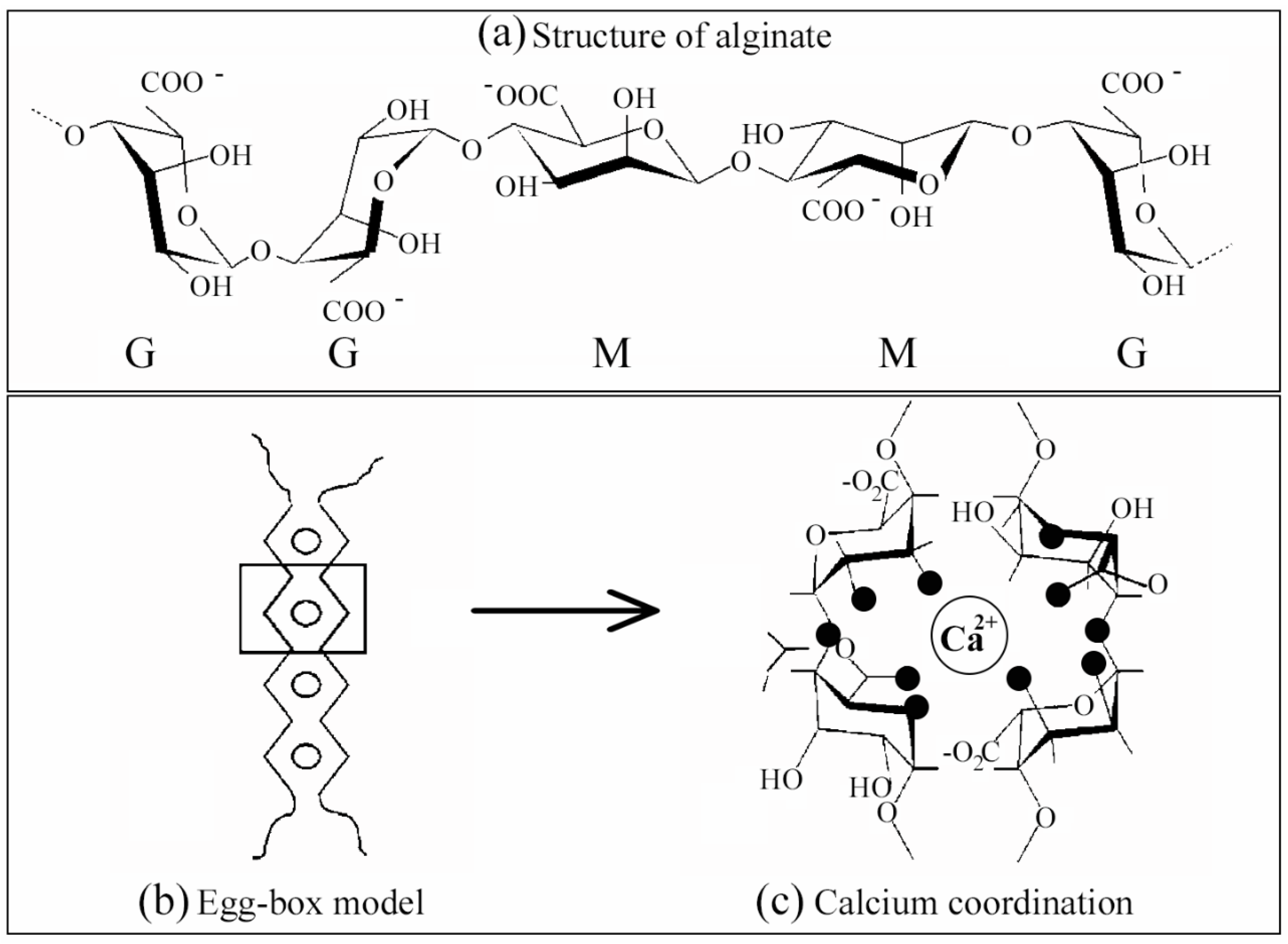
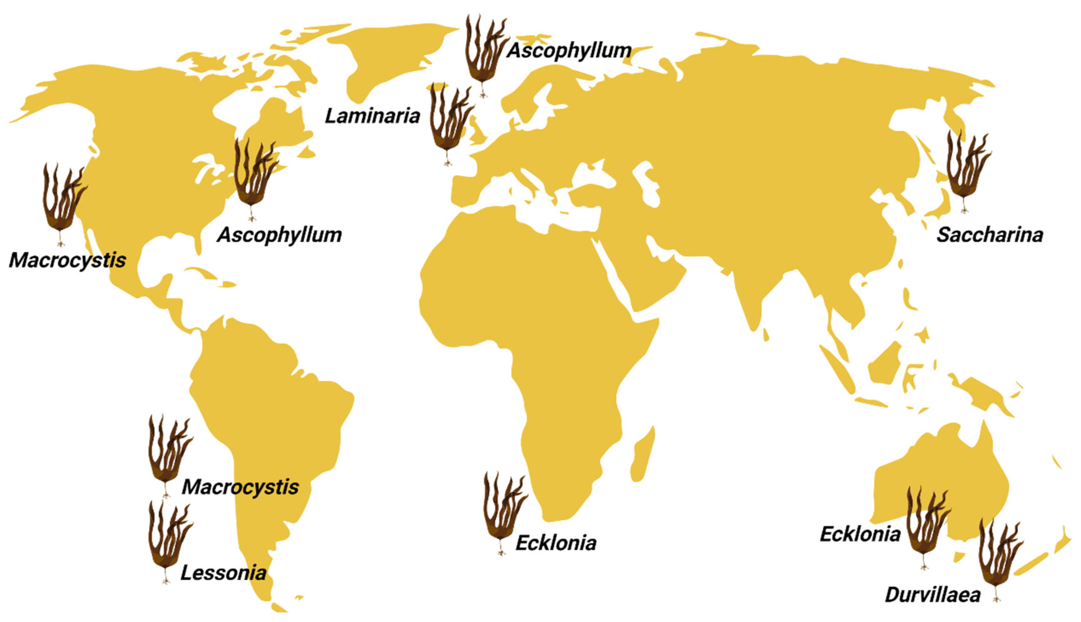
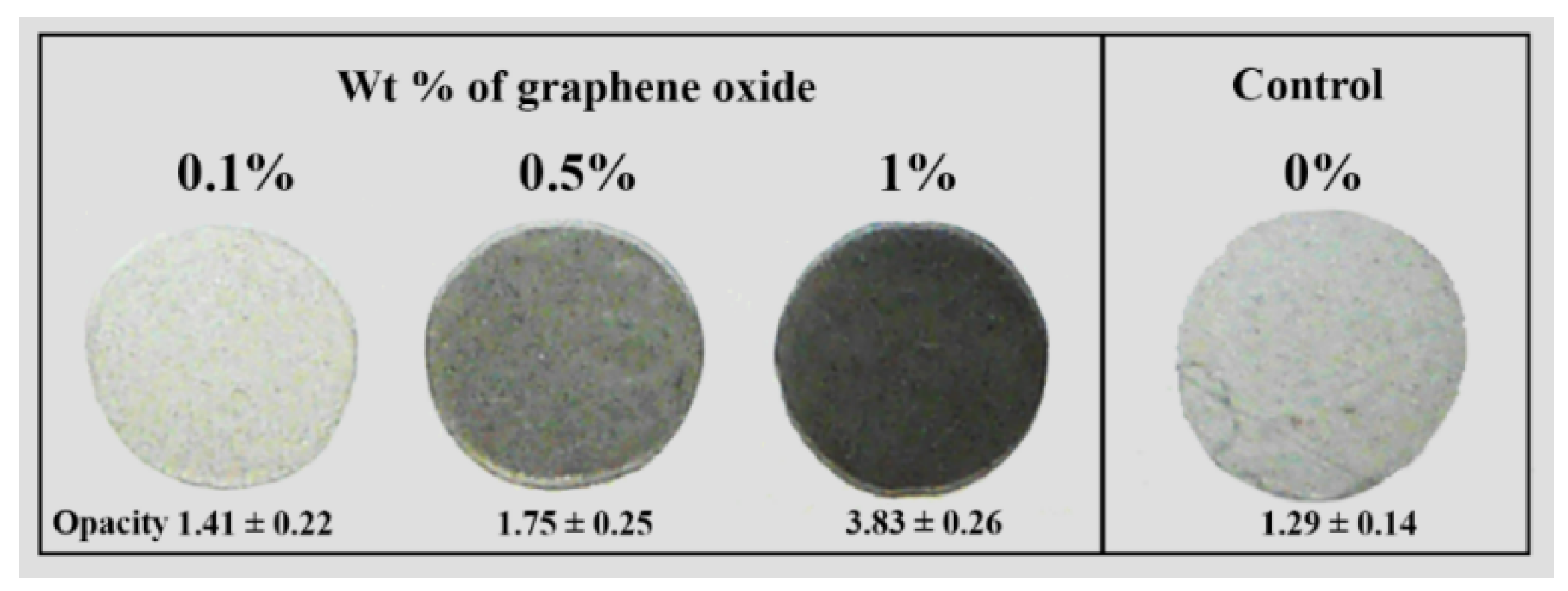
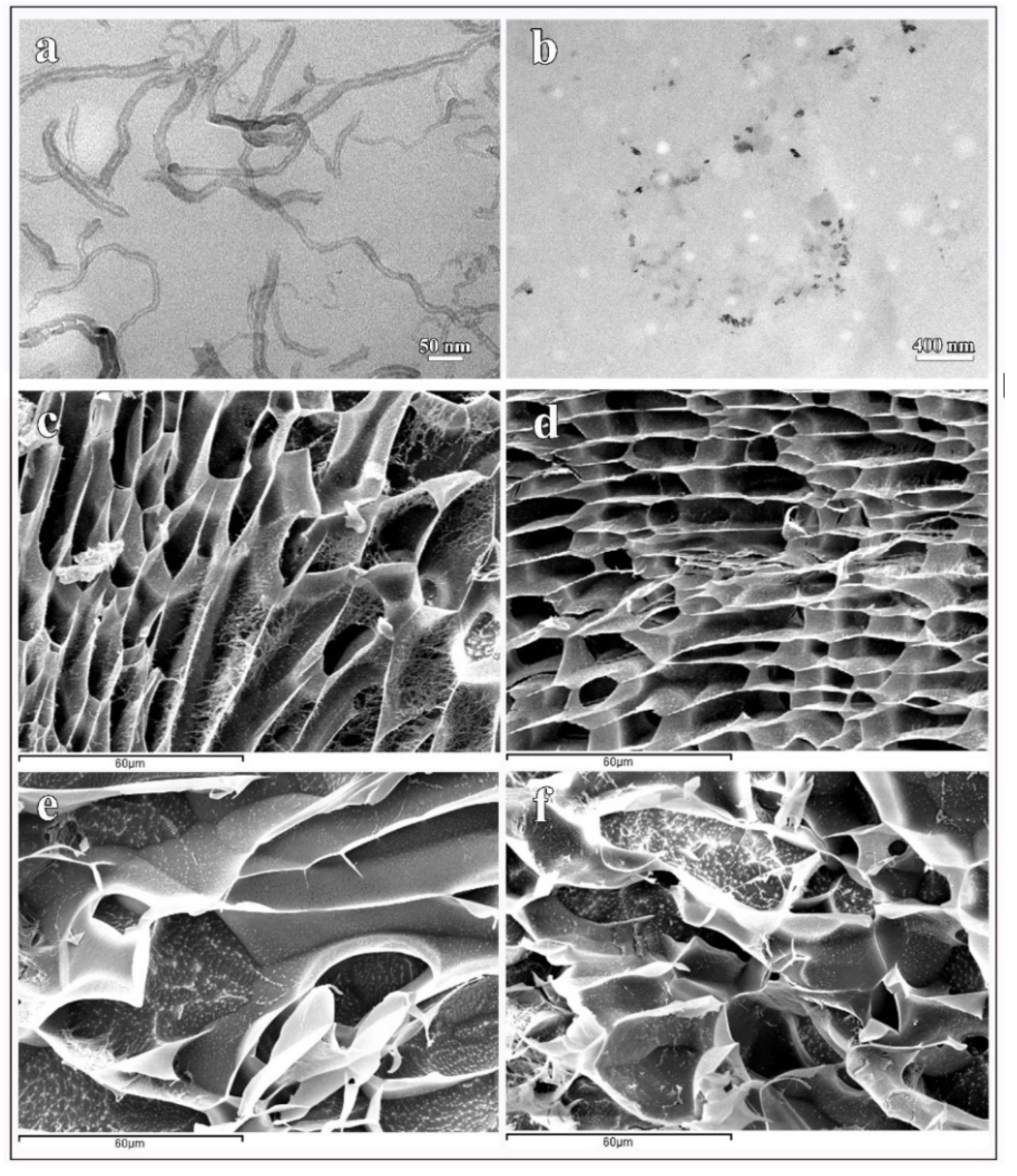
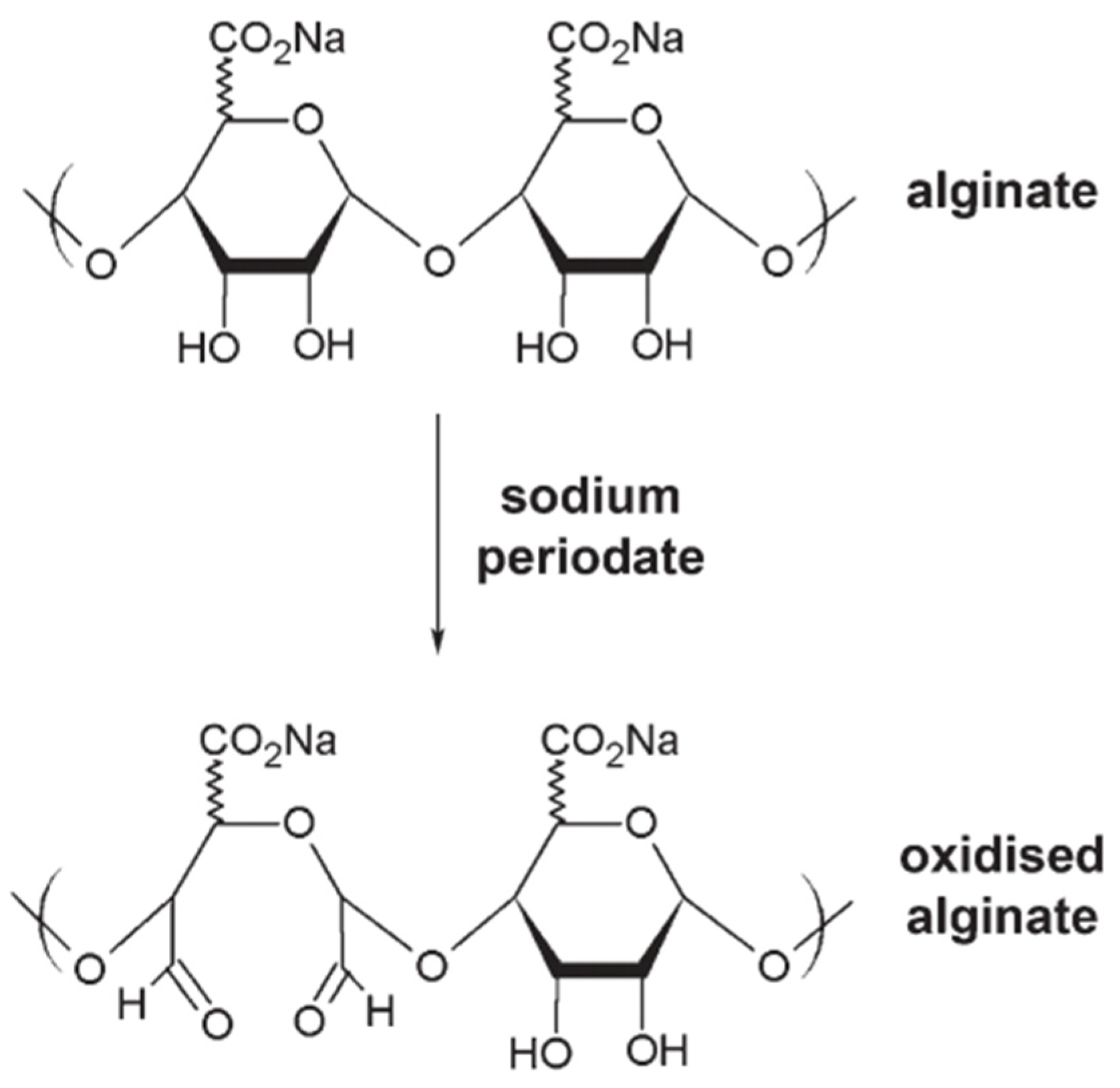
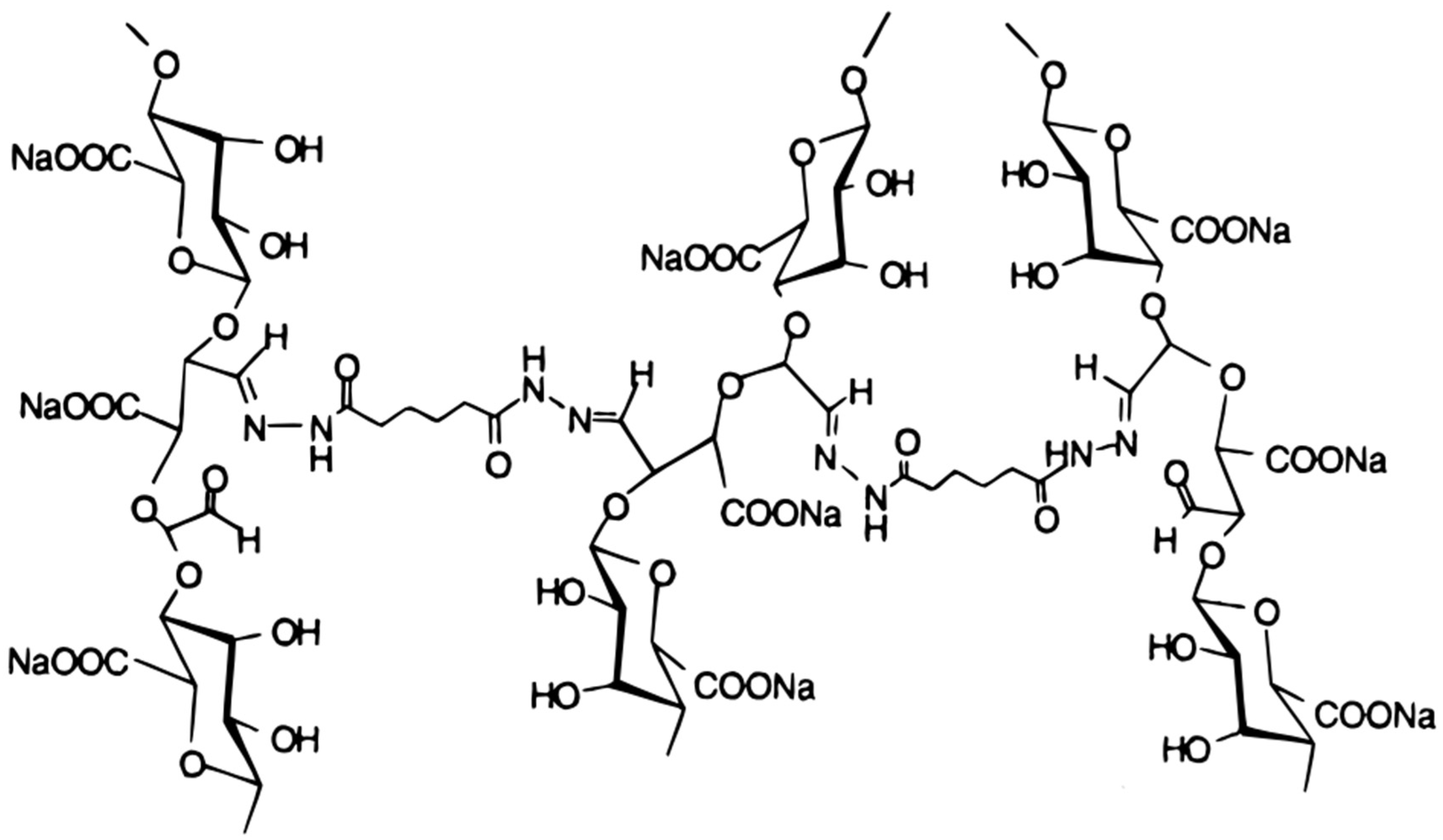
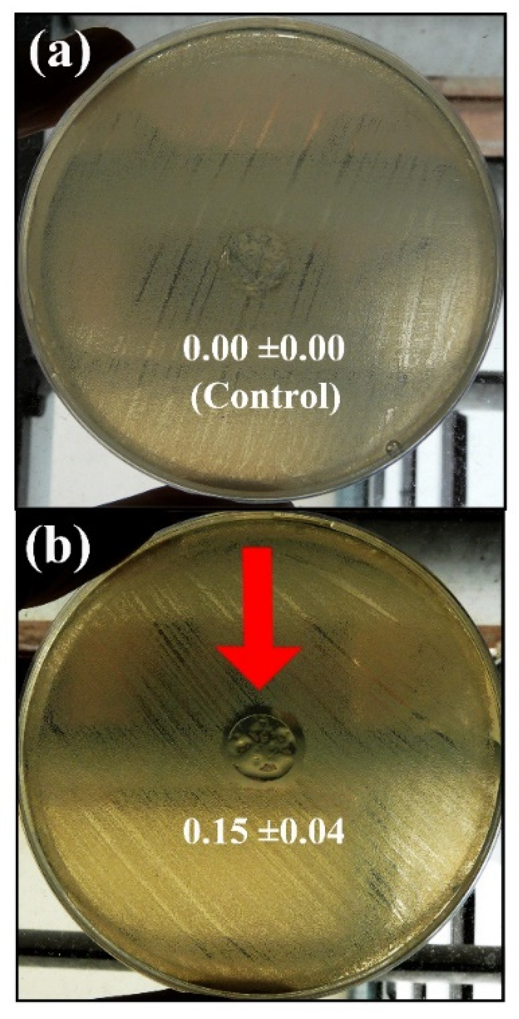
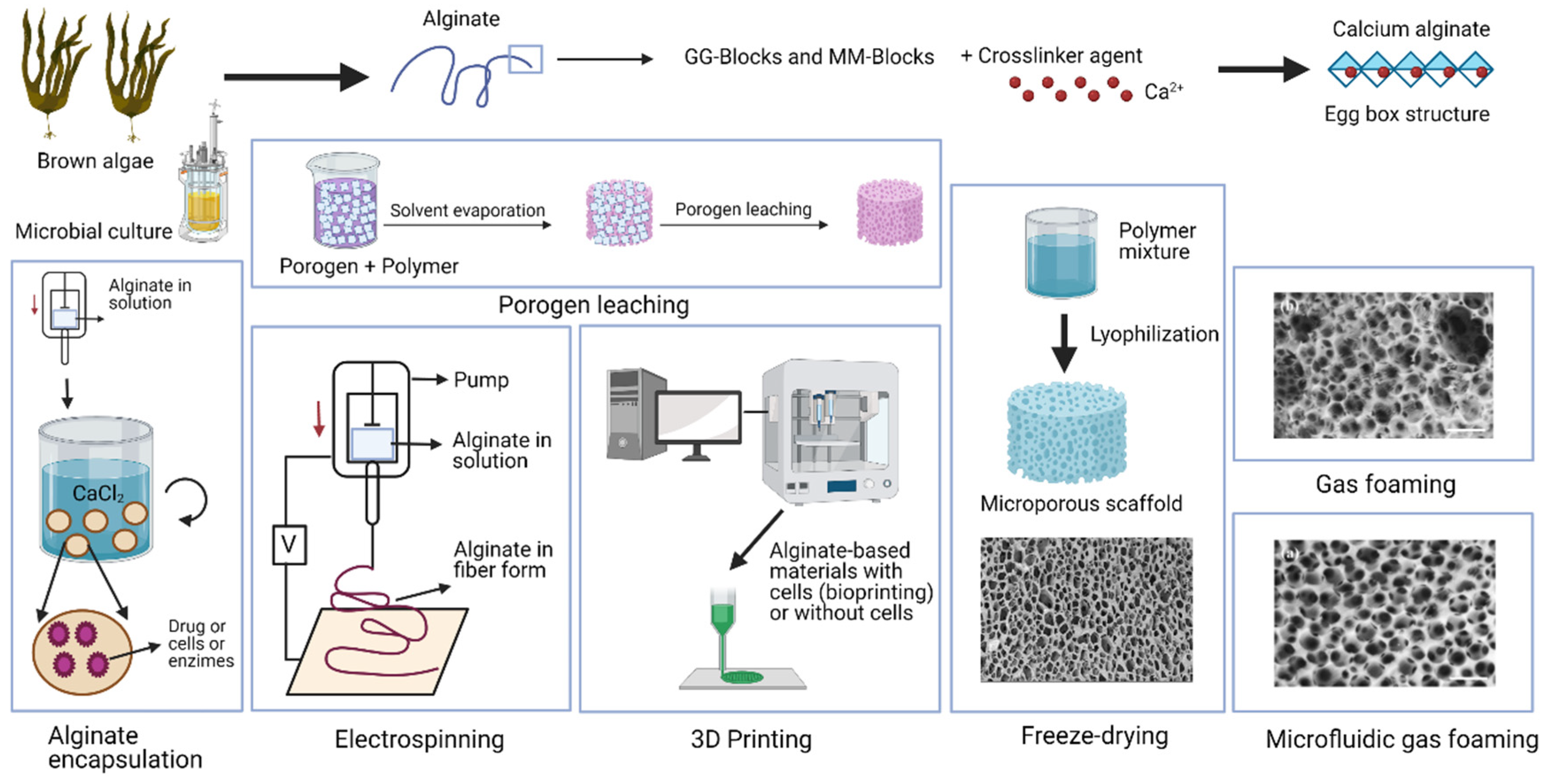
Table 1.
Application areas of alginate and its specific uses.
| Area | Specific Use | Reference |
|---|---|---|
| Biotechnology, bioengineering, biomedicine, and clinical | Dressings for wounds and burns | [6,7] |
| Heavy metal chelator | [8,9] | |
| Scaffolding in tissue engineering | [10,11,12,13] | |
| Controlled release | [14,15] | |
| 3D bio-printing | [16,17] | |
| Prosthesis, dental molds and impression materials | [18,19,20,21] | |
| Immobilization of enzymes and cells | [2,22] | |
| Pharmaceutical industry | Food supplements | [23] |
| Treatment for gastric reflux | [24,25] | |
| Cancer therapy | [26,27] | |
| Chemical, textile, packaging and construction industry | Cosmetics | [28,29] |
| Textile inks | [30,31] | |
| Detergents | [32] | |
| Adhesives | [33,34] | |
| Welding | [35,36,37] | |
| Building insulation | [38] | |
| Biodegradable packaging | [39] | |
| Food and drinks | Ice cream production | [40,41] |
| Binder and thickener | [42,43] | |
| Beer foam stabilizer | [44,45] | |
| Confectionery and gastronomy in general | [46,47] | |
| Aquaculture | Binder for food | [48,49] |
| Paper industry | Thickener | [39] |
| Arts and crafts | Taxidermy molds | [44,50] |
| Leisure industry | Protective masks | [51,52] |
Table 2.
The main alginate types from brown algae depend on the source, M/G ratio, molecular weight, and viscosity. Nd: not determined.
Table 2.
The main alginate types from brown algae depend on the source, M/G ratio, molecular weight, and viscosity. Nd: not determined.
| Source | M (%) | G (%) | Molecular Weight (kDa) | Viscosity (dL/g) | References |
|---|---|---|---|---|---|
| Laminaria hyperborea | 25–35 | 75–65 | 91.902 | 6.4 | [142,149,150] |
| Laminaria digitata | 53–60 | 40–47 | 114–132 | 2.4 | [90,113] |
| Macrocystis pyrifera | 61 | 39 | 146–264 | 12.1 | [142,151,152,153] |
| Fucus vesiculosus | 53.4–59 | 41–46.6 | 125–154.9 | 2.5 | [150,154,155] |
| Sargassum fluitans | 54.2 | 45.8 | 300 | 6.3 | [150,154,156] |
| Sargassum vulgare | 44–56 | 44–56 | 110–194 | 5.26–9.10 | [150,157,158] |
| Laminaria japonica | 65–72 | 28–35 | 770 | 15.4 | [44] |
| Ascophyllum nodosum | 46 | 54 | 177.3 | 2.8 | [142,150,155] |
| Saccharina longicruris | 41 | 59 | 106.6 | Nd | [142,155] |
| Durvillaea antartica | 68–71 | 29–32 | Nd | 7.82 | [136,142,159] |
Table 3.
Genes involved in the production of alginate by bacteria and their gene products.
| Gene | Product | Reference |
|---|---|---|
| algA | Phosphomannose isomerase/GDP-mannose pyrophosphorylase | [167,168,169] |
| algB | ntrC | [170,171] |
| algC | Phosphomannomutase | [172] |
| algD | GDP-mannose dehydrogenase | [173] |
| algF | O-Acetylation | [174] |
| algG | Mannuronan C-5 epimerase | [174,175] |
| algI | O-Acetylation | [174] |
| algL | Alginate lyase | [176,177] |
| algR1 | Regulatory molecules | [178] |
| algS | Anti σ factor | [179] |
Table 4.
Main alginate types produced from different bacterial cultures, M/G ratios, and characteristics of bacteria.
Table 4.
Main alginate types produced from different bacterial cultures, M/G ratios, and characteristics of bacteria.
| Source | M (%) | G (%) | Features | References |
|---|---|---|---|---|
| Pseudomonas aeruginosa | 70 | 30 | Mucoid biofilms | [184] |
| Pseudomonas fluorescens, | 60–73 | 27–40 | 1.4–1.8 of polydispersity index | [185,186,187] |
| Azotobacter vinelandii | 6–75 | 25–94 | Encystment process or biofilm | [185,186] |
| Pseudomonas putida | 78–63 | 22–37 | 1.5–1.8 of polydispersity index | [186,187] |
| Pseudomonas mendocina | 74 | 26 | Mucoid biofilms | [186,188] |
Table 5.
Alginate-based scaffolds: fabrication method, materials combined with alginate, pore size/shape, porosity, regenerative field, year, and reference.
Table 5.
Alginate-based scaffolds: fabrication method, materials combined with alginate, pore size/shape, porosity, regenerative field, year, and reference.
| Scaffold Fabrication Method | Materials Combined with Alginate | Pore Size/Shape | Porosity | Regenerative Field | Year | Ref. |
|---|---|---|---|---|---|---|
| Freeze-drying method | None | 200–300 µm | 90% | Tissue regeneration | 2002 | [433] |
| Hydroxyapatite | 150 µm | >82% | Bone | 2004 | [403] | |
| Chitosan | 200 µm | 84–88% | Cartilage | 2008 | [65] | |
| Sulfate | 120 ± 30 µm | >90% | Vascularization | 2009 | [445] | |
| Poly (lactic-co-glycolic acid)/calcium phosphate | 100–200 µm | 89.24% | Bone | 2009 | [437] | |
| RGD | 88 µm | >90% | Cartilage | 2010 | [434] | |
| RGD | 50–100 µm | >90% | Cardiac tissue engineering | 2011 | [435] | |
| Curcumin, chitosan and collagen | 50–250 µm | - | Diabetic wound healing | 2016 | [436] | |
| Collagen | 200–700 µm | 65–90% | Stem cell culture | 2018 | [417] | |
| PCL:gelatin electrospun mat, and kartogenin-PLGA nanoparticles | 78.6 µm | 92.4% | Tissue engineering | 2021 | [443] | |
| 3D printing/Bioprinting | MBG | 300–420 µm | 49–70% | Bone | 2012 | [429] |
| PCL | 388–499 µm | - | Bone | 2012 | [422] | |
| Calcium phosphate | 200–900 µm | 48–75% | Osteochondral regeneration | 2013 | [430] | |
| β-TCP | 551–875 µm | 23–52% | Bone tissue engineering | 2014 | [446] | |
| Tricalcium phosphate (TCP) | - | >80% | Bone | 2016 | [447] | |
| Gelatin | - | 40–75% | Tissue regeneration | 2016 | [448] | |
| BFP1 | - | - | Bone regeneration | 2017 | [449] | |
| Graphene oxide | - | - | Chondroinductive | 2020 | [431] | |
| Gelatin | <500 µm | 60–70% | Bone regeneration | 2021 | [450] | |
| Polyethylene glycol | 291.4 μm | - | Delivery of insulin | 2022 | [451] | |
| Electrospinning | PEO | - | - | Tissue regeneration | 2010 | [408] |
| Chitosan and PEO | - | - | Tissue regeneration | 2011 | [409] | |
| Gelatin | - | - | Corneal tissue engineering | 2013 | [406] | |
| PCL and ethanol treatment | - | - | Tissue regeneration | 2013 | [228] | |
| PCL | 821 ± 55 µm | 92% | Bone | 2014 | [410] | |
| Magnesium oxide | 2–50 µm | Low | Tissue regeneration | 2017 | [411] | |
| Porogen leaching | Poly(D, L-lactic acid) | 450–900 µm | 84.24–90.75% | Bone | 2008 | [452] |
| Gelatin | 204 ± 58 µm | 97.26 ± 0.18% | Cell culture for regeneration | 2015 | [13] | |
| Collagen | 700 µm | - | Cell cultures | 2018 | [417] | |
| Gelatin/PVA | 104.5 ± 15.9 µm | 74.5 ± 15.9% | Meniscus fibrocartilage | 2018 | [453] | |
| Vaterite/Crystals | 10–500 µm | - | Tissue regeneration | 2019 | [454] | |
| Four-step process: preparation, cross-linking, freezing and lyophilization | - | 50–200 µm | >90% | Vascularization and generation of embryos | 2004 | [455] |
| Chitosan | 100–300 µm | - | Cartilage | 2005 | [438] | |
| Solution and crosslinking | Fibroblast growth factor | 100–500 µm | >90% | Vascularization | 2003 | [402] |
| Thermally induced phase separation and subsequent sublimation of the solvent | Chitosan | 100–300 µm | 91.94 ± 0.9% | Bone | 2005 | [439] |
| Co-precipitation | HAp/chitosan | 50–100 µm | 79–85% | Bone and other tissues | 2008 | [440] |
| Sol–gel synthesis Surfactant foaming | Bioactive glass/polyvinyl alcohol | 200–500 µm | - | Trabecular bone | 2009 | [456] |
| Homogenizing interpolyelectrolyte complex method | Chitosan on PEC gel | 100 µm | - | Release of growth factor for tissue regeneration | 2009 | [457] |
| Lyophilization | Chitosan/Hydroxyapatite | 80–200 µm | >70% | Tissue regeneration | 2010 | [415] |
| Core/shell nozzle of a cryogenic co-extrusion process | Collagen | 100–200 µm | >90% | Skin tissue regeneration | 2011 | [441] |
| Modified Solid-Freeform | Cells (MC3T3-E1) | 300 µm | - | Tissue regeneration in general | 2012 | [458] |
| Three monitorized precision linear stages | Chitosan | - | 66% | - | 2014 | [459] |
| Binary polymer system | Felodipine Fibroin | - | 49–62% | Silk fibroin | 2020 | [460] |
| Solvent casting technique | TiO2/Chitosan | None | - | Bone regeneration | 2020 | [461] |
| 3D Printing (FDM)/freeze-drying/coating | PLA and hydroxyapatite | Circle | 44–36% | Bone regeneration | 2021 | [442] |
| 3D printing and impregnating techniques | Chitosan/alginate/hydroxyapatite | 2–3 mm | - | Cartilage regeneration | 2022 | [444] |
Publisher’s Note: MDPI stays neutral with regard to jurisdictional claims in published maps and institutional affiliations. |
© 2022 by the authors. Licensee MDPI, Basel, Switzerland. This article is an open access article distributed under the terms and conditions of the Creative Commons Attribution (CC BY) license (https://creativecommons.org/licenses/by/4.0/).
Share and Cite
MDPI and ACS Style
Hurtado, A.; Aljabali, A.A.A.; Mishra, V.; Tambuwala, M.M.; Serrano-Aroca, Á. Alginate: Enhancement Strategies for Advanced Applications. Int. J. Mol. Sci. 2022, 23, 4486. https://doi.org/10.3390/ijms23094486
AMA Style
Hurtado A, Aljabali AAA, Mishra V, Tambuwala MM, Serrano-Aroca Á. Alginate: Enhancement Strategies for Advanced Applications. International Journal of Molecular Sciences. 2022; 23(9):4486. https://doi.org/10.3390/ijms23094486
Chicago/Turabian StyleHurtado, Alejandro, Alaa A. A. Aljabali, Vijay Mishra, Murtaza M. Tambuwala, and Ángel Serrano-Aroca. 2022. "Alginate: Enhancement Strategies for Advanced Applications" International Journal of Molecular Sciences 23, no. 9: 4486. https://doi.org/10.3390/ijms23094486
Note that from the first issue of 2016, this journal uses article numbers instead of page numbers. See further details here.