
Inorganic Nanoparticles and Composite Films for Antimicrobial Therapies


 ,
,  , and
, and
Abstract
:1. Introduction
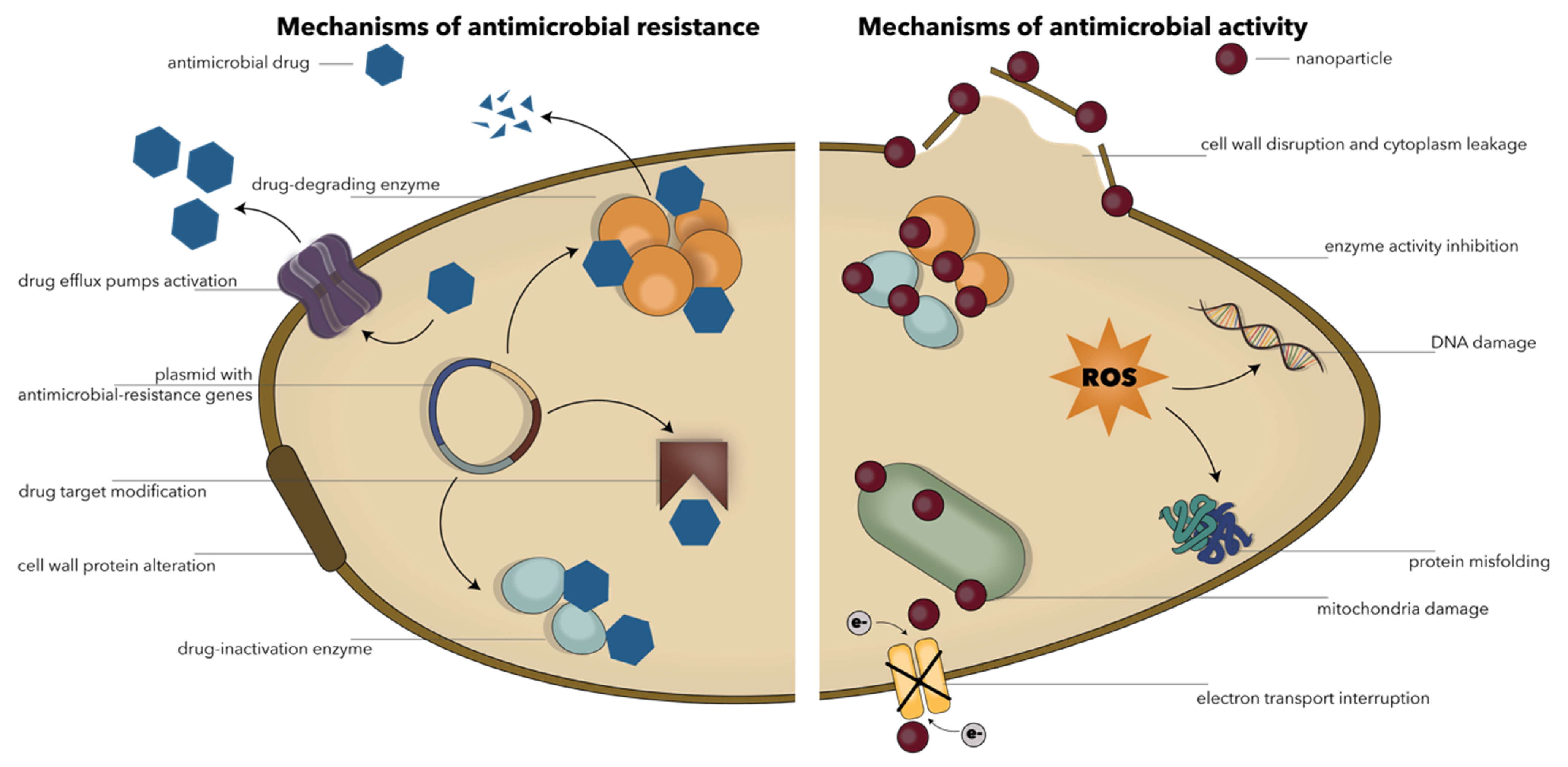
2. Inorganic Nanoparticles with Antimicrobial Properties
2.1. Gold Nanoparticles
2.2. Silver Nanoparticles
2.3. Copper Nanoparticles
2.4. Zinc Oxide Nanoparticles
2.5. Titanium Oxide Nanoparticles
2.6. Magnesium Oxide Nanoparticles
2.7. Iron Oxide Nanoparticles
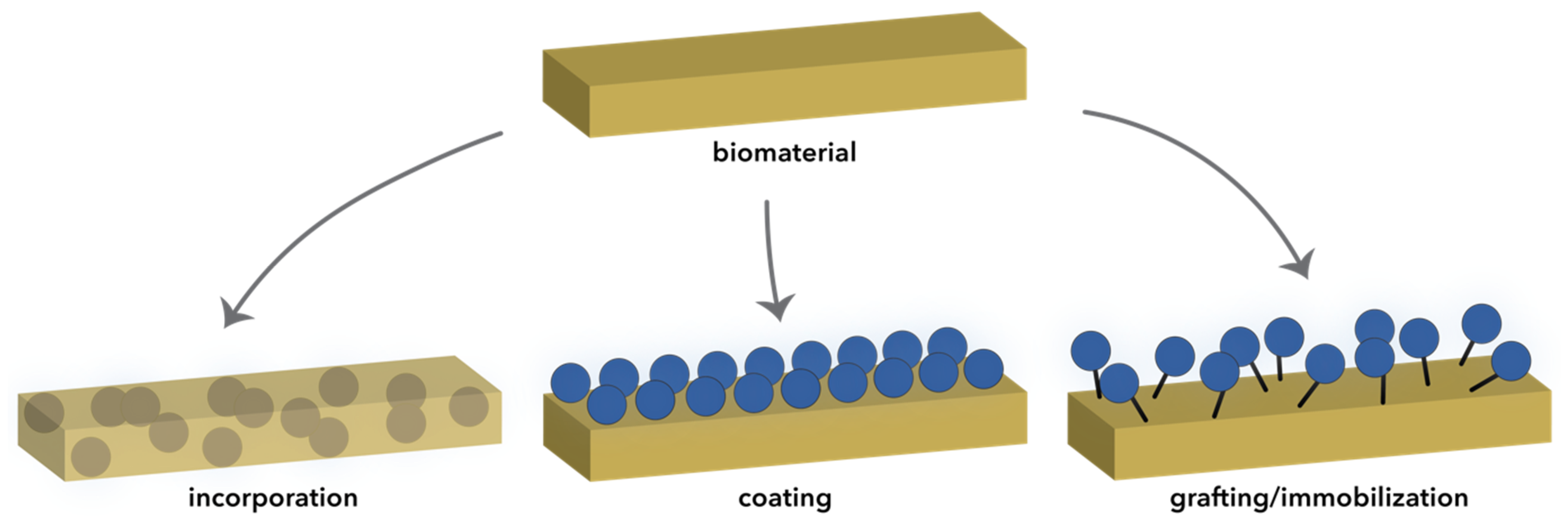
3. Inorganic Nanoparticle-Based Composite Films for Antimicrobial Applications
4. Conclusions and Future Perspectives
Author Contributions
Funding
Institutional Review Board Statement
Informed Consent Statement
Data Availability Statement
Conflicts of Interest
References
- Mitra, S.B. Nanoparticles for dental materials: Synthesis, analysis, and applications. In Emerging Nanotechnologies in Dentistry, 2nd ed.; Elsevier: Amsterdam, The Netherlands, 2018; pp. 17–39. [Google Scholar] [CrossRef]
- Satalkar, P.; Elger, B.S.; Shaw, D.M. Defining Nano, Nanotechnology and Nanomedicine: Why Should It Matter? Sci. Eng. Ethics 2015, 22, 1255–1276. [Google Scholar] [CrossRef]
- Pogribna, M.; Hammons, G. Epigenetic Effects of Nanomaterials and Nanoparticles. J. Nanobiotechnol. 2021, 19, 1–18. [Google Scholar] [CrossRef] [PubMed]
- Nanomaterials definition matters. In Nature Nanotechnology; Nature Publishing Group: Berlin, Germany, 2019; Volume 14, p. 193.
- Khan, I.; Saeed, K.; Khan, I. Nanoparticles: Properties, applications and toxicities. Arab. J. Chem. 2019, 12, 908–931. [Google Scholar] [CrossRef]
- Ahlawat, J.; Hooda, R.; Sharma, M.; Kalra, V.; Rana, J.S.; Batra, B. Nanoparticles in Biomedical Applications; Springer: Cham, Switzerland, 2020; pp. 227–250. [Google Scholar] [CrossRef]
- McNamara, K.; Tofail, S.A.M. Nanoparticles in biomedical applications. In Advances in Physics: X; Taylor and Francis Ltd.: Abingdon, UK, 2017; Volume 2, pp. 54–88. [Google Scholar]
- Kataria, S.; Jain, M.; Rastogi, A.; Živčák, M.; Brestic, M.; Liu, S.; Tripathi, D.K. Role of nanoparticles on photosynthesis: Avenues and applications. In Nanomaterials in Plants, Algae and Microorganisms: Concepts and Controversies: Volume 2; Elsevier: Amsterdam, The Netherlands, 2018; pp. 103–127. [Google Scholar] [CrossRef]
- Husain, Q. An overview on the green synthesis of nanoparticles and other nano-materials using enzymes and their potential applications. Biointerface Res. Appl. Chem. 2019, 9, 4255–4271. [Google Scholar] [CrossRef]
- Gouda, S.; Kerry, R.G.; Das, G.; Patra, J.K. Synthesis of nanoparticles utilizing sources from the mangrove environment and their potential applications: An overview. In Nanomaterials in Plants, Algae and Microorganisms: Concepts and Controversies: Volume 2; Elsevier: Amsterdam, The Netherlands, 2018; pp. 219–235. [Google Scholar] [CrossRef]
- Bundschuh, M.; Filser, J.; Lüderwald, S.; McKee, M.S.; Metreveli, G.; Schaumann, G.E.; Schulz, R.; Wagner, S. Nanoparticles in the environment: Where do we come from, where do we go to? Environ. Sci. Eur. 2018, 30, 1–17. [Google Scholar] [CrossRef] [PubMed] [Green Version]
- Khan, H.; Sakharkar, M.; Nayak, A.; Kishore, U.; Khan, A. Nanoparticles for biomedical applications: An overview. In Nanobiomaterials: Nanostructured Materials for Biomedical Applications; Elsevier: Amsterdam, The Netherlands, 2018; pp. 357–384. [Google Scholar]
- Zia-Ur-Rehman, M.; Qayyum, M.F.; Akmal, F.; Maqsood, M.A.; Rizwan, M.; Waqar, M.; Azhar, M. Recent Progress of Nanotoxicology in Plants. In Nanomaterials in Plants, Algae, and Microorganisms; Elsevier: Amsterdam, The Netherlands, 2018; Volume 1, pp. 143–174. [Google Scholar]
- Ojha, S.; Singh, D.; Sett, A.; Chetia, H.; Kabiraj, D.; Bora, U. Nanotechnology in Crop Protection. In Nanomaterials in Plants, Algae, and Microorganisms; Elsevier: Amsterdam, The Netherlands, 2018; Volume 1, pp. 345–391. [Google Scholar]
- Pathak, J.; Rajneesh; Ahmed, H.; Singh, D.K.; Pandey, A.; Singh, S.P.; Sinha, R.P. Recent developments in green synthesis of metal nanoparticles utilizing cyanobacterial cell factories. In Nanomaterials in Plants, Algae and Microorganisms: Concepts and Controversies: Volume 2; Elsevier: Amsterdam, The Netherlands, 2018; pp. 237–265. [Google Scholar] [CrossRef]
- Malakar, A.; Snow, D.D. Nanoparticles as sources of inorganic water pollutants. In Inorganic Pollutants in Water; Elsevier: Amsterdam, The Netherlands, 2020; pp. 337–370. [Google Scholar] [CrossRef]
- Ijaz, I.; Gilani, E.; Nazir, A.; Bukhari, A. Detail review on chemical, physical and green synthesis, classification, characterizations and applications of nanoparticles. Green Chem. Lett. Rev. 2020, 13, 59–81. [Google Scholar] [CrossRef]
- Ealias, A.M.; Saravanakumar, M.P. A review on the classification, characterisation, synthesis of nanoparticles and their ap-plication. In IOP Conference Series: Materials Science and Engineering; Institute of Physics Publishing: Bristol, UK, 2017; Volume 263, p. 032019. [Google Scholar]
- Jeevanandam, J.; Barhoum, A.; Chan, Y.S.; Dufresne, A.; Danquah, M.K. Review on nanoparticles and nanostructured mate-rials: History, sources, toxicity and regulations. Beilstein J. Nanotechnol. 2018, 9, 1050–1074. [Google Scholar] [CrossRef] [Green Version]
- Eleraky, N.E.; Allam, A.; Hassan, S.B.; Omar, M.M. Nanomedicine Fight against Antibacterial Resistance: An Overview of the Recent Pharmaceutical Innovations. Pharmaceutics 2020, 12, 142. [Google Scholar] [CrossRef] [Green Version]
- Teleanu, D.M.; Chircov, C.; Grumezescu, A.M.; Volceanov, A.; Teleanu, R.I. Impact of Nanoparticles on Brain Health: An Up to Date Overview. J. Clin. Med. 2018, 7, 490. [Google Scholar] [CrossRef] [Green Version]
- Astruc, D. Introduction: Nanoparticles in Catalysis. Chem. Rev. 2020, 120, 461–463. [Google Scholar] [CrossRef] [Green Version]
- Rahman, M.M.; Ferdous, K.S.; Ahmed, M. Emerging Promise of Nanoparticle-Based Treatment for Parkinson’s disease. Biointerface Res. Appl. Chem. 2020, 10, 7135–7151. [Google Scholar] [CrossRef]
- Sharifi, S.; Samani, A.A.; Ahmadian, E.; Eftekhari, A.; Derakhshankhah, H.; Jafari, S.; Mokhtarpour, M.; Vahed, S.Z.; Salatin, S.; Dizaj, S.M. Oral delivery of proteins and peptides by mucoadhesive nanoparticles. Biointerface Res. Appl. Chem. 2019, 9, 3849–3852. [Google Scholar] [CrossRef]
- Anderson, S.D.; Gwenin, V.V.; Gwenin, C.D. Magnetic Functionalized Nanoparticles for Biomedical, Drug Delivery and Imaging Applications. Nanoscale Res. Lett. 2019, 14, 1–16. [Google Scholar] [CrossRef] [Green Version]
- Kurtjak, M.; Aničić, N.; Vukomanovicć, M. Inorganic Nanoparticles: Innovative Tools for Antimicrobial Agents. In Antibacterial Agents; IntechOpen: London, UK, 2017. [Google Scholar] [CrossRef]
- Saidin, S.; Jumat, M.A.; Amin, N.A.A.M.; Al Hammadi, A.S.S. Organic and inorganic antibacterial approaches in combating bacterial infection for biomedical application. Mater. Sci. Eng. C 2021, 118, 111382. [Google Scholar] [CrossRef]
- Vassallo, A.; Silletti, M.F.; Faraone, I.; Milella, L. Nanoparticulate Antibiotic Systems as Antibacterial Agents and Antibiotic Delivery Platforms to Fight Infections. J. Nanomater. 2020, 2020, 1–31. [Google Scholar] [CrossRef]
- Malaekeh-Nikouei, B.; Bazzaz, B.S.F.; Mirhadi, E.; Tajani, A.S.; Khameneh, B. The role of nanotechnology in combating biofilm-based antibiotic resistance. J. Drug Deliv. Sci. Technol. 2020, 60, 101880. [Google Scholar] [CrossRef]
- Rodrigues, I.D.A.; Ferrari, R.G.; Panzenhagen, P.H.N.; Mano, S.B.; Conte-Junior, C.A. Antimicrobial resistance genes in bacteria from animal-based foods. In Advances in Applied Microbiology; Elsevier: Amsterdam, The Netherlands, 2020; Volume 112, pp. 143–183. [Google Scholar]
- Wester, A.L.; Gopinathan, U.; Gjefle, K.; Solberg, S.Ø.; Røttingen, J.-A. Antimicrobial Resistance in a One Health and One World Perspective—Mechanisms and Solutions. In International Encyclopedia of Public Health; Elsevier: Amsterdam, The Netherlands, 2016; pp. 140–153. [Google Scholar] [CrossRef]
- Majumder, M.A.A.; Singh, K.; Hilaire, M.G.S.; Rahman, S.; Sa, B.; Haque, M. Antimicrobial resistance and global public health crisis: Are we educating enough our future prescribers to combat this global threat? In Expert Review of Anti-Infective Therapy; Taylor and Francis Ltd.: Abingdon, UK, 2020. [Google Scholar]
- Morrison, L.; Zembower, T.R. Antimicrobial Resistance. Gastrointest. Endosc. Clin. N. Am. 2020, 30, 619–635. [Google Scholar] [CrossRef]
- Rice, L.B. Antimicrobial Stewardship and Antimicrobial Resistance. Med. Clin. N. Am. 2018, 102, 805–818. [Google Scholar] [CrossRef]
- Riduan, S.N.; Armugam, A.; Zhang, Y. Antibiotic resistance mitigation: The development of alternative general strategies. J. Mater. Chem. B 2020, 8, 6317–6321. [Google Scholar] [CrossRef]
- Christaki, E.; Marcou, M.; Tofarides, A. Antimicrobial Resistance in Bacteria: Mechanisms, Evolution, and Persistence. J. Mol. Evol. 2020, 88, 26–40. [Google Scholar] [CrossRef] [PubMed]
- Yi, C.; Yang, Y.; Liu, B.; He, J.; Nie, Z. Polymer-guided assembly of inorganic nanoparticles. Chem. Soc. Rev. 2020, 49, 465–508. [Google Scholar] [CrossRef]
- Videira-Quintela, D.; Martin, O.; Montalvo, G. Recent advances in polymer-metallic composites for food packaging applica-tions. In Trends in Food Science and Technology; Elsevier Ltd.: Amsterdam, The Netherlands, 2021; Volume 109, pp. 230–244. [Google Scholar]
- Dos Santos, C.A.; Ingle, A.P.; Rai, M. The emerging role of metallic nanoparticles in food. Appl. Microbiol. Biotechnol. 2020, 104, 2373–2383. [Google Scholar] [CrossRef] [PubMed]
- Vasile, C. Polymeric Nanocomposites and Nanocoatings for Food Packaging: A Review. Materials 2018, 11, 1834. [Google Scholar] [CrossRef] [PubMed] [Green Version]
- Spirescu, V.; Chircov, C.; Grumezescu, A.; Andronescu, E. Polymeric Nanoparticles for Antimicrobial Therapies: An up-to-date Overview. Polymers 2021, 13, 724. [Google Scholar] [CrossRef] [PubMed]
- Samrot, A.V.; Sahithya, C.S.; Sruthi, P.D.; Selvarani, A.J.; Raji, P.; Prakash, P.; Ponnaiah, P.; Petchi, I.; Pattammadath, S.; Purayil, S.K.; et al. Itraconazole Coated Super Paramagnetic Iron Oxide Nanoparticles for Antimicrobial Studies. Biointerface Res. Appl. Chem. 2020, 10, 6218–6225. [Google Scholar] [CrossRef]
- Thiruvengadam, V.; Bansod, A.V. Characterization of Silver Nanoparticles Synthesized using Chemical Method and its Antibacterial Property. Biointerface Res. Appl. Chem. 2020, 10, 7257–7264. [Google Scholar] [CrossRef]
- Pontes, D.S.; De Araujo, R.S.A.; Dantas, N.; Scotti, L.; Scotti, M.T.; De Moura, R.O.; Mendonca-Junior, F.J.B. Genetic Mechanisms of Antibiotic Resistance and the Role of Antibiotic Adjuvants. Curr. Top. Med. Chem. 2018, 18, 42–74. [Google Scholar] [CrossRef]
- Medina, E.; Pieper, D.H. Tackling Threats and Future Problems of Multidrug-Resistant Bacteria. In How to Overcome the Anti-biotic Crisis: Facts, Challenges, Technologies and Future Perspectives; Stadler, M., Dersch, P., Eds.; Springer International Publishing: Cham, Switzerland, 2016; pp. 3–33. [Google Scholar] [CrossRef]
- Boucher, H.W.; Talbot, G.H.; Bradley, J.S.; Edwards, J.E.; Gilbert, D.; Rice, L.B.; Scheld, M.; Spellberg, B.; Bartlett, J. Bad Bugs, No Drugs: No ESKAPE! An Update from the Infectious Diseases Society of America. Clin. Infect. Dis. 2009, 48, 1–12. [Google Scholar] [CrossRef] [Green Version]
- Patil, A.; Banerji, R.; Kanojiya, P.; Saroj, S.D. Foodborne ESKAPE Biofilms and Antimicrobial Resistance: Lessons Learned from Clinical Isolates. Pathog. Global Health 2021, 10, 1–18. [Google Scholar] [CrossRef]
- Santajit, S.; Indrawattana, N. Mechanisms of Antimicrobial Resistance in ESKAPE Pathogens. BioMed Res. Int. 2016, 2016, 1–8. [Google Scholar] [CrossRef] [Green Version]
- Mitevska, E.; Wong, B.; Surewaard, B.; Jenne, C. The Prevalence, Risk, and Management of Methicillin-Resistant Staphylococcus aureus Infection in Diverse Populations across Canada: A Systematic Review. Pathogens 2021, 10, 393. [Google Scholar] [CrossRef]
- Behzadi, P.; Baráth, Z.; Gajdács, M. It’s Not Easy Being Green: A Narrative Review on the Microbiology, Virulence and Therapeutic Prospects of Multidrug-Resistant Pseudomonas aeruginosa. Antibiotics 2021, 10, 42. [Google Scholar] [CrossRef] [PubMed]
- Sarshar, M.; Behzadi, P.; Scribano, D.; Palamara, A.; Ambrosi, C. Acinetobacter baumannii: An Ancient Commensal with Weapons of a Pathogen. Pathogens 2021, 10, 387. [Google Scholar] [CrossRef] [PubMed]
- Elemam, A.; Rahimian, J.; Mandell, W. Infection with PanresistantKlebsiella pneumoniae:A Report of 2 Cases and a Brief Review of the Literature. Clin. Infect. Dis. 2009, 49, 271–274. [Google Scholar] [CrossRef] [PubMed] [Green Version]
- Reygaert, W.C. An overview of the antimicrobial resistance mechanisms of bacteria. AIMS Microbiol. 2018, 4, 482–501. [Google Scholar] [CrossRef] [PubMed]
- Ogawara, H. Comparison of Antibiotic Resistance Mechanisms in Antibiotic-Producing and Pathogenic Bacteria. Molecules 2019, 24, 3430. [Google Scholar] [CrossRef] [Green Version]
- Munita, J.M.; Arias, C.A. Mechanisms of Antibiotic Resistance. Microbiol. Spectr. 2016, 4. [Google Scholar] [CrossRef] [PubMed] [Green Version]
- Gajdács, M. The Concept of an Ideal Antibiotic: Implications for Drug Design. Molecules 2019, 24, 892. [Google Scholar] [CrossRef] [Green Version]
- Da Cunha, B.R.; Fonseca, L.P.; Calado, C.R.C. Antibiotic Discovery: Where Have We Come from, Where Do We Go? Antibiotics 2019, 8, 45. [Google Scholar] [CrossRef] [Green Version]
- Liu, Y.; Tong, Z.; Shi, J.; Li, R.; Upton, M.; Wang, Z. Drug repurposing for next-generation combination therapies against multidrug-resistant bacteria. Theranostics 2021, 11, 4910–4928. [Google Scholar] [CrossRef]
- Peterson, E.; Kaur, P. Antibiotic Resistance Mechanisms in Bacteria: Relationships Between Resistance Determinants of Antibiotic Producers, Environmental Bacteria, and Clinical Pathogens. Front. Microbiol. 2018, 9, 2928. [Google Scholar] [CrossRef]
- Sánchez-López, E.; Gomes, D.; Esteruelas, G.; Bonilla, L.; Lopez-Machado, A.L.; Galindo, R.; Cano, A.; Espina, M.; Ettcheto, M.; Camins, A.; et al. Metal-Based Nanoparticles as Antimicrobial Agents: An Overview. Nanomaterials 2020, 10, 292. [Google Scholar] [CrossRef] [PubMed] [Green Version]
- Lyddiard, D.; Jones, G.L.; Greatrex, B.W. Keeping it simple: Lessons from the golden era of antibiotic discovery. FEMS Microbiol. Lett. 2016, 363. [Google Scholar] [CrossRef] [PubMed] [Green Version]
- Wang, L.; Hu, C.; Shao, L. The antimicrobial activity of nanoparticles: Present situation and prospects for the future. Int. J. Nanomed. 2017, 12, 1227–1249. [Google Scholar] [CrossRef] [PubMed] [Green Version]
- Fernando, S.; Gunasekara, T.; Holton, J. Antimicrobial Nanoparticles: Applications and mechanisms of action. Sri Lankan J. Infect. Dis. 2018, 8, 2. [Google Scholar] [CrossRef]
- Varier, K.M.; Gudeppu, M.; Chinnasamy, A.; Thangarajan, S.; Balasubramanian, J.; Li, Y.; Gajendran, B. Nanoparticles: Antimicrobial Applications and Its Prospects; Springer: Cham, Switzerland, 2019; pp. 321–355. [Google Scholar] [CrossRef]
- Lee, N.-Y.; Ko, W.-C.; Hsueh, P.-R. Nanoparticles in the Treatment of Infections Caused by Multidrug-Resistant Organisms. Front. Pharmacol. 2019, 10, 1153. [Google Scholar] [CrossRef] [PubMed] [Green Version]
- Lam, S.J.; Wong, E.H.; Boyer, C.; Qiao, G.G. Antimicrobial polymeric nanoparticles. Prog. Polym. Sci. 2018, 76, 40–64. [Google Scholar] [CrossRef]
- Ali, R.; Batool, T.; Manzoor, B.; Waseem, H.; Mehmood, S.; Kabeer, A.; Ali, Z.; Habib, S.; Rashid, U.; Iqbal, M.J. Nanobi-otechnology-based drug delivery strategy as a potential weapon against multiple drug-resistant pathogens. In Antibiotics and Antimicrobial Resistance Genes in the Environment: Volume 1; The Advances in Environmental Pollution Research Series; Elsevier: Amsterdam, The Netherlands, 2019; pp. 350–368. [Google Scholar] [CrossRef]
- Crane, J.K. Metal Nanoparticles in Infection and Immunity. Immunol. Investig. 2020, 49, 794–807. [Google Scholar] [CrossRef] [PubMed]
- Chen, H.; Zhou, K.; Zhao, G. Gold nanoparticles: From synthesis, properties to their potential application as colorimetric sensors in food safety screening. Trends Food Sci. Technol. 2018, 78, 83–94. [Google Scholar] [CrossRef]
- Optical, Structural, and Antimicrobial Study of Gold nanoparticles Synthesized Using an Aqueous Extract of Mimusops elengi Raw Fruits. Biointerface Res. Appl. Chem. 2020, 10, 7085–7096. [CrossRef]
- De Souza, C.D.; Nogueira, B.R.; Rostelato, M.E.C. Review of the methodologies used in the synthesis gold nanoparticles by chemical reduction. J. Alloys Compd. 2019, 798, 714–740. [Google Scholar] [CrossRef]
- Fahmy, H.M.; El-Feky, A.S.; El-Daim, T.M.A.; El-Hameed, M.M.A.; Gomaa, D.A.; Hamad, A.M.; Elfky, A.A.; Elkomy, Y.H.; Farouk, N.A.; El-Feky, A.S.; et al. Eco-Friendly Methods of Gold Nanoparticles Synthesis. Nanosci. Nanotechnol. Asia 2019, 9, 311–328. [Google Scholar] [CrossRef]
- Behzad, F.; Naghib, S.M.; Kouhbanani, M.A.J.; Tabatabaei, S.N.; Zare, Y.; Rhee, K.Y. An overview of the plant-mediated green synthesis of noble metal nanoparticles for antibacterial applications. J. Ind. Eng. Chem. 2021, 94, 92–104. [Google Scholar] [CrossRef]
- Rónavári, A.; Igaz, N.; Adamecz, D.; Szerencsés, B.; Molnar, C.; Kónya, Z.; Pfeiffer, I.; Kiricsi, M. Green Silver and Gold Nanoparticles: Biological Synthesis Approaches and Potentials for Biomedical Applications. Molecules 2021, 26, 844. [Google Scholar] [CrossRef]
- Ortiz-Benítez, E.A.; Velázquez-Guadarrama, N.; Figueroa, N.V.D.; Quezada, H.; Olivares-Trejo, J.D.J. Antibacterial mechanism of gold nanoparticles onStreptococcus pneumoniae. Metallomics 2019, 11, 1265–1276. [Google Scholar] [CrossRef] [PubMed]
- Mohamed, M.M.; Fouad, S.A.; Elshoky, H.A.; Mohammed, G.M.; Salaheldin, T.A. Antibacterial effect of gold nanoparticles against Corynebacterium pseudotuberculosis. Int. J. Vet. Sci. Med. 2017, 5, 23–29. [Google Scholar] [CrossRef] [PubMed] [Green Version]
- Shamaila, S.; Zafar, N.; Riaz, S.; Sharif, R.; Nazir, J.; Naseem, S. Gold Nanoparticles: An Efficient Antimicrobial Agent against Enteric Bacterial Human Pathogen. Nanomaterials 2016, 6, 71. [Google Scholar] [CrossRef] [Green Version]
- Tao, C. Antimicrobial activity and toxicity of gold nanoparticles: Research progress, challenges and prospects. Lett. Appl. Microbiol. 2018, 67, 537–543. [Google Scholar] [CrossRef]
- Vanlalveni, C.; Lallianrawna, S.; Biswas, A.; Selvaraj, M.; Changmai, B.; Rokhum, S.L. Green synthesis of silver nanoparticles using plant extracts and their antimicrobial activities: A review of recent literature. RSC Adv. 2021, 11, 2804–2837. [Google Scholar] [CrossRef]
- Hamad, A.; Khashan, K.S.; Hadi, A. Silver Nanoparticles and Silver Ions as Potential Antibacterial Agents. J. Inorg. Organomet. Polym. Mater. 2020, 30, 4811–4828. [Google Scholar] [CrossRef]
- Ratnasari, A.; Endarko, E.; Syafiuddin, A. A Green Method for the Enhancement of Antifungal Properties of Various Textiles Functionalized with Silver Nanoparticles. Biointerface Res. Appl. Chem. 2020, 10, 7284–7294. [Google Scholar] [CrossRef]
- Javed, B.; Ikram, M.; Farooq, F.; Sultana, T.; Mashwani, Z.-U.-R.; Raja, N.I. Biogenesis of silver nanoparticles to treat cancer, diabetes, and microbial infections: A mechanistic overview. Appl. Microbiol. Biotechnol. 2021, 105, 2261–2275. [Google Scholar] [CrossRef] [PubMed]
- Ciriminna, R.; Albo, Y.; Pagliaro, M. New Antivirals and Antibacterials Based on Silver Nanoparticles. ChemMedChem 2020, 15, 1619–1623. [Google Scholar] [CrossRef] [PubMed]
- De Lacerda Coriolano, D.; de Souza, J.B.; Bueno, E.V.; Medeiros, S.M.d.F.R.d.S.; Cavalcanti, I.D.L.; Cavalcanti, I.M.F. Anti-bacterial and antibiofilm potential of silver nanoparticles against antibiotic-sensitive and multidrug-resistant Pseudomonas aeruginosa strains. Braz. J. Microbiol. 2020, 52, 267–278. [Google Scholar] [CrossRef] [PubMed]
- Camacho-Jiménez, L.; Álvarez-Sánchez, A.R.; Mejía-Ruíz, C.H. Silver nanoparticles (AgNPs) as antimicrobials in marine shrimp farming: A review. Aquac. Rep. 2020, 18, 100512. [Google Scholar] [CrossRef]
- Ramzan, H.; Yousaf, Z. Chapter 4—Green Fabrication of Metallic Nanoparticles; Grumezescu, A.M., Ed.; William Andrew Publishing: Norwich, NY, USA, 2018; pp. 137–183. [Google Scholar] [CrossRef]
- Ramzan, H.; Yousaf, Z. Green fabrication of metallic nanoparticles. In Inorganic Frameworks as Smart Nanomedicines; Elsevier: Amsterdam, The Netherlands, 2018; pp. 137–183. [Google Scholar]
- Jadoun, S.; Arif, R.; Jangid, N.K.; Meena, R.K. Green synthesis of nanoparticles using plant extracts: A review. Environ. Chem. Lett. 2021, 19, 355–374. [Google Scholar] [CrossRef]
- Gollapudi, V.R.; Mallavarapu, U.; Seetha, J.; Duddela, V.; Amara, V.R.; Vatti, C.S.; Anumakonda, V. In situ generation of antibacterial bimetallic silver and copper nanocomposites using Tinospora cordifolia leaf extract as bio reductant. Biointerface Res. Appl. Chem. 2020, 10, 5569–5574. [Google Scholar] [CrossRef]
- Batool, M.; Khurshid, S.; Qureshi, Z.M.; Daoush, W.M.; Hashmi, F.; Mehboob, N. Effective adsorptive removal of azodyes on synthesized copper oxide nanoparticles. Biointerface Res. Appl. Chem. 2020, 10, 5369–5375. [Google Scholar] [CrossRef]
- Parveen, F.; Sannakki, B.; Mandke, M.V.; Pathan, H.M. Copper nanoparticles: Synthesis methods and its light harvesting performance. Sol. Energy Mater. Sol. Cells 2016, 144, 371–382. [Google Scholar] [CrossRef]
- Shnoudeh, A.J.; Hamad, I.; Abdo, R.W.; Qadumii, L.; Jaber, A.Y.; Surchi, H.S.; Alkelany, S.Z. Synthesis, Characterization, and Applications of Metal Nanoparticles. In Biomaterials and Bionanotechnology; Elsevier: Amsterdam, The Netherlands, 2019; pp. 527–612. [Google Scholar] [CrossRef]
- DeAlba-Montero, I.; Guajardo-Pacheco, J.; Morales-Sánchez, E.; Araujo-Martínez, R.; Loredo-Becerra, G.M.; Martínez-Castañón, G.-A.; Ruiz, F.; Jasso, M.E.C. Antimicrobial Properties of Copper Nanoparticles and Amino Acid Chelated Copper Nanoparticles Produced by Using a Soya Extract. Bioinorg. Chem. Appl. 2017, 2017, 1–6. [Google Scholar] [CrossRef]
- Parthasarathy, S.; Jayacumar, S.; Chakraborty, S.; Soundararajan, P.; Joshi, D.; Gangwar, K.; Bhattacharjee, A.; Pandima, M.; Venkatesh, D. Fabrication and Characterization of Copper Nanoparticles by Green Synthesis Approach Using Plectranthus Amboinicus Leaves Extract. Res. Sq. 2020. [Google Scholar] [CrossRef]
- Das, B.; Patra, S. Antimicrobials: Meeting the Challenges of Antibiotic Resistance through Nanotechnology. In Nanostructures for Antimicrobial Therapy: Nanostructures; Therapeutic Medicine Series; Elsevier: Amsterdam, The Netherlands, 2017; pp. 1–22. [Google Scholar] [CrossRef]
- Borah, S.; Tripathy, S.; Bhuyan, B.; Kaishap, P.P.; Sharma, H.K.; Choudhury, G.; Saikia, L. Metal nanoparticles as potent antimicrobial nanomachetes with an emphasis on nanogold and nanosilver. In Design of Nanostructures for Versatile Therapeutic Applications; Elsevier: Amsterdam, The Netherlands, 2018; pp. 487–521. [Google Scholar] [CrossRef]
- Chudobova, D.; Cihalova, K.; Kopel, P.; Melichar, L.; Ruttkay-Nedecky, B.; Vaculovicova, M.; Adam, V.; Kizek, R. Complexes of Metal-Based Nanoparticles with Chitosan Suppressing the Risk of Staphylococcus aureus and Escherichia coli Infections. In Nanotechnology in Diagnosis, Treatment and Prophylaxis of Infectious Diseases; Elsevier: Amsterdam, The Netherlands, 2015; pp. 217–232. [Google Scholar] [CrossRef]
- Chen, J.; Mao, S.; Xu, Z.; Ding, W. Various antibacterial mechanisms of biosynthesized copper oxide nanoparticles against soilborneRalstonia solanacearum. RSC Adv. 2019, 9, 3788–3799. [Google Scholar] [CrossRef] [Green Version]
- Sistemática, D.d.C.R.R.; Gabriela, S.-S.; Daniela, F.-R.; Helia, B.-T. Copper Nanoparticles as Potential Antimicrobial Agent in Disinfecting Root Canals. A Systematic Review. Int. J. Odontostomat. 2016, 10, 547–554. [Google Scholar]
- Vincent, M.; Duval, R.E.; Hartemann, P.; Engels-Deutsch, M. Contact killing and antimicrobial properties of copper. J. Appl. Microbiol. 2018, 124, 1032–1046. [Google Scholar] [CrossRef] [Green Version]
- Singh, T.A.; Das, J.; Sil, P.C. Zinc oxide nanoparticles: A comprehensive review on its synthesis, anticancer and drug delivery applications as well as health risks. Adv. Colloid Interface Sci. 2020, 286, 102317. [Google Scholar] [CrossRef] [PubMed]
- Yusof, H.M.; Mohamad, R.; Zaidan, U.H.; Rahman, N.A.A. Microbial synthesis of zinc oxide nanoparticles and their potential application as an antimicrobial agent and a feed supplement in animal industry: A review. J. Anim. Sci. Biotechnol. 2019, 10, 1–22. [Google Scholar] [CrossRef]
- Droepenu, E.K.; Wee, B.S.; Chin, S.F.; Kok, K.Y.; Asare, E.A. Synthesis and characterization of single phase ZnO nanostructures via solvothermal method: Influence of alkaline source. Biointerface Res. Appl. Chem. 2020, 10, 5648–5655. [Google Scholar] [CrossRef]
- Singh, A.; Singh, N.B.; Afzal, S.; Singh, T.; Hussain, I. Zinc oxide nanoparticles: A review of their biological synthesis, antimicrobial activity, uptake, translocation and biotransformation in plants. J. Mater. Sci. 2018, 53, 185–201. [Google Scholar] [CrossRef]
- Sirelkhatim, A.; Mahmud, S.; Seeni, A.; Kaus, N.H.M.; Ann, L.C.; Bakhori, S.K.M.; Hasan, H.; Mohamad, D. Review on Zinc Oxide Nanoparticles: Antibacterial Activity and Toxicity Mechanism. Nano Micro Lett. 2015, 7, 219–242. [Google Scholar] [CrossRef] [Green Version]
- Abebe, B.; Zereffa, E.A.; Tadesse, A.; Murthy, H.C.A. A Review on Enhancing the Antibacterial Activity of ZnO: Mechanisms and Microscopic Investigation. Nanoscale Res. Lett. 2020, 15, 1–19. [Google Scholar] [CrossRef]
- Tiwari, V.; Mishra, N.; Gadani, K.; Solanki, P.S.; Shah, N.A.; Tiwari, M. Mechanism of Anti-bacterial Activity of Zinc Oxide Nanoparticle Against Carbapenem-Resistant Acinetobacter baumannii. Front. Microbiol. 2018, 9, 1218. [Google Scholar] [CrossRef] [Green Version]
- Da Silva, B.L.; Abuçafy, M.P.; Manaia, E.B.; Junior, J.A.O.; Chiari-Andréo, B.G.; Pietro, R.C.R.; Chiavacci, L.A. Relationship Between Structure and Antimicrobial Activity of Zinc Oxide Nanoparticles: An Overview. Int. J. Nanomed. 2019, 14, 9395–9410. [Google Scholar] [CrossRef] [PubMed] [Green Version]
- Jin, S.-E.; Jin, H.-E. Antimicrobial Activity of Zinc Oxide Nano/Microparticles and Their Combinations against Pathogenic Microorganisms for Biomedical Applications: From Physicochemical Characteristics to Pharmacological Aspects. Nanomaterials 2021, 11, 263. [Google Scholar] [CrossRef] [PubMed]
- Ekielski, A. Interactions Between Food Ingredients and Nanocomponents Used for Composite Packaging. In Encyclopedia of Food Chemistry; Elsevier: Amsterdam, The Netherlands, 2018; pp. 669–674. [Google Scholar] [CrossRef]
- Jafarzadeh, S.; Salehabadi, A.; Jafari, S.M. Metal nanoparticles as antimicrobial agents in food packaging. In Handbook of Food Nanotechnology; Elsevier: Amsterdam, The Netherlands, 2020; pp. 379–414. [Google Scholar] [CrossRef]
- Waghmode, M.S.; Gunjal, A.B.; Mulla, J.A.; Patil, N.N.; Nawani, N.N. Studies on the titanium dioxide nanoparticles: Biosynthesis, applications and remediation. SN Appl. Sci. 2019, 1, 310. [Google Scholar] [CrossRef] [Green Version]
- Ejtemaee, P.; Khamehchi, E. Experimental investigation of rheological properties and formation damage of water-based drilling fluids in the presence of Al2O3, Fe3O4, and TiO2 nanoparticles. Biointerface Res. Appl. Chem. 2020, 10, 5886–5894. [Google Scholar] [CrossRef]
- Azizi-Lalabadi, M.; Ehsani, A.; Divband, B.; Alizadeh-Sani, M. Antimicrobial activity of Titanium dioxide and Zinc oxide nanoparticles supported in 4A zeolite and evaluation the morphological characteristic. Sci. Rep. 2019, 9, 17439. [Google Scholar] [CrossRef] [Green Version]
- de Dicastillo, C.L.; Correa, M.G.; Martínez, F.B.; Streitt, C.; Galotto, M.J. Antimicrobial Effect of Titanium Dioxide Nanoparticles. In Antimicrobial Resistance; IntechOpen: London, UK, 2021. [Google Scholar] [CrossRef] [Green Version]
- Nguyen, V.T.; Vu, V.T.; Nguyen, T.H.; Nguyen, T.A.; Tran, V.K.; Nguyen-Tri, P. Antibacterial Activity of TiO2- and ZnO-Decorated with Silver Nanoparticles. J. Compos. Sci. 2019, 3, 61. [Google Scholar] [CrossRef] [Green Version]
- Alizadeh-Sani, M.; Hamishehkar, H.; Khezerlou, A.; Maleki, M.; Azizi-Lalabadi, M.; Bagheri, V.; Safaei, P.; Azimi, T.; Hashemi, M.; Ehsani, A. Kinetics Analysis and Susceptibility Coefficient of the Pathogenic Bacteria by Titanium Dioxide and Zinc Oxide Nanoparticles. Adv. Pharm. Bull. 2019, 10, 56–64. [Google Scholar] [CrossRef] [Green Version]
- Akhtar, S.; Shahzad, K.; Mushtaq, S.; Ali, I.; Rafe, M.H.; Fazal-Ul-Karim, S.M. Antibacterial and antiviral potential of colloidal Titanium dioxide (TiO2) nanoparticles suitable for biological applications. Mater. Res. Express 2019, 6, 105409. [Google Scholar] [CrossRef]
- Alhadrami, H.A.; Shoudri, R.A. Titanium Oxide (TiO2) Nanoparticles for Treatment of Wound Infection. J. Pure Appl. Microbiol. 2021, 15, 437–451. [Google Scholar] [CrossRef]
- Cai, L.; Chen, J.; Liu, Z.; Wang, H.; Yang, H.; Ding, W. Magnesium Oxide Nanoparticles: Effective Agricultural Antibacterial Agent Against Ralstonia solanacearum. Front. Microbiol. 2018, 9, 790. [Google Scholar] [CrossRef] [PubMed]
- Noori, A.J.; Kareem, F.A. The effect of magnesium oxide nanoparticles on the antibacterial and antibiofilm properties of glass-ionomer cement. Heliyon 2019, 5, e02568. [Google Scholar] [CrossRef] [PubMed] [Green Version]
- Shkodenko, L.; Kassirov, I.; Koshel, E. Metal Oxide Nanoparticles Against Bacterial Biofilms: Perspectives and Limitations. Microorganisms 2020, 8, 1545. [Google Scholar] [CrossRef] [PubMed]
- Kadhem, E.A.; Zghair, M.H.; Tizkam, H.H.; Alahmad, S. Antibacterial activity of magnesium oxide nanoparticles prepared by Calcination method. Int. J. Pharm. Qual. Assur. 2019, 10, 1–8. [Google Scholar] [CrossRef]
- Maji, J.; Pandey, S.; Basu, S. Synthesis and evaluation of antibacterial properties of magnesium oxide nanoparticles. Bull. Mater. Sci. 2019, 43, 25. [Google Scholar] [CrossRef]
- Hassabo, A.G.; Mohamed, A.L. Novel flame retardant and antibacterial agent containing MgO NPs, phosphorus, nitrogen and silicon units for functionalise cotton fabrics. Biointerface Res. Appl. Chem. 2019, 9, 4272–4278. [Google Scholar] [CrossRef]
- Andra, S.; Balu, S.K.; Jeevanandham, J.; Muthalagu, M.; Vidyavathy, M.; Chan, Y.S.; Danquah, M.K. Phytosynthesized metal oxide nanoparticles for pharmaceutical applications. Naunyn-Schmiedeberg’s Arch. Pharmacol. 2019, 392, 755–771. [Google Scholar] [CrossRef]
- Nguyen, N.-Y.T.; Grelling, N.; Wetteland, C.L.; Rosario, R.; Liu, H. Antimicrobial Activities and Mechanisms of Magnesium Oxide Nanoparticles (nMgO) against Pathogenic Bacteria, Yeasts, and Biofilms. Sci. Rep. 2018, 8, 1–23. [Google Scholar] [CrossRef] [Green Version]
- He, Y.; Ingudam, S.; Reed, S.; Gehring, A.; Strobaugh, T.P.; Irwin, P. Study on the mechanism of antibacterial action of magnesium oxide nanoparticles against foodborne pathogens. J. Nanobiotechnol. 2016, 14, 54. [Google Scholar] [CrossRef] [Green Version]
- Chaudhary, R.G.; Bhusari, G.S.; Tiple, A.D.; Rai, A.R.; Somkuvar, S.R.; Potbhare, A.K.; Lambat, T.L.; Ingle, P.P.; Abdala, A.A. Metal/Metal Oxide Nanoparticles: Toxicity, Applications, and Future Prospects. Curr. Pharm. Des. 2019, 25, 4013–4029. [Google Scholar] [CrossRef]
- Lin, J.; Nguyen, N.-Y.T.; Zhang, C.; Ha, A.; Liu, H.H. Antimicrobial Properties of MgO Nanostructures on Magnesium Substrates. ACS Omega 2020, 5, 24613–24627. [Google Scholar] [CrossRef]
- Sharma, G.; Soni, R.; Jasuja, N.D. Phytoassisted synthesis of magnesium oxide nanoparticles with Swertia chirayaita. J. Taibah Univ. Sci. 2017, 11, 471–477. [Google Scholar] [CrossRef] [Green Version]
- Gabrielyan, L.; Badalyan, H.; Gevorgyan, V.; Trchounian, A. Comparable antibacterial effects and action mechanisms of silver and iron oxide nanoparticles on Escherichia coli and Salmonella typhimurium. Sci. Rep. 2020, 10, 1–12. [Google Scholar] [CrossRef]
- Mihai, A.D.; Chircov, C.; Grumezescu, A.M.; Holban, A.M. Magnetite Nanoparticles and Essential Oils Systems for Advanced Antibacterial Therapies. Int. J. Mol. Sci. 2020, 21, 7355. [Google Scholar] [CrossRef]
- Alavi, M.; Rai, M. Recent advances in antibacterial applications of metal nanoparticles (MNPs) and metal nanocomposites (MNCs) against multidrug-resistant (MDR) bacteria. Expert Rev. Anti Infect. Ther. 2019, 17, 419–428. [Google Scholar] [CrossRef] [PubMed]
- Samrot, A.V.; Sahithya, C.S.; Selvarani, J.A.; Purayil, S.K.; Ponnaiah, P. A review on synthesis, characterization and potential biological applications of superparamagnetic iron oxide nanoparticles. Curr. Res. Green Sustain. Chem. 2021, 4, 100042. [Google Scholar] [CrossRef]
- Rajendrachari, S.; Ceylan, K.B. The activation energy and antibacterial investigation of spherical Fe3O4 nanoparticles prepared by Crocus sativus (Saffron) flowers. Biointerface Res. Appl. Chem. 2020, 10, 5951–5959. [Google Scholar] [CrossRef]
- Mohan, P.; Mala, R. Comparative antibacterial activity of magnetic iron oxide nanoparticles synthesized by biological and chemical methods against poultry feed pathogens. Mater. Res. Express 2019, 6, 115077. [Google Scholar] [CrossRef]
- Ye, Q.; Chen, W.; Huang, H.; Tang, Y.; Wang, W.; Meng, F.; Wang, H.; Zheng, Y. Iron and zinc ions, potent weapons against multidrug-resistant bacteria. Appl. Microbiol. Biotechnol. 2020, 104, 5213–5227. [Google Scholar] [CrossRef]
- Abbas, H.S.; Krishnan, A. Magnetic Nanosystems as a Therapeutic Tool to Combat Pathogenic Fungi. Adv. Pharm. Bull. 2020, 10, 512–523. [Google Scholar] [CrossRef]
- Gabrielyan, L.; Hovhannisyan, A.; Gevorgyan, V.; Ananyan, M.; Trchounian, A. Antibacterial effects of iron oxide (Fe3O4) nanoparticles: Distinguishing concentration-dependent effects with different bacterial cells growth and membrane-associated mechanisms. Appl. Microbiol. Biotechnol. 2019, 103, 2773–2782. [Google Scholar] [CrossRef]
- Malviya, R. Exploration of neem gum-chitosan and kheri gum-chitosan polyelectrolyte complex based film for transdermal delivery of protein/peptide. Biointerface Res. Appl. Chem. 2020, 10, 5860–5868. [Google Scholar] [CrossRef]
- Bakil, S.N.A.; Kamal, H.; Abdullah, H.Z.; Idris, M.I. Sodium Alginate-Zinc Oxide Nanocomposite Film for Antibacterial Wound Healing Applications. Biointerface Res. Appl. Chem. 2020, 10, 6289–6296. [Google Scholar] [CrossRef]
- Zhu, G.; Sun, Z.; Hui, P.; Chen, W.; Jiang, X. Composite Film with Antibacterial Gold Nanoparticles and Silk Fibroin for Treating Multidrug-Resistant, E. coli-Infected Wounds. ACS Biomater. Sci. Eng. 2020. [Google Scholar] [CrossRef]
- Gromovykh, T.; Vasil’, A.; Kov, N.; Sadykova, V.; Feldman, N.; Demchenko, A.; Lyundup, A.; Butenko, I.; Lutsenko, S. Creation of composites of bacterial cellulose and silver nanoparticles: Evaluation of antimicrobial activity and cytotoxicity. Int. J. Nanotechnol. 2019, 16, 408. [Google Scholar] [CrossRef]
- Chatchawanwirote, L.; Chuysinuan, P.; Thanyacharoen, T.; Ekabutr, P.; Supaphol, P. Green synthesis of photomediated silver nanoprisms via a light-induced transformation reaction and silver nanoprism-impregnated bacteria cellulose films for use as antibacterial wound dressings. J. Drug Deliv. Sci. Technol. 2019, 54, 101305. [Google Scholar] [CrossRef]
- Khamrai, M.; Banerjee, S.L.; Paul, S.; Ghosh, A.K.; Sarkar, P.; Kundu, P.P. A Mussel Mimetic, Bioadhesive, Antimicrobial Patch Based on Dopamine-Modified Bacterial Cellulose/rGO/Ag NPs: A Green Approach toward Wound-Healing Applications. ACS Sustain. Chem. Eng. 2019, 7, 12083–12097. [Google Scholar] [CrossRef]
- Limaye, M.V.; Gupta, V.; Singh, S.B.; Paik, G.R.; Singh, P. Antimicrobial Activity of Composite Consisting of Cellulose Nanofibers and Silver Nanoparticles. ChemistrySelect 2019, 4, 12164–12169. [Google Scholar] [CrossRef]
- Wang, Y.; Cai, R.; Tao, G.; Wang, P.; Zuo, H.; Zhao, P.; Umar, A.; He, H. A Novel AgNPs/Sericin/Agar Film with Enhanced Mechanical Property and Antibacterial Capability. Molecules 2018, 23, 1821. [Google Scholar] [CrossRef] [PubMed] [Green Version]
- Liu, L.; Cai, R.; Wang, Y.; Tao, G.; Ai, L.; Wang, P.; Yang, M.; Zuo, H.; Zhao, P.; Shen, H.; et al. Preparation and Characterization of AgNPs In Situ Synthesis on Polyelectrolyte Membrane Coated Sericin/Agar Film for Antimicrobial Applications. Materials 2018, 11, 1205. [Google Scholar] [CrossRef] [Green Version]
- Liu, L.; Cai, R.; Wang, Y.; Tao, G.; Ai, L.; Wang, P.; Yang, M.; Zuo, H.; Zhao, P.; He, H. Polydopamine-Assisted Silver Nanoparticle Self-Assembly on Sericin/Agar Film for Potential Wound Dressing Application. Int. J. Mol. Sci. 2018, 19, 2875. [Google Scholar] [CrossRef] [Green Version]
- Li, W.; Huang, Z.; Cai, R.; Yang, W.; He, H.; Wang, Y. Rational Design of Ag/ZnO Hybrid Nanoparticles on Sericin/Agarose Composite Film for Enhanced Antimicrobial Applications. Int. J. Mol. Sci. 2020, 22, 105. [Google Scholar] [CrossRef]
- Sionkowska, A.; Walczak, M.; Michalska-Sionkowska, M. Preparation and characterization of collagen/chitosan composites with silver nanoparticles. Polym. Compos. 2019, 41, 951–957. [Google Scholar] [CrossRef]
- Zhu, W.; Li, J.; Lei, J.; Li, Y.; Chen, T.; Duan, T.; Yao, W.; Zhou, J.; Yu, Y.; Liu, Y. Silver nanoparticles incorporated konjac glucomannan-montmorillonite nacre-like composite films for antibacterial applications. Carbohydr. Polym. 2018, 197, 253–259. [Google Scholar] [CrossRef]
- Olmos, D.; Pontes-Quero, G.M.; Corral, A.; González-Gaitano, G.; Gonzãlez-Benito, J. Preparation and Characterization of Antimicrobial Films Based on LDPE/Ag Nanoparticles with Potential Uses in Food and Health Industries. Nanomaterials 2018, 8, 60. [Google Scholar] [CrossRef] [Green Version]
- Li, L.; Wang, Y.; Zhu, Y. Facile preparation and good performance of nano-Ag/metallocene polyethylene antibacterial coatings. J. Coat. Technol. Res. 2018, 15, 593–602. [Google Scholar] [CrossRef]
- Lethongkam, S.; Daengngam, C.; Tansakul, C.; Siri, R.; Chumpraman, A.; Phengmak, M.; Voravuthikunchai, S.P. Prolonged inhibitory effects against planktonic growth, adherence, and biofilm formation of pathogens causing ventilator-associated pneumonia using a novel polyamide/silver nanoparticle composite-coated endotracheal tube. Biofouling 2020, 36, 292–307. [Google Scholar] [CrossRef]
- Hoş, A.; Tunç, K.; Olgun, U. Antibacterial nano biocomposite poly(ε-caprolactone) films with nano Ag-hydroxyapatite filler particles. Compos. Interfaces 2019, 27, 479–493. [Google Scholar] [CrossRef]
- Mourad, R.; Helaly, F.; Darwesh, O.; Sawy, S.E. Antimicrobial and physicomechanical natures of silver nanoparticles incorporated into silicone-hydrogel films. Contact Lens Anterior Eye 2019, 42, 325–333. [Google Scholar] [CrossRef]
- Cruz-Pacheco, A.F.; Muñoz-Castiblanco, D.T.; Cuaspud, J.A.G.; Paredes-Madrid, L.; Vargas, C.A.P.; Zambrano, J.J.M.; Gómez, C.A.P. Coating of Polyetheretherketone Films with Silver Nanoparticles by a Simple Chemical Reduction Method and Their Antibacterial Activity. Coatings 2019, 9, 91. [Google Scholar] [CrossRef] [Green Version]
- López-Saucedo, F.; Flores-Rojas, G.G.; Magariños, B.; Concheiro, A.; Alvarez-Lorenzo, C.; Bucio, E. Radiation grafting of poly(methyl methacrylate) and poly(vinylimidazole) onto polytetrafluoroethylene films and silver immobilization for antimicrobial performance. Appl. Surf. Sci. 2019, 473, 951–959. [Google Scholar] [CrossRef]
- Anancharoenwong, E.; Chueangchayaphan, W.; Rakkapao, N.; Marthosa, S.; Chaisrikhwun, B. Thermo-mechanical and antimicrobial properties of natural rubber-based polyurethane nanocomposites for biomedical applications. Polym. Bull. 2021, 78, 833–848. [Google Scholar] [CrossRef]
- Jamróz, E.; Kulawik, P.; Kopel, P.; Balková, R.; Hynek, D.; Bytesnikova, Z.; Gagic, M.; Milosavljevic, V.; Adam, V. Intelligent and active composite films based on furcellaran: Structural characterization, antioxidant and antimicrobial activities. Food Packag. Shelf Life 2019, 22, 100405. [Google Scholar] [CrossRef]
- Wahab, J.A.; Al Mamun, S. Polyacrylonitrile nanofiber mats containing titania/AgNP composite nanoparticles for antibacterial applications. Mater. Res. Express 2020, 7, 015416. [Google Scholar] [CrossRef]
- Maharubin, S.; Nayak, C.; Phatak, O.; Kurhade, A.; Singh, M.; Zhou, Y.; Tan, G. Polyvinylchloride coated with silver nanoparticles and zinc oxide nanowires for antimicrobial applications. Mater. Lett. 2019, 249, 108–111. [Google Scholar] [CrossRef]
- Zhu, K.; Tian, H.; Zheng, X.; Wang, L.; Wang, X. Triangular silver nanoparticles loaded on graphene oxide sheets as an antibacterial film. Mater. Lett. 2020, 275, 128162. [Google Scholar] [CrossRef]
- Sivaranjana, P.; Nagarajan, E.R.; Rajini, N.; Jawaid, M.; Rajulu, A.V. Formulation and characterization of in situ generated copper nanoparticles reinforced cellulose composite films for potential antimicrobial applications. J. Macromol. Sci. Part. A 2017, 55, 58–65. [Google Scholar] [CrossRef]
- Miranda, C.; Rodríguez-Llamazares, S.; Castaño, J.; Mondaca, M.A. Cu nanoparticles/PVC Composites: Thermal, Rheological, and Antibacterial Properties. Adv. Polym. Technol. 2016, 37, 937–942. [Google Scholar] [CrossRef]
- Kruk, T.; Gołda-Cępa, M.; Szczepanowicz, K.; Szyk-Warszyńska, L.; Brzychczy-Włoch, M.; Kotarba, A.; Warszyński, P. Nanocomposite multifunctional polyelectrolyte thin films with copper nanoparticles as the antimicrobial coatings. Colloids Surf. B Biointerfaces 2019, 181, 112–118. [Google Scholar] [CrossRef]
- Jayaramudu, T.; Varaprasad, K.; Pyarasani, R.D.; Reddy, K.K.; Kumar, K.D.; Akbari-Fakhrabadi, A.; Mangalaraja, R.; Amalraj, J. Chitosan capped copper oxide/copper nanoparticles encapsulated microbial resistant nanocomposite films. Int. J. Biol. Macromol. 2019, 128, 499–508. [Google Scholar] [CrossRef]
- Muñoz-Escobar, A.; Ruíz-Baltazar, Á.D.J.; Reyes-López, S.Y. Novel Route of Synthesis of PCL-CuONPs Composites with Antimicrobial Properties. Dose Response 2019, 17, 17. [Google Scholar] [CrossRef] [PubMed] [Green Version]
- Hezma, A.; Rajeh, A.; Mannaa, M.A. An insight into the effect of zinc oxide nanoparticles on the structural, thermal, mechanical properties and antimicrobial activity of Cs/PVA composite. Colloids Surf. A Physicochem. Eng. Asp. 2019, 581, 123821. [Google Scholar] [CrossRef]
- Ai, L.; Wang, Y.; Tao, G.; Zhao, P.; Umar, A.; Wang, P.; He, H. Polydopamine-Based Surface Modification of ZnO Nanoparticles on Sericin/Polyvinyl Alcohol Composite Film for Antibacterial Application. Molecules 2019, 24, 503. [Google Scholar] [CrossRef] [PubMed] [Green Version]
- Jayakumar, A.; Radoor, S.; Nair, I.C.; Siengchin, S.; Parameswaranpillai, J.; Radhakrishnan, E. Lipopeptide and zinc oxide nanoparticles blended polyvinyl alcohol-based nanocomposite films as antimicrobial coating for biomedical applications. Process Biochem. 2021, 102, 220–228. [Google Scholar] [CrossRef]
- Liu, Z.; Chen, C.; Jiang, R.; Zhao, J.; Ren, L. Shape memory composite film for bacteria killing and biofilm detaching. Mater. Lett. 2021, 286, 129186. [Google Scholar] [CrossRef]
- Al-Jumaili, A.; Mulvey, P.; Kumar, A.; Prasad, K.; Bazaka, K.; Warner, J.; Jacob, M.V. Eco-friendly nanocomposites derived from geranium oil and zinc oxide in one step approach. Sci. Rep. 2019, 9, 5973. [Google Scholar] [CrossRef] [PubMed]
- Hussein, E.M.; Desoky, W.M.; Hanafy, M.F.; Guirguis, O.W. Effect of TiO2 nanoparticles on the structural configurations and thermal, mechanical, and optical properties of chitosan/TiO2 nanoparticle composites. J. Phys. Chem. Solids 2021, 152, 109983. [Google Scholar] [CrossRef]
- Qu, L.; Chen, G.; Dong, S.; Huo, Y.; Yin, Z.; Li, S.; Chen, Y. Improved mechanical and antimicrobial properties of zein/chitosan films by adding highly dispersed nano-TiO2. Ind. Crop. Prod. 2019, 130, 450–458. [Google Scholar] [CrossRef]
- Hussein, A.A.; Sabr, O.H. Preparation and facilitation of antibacterial activity, hydrophilicity of piezo–PVDF/n-MgO film by electro-spinning and spin coated for wound dressing: A comparative study. J. Mech. Eng. Res. Dev. 2019, 42, 23–31. [Google Scholar] [CrossRef]
- Dacrory, S.; Moussa, M.; Turky, G.; Kamel, S. In situ synthesis of Fe3O4@ cyanoethyl cellulose composite as antimicrobial and semiconducting film. Carbohydr. Polym. 2020, 236, 116032. [Google Scholar] [CrossRef]


| NP Type | Synthesis Methods | Antimicrobial Mechanisms |
|---|---|---|
| AuNPs | chemical reduction and green synthesis methods |
|
| AgNPs | chemical reduction, sol–gel method, hydrothermal method, thermal decomposition, chemical vapor deposition, microwave-assisted combustion, and biogenic synthesis methods |
|
| CuNPs | physical methods, chemical or sonochemical reduction, thermal decomposition, electrochemical synthesis, hydrothermal processes, or microemulsions, and green synthesis methods |
|
| ZnO NPs | thermal decomposition, combustion, vapor transport, sol–gel method, hydrothermal method, co-precipitation, ultrasonication, and green synthesis methods |
|
| TiO2 NPs | sol–gel method, hydrothermal and solvothermal method, precipitation, electrochemical processes, and green synthesis methods |
|
| MgO NPs | combustion, calcination, sol–gel, hydrothermal method, co-oxidation, wet precipitation, and green synthesis methods |
|
| Fe3O4 NPs | physical, chemical, and biological methods |
|
| NP Type | NP Synthesis Method | NP Mean Size [nm] | Film Material Type | Film Synthesis Method | Microbial Species | Application | Ref. |
|---|---|---|---|---|---|---|---|
| AuNPs | chemical reduction | 2.44 | silk fibroin | solvent evaporation | E. coli multidrug-resistant E. coli | wound dressing | [143] |
| AgNPs | metal-vapor synthesis | 8–12 | bacterial cellulose | Gluconacetobacter hansenii cultivation | S. aureus acid-resistant B. coagulans E. coli A. niger C. albicans | medical material coatings | [144] |
| chemical reduction followed by light-induced transformation reaction | 31.62 | bacterial cellulose | n.r. | P. aeruginosa E. faecalis methicillin-resistant S. aureus E. coli | wound dressing | [145] | |
| green reduction | 15 | bacterial cellulose | Gluconacetobacter xylinus cultivation | S. aureus L. fusiformis E. coli P. aeruginosa | wound dressing | [146] | |
| chemical reduction | 20 | cellulose nanofibers and polyvinyl alcohol | n.r. | B. subtilis E. coli | biological applications | [147] | |
| UV-assisted in situ reduction | 20–80 | silk sericin and agar | solvent evaporation | S. aureus E. coli | wound dressings and tissue engineering | [148] | |
| UV-assisted in situ reduction | 50–80 | silk sericin and agar | solvent evaporation | S. aureus E. coli | wound dressings, artificial skin, and tissue engineering | [149] | |
| in situ reduction | 300–500 | silk sericin and agar | solvent evaporation | S. aureus E. coli | wound dressings and tissue engineering | [150] | |
| in situ reduction | n.r. | silk sericin and agar | solvent evaporation | S. aureus E. coli | antibacterial coatings wound dressing tissue engineering | [151] | |
| chemical reduction | n.r. | collagen and chitosan | solvent evaporation | S. aureus | wound dressings | [152] | |
| in situ reduction | 10–20 | konjac glucomannan and montmorillonite | self-assembly and vacuum filtration | S. aureus E. coli | biomedical applications | [153] | |
| commercial AgNPs | 50 | low-density polyethylene | powder hot pressing | E. coli | biomedical applications | [154] | |
| commercial AgNPs | 60–120 | metallocene polyethylene | solvent evaporation | S. aureus E. coli | medical device coatings | [155] | |
| green reduction | 60 | polyamide | dip-coating | S. aureus methicillin-resistant E. faecalis P. aeruginosa carbapenem-resistant K. pneumoniae extended-spectrum b-lactamase-producing K. pneumoniae A. baumannii carbapenem-resistant A. baumannii C. albicans | endotracheal tube coating | [156] | |
| chemical reduction | 0–30 | polycaprolactone | polymer melting | S. aureus S. epidermidis E. coli | medical implants | [157] | |
| in situ reduction | 2–100 | silicone | mold injection | S. aureus P. aeruginosa E. coli | contact lenses | [158] | |
| chemical reduction | <30 | polyetheretherketone | n.r. | E. coli S. marcescens B. licheniformis | biomedical applications | [159] | |
| chemical reduction | n.r. | polytetrafluorethylene | commercial films | S. aureus E. coli | medical device coatings | [160] | |
| chemical reduction | 10 | polyurethane | solvent evaporation | P. aeruginosa S. aureus | biomedical applications | [161] | |
| chemical reduction | n.r. | furcellaran and gelatin | solvent evaporation | S. aureus multidrug-resistant S. aureus E. coli | biomedical applications | [162] | |
| chemical reduction | 5.9 | polyacrylonitrile | electrospinning | S. aureus E. coli | biomedical applications | [163] | |
| commercial AgNPs | n.r. | polyvinyl chloride | solvent evaporation | S. aureus | urinary catheter coating | [164] | |
| photochemical reaction | 49.3–114 | graphene oxide | solvent evaporation | S. aureus E. coli | biomedical applications | [165] | |
| CuNPs | in situ green reduction | 60–69 | cellulose | solvent evaporation | E. coli | biomedical applications | [166] |
| commercial CuNPs | 50 | polyvinyl chloride resin | melt mixing | E. coli | medical device manufacturing | [167] | |
| chemical reduction | 50–70 | poly(diallyldimethylammonium chloride) and poly(sodium 4-styrenesulfonate) | layer-by-layer method | S. aureus | medical device coatings | [168] | |
| chemical reduction | 7 | chitosan | solvent evaporation | B. subtilis E. coli | biomedical applications | [169] | |
| CuO NPs | chemical reduction | 35 | polycaprolactone | electrospinning | S. mutans K. oxytoca S. aureus P. aeruginosa B. subtilis E. coli | wound dressings | [170] |
| ZnO NPs | sol–gel method | n.r. | chitosan and polyvinyl alcohol | solution casting | S. aureus E. coli C. albicans A. niger | biomedical applications | [171] |
| commercial ZnO NPs | 60–120 | silk sericin and polyvinyl alcohol | solvent evaporation | S. aureus E. coli | wound dressings | [172] | |
| commercial ZnO NPs | n.r. | polyvinyl alcohol | solvent evaporation | S. aureus K. pneumoniae P. aeruginosa | biomedical applications | [173] | |
| n.r. | n.r. | thermal-responsive shape memory polyurethanes | solution casting | S. aureus | antibiofilm platforms | [174] | |
| gas-phase NP nucleation | 60–80 | geranium essential oil | plasma polymerization | S. aureus E. coli | medical device and implant coatings | [175] | |
| TiO2 NPs | commercial TiO2 NPs | <100 | chitosan | solution casting | B. cereus S. aureus E. coli | biomedical applications | [176] |
| sol–gel method | 5.12–6.29 | zein and chitosan | solvent evaporation | S. enteritidis S. aureus E. coli | biomedical applications | [177] | |
| commercial TiO2 NPs | 21 | polyurethane | solvent evaporation | P. aeruginosa S. aureus | biomedical applications | [161] | |
| MgO NPs | commercial MgO NPs | 30–40 | polyvinylidene fluoride | electrospinning and spin coating | S. aureus E. coli | wound dressings | [178] |
| Fe3O4 NPs | in situ co-precipitation | >20 | cyanoethyl cellulose | solvent evaporation | S. aureus E. coli C. albicans A. niger | biomedical applications | [179] |
Publisher’s Note: MDPI stays neutral with regard to jurisdictional claims in published maps and institutional affiliations. |
© 2021 by the authors. Licensee MDPI, Basel, Switzerland. This article is an open access article distributed under the terms and conditions of the Creative Commons Attribution (CC BY) license (https://creativecommons.org/licenses/by/4.0/).
Share and Cite
Spirescu, V.A.; Chircov, C.; Grumezescu, A.M.; Vasile, B.Ș.; Andronescu, E. Inorganic Nanoparticles and Composite Films for Antimicrobial Therapies. Int. J. Mol. Sci. 2021, 22, 4595. https://doi.org/10.3390/ijms22094595
Spirescu VA, Chircov C, Grumezescu AM, Vasile BȘ, Andronescu E. Inorganic Nanoparticles and Composite Films for Antimicrobial Therapies. International Journal of Molecular Sciences. 2021; 22(9):4595. https://doi.org/10.3390/ijms22094595
Chicago/Turabian StyleSpirescu, Vera Alexandra, Cristina Chircov, Alexandru Mihai Grumezescu, Bogdan Ștefan Vasile, and Ecaterina Andronescu. 2021. "Inorganic Nanoparticles and Composite Films for Antimicrobial Therapies" International Journal of Molecular Sciences 22, no. 9: 4595. https://doi.org/10.3390/ijms22094595