Biomaterials Loaded with Growth Factors/Cytokines and Stem Cells for Cardiac Tissue Regeneration


Abstract
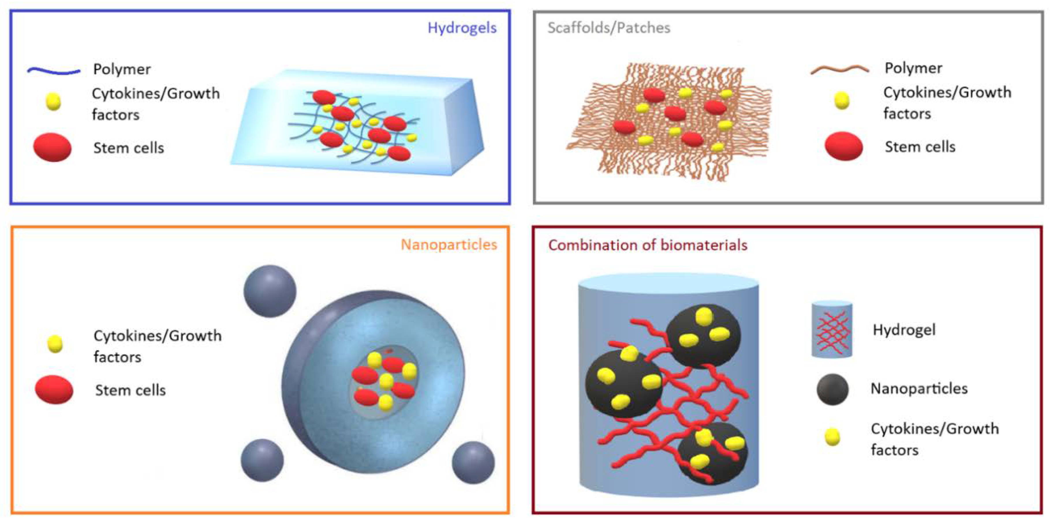
:1. Introduction
2. Biomaterials Loaded with Growth Factors and Cytokines for Cardiac Tissue Regeneration
3. Biomaterials Loaded with Stem Cells for Cardiac Tissue Regeneration
4. Conclusions
Author Contributions
Funding
Conflicts of Interest
References
- McClellan, M.; Brown, N.; Califf, R.M.; Warner, J.J. Call to action, urgent challenges in cardiovascular disease, a presidential advisory from the American Heart Association. Circulation 2019, 139, e44–e54. [Google Scholar] [CrossRef] [PubMed]
- Roth, G.A.; Johnson, C.; Abajobir, A.; Abd-Allah, F.; Abera, S.F.; Abyu, G.; Ahmed, M.; Aksut, B.; Alam, T.; Alam, K.; et al. Global, regional, and national burden of cardiovascular diseases for 10 causes, 1990 to 2015. J. Am. Coll. Cardiol. 2017, 70, 1–25. [Google Scholar] [CrossRef] [PubMed]
- Virani, S.S.; Alonso, A.; Benjamin, E.J.; Bittencourt, M.S.; Callaway, C.W.; Carson, A.P.; Chamberlain, A.M.; Chang, A.R.; Cheng, S.; Delling, F.N.; et al. Heart disease and stroke statistics—2020 update, a report from the American Heart Association. Circulation 2020, 141, E139–E596. [Google Scholar] [CrossRef]
- Swirski, F.K.; Matthias, N. Cardioimmunology: The immune system in cardiac homeostasis and disease. Nat. Rev. Immunol. 2018, 18, 733–744. [Google Scholar] [CrossRef] [PubMed]
- Saparov, A.; Ogay, V.; Nurgozhin, T.; Chen, W.C.; Mansurov, N.; Issabekova, A.; Zhakupova. Role of the immune system in cardiac tissue damage and repair following myocardial infarction. Inflamm. Res. 2017, 66, 739–751. [Google Scholar] [CrossRef] [PubMed]
- Andreadou, I.; Cabrera-Fuentes, H.A.; Devaux, Y.; Frangogiannis, N.G.; Frantz, S.; Guzik, T.; Liehn, E.A.; Gomes, C.P.; Schulz, R.; Hausenloy, D.J. Immune cells as targets for cardioprotection, new players and novel therapeutic opportunities. Cardiovasc. Res. 2019, 115, 1117–1130. [Google Scholar] [CrossRef] [Green Version]
- Hashimoto, H.; Eric, N.O.; Rhonda, B.D. Therapeutic approaches for cardiac regeneration and repair. Nat. Rev. Cardiol. 2018, 15, 585–600. [Google Scholar] [CrossRef]
- Rebouças, J.D.S.; Santos-Magalhães, N.S.; Formiga, F.R. Cardiac regeneration using growth factors, advances and challenges. Arq. Bras. Cardiol. 2016, 107, 271–275. [Google Scholar] [CrossRef]
- Zarrouk-Mahjoub, S.; Zaghdoudi, M.; Amira, Z.; Chebi, H.; Khabouchi, N.; Finsterer, J.; Mechmeche, R.; Ghazouani, E. Pro-and anti-inflammatory cytokines in post-infarction left ventricular remodeling. Int. J. Cardiol. 2016, 221, 632–636. [Google Scholar] [CrossRef]
- Ferrini, A.; Stevens, M.M.; Sattler, S.; Rosenthal, N. Toward regeneration of the heart, bioengineering strategies for immunomodulation. Front. Cardiovasc. Med. 2019, 6, 26. [Google Scholar] [CrossRef] [Green Version]
- Pascual-Gil, S.; Garbayo, E.; Díaz-Herráez, P.; Prosper, F.; Blanco-Prieto, M.J. Heart regeneration after myocardial infarction using synthetic biomaterials. J. Control. Release 2015, 203, 23–38. [Google Scholar] [CrossRef] [PubMed]
- Rocker, A.J.; Lee, D.J.; Shandas, R.; Park, D. Injectable Polymeric Delivery System for Spatiotemporal and Sequential Release of Therapeutic Proteins To Promote Therapeutic Angiogenesis and Reduce Inflammation. ACS Biomater. Sci. Eng. 2020, 6, 1217–1227. [Google Scholar] [CrossRef]
- Dormont, F.; Varna, M.; Couvreur, P. Nanoplumbers, biomaterials to fight cardiovascular diseases. Mater. Today 2018, 21, 122–143. [Google Scholar] [CrossRef]
- Nurkesh, A.; Jaguparov, A.; Jimi, S.; Saparov, A. Recent Advances in the Controlled Release of Growth Factors and Cytokines for Improving Cutaneous Wound Healing. Front. Cell Dev. Biol. 2020, 8, 638. [Google Scholar] [CrossRef]
- Saludas, L.; Pascual-Gil, S.; Prósper, F.; Garbayo, E.; Blanco-Prieto, M. Hydrogel based approaches for cardiac tissue engineering. Int. J. Pharm. 2017, 523, 454–475. [Google Scholar] [CrossRef] [PubMed]
- Mihic, A.; Cui, Z.; Wu, J.; Vlacic, G.; Miyagi, Y.; Li, S.H.; Lu, S.; Sung, H.W.; Weisel, R.D.; Li, R.K. A Conductive Polymer Hydrogel Supports Cell Electrical Signaling and Improves Cardiac Function after Implantation into Myocardial Infarct. Circulation 2015, 132, 772–784. [Google Scholar] [CrossRef] [Green Version]
- Wang, W.; Tan, B.; Chen, J.; Bao, R.; Zhang, X.; Liang, S.; Shang, Y.; Liang, W.; Cui, Y.; Fan, G.; et al. An injectable conductive hydrogel encapsulating plasmid DNA-eNOs and ADSCs for treating myocardial infarction. Biomaterials 2018, 160, 69–81. [Google Scholar] [CrossRef]
- Maghin, E.; Garbati, P.; Quarto, R.; Piccoli, M.; Bollini, S. Young at Heart, Combining Strategies to Rejuvenate Endogenous Mechanisms of Cardiac Repair. Front. Bioeng. Biotechnol. 2020, 8, 447. [Google Scholar] [CrossRef]
- Carvalho, E.; Verma, P.; Hourigan, K.; Banerjee, R. Myocardial infarction, stem cell transplantation for cardiac regeneration. Regen. Med. 2015, 10, 1025–1043. [Google Scholar] [CrossRef]
- Karantalis, V.; Hare, J.M. Use of mesenchymal stem cells for therapy of cardiac disease. Circ. Res. 2015, 116, 1413–1430. [Google Scholar] [CrossRef]
- Shafei, A.E.S.; Ali, M.A.; Ghanem, H.G.; Shehata, A.I.; Abdelgawad, A.A.; Handal, H.R.; Talaat, K.A.; Ashaal, A.E.; El-Shal, A.S. Mesenchymal stem cell therapy, A promising cellbased therapy for treatment of myocardial infarction. J. Gene Med. 2017, 19, e2995. [Google Scholar] [CrossRef] [PubMed]
- Katarzyna, R. Adult stem cell therapy for cardiac repair in patients after acute myocardial infarction leading to ischemic heart failure, an overview of evidence from the recent clinical trials. Curr. Cardiol. Rev. 2017, 13, 223–231. [Google Scholar] [CrossRef] [PubMed]
- Bar, A.; Cohen, S. Inducing Endogenous Cardiac Regeneration, Can Biomaterials Connect the Dots? Front. Bioeng. Biotechnol. 2020, 8, 126. [Google Scholar] [CrossRef] [PubMed] [Green Version]
- Domenech, M.; Polo-Corrales, L.; Ramirez-Vick, J.E.; Freytes, D.O. Tissue engineering strategies for myocardial regeneration, acellular versus cellular scaffolds? Tissue Eng. Part B Rev. 2016, 22, 438–458. [Google Scholar] [CrossRef] [PubMed] [Green Version]
- Ashtari, K.; Nazari, H.; Ko, H.; Tebon, P.; Akhshik, M.; Akbari, M.; Alhosseini, S.N.; Mozafari, M.; Mehravi, B.; Soleimani, M. Electrically conductive nanomaterials for cardiac tissue engineering. Adv. Drug Deliv. Rev. 2019, 144, 162–179. [Google Scholar] [CrossRef]
- Saghazadeh, S.; Rinoldi, C.; Schot, M.; Kashaf, S.S.; Sharifi, F.; Jalilian, E.; Nuutila, K.; Giatsidis, G.; Mostafalu, P.; Derakhshandeh, H. Drug delivery systems and materials for wound healing applications. Adv. Drug Deliv. Rev. 2018, 127, 138–166. [Google Scholar] [CrossRef]
- Wang, Z.; Wang, Z.; Lu, W.W.; Zhen, W.; Yang, D.; Peng, S. Novel biomaterial strategies for controlled growth factor delivery for biomedical applications. NPG Asia Mater. 2017, 9, e435. [Google Scholar] [CrossRef]
- Kakkar, A.; Traverso, G.; Farokhzad, O.C.; Weissleder, R.; Langer, R. Evolution of macromolecular complexity in drug delivery systems. Nat. Rev. Chem. 2017, 1, 1–17. [Google Scholar]
- Mansurov, N.; Chen, W.C.; Awada, H.; Huard, J.; Wang, Y.; Saparov, A. A controlled release system for simultaneous delivery of three human perivascular stem cell-derived factors for tissue repair and regeneration. J. Tissue Eng. Regen. Med. 2018, 12, e1164–e1172. [Google Scholar] [CrossRef]
- Jimi, S.; Jaguparov, A.; Nurkesh, A.; Sultankulov, B.; Saparov, A. Sequential Delivery of Cryogel Released Growth Factors and Cytokines Accelerates Wound Healing and Improves Tissue Regeneration. Front. Bioeng. Biotechnol. 2020, 8, 345. [Google Scholar] [CrossRef]
- Sultankulov, B.; Berillo, D.; Kauanova, S.; Mikhalovsky, S.; Mikhalovska, L.; Saparov, A. Composite Cryogel with Polyelectrolyte Complexes for Growth Factor Delivery. Pharmaceutics 2019, 11, 650. [Google Scholar] [CrossRef] [PubMed] [Green Version]
- Sultankulov, B.; Berillo, D.; Sultankulova, K.; Tokay, T.; Saparov, A. Progress in the Development of Chitosan-Based Biomaterials for Tissue Engineering and Regenerative Medicine. Biomolecules 2019, 9, 470. [Google Scholar] [CrossRef] [PubMed] [Green Version]
- Jo, H.; Gajendiran, M.; Kim, K. Influence of PEG chain length on colloidal stability of mPEGylated polycation based coacersomes for therapeutic protein delivery. J. Ind. Eng. Chem. 2020, 82, 234–242. [Google Scholar] [CrossRef]
- Li, J.; Mooney, D.J. Designing hydrogels for controlled drug delivery. Nat. Rev. Mater. 2016, 1, 1–17. [Google Scholar] [CrossRef]
- Carlini, A.S.; Gaetani, R.; Braden, R.L.; Luo, C.; Christman, K.L.; Gianneschi, N.C. Enzyme-responsive progelator cyclic peptides for minimally invasive delivery to the heart post-myocardial infarction. Nat. Commun. 2019, 10, 1–14. [Google Scholar] [CrossRef] [Green Version]
- Matsumura, Y.; Zhu, Y.; Jiang, H.; D’Amore, A.; Luketich, S.K.; Charwat, V.; Yoshizumi, T.; Sato, H.; Yang, B.; Uchibori, T. Intramyocardial injection of a fully synthetic hydrogel attenuates left ventricular remodeling post myocardial infarction. Biomaterials 2019, 217, 119289. [Google Scholar] [CrossRef] [PubMed]
- Blackburn, N.J.; Sofrenovic, T.; Kuraitis, D.; Ahmadi, A.; McNeill, B.; Deng, C.; Rayner, K.J.; Zhong, Z.; Ruel, M.; Suuronen, E.J. Timing underpins the benefits associated with injectable collagen biomaterial therapy for the treatment of myocardial infarction. Biomaterials 2015, 39, 182–192. [Google Scholar] [CrossRef] [PubMed]
- Lister, Z.; Rayner, K.J.; Suuronen, E.J. How Biomaterials Can Influence Various Cell Types in the Repair and Regeneration of the Heart after Myocardial Infarction. Front. Bioeng. Biotechnol. 2016, 4, 62. [Google Scholar] [CrossRef] [PubMed] [Green Version]
- Li, H.; Bao, M.; Nie, Y. Extracellular matrix-based biomaterials for cardiac regeneration and repair. Heart Fail. Rev. 2020. [Google Scholar] [CrossRef]
- McLaughlin, S.; McNeill, B.; Podrebarac, J.; Hosoyama, K.; Sedlakova, V.; Cron, G.; Smyth, D.; Seymour, R.; Goel, K.; Liang, W.; et al. Injectable human recombinant collagen matrices limit adverse remodeling and improve cardiac function after myocardial infarction. Nat. Commun. 2019, 10, 1–14. [Google Scholar] [CrossRef] [Green Version]
- Firoozi, S.; Pahlavan, S.; Ghanian, M.H.; Rabbani, S.; Tavakol, S.; Barekat, M.; Yakhkeshi, S.; Mahmoudi, E.; Soleymani, M.; Baharvand, H. A Cell-Free SDKP-Conjugated Self-Assembling Peptide Hydrogel Sufficient for Improvement of Myocardial Infarction. Biomolecules 2020, 10, 205. [Google Scholar] [CrossRef] [Green Version]
- Cui, Z.; Ni, N.C.; Wu, J.; Du, G.Q.; He, S.; Yau, T.M.; Weisel, R.D.; Sung, H.W.; Li, R.K. Polypyrrole-chitosan conductive biomaterial synchronizes cardiomyocyte contraction and improves myocardial electrical impulse propagation. Theranostics 2018, 8, 2752–2764. [Google Scholar] [CrossRef]
- Thiagarajan, H.; Thiyagamoorthy, U.; Shanmugham, I.; Nandagopal, G.D.; Kaliyaperumal, A. Angiogenic growth factors in myocardial infarction, a critical appraisal. Heart Fail. Rev. 2017, 22, 665–683. [Google Scholar] [CrossRef] [PubMed]
- Frangogiannis, N.G. The extracellular matrix in myocardial injury, repair, and remodeling. J. Clin. Investig. 2017, 127, 1600–1612. [Google Scholar] [CrossRef] [PubMed] [Green Version]
- Chen, B.; Huang, S.; Su, Y.; Wu, Y.J.; Hanna, A.; Brickshawana, A.; Graff, J.; Frangogiannis, N.G. Macrophage Smad3 protects the infarcted heart, stimulating phagocytosis and regulating inflammation. Circ. Res. 2019, 125, 55–70. [Google Scholar] [CrossRef] [PubMed]
- Yue, Y.; Wang, C.; Benedict, C.; Huang, G.; Truongcao, M.; Roy, R.; Cimini, M.; Garikipati, V.N.S.; Cheng, Z.; Koch, W.J. Interleukin-10 Deficiency Alters Endothelial Progenitor Cell–Derived Exosome Reparative Effect on Myocardial Repair via Integrin-Linked Kinase Enrichment. Circ. Res. 2020, 126, 315–329. [Google Scholar] [CrossRef]
- Jung, M.; Ma, Y.; Iyer, R.P.; DeLeon-Pennell, K.Y.; Yabluchanskiy, A.; Garrett, M.R.; Lindsey, M.L. IL-10 improves cardiac remodeling after myocardial infarction by stimulating M2 macrophage polarization and fibroblast activation. Basic Res. Cardiol. 2017, 112, 33. [Google Scholar] [CrossRef]
- Ferraro, B.; Leoni, G.; Hinkel, R.; Ormanns, S.; Paulin, N.; Ortega-Gomez, A.; Viola, J.R.; de Jong, R.; Bongiovanni, D.; Bozoglu, T.; et al. Pro-angiogenic macrophage phenotype to promote myocardial repair. J. Am. Coll. Cardiol. 2019, 73, 2990–3002. [Google Scholar] [CrossRef]
- Cahill, T.J.; Choudhury, R.P.; Riley, P.R. Heart regeneration and repair after myocardial infarction, translational opportunities for novel therapeutics. Nat. Rev. Drug Discov. 2017, 16, 699. [Google Scholar] [CrossRef]
- Shiraishi, M.; Shintani, Y.; Shintani, Y.; Ishida, H.; Saba, R.; Yamaguchi, A.; Adachi, H.; Yashiro, K.; Suzuki, K. Alternatively activated macrophages determine repair of the infarcted adult murine heart. J. Clin. Investig. 2016, 126, 2151–2166. [Google Scholar] [CrossRef] [Green Version]
- Oduk, Y.; Zhu, W.; Kannappan, R.; Zhao, M.; Borovjagin, A.V.; Oparil, S.; Zhang, J. VEGF nanoparticles repair the heart after myocardial infarction. Am. J. Physiol.-Heart C 2018, 314, H278–H284. [Google Scholar] [CrossRef] [PubMed]
- Hachim, D.; Whittaker, T.E.; Kim, H.; Stevens, M.M. Glycosaminoglycan-based biomaterials for growth factor and cytokine delivery: Making the right choices. J. Control. Release 2019, 313, 131–147. [Google Scholar] [CrossRef] [PubMed]
- Shi, J.; Fan, C.; Zhuang, Y.; Sun, J.; Hou, X.; Chen, B.; Xiao, Z.; Chen, Y.; Zhan, Z.; Zhao, Y. Heparan sulfate proteoglycan promotes fibroblast growth factor-2 function for ischemic heart repair. Biomater. Sci. 2019, 7, 5438–5450. [Google Scholar] [CrossRef] [PubMed]
- Fan, C.; Shi, J.; Zhuang, Y.; Zhang, L.; Huang, L.; Yang, W.; Chen, B.; Chen, Y.; Xiao, Z.; Shen, H. Myocardial-Infarction-Responsive Smart Hydrogels Targeting Matrix Metalloproteinase for On-Demand Growth Factor Delivery. Adv. Mater. 2019, 31, 1902900. [Google Scholar] [CrossRef] [PubMed]
- Itoh, N.; Ohta, H.; Nakayama, Y.; Konishi. Roles of FGF signals in heart development, health, and disease. Front. Cell Dev. Biol. 2016, 4, 110. [Google Scholar] [CrossRef] [Green Version]
- Teringova, E.; Tousek, P. Apoptosis in ischemic heart disease. J. Transl. Med. 2017, 15, 87. [Google Scholar] [CrossRef] [Green Version]
- Awada, H.K.; Johnson, N.R.; Wang, Y. Sequential delivery of angiogenic growth factors improves revascularization and heart function after myocardial infarction. J. Control. Release 2015, 207, 7–17. [Google Scholar] [CrossRef] [Green Version]
- Taimeh, Z.; Loughran, J.; Birks, E.J.; Bolli, R. Vascular endothelial growth factor in heart failure. Nat. Rev. Cardiol. 2013, 10, 519. [Google Scholar] [CrossRef]
- Corti, F.; Simons, M. Modulation of VEGF receptor 2 signaling by protein phosphatases. Pharmacol. Res. 2017, 115, 107–123. [Google Scholar] [CrossRef] [Green Version]
- Cassani, M.; Fernandes, S.; Vrbsky, J.; Ergir, E.; Cavalieri, F.; Forte, G. Combining Nanomaterials and Developmental Pathways to Design New Treatments for Cardiac Regeneration, The Pulsing Heart of Advanced Therapies. Front. Bioeng. Biotechnol. 2020, 8, 323. [Google Scholar] [CrossRef] [Green Version]
- Catoira, M.C.; Fusaro, L.; Di Francesco, D.; Ramella, M.; Boccafoschi, F. Overview of natural hydrogels for regenerative medicine applications. J. Mater. Sci. Mater. Med. 2019, 30, 115. [Google Scholar] [CrossRef] [PubMed] [Green Version]
- Gyles, D.A.; Castro, L.D.; Silva, J.O.C., Jr.; Ribeiro-Costa, R.M. A review of the designs and prominent biomedical advances of natural and synthetic hydrogel formulations. Eur. Polym. J. 2017, 88, 373–392. [Google Scholar] [CrossRef]
- Rufaihah, A.J.; Yasa, I.C.; Ramanujam, V.S.; Arularasu, S.C.; Kofidis, T.; Guler, M.O.; Tekinay, A.B. Angiogenic peptide nanofibers repair cardiac tissue defect after myocardial infarction. Acta Biomater. 2017, 58, 102–112. [Google Scholar] [CrossRef] [PubMed]
- Navaei, A.; Truong, D.; Heffernan, J.; Cutts, J.; Brafman, D.; Sirianni, R.W.; Vernon, B.; Nikkhah, M. PNIPAAm-based biohybrid injectable hydrogel for cardiac tissue engineering. Acta Biomater. 2016, 32, 10–23. [Google Scholar] [CrossRef]
- Yuan, Z.; Tsou, Y.; Zhang, X.; Huang, S.; Yang, Y.; Gao, M.; Ho, W.; Zhao, Q.; Ye, X.; Xu, X. Injectable citrate-based hydrogel as an angiogenic biomaterial improves cardiac repair after myocardial infarction. ACS Appl. Mater. Interfaces 2019, 11, 38429–38439. [Google Scholar] [CrossRef]
- Waters, R.; Alam, P.; Pacelli, S.; Chakravarti, A.; Ahmed, R.; Paul, A. Stem cell-inspired secretome-rich injectable hydrogel to repair injured cardiac tissue. Acta Biomater. 2018, 69, 95–106. [Google Scholar] [CrossRef]
- Ho, Y.T.; Poinard, B.; Kah, J.C.Y. Nanoparticle drug delivery systems and their use in cardiac tissue therapy. Nanomedicine 2016, 11, 693–714. [Google Scholar] [CrossRef]
- Nguyen, M.M.; Carlini, A.; Chien, M.; Sonnenberg, S.; Luo, C.; Braden, R.; Osborn, K.; Li, Y.; Gianneschi, N.; Christman, K. Enzyme-Responsive Nanoparticles for Targeted Accumulation and Prolonged Retention in Heart Tissue after Myocardial Infarction. Adv. Mater. 2015, 27, 5547–5552. [Google Scholar] [CrossRef]
- Somasuntharam, I.; Yehl, K.; Carroll, S.; Maxwell, J.; Martinez, M.; Che, P.; Brown, M.; Salaita, K.; Davis, M. Knockdown of TNF-α by DNAzyme gold nanoparticles as an anti-inflammatory therapy for myocardial infarction. Biomaterials 2016, 83, 12–22. [Google Scholar] [CrossRef] [Green Version]
- Qi, Q.; Lu, L.; Li, H.; Yuan, Z.; Chen, G.; Lin, M.; Ruan, Z.; Ye, X.; Xiao, Z.; Zhao, Q. Spatiotemporal delivery of nanoformulated liraglutide for cardiac regeneration after myocardial infarction. Int. J. Nanomed. 2017, 12, 4835–4848. [Google Scholar] [CrossRef] [Green Version]
- Steele, A.N.; Paulsen, M.; Wang, H.; Stapleton, L.; Lucian, H.; Eskandari, A.; Hironaka, C.; Farry, J.; Baker, S.; Thakore, A.; et al. Multi-phase catheter-injectable hydrogel enables dual-stage protein-engineered cytokine release to mitigate adverse left ventricular remodeling following myocardial infarction in a small animal model and a large animal model. Cytokine 2020, 127, 154974. [Google Scholar] [CrossRef] [PubMed]
- Park, S.; Nguyen, N.; Pezhouman, A.; Ardehali, R. Cardiac fibrosis, potential therapeutic targets. Transl. Res. 2019, 209, 121–137. [Google Scholar] [CrossRef] [PubMed]
- Lin, X.; Liu, Y.; Bai, A.; Cai, H.; Bai, Y.; Jiang, W.; Yang, H.; Wang, X.; Yang, L.; Sun, N.; et al. A viscoelastic adhesive epicardial patch for treating myocardial infarction. Nat. Biomed. Eng. 2019, 3, 632–643. [Google Scholar] [CrossRef] [PubMed]
- Wan, L.; Chen, Y.; Wang, Z.; Wang, W.; Schmull, S.; Dong, J.; Xue, S.; Imboden, H.; Li, J. Human heart valve-derived scaffold improves cardiac repair in a murine model of myocardial infarction. Sci. Rep. 2017, 7, 1–11. [Google Scholar]
- Rodness, J.; Mihic, A.; Miyagi, Y.; Wu, J.; Weisel, R.; Li, R. VEGF-loaded microsphere patch for local protein delivery to the ischemic heart. Acta Biomater. 2016, 45, 169–181. [Google Scholar] [CrossRef]
- Fan, C.; Tang, Y.; Zhao, M.; Lou, X.; Pretorius, D.; Menasche, P.; Zhu, W.; Zhang, J. CHIR99021 and fibroblast growth factor 1 enhance the regenerative potency of human cardiac muscle patch after myocardial infarction in mice. J. Mol. Cell. Cardiol. 2020, 141, 1–10. [Google Scholar] [CrossRef]
- Donndorf, P.; Strauer, B.E.; Haverich, A.; Steinhoff, G. Stem cell therapy for the treatment of acute myocardial infarction and chronic ischemic heart disease. Curr. Pharm. Biotechnol. 2013, 14, 12–19. [Google Scholar]
- Saparov, A.; Chen, C.W.; Beckman, S.A.; Wang, Y.; Huard, J. The role of antioxidation and immunomodulation in postnatal multipotent stem cell-mediated cardiac repair. Int. J. Mol. Sci. 2013, 14, 16258–16279. [Google Scholar] [CrossRef] [Green Version]
- Luo, L.; Tang, J.; Nishi, K.; Yan, C.; Dinh, P.; Cores, J.; Kudo, T.; Zhang, J.; Li, T.; Cheng, K. Fabrication of synthetic mesenchymal stem cells for the treatment of acute myocardial infarction in mice. Circ. Res. 2017, 120, 1768–1775. [Google Scholar] [CrossRef] [Green Version]
- Chang, M.L.; Chiu, Y.J.; Li, J.S.; Cheah, K.P.; Lin, H.H. Analyzing Impetus of Regenerative Cellular Therapeutics in Myocardial Infarction. J. Clin. Med. 2020, 9, 1277. [Google Scholar] [CrossRef]
- Chen, C.W.; Okada, M.; Proto, J.; Gao, X.; Sekiya, N.; Beckman, S.; Corselli, M.; Crisan, M.; Saparov, A.; Tobita, K.; et al. Human pericytes for ischemic heart repair. Stem cells 2013, 31, 305–316. [Google Scholar] [CrossRef] [PubMed] [Green Version]
- Jiang, Y.; Lian, X.L. Heart regeneration with human pluripotent stem cells, Prospects and challenges. Bioact. Mater. 2020, 5, 74–81. [Google Scholar] [CrossRef] [PubMed]
- Parizadeh, S.M.; Jafarzadeh-Esfehani, R.; Ghandehari, M.; Parizadeh, M.R.; Ferns, G.A.; Avan, A.; Hassanian, S.M. Stem cell therapy, A novel approach for myocardial infarction. J. Cell. Physiol. 2019, 234, 16904–16912. [Google Scholar] [CrossRef] [PubMed]
- Higuchi, A.; Ku, N.; Tseng, Y.; Pan, C.; Li, H.; Kumar, S.; Ling, Q.; Chang, Y.; Alarfaj, A.; Munusamy, M.; et al. Stem cell therapies for myocardial infarction in clinical trials, bioengineering and biomaterial aspects. Lab. Investig. 2017, 97, 1167–1179. [Google Scholar] [CrossRef] [PubMed]
- Karantalis, V.; DiFede, D.; Gerstenblith, G.; Pham, S.; Symes, J.; Zambrano, J.; Fishman, J.; Pattany, P.; McNiece, I.; Conte, J.; et al. Autologous mesenchymal stem cells produce concordant improvements in regional function, tissue perfusion, and fibrotic burden when administered to patients undergoing coronary artery bypass grafting, the Prospective Randomized Study of Mesenchymal Stem Cell Therapy in Patients Undergoing Cardiac Surgery (PROMETHEUS) trial. Circ. Res. 2014, 114, 1302–1310. [Google Scholar] [PubMed] [Green Version]
- Luger, D.; Lipinski, M.; Westman, P.; Glover, D.; Dimastromatteo, J.; Frias, J.; Albelda, M.; Sikora, S.; Kharazi, A.; Vertelov, G.; et al. Intravenously delivered mesenchymal stem cells, systemic anti-inflammatory effects improve left ventricular dysfunction in acute myocardial infarction and ischemic cardiomyopathy. Circ. Res. 2017, 120, 1598–1613. [Google Scholar] [CrossRef]
- Bao, L.; Meng, Q.; Li, Y.; Deng, S.; Yu, Z.; Liu, Z.; Zhang, L.; Fan, H. C-Kit Positive cardiac stem cells and bone marrow–derived mesenchymal stem cells synergistically enhance angiogenesis and improve cardiac function after myocardial infarction in a paracrine manner. J. Card. Fail. 2017, 23, 403–415. [Google Scholar] [CrossRef]
- Kanda, P.; Davis, D.R. Cellular mechanisms underlying cardiac engraftment of stem cells. Expert Opin. Biol. Ther. 2017, 17, 1127–1143. [Google Scholar] [CrossRef]
- Yao, Y.; Liao, W.; Yu, R.; Du, Y.; Zhang, T.; Peng, Q. Potentials of combining nanomaterials and stem cell therapy in myocardial repair. Nanomedicine (Lond) 2018, 13, 1623–1638. [Google Scholar] [CrossRef]
- Amer, M.H.; Rose, F.; White, L.; Shakesheff, K. A detailed assessment of varying ejection rate on delivery efficiency of mesenchymal stem cells using narrow-bore needles. Stem cells Transl. Med. 2016, 5, 366–378. [Google Scholar] [CrossRef] [Green Version]
- Aguado, B.A.; Mulyasasmita, W.; Su, J.; Lampe, K.; Heilshorn, S. Improving viability of stem cells during syringe needle flow through the design of hydrogel cell carriers. Tissue Eng. Part A 2012, 18, 806–815. [Google Scholar] [CrossRef] [PubMed] [Green Version]
- Park, T.Y.; Oh, J.; Cho, J.; Sim, S.; Lee, J.; Cha, H. Stem cell-loaded adhesive immiscible liquid for regeneration of myocardial infarction. J. Control. Release 2020, 321, 602–615. [Google Scholar] [CrossRef] [PubMed]
- Chen, Y.; Li, C.; Li, C.; Chen, J.; Li, Y.; Xie, H.; Lin, C.; Fan, M.; Guo, Y.; Gao, E.; et al. Tailorable Hydrogel Improves Retention and Cardioprotection of Intramyocardial Transplanted Mesenchymal Stem Cells for the Treatment of Acute Myocardial Infarction in Mice. J. Am. Heart Assoc. 2020, 9, e013784. [Google Scholar] [CrossRef] [PubMed]
- Blocki, A.; Beyer, S.; Dewavrin, J.; Goralczyk, A.; Wang, Y.; Peh, P.; Ng, M.; Moonshi, S.; Vuddagiri, S.; Raghunath, M.; et al. Microcapsules engineered to support mesenchymal stem cell (MSC) survival and proliferation enable long-term retention of MSCs in infarcted myocardium. Biomaterials 2015, 53, 12–24. [Google Scholar] [CrossRef]
- Gallagher, L.B.; Dolan, E.; O’Sullivan, J.; Levey, R.; Cavanagh, B.; Kovarova, L.; Pravda, M.; Velebny, V.; Farrell, T.; O’Brien, F.; et al. Pre-culture of mesenchymal stem cells within RGD-modified hyaluronic acid hydrogel improves their resilience to ischaemic conditions. Acta Biomater. 2020, 107, 78–90. [Google Scholar] [CrossRef]
- Chow, A.; Stuckey, D.; Kidher, E.; Rocco, M.; Jabbour, R.; Mansfield, C.; Darzi, A.; Harding, S.; Stevens, M.; Athanasiou, T. Human Induced Pluripotent Stem Cell-Derived Cardiomyocyte Encapsulating Bioactive Hydrogels Improve Rat Heart Function Post Myocardial Infarction. Stem Cell Rep. 2017, 9, 1415–1422. [Google Scholar] [CrossRef] [Green Version]
- Cai, H.; Wu, F.; Wang, Q.; Xu, P.; Mou, F.; Shao, S.; Luo, Z.; Zhu, J.; Xuan, S.; Lu, R.; et al. Self-assembling peptide modified with QHREDGS as a novel delivery system for mesenchymal stem cell transplantation after myocardial infarction. Faseb J. 2019, 33, 8306–8320. [Google Scholar] [CrossRef]
- Harel, S.; Mayaki, D.; Sanchez, V.; Hussain, S. NOX2, NOX4, and mitochondrial-derived reactive oxygen species contribute to angiopoietin-1 signaling and angiogenic responses in endothelial cells. Vasc. Pharm. 2017, 92, 22–32. [Google Scholar] [CrossRef]
- Lee, A.S.; Inayathullah, M.; Lijkwan, M.; Zhao, X.; Sun, W.; Park, S.; Hong, W.; Parekh, M.; Malkovskiy, A.; Lau, E.; et al. Prolonged survival of transplanted stem cells after ischaemic injury via the slow release of pro-survival peptides from a collagen matrix. Nat. Biomed. Eng. 2018, 2, 104–113. [Google Scholar] [CrossRef] [Green Version]
- Ottersbach, A.; Mykhaylyk, O.; Heidsieck, A.; Eberbeck, D.; Rieck, S.; Zimmermann, K.; Breitbach, M.; Engelbrecht, B.; Brügmann, T.; Hesse, M.; et al. Improved heart repair upon myocardial infarction, Combination of magnetic nanoparticles and tailored magnets strongly increases engraftment of myocytes. Biomaterials 2018, 155, 176–190. [Google Scholar] [CrossRef]
- Choe, G.; Kim, S.; Park, J.; Park, J.; Kim, S.; Kim, Y.; Ahn, Y.; Jung, D.; Williams, D.; Lee, J. Anti-oxidant activity reinforced reduced graphene oxide/alginate microgels, Mesenchymal stem cell encapsulation and regeneration of infarcted hearts. Biomaterials 2019, 225, 119513. [Google Scholar] [CrossRef] [PubMed]
- Kai, D.; Wang, Q.; Wang, H.; Prabhakaran, M.; Zhang, Y.; Tan, Y.; Ramakrishna, S. Stem cell-loaded nanofibrous patch promotes the regeneration of infarcted myocardium with functional improvement in rat model. Acta. Biomater. 2014, 10, 2727–2738. [Google Scholar] [CrossRef] [PubMed]
- Wang, Q.L.; Wang, H.; Li, Z.; Wang, Y.; Wu, X.; Tan, Y. Mesenchymal stem cell-loaded cardiac patch promotes epicardial activation and repair of the infarcted myocardium. J. Cell Mol. Med. 2017, 21, 1751–1766. [Google Scholar] [CrossRef]
- Chen, J.; Zhan, Y.; Wang, Y.; Han, D.; Tao, B.; Luo, Z.; Ma, S.; Wang, Q.; Li, X.; Fan, L.; et al. Chitosan/silk fibroin modified nanofibrous patches with mesenchymal stem cells prevent heart remodeling post-myocardial infarction in rats. Acta. Biomater. 2018, 80, 154–168. [Google Scholar] [CrossRef] [PubMed]
- Gaetani, R.; Feyen, D.; Verhage, V.; Slaats, R.; Messina, E.; Christman, K.; Giacomello, A.; Doevendans, P.; Sluijter, J. Epicardial application of cardiac progenitor cells in a 3D-printed gelatin/hyaluronic acid patch preserves cardiac function after myocardial infarction. Biomaterials 2015, 61, 339–348. [Google Scholar] [CrossRef]
- Su, T.; Huang, K.; Daniele, M.; Hensley, M.; Young, A.; Tang, J.; Allen, T.; Vandergriff, A.; Erb, P.; Ligler, F.; et al. Cardiac Stem Cell Patch Integrated with Microengineered Blood Vessels Promotes Cardiomyocyte Proliferation and Neovascularization after Acute Myocardial Infarction. ACS Appl. Mater. Interfaces 2018, 10, 33088–33096. [Google Scholar] [CrossRef]
- Dong, Y.; Hong, M.; Dai, R.; Wu, H.; Zhu, P. Engineered bioactive nanoparticles incorporated biofunctionalized ECM/silk proteins based cardiac patches combined with MSCs for the repair of myocardial infarction, In vitro and in vivo evaluations. Sci. Total Environ. 2020, 707, 135976. [Google Scholar] [CrossRef]
- Gao, L.; Kupfer, M.; Jung, J.; Yang, L.; Zhang, P.; Da Sie, Y.; Tran, Q.; Ajeti, V.; Freeman, B.; Fast, V.; et al. Myocardial Tissue Engineering With Cells Derived From Human-Induced Pluripotent Stem Cells and a Native-Like, High-Resolution, 3-Dimensionally Printed Scaffold. Circ. Res. 2017, 120, 1318–1325. [Google Scholar] [CrossRef] [Green Version]
- Tang, J.; Wang, J.; Huang, K.; Ye, Y.; Su, T.; Qiao, L.; Hensley, M.; Caranasos, T.; Zhang, J.; Gu, Z.; et al. Cardiac cell-integrated microneedle patch for treating myocardial infarction. Sci. Adv. 2018, 4, eaat9365. [Google Scholar] [CrossRef] [Green Version]
- Park, S.J.; Kim, R.; Park, B.; Lee, S.; Choi, S.; Park, J.; Choi, J.; Kim, S.; Jang, J.; Cho, D.; et al. Dual stem cell therapy synergistically improves cardiac function and vascular regeneration following myocardial infarction. Nat. Commun. 2019, 10, 3123. [Google Scholar] [CrossRef] [Green Version]
- Yeung, E.; Fukunishi, T.; Bai, Y.; Bedja, D.; Pitaktong, I.; Mattson, G.; Jeyaram, A.; Lui, C.; Ong, C.; Inoue, T.; et al. Cardiac regeneration using human-induced pluripotent stem cell-derived biomaterial-free 3D-bioprinted cardiac patch in vivo. J. Tissue Eng. Regen. Med. 2019, 13, 2031–2039. [Google Scholar] [CrossRef]
- Melhem, M.R.; Park, J.; Knapp, L.; Reinkensmeyer, L.; Cvetkovic, C.; Flewellyn, J.; Lee, M.; Jensen, T.; Bashir, R.; Kong, H.; et al. 3D Printed Stem-Cell-Laden, Microchanneled Hydrogel Patch for the Enhanced Release of Cell-Secreting Factors and Treatment of Myocardial Infarctions. ACS Biomater. Sci. Eng. 2017, 3, 1980–1987. [Google Scholar] [CrossRef]
- Mayfield, A.E.; Tilokee, E.; Latham, N.; McNeill, B.; Lam, B.; Ruel, M.; Suuronen, E.; Courtman, D.; Stewart, D.; Davis, D. The effect of encapsulation of cardiac stem cells within matrix-enriched hydrogel capsules on cell survival, post-ischemic cell retention and cardiac function. Biomaterials 2014, 35, 133–142. [Google Scholar] [CrossRef] [PubMed] [Green Version]
- Ahmadi, A.L.; Thorn, S.; Alarcon, E.; Kordos, M.; Padavan, D.; Hadizad, T.; Cron, G.; Beanlands, R.; DaSilva, J.; Ruel, M.; et al. PET imaging of a collagen matrix reveals its effective injection and targeted retention in a mouse model of myocardial infarction. Biomaterials 2015, 49, 18–26. [Google Scholar] [CrossRef] [PubMed]
- Han, J.; Kim, B.; Shin, J.; Ryu, S.; Noh, M.; Woo, J.; Park, J.; Lee, Y.; Lee, N.; Hyeon, T.; et al. Iron oxide nanoparticle-mediated development of cellular gap junction crosstalk to improve mesenchymal stem cells’ therapeutic efficacy for myocardial infarction. ACS Nano 2015, 9, 2805–2819. [Google Scholar] [CrossRef] [PubMed]
- Lemcke, H.; Gaebel, R.; Skorska, A.; Voronina, N.; Lux, C.; Petters, J.; Sasse, S.; Zarniko, N.; Steinhoff, G.; David, R. Mechanisms of stem cell based cardiac repair-gap junctional signaling promotes the cardiac lineage specification of mesenchymal stem cells. Sci. Rep. 2017, 7, 9755. [Google Scholar] [CrossRef]
- Marcu, I.C.; Illaste, A.; Heuking, P.; Jaconi, M.; Ullrich, N. Functional Characterization and Comparison of Intercellular Communication in Stem Cell-Derived Cardiomyocytes. Stem Cells 2015, 33, 2208–2218. [Google Scholar] [CrossRef] [Green Version]
- Sottas, V.; Wahl, C.; Trache, M.; Bartolf-Kopp, M.; Cambridge, S.; Hecker, M.; Ullrich, N. Improving electrical properties of iPSC-cardiomyocytes by enhancing Cx43 expression. J. Mol. Cell Cardiol. 2018, 120, 31–41. [Google Scholar] [CrossRef]
- Liang, W.; Chen, J.; Li, L.; Li, M.; Wei, X.; Tan, B.; Shang, Y.; Fan, G.; Wang, W.; Liu, W. Conductive Hydrogen Sulfide-Releasing Hydrogel Encapsulating ADSCs for Myocardial Infarction Treatment. ACS Appl. Mater. Interfaces 2019, 11, 14619–14629. [Google Scholar] [CrossRef]
- Tang, J.; Su, T.; Huang, K.; Dinh, P.; Wang, Z.; Vandergriff, A.; Hensley, M.; Cores, J.; Allen, T.; Li, T.; et al. Targeted repair of heart injury by stem cells fused with platelet nanovesicles. Nat. Biomed. Eng. 2018, 2, 17–26. [Google Scholar] [CrossRef]
- Yokoyama, R.; Ii, M.; Tabata, Y.; Hoshiga, M.; Ishizaka, N.; Asahi, M. Cardiac Regeneration by Statin-Polymer Nanoparticle-Loaded Adipose-Derived Stem Cell Therapy in Myocardial Infarction. Stem Cells Transl. Med. 2019, 8, 1055–1067. [Google Scholar] [CrossRef] [Green Version]
- Yao, X.; Liu, Y.; Gao, J.; Yang, L.; Mao, D.; Stefanitsch, C.; Li, Y.; Zhang, J.; Ou, L.; Kong, D.; et al. Nitric oxide releasing hydrogel enhances the therapeutic efficacy of mesenchymal stem cells for myocardial infarction. Biomaterials 2015, 60, 130–140. [Google Scholar] [CrossRef] [PubMed]
- Díaz-Herráez, P.; Saludas, L.; Pascual-Gil, S.; Simón-Yarza, T.; Abizanda, G.; Prósper, F.; Garbayo, E.; Blanco-Prieto, M. Transplantation of adipose-derived stem cells combined with neuregulin-microparticles promotes efficient cardiac repair in a rat myocardial infarction model. J. Control. Release 2017, 249, 23–31. [Google Scholar] [CrossRef]
- Chung, H.J.; Kim, J.; Kim, H.; Kyung, H.; Katila, P.; Lee, J.; Yang, T.; Yang, Y.; Lee, S. Epicardial delivery of VEGF and cardiac stem cells guided by 3-dimensional PLLA mat enhancing cardiac regeneration and angiogenesis in acute myocardial infarction. J. Control. Release 2015, 205, 218–230. [Google Scholar] [CrossRef] [PubMed]
- Kumar, M.; Kasala, E.; Bodduluru, L.; Dahiya, V.; Sharma, D.; Kumar, V.; Lahkar, M. Animal models of myocardial infarction, mainstay in clinical translation. Regul. Toxicol. Pharmacol. 2016, 76, 221–230. [Google Scholar] [CrossRef] [PubMed]
- Zaragoza, C.; Gomez-Guerrero, C.; Martin-Ventura, J.; Blanco-Colio, L.; Lavin, B.; Mallavia, B.; Tarin, C.; Mas, S.; Ortiz, A.; Egido, J. Animal models of cardiovascular diseases. J. Biomed. Biotechnol. 2011, 2011, 497841. [Google Scholar] [CrossRef]
- Seyhan, A.A. Lost in translation, the valley of death across preclinical and clinical divide–identification of problems and overcoming obstacles. Transl. Med. Commun. 2019, 4, 1–19. [Google Scholar] [CrossRef] [Green Version]
- Camacho, P.; Fan, H.; Liu, Z.; He, J.Q. Small mammalian animal models of heart disease. Am. J. Cardiovasc. Dis. 2016, 6, 70. [Google Scholar]


| Biomaterial | Growth Factors/Cytokine | Effect | References |
|---|---|---|---|
| Heparan sulfate proteoglycans | bFGF | Extended bioavailability of the growth factor by protecting it from degradation, and improved angiogenesis and cardiac function | [53] |
| Glutathione-modified collagen hydrogel | bFGF fused with glutathione-S-transferase and MMP-2/9 cleavable peptide TIMP | Decreased collagen deposition, increased vascularization, and improved heart function | [54] |
| Hydroxyethyl methacrylate hyaluronic acid hydrogel | Neuregulin-1β | Improved ventricular function and structure | [38] |
| Fibrin gel/heparine coacervate | VEGF and PDGF | Improved angiogenesis and cardiac function, and reduced scar formation and inflammation | [57] |
| Citrate-based polyester hydrogel | Mydgf | Reduced cell apoptosis and scar formation, and improved angiogenesis and cardiac function | [65] |
| Laponite/gelatin hydrogel | ADSC secretome | Improved angiogenesis, ejection fraction, and cardiac output, and reduced fibrosis | [66] |
| Poly(lactic-co-glycolic acid)–poly(ethylene glycol) nanoparticles | Liraglutide | Improved heart function, attenuated adverse cardiac remodeling, stimulated angiogenesis, and suppressed cardiomyocyte apoptosis | [70] |
| A sulfonated hydrogel and poly(ethylene glycol)-blockpoly(serinol hexamethylene urea)-block-poly(ethylene glycol) micelle nanoparticles | VEGF, IL-10, and PDGF | Improved angiogenesis and demonstrated potential amelioration of inflammation to optimize cardiac repair post-MI | [12] |
| Hyaluronic acid-based hydrogel | HGFdf and ESA | Decreased infarct size, and improved angiogenesis and heart function | [71] |
| Calcium-alginate microsphere patch | VEGF | Improved tissue regeneration and cardiac function, and increased capillary density | [75] |
| Human cardiomyocyte patch with polylactic-co-glycolic acid nanoparticles | FGF1 and CHIR99021 | Reduced infarction size and improved angiogenesis and cardiac function. The combination of factors reduced apoptosis and increased proliferation of transplanted cardiomyocytes | [76] |
| Biomaterial | Stem Cells | Effect | References |
|---|---|---|---|
| Mussel adhesive protein/HA coacervate | MSCs | Increased MSCs survival and retention | [92] |
| Collagen-based hydrogel | ADSCs | Improved engraftment of stem cells | [93] |
| Microcapsules made of agarose and ECM components | MSCs | Increased MSCs survival | [94] |
| Arginylglycylaspartic acid (RGD) modified HA hydrogel | MSCs | Increased MSCs survival | [95] |
| Erythropoietin linked hydrogel | iPSCs | Improved the post-MI heart recovery | [96] |
| Synthetic SAP and angiopoetin-1-derived pro-survival peptide QHREDGS | MSCs | Increased cells survival and cardiac function | [97] |
| Collagen–dendrimer | CPCs | Increased long-term survival | [99] |
| Silica-coated SOMag5 magnetic nanoparticles | Embryonic cardiomyocytes, embryonic stem cell-derived cardiomyocytes and BMSCs | Improved cell engraftment | [100] |
| Graphene oxide/alginate microgel | MSCs | Improved MI recovery | [101] |
| Poly(ε-caprolactone)/gelatin patch | MSCs | Increased angiogenesis, lymphangiogenesis, cardiomyogenesis, and paracrine factors released by stem cells and reduced scar size | [103] |
| Chitosan and silk fibroin microfibrous cardiac patch | MSCs | Increased MSC survival | [104] |
| Hyaluronic acid/gelatin cardiac patch | CPCs | Increased long-term CPCs survival and differentiation | [105] |
| Vascularized fibrin hydrogel patch | CSCs | Increased cell survival | [106] |
| Gold nanoparticles coated with a combination of ECM and silk proteins | MSCs | Increased cell survival and retention and decreased infarct size | [107] |
| ECM scaffold with the usage of methacrylated gelatin | iPSC-derived cardiomyocytes, smooth muscle cells and endothelial cells | Reduced infarct size and improved cell proliferation, cardiac function, and angiogenesis | [108] |
| Poly(vinyl alcohol) microneedle patch | CSCs | Improved angiogenesis, reduced fibrosis, and repaired left ventricular wall | [109] |
| Polycaprolactone patch | MSCs and iPSC-derived cardiomyocytes | Improved MI recovery and angiogenesis | [110] |
| Microchanneled poly(ethylene glycol) dimethacrylate hydrogel patch | MSCs | Improved cardiac function | [112] |
| Agarose hydrogel microcapsules supplemented with fibronectin and fibrinogen | CSCs | Improved production of pro-angiogenic/cardioprotective cytokines, angiogenesis, and angiogenic cells recruitment after direct intramyocardial injection | [113] |
| Iron nanoparticles | MSCs | Reduced the infarct size, prevented fibrosis, decreased apoptosis of myocardial cells, increased angiogenesis, and improved cardiac function | [115] |
| Statin-conjugated poly(lactic-co-glycolic acid) nanoparticles | ADSCs | Increased the ejection fraction and several other parameters which reflect the left ventricular function. Inhibited local inflammation, promoted recruitment of circulating stem cells, and stimulated their differentiation to cardiomyocytes and angiogenesis | [121] |
| Tetraaniline-polyethylene glycol diacrylate and thiolated hyaluronic acid conductive hydrogel | ADSCs | Improved neovascularization, regeneration of the damaged myocardium, and post-infarction cardiac function | [17] |
| Naphthalene hydrogel | MSCs | Increased cells survival, and stimulated the synthesis of angiogenic factors VEGF and SDF-1α | [122] |
| Poly(lactic-co-glycolic acid) microparticles | ADSCs | Improved cells survival, and induced the shift of macrophage found in the infarcted myocardium from pro-inflammatory M1 to regenerative M2 phenotype | [123] |
| Poly(l-lactic acid) mat | CSCs | Improved angiogenic and cardiomyogenic effects | [124] |
© 2020 by the authors. Licensee MDPI, Basel, Switzerland. This article is an open access article distributed under the terms and conditions of the Creative Commons Attribution (CC BY) license (http://creativecommons.org/licenses/by/4.0/).
Share and Cite
Smagul, S.; Kim, Y.; Smagulova, A.; Raziyeva, K.; Nurkesh, A.; Saparov, A. Biomaterials Loaded with Growth Factors/Cytokines and Stem Cells for Cardiac Tissue Regeneration. Int. J. Mol. Sci. 2020, 21, 5952. https://doi.org/10.3390/ijms21175952
Smagul S, Kim Y, Smagulova A, Raziyeva K, Nurkesh A, Saparov A. Biomaterials Loaded with Growth Factors/Cytokines and Stem Cells for Cardiac Tissue Regeneration. International Journal of Molecular Sciences. 2020; 21(17):5952. https://doi.org/10.3390/ijms21175952
Chicago/Turabian StyleSmagul, Saltanat, Yevgeniy Kim, Aiganym Smagulova, Kamila Raziyeva, Ayan Nurkesh, and Arman Saparov. 2020. "Biomaterials Loaded with Growth Factors/Cytokines and Stem Cells for Cardiac Tissue Regeneration" International Journal of Molecular Sciences 21, no. 17: 5952. https://doi.org/10.3390/ijms21175952