Article

Novel Diels–Alder Type Adducts from Morus alba Root Bark Targeting Human Monoamine Oxidase and Dopaminergic Receptors for the Management of Neurodegenerative Diseases

1
Department of Food and Life Science, Pukyong National University, Busan 48513, Korea
2
Department of Food Science and Human Nutrition, Jeonbuk National University, Jeonju 54896, Korea
*
Authors to whom correspondence should be addressed.
These authors contributed equally to this work.
Int. J. Mol. Sci. 2019, 20(24), 6232; https://doi.org/10.3390/ijms20246232
Submission received: 5 November 2019 / Revised: 6 December 2019 / Accepted: 9 December 2019 / Published: 10 December 2019
(This article belongs to the Special Issue Genetic Variants in Dopamine Receptors and Neurodegenerative Diseases)

Abstract

:
In this study, we delineate the human monoamine oxidase (hMAO) inhibitory potential of natural Diels–Alder type adducts, mulberrofuran G (1), kuwanon G (2), and albanol B (3), from Morus alba root bark to characterize their role in Parkinson’s disease (PD) and depression, focusing on their ability to modulate dopaminergic receptors (D1R, D2LR, D3R, and D4R). In hMAO-A inhibition, 13 showed mild effects (50% inhibitory concentration (IC50): 54‒114 μM). However, 1 displayed moderate inhibition of the hMAO-B isozyme (IC50: 18.14 ± 1.06 μM) followed by mild inhibition by 2 (IC50: 57.71 ± 2.12 μM) and 3 (IC50: 90.59 ± 1.72 μM). Our kinetic study characterized the inhibition mode, and the in silico docking predicted that the moderate inhibitor 1 would have the lowest binding energy. Similarly, cell-based G protein-coupled receptors (GPCR) functional assays in vector-transfected cells expressing dopamine (DA) receptors characterized 13 as D1R/D2LR antagonists and D3R/D4R agonists. The half-maximum effective concentration (EC50) of 13 on DA D3R/D4R was 15.13/17.19, 20.18/21.05, and 12.63/‒ µM, respectively. Similarly, 13 inhibited 50% of the DA response on D1R/D2LR by 6.13/2.41, 16.48/31.22, and 7.16/18.42 µM, respectively. A computational study revealed low binding energy for the test ligands. Interactions with residues Asp110, Val111, Tyr365, and Phe345 at the D3R receptor and Asp115 and His414 at the D4R receptor explain the high agonist effect. Likewise, Asp187 at D1R and Asp114 at D2LR play a crucial role in the antagonist effects of the ligand binding. Our overall results depict 13 from M. alba root bark as good inhibitors of hMAO and potent modulators of DA function as D1R/D2LR antagonists and D3R/D4R agonists. These active constituents in M. alba deserve in-depth study for their potential to manage neurodegenerative disorders (NDs), particularly PD and psychosis.

1. Introduction

Monoamine oxidase (MAO) is a flavoenzyme in the outer mitochondrial membrane of neuronal and non-neuronal cells that has a vital role in the etiology of age-regulated neurodegenerative disorders (NDs). MAO catalyzes the oxidative deamination of monoamine neurotransmitters, dietary amines, and xenobiotics and regulates their levels and functions in the brain. During oxidative deamination, MAO liberates hydrogen peroxide, the reactive oxygen species (ROS) most potent in causing oxidative stress and mitochondrial dysfunction [1]. Though the etiology of NDs remains unclear, apoptosis, oxidative stress, mitochondrial dysfunction, inflammation, an impaired ubiquitin-proteasome system, and excitotoxicity are common disease-modifying factors [2]. Two isoforms (MAO-A and MAO-B) with specific functions have been identified in different brain regions and cell types [3].
MAO-A displays a higher affinity for serotonin (5HT) and norepinephrine, whereas MAO-B prefers phenylethylamine. Dopamine (DA) and tyramine are common substrates for both isozymes [4]. MAO-A is associated with the onset of psychiatric disorders (Figure 1), including depression, and antisocial aggressive impulsive behaviors through its ability to decrease neurotransmitter levels (DA and serotonin) [5,6]. During a normal physiological state, DA levels in substantia nigra pars compacta (SNpc) are regulated as an equilibrium between synthesis, synaptic vesicle loading, uptake, and catabolism. MAO enzyme mediates oxidative deamination of DA to DOPAL along with H2O2 generation, leading DA deficit and oxidative stress state. And MAO-A inhibition prevents the deamination of neurotransmitters, reduces oxidative stress, and increases the availability of neurotransmitters within noradrenergic and serotonergic neurons of the CNS to regulate neuron signaling via their respective receptors [4,7]. Similarly, MAO-B metabolizes DA to DOPAC and catechol-O-methyltransferase (COMT) degrades it to homovanillic acid (HVA) in astrocyte [8,9]. Therefore, MAO inhibitors function as neuroprotective agents against age-related NDs.
The concept of precision medicine relies on protein targeting, and G protein-coupled receptors (GPCRs) are the largest family of target receptors and membrane proteins. At present, 34% of FDA-approved drugs target GPCRs [10]. GPCRs are widely expressed and activated by a broad range of ligands, including neurotransmitters, hormones, and ions, as well as sensory signals [11]. Neurotransmitters bind to their specific receptors at the postsynaptic cleft and trigger or inhibit neuronal functions and signals by regulating the activity of ion channels. In NDs, especially Parkinson’s disease (PD), the selective loss of dopaminergic neurons in the SNpc produces DA deficiency, which triggers cell-specific alterations in intrinsic excitability and synaptic plasticity [12]. Therefore, regulating DA levels or DA receptor signaling is a standard approach to PD treatment. Numerous neurotransmitters and their analogs have therapeutic properties, serve as medicaments for various diseases, and have been the subject of extensive pharmacological studies [13]. In this study, we discuss the critical physicochemical interactions between our test ligands and different residue side chains and the adjacent amino acids.
Morus alba Linn, commonly known as mulberry, is a perennial woody plant of the family Moraceae that is widely cultivated in tropical, subtropical, and temperate zones in Asia, Europe, and North and South America. The leaves of this plant are used as feed for animals and sericulture, the fruit is used as food, and the wood as timber. Furthermore, in traditional Chinese medicine, the leaves, twigs, fruit, and root bark are used as antioxidant, anti-inflammatory, anti-hypertensive, hypoglycemic, immunomodulatory, hypolipidemic, antibacterial, and anti-tumor agents [14]. The plant thus has unique medicinal and ethnic values. It is rich in flavonoids, alkaloids, steroids, and coumarins. Diels–Alder-type adducts are prototypical metabolites in the root bark [15]. In a previous study, mulberry fruit extract protected dopaminergic neurons in in vitro and in vivo PD models by regulating ROS generation through its antioxidant and anti-apoptotic effects [16]. A crude water extract of M. alba leaf ameliorated alterations in the retinal neurotransmitters adrenaline, DA, gamma-aminobutyric acid, histamine, noradrenaline, and serotonin in the pups of diabetic and hypercholesterolemic mother rats [17] and ameliorated kidney damage in diabetic rats by suppressing inflammation and fibrosis via peroxisome proliferator-activated receptor γ (PPARγ) modulation [18]. Similarly, a leaf-ethanol extract possessed anxiolytic and muscle-relaxant activities, probably via a γ-aminobutyric acid A-benzodiazepine (GABAA-BZD) mechanism [19]. No previous reports have considered the root bark of M. alba. In our recent work, we reported the antidiabetic [20,21], anti-Alzheimer’s disease activity [22,23], and antioxidant and anti-browning property [24] of Diels–Alder-type adducts and arylbenzofurans from M. alba root bark. More recently, kuwanon G and albanin G from the root bark were hypothesized as the components responsible for the appetite suppression activity of root-bark extract via cannabinoid (CB1) receptor antagonism [25]. In the present study, we characterize the multi-target effects of Diels–Alder-type adducts, mulberrofuran G (1), kuwanon G (2), and albanol B (3) (Figure 2), via human monoamine oxidase (hMAO) inhibition and the modulation of dopaminergic receptors (D1R, D2R, D3R, and D4R), and we use a molecular simulation to explore the action mechanism of the ligand–receptor interaction.

2. Results

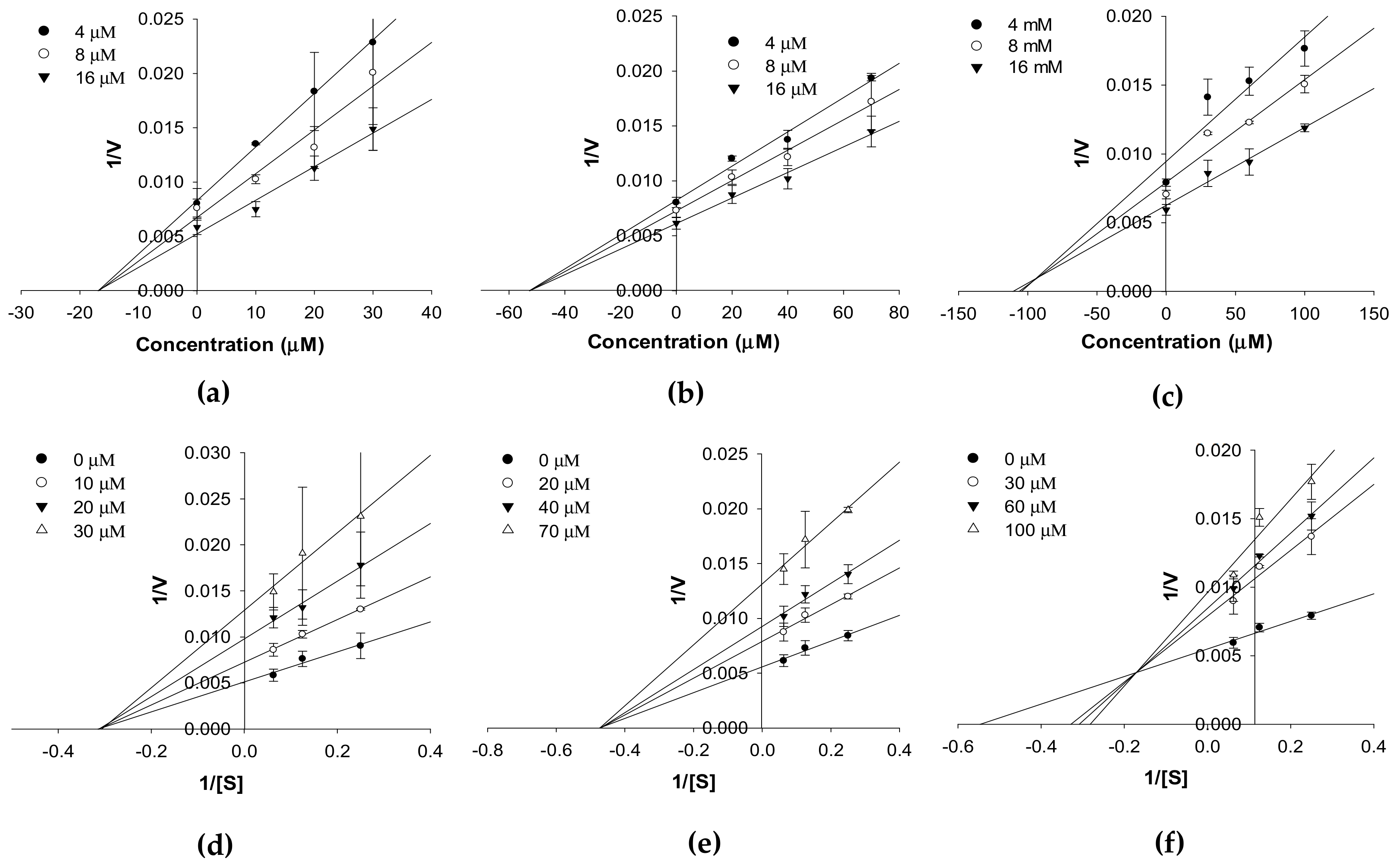
2.1. In Vitro hMAO Inhibition and Enzyme Kinetics

The in vitro hMAO inhibition potentials of 13 and the reference compound selegiline was evaluated via a chemiluminescent assay in a white, opaque, 96-well plate using the MAO-Glo kit (Promega, Madison, WI). At first, 13 were screened for hMAO activity at 100 µg/mL and the % inhibition was 93.87%, 99.05%, and 74.85%, respectively. Then the compounds were retested at different micromolar concentrations in triplicates and the 50% inhibitory concentration (IC50) values obtained from the log-dose inhibition curve are tabulated in Table 1.
As shown there, 13 displayed mild inhibition of hMAO-A activity. Among the test compounds, 1 showed the best inhibition, with an IC50 value of 54.79 ± 0.03 µM, followed by 2 (IC50: 70.16 ± 2.60 µM) and 3 (IC50: 114.31 ± 2.30 µM). The inhibition potentials of 13 were better against hMAO-B, though the pattern of inhibition was similar: 1 showed moderate inhibition effect, with an IC50 value of 18.14 ± 1.06 µM, and compounds 2 and 3 mildly inhibited hMAO-B, with IC50 values of 57.71 ± 2.12 and 90.59 ± 1.72 µM, respectively. The reference inhibitor selegiline inhibited the activity of isozymes -A and -B at IC50 values of 12.51 ± 1.11 and 0.30 ± 0.01 µM, respectively. However, compared to the reference reversible hMAO-A inhibitor (harmine, IC50: 0.006 µM) [26] and reversible hMAO-B inhibitor (safinamide, IC50: 0.00512 µM) [27], the potency of 13 is significantly weaker.
The enzyme inhibition patterns of compounds at different substrate concentrations in the kinetic study are tabulated in Table 1 and represented in Figure 3 and Figure 4. Compounds 13 competitively inhibited hMAO-A isozyme activity with Ki values of 26.96 ± 3.98, 28.29 ± 2.02, and 46.93 ± 4.12 µM, respectively (Table 1 and Figure 3a–c). The Lineweaver–Burk plots (1/V vs. 1/[S]) for hMAO-A isozyme activity (Figure 3d–f) reveal an increase in Km with an increase in the concentrations of 13, whereas 1/Vmax remained constant. Meanwhile, 1 and 2 were noncompetitive inhibitors (Vmax value decreased in a concentration-dependent manner without changing the Km value), and 3 was a mixed type inhibitor (increase in inhibitor concentration increased the Km value but decreased the Vmax value) of the hMAO-B isozyme (Figure 4c,f). From a Secondary plot (plot not shown here), the binding constants of 3 with a free enzyme (Kic) and with enzyme-substrate complex (Kiu) identified were 55.19 ± 7.97 and 186.2 ± 10.26 µM, respectively. Likewise, the Ki value of 1 and 2 for hMAO-B inhibition was 17.01 ± 3.31 and 52.09 ± 5.56 µM, respectively.

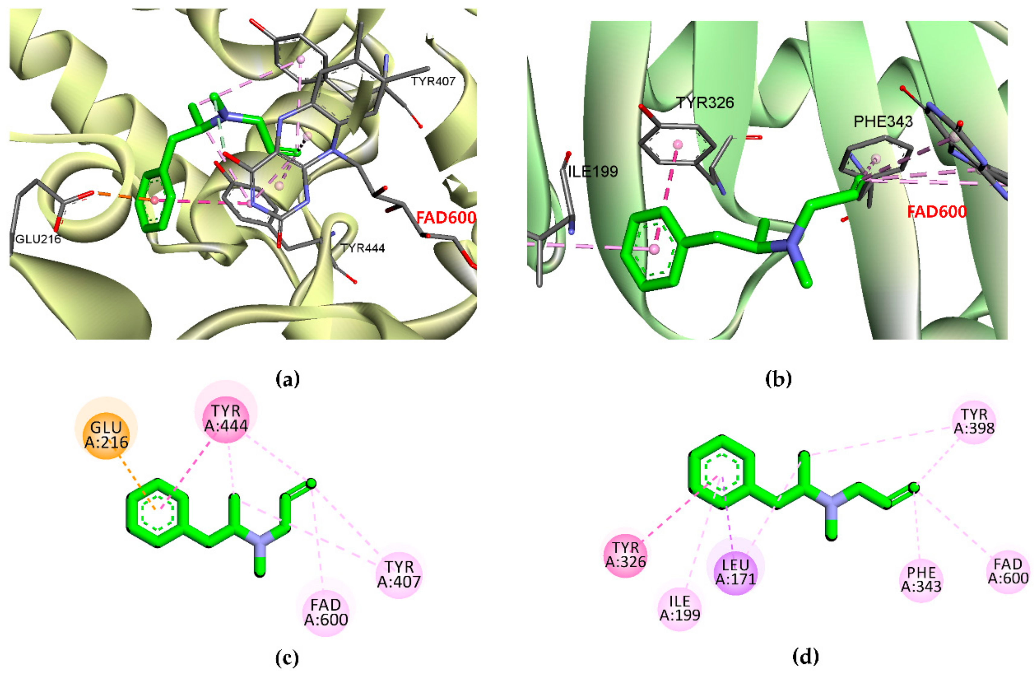
2.2. In Silico Docking Simulation of hMAO

Computational modeling was performed to obtain insights into the binding affinity between ligands and the enzyme using AutoDock 4.2. To validate the docking result, the reference inhibitor selegiline as well as reversible inhibitor harmine (for hMAO-A) and safinamide (for hMAO-B) were docked into the active site cavity of the hMAO enzyme, and the ligands were re-docked. The results of the simulation study are tabulated in Table 2 and Table 3 and represented in Figure 5, Figure 6 and Figure 7.
As shown in Table 2, the test ligand (13)–hMAO-A complexes showed lower binding energies (−6.74 to −9.54 kcal/mol) than the reference ligand selegiline (−6.54 kcal/mol) and harmine (−6.46 kcal/mol). Ligand 1 posed in the active site by interacting with Gly110, Thr336, Ile207, Gly214, and Ser209 via a hydrogen bond (Figure 5). Meanwhile, ligands 2 and 3 shared Met300 and Gly404 as common H-bond interacting residues. Reversible inhibitor harmine interacted with flavin adenine dinucleotide (FAD)600, Ile335, and Tyr444 residues at the active site cavity, which were not observed for test ligand-binding. In the case of hMAO-B, ligands 13 showed high affinity with binding energies (−11.09, −12.65, and −10.05 kcal/mol, respectively) by forming three and five H-bond interactions, respectively (Figure 6). With the lowest binding energy, ligand 1 stably positioned in the hMAO-B active site by interacting with His115, Pro476, and Glu483 via H-bonds. Moreover, 1 interacted with peripheral residues, including Phe103, Val106, and Ile477. Interacting residues Val106 and Phe103 were shared by all three ligands as a noncompetitive inhibitor. Ligand 2 shared the most abundant H-bond interaction residues: Pro103, Asn116, Glu483, Phe103, and Thr478. Ligand 3 also showed high affinity via H-bond interactions with Thr195, Pro104, Asn116, Thr478, and Gly193. Selegiline interacted with Ile198 and safinamide with Ile199, Cys172, Tyr326, and Thr201 via H-bonds in the active site of hMAO-B (Figure 7).

2.3. Cell-Based Functional GPCR Assays

To characterize the possible role of compounds 13 in neuronal diseases, we first screened their functional activity at 100 µM on DA (D1, D2, D3, D4) receptors by measuring their effects on secondary messengers (cAMP modulation or Ca2+ ion mobilization) in transfected cell lines expressing human cloned receptors of interest. The data in Table 4 represent the agonist/antagonist effects of 13 at 100 µM on the various receptors.
As shown in the table, 13 exhibited a full antagonist effect on the D1/D2 receptors and a full agonist effect on the D3/D4 receptors. The agonist effects of 13 at 100 µM on D3R/D4R were 119.9/86.30, 124.3/90.45, and 102.8/46.10%, respectively. Similarly, at 100 µM, 13 inhibited the DA response on D1R/D2LR by 87.65/101.30, 98.85/99.15, and 67.80/78.55%, respectively. Figure 8 shows the concentration-dependent functional effect of 13 on the DA receptor subtypes with corresponding EC50/IC50 values.
As shown there, all compounds showed promising antagonist effects on D1R/D2LR, with IC50 values in the range of 2.41‒31.22 µM (Figure 8c,d). The rank order for the antagonist effect was D2LR > D1R for 1 and D1R > D2LR for 2 and 3. Compared to 1 and 2, the dose–response curve for 3 (Figure 8c) looks unusual due to relatively higher standard deviation in response at 50 µM concentration. Similarly, 13 showed an agonist effect on D3R/D4R, with EC50 values in the range of 12.63‒21.05 µM (Figure 8a,b), and the rank order for the agonist effect was D3R = D4R for 1 and 2 and D3R > D4R for 3. These results indicate that compounds 13 mediate the DA function by acting as D1R/D2LR antagonists and D3R/D4R agonists.

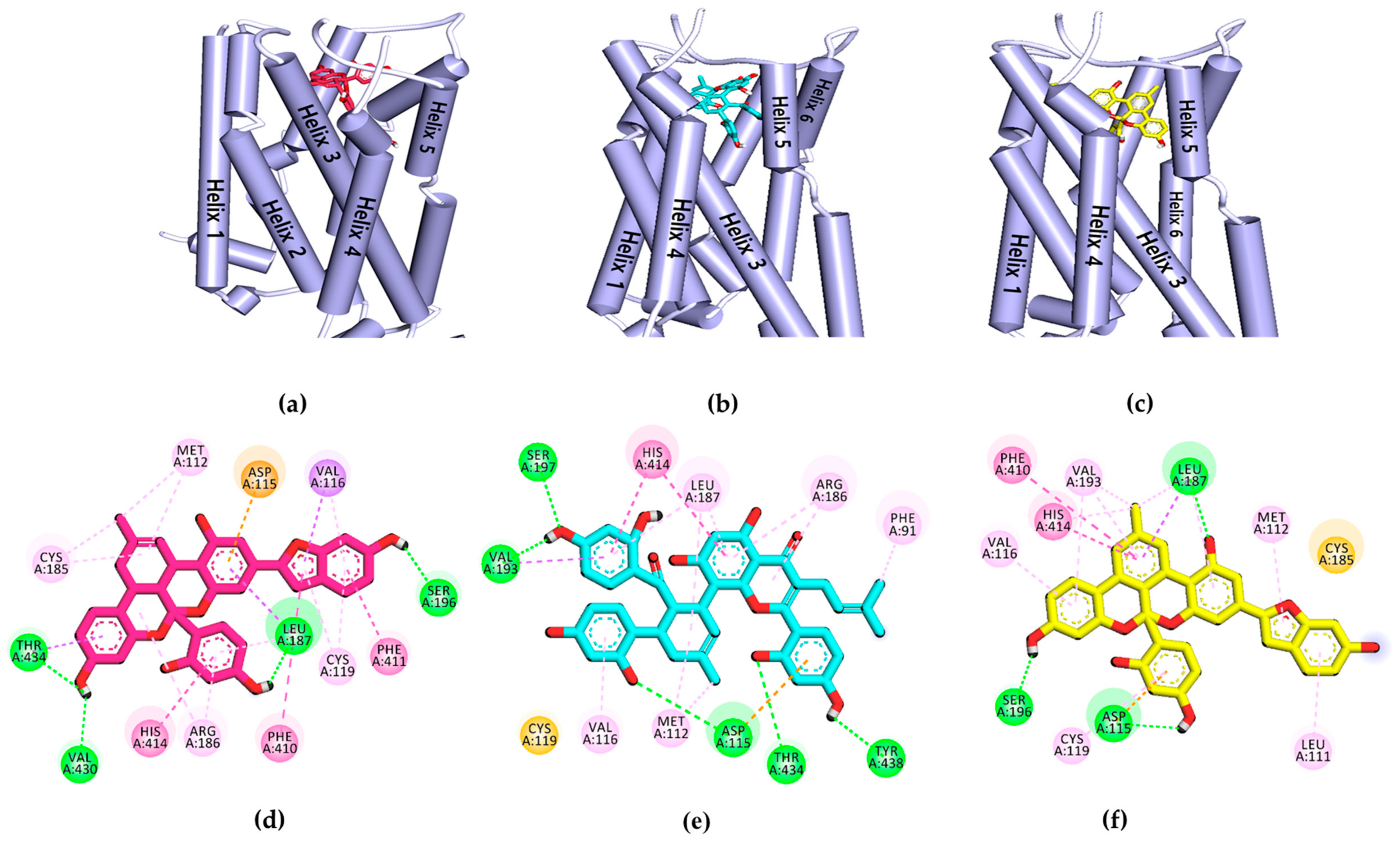
2.4. In Silico Docking Simulation of Dopamine Receptors

To validate the results of the functional assays and investigate and identify the ligand–receptor interactions for novel lead discovery, we carried out a computational docking simulation using AutoDock 4.2 (Figure 9, Figure 10, Figure 11 and Figure 12). Since the effect of 13 on DA receptors was promising, a simulation study was carried out on DA receptors. The binding affinities of reference ligands for each receptor were also evaluated to better understand and validate the docking results. The homology model of DA receptor subtype D1R was based on the structure of the β2-adrenergic receptor because it has a higher similarity to the DA D1 receptor in the binding site region and sequence identity [28]. Subtypes D2LR, D3R, and D4R were obtained from the protein data bank (PDB) IDs for 6CM4, 3PBL, and 5WIV, respectively. The results of the docking simulation are tabulated in Table 5, Table 6, Table 7 and Table 8 and represented in Figure 9, Figure 10, Figure 11 and Figure 12. The dotted colored lines in Figure 9, Figure 10, Figure 11 and Figure 12 represent specific interactions (green line: H-bond; purple line: π-sigma; pale pink: π-alkyl, alkyl; pink: π-π-T-shaped, π-π stacked; orange: π-anion).
As shown in Table 5, the 1‒D1R complex exhibited four strong H-bond interactions with Lys81, Leu291, Ser188, and Asp314 with low binding energy (−9.22 kcal/mol). The ligand 2‒D1R complex (−7.1 kcal/mol) interacted with Ser202 and Asp103 in H-bonds, similar to the reference antagonist SCH-23390 and agonist DA. Ligand 3, with a binding energy of −9.2 kcal/mol, shared Ser188 and Asp187 with D1R via H-bonds.
Furthermore, Asp187, Leu295, Phe306, Pro171, Ala192, and Ala195 were revealed as hydrophobic residues in the ligand 3‒D1R complex (Figure 9). Figure 10 provides a close-up view of ligands 13 binding at the active site of D2LR. As shown in Table 6, ligands 13 bound strongly to the active site of D2LR with low binding energies (−8.11 to −10.45 kcal/mol). Risperidone and butaclamol are D2LR agonist and antagonist and they bound to the active site of the receptor with binding energies −12.7 and −6.9 kcal/mol, respectively, by forming salt-bridge with Asp114.
Though ligands 13 did not form a salt-bridge with Asp114, they showed H-bond and π-anion interactions with the residue. Furthermore, the number of H-bond interactions was higher for test ligands compared to resperidone and butaclamol.
Specifically, Asp114 was an H-bond and electrostatic residue for both ligands 1 and 3, indicating high affinity with the receptor, whereas Ile184 was a crucially active residue in the second extracellular loop of D2LR, forming a π-alkyl interaction with 2. Phe189 and Val190 are necessary key residues in antagonist-ligand binding, and they were well observed in the 2‒D2LR and 3‒D2LR complexes.
Among the test ligands, 3 showed the highest affinity (−10.41 kcal/mol) for ligand‒D3R interactions (Table 7). Ligand 1 had a slightly higher binding energy than 3 but was comparable to that of reference agonist DA (−6.9 kcal/mol).
The key conserved interacting residue, Asp110 in the transmembrane III of D3R, could be seen in all three ligands‒D3R complexes via electrostatic interaction (Figure 11). Other important residues, such as Phe345 and Tyr365, fit tightly into ligands 13 via π-π hydrophobic bonds. Val111 at helix III was also observed forming a π-alkyl interaction with both ligands 1 and 3. Ligands 13 also interacted with neighboring residues, including His349, Ile183, Thr369, Val86, and Cys114. Similarly, at D4R, all the test ligands showed strong interactions with lower binding energies (−12.42 to −9.67 kcal/mol) than the reference drugs (Table 8).
DA, nemonapride, and clozapine were used as the reference agonist and antagonist and had binding energies of −6.1, −13.08, and −10.14 kcal/mol, respectively.
One of the most crucial residues in stimulating D4R, Asp115 interacted with all three ligands in π-anion form at helix III, whereas Ser197 interacted only with 1 and 3 on in helix V (Figure 12). Ligand 3 showed an H-bond interaction with Asp115 and Ser196, which is probably why it had the lowest binding energy (−12.42 kcal/mol) among the three ligands. Similarly, Val116, His414, and Leu187 were common interaction sites for three ligands at D4R. Other surrounding residues, including Met112, Thr434, Arg186, Phe410, Cys119, and Val193, were involved in hydrophobic interactions with the ligands.

3. Discussion

In the present study, we tested Diels–Alder type adducts 13 from the root bark of M. alba and found that they exhibit a mild-to-moderate hMAO inhibition effect. The inhibition effect was slightly higher on the hMAO-B isozyme (IC50: 18.14 to 90.59 µM) than on hMAO-A (IC50: 54.79 to 114.31 µM). In particular, ligand 1 demonstrated a moderate inhibition effect on the hMAO-B isozyme, with an IC50 value of 18.14 ± 1.06 µM. Among test compounds 13, 1 and 3 are fused benzofurans, and 2 is a mono-isoprenyl substituted flavone. The structural difference between 1 and 3 is the methyl cyclohexene in 1 and methylbenzene in 3. The methyl cyclohexene group of 1 was involved in specific alkyl interactions with Leu337, Ile335, and Met350 and this moiety is facing toward FAD at the hMAO-A active site (Figure 5). However, the methylbenzene of 3 was not involved in any interactions, which explains why 1 had better binding affinity and inhibition potency on hMAO-A than 3. Likewise, in hMAO-B inhibition, H-bond interaction of 1 with His115, Pro476, and Glu483 might explain for better binding affinity and activity compared to 3. In addition, 1 and 2 bound in a similar pose and this might explain the same binding mode for both the ligands. The test ligand activity (Ki values) did not show a strong correlation with the docking score (binding energies). This variation might be attributed to the physicochemical properties of ligands, especially logP which was predicted high in the range of 6.7 to 7.3 from web-based software PreADMET (v2.0, YONSEI University, Seoul, Korea) (data not reported here). Overall, the results of the hMAO inhibition assay reveal that these Diels–Alder type adducts, especially 1, might have therapeutic value in managing PD. However, this treatment approach is just symptomatic, restoring dopaminergic function in the striatum [29]. Therefore, the discovery of new natural DA agonists is promising.
Depending on their stimulation or inhibition of adenylyl cyclase and modulation of cAMP levels, DA receptors are categorized into two classes: D1-like (D1R and D5R) and D2-like (D2R, D3R, and D4R). These DA receptors have specific anatomical distributions and specifically mediate DA action [30]. Several studies have pointed to DA receptor antagonists as a promising approach to managing heroin addiction. For instance, a D1R antagonist (SCH 23390) and D2R antagonists (haloperidol and raclopride) attenuate heroin-induced reinstatement [31,32], and a D3R antagonist (SB-277011A) blocks the acquisition and expression of the conditioned place preference response to heroin [33]. Similarly, a natural alkaloid l-tetrahydropalmatine is a D1R/D2R antagonist with an anti-addiction property [34], and govadine (D1R/D2R antagonist) demonstrated antipsychotic properties in conjunction with pro-cognitive effects in rats [35]. The extent of D2R binding affinity and antagonizing ability represent the clinical efficacy of antipsychotic drugs [36]. Previously, a root ethanol extract of M. alba mediated skin wound healing by upregulating the mRNA levels of chemokine receptor 4, one of the GPCRs [37]. Other than that, no previous studies have reported on GPCR modulation by an M. alba root extract or its metabolites.
To evaluate the functional effects of adducts 13 on DA (D1, D2, D3, and D4) receptors, we conducted a cell-based GPCR functional assay. As shown in Figure 8c,d, 13 potently and concentration-dependently inhibited the agonist response of DA at D1R and D2LR. Even at 25 µM, 13 inhibited the DA response on D1R/D2LR by 92.32/97.16, 91.09/33.69, and 66.82/81.11%, respectively. Unlike sigmoidal dose–response curves of 1 and 2 at D1R, compound 3 showed an unusual non-sigmoidal curve (Figure 8c). A higher standard deviation in response at 50 µM concentration led to unusual appearance. While self-association into colloidal particles at a higher concentration or multi-target actions [38,39,40] explains the possible reason for the observed non-sigmoidal dose–response curve of the compound 3. Likewise, in the D2LR agonist assay, 2 showed nonspecific interference (NSI) in the assay system. This NSI might be attributed to aggregation/colloid formation or chemical reactivity because these are significant sources of nonspecific bioactivity particularly in high throughput screening (HTS) [41]. NSI by aggregates and colloids is detergent sensitive, so it will be confirmed in the coming report. We compared the binding affinity and interacting residues of test compounds 13 with those of a reference agonist (DA) and antagonist (SCH 23390) via a molecular docking simulation. As shown in Table 5 and Table 6, 13 showed a high binding affinity (the binding energies of 13 were lower than those of the reference drugs at D1R and D2R, except for risperidone at D2R). Test ligands 2 shared common H-bond interaction residues, Asp103 and Ser202 with the reference antagonist (SCH 23390). Furthermore, 13 displayed additional H-bond interactions with serine residues (Ser107, Ser188, and Ser198) and aspartic acid residues (Asp314 and Asp187). Similarly, at D2R, 13 interacted with the key interacting residues Asp114, Trp100, and Phe389 [42]. All three ligands bound to D2LR with high affinity and the binding energy was lower than that of the reference antagonist butaclamol. Interactions with Asp114, Cys118, Phe198, Phe389, Trp386, and Tyr416 were common among the test ligands and butaclamol (Table 6). Additional H-bond interactions between Ser197 and 1, Tyr408 and 2, and Trp100 and 3 were also observed. Residue Ser197 is a conserved-essential residue within the binding site for binding the D2R antagonist risperidone [43], which was also observed for test ligand 2 binding. Tyr408 is located deep in the binding site, whereas Trp100 is at the periphery of the binding site of D2R [44], and they were both involved in the binding of test ligands 2 and 3. According to Salmas et al. [45], Phe389, Phe390, and Trp386 in TM6 are main residues for D2R-antagonists. Meanwhile, Phe189, Phe198, and Val190 are necessary as key residues for antagonist ligands binding. Here, Phe389 and Val190 are interacting with ligand 2 whereas Val190 and Phe189 are bound to ligand 3 as hydrophobic bond. Using those findings, we characterized 13 as D1R/D2LR antagonists. In a previous study, M. alba leaf extract possessed D2R-mediated anti-dopaminergic activity, suggesting a possible clinical application for M. alba leaves in psychiatric disorders [46]. Our findings suggest that 13 could have antipsychotic effects.
The test compounds showed an agonist effect on D3R and D4R. As shown in Figure 8a,b, 13 showed a potent agonist effect on D3R and D4R. D3R is prominently distributed within the limbic system and mediates the psychiatric manifestation of DA receptor stimulation. Therefore, DA receptor agonists with high affinity for D3R have an antidepressant effect [47]. Similarly, Levant et al. suggested that D3R-stimulation (rather than D2R-stimulation) might mediate the antiparkinsonian effects of DA receptor agonists with a high preference for D2R [48]. Rotigotine is an FDA-approved, full DA agonist (rank order: D3R >  D2LR  >  D1R  =  D5R  >  D4.4R) developed as a transdermal patch for the treatment of PD [1,49].
A previously conducted survey reported that more than 110 patent applications had been submitted concerning selective D3R ligands [50]. Unfortunately, none of them has yet received clinical approval due to failures of the pharmacokinetics or safety profiles [51]. Similarly, D4R agonism has been implicated in the management of cognitive deficits associated with schizophrenia [52] and attention-deficit/hyperactivity disorder [53] and also to reduce the adverse effects of opioids [54].
The results of the functional assays in this study show that the ligands 13 have concentration-dependent agonist effects on D3R and D4R (rank order: D3R > D4R). Even at 25 µM, 13 showed potent agonist responses on DA D3R/D4R of 71.92/63.00, 64.99/58.66, and 94.93/‒%, respectively. The agonist effect of 3 on D4R was mild (% stimulation of agonist response of 44.85% at 100 µM). The antagonist effect on these receptor subtypes was negligible. We also used molecular docking simulations to compare the binding affinity and interacting residues between test compounds 13 and D3R (Table 7) with those of a reference agonist DA and antagonists (eticlopride and (+)-butaclamol). Likewise, docking simulations of 13 and D4R was compared with those of reference agonists DA and nemonapride, and an antagonist clozapine (Table 8). As tabulated in Table 7 and Table 8, the binding energies of 13 on D3R/D4R were comparable to the reference ligands. Interestingly, our prediction demonstrated that they had lower binding energy at D4R than at D3R. Interaction with Asp110 on D3R and Asp115 on D4R was in common with the agonist DA. It was reported earlier that a salt-bridge to the carboxylic acid group of the Asp110 on hD3R and the Asp115 on hD4R is critical to high-affinity ligand binding to dopaminergic receptors [55]. In this study, though ligands 13 did not form a salt-bridge with those receptors, they did form strong electrostatic interactions (Pi-Anion). In addition to their electrostatic interactions with Asp115 on D4R, 2 and 3 formed H-bond interactions with carboxylic acid group of Asp115.
At a molecular level, D1-like (D1 and D5) receptor signaling is mediated chiefly by the heterotrimeric G proteins Gαs/olf, which cause sequential activation of adenylate cyclase, cyclic AMP-dependent protein kinase, and the protein phosphatase-1 inhibitor DARPP-32 [56]. A recent study showed that hypersensitivity of D1R is responsible for l-DOPA-induced activation of mTORC1 signaling, and D1R antagonist (SCH23390) blocked the l-DOPA-induced phosphorylation of p70 S6 kinase (S6K), ribosomal protein S6, and eukaryotic translation initiation factor 4E (eIF4E)-binding protein 1 (4E-BP1) in 6-OHDA–lesioned mice [57]. Moreover, DA through D1R induces ERK stimulation via a cAMP/protein kinase A (PKA)/Rap1/B-Raf/MAPK/ERK kinase (MEK) pathway and SCH 23,390 completely blocks the p-ERK1/2 levels induced by DA [58].
Likewise, D2-like (D2, D3, and D4) receptor signaling is mediated by the heterotrimeric G proteins Gαi/o, which causes inhibition of adenylate cyclase thereby decreasing the phosphorylation of PKA substrates. Binding of DA to DA receptors regulate signaling via cAMP response element-binding protein (CREB), glutamate receptors, GABA receptors, and ion channels (e.g., calcium and potassium) [59]. Previous study reports that stimulation of D2-like receptors decreases PKA-stimulated phosphorylation of DARPP-32 at Thr34 and increases phosphorylation at Thr75 [60,61]. Even though DARPP-32 is an important modulator and/or effector of DA receptors signaling, it is not the only modulator of DA-mediated activities [62]. The test compounds of the present study showed a unique profile, i.e., moderate hMAO inhibition with good D1R/D2LR antagonist and D3R/D4R agonist effect. So, what could be the underlying mechanism and in vivo effect is very interesting and need to be studied shortly.

4. Materials and Methods

4.1. Chemicals and Reagents

Mulberrofuran G (1), kuwanon G (2), and albanol B (3) were isolated and identified from the root bark of M. alba Linn following a method described previously [63]. The purity of these compounds was considered to be >98% as evidenced by spectral data. A MAO-GloTM assay kit was purchased from Promega (Promega Corporation, Madison, WI, USA). Transfected Chinese hamster ovary (CHO) cells were obtained from Eurofins Scientific (Le Bois I’Eveque, France). Hank’s balanced salt solution (HBSS), Dulbecco’s modified Eagle medium, and 4-(2-hydroxyethyl)-1-piperazineethanesulfonic acid (HEPES) buffer were obtained from Invitrogen (Carlsbad, CA, USA). The hMAO isozymes and reference drugs selegiline, DA, serotonin, butaclamol, SCH 23390, clozapine, and (S)-WAY-100635 were purchased from Sigma-Aldrich (St. Louis, MO, USA).

4.2. In Vitro Human MAO Inhibition and Enzyme Kinetics

The potential of the test compounds for human MAO inhibition was evaluated via a chemiluminescence technique using the MAO-Glo kit (Promega, Madison, WI, USA). Detailed experimental conditions and procedures were reported previously [64,65]. The test compounds were evaluated at a concentration of 6, 30, and 120 µM. Selegiline was used as a positive control. The kinetic analysis of hMAO inhibition was analyzed at different concentrations of hMAO substrate depending on the isozyme (40, 80, and 160 µM for hMAO-A and 4, 8, and 16 µM for hMAO-B) following the same method of enzyme inhibition. The concentrations of the test compounds for the kinetic study are presented in Figure 2 and Figure 3. Kinetic parameters were analyzed using SigmaPlot (v12.0, SPP Inc., Chicago, IL, USA).

4.3. Cell-Based Functional GPCR Assay

Cell-based functional GPCR assays were conducted in CHO cells transfected with a plasmid containing the GPCR gene of interest. The functional activity of the test compounds (agonist or antagonist) was evaluated by measuring their effects on cAMP modulation or Ca2+ ion mobilization, depending on the receptor type. All assays were performed at Eurofins Cerep (Le Bois I’Eveque, France) following their in-house protocol, as stated in our previous reports [66,67,68].

4.4. Measurement of cAMP Level

The functional activity of the test compounds on D1R, D3R, and D4R was assessed by evaluating the effect on cAMP modulation. For this, stable transfectants (CHO-D1R, CHO-D3R, and CHO-D4R) were suspended in HBSS (Invitrogen, Carlsbad, CA, USA) containing 20 mM HEPES buffer and 500 μM 3-isobutyl-1-methylxanthine, distributed into microplates (5 × 10³ cells/well), and incubated for 30 min at room temperature (RT) in the absence (control) or presence of the test compounds (6.25, 12.5, 25, 50, and 100 μM) or reference agonist (DA). In the D3R and D4R assays, the adenylyl cyclase activator NKH 477 was added at a final concentration of 1.5 and 0.7 μM and incubated for 30 and 10 min, respectively, at 37 °C. Then, the cells were lysed and a fluorescence acceptor (D2-labeled cAMP) and fluorescence donor (anti-cAMP antibody with europium cryptate) were added. The fluorescence transfer was measured at λex = 337 nm and λem = 620 and 665 nm using a microplate reader (Envision, Perkin Elmer, Waltham, MA, USA) after 60 min of incubation at RT. Agonist effects are expressed as the % of the control response to 10 μM DA for D1R and 300 nM DA for D3R/D4R. Similarly, antagonist effects are expressed as the % inhibition of the control response to DA 300 nM for D1R, 10 nM for D3R, and 100 nM for D4R. The reference agonist DA and antagonists SCH 23390, (+)-butaclamol, and clozapine were used to validate the study.

4.5. Measurement of Intracellular [Ca2+] Levels

The functional activity of the test compounds on D2R was tested by fluorimetrically evaluating their effect on cytosolic Ca2+ ion mobilization. In brief, CHO-D2LR cells were separately suspended in HBSS (Invitrogen, Carlsbad, CA, USA) complemented with 20 mM HEPES buffer and distributed into microplates (1 × 10⁵ cells/well). Then, a fluorescent probe (Fluo8, AAT Bioquest) mixed with probenecid in HBSS (Invitrogen, Carlsbad, CA, USA) supplemented with 20 M HEPES (Invitrogen) (pH 7.4) was added to each well, and the cells were allowed to equilibrate for 60 min at 37 °C. Thereafter, the plates were positioned in a microplate reader (FlipR Tetra, Molecular Device), and compounds 13 (6.25, 12.5, 25, 50, and 100 μM), reference agonist, or HBSS (basal control) were added. We then measured the fluorescent intensity, which varied in proportion to the free cytosolic Ca2+ ion concentration. Agonist effects are expressed as the % of the control response to 10 μM DA. Similarly, antagonist effects are expressed as the % inhibition of the control response to 700 nM DA. Reference agonist (DA) and antagonist (butaclamol) were used to validate the study.

4.6. Homology Modeling

The primary sequence of the human DA D1 receptor was obtained from UniProt (ID: P21728, DRD1_HUMAN). The β2R (β2 adrenergic receptor) has a higher similarity to DA D1R in the binding site region and sequence identity [28]. Hence, the model was built on the template of the β2R crystal structure from the RCSB protein data bank (PDB) using ID 2RH1 with SWISS-MODEL. Refining the model was conducted using the ModRefiner sever [69].

4.7. In Silico Molecular Docking Simulation

Automated single docking simulations were carried out with AutoDock 4.2 [70]. X-ray crystallographic structures of hMAO-A, hMAO-B, hD2LR, hD3R, and hD4R were obtained from the PDB with IDs 2BXR, 2BYB, 6CM4, 3PBL, and 5WIV, respectively. The 3D chemical structures of the three test compounds were obtained from PubChem Compound (NCBI, CIDs 196583, 5281667, and 480,819 for compounds 13, respectively). The crystal structures of the reference compounds, selegiline, harmine, DA, SCH 23390, risperidone, butaclamol, eticlopride, nemonapride, and clozapine were also obtained from NCBI under CIDs 26758, 5280953, 681, 5018, 5073, 37461, 57267, 156333, and 135398737, respectively. Water and ligand molecules were removed using Discovery Studio (v17.2, Accelrys, San Diego, CA, USA). In the case of the hMAO isozymes, the cofactor flavin adenine dinucleotide (FAD) was retained. The Lamarckian genetic algorithm method in AutoDock 4.2 was applied. For the docking calculations, Gasteiger charges were added by default, and all the torsions were allowed to rotate. The grid maps were generated with the AutoGrid program. The docking protocol for rigid and flexible ligand docking consisted of 10 independent genetic algorithms, and other parameters were set using the defaults in the AutoDock Tools. The docking results were visualized using Discovery Studio.

5. Conclusions

This study is the first to report the therapeutic potential of natural Diels–Alder type adducts, mulberrofuran G (1), kuwanon G (2), and albanol B (3) from M. alba root bark in neurodegenerative diseases. Our investigations identified 13 as novel multi-target-directed ligands for the management of neurodegenerative diseases via hMAO inhibition and dopaminergic receptor modulation. Specifically, cell-based GPCR functional assays in vector-transfected CHO cells expressing DA receptors characterized 13 as potent D1R/D2LR antagonists and D3R/D4R agonists. The assay results were further supported by molecular docking studies, which predicted tight binding between the test ligands and the receptors. Overall, the results of this study provide evidence that ligands 13 from M. alba could be developed into neuronal drugs targeting DA receptors. Further in vivo studies are warranted to fully and precisely characterize the mechanism of action via a signal transduction pathway.

Author Contributions

Writing—original manuscript, in vitro assays, P.P.; enzyme kinetics and computational studies, S.E.P.; writing—reviewing, S.H.S.; writing—review and editing and supervision, H.A.J. and J.S.C. All authors read and approved the final manuscript.

Funding

This research was supported by the Basic Science Research Program through the National Research Foundation of Korea (NRF) funded by the Ministry of Science (2017R1A2B4005845).

Conflicts of Interest

The authors declare no conflict of interest.

References

  1. Adachi, N.; Yoshimura, A.; Chiba, S.; Ogawa, S.; Kunugi, H. Rotigotine, a dopamine receptor agonist, increased BDNF protein levels in the rat cortex and hippocampus. Neurosci. Lett. 2018, 662, 44–50. [Google Scholar] [CrossRef] [PubMed]
  2. Naoi, M.; Maruyama, W. Monoamine oxidase inhibitors as neuroprotective agents in age-dependent neurodegenerative disorders. Curr. Pharm. Des. 2010, 16, 2799–2817. [Google Scholar] [CrossRef] [PubMed]
  3. Riederer, P.; Konradi, C.; Schay, V.; Kienzl, E.; Birkmayer, G.; Danielczyk, W.; Sofic, E.; Youdim, M.B. Localization of MAO-A and MAO-B in human brain: A step in understanding the therapeutic action of l-deprenyl. Adv. Neurol. 1987, 45, 111–118. [Google Scholar] [PubMed]
  4. Bortolato, M.; Chen, K.; Shih, J.C. Monoamine oxidase inactivation: From pathophysiology to therapeutics. Adv. Drug Deliv. Rev. 2008, 60, 1527–1533. [Google Scholar] [CrossRef] [Green Version]
  5. Brunner, H.G.; Nelen, M.; Breakefield, X.O.; Ropers, H.H.; van Oost, B.A. Abnormal behavior associated with a point mutation in the structural gene for monoamine oxidase A. Science 1993, 262, 578–580. [Google Scholar] [CrossRef] [Green Version]
  6. Meyer, J.H.; Ginovart, N.; Boovariwala, A.; Sagrati, S.; Hussey, D.; Garcia, A.; Young, T.; Praschak-Rieder, N.; Wilson, A.A.; Houle, S. Elevated monoamine oxidase A levels in the brain: An explanation for the monoamine imbalance of major depression. Arch. Gen. Psychiatry 2006, 63, 1209–1216. [Google Scholar] [CrossRef] [Green Version]
  7. Finberg, J.P.M.; Rabey, J.M. Inhibitors of MAO-A and MAO-B in psychiatry and neurology. Front. Pharmacol. 2016, 7, 340. [Google Scholar] [CrossRef] [Green Version]
  8. Winner, B.M.; Zhang, H.; Farthing, M.M.; Karchalla, L.M.; Lookingland, K.J.; Goudreau, J.L. Metabolism of dopamine in nucleus accumbens astrocytes is preserved in aged mice exposed to MPTP. Front. Aging Neurosci. 2017, 9, 410. [Google Scholar] [CrossRef] [Green Version]
  9. Colzi, A.; d’Agostini, F.; Kettler, R.; Borroni, E.; Da Prada, M. Effect of selective and reversible MAO inhibitors on dopamine outflow in rat striatum: A microdialysis study. J. Neural. Transm. Suppl. 1990, 32, 79–84. [Google Scholar]
  10. Hauser, A.S.; Attwood, M.M.; Rask-Andersen, M.; Schiöth, H.B.; Gloriam, D.E. Trends in GPCR drug discovery: New agents, targets and indications. Nat. Rev. Drug Discov. 2017, 16, 829–842. [Google Scholar] [CrossRef]
  11. Bräuner-Osborne, H.; Rosenkilde, M.M.; Gether, U.; Gloriam, D.E. G protein-coupled receptor pharmacology—The next generation. Basic Clin. Pharmacol. Toxicol. 2019. [Google Scholar] [CrossRef] [PubMed]
  12. Zhai, S.; Tanimura, A.; Graves, S.M.; Shen, W.; Surmeier, D.J. Striatal synapses, circuits, and Parkinson’s disease. Curr. Opin. Neurobiol. 2018, 48, 9–16. [Google Scholar] [CrossRef] [PubMed]
  13. Levite, M. Neurotransmitters activate T-cells and elicit crucial functions via neurotransmitter receptors. Curr. Opin. Pharmacol. 2008, 8, 460–471. [Google Scholar] [CrossRef] [PubMed]
  14. Yang, S.; Wang, B.L.; Li, Y. Advances in the pharmacological study of Morus alba L. Yao Xue Xue Bao 2014, 49, 824–831. [Google Scholar] [PubMed]
  15. Wei, H.; Zhu, J.-J.; Liu, X.-Q.; Feng, W.-H.; Wang, Z.-M.; Yan, L.-H. Review of bioactive compounds from root barks of Morus plants (Sang-Bai-Pi) and their pharmacological effects. Cogent Chem. 2016, 2, 1212320. [Google Scholar] [CrossRef]
  16. Kim, H.G.; Ju, M.S.; Shim, J.S.; Kim, M.C.; Lee, S.-H.; Huh, Y.; Kim, S.Y.; Oh, M.S. Mulberry fruit protects dopaminergic neurons in toxin-induced Parkinson’s disease models. Br. J. Nutr. 2010, 104, 8–16. [Google Scholar] [CrossRef] [PubMed] [Green Version]
  17. El-Sayyad, H.I.H.; El-Sherbiny, M.A.; Sobh, M.A.; Abou-El-Naga, A.M.; Ibrahim, M.A.N.; Mousa, S.A. Protective effects of Morus alba leaves extract on ocular functions of pups from diabetic and hypercholesterolemic mother rats. Int. J. Biol. Sci. 2011, 7, 715. [Google Scholar] [CrossRef] [PubMed] [Green Version]
  18. Gurukar, M.S.A.; Chilkunda, N.D. Morus alba leaf bioactives modulate peroxisome proliferator activated receptor γ in the kidney of diabetic rat and impart beneficial effect. J. Agric. Food Chem. 2018, 66, 7923–7934. [Google Scholar] [CrossRef]
  19. Yadav, A.; Kawale, L.; Nade, V. Effect of Morus alba L. (mulberry) leaves on anxiety in mice. Indian J. Pharmacol. 2008, 40, 32. [Google Scholar] [CrossRef] [Green Version]
  20. Paudel, P.; Yu, T.; Seong, S.H.; Kuk, E.B.; Jung, H.A.; Choi, J.S. Protein tyrosine phosphatase 1B inhibition and glucose uptake potentials of mulberrofuran G, albanol B, and kuwanon G from root bark of Morus alba L. in insulin-resistant HepG2 cells: An in vitro and in silico study. Int. J. Mol. Sci. 2018, 19, 1542. [Google Scholar] [CrossRef] [Green Version]
  21. Ha, M.T.; Seong, S.H.; Nguyen, T.D.; Cho, W.-K.; Ah, K.J.; Ma, J.Y.; Woo, M.H.; Choi, J.S.; Min, B.S. Chalcone derivatives from the root bark of Morus alba L. act as inhibitors of PTP1B and α-glucosidase. Phytochemistry 2018, 155, 114–125. [Google Scholar] [CrossRef] [PubMed]
  22. Paudel, P.; Seong, S.H.; Zhou, Y.; Ha, M.T.; Min, B.S.; Jung, H.A.; Choi, J.S. Arylbenzofurans from the root bark of Morus alba as triple inhibitors of cholinesterase, β-site amyloid precursor protein cleaving enzyme 1, and glycogen synthase kinase-3β: Relevance to Alzheimer’s disease. ACS Omega 2019, 4, 6283–6294. [Google Scholar] [CrossRef] [PubMed] [Green Version]
  23. Seong, S.H.; Ha, M.T.; Min, B.S.; Jung, H.A.; Choi, J.S. Moracin derivatives from Morus Radix as dual BACE1 and cholinesterase inhibitors with antioxidant and anti-glycation capacities. Life Sci. 2018, 210, 20–28. [Google Scholar] [CrossRef] [PubMed]
  24. Paudel, P.; Seong, S.H.; Wagle, A.; Min, B.S.; Jung, H.A.; Choi, J.S. Antioxidant and anti-browning property of 2-arylbenzofuran derivatives from Morus alba Linn root bark. Food Chem. 2019, 125739. [Google Scholar] [CrossRef] [PubMed]
  25. Yimam, M.; Jiao, P.; Hong, M.; Brownell, L.; Lee, Y.-C.; Kim, H.-J.; Nam, J.-B.; Kim, M.-R.; Jia, Q. Morus alba, a medicinal plant for appetite suppression and weight loss. J. Med. Food. 2019, 22, 741–751. [Google Scholar] [CrossRef] [PubMed]
  26. Wagmann, L.; Brandt, S.D.; Kavanagh, P.V.; Maurer, H.H.; Meyer, M.R. In vitro monoamine oxidase inhibition potential of alpha-methyltryptamine analog new psychoactive substances for assessing possible toxic risks. Toxicol. Lett. 2017, 272, 84–93. [Google Scholar] [CrossRef] [Green Version]
  27. Tzvetkov, N.T.; Hinz, S.; Küppers, P.; Gastreich, M.; Müller, C.E. Indazole-and indole-5-carboxamides: Selective and reversible monoamine oxidase B inhibitors with subnanomolar potency. J. Med. Chem. 2014, 57, 6679–6703. [Google Scholar] [CrossRef]
  28. Kołaczkowski, M.; Bucki, A.; Feder, M.; Pawłowski, M. Ligand-optimized homology models of D1 and D2 dopamine receptors: Application for virtual screening. J. Chem. Inf. Model. 2013, 53, 638–648. [Google Scholar] [CrossRef]
  29. Gerlach, M.; Double, K.; Arzberger, T.; Leblhuber, F.; Tatschner, T.; Riederer, P. Dopamine receptor agonists in current clinical use: Comparative dopamine receptor binding profiles defined in the human striatum. J. Neural. Transm. 2003, 110, 1119–1127. [Google Scholar] [CrossRef]
  30. Jaber, M.; Robinson, S.W.; Missale, C.; Caron, M.G. Dopamine receptors and brain function. Neuropharmacology 1996, 35, 1503–1519. [Google Scholar] [CrossRef]
  31. Ettenberg, A.; MacConell, L.A.; Geist, T.D. Effects of haloperidol in a response-reinstatement model of heroin relapse. Psychopharmacology 1996, 124, 205–210. [Google Scholar] [CrossRef] [PubMed]
  32. Shaham, Y.; Stewart, J. Effects of opioid and dopamine receptor antagonists on relapse induced by stress and re-exposure to heroin in rats. Psychopharmacology 1996, 125, 385–391. [Google Scholar] [CrossRef] [PubMed]
  33. Ashby, C.R.; Paul, M.; Gardner, E.L.; Heidbreder, C.A.; Hagan, J.J. Acute administration of the selective D3 receptor antagonist SB-277011A blocks the acquisition and expression of the conditioned place preference response to heroin in male rats. Synapse 2003, 48, 154–156. [Google Scholar] [CrossRef] [PubMed]
  34. Wang, J.B.; Mantsch, J.R. l-tetrahydropalamatine: A potential new medication for the treatment of cocaine addiction. Future Med. Chem. 2012, 4, 177–186. [Google Scholar] [CrossRef] [Green Version]
  35. Lapish, C.C.; Belardetti, F.; Ashby, D.M.; Ahn, S.; Butts, K.A.; So, K.; Macrae, C.M.; Hynd, J.J.; Miller, J.J.; Phillips, A.G. A preclinical assessment of dl-govadine as a potential antipsychotic and cognitive enhancer. Int. J. Neuropsychopharmacol. 2012, 15, 1441–1455. [Google Scholar] [CrossRef] [Green Version]
  36. Masri, B.; Salahpour, A.; Didriksen, M.; Ghisi, V.; Beaulieu, J.-M.; Gainetdinov, R.R.; Caron, M.G. Antagonism of dopamine D2 receptor/β-arrestin 2 interaction is a common property of clinically effective antipsychotics. Proc. Natl. Acad. Sci. USA 2008, 105, 13656–13661. [Google Scholar] [CrossRef] [Green Version]
  37. Kim, K.H.; Chung, W.S.; Kim, Y.; Kim, K.S.; Lee, I.S.; Park, J.Y.; Jeong, H.S.; Na, Y.C.; Lee, C.H.; Jang, H.J. Transcriptomic analysis reveals wound healing of Morus alba root extract by up-regulating keratin filament and CXCL12/CXCR4 signaling. Phytother. Res. 2015, 29, 1251–1258. [Google Scholar] [CrossRef]
  38. Shoichet, B.K. Interpreting steep dose-response curves in early inhibitor discovery. J. Med. Chem. 2006, 49, 7274–7277. [Google Scholar] [CrossRef]
  39. McGovern, S.L.; Caselli, E.; Grigorieff, N.; Shoichet, B.K. A common mechanism underlying promiscuous inhibitors from virtual and high-throughput screening. J. Med. Chem. 2002, 45, 1712–1722. [Google Scholar] [CrossRef]
  40. Coan, K.E.D.; Shoichet, B.K. Stoichiometry and physical chemistry of promiscuous aggregate-based inhibitors. J. Am. Chem. Soc. 2008, 130, 9606–9612. [Google Scholar] [CrossRef] [Green Version]
  41. Jadhav, A.; Ferreira, R.S.; Klumpp, C.; Mott, B.T.; Austin, C.P.; Inglese, J.; Thomas, C.J.; Maloney, D.J.; Shoichet, B.K.; Simeonov, A. Quantitative analyses of aggregation, autofluorescence, and reactivity artifacts in a screen for inhibitors of a thiol protease. J. Med. Chem. 2010, 53, 37–51. [Google Scholar] [CrossRef] [PubMed] [Green Version]
  42. Zhang, C.; Li, Q.; Meng, L.; Ren, Y. Design of novel dopamine D2 and serotonin 5-HT2A receptors dual antagonists toward schizophrenia: An integrated study with QSAR, molecular docking, virtual screening and molecular dynamics simulations. J. Biomol. Struct. Dyn. 2019, 1–26. [Google Scholar] [CrossRef]
  43. Kalani, M.Y.S.; Vaidehi, N.; Hall, S.E.; Trabanino, R.J.; Freddolino, P.L.; Kalani, M.A.; Floriano, W.B.; Kam, V.W.T.; Goddard, W.A. The predicted 3D structure of the human D2 dopamine receptor and the binding site and binding affinities for agonists and antagonists. Proc. Natl. Acad. Sci. USA 2004, 101, 3815–3820. [Google Scholar] [CrossRef] [PubMed] [Green Version]
  44. Ramdani, E.D.; Yanuar, A.; Tjandrawinata, R.R. Comparison of dopamine D2 receptor (homology model and X-ray structure) and virtual screening protocol validation for the antagonism mechanism. J. Appl. Pharm. Sci. 2019, 9, 17–22. [Google Scholar]
  45. Salmas, R.E.; Yurtsever, M.; Stein, M.; Durdagi, S. Modeling and protein engineering studies of active and inactive states of human dopamine D2 receptor (D2R) and investigation of drug/receptor interactions. Mol. Divers. 2015, 19, 321–332. [Google Scholar] [CrossRef]
  46. Yadav, A.V.; Nade, V.S. Anti-dopaminergic effect of the methanolic extract of Morus alba L. leaves. Indian J. Pharmacol. 2008, 40, 221. [Google Scholar] [CrossRef] [Green Version]
  47. Gurevich, E.V.; Joyce, J.N. Distribution of dopamine D3 receptor expressing neurons in the human forebrain: Comparison with D2 receptor expressing neurons. Neuropsychopharmacology 1999, 20, 60–80. [Google Scholar] [CrossRef] [Green Version]
  48. Levant, B.; Ling, Z.D.; Carvey, P.M. Dopamine D3 Receptors. CNS Drugs 1999, 12, 391–402. [Google Scholar] [CrossRef]
  49. Scheller, D.; Ullmer, C.; Berkels, R.; Gwarek, M.; Lübbert, H. The in vitro receptor profile of rotigotine: A new agent for the treatment of Parkinson’s disease. Naunyn-Schmiedeberg’s Arch. Pharmacol. 2009, 379, 73–86. [Google Scholar] [CrossRef]
  50. Sokoloff, P.; Leriche, L.; Diaz, J.; Louvel, J.; Pumain, R. Direct and indirect interactions of the dopamine D3 receptor with glutamate pathways: Implications for the treatment of schizophrenia. Naunyn-Schmiedeberg’s Arch. Pharmacol. 2013, 386, 107–124. [Google Scholar] [CrossRef] [Green Version]
  51. Leggio, G.M.; Bucolo, C.; Platania, C.B.M.; Salomone, S.; Drago, F. Current drug treatments targeting dopamine D3 receptor. Pharmacol. Ther. 2016, 165, 164–177. [Google Scholar] [CrossRef] [PubMed]
  52. Huang, M.; Kwon, S.; He, W.; Meltzer, H.Y. Neurochemical arguments for the use of dopamine D4 receptor stimulation to improve cognitive impairment associated with schizophrenia. Pharmacol. Biochem. Behav. 2017, 157, 16–23. [Google Scholar] [CrossRef] [PubMed]
  53. Tomlinson, A.; Grayson, B.; Marsh, S.; Hayward, A.; Marshall, K.M.; Neill, J.C. Putative therapeutic targets for symptom subtypes of adult ADHD: D4 receptor agonism and COMT inhibition improve attention and response inhibition in a novel translational animal model. Eur. Neuropsychopharmacol. 2015, 25, 454–467. [Google Scholar] [CrossRef] [PubMed]
  54. Negrete-Díaz, J.V.; Sumilov, K.; Real, M.Á.; Medina-Luque, J.; Valderrama-Carvajal, A.; Flores, G.; Rodríguez-Moreno, A.; Rivera, A. Pharmacological activation of dopamine D4 receptor modulates morphine-induced changes in the expression of GAD65/67 and GABAB receptors in the basal ganglia. Neuropharmacology 2019, 152, 22–29. [Google Scholar] [CrossRef]
  55. Chien, E.Y.; Liu, W.; Zhao, Q.; Katritch, V.; Han, G.W.; Hanson, M.A.; Shi, L.; Newman, A.H.; Javitch, J.A.; Cherezov, V. Structure of the human dopamine D3 receptor in complex with a D2/D3 selective antagonist. Science 2010, 330, 1091–1095. [Google Scholar] [CrossRef] [Green Version]
  56. Neve, K.A.; Seamans, J.K.; Trantham-Davidson, H. Dopamine receptor signaling. J. Recept. Signal. Transduct. 2004, 24, 165–205. [Google Scholar] [CrossRef]
  57. Santini, E.; Heiman, M.; Greengard, P.; Valjent, E.; Fisone, G. Inhibition of mTOR signaling in Parkinson’s disease prevents l-DOPA–induced dyskinesia. Sci. Signal. 2009, 2, ra36. [Google Scholar] [CrossRef] [Green Version]
  58. Chen, J.; Rusnak, M.; Lombroso, P.J.; Sidhu, A. Dopamine promotes striatal neuronal apoptotic death via ERK signaling cascades. Eur. J. Neurosci. 2009, 29, 287–306. [Google Scholar] [CrossRef] [Green Version]
  59. Greengard, P. The neurobiology of slow synaptic transmission. Science 2001, 294, 1024–1030. [Google Scholar] [CrossRef] [Green Version]
  60. Nishi, A.; Bibb, J.A.; Snyder, G.L.; Higashi, H.; Nairn, A.C.; Greengard, P. Amplification of dopaminergic signaling by a positive feedback loop. Proc. Natl. Acad. Sci. USA 2000, 97, 12840–12845. [Google Scholar] [CrossRef] [Green Version]
  61. Nishi, A.; Snyder, G.L.; Greengard, P. Bidirectional regulation of DARPP-32 phosphorylation by dopamine. J. Neurosci. 1997, 17, 8147–8155. [Google Scholar] [CrossRef] [PubMed] [Green Version]
  62. Klein, M.O.; Battagello, D.S.; Cardoso, A.R.; Hauser, D.N.; Bittencourt, J.C.; Correa, R.G. Dopamine: Functions, signaling, and association with neurological diseases. Cell. Mol. Neurobiol. 2019, 39, 31–59. [Google Scholar] [CrossRef] [PubMed]
  63. Kuk, E.B.; Jo, A.R.; Oh, S.I.; Sohn, H.S.; Seong, S.H.; Roy, A.; Choi, J.S.; Jung, H.A. Anti-Alzheimer’s disease activity of compounds from the root bark of Morus alba L. Arch. Pharm. Res. 2017, 40, 338–349. [Google Scholar] [CrossRef] [PubMed]
  64. Paudel, P.; Seong, S.H.; Jung, H.A.; Choi, J.S. Rubrofusarin as a dual protein tyrosine phosphate 1B and human monoamine oxidase-A inhibitor: An in vitro and in silico study. ACS Omega 2019, 4, 11621–11630. [Google Scholar] [CrossRef] [PubMed] [Green Version]
  65. Paudel, P.; Seong, S.H.; Shrestha, S.; Jung, H.A.; Choi, J.S. In vitro and in silico human monoamine oxidase inhibitory potential of anthraquinones, naphthopyrones, and naphthalenic lactones from Cassia obtusifolia Linn seeds. ACS Omega 2019, 4, 16139–16152. [Google Scholar] [CrossRef] [PubMed] [Green Version]
  66. Paudel, P.; Seong, S.H.; Wu, S.; Park, S.; Jung, H.A.; Choi, J.S. Eckol as a potential therapeutic against neurodegenerative diseases targeting dopamine D3/D4 receptors. Mar. Drugs 2019, 17, 108. [Google Scholar] [CrossRef] [PubMed] [Green Version]
  67. Paudel, P.; Seong, S.H.; Jung, H.A.; Choi, J.S. Characterizing fucoxanthin as a selective dopamine D3/D4 receptor agonist: Relevance to Parkinson’s disease. Chem. Biol. Interact. 2019, 310, 108757. [Google Scholar] [CrossRef]
  68. Seong, S.H.; Paudel, P.; Choi, J.-W.; Ahn, D.H.; Nam, T.-J.; Jung, H.A.; Choi, J.S. Probing multi-target action of phlorotannins as new monoamine oxidase inhibitors and dopaminergic receptor modulators with the potential for treatment of neuronal disorders. Mar. Drugs 2019, 17, 377. [Google Scholar] [CrossRef] [Green Version]
  69. Xu, D.; Zhang, Y. Improving the physical realism and structural accuracy of protein models by a two-step atomic-level energy minimization. Biophys. J. 2011, 101, 2525–2534. [Google Scholar] [CrossRef] [Green Version]
  70. Goodsell, D.S.; Morris, G.M.; Olson, A.J. Automated docking of flexible ligands: Applications of AutoDock. J. Mol. Recognit. 1996, 9, 1–5. [Google Scholar] [CrossRef]
Figure 1. Activity of monoamine oxidase (MAO) enzyme in neuronal cells.
Ijms 20 06232 g001
Figure 2. Structures of compounds isolated from Morus alba.
Ijms 20 06232 g002
Figure 3. Dixon plot (a‒c) and Lineweaver–Burk plot (d‒f) of hMAO-A inhibition by compounds 13, respectively.
Ijms 20 06232 g003
Figure 4. Dixon plot (a‒c) and Lineweaver–Burk plot (d‒f) of hMAO-B inhibition by compounds 13, respectively.
Ijms 20 06232 g004
Figure 5. (a) hMAO-A inhibition mode of 13 and selegiline. (b–d) 2D ligand interaction diagram of hMAO-A inhibition by 13, respectively.
Ijms 20 06232 g005
Figure 6. (a) hMAO-B inhibition mode of 13 and selegiline. (b–d) 2D ligand interaction diagram of hMAO-B inhibition by 13, respectively.
Ijms 20 06232 g006
Figure 7. (a) hMAO-A and (b) hMAO-B inhibition mode of selegiline with flavin adenine dinucleotide (FAD). (c,d) 2D ligand interaction diagram of hMAO-A and hMAO-B inhibition by selegiline.
Ijms 20 06232 g007
Figure 8. Concentration-dependent % of control agonist response on human dopamine D3 receptor (hD3R) (a) and human dopamine D4 receptor (hD4R) (b), and % inhibition of control agonist response on human dopamine D1 receptor (hD1R) (c) and hD2LR (d) of test compounds 13.
Ijms 20 06232 g008
Figure 9. (a–c) Molecular docking simulation of 13 with human dopamine D1 receptor (hD1R). (d–f) 2D diagram of the ligand binding sites.
Ijms 20 06232 g009
Figure 10. (a–c) Molecular docking simulation of 13 with human dopamine D2L receptor (hD2LR). (d–f) 2D diagram of the ligand-binding sites.
Ijms 20 06232 g010
Figure 11. (a–c) Molecular docking simulation of 13 with human dopamine D3 receptor (hD3R). (d–f) 2D diagram of the ligand-binding sites.
Ijms 20 06232 g011
Figure 12. (a–c) Molecular docking simulation of 13 with human dopamine D4 receptor (hD4R). (d–f) 2D diagram of the ligand-binding sites.
Ijms 20 06232 g012
Table 1. Human monoamine oxidase (hMAO) inhibitory potential of compounds from Morus alba.
CompoundsHuman Monoamine Oxidase A (hMAO-A)
IC50 (μM, Mean ± SD) aKi Value bInhibition Type c
154.79 ± 0.0326.96 ± 3.98Competitive
270.16 ± 2.6028.29 ± 2.02Competitive
3114.31 ± 2.3046.93 ± 4.12Competitive
Selegiline d12.51 ± 1.11NTNT
Harmine d, e0.006 [26]NTNT
Human Monoamine Oxidase B (hMAO-B)
118.14 ± 1.0617.01 ± 3.31Noncompetitive
257.71 ± 2.1252.09 ± 5.56Noncompetitive
390.59 ± 1.7255.19 ± 7.79 f/186.2 ± 10.26 gMixed
Selegiline d0.30 ± 0.01NTNT
Safinamide d,e0.00512 [27]NTNT
NT: Not tested. a The 50% inhibitory concentration (IC50) values (μM) were calculated from a log dose-inhibition curve and expressed as the mean ± SD of triplicate experiments. b The hMAO inhibition constant (Ki) was determined using a Dixon plot. c The hMAO inhibition type was determined using Lineweaver–Burk plots and Dixon plots. d Reference inhibitor. e Values extracted from the literature. f, g Kic and Kiu values, respectively.
Table 2. Binding site residues and docking scores of 13 and reference inhibitors in human monoamine oxidase A (hMAO-A) (2BXR) obtained using Autodock 4.2.
CompoundBinding Energy (kcal/mol) aH-bond Interacting Residues bHydrophobic Interacting Residues bElectrostatic Interacting Residues b
1−9.54Gly110, Thr336, Ile207, Gly214, Ser209Val210 (Pi-Sigma, Pi-Alkyl), Ile325 (Pi-Sigma), Phe208 (Pi-Pi Stacked, Pi-Pi T-Shaped), Ile358 (Alkyl), Leu337 (Alkyl), Ile335 (Alkyl), Met350 (Alkyl), Val93 (Pi-Alkyl),-
2−6.74Met300, Leu298, Asp359, Gly404, Cys398, Trp397, Glu400Ala302 (Pi-Alkyl, Alkyl)-
3−8.62Gln296, Ile295, Gly404, Tyr410, Met300, Thr183, Ser184Pro299, Ala279, Ala302 (Pi-Alkyl)Glu188 (Pi-Anion)
Selegiline−6.54-Ile335 (Pi-Sigma), Leu337 (Pi-Alkyl), FAD600 (Pi-Alkyl), Tyr407 (Pi-Alkyl), Tyr444 (Pi-Alkyl)-
HRM c(Harmine)−6.46FAD600Tyr444 (Pi-Sigma), FAD600 (Pi-Sigma, Pi-Pi T-shaped, Pi-Alkyl), Tyr444 (Pi-Pi Stacked), Phe352 (Pi-Pi T-shaped), Tyr407 (Pi-Alkyl), Ile335 (Pi-Alkyl)-
a Estimated binding free energy of the ligand–receptor complex. b The number of hydrogen bonds and all amino acid residues from the enzyme–inhibitor complex was determined with the AutoDock 4.2 program. c 7-Methoxy-1-methyl-9H-pyrido [3,4-b]indole.
Table 3. Binding site residues and docking scores of 13 and reference inhibitors in human monoamine oxidase B (hMAO-B) (2BYB) obtained using Autodock 4.2.
CompoundBinding Energy (kcal/mol) aH-bond Interacting Residues bHydrophobic Interacting Residues bElectrostatic Interacting Residues b
1−11.09His115, Pro476, Glu483Phe103 (Pi-Pi Stacked, Pi-Pi T-shaped, Pi-Alkyl), Val106 (Pi-Alkyl), Ile477 (Pi-Alkyl)Glu483(Pi-Anion)
2−12.65Pro104, Asn116, Glu483, Phe103, Thr478Tyr112 (Pi-Sigma), Phe103 (Pi-Pi Stacked), Val106 (Alkyl, Pi-Alkyl), Pro102 (Alkyl, Pi-Alkyl), Tyr112 (Pi-Alkyl), Trp119 (Pi-Alkyl), Pro104 (Pi-Alkyl), Leu164 (Pi-Alkyl)Glu483(Pi-Anion)
3−10.05Thr195, Pro104, Asn116, Thr478, Gly193Ile477 (Pi-Sigma), Trp119 (Pi-Pi Stacked), Phe103 (Pi-Pi T-shaped), Thr195 (Amide-Pi Stacked), Gly194 (Amide-Pi Stacked), Arg120 (Alkyl, Pi-Alkyl), Val106 (Pi-Alkyl)Asp123(Pi-Anion), Glu483(Pi-Anion)
Selegiline c−7.06Ile198Tyr398 (Pi-Pi Stacked), Tyr435 (Pi-Pi Stacked), FAD600 (Pi-Pi T-shaped), Leu171 (Alkyl), Cys172 (Alkyl), Phe188 (Pi-Alkyl)-
Safinamide c−9.86Cys172, Ile199, Tyr326, Thr201Leu171 (Pi-Sigma, Pi-Alkyl), Tyr398 (Pi-Pi Stacked), Tyr326 (Pi-Pi T-shaped), Ile199 (Pi-Alkyl)-
a Estimated binding free energy of the ligand–receptor complex. b The number of hydrogen bonds and all amino acid residues from the enzyme–inhibitor complex were determined with the AutoDock 4.2 program. c Reference inhibitors.
Table 4. Efficacy values (% stimulation and % inhibition) of Diels–Alder type adducts (13) from M. alba on DA (D1, D2L, D3, and D4) receptors.
Receptors123Reference Drugs
% Stimulation a
(% Inhibition b)
% Stimulation a
(% Inhibition b)
% Stimulation a
(% Inhibition b)
EC50 c
(IC50 d)
D1 (h)17.2 ± 8.4
(87.65 ± 1.19)
0.85 ± 0.24
(98.85 ± 1.79)
INTER
(67.80 ± 9.05)
28
(2.8)
D2L (h)7.10 ± 1.47
(101.30 ± 0.16)
NSI
(99.15 ± 0.77)
4.10 ± 1.06
(78.55 ± 3.61)
12
(28)
D3 (h)119.9 ± 2.44
(−28.7 ± 11.15)
124.3 ± 0.76
(−27.4 ± 7.79)
102.8 ± 1.36
(−13.4 ± 1.87)
4.1
(20)
D4 (h)86.30 ± 0.99
(−20.8 ± 6.93)
90.45 ± 0.14
(−29.6 ± 7.21)
46.10 ± 1.76
(26.9 ± 5.09)
21
(150)
a, b % Stimulation and % inhibition, respectively, of control agonist response at 100 µM of test compounds. c EC50 (nM) values of standard agonist DA. d IC50 (nM) values of standard antagonists (D1: SCH-23390, D2L: butaclamol, D3: (+)-butaclamol, D4: clozapine. INTER: Test compound interfered with the assay detection method. NSI: Test compound interfered nonspecifically in the assay.
Table 5. Binding sites and docking scores of compounds on hD1R.
TargetCompoundsBinding Energy (kcal/mol)H-bond Interaction ResiduesHydrophobic Interacting ResiduesElectrostatic Interacting Residues
hD1RDopamine a
(agonist)
−5.59Asp103 (Salt bridge), Ser202, Asn292, Ser199Phe289 (Pi-Pi T-shaped), Ile104 (Pi-Alkyl)Phe288(Pi-Cation)
SCH23390 a
(antagonist)
− 6.94Asp103 (Salt bridge), Ser199, Ser202Leu190 (Pi-sigma), Phe288 (Pi-Pi T-shaped), Ile104 (Pi-Alkyl), Ala195 (Pi-Alkyl)-
1−9.22Lys81, Leu291, Asp314, Ser188Leu295 (Pi-sigma), Phe313 (Pi-Pi Stacked), Phe306 (Pi-Pi T-shaped), Ser188 (Amide-Pi Stacked), Leu295 (Pi-Alkyl), Leu291 (Pi-Alkyl)Lys81(Pi-Cation), Asp314(Pi-Anion)
2−7.1Lys81, Ser107, Ser202, Asp187, Asp103, Ser198Val100 (Pi-sigma), Val317 (Pi-Sigma, Pi-Alkyl), Phe313 (Pi-Pi T-shaped), Leu190 (Alkyl), Cys186 (Alkyl), Phe288 (Pi-Alkyl), Ile104 (Pi-Alkyl)Asp187 (Pi-Anion)
3−9.2Asp187, Ser188Asp187 (Pi-Sigma). Leu295 (Pi-Sigma, Pi-Alkyl), Phe30 6 (Pi-Pi T-shaped), Pro171 (Pi-Alkyl), Arg192 (Pi-Alkyl), Ala195 (Pi-Alkyl)-
a Reference ligand for hD1R.
Table 6. Binding sites and docking scores of compounds on hD2LR.
TargetCompoundsBinding Energy (kcal/mol)H-bond Interaction ResiduesHydrophobic Interacting ResiduesElectrostatic Interacting Residues
hD2LRDopamine a
(agonist)
−6.98Asp114 (Salt bridge), Tyr416, Thr119Trp386 (Pi-Pi T-shaped), Val115 (Pi-Alkyl)-
Risperidone a
(agonist)
−12.7Asp114 (salt bridge), Thr119Trp100 (Pi-Pi T-shaped, Pi-Alkyl), Trp386 (Pi-Pi T-shaped), Val91(Alkyl), Leu94 (Alkyl), Val115 (Alkyl, Pi-Alkyl), Val111 (Alkyl), Ile184 (Alkyl), Phe110 (Pi-Alkyl), Phe389 (Pi-Alkyl), Cys118 (Pi-Alkyl), Ala122 (Pi-Alkyl)-
Butaclamol a
(antagonist)
−6.9Asp114 (Salt bridge), Ser193Phe389 (Pi-Pi Stacked, Pi-Pi T-shaped, Pi-Alkyl), Tyr416 (Pi-Pi Stacked), Cys118 (Alkyl), Phe198 (Pi-Alkyl), Trp386 (Pi-Alkyl), Phe390 (Pi-Alkyl)-
1−8.11Ser197, Asp114, Thr412,Thr412 (Pi-Sigma), Phe110 (Pi-Sigma),
Trp110 (Pi-Pi T-shaped, Pi-Alkyl), Trp386 (Pi-Pi T-shaped), Tyr416 (Pi-Pi- T-shaped), Val111 (Alkyl), Ile184 (Alkyl),
Asp114 (Pi-Anion)
2−8.23Asn396, Tyr408, Ile184Tyr408 (Pi-Pi Stacked), Tyr100 (Pi-Pi T-shaped), Phe389 (Pi-Alkyl), Tyr416 (Pi-Alkyl), Ile184 (Pi-Alkyl), Val190 (Pi-Alkyl)-
3−10.45Trp100, Cys118, Ser193, Asp114Ile184 (Pi-Sigma, Alkyl), Trp100 (Pi-Pi T-shaped), Trp386 (Pi-Pi T-shaped), Val190 (Alkyl), Phe189 (Pi-Alkyl), Val115 (Pi-Alkyl)Asp114 (Pi-Anion)
a Reference ligand for hD2LR.
Table 7. Binding sites and docking scores of compounds on hD3R.
TargetCompoundsBinding Energy (kcal/mol)H-bond Interaction ResiduesHydrophobic Interacting ResiduesElectrostatic Interacting Residues
hD3RDopamine a
(agonist)
−5.72Asp110 (Salt bridge), Tyr373, Val111, Thr115, Ser196Val111 (Pi-Alkyl), Cys114 (Pi-Alkyl)
Eticlopride a
(antagonist)
−9.22Asp110 (Salt bridge), Tyr373Phe345 (Pi-Pi T-shaped), Ile183 (Alkyl, Pi-Alkyl), Val189 (Alkyl), VAl111 (Pi-Alkyl)
(+)-butaclamol a
(antagonist)
−10.69Asp110(Salt bridge)Val111 (Alkyl), Cys114 (Alkyl), Trp342 (Pi-Alkyl), Phe345 (Pi-Alkyl), Phe346 (Pi-Alkyl), Val86 (Pi-Alkyl)
1−5.89Tyr365, Cys181, Ser366, Thr369Ile183 (Pi-Sigma), Phe345 (Pi-Pi T-shaped), His349 (Pi-Pi T-shaped), Tyr365 (Pi-Pi T-shaped), Val86 (Alkyl, Pi-Alkyl), Leu89 (Alkyl), Phe106 (PI-Alkyl), Val107 (Pi-Alkyl), Val111 (Pi-Alkyl)Asp110 (Pi-Anion)
2−7.45Tyr365, Thr369, Cys181,Thr369 (Pi-Sigma), Phe345(Pi-Pi Stacked, Pi-Alkyl), Phe106 (Pi-Pi T-shaped), Tyr365 (Pi-Pi T-shaped), Val86 (Alkyl), Leu89 (Alkyl), Phe346 (Pi-Alkyl), Val107 (PI-Alkyl)Asp110 (Pi-Anion),
3−10.41Ile183, Val110, Thr115Leu89 (Pi-Sigma), Thr359 (Pi-Sigma), Phe345 (Pi-Pi Stacked), Tyr365 (PI-Pi T-shaped), Val86 (Alkyl, Pi-Alkyl), Tyr36 (Pi-Alkyl), Val111 (Pi-Alkyl), Cys114 (Pi-Alkyl),Asp110 (Pi-Anion)
a Reference ligand for hD3R.
Table 8. Binding sites and docking scores of compounds on hD4R.
TargetCompoundsBinding Energy (kcal/mol)H-bond Interaction ResiduesHydrophobic Interacting ResiduesElectrostatic Interacting Residues
hD4RDopamine a
(agonist)
−6.1Asp115(Salt bridge), Thr120, Ser196, Tyr438Cys119(Pi-Alkyl), Val116(Pi-Alkyl), Phe411(Pi-Pi T-shaped)
Nemonapride a
(agonist)
−13.08Asp115(Salt bridge), Tyr438, Ser196Val116 (Pi-Sigma), Phe91 (Pi-Pi T-shaped), Phe410 (Pi-Pi T-shaped), Leu90 (Amide-Pi Stacked), Val193 (Alkyl), Leu111 (Pi-Alkyl)
Clozapine a
(antagonist)
−10.14Asp115(Salt bridge)Leu187(Pi-Sigma), Phe410(Pi-PI T-shaped), His414(Pi-Pi T-shaped), Val116(Alkyl, Pi-Alkyl), Val193(Pi-Alkyl)
1−9.67Ser196, Leu187, Val430, Thr434Val116 (Pi-Sigma, Pi-Alkyl), Leu187 (Pi-Sigma), Thr434 (Pi-Sigma), Phe411 (Pi-Pi T-shaped), His414 (PI-Pi T-shaped), Phe410 (Pi-Pi T-shaped), Met112 (Alkyl), Cys185 (Alkyl), Cys119 (Alkyl, Pi-Alkyl), Arg186 (Pi-Alkyl)Asp115 (Pi-Anion)
2−10.34Ser197, Thr434, Asp115, Tyr438Val193 (Pi-sigma), His414 (Pi-Pi Stacked, Pi-Pi T-shaped), Met112 (Alkyl), Leu187 (Alkyl, Pi-Alkyl), Phe91 (Pi-Alkyl), Arg186 (Pi-Alkyl),Val116 (Pi-Alkyl)Asp115 (Pi-Anion)
3−12.42Leu187, Asp115, Ser196Leu187 (Pi-Sigma, Alkyl, Pi-Alkyl), Phe410 (Pi-Pi T-shaped), His414 (Pi-Pi T-shaped, Pi-Alkyl), Val193 (Alkyl, Pi-Alkyl), Val116 (Pi-Alkyl)Asp115 (Pi-Anion)
a Reference ligand for hD4R.

