Article

Genome-Wide Analysis Reveals the Role of Mediator Complex in the Soybean—Phytophthora sojae Interaction

National Center for Soybean Improvement, Key Laboratory of Biology and Genetics and Breeding for Soybean, Ministry of Agriculture, State Key Laboratory for Crop Genetics and Germplasm Enhancement, College of Agriculture, Nanjing Agricultural University, Nanjing 210095, China
*
Authors to whom correspondence should be addressed.
These authors contribute equally to the work.
Int. J. Mol. Sci. 2019, 20(18), 4570; https://doi.org/10.3390/ijms20184570
Submission received: 6 August 2019 / Revised: 7 September 2019 / Accepted: 9 September 2019 / Published: 15 September 2019
(This article belongs to the Section Molecular Biology)

Abstract

:
The mediator complex is an essential link between transcription factors and RNA polymerase II, and mainly functions in the transduction of diverse signals to genes involved in different pathways. Limited information is available on the role of soybean mediator subunits in growth and development, and their participation in defense response regulation. Here, we performed genome-wide identification of the 95 soybean mediator subunits, which were unevenly localized on the 20 chromosomes and only segmental duplication events were detected. We focused on GmMED16-1, which is highly expressed in the roots, for further functional analysis. Transcription of GmMED16-1 was induced in response to Phytophthora sojae infection. Agrobacterium rhizogenes mediated soybean hairy root transformation was performed for the silencing of the GmMED16-1 gene. Silencing of GmMED16-1 led to an enhanced susceptibility phenotype and increased accumulation of P. sojae biomass in hairy roots of transformants. The transcript levels of NPR1, PR1a, and PR5 in the salicylic acid defense pathway in roots of GmMED16-1-silenced transformants were lower than those of empty-vector transformants. The results provide evidence that GmMED16-1 may participate in the soybean–P. sojae interaction via a salicylic acid-dependent process.

1. Introduction

The mediator complex, a component of the RNA polymerase II transcription system, is a crucial regulatory element of the transcriptional machinery and is likely to facilitate a variety of functional interactions. The earliest evidence for Mediator came from biochemical studies in yeast. It was first found as an activator which interferes with the activation of pol II transcription. [1]. The first mammal mediator-like complex purified was the translocon-associated protein complex from humans [2]. A set of approximately 30 consensuses were identified and the sequences of constituent subunits from 70 eukaryotes were analyzed using bioinformatic approaches [3,4,5]. Electron micrographs of the holoenzyme reveal that the mediator complex shows an extended conformation with head, middle, and tail modules [6]. In addition to the three core modules, a fourth separate regulatory module of the mediator complex, termed “cyclin-dependent kinase” (CDK), has been identified [7]. The head and middle modules interact with the promoters of RNA polymerase II and general transcription factors. The tail module binds to the enhancer of sequence-specific transcription factors, whereas the kinase module reversibly associates with the remainder of the mediator complex to regulate transcription [8,9,10].
Mediator complex subunits were first biochemically identified in fungi and metazoans, and in plants were first detected in Arabidopsis (Arabidopsis thaliana). The plant mediator complex is composed of 32 mediator (MED) subunits, one cyclin-dependent kinase 8 (CDK8), and one cyclin C (CYC) [5]. MED12, MED13, and CYC are located in the kinase module and mutation of these three subunits results in sensitivity or resistance to different pathogens in Arabidopsis [11]. Two tail subunits of the mediator complex, MED14 and MED15, have been reported to be critical regulators of the salicylic acid (SA) response as well as the systemic acquired resistance (SAR) signaling pathway, which is similar to NPR1 [12,13]. MED18 is the direct target of four plant defense regulation genes, namely TRX-h5, GRXS13, GRX480, and PTR3 [14]. MED19a is degraded via the proteasome-dependent pathway by interacting with HaRxL44 under pathogen signals [15]. MED21 interacts with the plant defense regulator HUB1 [16]. MED25 and MED 8 are involved in the jasmonate (JA)-dependent defense pathway [17,18,19]. Recent research has revealed that MED25 and MED30 may regulate flowering time in the model plant Arabidopsis [20,21].
Soybean (Glycine max) is a commercially important oilseed and vegetable crop grown worldwide. Phytophthora sojae is a soil-borne plant pathogen belonging to the class Oomycota that shows a restricted host range, including soybean as its primary host. It causes root and stem rot, and pre- and post-emergence damping-off, which leads to an annual yield loss as high as $200 million in the USA and $1–2 billion worldwide [22]. The pathogen can infect seeds, roots, stems, and leaves in all major soybean growing regions. To protect soybean against the disease, an environmental-friendly method is to breed disease-resistant cultivars. To date, numerous defense-associated genes have been reported to enhance resistance to P. sojae in soybean. Silencing of GmSGT1 contributes to race-specific resistance in soybean [23]. GmIFR encodes a NAD(P)H-dependent oxidoreductase and overexpression of GmIFR in soybean enhances resistance to P. sojae by reducing the accumulation of reactive oxygen species [24]. Overexpression of a soybean ethylene-responsive factor (ERF) transcription factor, GmERF5, enhances resistance to P. sojae in soybean by interaction with a basic helix–loop–helix (bHLH) transcription factor (GmbHLH) and an eukaryotic translation initiation factor (GmEIF) [25]. An additional ERF transcription factor, GmERF113, is also reported to play a crucial role in the defense of soybean against P. sojae infection. Overexpression of GmERF113 in a susceptible soybean cultivar resulted in increased resistance to P. sojae and positively regulated expression of the pathogenesis-related genes PR1 and PR10-1 [26]. A soybean dirigent gene, GmDIR22, contributes to the regulation of lignan biosynthesis and to resistance to P. sojae [27]. GmPIB1, a bHLH transcription factor in soybean, has been reported to enhance resistance to P. sojae by repressing expression of the promoter GmSPOD1 [28]. The glycinol 2-dimethylallyl transferase GmPT01 may be involved in conferring partial resistance to stem and root rot disease in soybean [29]. GmDAD1, a conserved defender against cell death 1 (DAD1) from soybean, is reported to play a critical role in defense against Phytophthora pathogens and might participate in the endoplasmic reticulum stress signaling pathway [30]. GmBTB/POZ, a novel BTB/POZ domain-containing nuclear protein, plays a positive role in P. sojae resistance and the defense response in soybean via a process that might be dependent on SA [31].
The former name for Mediator 16 (MED16), a tail subunit in the mediator complex, is Sensitive to Freezing 6 (SFR6). Increasing evidence indicates that MED16 plays a crucial role in regulating cold acclimation, drought and osmotic-stress tolerance, development, flowering time, and the circadian clock [32,33]. SFR6/MED16 also contributes to the control of defense-related gene expression mediated by SA- and JA-responsive pathways [34]. Research on the med16-1 (ien1) mutant, which is insensitive to exogenous NAD+, revealed the role of MED16 in SA-mediated SAR and JA/ethylene-induced defense pathways [35]. MED16 can also interact with WRKY33 and is required for WRKY33-activated transcription of PDF1.2 and ORA59, in which it acts as a central regulator of basal resistance against Sclerotinia sclerotiorum in Arabidopsis [36]. In addition, previous studies show that genetic interaction of the mediator complex subunits MED2, MED5, MED16, and MED23 is involved in the regulation of phenylpropanoid biosynthesis [37].
Except for the identification together with other 14 plant species, relatively little information is available on the organization, phylogenetic relationships, and structure of the mediator complex subunits in soybean, and no analysis of the function of a subunit in soybean growth and development has been conducted. Many mediator complex subunits, including MED16, have been reported to participate in the plant defense response in other species. In this study, we performed a genome-wide analysis of mediator complex subunits in soybean. The chromosomal distribution, gene duplication, phylogenetic relationships, and gene structures of the soybean mediator subunits were analyzed. Furthermore, we revealed the roles of GmMED16-1 in soybean–P. sojae interaction. The hairy roots of GmMED16-1-silenced transformants showed enhanced susceptibility to P. sojae compared with that of non-silenced plants. Collectively, the results suggest that GmMED16-1 positively regulates resistance to P. sojae infection in soybean.

2. Results

2.1. Genome-wide Identification of Soybean Mediator Complex Subunits

Using a bioinformatic approach, 95 mediator complex subunits were identified in soybean based on the domains of mediator proteins predicted with the Pfam database. The nomenclature of the soybean mediator subunits is listed in Table S1. Characteristics of the 95 mediator subunits, including peptide length, molecular weight, number of transmembrane helices, and isoelectric point (pI) were predicted using online database (Table S2). The subunits comprised 115 to 2266 amino acids and 11 of the 95 subunits contained 1–3 transmembrane helices. The predicted molecular weight of the subunits ranged from 13.11 to 251.23 kDa and the predicted pI values ranged between 4.56 and 10.05.
The subcellular localization of the mediator subunits was predicted and included extracellular, endoplasmic reticulum, plasma membrane, chloroplast, cytoplasm, and nucleus (Table S2). The predictions showed that the majority of the mediator subunits were localized in the nucleus (46/95, 48.4%), and few of the subunits were predicted to be localized in the extracellular space, endoplasm reticulum, and plasma membrane (1 (1.1%), 2 (2.1%), and 6 (6.3%), respectively). Thirteen (13.7%) subunits were predicted to be localized in chloroplasts and 27 (28.4%) in the cytoplasm.

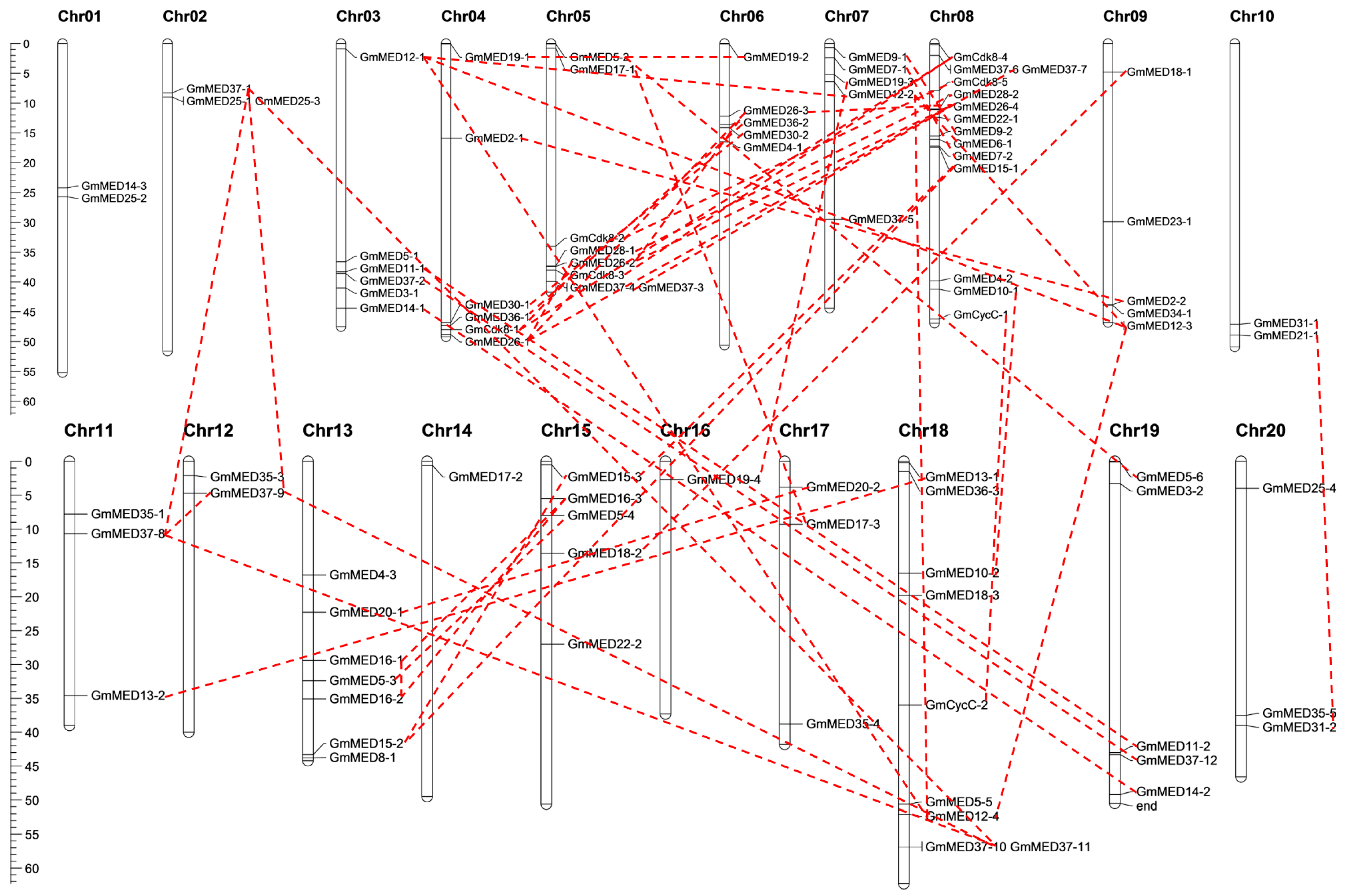
2.2. Chromosomal Locations and Duplication Patterns of Soybean Mediator Complex Subunits

On the basis of their physical positions, the 95 subunits of the mediator complex were unevenly distributed on the 20 chromosomes of soybean (Figure 1, Table S1). The number of subunits located on the different chromosomes ranged from 1 to 14. Chromosome 8 carried the highest number of subunits, whereas chromosomes 14 and 18 each carried only one of the 95 subunits. Eleven of the 20 chromosomes carried at least five subunits and accounted for more than three-quarters of the total number of subunits. The remaining nine chromosomes carried no more than three subunits on each chromosome.
We investigated the existence of duplications within the soybean mediator complex. Both tandem duplications and segmental duplications were analyzed and only segmental duplications were detected for 65 of the 95 subunits (Table S3). Segmental duplications were detected on all of the soybean chromosomes except chromosomes 1 and 14 (Figure 1). No tandem duplication was identified among the mediator complex subunits.

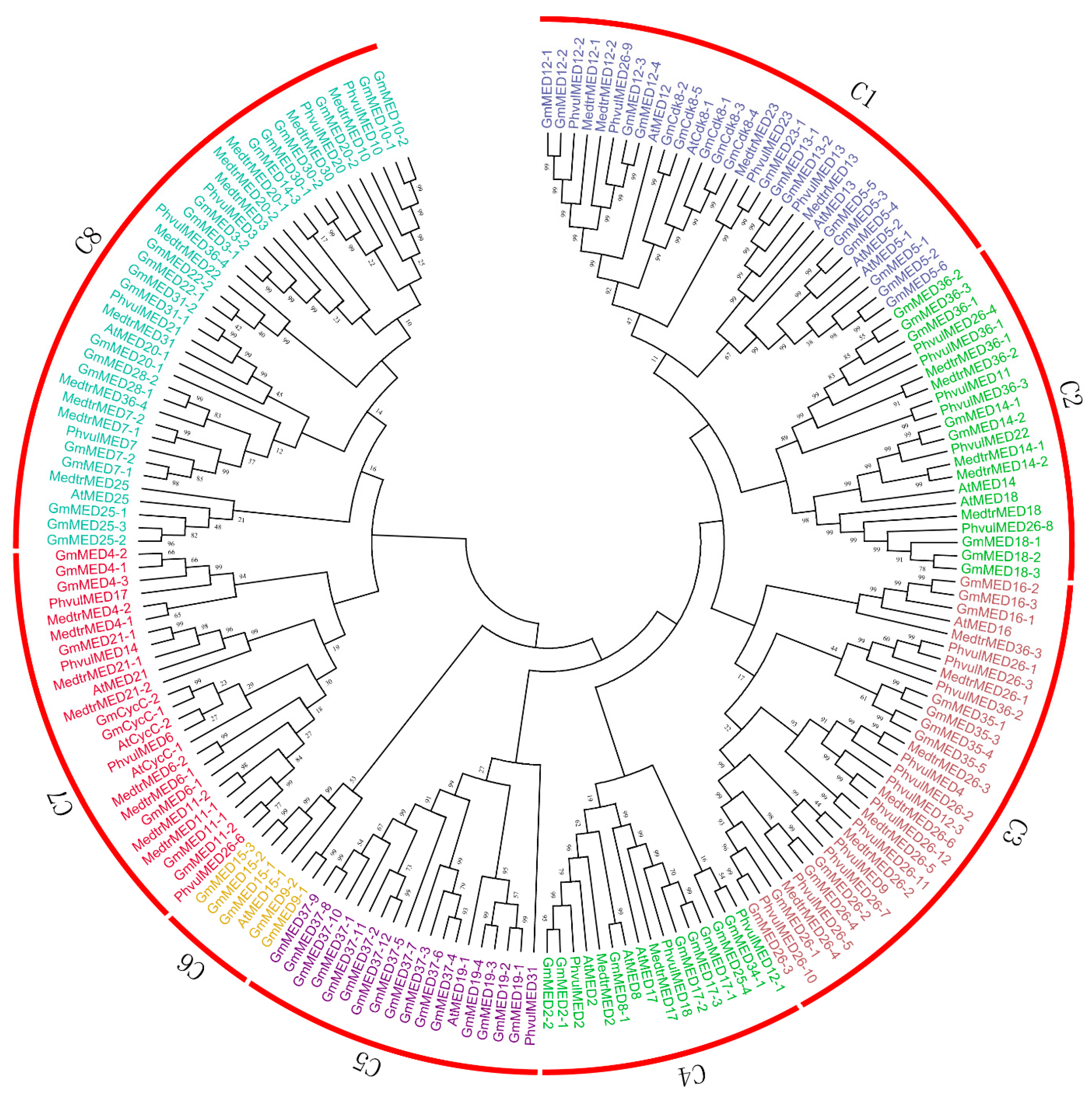
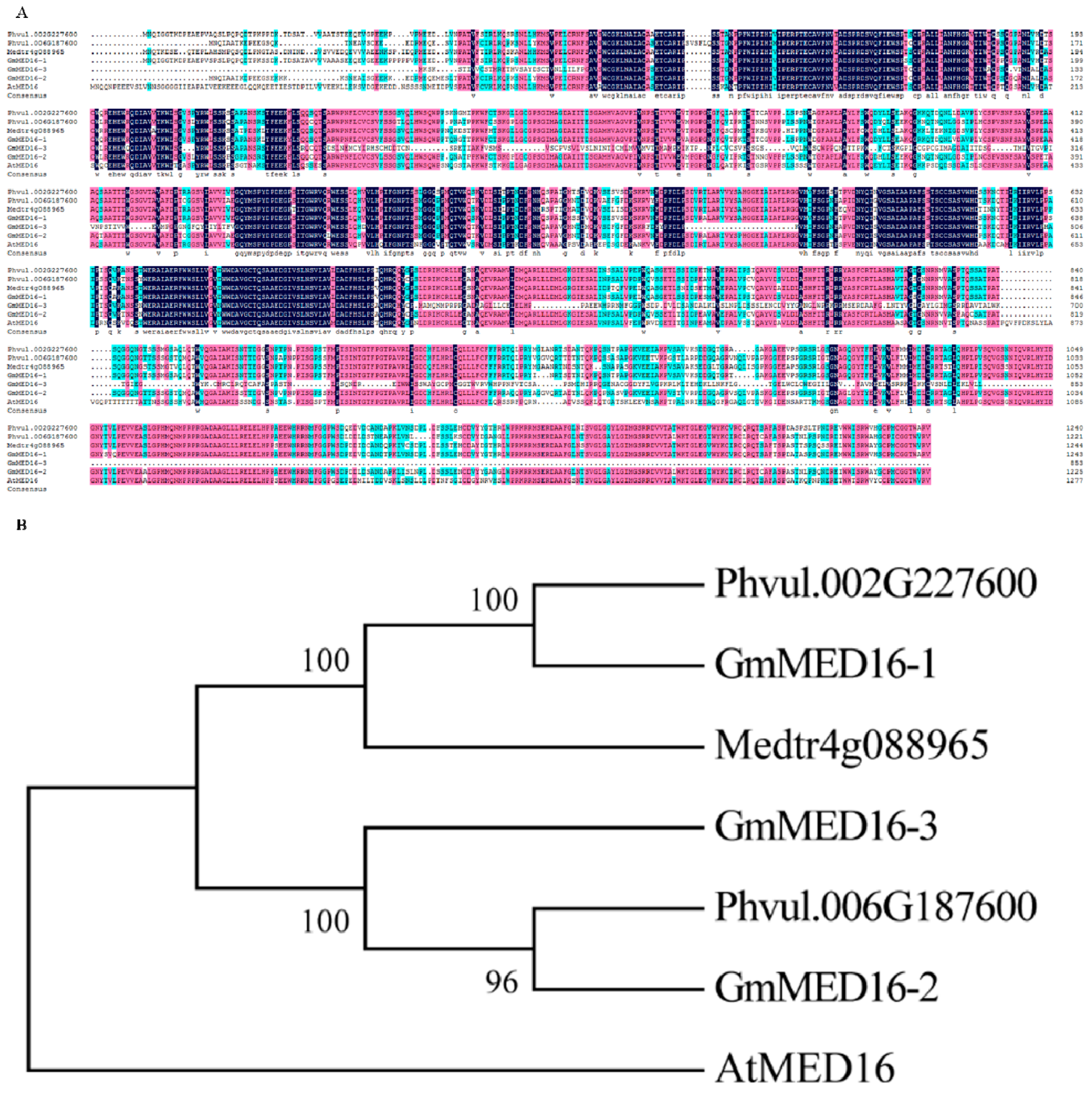
2.3. Phylogenetic Analysis of Soybean Mediator Complex Subunits

On the basis of conserved mediator domains reported previously [5], 36 conserved subunits were detected in the genome of Phaseolus vulgaris and 37 were identified in the Medicago truncatula genome. Among the mediator subunits identified in Arabidopsis, 18 are reported to participate in transcription regulation. Together with the 95 subunits from soybean, in total 186 mediator complex subunits from the four species were used to reconstruct a phylogenetic tree (Figure 2, Data sheets S1 and S2).
The mediator complex subunits from the former two plant species were used to analyze sequence conservation in the Leguminosae family, and the subunits selected from Arabidopsis were used to predict the putative function of the soybean mediator subunits. The mediator complex subunits were classified into eight groups, which were designated subfamily C1 to C8 and differed in the number of subunits per group (Figure 2). Almost all subfamilies contained subunits from P. vulgaris and M. truncatula as well as Arabidopsis, which thus indicated a high degree of conservation in the evolutionary relationship among leguminous plants, and permitted prediction of the putative function of the mediator complex subunits in soybean with greater confidence.

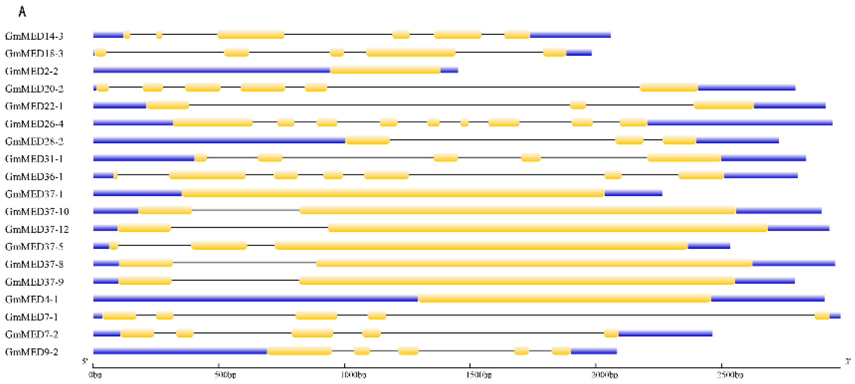
2.4. Gene Structure of Soybean Mediator Complex Subunits

For an improved understanding of the characteristics of the soybean mediator complex subunits, the gene structure was analyzed by comparing the full-length genomic sequence with the corresponding coding sequence (Data sheet S1). Exons, introns, and upstream/downstream regions of the 95 mediator subunits were predicted using the GSDS 2.0 database [38]. The number of exons and introns per subunit ranged from 1 to 22, therefore the subunits were grouped by the length of the genomic sequence and the gene structure was analyzed (Figure 3). The gene structure of the soybean mediator subunits, including the length and the numbers of exons and introns, showed highest similarity within the same subunits with the difference in the suffix of -1, -2, -3 et al., and within a group the subunits differed in the length of the introns and the upstream/downstream regions.

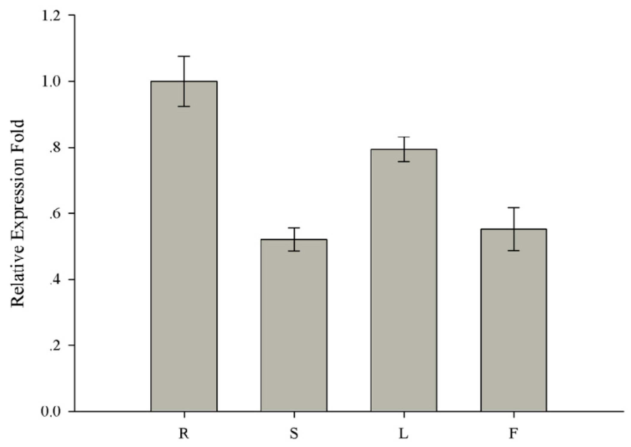
2.5. Sequence Analysis and Expression Pattern of GmMED16-1 in Soybean

It was previously reported that MED16 plays a crucial role in the defense response in Arabidopsis [35,36]. Three MED16 homologs were detected in soybean, namely GmMED16-1, GmMED16-2, and GmMED16-3. On the basis of the sequence similarity of the MED16 protein, two MED16 proteins were identified in P. vulgaris and one in M. truncatula. Together with AtMED16, in total seven MED16 proteins were used for sequence alignment and phylogenetic analysis (Figure 4). Sequence alignment revealed that, with the exception of GmMED16-3, the remaining six proteins showed high sequence similarity (Figure 4A). Phylogenetic analysis revealed that the sequence of MED16 proteins was highly conserved among legume species (Figure 4B).
To investigate the physiological role of soybean MED16 genes, the expression pattern of GmMED16-1 gene in the roots, stems, leaves, and flowers of soybean were analyzed by quantitative real-time PCR (qRT-PCR). The relative expression level of GmMED16-1 was highest in the root (Figure 5). Given that Phytophthora root rot of soybean is a destructive soil-borne disease, GmMED16-1 (Glyma.13G181200) was chosen for further expression analysis in response to P. sojae infection.

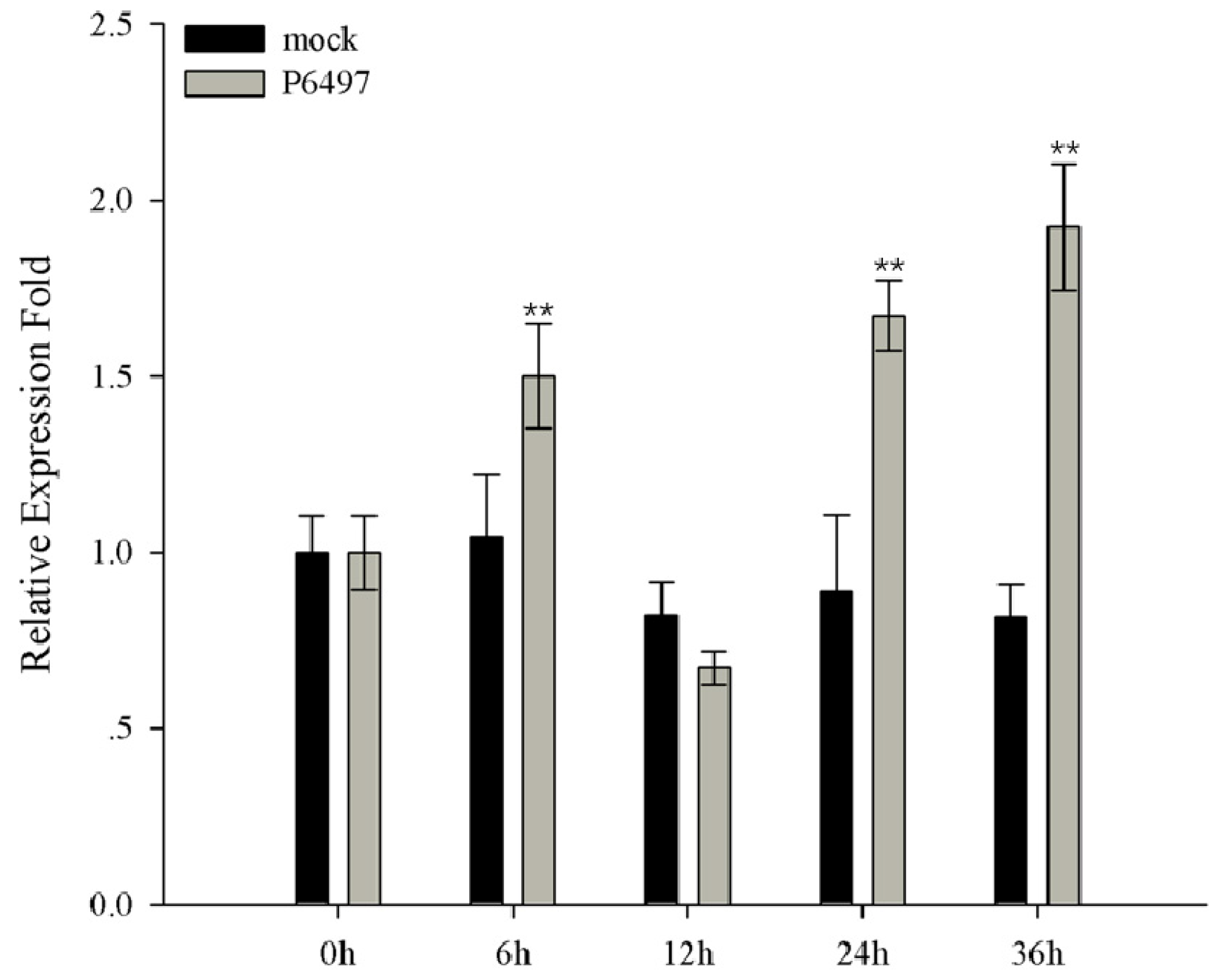
2.6. Expression Profile of GmMED16-1 under Phytophthora sojae Infection

To determine whether GmMED16-1 is induced in response to Phytophthora sojae infection in soybean, the cotyledon of the seven-day-old seedlings was inoculated with P. sojae and the transcript level of GmMED16-1 was analyzed by qRT-PCR. After inoculation, the transcript level of GmMED16-1 initially increased at 6 h post-inoculation (hpi), then decreased at 12 hpi, and subsequently rebound at 24 hpi. The highest transcript level was recorded at 36 hpi (Figure 6).

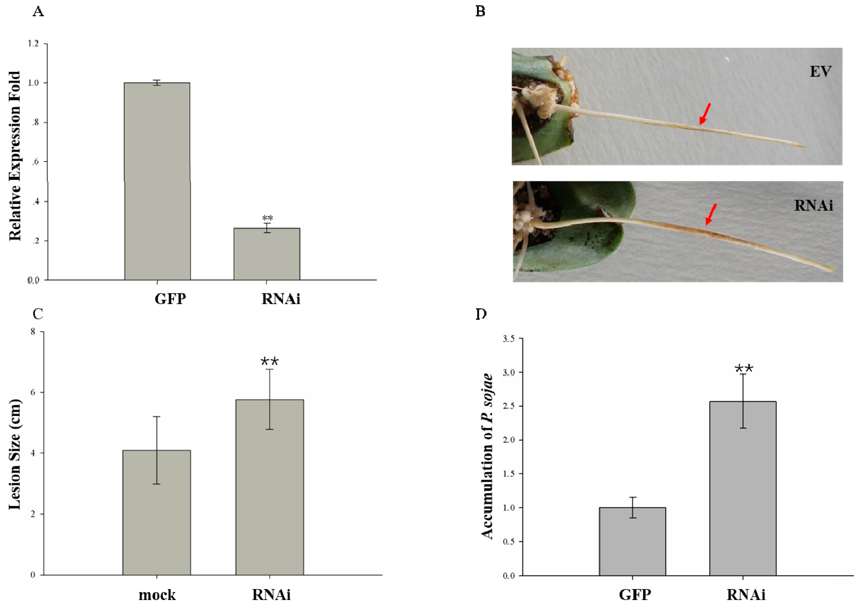
2.7. GmMED16-1 Silencing Reduced Resistance to Phytophthora sojae in Hairy Roots of Transformants

Given that GmMED16-1 expression was highest in the root (Figure 5), we investigated whether GmMED16-1 was involved in soybean–P. sojae interaction using Agrobacterium rhizogenes mediated hairy root transformation using RNA interference (RNAi) in soybean cotyledons. A 365 bp conserved DNA fragment was selected to specifically silence GmMED16-1. The pHellsGate12:GFP:GmMED16-1 RNAi vector was constructed using the Gateway technology and the plasmid was transformed into Agrobacterium rhizogenes strain K599 by electroporation. Hairy roots that exhibited GFP fluorescence were first used to detect the RNAi efficiency. The results of qRT-PCR analysis indicated that the RNAi efficiency attained 70%, thus the expression level of the transgene in the RNAi hairy root was only 30% that of the mock roots (Figure 7A).
The RNAi-positive hairy roots were used for a P. sojae infection assay. The GmMED16-1 RNAi roots resulted in long, water-soaked lesions following inoculation with P. sojae, whereas hairy roots transformed with the control vector showed dark brown spots at the infection site (Figure 7B). Furthermore, the lesion size on the RNAi hairy roots was significant longer compared with that of hairy roots transformed with the control vector (Figure 7C). We also detected the accumulated biomass of P. sojae in the RNAi hairy roots and the hairy roots expressing the control vector. At 48 hpi, the accumulated biomass of P. sojae was significantly higher in RNAi hairy roots compared with the hairy roots expressing the control vector (Figure 7D). These results suggested that GmMED16-1 plays a positive role in soybean–P. sojae interaction.

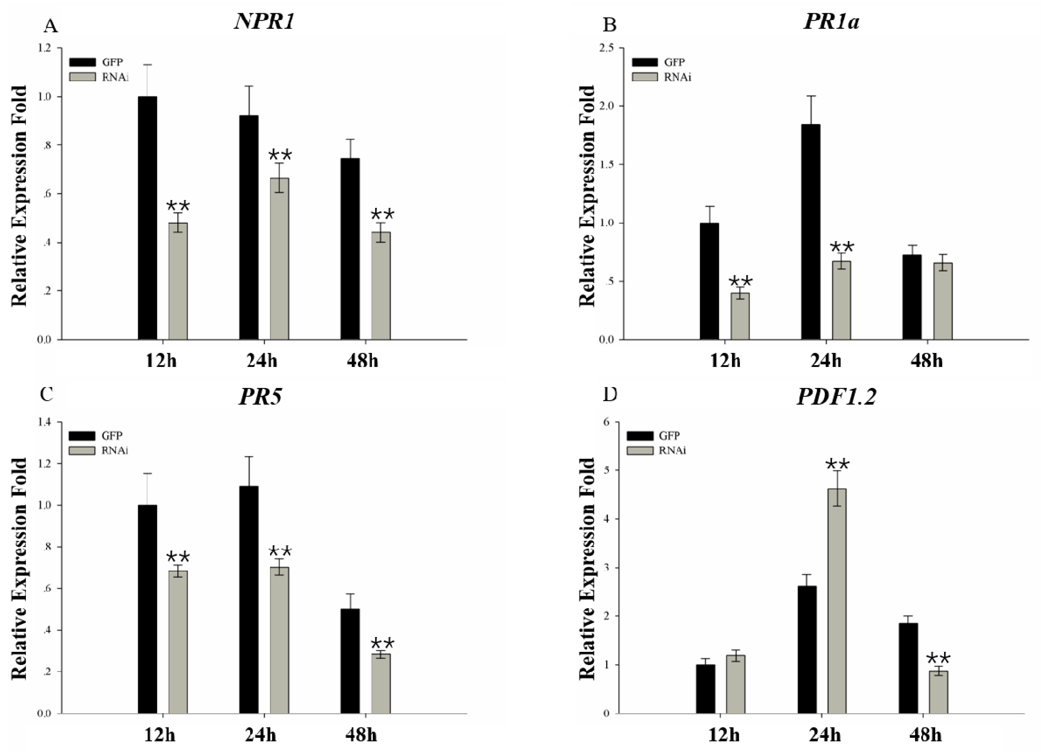
2.8. Expression Analysis of Plant Defense-related Genes in Response to Phytophthora sojae Inoculation

Plant hormones have been reported to play vital roles in the plant defense system. To further examine the function of GmMED16-1 in soybean in response to Phytophthora sojae inoculation, expression level of genes involved in the SA and JA signaling pathways was analyzed. Expression of genes involved in the SA signaling pathway (NPR1, PR1a, and PR5) showed a lower level in the RNAi hairy roots than the mock (Figure 8A–C). Expression of PDF1.2, which participates in the JA signaling pathway, was higher in the RNAi hairy roots than the mock at 24 hpi, whereas at 48 hpi the expression level had decreased (Figure 8D). These results suggested that GmMED16-1 may participate in the soybean–P. sojae interaction by means of the SA signaling pathway.

3. Discussion

In the present research, 95 mediator complex subunits were identified in soybean. The mediator complex subunits of soybean, together with those of Arabidopsis, P. vulgaris, and M. truncatula, were classified into eight subfamilies, which displayed substantial differences in gene structure. Among the soybean subunits, GmMED16-1 was chosen for further functional analysis. GmMED16-1 was induced in response to P. sojae infection (Figure 6). We also confirmed that silencing of GmMED16-1 using RNAi technology led to enhanced susceptibility of the transgenic hairy root to P. sojae infection. Expression analysis of genes involved in the SA and JA signaling pathways suggested that GmMED16-1 may participate in the soybean—P. sojae interaction via a SA-dependent process.
Mathur et al. identified mediator complex genes in 16 plant species and analyzed the structure, phylogenetic relationships, and expression profiles of representative genes in the dicotyledon model plant Arabidopsis and the monocotyledon model plant rice during reproduction and under abiotic stress [5]. In the present study, we identified 95 mediator complex subunits based on the current assembly of the soybean genome. Some in silico parameter, like peptide length, molecular weight, numbers of transmembrane helices, isoelectric point and the subcellular localization were predicted using the online database. These results will provide a foundation for the further functional analysis of the soybean mediator subunits. Besides, chromosomal location as well as the duplication patterns were analysis and results showed that the 95 subunits unevenly located on the 20 chromosomes of soybean and only segmental duplications were found. Gene duplications are considered to be one of the primary driving forces in the evolution of genomes and genetic systems [39]. Segmental duplications, one of the main causes of gene family expansion in plant, multiple genes through polyploidy followed by chromosome rearrangements [40]. It occurs most frequently in plants because most plants are diploidized polyploids and retain numerous duplicated chromosomal blocks within their genomes [41]. Researches showed that the soybean expansin gene superfamily has expanded through the duplication events [42].
We analyzed the chromosomal location, duplication, phylogenetic relationships, and gene structure of the mediator complex subunits in soybean in comparison with subunits from P. vulgaris, M. truncatula, and Arabidopsis. The 186 mediator complex subunits analyzed were classified into eight subfamilies. AtMED12 and AtCDK8, which were classified in subfamily C1, are CDK module subunits involved in plant defense pathways and phosphorylation of the CDK module, which is important in defense gene regulation [11]. MED14 belonged to subfamily C2 and has been reported to participate in plant defense via the SA-induced pathogen defense response and SAR signaling pathway [13]. MED18, which was also classified in subfamily C2, is involved in plant sensitivity to necrotrophic fungal pathogens [14]. AtMED16 was classified in subfamily C3 and affects the expression level of defense-related genes in the SA and JA pathways as well as SA-mediated SAR [35]. Recently, it was observed that MED16 regulates resistance to S. sclerotiorum by governing both JA/ethylene-mediated and WRKY33-activated defense signaling [36]. In addition, MED16 is involved in defense signaling crosstalk together with MED14 and MED15 in Arabidopsis [43]. MED19a, a member of subfamily C4, is a positive regulator of plant defense against the oomycete downy mildew pathogen Hyaloperonospora arabidopsidis (Hpa) and participates in the JA/ethylene signaling pathway as well as SA-triggered immunity in Arabidopsis [15]. MED15, which is a critical regulator of SA response in plant defense, also influences the flowering-time phenotype in Arabidopsis [12]. The AtCycC-1 and AtCycC-2 genes classified in subfamily C7 are involved in plant defense pathways and phosphorylation of the CYC module, which is important in defense gene regulation [11].
Phytophthora sojae is a soil-borne oomycete pathogen and it mainly causes Phytophthora root and stem rot under field conditions. As an essential component of the transcriptional mechanism, the mediator complex plays an important role in plant–pathogen interaction in Arabidopsis. Five individual subunits of the mediator complex have been functionally characterized to participate in plant pathogen resistance, namely MED25/PFT1, MED21, MED15/NRB4, MED16/SFR6, and MED8 [32]. Among these genes, MED16 is a key component of basal resistance against the necrotrophic fungal pathogen S. sclerotiorum [36]. In addition, MED19 is a positive regulator of plant resistance against the oomycete pathogen Hpa, leading to proteasome-dependent degradation of MED19a [15]. In the present research, GmMED16-1 expression was induced in response to P. sojae infection. In addition, the lesion size and biomass accumulation of P. sojae in RNAi hairy roots showed a significant difference compared with hairy roots transformed with the control vector. These results indicate that GmMED16-1 is a positive regulator of the soybean—P. sojae interaction.
Plant pathogens are often categorized as biotrophs and necrotrophs on the basis of their lifestyles. Biotrophs are pathogens that derive nutrients from living host tissues, whereas necrotrophs are pathogens that derive nutrients from dead or dying cells [44]. Phytohormones, including SA, ethylene, JA, abscisic acid, auxin, brassinosteroids, gibberellic acid, cytokinin, and the recently identified strigolactones, orchestrate effective defense responses by activation of defense gene expression [45]. Among these phytohormones, SA and JA are crucial in the regulation of signaling networks for basal resistance against multiple pathogens [46,47]. The SA signaling pathway positively induces plant defense against biotrophic pathogens that feed and reproduce on living host cells, whereas JA signaling pathways are required for resistance predominantly against necrotrophic pathogens and herbivorous insects, which derive nutrients from living host cells, commonly through specialized feeding structures [44,48,49]. Phytophthora sojae is described as a hemibiotrophic pathogen and secretes effector proteins (coded by Avr genes) to manipulate and invade living host cells during the initial biotrophic stage of infection [50]. In Arabidopsis, SFR6/MED16 controls SA- and JA-mediated defense gene expression and is essential for resistance to the hemibiotrophic pathogen Pseudomonas syringae [34]. The present results showed that the expression level of three genes involved in the SA signaling pathway (NPR1, PR1a, and PR5) was lower in RNAi hairy roots than the mock roots (Figure 8). The expression pattern of plant defense-related genes in the RNAi roots was opposite to that observed in the mock roots in response to P. sojae infection. These results suggested that GmMED16-1 may participate in the soybean–P. sojae interaction via the SA signaling pathway. Similar findings have been observed in Arabidopsis. MED19a, a positive regulator of resistance to the oomycete Hpa, interacts with HaRxL44, which alters the balance of defense transcription from SA-responsive defense to JA/ethylene-signalling, and enhances susceptibility to biotrophs by attenuating SA-dependent gene expression [15].

4. Materials and Methods

4.1. Identification of Mediator Subunits in Soybean

Conserved pfam domains have already been predicted [5] and the seed files of these domains were downloaded from the Pfam database (http://pfam.xfam.org/) [51]. They were used to search the soybean genome as well as the P. vulgaris and M. truncatula by the software hummer 3.0. A total of 95 subunits of the soybean mediator were finally identified from the newest soybean genome database. Besides, 37 conserved subunits were found in M. truncatula and 36 P. vulgaris were found. Data files containing the information of the final 95 soybean mediator subunits (including their corresponding physical locations on soybean chromosomes, genomic sequences, coding sequences, and protein sequences) were downloaded from Phytozome v11.0 (http://phytozome.jgi.doe.gov/pz/portal.html) [52] (Table S1, Data sheet S1). Theoretical isoelectric point (pI) and molecular weight (MW) of soybean mediator subunits were computed by ExPASy “Compute pI/Mw” tool (http://www.expasy.ch/tools/pi_tool.html) [53,54,55]. The subcellular localizations of the mediator subunits were predicted using WoLF PSORT (http://www.genscript.com/wolf-psort.html) [56]. The numbers of transmembrane helices in these proteins were predicted by TMHMM Server v.2.0 (http://www.cbs.dtu.dk/services/TMHMM/) [57].

4.2. Phylogenetic and Structural Analysis of Mediator Subunits in Soybean

The protein sequence of 95 soybean mediator subunits (Data sheet S1), 37 conserved subunits in M. truncatula and 36 conserved P. vulgaris subunits as well as 18 subunits from Arabidopsis (Data sheet S2) together were used for multiple sequence alignments by Clustal W. Then the evolutionary analyses were conducted with MEGA 6.0 [58]. The unrooted phylogenetic tree was then constructed using the neighbor-joining method [59]. Phylogeny test using the bootstrap method and the number of bootstrap replications was 1000. The evolutionary distances were computed using the p-distance method [60]. Gene structure analysis was performed in the Gene Structure Display Server (GSDS) database 2.0 (http://gsds.cbi.pku.edu.cn/) [38] with genomic sequences and coding sequences (Data sheet S1) with default settings.

4.3. Chromosomal Locations and Gene Duplication Analysis

The chromosomal locations of soybean mediator subunits were illustrated by MapChart [61] based on the physical locations downloaded from Phytozome v11.0 [52] (Table S1). Segmental and tandem duplication events of these soybean mediator subunits were identified using the Multiple Collinearity Scan toolkit (MCScan) [62] from the Plant Genome Duplication Database (http://chibba.agtec.uga.edu/duplication/) [63] with relevant parameters: BLASTP was used to search for potential anchors (E <1 × 10⁻⁵, top five matches) between every possible homologous pair, and these pairs were used as the input for MCscan. Syntonic blocks were identified using the E-value ≤ 1 × 10⁻¹⁰ as a significance cutoff. Tandem duplication was defined as homologous genes with less than ten gene loci in between and > 50% similarity at protein level on a single chromosome [64].

4.4. Plant Materials and Phytophthora sojae Inoculation

Soybean (Glycine max (L.) Merr.) cultivar Williams 82 was used for the experiment. Soybean seedings were grown in vermiculite substrate in the greenhouse at 25 °C under 16-h-light/8-h-dark cycle for seven days. Then, the leaves were used for P. sojae (P6497) infection. The inoculated leaves were placed in the petri dishes, on the surface of the wet filter paper to retain moisture. The petri dishes were cultured in the incubator during the infection of P6497. The mock control was also cultured with the V8 medium without P. sojae and samples were collected from the inoculated area as well as the mock control at 0, 6, 12, 24, and 36 h post-infection (hpi). All samples collected above were rapidly frozen in liquid nitrogen and then stored at −70 °C for RNA extraction.

4.5. RNA Isolation and Quantitative Real-time PCR

Total RNA of the samples was extracted using the RNAsimple Total RNA Kit (Tiangen, Beijing, China), gDNA elimination and reverse transcription of the first strand cDNA were performed with the PrimeScriptTM RT reagent Kit with gDNA Eraser (TaKaRa, Dalian, China). Quantitative real-time PCR was carried out with the LightCycler® 480 II real-time PCR system (Roche, Basel, Switzerland) using the SYBR® Premix Ex TaqTM II (TaKaRa) according to the manufacturers instructions. The relative expression levels of target genes were calculated using the 2–ΔΔCT method [65]. The GmCons4 (GenBank: BU578186.1) gene was selected as the reference for the qRT-PCR. All the nucleic acid sequences of the primers are listed in Table S4.

4.6. Construction of pHellsGate12: GFP: GmMED16-1 RNAi Vector

A 365bp conserved fragment of the GmMED16-1 gene was first cloned into the Gateway entry vector pDONR221, then constructed into pHellsGate12: GFP through an LR recombination reaction between the entry clone and the destination vector (Invitrogen, Shanghai, China). The vector pHellsGate12: GFP which was modified from pHellsGate12 were used for RNAi construction [23,66]. The constructed pHellsGate12: GFP: GmMED16-1 was validated by PCR as well as sequencing.

4.7. Agrobacterium Culture, Soybean Hairy Root Transformation

The plasmid vectors pHellsGate12: GFP: GmMED16-1 and the empty vector were transformed into A. rhizogenes K599 strain by electroporation. The positive clones were selected on the LB medium (with 50 μg/mL kanamycin and 50 μg/mL streptomycin) after 48 h at 28 °C and the individual clones were validated by colony PCR. The single positive colony was shaken in 4 mL at 28 °C, and the bacteria cultures were centrifuged at 4000 rpm for 5 min and resuspended with 10 mM MgCl2. The final OD600 of bacteria was adjusted to approximately 0.5 to make it suitable for the hair root transformation on the soybean cotyledon.
The soybean hairy root transformation was performed as the described procedures with some modification [23,67]. Soybean cotyledons were removed from the 5-days-old seedlings gently and a circular cut (approximately 0.4 cm in diameter) was made near the petiole end. The cut cotyledons were then place in the Petri dishes containing 0.6% agar medium immediately. Approximately 20 μL of the A. rhizogenes suspension were pipetted into the cut surface. These cotyledons were placed in incubator at 25 °C in a dark condition. Seven days later, the callus was emerged at the cut site in the cptyledon. After 3 weeks, transformed hairy roots were seen along a callus ridge on the inoculated cotyledons, and the positive transformed hairy roots were then used for P. sojae inoculation and the expression pattern of some genes involved plant hormone signaling pathways.

4.8. Assay of P. sojae Infection of the Soybean Hairy Roots

The positive roots were verified by GFP fluorescence and qRT-PCR. Then the positive ones with similar length were for the inoculation with zoospore suspension (about 104 zoospores/mL) of P. sojae P6497. Lesion length was measured by venire caliper. The biomass of P. sojae was determined by q-PCR using primers from P. sojae and soybean. Student’s t test was performed to determine the significance of differences between control and the RNAi roots. For gene involved plant hormone signaling pathways in the transgenic hair roots, the infected roots were mixed and collected at 12, 24 and 48 hpi.

Supplementary Materials

The following are available online at https://www.mdpi.com/1422-0067/20/18/4570/s1.

Author Contributions

D.X., N.G. J.-M.Z. and H.X. conceived and designed the experiment; D.X. and D.-L.J. analyzed the soybean genome data and performed the bioinformatics analysis; D.X., N.G., X.-L.Z. and Y.-P.B. performed P. sojae inoculation, RNA isolation and qRT-PCR; D.X., N.G., and H.-T.W. performed the RNAi experiments. D.X. drafted the manuscript; N.G., X.-T.W., J.-M.Z., R.-Z.G. and H.X. revised the manuscript. All authors read and approved the final manuscript.

Funding

This work was supported by the Fundamental Research Funds for the Central University (Y0201700146), Genetically Modified Organisms Breeding Major Projects (2016ZX08004002), the Modern Agroindustry Technology Research System of China (CARS-04-PS10), the Program for Changjiang Scholars and Innovative Research Team in University (PCSIRT_17R55) and the Jiangsu Collaborative Innovation Center for Modern Crop Production.

Acknowledgments

The authors would like to thank Daolong Dou at the College of Plant Production, Nanjing Agricultural University for providing the vector for RNAi used in the experiment and Robert McKenzie, from Liwen Bianji, Edanz Group China (www.liwenbianji.cn/ac), for editing the language of this manuscript.

Conflicts of Interest

The authors declare that they have no competing interests.

References

  1. Gill, G.; Ptashne, M. Negative effect of the transcriptional activator GAL4. Nature 1988, 334, 721–724. [Google Scholar] [CrossRef] [PubMed]
  2. Gu, W.; Malik, S.; Ito, M.; Yuan, C.X.; Fondell, J.D.; Zhang, X.L.; Martinez, E.; Qin, J.; Roeder, R.G. A novel human SRB/MED-containing cofactor complex, SMCC, involved in transcription regulation. Mol. Cell 1999, 3, 97–108. [Google Scholar] [CrossRef]
  3. Sato, S.; Tomomori-Sato, C.; Parmely, T.J.; Florens, L.; Zybailov, B.; Swanson, S.K.; Banks, C.A.S.; Jin, J.J.; Cai, Y.; Washburn, M.P.; et al. A set of consensus mammalian Mediator subunits identified by multidimensional protein identification technology. Mol. Cell 2004, 14, 685–691. [Google Scholar] [CrossRef] [PubMed]
  4. Bourbon, H.-M. Comparative genomics supports a deep evolutionary origin for the large, four-module transcriptional mediator complex. Nucleic Acids Res. 2008, 36, 3993–4008. [Google Scholar] [CrossRef] [PubMed] [Green Version]
  5. Mathur, S.; Vyas, S.; Kapoor, S.; Tyagi, A.K. The Mediator Complex in Plants: Structure, Phylogeny, and Expression Profiling of Representative Genes in a Dicot (Arabidopsis) and a Monocot (Rice) during Reproduction and Abiotic Stress. Plant Physiol. 2011, 157, 1609–1627. [Google Scholar] [CrossRef] [PubMed]
  6. Asturias, F.J.; Jiang, Y.W.; Myers, L.C.; Gustafsson, C.M.; Kornberg, R.D. Conserved structures of mediator and RNA polymerase II holoenzyme. Science 1999, 283, 985–987. [Google Scholar] [CrossRef]
  7. Borggrefe, T.; Davis, R.; Erdjument-Bromage, H.; Tempst, P.; Kornberg, R.D. A complex of the Srb8, -9, -10, and -11 transcriptional regulatory proteins from yeast. J. Biol. Chem. 2002, 277, 44202–44207. [Google Scholar] [CrossRef]
  8. Cevher, M.A.; Shi, Y.; Li, D.; Chait, B.T.; Malik, S.; Roeder, R.G. Reconstitution of active human core Mediator complex reveals a critical role of the MED14 subunit. Nat. Struct. Mol. Biol. 2014, 21, 1028–1034. [Google Scholar] [CrossRef]
  9. Tsai, K.L.; Tomomori-Sato, C.; Sato, S.; Conaway, R.C.; Conaway, J.W.; Asturias, F.J. Subunit Architecture and Functional Modular Rearrangements of the Transcriptional Mediator Complex. Cell 2014, 157, 1430–1444. [Google Scholar] [CrossRef] [Green Version]
  10. Plaschka, C.; Lariviere, L.; Wenzeck, L.; Seizl, M.; Hemann, M.; Tegunov, D.; Petrotchenko, E.V.; Borchers, C.H.; Baumeister, W.; Herzog, F.; et al. Architecture of the RNA polymerase II-Mediator core initiation complex. Nature 2015, 518, 376–380. [Google Scholar] [CrossRef]
  11. Zhu, Y.; Schluttenhoffer, C.M.; Wang, P.; Fu, F.; Thimmapuram, J.; Zhu, J.-K.; Lee, S.Y.; Yun, D.-J.; Mengiste, T. CYCLIN-DEPENDENT KINASE8 Differentially Regulates Plant Immunity to Fungal Pathogens through Kinase-Dependent and -Independent Functions in Arabidopsis. Plant Cell 2014, 26, 4149–4170. [Google Scholar] [CrossRef] [PubMed]
  12. Canet, J.V.; Dobon, A.; Tornero, P. Non-recognition-of-BTH4, an Arabidopsis mediator subunit homolog, is necessary for development and response to salicylic acid. Plant Cell 2012, 24, 4220–4235. [Google Scholar] [CrossRef] [PubMed]
  13. Zhang, X.; Yao, J.; Zhang, Y.; Sun, Y.; Mou, Z. The Arabidopsis Mediator complex subunits MED14/SWP and MED16/SFR6/IEN1 differentially regulate defense gene expression in plant immune responses. Plant J. 2013, 75, 484–497. [Google Scholar] [CrossRef] [PubMed]
  14. Lai, Z.; Schluttenhofer, C.M.; Bhide, K.; Shreve, J.; Thimmapuram, J.; Lee, S.Y.; Yun, D.J.; Mengiste, T. MED18 interaction with distinct transcription factors regulates multiple plant functions. Nat Commun 2014, 5, 3064. [Google Scholar] [CrossRef] [PubMed] [Green Version]
  15. Caillaud, M.C.; Asai, S.; Rallapalli, G.; Piquerez, S.; Fabro, G.; Jones, J.D. A downy mildew effector attenuates salicylic acid-triggered immunity in Arabidopsis by interacting with the host mediator complex. PLoS Biol. 2013, 11, e1001732. [Google Scholar] [CrossRef] [PubMed]
  16. Dhawan, R.; Luo, H.; Foerster, A.M.; Abuqamar, S.; Du, H.N.; Briggs, S.D.; Mittelsten Scheid, O.; Mengiste, T. HISTONE MONOUBIQUITINATION1 interacts with a subunit of the mediator complex and regulates defense against necrotrophic fungal pathogens in Arabidopsis. Plant Cell 2009, 21, 1000–1019. [Google Scholar] [CrossRef] [PubMed]
  17. Ou, B.; Yin, K.Q.; Liu, S.N.; Yang, Y.; Gu, T.; Wing Hui, J.M.; Zhang, L.; Miao, J.; Kondou, Y.; Matsui, M.; et al. A high-throughput screening system for Arabidopsis transcription factors and its application to Med25-dependent transcriptional regulation. Mol. Plant 2011, 4, 546–555. [Google Scholar] [CrossRef]
  18. Cevik, V.; Kidd, B.N.; Zhang, P.; Hill, C.; Kiddle, S.; Denby, K.J.; Holub, E.B.; Cahill, D.M.; Manners, J.M.; Schenk, P.M.; et al. MEDIATOR25 Acts as an Integrative Hub for the Regulation of Jasmonate-Responsive Gene Expression in Arabidopsis. Plant Physiol. 2012, 160, 541–555. [Google Scholar] [CrossRef]
  19. Kidd, B.N.; Edgar, C.I.; Kumar, K.K.; Aitken, E.A.; Schenk, P.M.; Manners, J.M.; Kazan, K. The Mediator Complex Subunit PFT1 Is a Key Regulator of Jasmonate-Dependent Defense in Arabidopsis. Plant Cell 2009, 21, 2237–2252. [Google Scholar] [CrossRef]
  20. Yao, T.; Park, B.S.; Mao, H.Z.; Seo, J.S.; Ohama, N.; Li, Y.; Yu, N.; Mustafa, N.F.B.; Huang, C.H.; Chua, N.H. Regulation of flowering time by SPL10/MED25 module in Arabidopsis. New Phytol. 2019, 224, 493–504. [Google Scholar] [CrossRef]
  21. Jaskolowski, A.; Inigo, S.; Arellano, S.M.; Arias, L.A.; Fiol, D.F.; Sede, A.R.; Oldra, M.B.; Lorenzi, H.; Muschietti, J.P.; Pagnussat, G.C.; et al. The MED30 subunit of mediator complex is essential for early plant development and promotes flowering in Arabidopsis thaliana. Development 2019, 146, dev175224. [Google Scholar] [CrossRef] [PubMed]
  22. Tyler, B.M. Phytophthora sojae: Root rot pathogen of soybean and model oomycete. Mol. Plant Pathol. 2007, 8, 1–8. [Google Scholar] [CrossRef] [PubMed]
  23. Yan, Q.; Cui, X.; Su, L.; Xu, N.; Guo, N.; Xing, H.; Dou, D. GmSGT1 is differently required for soybean Rps genes-mediated and basal resistance to Phytophthora sojae. Plant Cell Rep. 2014, 33, 1275–1288. [Google Scholar] [CrossRef] [PubMed]
  24. Cheng, Q.; Li, N.; Dong, L.; Zhang, D.; Fan, S.; Jiang, L.; Wang, X.; Xu, P.; Zhang, S. Overexpression of Soybean Isoflavone Reductase (GmIFR) Enhances Resistance to Phytophthora sojae in Soybean. Front. Plant Sci. 2015, 6, 1024. [Google Scholar] [CrossRef] [PubMed]
  25. Dong, L.; Cheng, Y.; Wu, J.; Cheng, Q.; Li, W.; Fan, S.; Jiang, L.; Xu, Z.; Kong, F.; Zhang, D.; et al. Overexpression of GmERF5, a new member of the soybean EAR motif-containing ERF transcription factor, enhances resistance to Phytophthora sojae in soybean. J. Exp. Bot. 2015, 66, 2635–2647. [Google Scholar] [CrossRef] [PubMed]
  26. Zhao, Y.; Chang, X.; Qi, D.; Dong, L.; Wang, G.; Fan, S.; Jiang, L.; Cheng, Q.; Chen, X.; Han, D.; et al. A Novel Soybean ERF Transcription Factor, GmERF113, Increases Resistance to Phytophthora sojae Infection in Soybean. Front. Plant Sci. 2017, 8, 299. [Google Scholar] [CrossRef] [PubMed]
  27. Li, N.; Zhao, M.; Liu, T.; Dong, L.; Cheng, Q.; Wu, J.; Wang, L.; Chen, X.; Zhang, C.; Lu, W.; et al. A Novel Soybean Dirigent Gene GmDIR22 Contributes to Promotion of Lignan Biosynthesis and Enhances Resistance to Phytophthora sojae. Front. Plant Sci. 2017, 8, 1185. [Google Scholar] [CrossRef] [PubMed]
  28. Cheng, Q.; Dong, L.; Gao, T.; Liu, T.; Li, N.; Wang, L.; Chang, X.; Wu, J.; Xu, P.; Zhang, S. The bHLH transcription factor GmPIB1 facilitates resistance to Phytophthora sojae in Glycine max. J. Exp. Bot. 2018, 69, 2527–2541. [Google Scholar] [CrossRef]
  29. Sukumaran, A.; McDowell, T.; Chen, L.; Renaud, J.; Dhaubhadel, S. Isoflavonoid-specific prenyltransferase gene family in soybean: GmPT01, a pterocarpan 2-dimethylallyltransferase involved in glyceollin biosynthesis. Plant J. Cell Mol. Biol. 2018, 96, 966–981. [Google Scholar] [CrossRef]
  30. Yan, Q.; Si, J.; Cui, X.; Peng, H.; Jing, M.; Chen, X.; Xing, H.; Dou, D. GmDAD1, a Conserved Defender Against Cell Death 1 (DAD1) From Soybean, Positively Regulates Plant Resistance Against Phytophthora Pathogens. Front. Plant Sci. 2019, 10, 107. [Google Scholar] [CrossRef]
  31. Zhang, C.; Gao, H.; Li, R.; Han, D.; Wang, L.; Wu, J.; Xu, P.; Zhang, S. GmBTB/POZ, a novel BTB/POZ domain-containing nuclear protein, positively regulates the response of soybean to Phytophthora sojae infection. Mol. Plant Pathol. 2019, 20, 78–91. [Google Scholar] [CrossRef] [PubMed]
  32. An, C.; Mou, Z. The function of the Mediator complex in plant immunity. Plant Signal. Behav. 2013, 8, e23182. [Google Scholar] [CrossRef] [PubMed] [Green Version]
  33. Liu, Z.; Chen, G.; Gao, F.; Xu, R.; Li, N.; Zhang, Y.; Li, Y. Transcriptional Repression of the APC/C Activator Genes CCS52A1/A2 by the Mediator Complex Subunit MED16 Controls Endoreduplication and Cell Growth in Arabidopsis. Plant Cell 2019, 1899–1912. [Google Scholar] [CrossRef] [PubMed]
  34. Wathugala, D.L.; Hemsley, P.A.; Moffat, C.S.; Cremelie, P.; Knight, M.R.; Knight, H. The Mediator subunit SFR6/MED16 controls defence gene expression mediated by salicylic acid and jasmonate responsive pathways. New Phytol. 2012, 195, 217–230. [Google Scholar] [CrossRef] [PubMed]
  35. Zhang, X.; Wang, C.; Zhang, Y.; Sun, Y.; Mou, Z. The Arabidopsis mediator complex subunit16 positively regulates salicylate-mediated systemic acquired resistance and jasmonate/ethylene-induced defense pathways. Plant Cell 2012, 24, 4294–4309. [Google Scholar] [CrossRef]
  36. Wang, C.; Yao, J.; Du, X.; Zhang, Y.; Sun, Y.; Rollins, J.A.; Mou, Z. The Arabidopsis Mediator Complex Subunit16 Is a Key Component of Basal Resistance against the Necrotrophic Fungal Pathogen Sclerotinia sclerotiorum. Plant Physiol. 2015, 169, 856–872. [Google Scholar] [CrossRef]
  37. Dolan, W.L.; Dilkes, B.P.; Stout, J.M.; Bonawitz, N.D.; Chapple, C. Mediator Complex Subunits MED2, MED5, MED16, and MED23 Genetically Interact in the Regulation of Phenylpropanoid Biosynthesis. Plant Cell 2017, 29, 3269–3285. [Google Scholar] [CrossRef]
  38. Hu, B.; Jin, J.; Guo, A.Y.; Zhang, H.; Luo, J.; Gao, G. GSDS 2.0: An upgraded gene feature visualization server. Bioinformatics 2015, 31, 1296–1297. [Google Scholar] [CrossRef]
  39. Moore, R.C.; Purugganan, M.D. The early stages of duplicate gene evolution. Proc Natl Acad Sci USA 2003, 100, 15682–15687. [Google Scholar] [CrossRef] [Green Version]
  40. Yu, J.; Wang, J.; Lin, W.; Li, S.; Li, H.; Zhou, J.; Ni, P.; Dong, W.; Hu, S.; Zeng, C.; et al. The Genomes of Oryza sativa: A history of duplications. PLoS Biol. 2005, 3, e38. [Google Scholar] [CrossRef]
  41. Cannon, S.B.; Mitra, A.; Baumgarten, A.; Young, N.D.; May, G. The roles of segmental and tandem gene duplication in the evolution of large gene families in Arabidopsis thaliana. BMC Plant Biol. 2004, 4, 10. [Google Scholar] [CrossRef] [PubMed]
  42. Zhu, Y.; Wu, N.; Song, W.; Yin, G.; Qin, Y.; Yan, Y.; Hu, Y. Soybean (Glycine max) expansin gene superfamily origins: Segmental and tandem duplication events followed by divergent selection among subfamilies. BMC Plant Biol. 2014, 14, 93. [Google Scholar] [CrossRef]
  43. Wang, C.; Du, X.; Mou, Z. The Mediator Complex Subunits MED14, MED15, and MED16 Are Involved in Defense Signaling Crosstalk in Arabidopsis. Front. Plant Sci. 2016, 7, 1947. [Google Scholar] [CrossRef] [PubMed]
  44. Glazebrook, J. Contrasting mechanisms of defense against biotrophic and necrotrophic pathogens. Annu. Rev. Phytopathol. 2005, 43, 205–227. [Google Scholar] [CrossRef] [PubMed]
  45. Li, N.; Han, X.; Feng, D.; Yuan, D.; Huang, L.J. Signaling Crosstalk between Salicylic Acid and Ethylene/Jasmonate in Plant Defense: Do We Understand What They Are Whispering? Int. J. Mol. Sci. 2019, 20, 671. [Google Scholar] [CrossRef] [PubMed]
  46. Robert-Seilaniantz, A.; Grant, M.; Jones, J.D. Hormone crosstalk in plant disease and defense: More than just jasmonate-salicylate antagonism. Annu. Rev. Phytopathol. 2011, 49, 317–343. [Google Scholar] [CrossRef] [PubMed]
  47. Pieterse, C.M.; Van der Does, D.; Zamioudis, C.; Leon-Reyes, A.; Van Wees, S.C. Hormonal modulation of plant immunity. Annu. Rev. Cell Dev. Biol. 2012, 28, 489–521. [Google Scholar] [CrossRef] [PubMed]
  48. Spoel, S.H.; Johnson, J.S.; Dong, X. Regulation of tradeoffs between plant defenses against pathogens with different lifestyles. Proc Natl Acad Sci USA 2007, 104, 18842–18847. [Google Scholar] [CrossRef] [Green Version]
  49. Bari, R.; Jones, J.D. Role of plant hormones in plant defence responses. Plant Mol. Biol. 2009, 69, 473–488. [Google Scholar] [CrossRef]
  50. Rasoolizadeh, A.; Labbe, C.; Sonah, H.; Deshmukh, R.K.; Belzile, F.; Menzies, J.G.; Belanger, R.R. Silicon protects soybean plants against Phytophthora sojae by interfering with effector-receptor expression. Bmc Plant Biol. 2018, 18, 97. [Google Scholar] [CrossRef]
  51. Finn, R.D.; Coggill, P.; Eberhardt, R.Y.; Eddy, S.R.; Mistry, J.; Mitchell, A.L.; Potter, S.C.; Punta, M.; Qureshi, M.; Sangrador-Vegas, A.; et al. The Pfam protein families database: Towards a more sustainable future. Nucleic Acids Res. 2016, 44, D279–D285. [Google Scholar] [CrossRef] [PubMed]
  52. Goodstein, D.M.; Shu, S.; Howson, R.; Neupane, R.; Hayes, R.D.; Fazo, J.; Mitros, T.; Dirks, W.; Hellsten, U.; Putnam, N.; et al. Phytozome: A comparative platform for green plant genomics. Nucleic Acids Res. 2012, 40, D1178–D1186. [Google Scholar] [CrossRef] [PubMed]
  53. Bjellqvist, B.; Hughes, G.J.; Pasquali, C.; Paquet, N.; Ravier, F.; Sanchez, J.C.; Frutiger, S.; Hochstrasser, D. The focusing positions of polypeptides in immobilized pH gradients can be predicted from their amino acid sequences. Electrophoresis 1993, 14, 1023–1031. [Google Scholar] [CrossRef] [PubMed]
  54. Bjellqvist, B.; Basse, B.; Olsen, E.; Celis, J.E. Reference points for comparisons of two-dimensional maps of proteins from different human cell types defined in a pH scale where isoelectric points correlate with polypeptide compositions. Electrophoresis 1994, 15, 529–539. [Google Scholar] [CrossRef] [PubMed]
  55. Wilkins, M.R.; Gasteiger, E.; Bairoch, A.; Sanchez, J.C.; Williams, K.L.; Appel, R.D.; Hochstrasser, D.F. Protein identification and analysis tools in the ExPASy server. Methods Mol. Biol. 1999, 112, 531–552. [Google Scholar] [PubMed]
  56. Horton, P.; Park, K.J.; Obayashi, T.; Fujita, N.; Harada, H.; Adams-Collier, C.J.; Nakai, K. WoLF PSORT: Protein localization predictor. Nucleic Acids Res. 2007, 35, W585–W587. [Google Scholar] [CrossRef]
  57. Krogh, A.; Larsson, B.; von Heijne, G.; Sonnhammer, E.L. Predicting transmembrane protein topology with a hidden Markov model: Application to complete genomes. J. Mol. Biol. 2001, 305, 567–580. [Google Scholar] [CrossRef]
  58. Tamura, K.; Stecher, G.; Peterson, D.; Filipski, A.; Kumar, S. MEGA6: Molecular Evolutionary Genetics Analysis version 6.0. Mol. Biol. Evol. 2013, 30, 2725–2729. [Google Scholar] [CrossRef]
  59. Saitou, N.; Nei, M. The neighbor-joining method: A new method for reconstructing phylogenetic trees. Mol. Biol. Evol. 1987, 4, 406–425. [Google Scholar]
  60. Nei, M.; Kumar, S. Molecular Evolution and Phylogenetics. Mol. Evol. Phylogenetics 2000, 31, 193–196. [Google Scholar]
  61. Voorrips, R.E. MapChart: Software for the graphical presentation of linkage maps and QTLs. J. Hered. 2002, 93, 77–78. [Google Scholar] [CrossRef] [PubMed]
  62. Wang, Y.; Tang, H.; Debarry, J.D.; Tan, X.; Li, J.; Wang, X.; Lee, T.H.; Jin, H.; Marler, B.; Guo, H.; et al. MCScanX: A toolkit for detection and evolutionary analysis of gene synteny and collinearity. Nucleic Acids Res. 2012, 40, e49. [Google Scholar] [CrossRef]
  63. Lee, T.H.; Tang, H.; Wang, X.; Paterson, A.H. PGDD: A database of gene and genome duplication in plants. Nucleic Acids Res. 2013, 41, D1152–D1158. [Google Scholar] [CrossRef] [PubMed]
  64. Gupta, S.; Garg, V.; Kant, C.; Bhatia, S. Genome-wide survey and expression analysis of F-box genes in chickpea. BMC Genom. 2015, 16, 67. [Google Scholar] [CrossRef] [PubMed]
  65. Livak, K.J.; Schmittgen, T.D. Analysis of relative gene expression data using real-time quantitative PCR and the 2(-Delta Delta C(T)) Method. Methods 2001, 25, 402–408. [Google Scholar] [CrossRef]
  66. Helliwell, C.A.; Waterhouse, P.M. Constructs and methods for hairpin RNA-mediated gene silencing in plants. Methods Enzymol. 2005, 392, 24–35. [Google Scholar]
  67. Cui, X.; Yan, Q.; Gan, S.; Xue, D.; Dou, D.; Guo, N.; Xing, H. Overexpression of gma-miR1510a/b suppresses the expression of a NB-LRR domain gene and reduces resistance to Phytophthora sojae. Gene 2017, 621, 32–39. [Google Scholar] [CrossRef]
Figure 1. Chromosomal location and duplications of soybean mediator complex subunits. The chromosome number is indicated above each bar and the scale on the left is in MegaBases (Mb). The chromosome size is indicated by its relative length using the information from Phytozome and SoyBase. Each pair of segmental duplication is indicated by the dashed lines.
Ijms 20 04570 g001
Figure 2. Phylogenetic tree for mediator complex subunits of soybean, Arabidopsis, Phaseolus vulgaris, and Medicago truncatula. The phylogenetic tree was constructed by MEGA 6.0 using the Neighbor-Joining method. The evolutionary distances were computed using the p-distance method. Bootstrap values in percentage (1000 replicates) are indicated on the nodes. Different subfamilies are highlighted using different colors and marked with red arcs outside of the cycle tree.
Ijms 20 04570 g002
Figure 3. Gene structure of soybean mediator complex subunits. The structures of the 95 soybean mediator subunits were plotted using yellow boxes representing CDS (coding DNA sequence, exons), black lines representing introns and blue boxes indicating upstream/downstream sequences. The genes are listed by the length of the genomic sequence, from A to E represents sequence difference in size. A: <3 kb; B: 3 kb–5 kb; C: 5 kb–10 kb; D: 10 kb–15 kb; E: >15 kb The scale representing the length of each gene is on the bottom.
Ijms 20 04570 g003aIjms 20 04570 g003b
Figure 4. Sequence alignment and phylogenetic analysis of MED16 proteins from soybean, Arabidopsis, Phaseolus vulgaris, and Medicago truncatula. (A) Sequence aligment was performed using MED16 proteins from Arabidopsis, Phaseolus vulgaris, Medicago truncatula and Glycine max. Amino acid marked with boxes of different colors represents the similarity difference among these sequences. Dark blue represents 100%, pink represents >80%, cyan represents >60%. (B) Sequences used for phylogenetics analysis were same as these in alignment. The phylogenetic tree was constructed by MEGA 6.0 using the Neighbor-Joining method. The evolutionary distances were computed using the p-distance method. Bootstrap values in percentage (1000 replicates) are indicated on the nodes.
Ijms 20 04570 g004
Figure 5. Expression profiles of GmMED16-1 in various soybean tissues. Expression of GmMED16-1 was detected in various soybean tissues under field grown conditions. Samples were collected in the flowering stages. The expression levels are normalized to GmCons4 gene as an endogenous control. R: roots, S: stems, L: leaves, F: flowers.
Ijms 20 04570 g005
Figure 6. Expression profiles of GmMED16-1 under Phytophthora sojae inoculation. Leaves from the seven days old seedlings were used for P. sojae infection. The mock control was treated with the V8 medium without P. sojae. Samples were collected from the inoculated area as well as the mock control at 0, 6, 12, 24, and 36 h post-infection (hpi). The expression levels are normalized to GmCons4 gene as an endogenous control. ** indicate significant difference from control at p < 0.01, respectively.
Ijms 20 04570 g006
Figure 7. Silencing GmMED16-1 makes the soybean hairy roots susceptible to P. sojae.(A) Expression pattern of GmMED16-1 in the mock and RNAi hairy roots; (B) Phenotype of the mock and RNAi hairy roots inoculated by mycelial pellets of P. sojae, the red arrow indicates the water-soked lesions after P. sojae inoculation of the transgenic hairy roots. EV represents the mock control and RNAi represents the gene silencing ones. (C) Lesion size of the mock and RNAi hairy roots inoculated by mycelial pellets of P. sojae; (D) The biomass accumulation of P. sojae zoospores in the mock and RNAi hairy roots treated by the zoospores was quantified by qRT-PCR. GmCons4 was used as the reference gene. The error bars indicate the standard deviation from three replicates. ** indicate significant difference from control at p < 0.01, respectively.
Ijms 20 04570 g007
Figure 8. Expression profiles of NPR1, PR1a, PR5 and PDF1.2 in the transgenic hairy roots. Expression of marker genes NPR1, PR1a and PR5 in the SA signaling pathway and PDF1.2 in the JA pathway was detected in the RNAi hairy roots as well as the mock ones. The hairy roots were inoculated with zoospore suspension (about 104 zoospores/mL) of P. sojae P6497. Samples were collected at 12, 24 and 48 hpi. The expression levels are normalized to GmCons4 gene as an endogenous control. ** indicate significant difference from control at p < 0.01, respectively.
Ijms 20 04570 g008

