Article

A Novel CreA-Mediated Regulation Mechanism of Cellulase Expression in the Thermophilic Fungus Humicola insolens

1
Biotechnology Research Institute, Chinese Academy of Agricultural Sciences, No.12 Zhongguancun South St., Haidian District, Beijing 100081, China
2
Beijing Key Lab of Plant Resource Research and Development, Beijing Technology and Business University, No.11 Fucheng Road, Haidian District, Beijing 100048, China
*
Authors to whom correspondence should be addressed.
These authors contributed equally to this work.
Int. J. Mol. Sci. 2019, 20(15), 3693; https://doi.org/10.3390/ijms20153693
Submission received: 25 June 2019 / Revised: 23 July 2019 / Accepted: 26 July 2019 / Published: 28 July 2019

Abstract

:
The thermophilic fungus Humicola insolens produces cellulolytic enzymes that are of great scientific and commercial interest; however, few reports have focused on its cellulase expression regulation mechanism. In this study, we constructed a creA gene (carbon catabolite repressor gene) disruption mutant strain of H. insolens that exhibited a reduced radial growth rate and stouter hyphae compared to the wild-type (WT) strain. The creA disruption mutant also expressed elevated pNPCase (cellobiohydrolase activities), pNPGase (β-glucosidase activities), and xylanase levels in non-inducing fermentation with glucose. Unlike other fungi, the H. insolens creA disruption mutant displayed lower FPase (filter paper activity), CMCase (carboxymethyl cellulose activity), pNPCase, and pNPGase activity than observed in the WT strain when fermentation was induced using Avicel, whereas its xylanase activity was higher than that of the parental strain. These results indicate that CreA acts as a crucial regulator of hyphal growth and is part of a unique cellulase expression regulation mechanism in H. insolens. These findings provide a new perspective to improve the understanding of carbon catabolite repression regulation mechanisms in cellulase expression, and enrich the knowledge of metabolism diversity and molecular regulation of carbon metabolism in thermophilic fungi.

1. Introduction

The thermophilic fungus Humicola insolens produces an array of enzymes to degrade cellulose and hemicellulose [1,2,3,4,5]. Compared to mesophilic fungi, H. insolens exhibits a higher optimal growth temperature (45 °C), faster growth rate, and stronger secretion capacity of cellulases [6]. Cellulases produced by H. insolens are active under high temperatures and neutral conditions, and exhibit good thermostability and a wide pH adaptation range, resulting in great application potential in biomass degradation and in the textile, brewing, and feed industries [2,6,7,8]. H. insolens also can produce large amounts of foreign proteins. Thus, H. insolens is expected to become an excellent cellulase industrial production strain and protein expression system. However, its genetic background remains unclear and few studies have explored its cellulase expression regulation mechanisms [5,9].
Cellulase gene transcription in filamentous fungi has been shown to be regulated by several transcription factors, including CreA (carbon catabolite repressor A), Ace1 (activator of cellulases 1), Xyr1 (xylanase regulator 1), Ace2 (activator of cellulases 2), and the Hap (heme activator protein) 2/3/5 complex [10,11,12,13,14]. CreA, a core responder in carbon metabolite repression (CCR), has been studied widely as it is a major transcription factor involved in cellulase and hemicellulase expression regulation in various filamentous fungi, including Trichoderma reesei [15], Neurospora crassa [16], Aspergillus nidulans [17], and Penicillium oxalicum [18]. In filamentous fungi, CreA inhibits cellulase expression at the transcriptional level when glucose is present in the medium. Apart from directly regulating cellulase gene transcription, CreA also affects transcriptional activators, such as Xyr1, and cellulase transporters [19,20]. Deletion of the creA gene in filamentous fungi has been shown to significantly improve cellulase production [18,21,22].
In this study, to explore the regulation by creA gene during H. insolens cellulase production, we constructed a creA gene disruption mutant strain and a creA gene complement strain. This is the first report of gene knockout in H. insolens. We also explored creA gene function in H. insolens through an intensive analysis of differences in phenotypes, hyphal morphology, and cellulase production between the wild-type (WT) strain, the creA gene disruption mutant, and creA complement strains. Findings from this study enrich our understanding of the molecular mechanisms of efficient lignocellulose decomposition by H. insolens, and constitute a valuable foundation for its further genetic improvement to enhance cellulase production.

2. Results

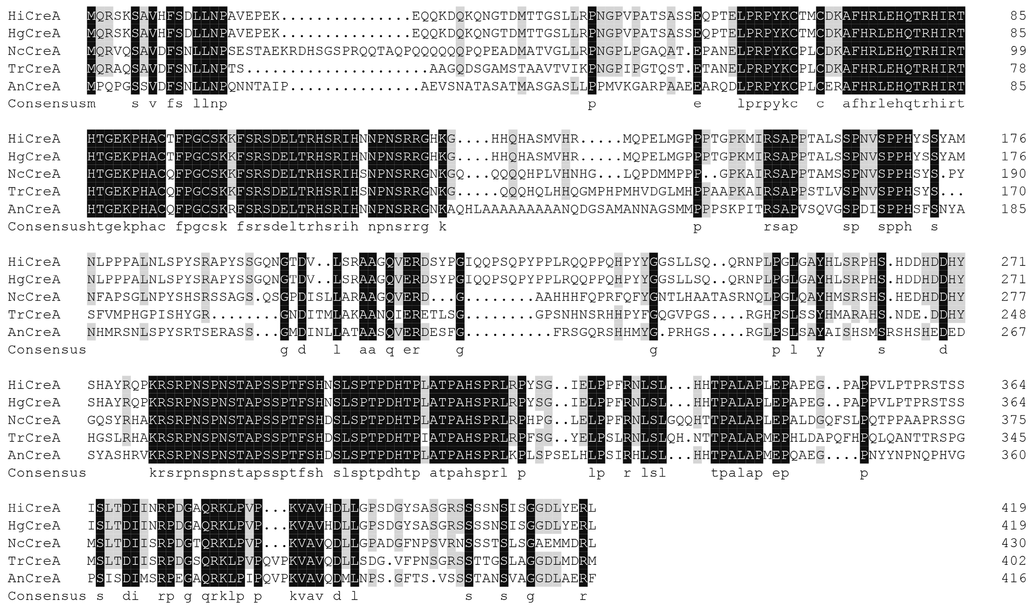
2.1. Isolation and Sequence Analysis of creA from H. insolens Y1

A 1260-bp, full-length creA gene was isolated from H. insolens Y1; it encoded a predicted polypeptide of 419 amino acids in length. The deduced CreA protein contains two C2H2 Zn-finger domains (residues 66–86 and 94–116). Based on sequence alignment analysis, the deduced CreA amino acid sequence shares 100, 56, 50, and 44% identity with CreA proteins from Humicola grisea (O93781), N. crassa (O59958), T. reesei (G0RB08), and A. nidulans (Q01981), respectively (Figure 1).

2.2. Disruption and Complementation of creA in H. insolens Y1

To determine the gene function of creA in H. insolens Y1, creA was knocked out using positive and negative screening methods with geneticin and hygromycin B resistance genes as selection markers (Figure 2a). Transformants that had acquired resistance to geneticin but could not grow in the presence of hygromycin B were selected as potential creA disruption mutants. Genomic DNA was extracted from the potential mutants, and primers within and outside the creA gene were used to verify that disruption had occurred. A 558-bp specific fragment was amplified by polymerase chain reaction (PCR) from the H. insolens WT strain, but not from the creA disruption mutant, using the creAIS/creAIA primer pair; 4742- and 3171-bp specific fragments were amplified from the creA disruption mutant and WT strains, respectively, using the creAS/creAA primer pair (Figure 2b). In addition, absolute quantitative PCR was performed to analyze the insertion copy number of the deletion cassette in the creA disruption mutant, and the results confirmed the single insertion of the deletion cassette (data not shown). To further determine the function of creA in H. insolens, creA complement strains were also constructed by introducing the entire creA gene into the disruption mutant.

2.3. Disruption of creA Affected the H. insolens Phenotype and Mycelium Morphology

To determine the effect of the creA gene on the H. insolens phenotype and mycelium morphology, the WT H. insolens Y1 strain, the creA disruption mutant, and the creA complement strains were cultured on modified Melin–Norkrans (MMN) and potato dextrose agar (PDA) plates. After three days of growth at 42 °C, no substantial differences in colony size or mycelium morphology were observed between the WT and creA complement strains, whereas the creA disruption mutant exhibited slower radial growth and smaller colonies than the WT (Figure 3a). The creA disruption mutant showed stouter hyphae and more branches on PDA plates at 24 h under microscopic observation (Figure 3b).

2.4. Disruption of creA Revealed a Novel Regulation Mechanism for H. insolens Cellulase Production

To verify the role of the creA gene in H. insolens cellulase production, cellulase production by the WT, the creA disruption mutant, and the creA complement strains grown using shake-flask fermentation were evaluated. At 24 h in yeast extract peptone dextrose (YPD) medium, the FPase activity of the creA disruption mutant was similar to that of the WT and creA complement strains; CMCase activity was observed to follow a similar trend. In contrast, pNPCase, pNPGase, and xylanase activities were 518.35, 119.58, and 42.68% higher than that of the WT strain, respectively (Figure 4).
The levels of cellulase activity and secreted proteins in MMN medium were recorded over a period of six days. Throughout the fermentation period, no significant difference was detected between the parental and creA complement strains (Figure 5). In contrast, the biomass of the creA disruption mutant was slightly lower than that of the WT strain at five to six days of fermentation (Figure 5a). The creA disruption mutant exhibited lower FPase activity than the WT strain after four days of cultivation. After six days of fermentation, the maximum FPase activity of the creA disruption mutant was observed at 7.89 U/mL, representing 80.43% of the 9.81 U/mL shown by the WT strain (Figure 5b). The extracellular CMCase, pNPCase, pNPGase, and xylanase activities were also determined. The maximum CMCase, pNPCase, and pNPGase activities in the creA disruption mutant were significantly reduced to 70.40, 71.22, and 41.58% of that of the parental strain, respectively (Figure 5c–e). In contrast, the xylanase activity was 21.97% higher than that of the parental strain (Figure 5f).
Extracellular protein secretion profiles were then examined by SDS-PAGE. The protein yield of the creA disruption mutant was significantly greater than that of the WT strain in YPD medium fermentation (Figure S1). SDS-PAGE results showed a significant decrease in the total secreted proteins of the creA disruption mutant; notably, the creA disruption mutant band at around 40 kDa was much weaker than that of the WT strain in MMN medium fermentation (Figure S1).

2.5. Regulation of Cellulolytic Gene Expression by CreA in H. insolens

qRT-PCRs were performed to determine the influence of creA disruption on the transcriptional level of cellulolytic genes. When cultured for 24 h in YPD medium, the transcriptional levels of all the target genes (including the β-glucosidase genes bgl3B and bgl3C, cellobiohydrolase genes cel6A and cel7A, endoglucanase genes cel6B and cel7B, and xylanase genes xynA and xynC) were enhanced in the creA disruption mutant except bgl3A and xynB (Figure 6a).
The transcriptional levels of cellulolytic genes in the creA disruption mutant and WT H.insolens strain under inducing condition were also investigated. Consistent with the enzyme activities, all the xylanase genes—xynA, xynB, and xynC—showed much higher mRNA levels in the creA disruption mutant than that in WT strain when the strains were cultured for 48 h in MMN medium (Figure 6b). On the contrary, the transcriptional levels of all the cellulase genes except cel7A were significantly decreased in the creA disruption mutant.

3. Discussion

H. insolens is a potential industrial cellulase producer due to its powerful ability to degrade polysaccharide biomass constituents such as cellulose and hemicellulose. To date, research on H. insolens has mainly focused on the purification of extracellular enzymes and the isolation and heterologous expression of related carbohydrate hydrolases, with few reports on the molecular mechanisms of cellulase expression regulation. In the present study, we performed the first reported knockout of the creA gene and determined its function in H. insolens. The results of this study provide further opportunities for studying the functions of H. insolens genes, and will help to reveal their cellulase regulation mechanisms.
CreA from H. insolens shows high identity with those from H. grisea, N. crassa, T. reesei, and A. nidulans. As previously shown in creA disruption strains of Penicillium oxalicum, Penicillium expansum, and T. reesei [18,22,23,24], the H. insolens creA disruption strain significantly affected hyphal growth rate and development. Previous findings have suggested that CreA affects morphology and sporulation by interacting with RAS1 in T. reesei [23]. As a regulator, RAS1 affects hyphal morphology, reproductive development, and cell wall integrity in N. crassa and A. fumigatus through interaction with related genes [25,26]. The results of transcriptome analysis in P. expansum suggest that the alteration of carbon/nitrogen metabolism in the ΔcreA mutant may contribute to the deviant fungal growth [24].
CreA also plays an important role in cellulase and hemicellulase production in H. insolens. In YPD medium fermentation with glucose as a carbon source, the extracellular protein yield and pNPCase, pNPGase, and xylanase activities were higher in the creA disruption mutant than in the WT strain. These results suggest that creA disruption results in de-repression of cellulase and hemicellulase gene expression under non-inducing conditions. In contrast to results reported for other fungi [16,18,22], the H. insolens creA disruption mutant strain cultured under Avicel-inducing conditions exhibited significantly lower cellulase expression but higher xylanase production than the WT strain. The transcriptional levels of cellulolytic genes in creA disruption mutant and WT H. insolens strain under non-inducing and inducing conditions also matched well with the cellulase activities. These results indicate that H. insolens CreA plays a similar role in hemicellulase production to that in cellulase production, via a different regulation mechanism. CreA may inhibit cellulase gene transcription in the presence of glucose, and may also be indispensable for cellulase hyperproduction in H. insolens. We speculate that these results may be partially due to the influence of creA disruption on Xyr1, an important transcription cellulase and hemicellulose activator. Competition between CreA and Xyr1 for binding to sites on cellulase gene promoters has been reported [27,28,29]. In the creA disruption mutant, hemicellulase gene promoters recruited more Xyr1, in turn increasing their transcriptional levels. In contrast, the expression of xyr1 decreased when creA was deleted in H. insolens, as previously described in T. reesei [20]. Limited Xyr1 preferentially activated hemicellulases such as xylanase; however, the difficulty of fully activating cellulases resulted in a reduction in cellulase production. In addition, due to the complexity of the regulation network of non-glucose source utilization, creA disruption may exert a complex influence on many regulation elements and transporters involved in cellulase expression. In future work, we plan to perform RNA sequencing, electrophoretic mobility shift assays (EMSAs), and DNaseI footprinting assays to elucidate the specific regulation mechanisms for creA in H. insolens.

4. Materials and Methods

4.1. Strains, Media, and Culture Conditions

H. insolens strain Y1 CGMCC 4573 was cultured on potato dextrose agar (PDA) plates for 7 days at 42 °C to induce sporulation. Shake-flask fermentation was conducted at 42 °C for 6 days in modified Melin–Norkrans (MMN) medium (0.1% tryptone, 2% yeast extract, 0.06% MgSO4·7H2O, 2% Avicel). Yeast extract peptone dextrose (YPD) medium (2% tryptone, 1% yeast extract, 2% glucose) was used to culture hyphae. Agrobacterium tumefaciens strain AGL-1 grown at 28 °C was used to mediate DNA transformation of H. insolens Y1.

4.2. Molecular Manipulation

H. insolens Y1 genomic DNA was extracted from mycelia grown on PDA plates. The primers used in this experiment are listed in Table S1. Polymerase chain reaction (PCR) amplification was performed using an Eppendorf Mastercycler and KOD FX Polymerase (Toyobo). Primer synthesis and DNA sequencing were performed by Sangon Biotech Corporation (Shanghai, China).

4.3. Sequence Analysis

The predicted amino acid sequence of the CreA protein was analyzed using FGENESH (http://linux1.softberry.com/berry.phtml). An online conserved domain search tool (https://www.ncbi.nlm.nih.gov/Structure/cdd/wrpsb.cgi) and InterProScan (http://www.ebi.ac.uk/Tools/pfa/iprscan/) were used to analyze the CreA conserved domains and protein functions. Protein sequence alignment was performed using ClustalW (http://www.ebi.ac.uk/clustalW/).

4.4. Construction of H. insolens Y1 creA Disruption Mutant and Complementation Strains

The entire Pgpd-neo-Tgpd cassette was excised from pP-neo-T using SmaI and XhoI subjected to end filling, and then ligated into the SwaI site of pAg1-hyg [9] to yield pAg1-HygNeo. The creAL fragment was amplified from WT H. insolens genomic DNA using the creALS/creALA primer pair, and then ligated into the BglII site of pAg1-Hyg-Neo to generate pAg1-HygNeo-creAL. The creAR fragment was amplified in the same manner using the creARS/creARA primer pair, and then ligated into the PacI site of pAg1-HygNeo-creAL to generate pAg1-HygNeocreA. pAg1-HygNeocreA was then transformed into an H. insolens strain using a Agrobacterium tumefaciens-mediated transformation (ATMT) system as previously described [9] to obtain creA disruption mutants.
The creA WT allele complementation cassette was amplified from WT H. insolens genomic DNA using the creAS/creAA primer pair. The creA cassette was then ligated into the PacI site of pAg1-hyg to yield pAg1-Hyg-creAC. The resulting pAg1-Hyg-creAC was transformed into a creA disruption mutant to obtain creA complement strains. The sequences of all of the primers used in this study are shown in Table S1.

4.5. Phenotypic Observation and Microscopic Observation

H. insolens WT, the creA disruption mutant, and creA complementation strains were cultured following inoculation of 2 × 10⁵ spores onto MMN, PDA, and YPD plates for 3 days at 42 °C, and the phenotypes of the resulting colonies were observed. Hyphal morphology was observed by microscopy (Eclipse Ni-U, Nikon, Tokyo, Japan) after spore germination or 24 h on cellophane-coated PDA and YPD plates.

4.6. Enzyme Activity Assay and Sodium Dodecyl Sulfate–Polyacrylamide Gel Electrophoresis (SDS–PAGE)

H. insolens WT, the creA disruption mutant, and creA complement strains were each cultured, starting with inoculation of the same number of spores, in 40 mL YPD medium for 24 h at 42 °C. Homogeneous medium (1 mL) was sampled from the YPD medium to detect enzyme activity and protein expression. After centrifuging at 12,000 rpm for 10 min and washing with ddH2O, the thalli in the remaining culture medium were transferred to MMN medium for shake-flask fermentation at 42° C for 6 days. The cellulase activities in the supernatant of each strain were assayed every 24 h. Filter paper cellulase (FPase), carboxymethylcellulose cellulose (CMCase), p-nitrophenyl-β-cellobioside cellulase (pNPCase), p-nitrophenyl-β-glucopyranoside cellulase (pNPGase), and xylanase enzyme activities were assayed as described [30].
SDS–PAGE was performed using 12% (w/v) polyacrylamide gels. Proteins were stained with Coomassie Brilliant Blue G-250. The H. insolens biomass in the MMN fermentation medium was measured as previously described [27].

4.7. Real-time Quantitative PCR Analysis

Absolute quantitative PCR was performed to analyze the integrated copy number of the deletion cassette in disruption mutant according to the reported method [31,32,33]. The copy number of the G418 resistance gene (neo) in creA disruption cassette in the creA disruption mutant was determined. The standard curve was measured using a plasmid (M13-PTgpd-neo) which contains single neo gene as the template.
The relative transcriptional level of cellulytic genes in H. insolens was determined by qRT-PCR. β-actin gene was used as an internal control for normalization. Reactions were performed using 2×ChamQ Universal SYBR qPCR Master Mix kit (Vazyme, Nanjing, China) as described by the manufacturer’s protocol. Data analysis was done with the 2−△△Ct method.
The qPCR experiment was performed on the LightCycler 96 System as follows: 1 cycle of 94 °C for 5 min, followed by 40 cycles of 94 °C for 5 s, 58 °C for 15 s, and 72 °C for 20 s. The primers for qPCR are shown in Table S1. Three biological replicates were performed with three technical replicates for each biological replicate.

4.8. Nucleotide Sequence

The nucleotide sequence of the creA gene of H. insolens Y1 was deposited into the GenBank database under accession number MK907677.

Supplementary Materials

Supplementary materials can be found at https://www.mdpi.com/1422-0067/20/15/3693/s1.

Author Contributions

B.L., W.Z., and X.X. conceived and designed the experiments; C.F., J.L., and Y.C. performed the experiments; C.F., X.X., and Y.Z. analyzed the data; L.S and W.Z. contributed reagents/materials/analysis tools; C.F. and X.X. wrote the paper.

Funding

This research was funded by the National Natural Science Foundation of China (grant number 31600065) and the Open Research Fund Program of the Beijing Key Laboratory of Plant Resource Research and Development, Beijing Technology and Business University (grant number 2016-YB3).

Conflicts of Interest

The authors declare no conflict of interest.

References

  1. Matsumoto, H.; Koganei, K.; Nishida, N.; Koyama, Y.; Saito, S.; Kataoka, H.; Ogihara, J.; Kasumi, T. Cell dispersion culture for the effective growth of Humicola insolens and efficient enzyme production. J. Biosci. Bioeng. 2013, 117, 257–262. [Google Scholar] [CrossRef] [PubMed]
  2. Schulein, M. Enzymatic properties of cellulases from Humicola insolens. J. Biotechnol. 1997, 57, 71–81. [Google Scholar] [CrossRef]
  3. Souza, F.H.M.; Inocentes, R.F.; Ward, R.J.; Jorge, J.A.; Furriel, R.P.M. Glucose and xylose stimulation of a beta-glucosidase from the thermophilic fungus Humicola insolens: A kinetic and biophysical study. J. Mol. Catal. B. Enzym. 2013, 94, 119–128. [Google Scholar] [CrossRef]
  4. Wu, I.; Arnold, F.H. Engineered thermostable fungal Cel6A and Cel7A cellobiohydrolases hydrolyze cellulose efficiently at elevated temperatures. Biotechnol. Bioeng. 2013, 110, 1874–1883. [Google Scholar] [CrossRef] [PubMed]
  5. Xu, X.; Li, J.; Zhang, W.; Huang, H.; Shi, P.; Luo, H.; Liu, B.; Zhang, Y.; Zhang, Z.; Fan, Y. A neutral thermostable β-1,4-glucanase from Humicola insolens Y1 with potential for applications in various industries. PLoS ONE 2015, 10, e0124925. [Google Scholar] [CrossRef] [PubMed]
  6. Maheshwari, R.; Bharadwaj, G.; Bhat, M.K. Thermophilic fungi: Their physiology and enzymes. Microbiol. Mol. Biol. Rev. 2000, 64, 461–488. [Google Scholar] [CrossRef]
  7. Meleiro, L.P.; Zimbardi, A.L.; Souza, F.H.; Masui, D.C.; Silva, T.M.; Jorge, J.A.; Furriel, R.P. A novel beta-glucosidase from Humicola insolens with high potential for untreated waste paper conversion to sugars. Appl. Biochem. Biotechnol. 2014, 173, 391–408. [Google Scholar] [CrossRef]
  8. Xia, W.; Shi, P.; Xu, X.; Qian, L.; Cui, Y.; Xia, M.; Yao, B. High level expression of a novel family 3 neutral beta-xylosidase from Humicola insolens Y1 with high tolerance to D-xylose. PLoS ONE 2015, 10, e0117578. [Google Scholar]
  9. Xu, X.X.; Li, J.Y.; Shi, P.J.; Ji, W.L.; Liu, B.; Zhang, Y.H.; Yao, B.; Fan, Y.L.; Zhang, W. The use of T-DNA insertional mutagenesis to improve cellulase production by the thermophilic fungus Humicola insolens Y1. Sci. Rep. 2016, 6, 9. [Google Scholar] [CrossRef]
  10. Furukawa, T.; Shida, Y.; Kitagami, N.; Mori, K.; Kato, M.; Kobayashi, T.; Okada, H.; Ogasawara, W.; Morikawa, Y. Identification of specific binding sites for XYR1, a transcriptional activator of cellulolytic and xylanolytic genes in Trichoderma reesei. Fungal. Genet. Biol. 2009, 46, 564–574. [Google Scholar] [CrossRef]
  11. Ling, M.; Qin, Y.; Li, N.; Liang, Z. Binding of two transcriptional factors, Xyr1 and ACEI, in the promoter region of cellulase cbh1 gene. Biotechnol. Lett. 2009, 31, 227–231. [Google Scholar] [CrossRef]
  12. Stricker, A.R.; Grosstessnerhain, K.; Würleitner, E.; Mach, R.L. Xyr1 (xylanase regulator 1) regulates both the hydrolytic enzyme system and D-xylose metabolism in Hypocrea jecorina. Eukaryot. Cell 2006, 5, 2128–2137. [Google Scholar] [CrossRef]
  13. Kubicek, C.P.; Mikus, M.; Schuster, A.; Schmoll, M.; Seiboth, B. Metabolic engineering strategies for the improvement of cellulase production by Hypocrea jecorina. Biotechnol. Biofuels. 2009, 2, 19. [Google Scholar] [CrossRef]
  14. Antonella, A.; Simona, G.; Vincenza, F. Regulation of cellulase and hemicellulase gene expression in fungi. Curr. Genomics 2013, 14, 230–249. [Google Scholar]
  15. Antonieto, A.C.; dos Santos Castro, L.; Silva-Rocha, R.; Persinoti, G.F.; Silva, R.N. Defining the genome-wide role of CRE1 during carbon catabolite repression in Trichoderma reesei using RNA-Seq analysis. Fungal. Genet. Biol. 2014, 73, 93–103. [Google Scholar] [CrossRef]
  16. Sun, J.; Glass, N.L. Identification of the CRE-1 cellulolytic regulon in Neurospora crassa. PLoS ONE 2011, 6, e25654. [Google Scholar] [CrossRef]
  17. Ries, L.N.; Beattie, S.R.; Espeso, E.A.; Cramer, R.A.; Goldman, G.H. Diverse regulation of the CreA carbon catabolite repressor in Aspergillus nidulans. Genetics 2016, 203, 335–352. [Google Scholar] [CrossRef]
  18. Li, Z.; Yao, G.; Wu, R.; Gao, L.; Kan, Q.; Liu, M.; Yang, P.; Liu, G.; Qin, Y.; Song, X. Synergistic and dose-controlled regulation of cellulase gene expression in Penicillium oxalicum. PLoS Genet. 2015, 11, e1005509. [Google Scholar] [CrossRef]
  19. Mach-Aigner, A.R.; Pucher, M.E.; Steiger, M.G.; Bauer, G.E.; Preis, S.J.; Mach, R.L. Transcriptional regulation of xyr1, encoding the main regulator of the xylanolytic and cellulolytic enzyme system in Hypocrea jecorina. Appl. Environ. Microbiol. 2008, 74, 6554. [Google Scholar] [CrossRef]
  20. Portnoy, T.; Margeot, A.; Seidlseiboth, V.; Le, C.S.; Ben, C.F.; Linke, R.; Seiboth, B.; Kubicek, C.P. Differential regulation of the cellulase transcription factors XYR1, ACE2, and ACE1 in Trichoderma reesei strains producing high and low levels of cellulase. Eukaryot. Cell 2011, 10, 262–271. [Google Scholar] [CrossRef]
  21. Adnan, M.; Zheng, W.; Islam, W.; Arif, M.; Abubakar, Y.; Wang, Z.; Lu, G. Carbon catabolite repression in filamentous fungi. Int. J. Mol. Sci. 2018, 19, 48. [Google Scholar] [CrossRef]
  22. Nakari-Setala, T.; Paloheimo, M.; Kallio, J.; Vehmaanpera, J.; Penttila, M.; Saloheimo, M. Genetic modification of carbon catabolite repression in Trichoderma reesei for improved protein production. Appl. Environ. Microbiol. 2009, 75, 4853–4860. [Google Scholar] [CrossRef]
  23. Portnoy, T.; Margeot, A.; Linke, R.; Atanasova, L.; Fekete, E.; Sándor, E.; Hartl, L.; Karaffa, L.; Druzhinina, I.S.; Seiboth, B. The CRE1 carbon catabolite repressor of the fungus Trichoderma reesei: A master regulator of carbon assimilation. Bmc. Genomics 2011, 12, 269. [Google Scholar] [CrossRef]
  24. Tannous, J.; Kumar, D.; Sela, N.; Sionov, E.; Prusky, D.; Keller, N.P. Fungal attack and host defence pathways unveiled in near-avirulent interactions of Penicillium expansum creA mutants on apples. Mol. Plant Pathol. 2018, 19, 2635–2650. [Google Scholar] [CrossRef]
  25. Fortwendel, J.R.; Fuller, K.K.; Stephens, T.J.; Bacon, W.C.; Askew, D.S.; Rhodes, J.C. Aspergillus fumigatus RasA regulates asexual development and cell wall integrity. Eukaryot. Cell 2008, 7, 1530. [Google Scholar] [CrossRef]
  26. Kana-Uchi, A.; Yamashiro, C.T.; Tanabe, S.; Murayama, T. A ras homologue of Neurospora crassa regulates morphology. Mol. Gen. Genet. 1997, 254, 427–432. [Google Scholar]
  27. Castro, L.D.S.; Antoniêto, A.C.C.; Pedersoli, W.R.; Silva-Rocha, R.; Persinoti, G.F.; Silva, R.N. Expression pattern of cellulolytic and xylanolytic genes regulated by transcriptional factors XYR1 and CRE1 are affected by carbon source in Trichoderma reesei. Gene. Expr. Patterns 2014, 14, 88–95. [Google Scholar] [CrossRef]
  28. Kiesenhofer, D.P.; Mach, R.L.; Machaigner, A.R. Influence of cis element arrangement on promoter strength in Trichoderma reesei. Appl. Environ. Microbiol. 2018, 84, e01742-17. [Google Scholar] [CrossRef]
  29. Silvarocha, R.; Castro, L.S.; Antoniêto, A.C.; Guazzaroni, M.E.; Persinoti, G.F.; Silva, R.N. Deciphering the cis-regulatory elements for XYR1 and CRE1 regulators in Trichoderma reesei. PLoS ONE 2014, 9, e99366. [Google Scholar]
  30. Fan, C.; Xu, X.; Song, L.; Guan, W.; Li, J.; Liu, B.; Shi, P.; Zhang, W. The use of Agrobacterium-mediated insertional mutagenesis sequencing to identify novel genes of Humicola insolens involved in cellulase production. Biotech. 2018, 8, 153. [Google Scholar] [CrossRef]
  31. Xue, X.; Wu, Y.; Qin, X.; Ma, R.; Luo, H.; Su, X.; Yao, B. Revisiting overexpression of a heterologous β-glucosidase in Trichoderma reesei: fusion expression of the Neosartorya fischeri Bgl3A to cbh1 enhances the overall as well as individual cellulase activities. Microb. Cell. Fact. 2016, 15, 122. [Google Scholar] [CrossRef]
  32. Solomon, P.; Ipcho, S.V.; Hane, J.; Tan, K.C.; Oliver, R. A quantitative PCR approach to determine gene copy number. Fungal Genet. Rep 2008, 55, 5–8. [Google Scholar] [CrossRef] [Green Version]
  33. D’haene, B.; Vandesompele, J.; Hellemans, J. Accurate and objective copy number profiling using real-time quantitative PCR. Methods 2010, 50, 262–270. [Google Scholar] [CrossRef]
Figure 1. Multiple protein sequence alignment of the deduced CreA sequence from H. insolens with four CreA counterparts. The sequences shown are CreA from H. insolens, HgCreA from H. grisea (O93781), NcCreA from N. crassa (O59958), TrCreA from T. reesei (G0RB08), and AnCreA from A. nidulans (Q01981). Identical and conserved residues are shaded in black and gray, respectively.
Ijms 20 03693 g001
Figure 2. Verification of the creA disruption mutant. (a) Schematic diagram of the creA gene knockout mediated by homologous recombination. hyg, hygromycin B resistance gene expression cassette; neo, geneticin resistance gene expression cassette. (b) Polymerase chain reaction (PCR) analysis of the creA disruption mutant using the primer pairs creAS/creAA (PCR1) and creAIS/creAIA (PCR2). M, 1000-bp DNA ladder; WT, wild type; ΔcreA, creA disruption mutant; P, positive control with the plasmid pAg1-HygNeocreA as the template; NC, negative control.
Ijms 20 03693 g002
Figure 3. Characterization of the creA disruption mutant growth and hyphal morphology. (a) Colony growth and sporulation of the creA disruption mutant, creA complement strains, and the wild-type (WT) strain on modified Melin–Norkrans (MMN) and potato dextrose agar (PDA) plates at 42 °C for 3 days. (b) Hyphae observed under a microscope. All strains were grown on PDA plates at 42 °C for 24 h. creAC, creA complement strain.
Ijms 20 03693 g003
Figure 4. Cellulase activity of H. insolens WT, the creA disruption mutant, and creA complement strains in the presence of glucose. All strains were cultured in shake flasks at 42 °C for 24 h with 2.0% (w/v) glucose as the carbon source.
Ijms 20 03693 g004
Figure 5. Cellulase activities of H. insolens WT, creA disruption mutant, and creA complement strains under inducing conditions. Dry weight (a), FPase activity (b), CMCase activity (c), pNPCase activity (d), pNPGase activity (e), and xylanase activity (f) were measured from shake-flask fermentation cultures grown at 42 °C for 6 days using 2.0% (w/v) Avicel as the carbon source.
Ijms 20 03693 g005
Figure 6. Relative transcriptional level of cellulolytic genes in H. insolens WT and creA disruption mutant under non-inducing (a) or inducing (b) conditions.
Ijms 20 03693 g006

