Article

Mouse Mammary Tumor Virus (MMTV)-Like env Sequence in Brazilian Breast Cancer Samples: Implications in Clinicopathological Parameters in Molecular Subtypes

by
Nathália de Sousa Pereira
1,
Glauco Akelinghton Freire Vitiello
1,
Bruna Karina Banin-Hirata
1,
Glaura Scantamburlo Alves Fernandes
2,
Maria José Sparça Salles
2,
Marla Karine Amarante
1,* and
Maria Angelica Ehara Watanabe
1
1
Laboratory of DNA Polymorphisms and Immunology, Department of Pathological Sciences, Biological Sciences Center, Londrina State University, Londrina, PR 86057-970, Brazil
2
Department of General Biology, Biological Sciences Center, Londrina State University, Londrina, PR 86057-970, Brazil
*
Author to whom correspondence should be addressed.
Int. J. Environ. Res. Public Health 2020, 17(24), 9496; https://doi.org/10.3390/ijerph17249496
Submission received: 30 October 2020 / Revised: 11 December 2020 / Accepted: 15 December 2020 / Published: 18 December 2020
(This article belongs to the Special Issue Breast Cancer Genetics)

Abstract

:
Background: Breast cancer (BC) is a complex disease in which susceptibility and clinical course depend on multiple factors. Evidence suggests that a mouse mammary tumor virus (MMTV)-homolog may be present in human BCs; however, little is known about its clinical implications. Methods: MMTV-like env nucleotide-sequence was searched in tumor and tumor-adjacent tissues from 217 Brazilian BC patients through nested-PCR and confirmed through PCR-sequencing. Blood samples were also tested for patients with MMTV-like env gene-positive tumors. Correlations with clinicopathological parameters were evaluated. Results: MMTV-like env sequence was detected in tumor and tumor-adjacent tissue samples from 41/217 and 30/196 patients, respectively. In blood, MMTV-like was detected in 17/32 patients. In Luminal-B tumors, MMTV-like in tumor tissue was negatively correlated with tumor size and disease stage, whereas in HER2 tumors it anti-correlated with lymph node metastasis (LNM) and disease stage. Considering blood, MMTV-like env gene positivity negatively correlated with age in general BC, while in Luminal-A tumors it positively correlated with Ki67 but negatively correlated with age and LNM. The associations with decreased LNM frequency were independent of other prognostic factors. Conclusion: MMTV-like env positivity is associated with better prognostic parameters in BC subtypes, which might be explainable by its anti-metastatic potential and by putative activation of immune milieu.

1. Introduction

Breast cancer (BC) is a serious public health issue, considering the number of women who are diagnosed and deaths that occur annually from this disease worldwide [1], and its clinical course and outcome vary from patient to patient depending on a complex series of factors [2,3,4,5,6].
BC can be stratified though the expression of molecular markers, such as estrogen receptors (ER), progesterone receptors (RP), and human epidermal growth factor receptor 2 (HER2) overexpression or amplification, classifying the disease into four major molecular subtypes: Luminal A (LA; ER/PR + HER2-), Luminal-B HER2-positive (LB; ER/PR + HER2+), HER2-enriched (HER2; ER-PR-HER2+) and triple-negative (TN; ER-PR-HER2-) [7,8]. These subtypes have a direct impact on patients’ prognosis and therapeutic response and are used to guide BC treatment [9,10].
Cancers are recognized as multifactorial diseases emerging in the background of multiple genetic and environmental risk factors [11]. Several viruses are known to participate in the induction and in the progression of some neoplasia through different mechanisms, such as expression of oncogenic proteins, induction of cancer-promoting inflammation and insertional mutagenesis. Examples of consolidated oncogenic viruses include the human papilloma virus (HPV) in cervical cancer, the Hepatitis B virus (HBV) in hepatic cancer, the Epstein–Barr virus (EBV) in lymphoma, and the human T-cell Lymphotropic virus (HTLV) in leukemias [12]. Further, other viruses have emerged as potential risk factors for other cancers, including a proportion of human breast tumors [13,14].
The mouse mammary tumor virus (MMTV), from Betaretroviridae family, was firstly related as a carcinogenic agent by Bittner [15] and is currently considered the most common etiologic agent for BC and T-cell lymphomas in mice [16,17]. Since the discovery of MMTV, researchers have sought to investigate the participation of viruses in human mammary carcinogenesis. These investigations have led to the identification of antigens and sequences homologous to MMTV in human BC tissues by several groups worldwide, indicating that a similar virus, referred to as MMTV-like, might play a role in human BC [18]. Other groups, however, have failed to identify evidence for MMTV-like sequences in human BCs. These discrepancies may be attributable to technical, methodological, or epidemiological variations among studies [18].
Over the last years, several works have narrowed the link between MMTV-like and human BC [18]. Liu et al. [19] were able to isolate a complete provirus from human BC samples and Lawson et al. [20] found MMTV-like antigens and sequences in benign lesions from women that developed BC years later. Furthermore, MMTV-like sequences were found in other human biological samples, such as milk [21], saliva [22], and peripheral blood lymphoid cells [23,24], suggesting that viral transmission and spreading in humans are similar to that described in mice, occurring via breast-feeding and infection of mucosa-associated lymphocytes before reaching the breast tissue [25].
Despite these evidences, there is only one study investigating the presence of MMTV-like sequences in South America, which was conducted in Argentina [26]. Additionally, little is known about the implications of MMTV-like infection on disease prognosis and clinical presentation of BC, especially in BC molecular subtypes. Therefore, the present study aimed to evaluate the presence of MMTV-like env gene in breast tissue (tumor and tumor-adjacent tissue) and peripheral blood of Brazilian BC patients and to analyze its correlation with clinicopathological parameters within BC molecular subtypes.

2. Materials and Methods

2.1. Human Subjects

Following approval from the institutional Ethics Committee for research involving human beings (CAAE 47709015200005231), women diagnosed with BC were invited to participate in the research during the clinical care in the specialized services at Londrina Cancer Hospital (LCH) (Londrina, Paraná, Brazil). A free-informed consent form was signed by all donors prior to sample collection. All patients were treated by the Brazilian Public Health System (SUS).
All patients underwent mastectomy surgery, during which biological samples, consisting of small fragments of freshly excised tissue, were collected by the surgeon responsible from Londrina Cancer Hospital. After collection, the samples were sent to the Laboratory of DNA Polymorphisms and Immunology for processing. In total, fresh tumor tissues were collected from 217 BC patients. Tumor-adjacent tissues and blood samples were also collected at surgery for some of these patients.
Clinicopathological features retrieved from patients’ medical records included: age at diagnosis, tumor size, histopathological grade, lymph node metastasis (LNM) status, pathological disease stage and immunostaining for Ki67 (cellular proliferation index), p53, estrogen receptor (ER), progesterone receptor (PR) and human epidermal growth factor receptor 2 (HER2). Pathologic disease stage was determined according to the Union for International Control of Cancer (UICC) classification criteria [27]. Immunohistochemical analyses were performed in clinical routine in the Laboratory of Clinical Pathology of the Londrina Cancer Hospital, following standard protocols [28,29].

2.2. DNA Extraction

Tumor and tumor-adjacent tissues were mechanically dissociated under aseptic conditions and genomic DNA was obtained through the salting-out method [30]. For peripheral blood samples, DNA was obtained using the Biopur MiniSpin kit (Biometrix diagnostica®, Curitiba, PR, Brazil). All DNA samples were quantified in a NanoDrop-2000c Spectrophotometer (Thermo-Fisher Scientific, Wilmington, Delaware, EUA) at 260 nm wavelength. The 260/280 nm absorbance ratio was applied to assess protein contamination and only samples with a minimum of 1.7 ratio were used. DNA samples were stored at −20 °C and adjusted for a concentration of 200 ng/µL with ultrapure water prior to use.

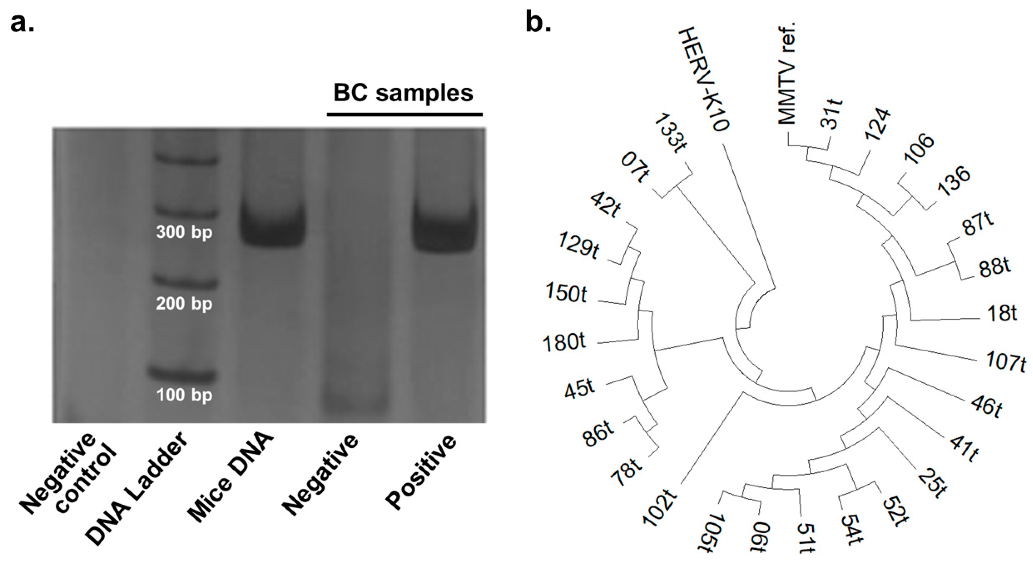
2.3. Nested PCR for MMTV-Like env Gene

MMTV-like env gene sequence was detected by nested polymerase chain reaction (PCR), using the four oligonucleotide primers described by Nartey et al. [21], according to the sequence deposited in the NCBI GenBank (Accession Number KJ831810). PCRs were performed using 16 ng/μL of template DNA in the first PCR round and 2 μL of PCR-product from the first round in the second reaction. In both reactions, reagents concentrations were as follows: 1 × PCR-Buffer (20 mM Tris-HCl pH 8.5, 50 mM KCl), 75 μM dNTP, 0.1 μM of each primer, 1.5 mM MgCl2, and 1.25 U Taq DNA polymerase, all from Invitrogen (Carlsbad, California, USA), and ultrapure water to complete a final volume of 25 μL. The reactions took place on a thermal cycler under the following conditions: 95 °C for 5 min followed by 35 cycles of 30 s at 95 °C, 30 s at 58 °C and 30 s and a final extension at 72 °C for 7 min. The amplified products were visualized through electrophoresis in 10% polyacrylamide gels stained with silver nitrate. Positive reactions were represented by the amplification of a 251 bp fragment after the second round of PCR (Figure 1a).
To control for false-positive and false negative results, a negative control without DNA addition and a positive control, consisting of pooled genomic DNA from mammary glands of lactating mice, were also tested in each PCR batch. Furthermore, all DNA samples in the present study were previously used in recent studies by our group [31,32], proving that all samples had amplifiable genomic DNA. In addition, positive samples were independently amplified at least three times and tumor tissues positive for MMTV-like env gene were sequenced to confirm the amplification of the target fragment.

2.4. Sequencing

Nested-PCR products for MMTV-like env were purified using PureLink™ PCR Purification Kit (Invitrogen), following the manufacturer’s instructions. Forward and reverse sequencing reactions were performed in duplicates using the BigDye® Terminator v3.1 kit (Applied Biosystems, Foster City, California, USA), 50 ng of template, and 5 pM of respective oligonucleotide primer in a final volume of 10 μL. PCR conditions were as follows: 10 s at 95 °C, 30 cycles of 20 s at 95 °C, 20 s at 50 °C, and 1 min at 60 °C. The sequencing fragments were analyzed through capillary electrophoresis in a 3500 XL Genetic Analyzer (Applied Biosystems).
The obtained sequences were compared against the NCBI nucleotide databank using the BLAST algorithm. Further, the sequences were aligned and compared with MMTV env (gPr73, accession number 149,186 in NCBI GenBank) and HERV-K10 (accession number AF164613.1 in NCBI GenBank) reference sequences using the MUSCLE algorithm running within MEGA7 software [33] and a phylogenetic tree was constructed using the Tamura–Nei model [34].

2.5. Statistical Analyses

To analyze the relationship between MMTV-like env and clinicopathological features, Kendall’s rank correlation tests were applied, considering Tau-b coefficient for square contingency tables (2 × 2) or Tau-c coefficients for rectangular contingency tables (2 × 3 and 2 × 4). Binary logistic regression analyses were applied to test the association between MMTV-like env and the presence of LNM adjusting for tumor size and proliferation index (Ki67). All statistical analyses were two-tailed with a significance level of 0.05 and were performed using SPSS software version 22.0 (IBM®, Chicago, IL, USA).

3. Results

3.1. Prevalence of MMTV-Like env in Breast Cancer Samples

A total of 217 BC samples were included in this work. Patients’ clinicopathological features according to BC subtypes are shown in Table 1.
MMTV-like env gene was detected in 18.9% (41/217) of tumor tissues analyzed (Table 2). Of these positive samples, 26 were sequenced and compared to sequences deposited on the NCBI nucleotide bank through the BLAST algorithm, confirming the amplification of MMTV-like env gene. Both murine and human sequences returned in the queries with similar scores and identities, confirming that human and murine-derived MMTV-like env sequences are genetically indistinguishable.
A representative electrophoretic profile for the nested-PCR to MMTV-like env gene is shown in Figure 1a. The obtained sequences were highly similar to the MMTV env reference gene, but not to the sequence of HERV-K10 which was previously suggested to be amplified through PCR protocols targeting MMTV sequences [35]. Differences in aligned nucleotide sequences among samples argued against the hypothesis of contamination of PCR reactions or extracted DNA (Figure 1b).
Tumors expressing HER2 showed a higher prevalence of MMTV-like env in tumor tissue, with the highest prevalence being observed in tumors from the HER2-enriched subtype (38.5%), followed by those from the Luminal-B subtype (25.0%). TN tumors, otherwise, showed the lower prevalence of MMTV-like env, with only one out of 26 samples (3.8%) being positive (Table 2).
Tumor-adjacent tissues were also tested and showed that MMTV-like env was present in 15.3% (30/196) samples (Table 2). The frequency of MMTV-like env was higher in the tumor than in adjacent tissue in general BC and subtypes, except for TN. Some samples were positive for MMTV-like env in adjacent tissue but negative in the tumor tissue. This occurred in 19 of 176 (10.8%) samples in the general group, in 8 of 112 (7.1%) of LA samples, in 3 of 18 (16.7%) LB samples, in 1 of 8 (12.5%) of HER2-enriched samples, and in 4 of 22 (18.2%) of TN samples.
As shown in the literature, MMTV-like sequences were also detected in other biological materials, including peripheral blood. Therefore, we decided to investigate the presence of the MMTV-like env in blood samples from patients that were positive in tumor tissue, identifying it in 17/32 (53.1%) of them (Table 2). According to the BC subgroup, MMTV-like env prevalence in blood was higher in hormonal-receptor-positive patients (13/24, 54.2%), especially in the LA group, where 57.1% of env-positive patients considering breast tissues, were also positive in peripheral blood.

3.2. Correlations between MMTV-Like env and Clinicopathological Features

Next, we sought to investigate if MMTV-like env presence in tumor tissue was correlated with prognostic clinicopathological features in BC and its subgroups (Table 3). Because of the low prevalence of MMTV-like env in TN tumors, correlations could not be analyzed in this subgroup.
Regarding the presence of env in tumor tissue, there was no correlation in general BC group and LA subgroup. However, in LB cancers, it was negatively correlated with tumor size (Tau-c = −0.465; p = 0.005) and disease stage (Tau-c = −0.382; p = 0.04), and in HER2-enriched BCs it negatively correlated with the presence of LNM (Tau-b = −0.559; p = 0.015) and with disease stage (Tau-c = −0.639; p = 0.001) (Table 3).
We also sought to investigate the correlation between MMTV-like positivity in blood and clinicopathological parameters. For this purpose, samples that were positive for env both in tumor tissue and peripheral blood were compared either with those that were negative for env in any tissue or with those that were positive in tumor tissue but not in blood.
In the general BC group env positivity in blood was negatively correlated with age considering any group of comparison, and a marked trend towards a negative correlation with LNM (Tau-b = −0.4, p = 0.051) was observed when samples positive for MMTV in blood and tumor were compared with samples that were positive only in tumor tissue (Table 4).
In LA subgroup, env positivity in blood and tumor tissue was negatively correlated with age at diagnosis and positively correlated with Ki67 when compared to samples that did not amplify the MMTV-like env in breast tissue. When considering only LA samples that were positive for the env in breast tissue, env positivity in peripheral blood was negatively correlated with LNM (Table 4).

3.3. MMTV-like is Independently Associated with Lymph Node Metastasis

LNM is a severe latter event in breast carcinogenesis that is associated with poor prognosis. Thus, we investigated whether the associations between MMTV-like env and the presence of LNM was independent of other clinicopathological features.
For this purpose, in groups where LNM correlated with MMTV-like env in tumor tissue (HER2) or in blood samples (LA), pairwise correlation analyses were performed to identify clinicopathological features correlated with LNM. Then logistic regression models including these factors along with MMTV-like env presence were tested.
In HER2+ BCs, no factor other than MMTV-like env in tumor tissue was correlated with LNM. Regression models considering only MMTV-like env in tumor could not be tested, because all samples positive for MMTV-like env in tumor tissue (n = 5) were negative for LNM in this group.
In the LA group, LNM was positively correlated with tumor size (Tau-c = 0.312; p < 0.001) and Ki67 (Tau-c = 0.185; p = 0.029). Therefore, logistic regression models were tested including MMTV-like env in blood (compared to tumor tissue env+ and blood env samples) along with tumor size (Table 5; Model 1), Ki67 (Table 5; Model 2) or both (Table 5; Model 3), as explanatory variables. In all models, MMTV-like env was the only factor that remained significant, while tumor size and Ki67 lost significance, indicating that MMTV-like env in blood is independently associated with LNM metastasis in this group.

4. Discussion

MMTV-like env gene was found in varying proportions in human BC tissue worldwide, ranging from 0 to 74% of cases, as analyzed by a recent review by Amarante et al. [18]. In the present study, we found MMTV-like env gene in 18.9% of BC tumor tissue in a Brazilian sample, which is similar to the findings from Chinese (16.8%) [36] and Tunisian (13.9%) [37] populations. The only study carried out in South America was conducted in Argentina and has found MMTV-like env sequence in 31.0% of BC samples [26]. On the other hand, there are several studies that were not able to evidence the presence of MMTV-like sequences in tumor tissue samples, as shown in studies carried out in Japanese [38] and Iranian [39] populations.
Of note, in the current work, we submitted our PCR amplicons to sequencing, and small variations in nucleotide sequences among samples were observed. Furthermore, the obtained sequences were highly similar both to mouse- and human-derived MMTV env sequences in NCBI nucleotide bank, consistent with previous findings [35]. Therefore, the current results are unlikely to be a result of PCR amplification of DNA derived from a single environmental source, as suggested in previous reports [40] and adds new data to previous literature that shows the presence of a virus highly homologous to MMTV in human BC samples with varying prevalence among populations worldwide [18].
Patients positive for MMTV-like env in tumor tissue were also tested for the presence of MMTV-like env in peripheral blood and MMTV-like env was detected in 17/32 (53.1%) of these samples in the general group, and luminal tumors showed the highest proportion of positivity for env in blood samples (54.2%). Both results are in accordance with previous studies that identified the presence of MMTV-like sequences in peripheral blood cells of BC patients and showed that it was positively correlated with hormonal receptor expression, which is a hallmark of BC from luminal origin [23].
In previous reports, MMTV-related antigens were found both in T and B cell fractions of peripheral blood from BC patients [24], and active MMTV infection of lymphocytes was shown to be necessary to virus spread to mouse mammary tissue [41], indicating circulating lymphocytes as a source of MMTV-sequences in blood. Additionally, a previous study showed that MMTV-like sequences in blood were positively correlated with distant metastasis [23]. Altogether, these results indicate that hormonal receptors play a role in MMTV presence in the blood, which is conceivably attributable to increased viral production through the activation of estrogen-responsive elements in MMTV-LTR region in mammary cells [42,43,44] and that circulating tumor cells in metastatic tumors may also carry MMTV-like sequences in the blood.
Regarding correlations between MMTV-like env in tumor tissue and clinicopathological features, the meta-analysis by Wang et al. [45] reported no association between MMTV-like env and expression of ER, PR, HER2, p53 or histological grade. In agreement with these authors, we did not find any correlation between MMTV-like env in tumor tissue and clinicopathological parameters from the general BC group. However, in LB and HER2-enriched subtypes, we observed correlations between the presence of MMTV-like env gene in tumor tissue and clinicopathological parameters indicative of better prognosis, such as smaller tumor size, lower TNM staging, and lower frequency of LNM. BC molecular subtypes differ in relation to etiology, clinical aspects, and treatment protocols and response [7,8,46]. Our results suggest that MMTV-like may display subtype-specific effects in BC and reinforce the need to consider BC subtypes as separate entities when investigating biomarkers.
Correlations with clinicopathological parameters were also observed for MMTV-like env detection in peripheral blood samples. In the general BC and in LA groups, MMTV-like was mostly presented in blood from younger patients. In addition, in LA tumors, MMTV-like in blood correlated with higher proliferation index. We observed that patients who were positive for MMTV-like in tumor tissue and peripheral blood had a significant decrease in the frequency of LNM in comparison to patients that were positive for the env gene only in tumor tissue.
LNM is a latter event in carcinogenesis, being indicative of the ability of cancer cells to spread, which is classically related to poor BC prognosis [47,48]. Thus, the relationship between MMTV-like in breast tissue or peripheral blood and LNM was further evaluated by more accurate models, considering the influence of other clinicopathological parameters on LNM.
In HER2-enriched samples, MMTV-like env in tumor tissue was the only factor associated with LNM. In LA tumors, otherwise, MMTV-like env detection in blood was protective against LNM independently of tumor size and Ki67. It has been shown that MMTV does not encode miRNAs, but alters the expression of host miRNA network by upregulating the expression of oncogenic miRNAs from the miR-17–92 cluster, which has been shown to be a signature of BC, while downregulating the expression of miR-10b-5p, which is associated with metastasis promotion. These results indicate a putative mechanism by which MMTV-like can promote carcinogenesis while inhibiting metastasis [49], which can be consistent with the paradoxical results found in the LA subgroup, in which MMTV-like was positively correlated with proliferation index but negatively correlated with LNM.
Another possible mechanism explaining MMTV-like association with better prognosis in BC might involve the activation of antiviral immune responses in the tumor microenvironment. Indeed, BC shows increased expression of genes related to inflammation and viral infection, such as interferons (IFN) [50]. The immune response provided by type-I IFNs, which are classically activated during viral infections, has been shown to be an important suppressive factor in the metastatic process [51]. Furthermore, several studies have shown that both the expression of endogenous retroviruses or the infection with exogenous viruses indicate enhanced immunogenicity and overall better prognosis in multiple cancer types [52,53,54,55].
The APOBEC3 family of cytidine deaminases are also important components of anti-viral immunity that are induced by IFN signaling. These enzymes work by restricting viral elements through the promotion of mutations in viral nucleic acids. Recent evidence has shown that some of them are endogenous carcinogens by inducing mutations in host DNA in several cancers [56], especially in BC [57]. This mutagenic process, while increasing genomic instability and promoting cellular transformation, shows great potential to generate neoepitopes that can be recognized by the host immune system, being associated with high immunological infiltration in tumor tissue [58]. Strikingly, a germline deletion linking APOBEC3A and APOBEC3B loci, which enhances APOBEC3A expression and APOBEC-derived mutagenesis in cancer, has been associated with higher neoepitope loads and immunologic infiltrate [58] and negatively correlated with LNM in BC [59].
Of note, the link between viral infections, APOBEC3 expression, and mutagenesis has been recognized in several virus-associated tumors, such as HPV-positive cervical and oropharyngeal cancers [60] and EBV-positive gastric tumors [61]. Additionally, during MMTV infections in mice, both type I IFNs and APOBEC3 seem to participate in viral restriction [62,63]; and during infection in human breast cells, an APOBEC3G-mediated mutation in env is positively selected by increasing of MMTV-like infectivity [64], showing that APOBEC3 cluster is activated during MMTV infection both in mice and human breast cells.
Overall, these evidences indicate mechanisms by which MMTV-like could participate in BC initiation while being associated with favorable prognostic parameters. MMTV-like is a retrovirus and has the potential to upregulate IFN signaling and APOBEC-mediated mutagenesis in BC; and in this way it can increase genomic instability and tumor evolution [57] while promoting cancer cell apoptosis and immunological surveillance through recognition of viral antigens and APOBEC-generated neoepitopes by host immune system [55,58].

5. Conclusions

In conclusion, the present work has shown, for the first time, that MMTV-like DNA sequences are present in a proportion BC tissues from Brazilian patients, adding to a growing bunch of epidemiological evidence indicating that MMTV-like DNA sequences are present in human breast tissue of BC patients from different populations worldwide with variable prevalence [18]. Additionally, we were able to detect MMTV-like DNA sequences in peripheral blood samples of BC patients, indicating its potential as a circulating biomarker for BC. In addition, MMTV-like env sequence was associated with better prognostic parameters in specific BC subtypes, further strengthening its potential as a valuable marker indicating BC prognosis, beyond being a BC risk factor. Future studies may now focus on the elucidation of possible carcinogenic mechanisms and clinical implications of MMTV-like in human BC, once this knowledge can pave the way for the development of new preventive, prognostic and therapeutic approaches for human BC.

Author Contributions

Conceptualization: M.K.A. and M.A.E.W.; Methodology: M.K.A., M.A.E.W. and N.d.S.P.; Formal Analysis: N.d.S.P. and G.A.F.V.; Investigation: N.d.S.P., G.A.F.V. and B.K.B.-H.; Resources: M.A.E.W., G.S.A.F. and M.J.S.S.; Data Curation: N.d.S.P., G.A.F.V., M.K.A. and B.K.B.-H.; Writing–Original Draft Preparation: N.d.S.P.; Writing–Review & Editing: G.A.F.V., M.K.A. and M.A.E.W.; Visualization: N.d.S.P. and G.A.F.V.; Supervision: M.K.A. and M.A.E.W.; Project Administration: M.K.A. and M.A.E.W.; Funding Acquisition: M.K.A. and M.A.E.W. All authors have read and agreed to the published version of the manuscript.

Funding

This research was funded by the Brazilian National Council for Scientific and Technological Development (CNPq, Process number 304348/2019-8).

Acknowledgments

The authors would like to acknowledge the volunteers who made this study possible. Authors would also to express their gratitude for the support provided by the National Council for Scientific and Technological Development (CNPq, Brazil), the Coordination of Superior Level Staff Improvement (CAPES, Brazil) and by the Araucária Foundation for Scientific and Technological Development Support (FAADCT, Paraná).

Conflicts of Interest

The authors declare no conflict of interests.

References

  1. Bray, F.; Ferlay, J.; Soerjomataram, I.; Siegel, R.L.; Torre, L.A.; Jemal, A. Global cancer statistics 2018: GLOBOCAN estimates of incidence and mortality worldwide for 36 cancers in 185 countries. CA Cancer J. Clin. 2018, 68, 394–424. [Google Scholar] [CrossRef] [PubMed] [Green Version]
  2. Brewer, H.R.; Jones, M.E.; Schoemaker, M.J.; Ashworth, A.; Swerdlow, A.J. Family history and risk of breast cancer: An analysis accounting for family structure. Breast Cancer Res. Treat. 2017, 165, 193–200. [Google Scholar] [CrossRef] [PubMed] [Green Version]
  3. Horn, J.; Åsvold, B.O.; Opdahl, S.; Tretli, S.; Vatten, L.J. Reproductive factors and the risk of breast cancer in old age: A Norwegian cohort study. Breast Cancer Res. Treat. 2013, 139, 237–243. [Google Scholar] [CrossRef] [PubMed]
  4. Makarem, N.; Chandran, U.; Bandera, E.V.; Parekh, N. Dietary Fat in Breast Cancer Survival. Annu. Rev. Nutr. 2013, 33, 319–348. [Google Scholar] [CrossRef] [PubMed] [Green Version]
  5. Ban, K.A.; Godellas, C.V. Epidemiology of Breast Cancer. Surg. Oncol. Clin. N. Am. 2014, 23, 409–422. [Google Scholar] [CrossRef] [PubMed]
  6. Jung, S.; Wang, M.; Anderson, K.; Baglietto, L.; Bergkvist, L.; Bernstein, L.; van den Brandt, P.A.; Brinton, L.; Buring, J.E.; Eliassen, A.H.; et al. Alcohol consumption and breast cancer risk by estrogen receptor status: In a pooled analysis of 20 studies. Int. J. Epidemiol. 2015, 45, 916–928. [Google Scholar] [CrossRef] [Green Version]
  7. Cirqueira, M.B.; Moreira, M.A.R.; Soares, L.R.; Freitas-Júnior, R. Subtipos moleculares do câncer de mama. FEMINA 2011, 39, 499–503. [Google Scholar]
  8. Sorlie, T.; Perou, C.M.; Tibshirani, R.; Aas, T.; Geisler, S.; Johnsen, H.; Hastie, T.; Eisen, M.B.; van de Rijn, M.; Jeffrey, S.S.; et al. Gene expression patterns of breast carcinomas distinguish tumor subclasses with clinical implications. Proc. Natl. Acad. Sci. USA 2001, 98, 10869–10874. [Google Scholar] [CrossRef] [Green Version]
  9. Rakha, E.A.; Reis-Filho, J.S.; Baehner, F.L.; Dabbs, D.J.; Decker, T.; Eusebi, V.; Fox, S.; Ichihara, S.; Jacquemier, J.; Lakhani, S.R.; et al. Breast cancer prognostic classification in the molecular era: The role of histological grade. Breast Cancer Res. 2010, 12, 1–12. [Google Scholar] [CrossRef] [Green Version]
  10. Weigelt, B.; Geyer, F.C.; Reis-Filho, J.S. Histological types of breast cancer: How special are they? Mol. Oncol. 2010, 4, 192–208. [Google Scholar] [CrossRef] [Green Version]
  11. Tomasetti, C.; Vogelstein, B. Variation in cancer risk among tissues can be explained by the number of stem cell divisions. Science 2015, 347, 78–81. [Google Scholar] [CrossRef] [PubMed] [Green Version]
  12. Chang, Y.; Moore, P.S.; Weiss, R.A. Human oncogenic viruses: Nature and discovery. Philos. Trans. R. Soc. B Biol. Sci. 2017, 372, 20160264. [Google Scholar] [CrossRef] [PubMed] [Green Version]
  13. Etkind, P.; Du, J.; Khan, A.; Pillitteri, J.; Wiernik, P.H. Mouse mammary tumor virus-like ENV gene sequences in human breast tumors and in a lymphoma of a breast cancer patient. Clin. Cancer Res. 2000, 6, 1273–1278. [Google Scholar] [PubMed]
  14. Amarante, M.K.; Watanabe, M.A. The possible involvement of virus in breast cancer. J. Cancer Res. Clin. Oncol. 2008, 135, 329–337. [Google Scholar] [CrossRef]
  15. Bittner, J.J.; Zucker, T.F. Some Possible Effects of Nursing on the Mammary Gland Tumor Incidence in Mice. Science 1936, 84, 162. [Google Scholar] [CrossRef]
  16. Mason, A. Is there a breast cancer virus? Ochsner. J. 2000, 2, 36–39. [Google Scholar]
  17. Lawson, J.S.; Günzburg, W.H.; Whitaker, N.J. Viruses and human breast cancer. Future Microbiol. 2006, 1, 33–51. [Google Scholar] [CrossRef] [Green Version]
  18. Amarante, M.K.; Pereira, N.D.S.; Vitiello, G.A.F.; Watanabe, M.A. Involvement of a mouse mammary tumor virus (MMTV) homologue in human breast cancer: Evidence for, against and possible causes of controversies. Microb. Pathog. 2019, 130, 283–294. [Google Scholar] [CrossRef]
  19. Liu, B.; Wang, Y.; Melana, S.M.; Pelisson, I.; Najfeld, V.; Holland, J.F.; Pogo, B.G. Identification of a proviral structure in human breast cancer. Cancer Res. 2001, 61, 1754–1759. [Google Scholar]
  20. Lawson, J.S.; Mazzanti, C.; Civita, P.; Menicagli, M.; Ngan, C.C.; Whitaker, N.J.; Hochman, J.; Braitbard, O.; Yosufi, B.; Glenn, W.K. Association of Mouse Mammary Tumor Virus With Human Breast Cancer: Histology, Immunohistochemistry and Polymerase Chain Reaction Analyses. Front. Oncol. 2018, 8, 141. [Google Scholar] [CrossRef] [Green Version]
  21. Nartey, T.; Moran, H.; Marin, T.; Arcaro, K.F.; Anderton, D.L.; Etkind, P.; Holland, J.F.; Melana, S.M.; Pogo, B.G.T. Human Mammary Tumor Virus (HMTV) sequences in human milk. Infect. Agents Cancer 2014, 9, 20. [Google Scholar] [CrossRef] [Green Version]
  22. Mazzanti, C.M.; Lessi, F.; Armogida, I.; Zavaglia, K.; Franceschi, S.; Hamad, M.A.; Roncella, M.; Ghilli, M.; Boldrini, A.; Aretini, P.; et al. Human saliva as route of inter-human infection for mouse mammary tumor virus. Oncotarget 2015, 6, 18355–18363. [Google Scholar] [CrossRef] [PubMed]
  23. Naushad, W.; Ayub, S.; Sadia, H. Significant correlation of MMTV (Mouse mammary tumor virus) LTR gene with hormone receptor status in peripheral blood samples of breast cancer patients from North Pakistan. Int. J. Biosci. IJB 2017, 10, 399–405. [Google Scholar] [CrossRef]
  24. Lushnikova, A.A.; Kriukova, I.N.; Malivanova, T.F.; Makhov, P.B.; Polevaia, E.B. Correlation between expression of antigen immunologically related to gp52 MMTV and transcription of homologous ENV MMTV DNA sequences in peripheral blood lymphocytes from breast cancer patients. Mol. Genet. Microbiol. Virol. 1998, 3, 33–36. [Google Scholar]
  25. Lawson, J.S.; Glenn, W.K. Evidence for a causal role by mouse mammary tumour-like virus in human breast cancer. NPJ Breast Cancer 2019, 5, 1–10. [Google Scholar] [CrossRef] [PubMed] [Green Version]
  26. Melana, S.M.; Picconi, M.A.; Rossi, C.; Mural, J.; Alonio, L.V.; Teyssié, A.; Holland, J.F.; Pogo, B.G.T. Detection of murine mammary tumor virus (MMTV) env gene-like sequences in breast cancer from Argentine patients. Medicina 2002, 62, 323–327. [Google Scholar] [PubMed]
  27. Brierley, J.; Gospodarowicz, M.K.; Wittekind, C. TNM Classification of Malignant Tumours, 8th ed.; Blackwell Publishing Ltd.: Oxford, UK, 2017. [Google Scholar]
  28. Hammond, M.E.H.; Hayes, D.F.; Dowsett, M.; Allred, D.C.; Hagerty, K.L.; Badve, S.; Fitzgibbons, P.L.; Francis, G.; Goldstein, N.S.; Hayes, M.; et al. American Society of Clinical Oncology/College of American Pathologists guideline recommendations for immunohistochemical testing of estrogen and progesterone receptors in breast cancer (unabridged version). Arch. Pathol. Lab. Med. 2010, 134, e48–e72. [Google Scholar]
  29. Wolff, A.C.; Hammond, M.E.H.; Hicks, D.G.; Dowsett, M.; McShane, L.M.; Allison, K.H.; Allred, D.C.; Bartlett, J.M.; Bilous, M.; Fitzgibbons, P.; et al. Recommendations for Human Epidermal Growth Factor Receptor 2 Testing in Breast Cancer: American Society of Clinical Oncology/College of American Pathologists Clinical Practice Guideline Update. J. Clin. Oncol. 2013, 31, 3997–4013. [Google Scholar] [CrossRef]
  30. Kirby, L.T. DNA Fingerprint; Palgrave Macmillan: London, UK, 1990. [Google Scholar]
  31. Hirata, B.K.B.; Guembarovski, R.L.; Vitiello, G.A.F.; Guembarovski, A.L.; De Oliveira, K.B.; Watanabe, M.A. FOXP3Allelic Variants and Haplotype Structures Are Associated with Aggressive Breast Cancer Subtypes. Dis. Markers 2017, 2017, 1–8. [Google Scholar] [CrossRef] [Green Version]
  32. Vitiello, G.A.F.; Guembarovski, R.L.; Hirata, B.K.B.; Amarante, M.K.; De Oliveira, C.E.C.; De Oliveira, K.B.; Cebinelli, G.M.; Guembarovski, A.L.; Campos, C.Z.; Watanabe, M.A. Transforming growth factor beta 1 (TGFβ1) polymorphisms and haplotype structures have dual roles in breast cancer pathogenesis. J. Cancer Res. Clin. Oncol. 2018, 144, 645–655. [Google Scholar] [CrossRef]
  33. Kumar, S.; Stecher, G.; Tamura, K. MEGA7: Molecular Evolutionary Genetics Analysis Version 7.0 for Bigger Datasets. Mol. Biol. Evol. 2016, 33, 1870–1874. [Google Scholar] [CrossRef] [PubMed] [Green Version]
  34. Tamura, K.; Nei, M. Estimation of the number of nucleotide substitutions in the control region of mitochondrial DNA in humans and chimpanzees. Mol. Biol. Evol. 1993, 10, 512–526. [Google Scholar] [CrossRef] [PubMed]
  35. Mant, C.; Gillett, C.; D’Arrigo, C.; Cason, J. Human murine mammary tumour virus-like agents are genetically distinct from endogenous retroviruses and are not detectable in breast cancer cell lines or biopsies. Virology 2004, 318, 393–404. [Google Scholar] [CrossRef] [PubMed] [Green Version]
  36. Luo, T.; Wu, X.-T.; Zhang, M.-M.; Qian, K. Study of mouse mammary tumor virus-like gene sequences expressing in breast tumors of Chinese women. Sichuan da xue xue bao. Yi xue ban J. Sichuan Univ. Med Sci. Ed. 2006, 37, 851. [Google Scholar]
  37. Hachana, M.; Trimeche, M.; Ziadi, S.; Amara, K.; Gaddas, N.; Mokni, M.; Korbi, S. Prevalence and characteristics of the MMTV-like associated breast carcinomas in Tunisia. Cancer Lett. 2008, 271, 222–230. [Google Scholar] [CrossRef] [PubMed]
  38. Fukuoka, H.; Moriuchi, M.; Yano, H.; Nagayasu, T.; Moriuchi, H. No association of mouse mammary tumor virus-related retrovirus with Japanese cases of breast cancer. J. Med. Virol. 2008, 80, 1447–1451. [Google Scholar] [CrossRef]
  39. Oskouee, M.A.; Shahmahmoodi, S.; Jalilvand, S.; Mahmoodi, M.; Ziaee, A.-A.; Esmaeili, H.A.; Azad, T.M.; Yousefi, M.; Mollaei-Kandelous, Y.; Nategh, R. No Evidence of Mammary Tumor Virus env Gene-Like Sequences among Iranian Women with Breast Cancer. Intervirology 2014, 57, 353–356. [Google Scholar] [CrossRef]
  40. Perzova, R.; Abbott, L.; Benz, P.; Landas, S.; Khan, S.A.; Glaser, J.; Cunningham, C.K.; Poiesz, B.J. Is MMTV associated with human breast cancer? Maybe, but probably not. Virol. J. 2017, 14, 196. [Google Scholar] [CrossRef] [Green Version]
  41. Golovkina, T.V.; Dudley, J.P.; Ross, S.R. B and T cells are required for mouse mammary tumor virus spread within the mammary gland. J. Immunol. 1998, 161, 2375–2382. [Google Scholar]
  42. Glover, J.F.; Darbre, P.D. Multihormone regulation of MMTV-LTR in transfected T-47-D human breast cancer cells. J. Steroid Biochem. 1989, 32, 357–363. [Google Scholar] [CrossRef]
  43. Otten, A.D.; Sanders, M.M.; McKnight, G.S. The MMTV LTR Promoter is Induced by Progesterone and Dihydrotestosterone but not by Estrogen. Mol. Endocrinol. 1988, 2, 143–147. [Google Scholar] [CrossRef] [PubMed] [Green Version]
  44. Cato, A.; Henderson, D.; Ponta, H. The hormone response element of the mouse mammary tumour virus DNA mediates the progestin and androgen induction of transcription in the proviral long terminal repeat region. EMBO J. 1987, 6, 363–368. [Google Scholar] [CrossRef] [PubMed]
  45. Wang, F.; Hou, J.; Shen, Q.; Yue, Y.; Xie, F.; Wang, X.; Jin, H. Mouse mammary tumor virus-like virus infection and the risk of human breast cancer: A meta-analysis. Am. J. Transl. Res. 2014, 6, 248–266. [Google Scholar] [PubMed]
  46. Ward, S.; Pilgrim, H.; Hind, D. Trastuzumab for the treatment of primary breast cancer in HER2-positive women: A single technology appraisal. Heal. Technol. Assess. 2009, 13. [Google Scholar] [CrossRef] [PubMed] [Green Version]
  47. Carter, C.L.; Allen, C.; Henson, D.E. Relation of tumor size, lymph node status, and survival in 24,740 breast cancer cases. Cancer 1989, 63, 181–187. [Google Scholar] [CrossRef]
  48. Yamanouchi, K.; Kuba, S.; Eguchi, S. Hormone receptor, human epidermal growth factor receptor-2, and Ki-67 status in primary breast cancer and corresponding recurrences or synchronous axillary lymph node metastases. Surg. Today 2019, 50, 657–663. [Google Scholar] [CrossRef] [PubMed]
  49. Kincaid, R.P.; Panicker, N.G.; Lozano, M.M.; Sullivan, C.S.; Dudley, J.P.; Mustafa, F. MMTV does not encode viral microRNAs but alters the levels of cancer-associated host microRNAs. Virology 2018, 513, 180–187. [Google Scholar] [CrossRef]
  50. Fernandez-Cobo, M.; Melana, S.M.; Holland, J.F.; Pogo, B.G.T. Transcription profile of a human breast cancer cell line expressing MMTV-like sequences. Infect. Agents Cancer 2006, 1, 7. [Google Scholar] [CrossRef] [Green Version]
  51. Slaney, C.Y.; Möller, A.; Hertzog, P.J.; Parker, B.S. The role of Type I interferons in immunoregulation of breast cancer metastasis to the bone. OncoImmunology 2013, 2, e22339. [Google Scholar] [CrossRef] [Green Version]
  52. Attig, J.; Young, G.R.; Hosie, L.; Perkins, D.; Encheva-Yokoya, V.; Stoye, J.P.; Snijders, A.P.; Ternette, N.; Kassiotis, G. LTR retroelement expansion of the human cancer transcriptome and immunopeptidome revealed by de novo transcript assembly. Genome Res. 2019, 29, 1578–1590. [Google Scholar] [CrossRef]
  53. Kong, Y.; Rose, C.M.; Cass, A.A.; Williams, A.; Darwish, M.; Lianoglou, S.; Haverty, P.M.; Tong, A.-J.; Blanchette, C.; Albert, M.L.; et al. Transposable element expression in tumors is associated with immune infiltration and increased antigenicity. Nat. Commun. 2019, 10, 5228. [Google Scholar] [CrossRef] [PubMed]
  54. Laumont, C.M.; Vincent, K.; Hesnard, L.; Audemard, E.; Bonneil, É.; Laverdure, J.-P.; Gendron, P.; Courcelles, M.; Hardy, M.-P.; Côté, C.; et al. Noncoding regions are the main source of targetable tumor-specific antigens. Sci. Transl. Med. 2018, 10, eaau5516. [Google Scholar] [CrossRef] [PubMed] [Green Version]
  55. Varn, F.S.; Schaafsma, E.; Wang, Y.; Cheng, C. Genomic Characterization of Six Virus-Associated Cancers Identifies Changes in the Tumor Immune Microenvironment and Altered Genetic Programs. Cancer Res. 2018, 78, 6413–6423. [Google Scholar] [CrossRef] [PubMed] [Green Version]
  56. Chan, K.; Roberts, S.A.; Klimczak, L.J.; Sterling, J.F.; Saini, N.; Malc, E.P.; Kim, J.; Kwiatkowski, D.J.; Fargo, D.C.; Mieczkowski, P.A.; et al. An APOBEC3A hypermutation signature is distinguishable from the signature of background mutagenesis by APOBEC3B in human cancers. Nat. Genet. 2015, 47, 1067–1072. [Google Scholar] [CrossRef] [PubMed]
  57. Nik-Zainal, S.; Alexandrov, L.B.; Wedge, D.C.; Van Loo, P.; Greenman, C.D.; Raine, K.; Jones, D.; Hinton, J.; Marshall, J.; Stebbings, L.A.; et al. Mutational Processes Molding the Genomes of 21 Breast Cancers. Cell 2012, 149, 979–993. [Google Scholar] [CrossRef] [Green Version]
  58. Smid, M.; Rodríguez-González, F.G.; Sieuwerts, A.M.; Salgado, R.; Der Smissen, W.J.C.P.-V.; Van Der Vlugt-Daane, M.; Van Galen, A.; Nik-Zainal, S.; Staaf, J.; Brinkman, A.B.; et al. Breast cancer genome and transcriptome integration implicates specific mutational signatures with immune cell infiltration. Nat. Commun. 2016, 7, 12910. [Google Scholar] [CrossRef]
  59. Vitiello, G.A.F.; Pereira, N.D.S.; Amarante, M.K.; Banin-Hirata, B.K.; Campos, C.Z.; De Oliveira, K.B.; Losi-Guembarovski, R.; Watanabe, M.A. Germline APOBEC3B deletion influences clinicopathological parameters in luminal-A breast cancer: Evidences from a southern Brazilian cohort. J. Cancer Res. Clin. Oncol. 2020, 146, 1523–1532. [Google Scholar] [CrossRef]
  60. Vieira, V.C.; Leonard, B.; White, E.A.; Starrett, G.J.; Temiz, N.A.; Lorenz, L.D.; Lee, D.; Soares, M.A.; Lambert, P.F.; Howley, P.M.; et al. Human Papillomavirus E6 Triggers Upregulation of the Antiviral and Cancer Genomic DNA Deaminase APOBEC3B. mBio 2014, 5, e02234-14. [Google Scholar] [CrossRef] [Green Version]
  61. Bobrovnitchaia, I.; Valieris, R.; Drummond, R.D.; Lima, J.P.; Freitas, H.C.; Bartelli, T.F.; De Amorim, M.G.; Nunes, D.N.; Neto, E.D.; Da Silva, I.T. APOBEC-mediated DNA alterations: A possible new mechanism of carcinogenesis in EBV-positive gastric cancer. Int. J. Cancer 2019, 146, 181–191. [Google Scholar] [CrossRef] [Green Version]
  62. Okeoma, C.M.; Huegel, A.L.; Lingappa, J.; Feldman, M.D.; Ross, S.R. APOBEC3 Proteins Expressed in Mammary Epithelial Cells Are Packaged into Retroviruses and Can Restrict Transmission of Milk-Borne Virions. Cell Host Microbe 2010, 8, 534–543. [Google Scholar] [CrossRef] [Green Version]
  63. Arya, S.K.; Gordon, B. Interferon-mediated Inhibition of Mouse Mammary Tumour Virus (Type B) and Mouse Leukaemia Virus (Type C) in the Same Culture. J. Gen. Virol. 1981, 53, 383–387. [Google Scholar] [CrossRef] [PubMed]
  64. Konstantoulas, C.J.; Lamp, B.; Rümenapf, T.; Indik, S. Single amino acid substitution (G42E) in the receptor binding domain of mouse mammary tumour virus envelope protein facilitates infection of non-murine cells in a transferrin receptor 1-independent manner. Retrovirology 2015, 12, 1–19. [Google Scholar] [CrossRef] [PubMed] [Green Version]
Figure 1. Amplification and sequencing of MMTV-like env sequences. (a). Representative picture of polyacrylamide gel after electrophoresis showing a positive and a negative BC sample for MMTV-like env amplification along with negative and positive controls (pooled murine mammary gland DNA). (b). Phylogenetic tree showing that amplicons from BC samples (represented by numbers) vary in nucleotide sequence and are homologous to MMTV env reference sequence (MMTV ref.) but not to HERK-10 reference sequence.
Ijerph 17 09496 g001
Table 1. Patients’ clinicopathological features.
ParameterGeneral BC
(n = 217)
Luminal-A
(n = 139)
Luminal-B
(n = 24)
HER2
(n = 13)
TN
(n = 26)
Age (years)
Mean (SD)55.8 (13.1)56.7 (12.9)53.1 (10.5)55.4 (17.8)55.2 (14.4)
Median (IQR)54 (19)56 (19)49.5 (18)51.5 (15)53.5 (22)
<40 [n (%)]23 (10.6)11 (8.0)2 (8.3)3 (23.1)5 (19.2)
40–49 [n (%)] 59 (27.3)37 (26.8)10 (41.7)2 (15.4)5 (19.2)
50–59 [n (%)]50 (23.1)32 (23.2)4 (16.7)5 (38.5)6 (23.1)
60–69 [n (%)]50 (23.1)33 (23.9)7 (29.2)1 (7.7)6 (23.1)
70–79 [n (%)]25 (11.6)19 (13.8)1 (4.2)1 (7.7)2 (7.7)
>80 [n (%)]9 (4.2)6 (4.3)0 (0.0)1 (7.7)2 (7.7)
Missed [n (%)]11000
Histological Class [n (%)]
IDC195 (90.0)126 (90.6)22 (91.6)11 (84.6)25 (96.2)
ILC6 (2.8)4 (2.9)1 (4.2)0 (0.0)1 (3.8)
DCIS10 (4.6)5 (3.6)0 (0.0)2 (15.4)0 (0.0)
Other6 (2.8)4 (2.9)1 (4.2)0 (0.0)0 (0.0)
Estrogen/progesterone receptor [n (%)]
Positive167 (80.7)139 (100.0)24 (100.0)0 (0.0)0 (0.0)
Negative40 (19.3)0 (0.0)0 (0.0)13 (100.0)26 (100.0)
Missed100000
HER2 [n (%)]
Positive37 (18.3)0 (0.0)24 (100.0)13 (100.0)0 (0.0)
Negative165 (81.7)139 (100.0)0 (0.0)0 (0.0)26 (100.0)
Missed150000
Tumor size (cm)
Mean (SD)2.8 (1.9)2.6 (1.8)2.8 (2.3)3.0 (1.5)3.8 (2.0)
Median (IQR)2.2 (2)2.0 (2)2.2 (1)2.35 (2)3.5 (3)
0–1.5 [n (%)]57 (26.6)40 (29.0)5 (20.8)1 (8.3)4 (15.4)
1.51–3.0 [n (%)]101 (47.2)69 (50.0)14 (58.4)8 (66.7)6 (23.1)
>3.0 [n (%)]56 (26.2)29 (21.0)5 (20.8)3 (25.0)16 (61.5)
Missed31010
Histopathological grade [n (%)]
I28 (13.7)24 (18.5)1 (4.2)0 (0.0)0 (0.0)
II85 (41.7)63 (48.5)9 (37.5)4 (33.3)6 (23.1)
III91 (44.6)43 (33.1)14 (58.3)8 (66.7)20 (76.9)
Missed131010
Ki67 [n (%)]
Low48 (25.9)43 (34.4)3 (14.3)0 (0.0)1 (4.0)
Intermediate79 (42.7)60 (48.0)9 (42.9)4 (40.0)5 (20.0)
High58 (31.4)22 (17.6)9 (42.9)6 (60.0)19 (76.0)
Missed3214331
p53 mutation [n (%)]
Positive67 (34.4)31 (23.5)11 (55.0)8 (66.7)14 (53.8)
Negative128 (65.6)101 (76.5)9 (45.0)4 (33.3)12 (46.2)
Missed227410
Lymph node metastasis [n (%)]
Positive97 (46.6)62 (45.3)12 (54.5)3 (27.3)12 (46.2)
Negative111 (53.4)75 (54.7)10 (45.5)8 (72.7)14 (53.8)
Missed92220
Tumor stage [n (%)]
010 (5.9)5 (4.7)1 (5.3)2 (15.4)0 (0.0)
I36 (21.3)29 (27.4)3 (15.8)2 (15.4)2 (8.0)
II69 (40.8)40 (37.7)9 (47.4)6 (46.2)11 (44.0)
III44 (26.0)25 (23.6)6 (31.6)2 (15.4)10 (40.0)
IV10 (5.9)7 (6.6)0 (0.0)1 (7.7)2 (8.0)
Unknown4933501
Table 2. Prevalence of Mouse Mammary Tumour Virus (MMTV)-like env among BC subgroups.
Breast Cancer GroupMMTV-env Frequency [Positive/Tested (%)]
TumorAdjacentBlood
General BC41/217 (18.9)30/196 (15.3)17/32 (53.1)
Luminal A27/139 (19.4)14/123 (11.4)12/21 (57.1)
Luminal B6/24 (25.0)5/23 (21.7)1/2 (50.0)
HER2-Enriched5/13 (38.5)4/13 (30.8)1/3 (33.3)
Triple-Negative1/26 (3.8)4/25 (16.0)2/4 (50.0)
Table 3. Correlation analyses between MMTV-env in tumor tissue and clinicopathological parameters.
BC GroupParameterTau (p-Value)
General BCAge0.019 (0.764)
Tumor size0.012 (0.850)
Hist. grade0.048 (0.415)
Ki670.000 (0.994)
p530.084 (0.262)
LNM0.008 (0.903)
Stage−0.021 (0.753)
Luminal-AAge0.017 (0.843)
Tumor size0.085 (0.310)
Hist. grade0.031 (0.688)
Ki670.028 (0.710)
p530.188 (0.061)
LNM 0.066 (0.446)
Stage0.122 (0.149)
Luminal-BAge−0.215 (0.294)
Tumor size−0.465 (0.005*)
Hist. grade0.097 (0.559)
Ki67−0.058 (0.789)
p53−0.050 (0.823)
LNM−0.158 (0.460)
Stage−0.382 (0.042 *)
HER2-enrichedAge0.355 (0.203)
Tumor size−0.222 (0.474)
Hist. grade0.111 (0.652)
Ki67−0.160 (0.598)
p53−0.250 (0.401)
LNM−0.559 (0.015 *)
Stage−0.639 (0.001 *)
BC: breast cancer; LNM: lymph node metastasis. * p < 0.05.
Table 4. Correlation between MMTV-like env in peripheral blood and clinicopathological parameters.
BC GroupParameterCorrelation Coefficients [Tau (p-Value)]
Tu/No+Blood+ vs.
Tu/No
Tu/No+Blood+ vs.
Tu/No+Blood
General BCAge−0.138 (0.007 *)−0.480 (0.037 *)
Tumor size0.035 (0.526)0.000 (1.000)
Hist. grade−0.021 (0.699)−0.250 (0.280)
Ki670.062 (0.169)0.173 (0.468)
p53−0.106 (0.220)0.292 (0.210)
LNM−0.061 (0.417)−0.400 (0.051)
Stage0.077 (0.248)0.132 (0.515)
Luminal-AAge−0.187 (0.016 *)−0.498 (0.095)
Tumor size0.084 (0.221)0.071 (0.808)
Hist. grade−0.004 (0.948)0.000 (1.000)
Ki670.120 (0.042 *)0.284 (0.262)
p530.192 (0.118)0.289 (0.259)
LNM−0.113 (0.199)−0.764 (<0.001 *)
Stage0.037 (0.653)−0.015 (0.948)
BC: breast cancer; Tu: Tumor tissue; No: Normal adjacent tissue; LNM: lymph node metastasis. * p < 0.05.
Table 5. Association between MMTV-env in blood and LN metastasis adjusting for other clinicopathological features in luminal A breast cancers.
Factors in the ModelModels Tested
Model 1Model 2Model 3
MMTV in blood (p-Value)0.026 *0.026 *0.022 *
NegativeReferenceReferenceReference
Positive [OR (95%CI)]0.026 (0.001–0.481)0.057 (0.005–0.709)0.028 (0.001–0.600)
Tumor size (p-Value)0.073-0.147
OR (95%CI)1.872 (0.944–3.711)2.241 (0.753–6.667)
Ki67 (p-Value)-0.6540.860
Ki67 low [OR (95%CI)]ReferenceReference
Ki67 Intermediate [OR (95%CI)]2.926 (0.151–56.866)0.932 (0.034–25.367)
Ki67 High [OR (95%CI)]5.690 (0.139–233.359)0.184 (0.000–210.232)
OR: odds ratio; CI: confidence interval. * p < 0.05.
Publisher’s Note: MDPI stays neutral with regard to jurisdictional claims in published maps and institutional affiliations.

