Article

Protein Arginine Methyltransferase 5 Is Necessary for Embryonic Development in Medaka Oryzias latipes

1
Hubei Key Laboratory of Genetic Regulation and Integrative Biology, School of Life Sciences, Central China Normal University, Wuhan 430079, China
2
Hubei Key Laboratory of Environmental and Health Effects of Persistent Toxic Substances, Institute of Environment and Health, Jianghan University, Wuhan 430056, China
*
Author to whom correspondence should be addressed.
Fishes 2023, 8(1), 19; https://doi.org/10.3390/fishes8010019
Submission received: 9 November 2022 / Revised: 19 December 2022 / Accepted: 24 December 2022 / Published: 27 December 2022
(This article belongs to the Section Physiology and Biochemistry)

Abstract

:
Protein arginine methyltransferase 5 (Prmt5), conserved from yeast to humans, catalyzes arginine’s dimethylation in proteins. Prmt5 is necessary for embryonic development in mice because it maintains embryonic stem cells. However, the embryos of zebrafish (Danio rerio) remain viable with a deficiency in germ cells and sexual development after the knockout of prmt5. Therefore, it was considered whether prmt5 is dispensable during embryogenesis in fish. Medaka (Oryzias latipes), another model fish organism, was used in this experiment. The medaka prmt5 was mutated with Transcription Activator-Like Effector Nucleases (TALEN) causing the premature stopping of transcription. None of the homozygous prmt5 mutant fish were viable, only the heterozygous offspring survived. Quantitative reverse transcription-polymerase chain reaction (qPCR) results showed a significant decrease in octamer-binding transcription factor 4 (oct4), homeobox transcription factor nanog (nanog), vasa, B-cell CLL/lymphoma 2 (bcl2), and the ratio of bcl2 to bax (bcl2 associated x), and a significant increase in caspase3 and caspase8 in the embryos of the heterozygous prmt5 mutant compared with that of the wild type. The results showed that the mutation of prmt5 caused down-regulation of the genes functioning in stemness and up-regulation of the genes in the cascade of cell death. These results suggested that prmt5 is necessary for embryogenesis via maintaining stemness and repressing apoptosis in medaka.

Graphical Abstract

1. Introduction

Protein arginine methyltransferases (Prmts) are a family of proteins that catalyze the methylation of arginine in histone or none histone proteins [1]. Prmt5 is a major type 2 methyltransferase to catalyze ω-NG-monomethylarginine (MMA) as an intermediate and ω-NG, N′G-symmetric dimethylarginine (sDMA). In addition, Prmt5 is involved in many biological processes, such as DNA methylation, via the symmetric methylation of histone H4 arginine 3 (H4R3me2s) recruiting DNA methyltransferase (Dnmt) 3a [2] and small nuclear ribonucleoprotein (snRNP) biogenesis in the assembly pathway of Sm-class U snRNPs and the localization of the survival of motor neurons (SMN) in Cajal bodies [3,4]. It is also involved in cell survival through regulating the eukaryotic translation initiation factor 4E (eIF4E) expression and p53 translation [5]; apoptosis by regulating the p53 response [6,7]; tumorigenesis by regulating tumor suppressors such as p53 [5], polycomb repressor complex 2 (PRC2) [8], melanocyte inducing transcription factor (MITF), and p27 [9]; and cytokine interleukin 2 (IL-2) secretion by regulating the IL-2 gene expression in T lymphocytes [10].
Prmt5 is necessary for embryogenesis in mice. Homozygous embryos lacking Prmt5 die as zygotes [11]. Subsequent studies have shown that Prmt5 plays a major role in maintaining and inducing stem cells [12,13]. The loss of Prmt5 resulted in the abrogation of pluripotent cells in blastocysts. Prmt5 is upregulated with Stat3 in embryonic stem (ES) cells. Prmt5 methylates cytosolic histone H2A to maintain stemness by repressing differentiation genes in ES cells [12]. Prmt5 could reprogram mouse embryonic fibroblasts into pluripotent stem cells with Kruppel-like factor 4 (Klf4) and Oct3/4 [13]. In addition, Prmt5 is needed for myogenic differentiation (MyoD) induced myogenesis via the dimethylation of histone 3 arginine 8 (H3R8) [14] and the expression of myogenic microRNAs [15].
Prmt5 plays a major role in the functioning of germ cells. Prmt5 is required for primordial germ cell (PGC) formation by methylating Piwi proteins in Drosophila [16]. Prmt5 also plays a role in the maturation of spermatocytes in males and germ cell specification in females in Drosophila [17,18]. However, Prmt5 is not required for PGC specification [19], but it is involved in their protection [20] and in the survival of germ cells during spermatogenesis [21] in mice. Prmt5 functions in association with B lymphocyte induced maturation protein 1 (Blimp1) and Piwi proteins, MILI (miwi-like), MIWI (mouse piwi), and MIWI2 (mouse piwi 2), in mouse germ cells [22,23].
Prmt5 is highly conserved, showing high degrees of similarity from yeast to humans [24,25,26]. Zebrafish (Danio rerio) prmt5 functions in myogenesis with Prmt4 shown by knockdown with morpholinos. In addition, Prmt5 regulates the myod, myf5 (myogenic factor 5), and myogenin expression, thereby affecting slow and fast fiber formation [27]. Unlike mammals, zebrafish with a deficiency of prmt5 normally develop during embryogenesis and survive to adult, but are infertile in males due to a loss of germ cells [28]. Whether it is common that prmt5 is dispensable during embryonic development in fish is unknown to date.
Previously, prmt5 was identified in medaka Oryzais latipes, another model fish used in developmental biology and toxicology [25]. Medaka prmt5 is expressed ubiquitously in adult tissues and is a maternal factor in embryos [25,29]. Except for Mep50 (methylosome protein 50), other proteins such as Prdm (PR domain-containing protein) 1a, and Prdm1b, are identified binding with prmt5 in medaka [29,30]. Therefore, we considered whether medaka prmt5 functions similarly to its homologs in mammals or zebrafish. In this study, medaka prmt5 was mutated by Transcription Activator-Like Effector Nucleases (TALENs), a gene-editing technology. The mutation of prmt5 caused a developmentally severe problem in medaka embryos. The results showed that prmt5 is essential for embryonic development in medaka.

2. Materials and Methods

2.1. Ethical Statement

This study was carried out according to recommendations in the Regulation for the Management of Laboratory Animals of China’s Ministry of Science and Technology of China. The animal protocol for this study was approved by the Animal Care and Use Committee of Hubei Province in China (no. SYXK(E)2015-0012, August 2015).

2.2. Experimental Animal

A wild-type of medaka was used as the experimental fish, which was obtained from the Institute of Hydrobiology, Chinese Academy of Sciences, Wuhan, China. The fish, including wild-type and mutants, were maintained in our aquarium in Central China Normal University, Wuhan, China, under an artificial photoperiod of 14 h light and 10 h dark and an ambient temperature of 26.0 °C. The mature male and female fish (2 to 3-month-old) were mated in the tank. Spontaneously spawned eggs were collected daily and incubated in the embryonic rearing medium (0.1% NaCl, 0.003% KCl, 0.004% CaCl₂·2H2O, 0.016% MgSO₄·7H₂O with or without 0.0001% methylene blue) at an ambient temperature of 26.0 °C. The embryos in development were observed and recorded under a stereomicroscope.

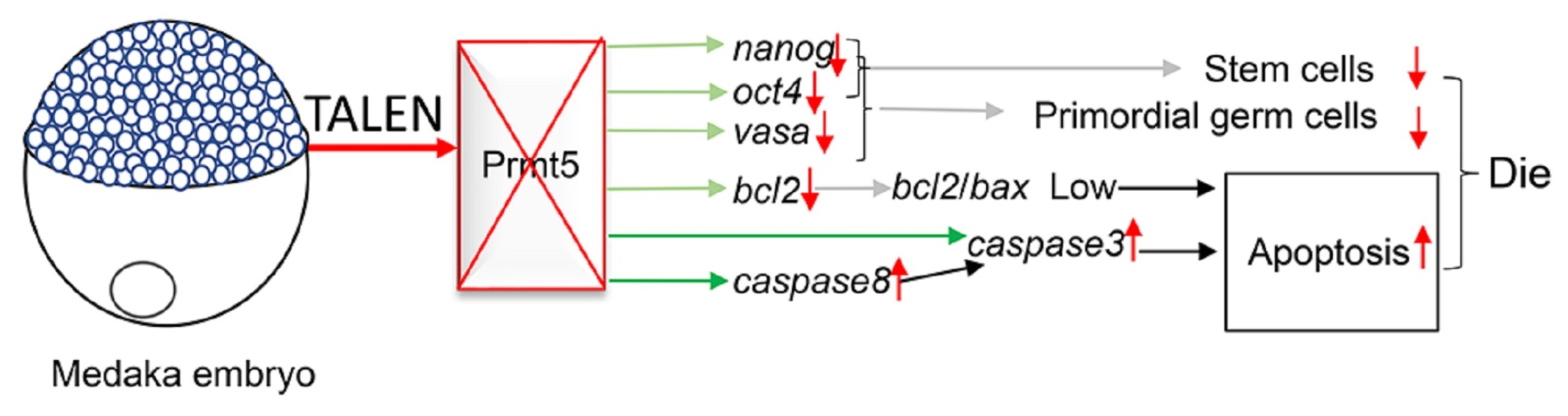
2.3. Gene Editing by TALENs

The specific TALEN target sites were identified online using TALEN Targeter (https://tale-nt.cac.cornell.edu/). The target sites were selected with the same criteria as described by Huang et al. [31,32]. The left and right TALEN targets, Tale-L and Tale-R, respectively, of medaka prmt5 are GGCGTCCGCCAGTA and TCATATCTCTCCCG (Figure 1). The gene-specific TALEN constructs were assembled using the Unit Assembly method [31,32]. The two TALEN backbones, pCS2-C-PEAS and pCS2-C-PERR, and the four single-unit vectors, pA, pT, pC, and pG, were gifted from Dr. Bo Zhang (Peking University, Beijing, China). The final constructs were confirmed by sequencing.
The 5′-capped mRNA was produced by in vitro transcription from the constructed TALEN plasmids using SP6 mMESSAGE mMACHINE Kit as per the protocol provided by the manufacturer (ThermoFisher Scientific, Shanghai, China). The concentration of the transcribed mRNA was determined by NanoDrop 2000 (ThermoFisher Scientific). The left and right TALEN mRNA of 120 pg each were co-injected into the cytoplasm of one-cell stage embryos of medaka by microinjection with a MPPI-3 pressure injector (Applied Scientific Instrumentation, Eugene, OR, USA).

2.4. Genomic DNA Extraction and Polymerase Chain Reaction (PCR)

The genomic DNA (gDNA) was obtained using the Universal Genomic DNA Kit (Cwbio, Beijing, China) according to the protocol provided by the manufacturer. The caudal fin was sampled after anesthesia of the fish with MS-222 (Sigma-Aldrich, Merck KGaA, Darmstadt, Germany). The sample DNA was used in PCR after extraction.
The PCR reaction was performed in a volume of 25 μL with 2 μL DNA, 1 μL each primer pair, dNTPs, buffer, and LaTaq (Takara, Beijing, China). The primers for medaka prmt5 were prmt5F and prmt5R (Table 1). The program for PCR was denaturing at 94 °C 3 min, 30 cycles of 94 °C 30 s, 56 °C 30 s, and 72 °C 30 s, and a final incubation at 72 °C 10 min.

2.5. Detection of Mutation by T7E1 (T7 Endonuclease I) or Sequencing

After the PCR reaction and purification by running on an agarose gel and gel extraction, the mutation of prmt5 was detected by T7E1 digestion or sequencing.
For T7E1 detection, the purified PCR products were incubated with a T7E1 buffer in a tube at 95 °C in a water bath for 5 min. The T7E1 enzyme (New England Biolabs, Beijing, China) was added to the tube after cooling at room temperature. The tube was mixed well and incubated at 37 °C for 30 min. The mixture was electrophoresed on an agarose gel of 2%.
For sequencing, the purified PCR products were sequenced directly and separately for each sample. The purified PCR products were also subcloned into a pGEMT-easy vector (Promega, Beijing, China). Three colonies were selected randomly for sequencing after ligation, a transformation of E. coli, and incubation on an X-Gal (5-Bromo-4-chloro-3-indolyl β-D-galactopyranoside) LB (Luria Broth) agar plate with ampicillin.

2.6. Detection of the Gene Expression by Quantitative RT-PCR

The total RNA from the embryos was extracted using an Ultrapure RNA kit (CoWin Biosciences, Beijing, China) following the protocol provided by the manufacturer. The cDNA (complementary DNA) was synthesized according to the protocol of the FastQuant RT kit (Tiangen Biotech, Beijing, China).
The quantitative RT-PCR (qPCR) of the triplicate samples was performed with a CFX96 real-time PCR detection system (BioRad Laboratories, Hercules, CA, USA) in a volume of 20 μL containing template cDNA, primers, and 2× SuperReal Pre Mix Plus kit (Tiangen). The cycling program was 95 °C 2 min followed by 39 cycles of 95 °C 10 s, 62 °C 30 s, and 65 °C 30 s. The relative expression of the genes in the samples was calibrated/normalized against RPS18 (ribosomal protein S18) using the 2−ΔΔCt method [33]. The primers are shown in Table 1. RPS18 was used as the internal control [34].

2.7. Statistical Analysis

Statistical analysis was performed with SPSS Statistics (IBM, Armonk, NY, USA). The gene expression values were calculated and described as mean ± standard errors with at least three independent experiments (n ≥ 3). The differences among the treatments were calculated by one-way analysis of variance (ANOVA).

3. Results

3.1. Mutation of Medaka Prmt5

After microinjection of the left and right TALEN mRNA into the zygotes, the embryos were incubated until four days post-fertilization (dpf). The live embryos at this stage were collected for detection of the editing efficiency. Twelve embryos were collected and detected by sequencing after DNA extraction, PCR, ligation into the pGEMT-easy vector, and transformation of E. coli. In totally, 20 colonies were selected and sequenced. Eight colonies were identified with insertion and deletion (indel) (Figure 1), which was also confirmed by the T7E1 method. The mutation efficiency was roughly 40%. The result showed that these TALEN constructs could efficiently induce indels in the target gene prmt5 of the medaka embryos.
The larvae from the microinjected embryos were cultivated into adults as F0. In total, 7 of 20 F0 fish were checked with different indels. The mutated fish with the reading shift mutation of prmt5 were mated with the wild-type fish to produce F1 fish. The F1 fish were checked individually using the T7E1 method and/or sequencing (Figure 2). The F1 fish with the deletion of four base pairs (bp) in the prmt5 gene were selected. Deletion of the four bp caused a reading frame shift and premature stopping of prmt5’s translation (Figure 1). The mutated prmt5 may produce a peptide of 34 amino acids (AA), and this peptide loses the main functional domains of Prmt5, PRMT5_TIM (TIM barrel domain), PRMT5 (PRMT5 arginine-N-methyltransferase), and PRMT5_C (PRMT5 oligomerization domain) (https://www.ncbi.nlm.nih.gov/cdd).
Male and female F1 fish with the four bp deletion in the prmt5 gene were mated to produce the homozygous F2. Surprisingly, about 30% (245/819, dead/total numbers) of the F2 embryos died during embryonic development (Table 2 and Figure 3). No homozygous mutated fish were detected in the adult F2 fish. So, the heterozygous F2 fish were mated with either wild type or heterozygous F2 fish to produce the next generation. The fish were checked and mated generation after generation until F12. Unfortunately, no homozygous mutant fish were obtained. Only the heterozygous fish were cultivated.
The mutant offspring embryos died before or/and after the gastrula stage (Table 2 and Figure 3). Some mutant offspring developed beyond the gastrula stage with minor eyes, abnormal body axis, or retarded development during organogenesis (Figure 3). Compared with the control embryos, the death rate (43/177, 245/819) of the F2-F3 offspring was significantly higher. It was found that 18.1–19.9% (32/177, 163/819) of the embryos died from the zygote to the gastrula stage. We considered whether the homozygous offspring died early or died during organogenesis. However, 83.3% (335/401) of the F8 embryos died in the gastrula stage due to some unknown reasons. The most inbred embryos of the heterozygous fish developed beyond the gastrula stage in F9-F11; the homozygous mutant could have died during organogenesis because no homozygote was obtained.
The genotypes of the mutant offspring at adulthood were checked. Because no homozygotes of the mutant were identified, the homozygotes were assumed to be dead. Theoretically, the heterozygotes should be 2/3, and the wild type should be 1/3. The heterozygotes and the wild-type numbers were not significantly different from the expected offspring values (Table 3). This confirmed the hypothesis that the homozygotes of the prmt5 mutant died during development.

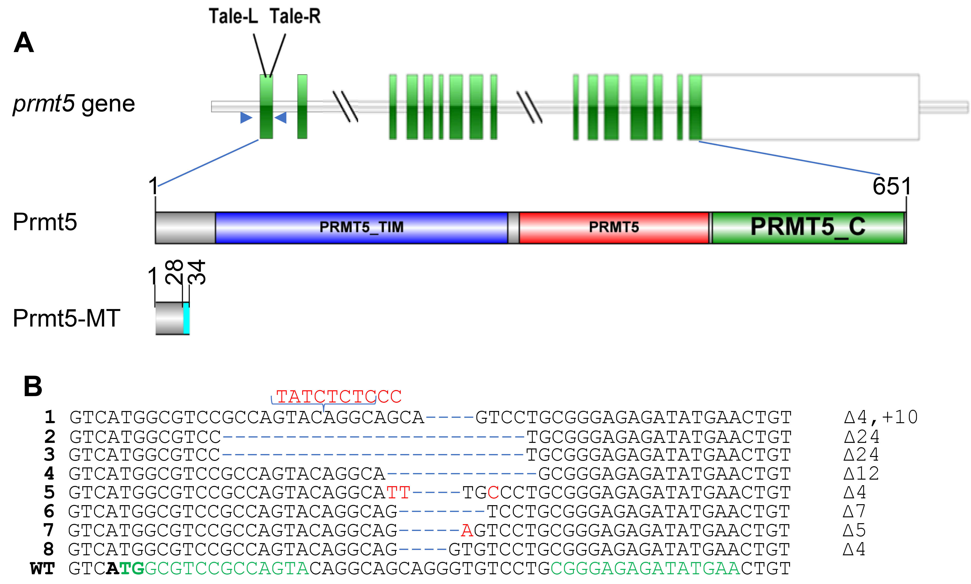
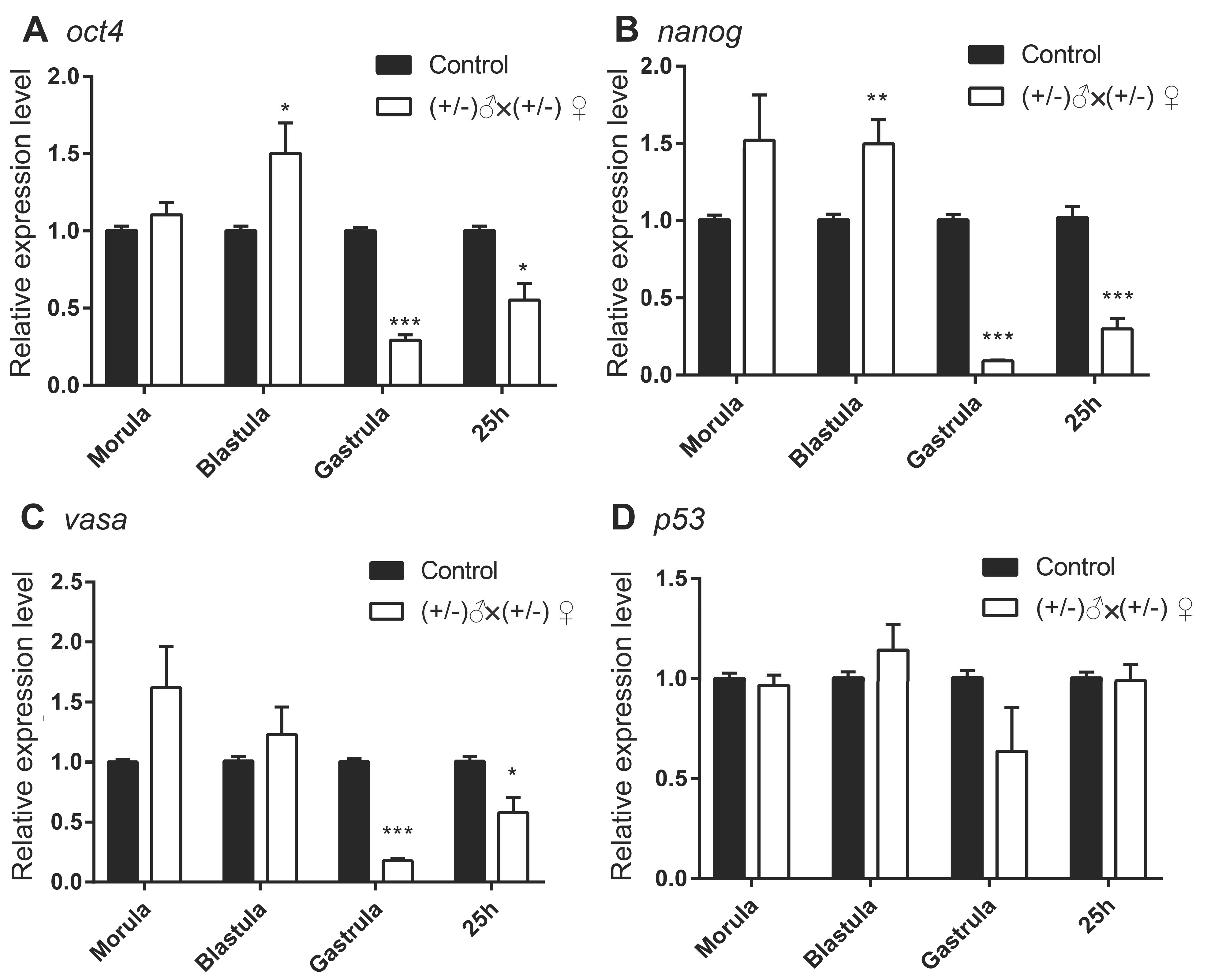
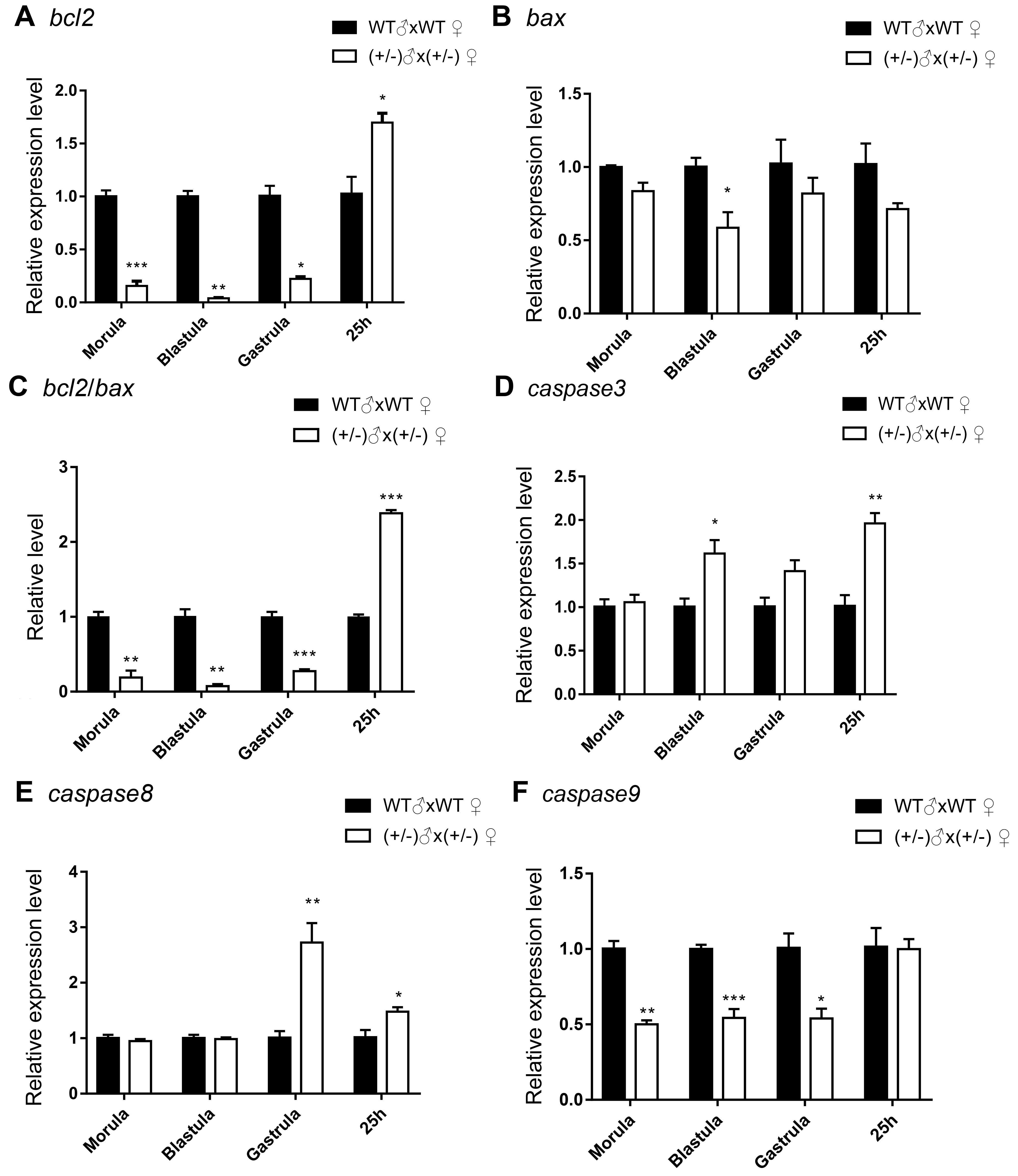
3.2. Gene Expression of the Inbred Offspring of the Heterozygous Mutant during Embryogenesis
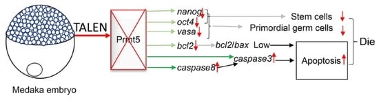
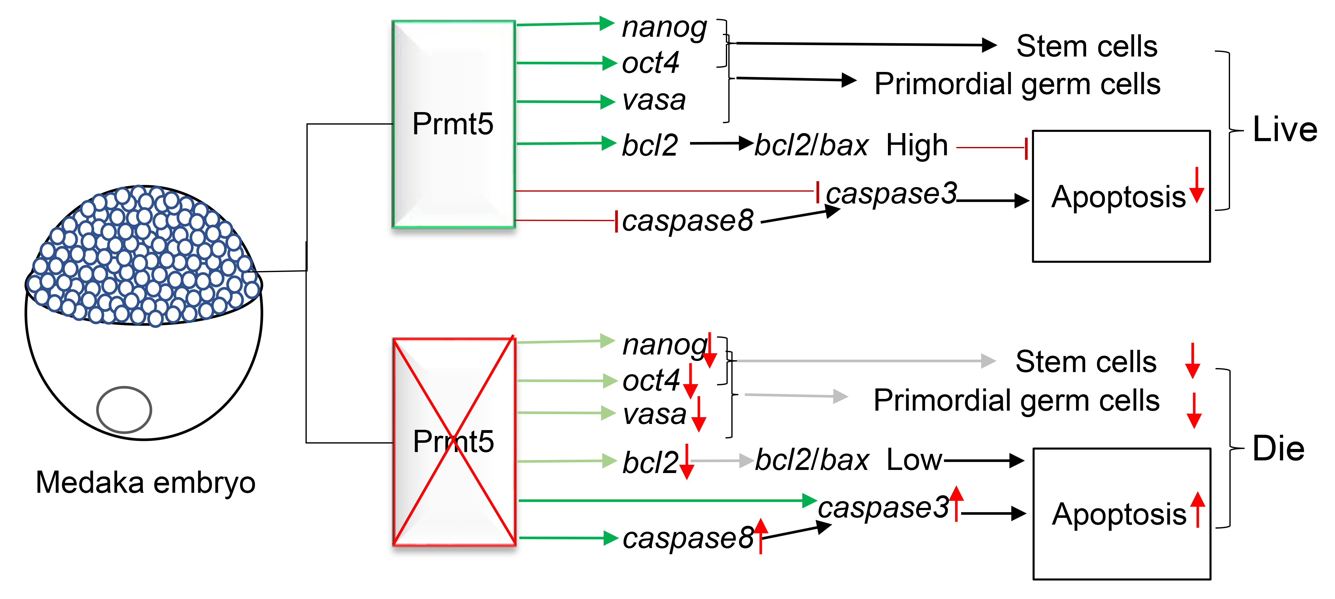
Several important genes were selected and detected in the embryos by qPCR to understand the reasons for the homozygous decease. The selected genes were oct4, nanog, and vasa related to ES cells and PGCs [35,36,37,38], p53 [39], bcl2, bax [40], caspase3 [41], caspase8 [42], and caspase9 [43] related to apoptosis or cell death. In addition, because of lethal homozygotes, the inbred offspring of the heterozygotes were collected and checked.
Compared with the expression levels in the wild type embryos, oct4 and nanog in the mutant offspring were higher at the blastula stage, but decreased significantly at the gastrula stage and 25 h post-fertilization (hpf) (Figure 4). In addition, vasa was significantly decreased at the gastrula and 25 hpf in the mutant offspring too. However, the p53 level in the mutant offspring was not different from that in the control (Figure 4).
Because the expression of p53 was not different in the mutant and control offspring, the expression of bcl2, bax, caspase3, caspase8, and caspase9 was checked (Figure 5). Comparing the expression in the control, the bcl2 was decreased in the morula to blastula, but increased in 25 hpf; the bax was decreased only in the blastula. The pattern of the ratio of bcl2/bax was similar to that of the bcl2 expression. The ratio was low from the morula to gastrula, but was high in 25 hpf in the mutant offspring. The caspase3 level increased in the blastula stage and 25 hpf. The caspase8 level increased in the gastrula stage to 25 hpf, but caspase9 levels were decreased in the morula to gastrula stage.

4. Discussion

In this study, the medaka prmt5 gene was mutated by gene editing using TALENs. The mutation of prmt5 resulted in a premature stopping mutation, which caused loss of function in the Prmt5 protein in homozygous offspring. As a result, the homozygotes of prmt5 mutant died during embryogenesis. Moreover, the genes related to stemness and apoptosis were down- or upregulated. These results suggest that prmt5 is necessary for the embryonic development of medaka by regulating cell stemness and apoptosis.
The mutation of prmt5 seriously affects embryogenesis through its effects on stem cells. A defect in Prmt5 is fatal for embryos in mice [11]. Prmt5 is essential for maintaining the ES cells by repressing the differentiation genes [12,13]. Contrarily, Prmt5 remarkably promotes the generation of induced pluripotent stem (iPS) cells from somatic cells in mice [13] and goats, Capra hircus [44]. Prmt5 promotes the culture of human iPS cells [45] and regulates the proliferation of human ES cells by increasing the expression of the G1 cell cycle inhibitor P57 [46]. Our results are similar to previous findings. The mutation of prmt5 caused homozygous death, and no homozygote was obtained in an extended period. The loss of Prmt5 decreased the genes related to PGCs and ES cells, such as nanog, oct4, and vasa. Oct4 and nanog are two factors expressed in the ES cells and PGCs of the medaka [37,47,48,49,50]. Although oct4 and nanog were increased in the blastula stage, these two factors decreased significantly in the mutant offspring from gastrula to 25 hpf compared with that in the control. A decrease in oct4 and nanog indicated that the stem cells were differentiated or dead in the embryos.
Along with oct4 and nanog, the germ cell marker, vasa, was also decreased in the mutant offspring. This result indicates the loss of PGCs in the offspring with a deficiency of prmt5. A conditional loss of Prmt5 in early PGCs caused complete male and female sterility in mice [20]. Prmt5 is necessary for H2A/H4R3me2s chromatin modification to repress long interspersed repetitive element (Line) 1 and intracisternal A particles (Iap) transposons in mouse PGCs. Prmt5 also participates in transposon silencing through the Piwi-interacting RNA (piRNA) pathway [20]. In zebrafish, the mutation of prmt5 caused a loss of germ cells by apoptosis [28]. Mutating prmt5 in zebrafish induces a decrease in vasa and zili (zebrafish piwi like) [28]. These results indicate a conserved function of prmt5 in germ cells. Medeka’s oct4 and nanog are also expressed in germ cells [37,47,48,49]. Medaka Nanog mediates PGC migration by regulating the expression of cxcr4b [49]. The disappearance of PGCs may be as a result of deficits in migration and apoptosis.
Prmt5 plays a significant role in organogenesis. The homozygous mutant of prmt5 may die in the early stages before gastrulation or at the late stages during organogenesis in medaka. The mutation of prmt5 decreases the expression of oct4 in the gastrula stage. A decrease in oct4 interferes with gastrulation, central nervous system development, and angiogenesis in medaka embryos [51]. In mice, Prmt5 regulates glial cell differentiation [52]. Prmt5 is essential for maintaining chondrogenic progenitor cells in the limb bud and for forming distinct cartilage identities in the knee and long bone in mice [53,54]. Prmt5 is also needed for myogenesis [14,27] and regulates muscle stem cells [55]. The mutation of prmt5 may also affect immune responses, such as the cholesterol biosynthesis-mediated Th17 responses [56]. Prmt5 is necessary for B cell development by preventing p53-dependent and p53-independent blocks [57], and is required for T cell survival and proliferation [58]. A variety of effects may contribute to the homozygous death in the prmt5 mutant of medaka.
Prmt5 is a survival factor. Prmt5 negatively regulates DNA damage-induced apoptosis in Caenorhabditis elegans [7]. Inactivation of C. elegans Prmt5 induces excessive apoptosis in germline upon irradiation by a CEP-1 (C. elegans p53 homolog)-dependent up-regulation of the cell death initiator EGL-1 (egl eg, g-l aying defective) [7]. In mice, a deficiency in Prmt5 causes cell-cycle arrest in G1. However, Prmt5 promotes p53 expression, and the induction of p53 targets MDM2 (mouse double minute 2) and p21 upon DNA damage [5]. Growth suppression mediated upon prmt5 knockdown is independent of p53, but is dependent on eIF4E [5]. Contrarily, Prmt5 down-regulates p53 and enhances iPS cell generation from dairy goat embryonic fibroblasts [44]. In this study, p53 expression was not affected in the prmt5 mutant offspring of medaka. This hints that the effect of prmt5 on the expression of p53 is different between species.
Bcl2 and Bax are a pair of factors for apoptosis and cell death [59,60]. Bcl2 functions as a repressor of cell death and Bax is a pro-apoptotic factor. The ratio of Bcl2/Bax determines the survival or death of cells following an apoptotic stimulus [59,60]. In the prmt5 mutant offspring, bcl2 was decreased from morula to blastula, and the ratio of bcl2/bax appeared in the same pattern. These results suggest a possible mechanism of prmt5 on cell survival through the regulation of bcl2 and the ratio of bcl2/bax in medaka embryos.
The caspases are aspartate-specific cysteine proteases (reviewed by Galluzzi et al.) [61]. The activation and function of caspases can be regulated by various molecules, such as the Bcl2 family proteins (reviewed by Fan et al.) [62]. The caspase cascade plays vital roles in apoptotic signaling. In the caspase family, caspase3 is an apoptosis executioner, and caspase8 and caspase9 are two apoptosis activators or initiators [61,62]. Caspase8 and caspase3 were upregulated in the blastula, gastrula, and 25 hpf in the prmt5 mutant offspring. However, the expression of caspase9 was decreased in the prmt5 mutant offspring from the morula to gastrula. These results suggest that Prmt5 can regulate the expression of the caspases in the embryos and that embryonic death is induced by apoptosis through caspase8 and caspase3 cascade after the mutation of prmt5.
In this study, the expression of the genes related to stemness, PGCs, and apoptosis was studied in the offspring embryos of the heterozygous prmt5 mutant fish due to homozygous death. The results are not the same as those in the homozygotes. To further study the role of prmt5 in medaka fish, conditional editing must be applied. However, the results from the offspring of the heterozygotes can provide some rough information to understand the functions of prmt5 in medaka embryos.
Taken together, prmt5 is necessary for embryonic development in medaka. The mutation of prmt5 can induce apoptosis in the embryos by decreasing bcl2/bax and increasing caspase 8 and caspase3. In addition, the mutation of prmt5 causes a decrease in the stemness of ES cells and PGCs through down-regulation of oct4, nanog, and vasa. A decrease in the stem cells and increase in cell apoptosis results in death of the embryos in the homozygous prmt5 mutant (Figure 6).

5. Conclusions

Prmt5 is necessary for embryonic development in medaka. A decrease in the stem cells and increase in cell apoptosis results in death of the embryos in the homozygous prmt5 mutant.

Author Contributions

Q.Z., X.Z. and H.Z.: conceptualization and designing the experiment; X.L., S.D., Q.Y. (Qing Yang), X.M., Z.L., Q.Y. (Qiting Yao), W.H., K.W., P.C. and G.F.: experimental set up and execution; X.L., Q.Y. (Qiting Yao) and M.C.: data analysis; X.L. and H.Z.: manuscript writing. All authors have read and agreed to the published version of the manuscript.

Funding

This work was supported by the Natural Science Foundation of China (grant no. 31672284, 31272645 to H.Z.) and the Open Fund of Hubei Key Laboratory of Environmental and Health Effects of Persistent Toxic Substances (no. PTS2021-01 to QZ).

Institutional Review Board Statement

This study was carried out according to recommendations in the Regulation for the Management of Laboratory Animals of China’s Ministry of Science and Technology of China. The animal protocol for this study was approved by the Animal Care and Use Committee of Hubei Province in China (No. SYXK(E)2015-0012).

Data Availability Statement

The data that support the findings of this study are available from the corresponding author upon reasonable request.

Conflicts of Interest

The authors declare no conflict of interest.

References

  1. Bedford, M.T.; Clarke, S.G. Protein arginine methylation in mammals: Who, what, and why. Mol. Cell 2009, 33, 1–13. [Google Scholar] [CrossRef] [PubMed] [Green Version]
  2. Zhao, Q.; Rank, G.; Tan, Y.T.; Li, H.; Moritz, R.L.; Simpson, R.J.; Cerruti, L.; Curtis, D.J.; Patel, D.J.; Allis, C.D.; et al. PRMT5-mediated methylation of histone H4R3 recruits DNMT3A, coupling histone and DNA methylation in gene silencing. Nat. Struct. Mol. Biol. 2009, 16, 304–311. [Google Scholar] [CrossRef] [PubMed] [Green Version]
  3. Boisvert, F.M.; Cote, J.; Boulanger, M.C.; Cleroux, P.; Bachand, F.; Autexier, C.; Richard, S. Symmetrical dimethylarginine methylation is required for the localization of SMN in Cajal bodies and pre-mRNA splicing. J. Cell Biol. 2002, 159, 957–969. [Google Scholar] [CrossRef] [Green Version]
  4. Neuenkirchen, N.; Chari, A.; Fischer, U. Deciphering the assembly pathway of Sm-class U snRNPs. FEBS Lett. 2008, 582, 1997–2003. [Google Scholar] [CrossRef] [Green Version]
  5. Scoumanne, A.; Zhang, J.; Chen, X. PRMT5 is required for cell-cycle progression and p53 tumor suppressor function. Nucleic Acids Res. 2009, 37, 4965–4976. [Google Scholar] [CrossRef] [PubMed] [Green Version]
  6. Jansson, M.; Durant, S.T.; Cho, E.C.; Sheahan, S.; Edelmann, M.; Kessler, B.; La Thangue, N.B. Arginine methylation regulates the p53 response. Nat. Cell Biol. 2008, 10, 1431–1439. [Google Scholar] [CrossRef]
  7. Yang, M.; Sun, J.; Sun, X.; Shen, Q.; Gao, Z.; Yang, C. Caenorhabditis elegans protein arginine methyltransferase PRMT-5 negatively regulates DNA damage-induced apoptosis. PLoS Genet. 2009, 5, e1000514. [Google Scholar] [CrossRef] [Green Version]
  8. Chung, J.; Karkhanis, V.; Tae, S.; Yan, F.; Smith, P.; Ayers, L.W.; Agostinelli, C.; Pileri, S.; Denis, G.V.; Baiocchi, R.A.; et al. Protein arginine methyltransferase 5 (PRMT5) inhibition induces lymphoma cell death through reactivation of the retinoblastoma tumor suppressor pathway and polycomb repressor complex 2 (PRC2) silencing. J. Biol. Chem. 2013, 288, 35534–35547. [Google Scholar]
  9. Nicholas, C.; Yang, J.; Peters, S.B.; Bill, M.A.; Baiocchi, R.A.; Yan, F.; Sïf, S.; Tae, S.; Gaudio, E.; Wu, X.; et al. PRMT5 is upregulated in malignant and metastatic melanoma and regulates expression of MITF and p27(Kip1). PLoS ONE 2013, 8, e74710. [Google Scholar] [CrossRef]
  10. Richard, S.; Morel, M.; Cléroux, P. Arginine methylation regulates IL-2 gene expression: A role for protein arginine methyltransferase 5 (PRMT5). Biochem. J. 2005, 388, 379–386. [Google Scholar] [CrossRef] [Green Version]
  11. Krause, C.D.; Yang, Z.H.; Kim, Y.S.; Lee, J.H.; Cook, J.R.; Pestka, S. Protein arginine methyltransferases: Evolution and assessment of their pharmacological and therapeutic potential. Pharmacol. Ther. 2007, 113, 50–87. [Google Scholar] [CrossRef] [PubMed]
  12. Tee, W.W.; Pardo, M.; Theunissen, T.W.; Yu, L.; Choudhary, J.S.; Hajkova, P.; Surani, M.A. Prmt5 is essential for early mouse development and acts in the cytoplasm to maintain ES cell pluripotency. Genes Dev. 2010, 24, 2772–2777. [Google Scholar] [CrossRef] [PubMed] [Green Version]
  13. Nagamatsu, G.; Kosaka, T.; Kawasumi, M.; Kinoshita, T.; Takubo, K.; Akiyama, H.; Sudo, T.; Kobayashi, T.; Oya, M.; Suda, T. A germ cell-specific gene, Prmt5, works in somatic cell reprogramming. J. Biol. Chem. 2011, 286, 10641–10648. [Google Scholar] [CrossRef] [PubMed] [Green Version]
  14. Dacwag, C.S.; Ohkawa, Y.; Pal, S.; Sif, S.; Imbalzano, A.N. The protein arginine methyltransferase Prmt5 is required for myogenesis because it facilitates ATP-dependent chromatin remodeling. Mol. Cell. Biol. 2007, 27, 384–394. [Google Scholar] [CrossRef] [Green Version]
  15. Mallappa, C.; Hu, Y.J.; Shamulailatpam, P.; Tae, S.; Sif, S.; Imbalzano, A.N. The expression of myogenic microRNAs indirectly requires protein arginine methyltransferase (Prmt)5 but directly requires Prmt4. Nucleic Acids Res. 2011, 39, 1243–1255. [Google Scholar] [CrossRef]
  16. Kirino, Y.; Kim, N.; de Planell-Saguer, M.; Khandros, E.; Chiorean, S.; Klein, P.S.; Rigoutsos, I.; Jongens, T.A.; Mourelatos, Z. Arginine methylation of Piwi proteins catalysed by dPRMT5 is required for Ago3 and Aub stability. Nat. Cell Biol. 2009, 11, 652–658. [Google Scholar] [CrossRef] [Green Version]
  17. Gonsalvez, G.B.; Rajendra, T.K.; Tian, L.; Matera, A.G. The Sm-protein methyltransferase, Dart5, is essential for germ-cell specification and maintenance. Curr. Biol. 2006, 16, 1077–1089. [Google Scholar] [CrossRef] [Green Version]
  18. Anne, J.; Ollo, R.; Ephrussi, A.; Mechler, B.M. Arginine methyltransferase Capsuleen is essential for methylation of spliceosomal Sm proteins and germ cell formation in Drosophila. Development 2007, 134, 137–146. [Google Scholar] [CrossRef] [Green Version]
  19. Li, Z.; Yu, J.; Hosohama, L.; Nee, K.; Gkountela, S.; Chaudhari, S.; Cass, A.A.; Xiao, X.; Clark, A.T. The Sm protein methyltransferase PRMT5 is not required for primordial germ cell specification in mice. EMBO J. 2015, 34, 748–758. [Google Scholar] [CrossRef] [Green Version]
  20. Kim, S.; Günesdogan, U.; Zylicz, J.J.; Hackett, J.A.; Cougot, D.; Bao, S.; Lee, C.; Dietmann, S.; Allen, G.E.; Sengupta, R.; et al. PRMT5 protects genomic integrity during global DNA demethylation in primordial germ cells and preimplantation embryos. Mol. Cell 2014, 56, 564–579. [Google Scholar] [CrossRef] [Green Version]
  21. Wang, Y.; Zhu, T.; Li, Q.; Liu, C.; Han, F.; Chen, M.; Zhang, L.; Cui, X.; Qin, Y.; Bao, S.; et al. Prmt5 is required for germ cell survival during spermatogenesis in mice. Sci. Rep. 2015, 5, 11031. [Google Scholar] [CrossRef]
  22. Ancelin, K.; Lange, U.C.; Hajkova, P.; Schneider, R.; Bannister, A.J.; Kouzarides, T.; Surani, M.A. Blimp1 associates with Prmt5 and directs histone arginine methylation in mouse germ cells. Nat. Cell Biol. 2006, 8, 623–630. [Google Scholar] [CrossRef]
  23. Vagin, V.V.; Wohlschlegel, J.; Qu, J.; Jonsson, Z.; Huang, X.; Chuma, S.; Girard, A.; Sachidanandam, R.; Hannon, G.J.; Aravin, A.A. Proteomic analysis of murine Piwi proteins reveals a role for arginine methylation in specifying interaction with Tudor family members. Genes Dev. 2009, 23, 1749–1762. [Google Scholar] [CrossRef] [PubMed] [Green Version]
  24. Hung, C.M.; Li, C. Identification and phylogenetic analyses of the protein arginine methyltransferase gene family in fish and ascidians. Gene 2004, 340, 179–187. [Google Scholar] [CrossRef] [PubMed]
  25. Chen, W.; Cao, M.; Yang, Y.; Nagahama, Y.; Zhao, H. Expression pattern of prmt5 in adult fish and embryos of medaka, Oryzias latipes. Fish Physiol. Biochem. 2009, 35, 325–332. [Google Scholar] [CrossRef] [PubMed]
  26. Wang, Y.C.; Li, C. Evolutionarily conserved protein arginine methyltransferases in non-mammalian animal systems. FEBS J. 2012, 279, 932–945. [Google Scholar] [CrossRef]
  27. Batut, J.; Duboé, C.; Vandel, L. The methyltransferases PRMT4/CARM1 and PRMT5 control differentially myogenesis in zebrafish. PLoS ONE 2011, 6, e25427. [Google Scholar] [CrossRef]
  28. Zhu, J.; Zhang, D.; Liu, X.; Yu, G.; Cai, X.; Xu, C.; Rong, F.; Ouyang, G.; Wang, J.; Xiao, W. Zebrafish prmt5 arginine methyltransferase is essential for germ cell development. Development 2019, 146, dev179572. [Google Scholar] [CrossRef] [Green Version]
  29. Cheng, N.; Guo, M.; Chang, P.; Zhang, X.; Zhang, R.; Qi, C.; Zhong, X.; Zhou, Q.; Zhao, H. Expression of mep50 in adult and embryos of medaka fish (Oryzias latipes). Fish Physiol. Biochem. 2016, 42, 1053–1061. [Google Scholar] [CrossRef]
  30. Shen, H.; Zhang, X.; Al Hafiz, M.A.; Liang, X.; Yao, Q.; Guo, M.; Xu, G.; Zhong, X.; Zhou, Q.; Zhao, H. The proteins interacting with Prmt5 in medaka (Oryzias latipes) identified by yeast two-hybridization. Protein Pept. Lett. 2020, 27, 971–978. [Google Scholar] [CrossRef]
  31. Huang, P.; Xiao, A.; Zhou, M.; Zhu, Z.; Lin, S.; Zhang, B. Heritable gene targeting in zebrafish using customized TALENs. Nat. Biotechnol. 2011, 29, 699–700. [Google Scholar] [CrossRef] [PubMed]
  32. Huang, P.; Xiao, A.; Tong, X.; Zu, Y.; Wang, Z.; Zhang, B. TALEN construction via “Unit Assembly” method and targeted genome modifications in zebrafish. Methods 2014, 69, 67–75. [Google Scholar] [CrossRef] [PubMed]
  33. Livak, K.J.; Schmittgen, T.D. Analysis of relative gene expression data using real-time quantitative PCR and the 2−ΔΔCT Method. Methods 2001, 25, 402–408. [Google Scholar] [CrossRef] [PubMed]
  34. Zhao, H.; Zhang, X.; Cheng, N.; Duan, J.; Wang, J.; Nagahama, Y.; Zhong, X.; Zhou, Q.; Wang, Y. Identification and expression profiles of prdm1 in medaka Oryzias latipes. Mol. Biol. Rep. 2014, 41, 617–626. [Google Scholar] [CrossRef] [PubMed]
  35. Shinomiya, A.; Tanaka, M.; Kobayashi, T.; Nagahama, Y.; Hamaguchi, S. The vasa-like gene, olvas, identifies the migration path of primordial germ cells during embryonic body formation stage in the medaka, Oryzias latipes. Dev. Growth Differ. 2000, 42, 317–326. [Google Scholar] [CrossRef]
  36. Camp, E.; Sánchez-Sánchez, A.V.; García-España, A.; Desalle, R.; Odqvist, L.; Enrique O’Connor, J.; Mullor, J.L. Nanog regulates proliferation during early fish development. Stem Cells 2009, 27, 2081–2091. [Google Scholar] [CrossRef]
  37. Wang, D.; Manali, D.; Wang, T.; Bhat, N.; Hong, N.; Li, Z.; Wang, L.; Yan, Y.; Liu, R.; Hong, Y. Identification of pluripotency genes in the fish medaka. Int. J. Biol. Sci. 2011, 7, 440–451. [Google Scholar] [CrossRef] [Green Version]
  38. Shen, J.; Yokota, S.; Yokoi, H.; Suzuki, T. Diethylnitrosamine-induced expression of germline-specific genes and pluripotency factors, including vasa and oct4, in medaka somatic cells. Biochem. Biophys. Res. Commun. 2016, 478, 858–863. [Google Scholar] [CrossRef]
  39. Krause, M.K.; Rhodes, L.D.; Van Beneden, R.J. Cloning of the p53 tumor suppressor gene from the Japanese medaka (Oryzias latipes) and evaluation of mutational hotspots in MNNG-exposed fish. Gene 1997, 189, 101–106. [Google Scholar] [CrossRef]
  40. Barjhoux, I.; Gonzalez, P.; Baudrimont, M.; Cachot, J. Molecular and phenotypic responses of Japanese medaka (Oryzias latipes) early life stages to environmental concentrations of cadmium in sediment. Environ. Sci. Pollut. Res. Int. 2016, 23, 17969–17981. [Google Scholar] [CrossRef]
  41. Iijima, N.; Yokoyama, T. Apoptosis in the medaka embryo in the early developmental stage. Acta Histochem. Cytochem. 2007, 40, 1–7. [Google Scholar] [CrossRef] [PubMed]
  42. Sakamaki, K.; Nozaki, M.; Kominami, K.; Satou, Y. The evolutionary conservation of the core components necessary for the extrinsic apoptotic signaling pathway, in medaka fish. BMC Genom. 2007, 8, 141. [Google Scholar] [CrossRef] [Green Version]
  43. Lee, J.W.; Choi, Y.C.; Kim, R.; Lee, S.K. Multiwall carbon nanotube-induced apoptosis and antioxidant gene expression in the gills, liver, and intestine of Oryzias latipes. BioMed Res. Int. 2015, 2015, 485343. [Google Scholar] [CrossRef] [PubMed] [Green Version]
  44. Chu, Z.; Niu, B.; Zhu, H.; He, X.; Bai, C.; Li, G.; Hua, J. PRMT5 enhances generation of induced pluripotent stem cells from dairy goat embryonic fibroblasts via down-regulation of p53. Cell Prolif. 2015, 48, 29–38. [Google Scholar] [CrossRef] [PubMed]
  45. Goyal, A.; Chavez, S.L.; Pera, R.A.R. Generation of human induced pluripotent stem cells using epigenetic regulators reveals a germ cell-like identity in partially reprogrammed colonies. PLoS ONE 2013, 8, e82838. [Google Scholar] [CrossRef] [PubMed]
  46. Gkountela, S.; Li, Z.; Chin, C.J.; Lee, S.A.; Clark, A.T. PRMT5 is required for human embryonic stem cell proliferation but not pluripotency. Stem Cell Rev. Rep. 2014, 10, 230–239. [Google Scholar] [CrossRef] [Green Version]
  47. Hong, Y.; Winkler, C.; Liu, T.; Chai, G.; Schartl, M. Activation of the mouse Oct4 promoter in medaka embryonic stem cells and its use for ablation of spontaneous differentiation. Mech. Dev. 2004, 121, 933–943. [Google Scholar] [CrossRef]
  48. Sánchez-Sánchez, A.V.; Camp, E.; García-España, A.; Leal-Tassias, A.; Mullor, J.L. Medaka Oct4 is expressed during early embryo development, and in primordial germ cells and adult gonads. Dev. Dyn. 2010, 239, 672–679. [Google Scholar] [CrossRef]
  49. Sánchez-Sánchez, A.V.; Camp, E.; Leal-Tassias, A.; Atkinson, S.P.; Armstrong, L.; Díaz-Llopis, M.; Mullor, J.L. Nanog regulates primordial germ cell migration through Cxcr4b. Stem Cells 2010, 28, 1457–1764. [Google Scholar] [CrossRef]
  50. Liu, R.; Li, M.; Li, Z.; Hong, N.; Xu, H.; Hong, Y. Medaka Oct4 is essential for pluripotency in blastula formation and ES cell derivation. Stem Cell Rev. Rep. 2015, 11, 11–23. [Google Scholar] [CrossRef]
  51. Sun, B.; Gui, L.; Liu, R.; Hong, Y.; Li, M. Medaka oct4 is essential for gastrulation, central nervous system development and angiogenesis. Gene 2020, 733, 144270. [Google Scholar] [CrossRef] [PubMed]
  52. Huang, J.; Vogel, G.; Yu, Z.; Almazan, G.; Richard, S. Type II arginine methyltransferase PRMT5 regulates gene expression of inhibitors of differentiation/DNA binding Id2 and Id4 during glial cell differentiation. J. Biol. Chem. 2011, 286, 44424–44432. [Google Scholar] [CrossRef] [PubMed] [Green Version]
  53. Norrie, J.L.; Li, Q.; Co, S.; Huang, B.L.; Ding, D.; Uy, J.C.; Ji, Z.; Mackem, S.; Bedford, M.T.; Galli, A.; et al. PRMT5 is essential for the maintenance of chondrogenic progenitor cells in the limb bud. Development 2016, 143, 4608–4619. [Google Scholar] [CrossRef] [PubMed] [Green Version]
  54. Ramachandran, J.; Liu, Z.; Gray, R.S.; Vokes, S.A. PRMT5 is necessary to form distinct cartilage identities in the knee and long bone. Dev. Biol. 2019, 456, 154–163. [Google Scholar] [CrossRef] [PubMed]
  55. Zhang, T.; Günther, S.; Looso, M.; Künne, C.; Krüger, M.; Kim, J.; Zhou, Y.; Braun, T. Prmt5 is a regulator of muscle stem cell expansion in adult mice. Nat. Commun. 2015, 6, 7140. [Google Scholar] [CrossRef] [Green Version]
  56. Webb, L.M.; Sengupta, S.; Edell, C.; Piedra-Quintero, Z.L.; Amici, S.A.; Miranda, J.N.; Bevins, M.; Kennemer, A.; Laliotis, G.; Tsichlis, P.N.; et al. Protein arginine methyltransferase 5 promotes cholesterol biosynthesis-mediated Th17 responses and autoimmunity. J. Clin. Investig. 2020, 130, 1683–1698. [Google Scholar] [CrossRef]
  57. Litzler, L.C.; Zahn, A.; Meli, A.P.; Hébert, S.; Patenaude, A.M.; Methot, S.P.; Sprumont, A.; Bois, T.; Kitamura, D.; Costantino, S.; et al. PRMT5 is essential for B cell development and germinal center dynamics. Nat. Commun. 2019, 10, 22. [Google Scholar] [CrossRef] [Green Version]
  58. Tanaka, Y.; Nagai, Y.; Okumura, M.; Greene, M.I.; Kambayashi, T. PRMT5 is required for T cell survival and proliferation by maintaining cytokine signaling. Front. Immunol. 2020, 11, 621. [Google Scholar] [CrossRef]
  59. Korsmeyer, S.J.; Shutter, J.R.; Veis, D.J.; Merry, D.E.; Oltvai, Z.N. Bcl-2/Bax: A rheostat that regulates an anti-oxidant pathway and cell death. Semin. Cancer Biol. 1993, 4, 327–332. [Google Scholar]
  60. Korsmeyer, S.J. Regulators of cell death. Trends Genet. 1995, 11, 101–105. [Google Scholar] [CrossRef]
  61. Galluzzi, L.; López-Soto, A.; Kumar, S.; Kroemer, G. Caspases connect cell-death signaling to organismal homeostasis. Immunity 2016, 44, 221–231. [Google Scholar] [CrossRef] [PubMed] [Green Version]
  62. Fan, T.J.; Han, L.H.; Cong, R.S.; Liang, J. Caspase family proteases and apoptosis. Acta Biochim. Biophys. Sin. (Shanghai) 2005, 37, 719–727. [Google Scholar] [CrossRef] [PubMed]
Figure 1. (A) Schematic structure of medaka prmt5 gene and proteins. The blocks indicate the exons. The open reading frame (ORF) is shown in green. The arrowheads present the position of the primers used for screening. The target sites of the Tale-L and Tale-R are shown. The full protein of Prmt5 in the length of 651 AA is shown with the functional domains. Prmt5-MT presents the mutant protein with 34 AA in length. The gray fragment is the same as it is in the full protein. The cyan fragment is deduced from the mutant sequence. (B) Alignment of the prmt5 sequences in the founder embryos indicating the insertion and deletion (indel) after microinjection of the TALEN constructs. WT, wild type. The green letters indicate the left and right TALEN targets. The dash or Δ indicate the deletion, the red or + indicate the insertion. The start codon is in bold.
Fishes 08 00019 g001
Figure 2. (A) A gel document presents the screening of the mutant by the T7E1 method. M, marker; WT, wild type; P, positive control; T1-T8 indicate the sample 1–8. The sizes of the marker and the fragments are indicated in bp beside the document. (B) The typical chromatogram of the partial sequence of prmt5. WT, wild type; MT, mutant. The deletion of the 4 bp in the wild type is highlighted in the red frame.
Fishes 08 00019 g002
Figure 3. The embryos of the control and the offspring of the heterozygous fish at different stages (3 hpf and 4 dpf). The framed embryos are shown in enlarged pictures. The mutant offspring could have died in 3 hpf, developed slowly with minor or pigment-less eyes, abnormal body axis, and/or headless in 4 dpf. Scale bar, 1000 or 250 μm.
Fishes 08 00019 g003
Figure 4. The relative expression of oct4 (A), nanog (B), vasa (C), and p53 (D) in the embryos from the morula stage to 25 hpf. The expression of each gene in the control at the same stage was set as 1. The asterisks show the significances. *, p< 0.05; **, p < 0.01; ***, p < 0.001.
Fishes 08 00019 g004
Figure 5. The relative expression of bcl2 (A), bax (B), caspase3 (D), caspase8 (E), and caspase9 (F), and the ratio of bcl2/bax (C) in the embryos from the morula stage to 25 hpf. The expression of each gene in control was set as 1 at the same stage. The asterisks show the significances. *, p < 0.05; **, p < 0.01; ***, p < 0.001.
Fishes 08 00019 g005
Figure 6. A diagram indicating the functions of Prmt5 in medaka embryos. In the wild type, Prmt5 is functional (in green frame) to maintain the nanog, oct4, and vasa expression in the stem and primordial germ cells. Prmt5 promotes the expression of bcl2, which results in a high bcl2/bax ratio and represses the expression of caspase8 and caspase3 to prevent apoptosis. So, the embryos of the wild type are live. The contrary events occurred in the homozygotes of the prmt5 mutant (in red frame) and the homozygous mutant embryos died.
Fishes 08 00019 g006
Table 1. The primers used in the experiments.
GenePrimerSequence (5′-3′)
prmt5prmt5FACCTGTAGCTGTTAATTTGATCTGC
prmt5RGAATCCAAAAGCAGAGCATCCT
β-actinβ-actinFCACACCTTCTACAATGAGCTG
β-actinRCCAGATCTGCTGGAAGGTGG
oct4oct4-qFTCTTTGGCGTAAACTCGTCTCA
oct4-qRCTTGCGTAAAACCCAAAGTGAT
nanognanog-qFTACTCCAAACGCCCCGAAAG
nanog-qRGTGTCCTTCTGATGCCTCCTAA
vasavasa-qFGCTCATCAACCAGATTTACCA
vasa-qRATCTCCCTCATCTGGTAGCCG
p53p53-qFTCTACAAGAAGACGGAGCACG
p53-qRACTGTAACACTCTGCCTTTTGGTAT
bcl2bcl2-qFTCGACAGTTTTCCCCTGCAA
bcl2-qRGAAACCCCCTGAACGGAACT
baxbax-qFGCGATCAAGGTAGCGAAAAAT
bax-qRCAGGTTTCTCCTGACCCGTT
caspase3caspase3-qFAACAAGACGCCGACCCTTAC
caspase3-qRTGTACCGTTACGAGGACCCA
caspase8caspase8-qFTGACCCTACCCTTTCCCAGT
caspase8-qRCACTTTGGGCTAGTGTGCCT
caspase9caspase9-qFAACTTCGTGGTGGAAGTCCG
caspase9-qRAGCTCCCAGCTGATCTGATCTT
RPS18s18-qFGTGTGGTGACCATCATGCAGAA
s18-qRTGGCAAGGACCTGGCTGTATT
Table 2. The death rate of the embryos in different stages and different generations.
GenerationPrmt5 (+/−) × (+/−)Wild Typep Value
All StagesTo GastrulaAll StagesTo Gastrula
F229.9%19.9%4.2%-2.3 × 10⁻²⁷
F324.3%18.1%5.7%-3.1 × 10⁻⁵
F8-83.3%-6.9%1.9 × 10−101
F9-2.4%-3.5%0.31
F10-2.4%-2.7%0.84
F11-1.9%-2.8%0.97
Table 3. The genotypes of the offspring with mutated prmt5.
Mating PatternThe Offspring Tested(+/−)(+/+)(−/−)p Value
F2 (+/−) × WT421626-0.12
F2 (+/−) × F2 (+/−)43281500.83
F3 (+/−) × F3( +/−)43251800.24
WT × F8 (+/−)331419-0.38
F9 (+/−) × F9 (+/−)46262000.14
F11 (+/−) × F11 (+/−)48331500.76
Total255142113-0.81
Note: WT, wild type. The p value was obtained by the Chi-square test with Excel, and the offspring of prmt5 (−/−) were expected not to survive.
Disclaimer/Publisher’s Note: The statements, opinions and data contained in all publications are solely those of the individual author(s) and contributor(s) and not of MDPI and/or the editor(s). MDPI and/or the editor(s) disclaim responsibility for any injury to people or property resulting from any ideas, methods, instructions or products referred to in the content.

