
On-Chip Liquid Chromatography


Graduate School of Pharmaceutical Sciences, University of Tokyo, Tokyo 1130033, Japan
Encyclopedia 2022, 2(1), 617-624; https://doi.org/10.3390/encyclopedia2010041
Submission received: 19 February 2022
/
Accepted: 3 March 2022
/
Published: 15 March 2022
(This article belongs to the Section Chemistry)
Definition
:On-chip liquid chromatography (LC) refers to LC technology that is miniaturized to fit on a microchip to enable fast, high-throughput analysis, with small sample volumes and low reagent consumption. Four different on-chip LC approaches have been developed to date: use of open-tubular, packed-particle, monolithic, and pillar array columns. These methods have been applied to proteomics as well as the analysis of small molecules and drugs in various biological samples. Recent advances in on-chip LC are summarized herein.
1. Introduction
Liquid chromatography (LC) is an indispensable separation technique with many biological, clinical, environmental, forensic, and pharmaceutical applications. Various stationary phases can be used for LC, and this technique provides highly reproducible results. In omics research, such as proteomics and metabolomics, LC coupled to mass spectrometry (LC-MS) is an effective analytical method for highly sensitive and accurate mass determination. However, it is still difficult to achieve satisfactory separation, particularly in complex mixtures such as biological samples (tissue, plasma, serum, and urine). In metabolomics, thousands of metabolites must be measured simultaneously. Furthermore, some important biological compounds are present only in small amounts, along with high concentrations of other endogenous compounds that may present interference. These challenges can be overcome through LC miniaturization.
In recent years, micro total analysis systems (microTAS), also known as lab-on-a-chip, have emerged as an attractive area of research. In a microTAS, sampling, sample transport, necessary chemical reactions, separation, and detection are all performed on a chip. Separation is one of the key processes in a microTAS. Therefore, extensive research has been conducted to realize on-chip separation using miniaturized LC.
Previous studies have examined on-chip separation using electrophoresis or electrochromatography. However, there are relatively few examples of chip-based LC. Four different on-chip LC approaches have been developed to date: use of open-tubular, packed-particle, monolithic, and pillar array columns (Figure 1). These techniques enable fast analysis, while offering the advantages of high throughput, very small sample volumes, and low reagent consumption. Each method is described in the following section.
2. Approaches
2.1. Open-Tubular Chromatography
Open-tubular chromatography is the simplest way to realize chromatography on a chip and is the first published example of on-chip LC; this technique was first reported by Manz et al. [1]. In their study, an open-tubular column with a length of 15 cm, width of 6 µm, and depth of 2 µm was fabricated, but no separations were attempted.
The channel wall of the chip was directly modified and used as the stationary phase to obtain the desired retention capacity. A notable advantage of open-tubular chromatography is the low backpressure. A device with an integrated micropump that employs sodium-silicate-based sol–gel membranes for generating pressure-driven flow was employed for reverse-phase LC separation of three rhodamine B derivatives. Separation was achieved in 60 s in an open-tubular channel [5]. An open-tubular column coated with a C8 stationary phase and pressure-driven gated injection were used to achieve baseline separation of phenol and 2,6-dimethylphenol; vitamin A and vitamin E were also partially separated [6].
The main disadvantage of this method is the low column capacity due to the low surface-to-volume ratio. To increase the capacity of open-tubular columns, the specific surface area of the stationary phase must be increased. Although not chip-based, a capillary application was used to increase the relevant surface area by introducing an open-tubular monolithic porous polymer layer through ultraviolet photoinitiated fabrication [7].
2.2. Packed-Particle Columns
In conventional LC, most columns are packed with particles. In on-chip LC, conventional-type stationary phases are transferred to the microfluidic chip. Hence, on-chip LC systems packed with particles represent one of the more straightforward options for achieving separation. Pre-prepared particles can flow into the microchannel on a chip, resulting in the formation of a packed bed. Numerous particle types with different modifications, e.g., C8, C18, ion exchange, hydrophilic interaction chromatography (HILIC), are available; these particles can be used to develop particle-packed chips. These particle-packed columns have a much larger specific surface area than open-tubular columns.
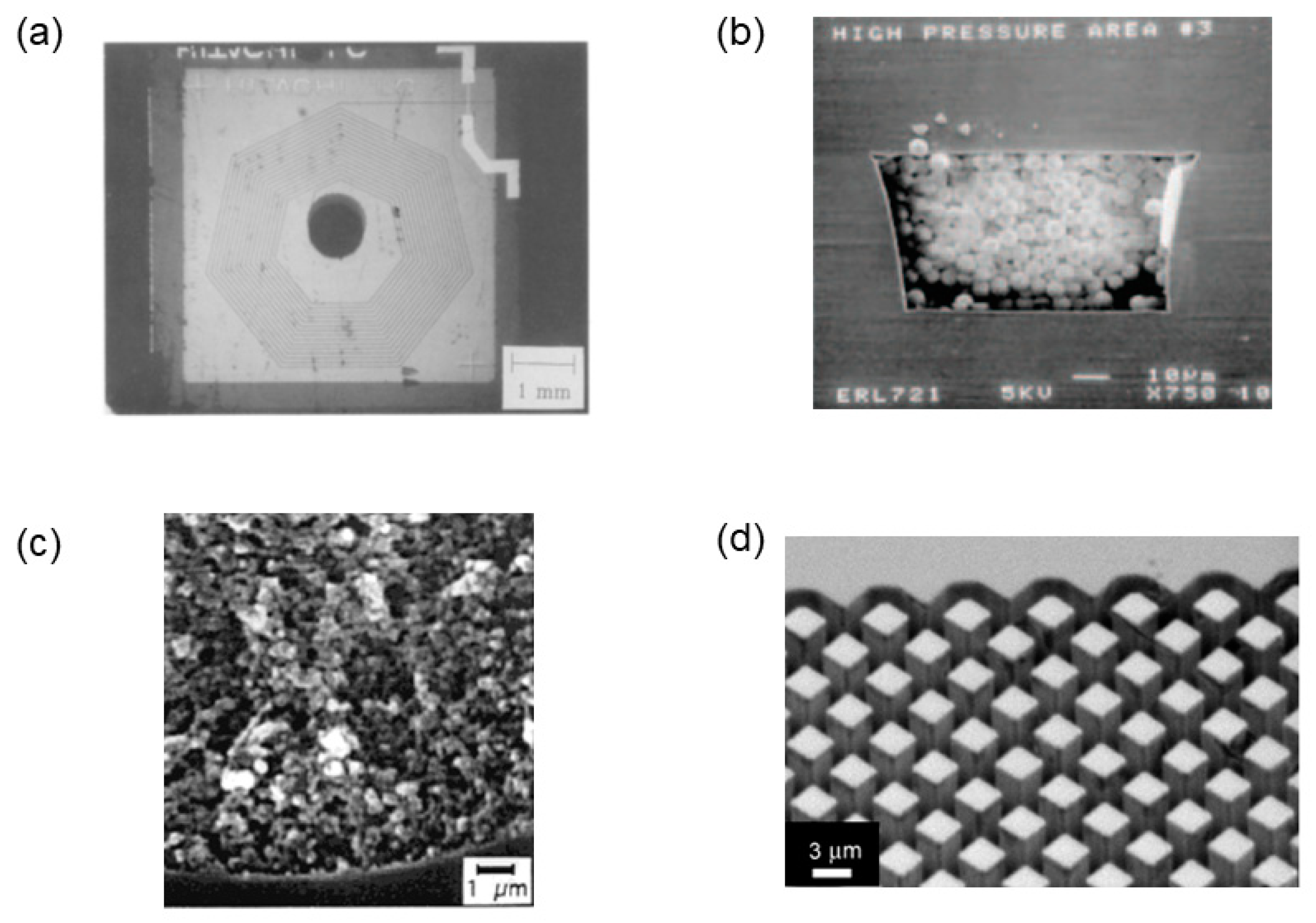
On-chip cation-exchange chromatography was used to separate hemoglobin A1c (HbA1c), one of the most important marker proteins for diabetes [8]. The chip was packed with cation exchange beads (particle diameter 30 μm), which could separate HbA1c from other Hbs.
The first commercially available chips were packed with particles. However, they have only been used for specific equipment configurations, for example, in combination with a mass spectrometer. The packed particles are required to eliminate the dead volumes that result from the connection between the column and the mass spectrometer.
The Agilent LC particle-packed chip includes an on-chip microvalve, an enrichment column, a packed-particle analytical column, and a nanospray emitter on a single multilayer polymer chip [2]. It has been extensively utilized for proteome research and for analyzing small molecules such as 7-aminoflunitrazepam in human urine, 8-isoprostaglandin F2α in human urine, hepcidin (a peptide marker of clinical disorders linked to iron metabolism) in human plasma, pharmaceutical drugs covering a wide range of hydrophilicities, fluoxetine, and norfluoxetine in rat serum, illicit drugs and metabolites in human hair, and 7-ethyl-10-hydroxycamptothecin (SN38) in mouse plasma.
The ionKey and iKey (Waters) chips are ceramic. These chips contain 1.7 μm silica-particles packed in a 150 or 300 μm microchannel, an electrical column heater, a nebulization gas channel, and a post-column mobile-phase-modifier channel. The post-column makeup-flow design allows the post-column addition of solvents to enhance the electrospray ionization and increase the sensitivity of the mass spectrometer. An Ionkey/MS system was used to quantify infliximab in rat serum [9]. Compared to conventional LC, the sensitivity was higher. An online analysis system for evaluating intestinal absorption and the formation of unknown products was developed by coupling a flow-through Transwell system with an online iKeyChip-QTOF-MS system [10].
cHiPLC (SCIEX (Eksigent)) is another commercially available chip-based platform, which acts as a “docking station” for up to three quartz microfluidic chips. The system’s flexible design and built-in 10-port nanovalve allow for easy switching between different modes, such as direct injection and trap loading. cHiPLC was recently used to investigate LGALS3BP/90 K glycoprotein in the cerebrospinal fluid of patients with neurological diseases [11].
The performance of particle-packed on-chip LC devices depends critically on the packing conditions of the particles. Because the chip channels are not cylindrical, as in a conventional LC column, and the pressure applied for packing is limited, it is challenging to achieve reproducible packing as in conventional LC columns. Some studies have shown that for particle-packed on-chip LC columns, high bed densities are critical for achieving good separation performance in noncylindrical packed beds [12], and that the desired high packing densities can be much more easily achieved using 3 μm particles than with the use of 5 μm particles [13].
The preparation of packed beds in chips requires the installation of packed particles in the microchannels. A common method for immobilizing the particles is to construct a frit structure inside the chip channels by the microfabrication of bottleneck-shaped channels or by synthesizing thin monolith layers. Porous polymeric monoliths have been utilized as retaining elements in common slurry-packing procedures [14]. By using laser-assisted photopolymerization, the frits can be precisely defined to have lengths less than 100 μm.
Particle-packed LC chips were recently reviewed in detail elsewhere [15].
2.3. Monoliths
Monoliths consist of a single rod of porous material that is easily synthesized in situ by a polymerization reaction. Unlike particle-packed columns, monoliths do not require frits and have high permeability. Compared to open-tubular columns, monoliths have a higher specific surface area and greater column capacity.
Solutions containing monomers and functional groups can be introduced into the microchannels for thermally or via photoinduced polymerization, resulting in a highly porous material. Ericson et al. first demonstrated anion-exchange chromatography of proteins by filling narrow channels with continuous polymer beds [3]. A monolithic nano-porous polymer chip was fabricated by packing the channels with poly(methyl acrylate) monoliths [16]. The monolith consisted of a number of globes, with a diameter of approximately 2 μm. Neurotransmitters, 5-hydroxyindole-3-acetic acid (5-HIAA) and 5-hydroxytryptamine (serotonin), were successfully separated.
Ion-exchange chromatographic separation of protein-isoforms and sensitive immunoassay detection were combined in a porous nitrocellulose monolith chip [17]. The chip could discriminate between transferrin isoforms differing by only 0.1 pH units in pI and was used to specifically quantify the separated transferrin isoforms.
The use of a reverse-phase polymethacrylate monolith (15 cm long) as the separation column and a 5 mm long methacrylate monolith as the solid-phase extraction (SPE) element for sample cleanup and enrichment enabled the separation of fluorescein-labeled model peptides [18]. Online removal of free fluorescein was achieved using the on-chip SPE column, resulting in a 150-fold improvement in the sensitivity.
A chip-based polymer monolithic column was coupled with a valveless gated sample injection method [19]. Two fluorescently labeled amines (butylamine and cyclohexylamine) were successfully separated in less than 220 s with a column length of 1 cm. The valveless gated sample injection method enabled nanoliter-scale sample injection without the need for a mechanical valve.
A microfluidic device was designed for comprehensive spatial two-dimensional LC [20]. A hydrophobic methacrylate ester-based monolithic stationary phase was polymerized in situ in the second-dimension channels. In addition, a photomask was used to localize monolith formation in the parallel second-dimension channels. The pore and globule sizes were in the range of 0.8–1.5 and 1–2 μm, respectively.
Recently, 3D printing technology has been used to fabricate LC chips. Selective laser melting was used to fabricate titanium-based LC columns [21,22]. In-column thermal polymerization of mechanically stable poly(BuMA-co-EDMA) monoliths was achieved, without any structural changes. The monolith in the complex internal channels (~0.9 mm I.D. and 600 mm length) was found to be mechanically stable, and was used to separate intact proteins, including the A and B components of β-lactoglobulin and peptides, from complex mixtures.
Considering its practical uses, the main disadvantage is column-to-column reproducibility because the polymerization reaction cannot be completely controlled. In addition, the overall size of the monolithic beds often decreases after polymerization.
2.4. Pillar Array
A pillar array column can also be used to achieve LC separation on a chip. Before introducing pillar array columns, the theoretical aspects of LC should be considered. According to Giddings’s theory, three processes contribute to band dispersion, and the plate height can be expressed as the sum of three terms. The reduced plate height depends on the reduced velocity, as shown in the following equation:
where A, B, and C are constants, h is the reduced plate height (plate height divided by dp, i.e., the particle diameter of the packing material), and v is the reduced velocity (expressed as udp/Dm, where u is the mobile phase linear velocity and Dm is the diffusion coefficient of the analyte in the mobile phase). The terms A, B, and C relate to flow in interparticle spaces, axial diffusion, and slow equilibration between the mobile and stationary phases, respectively. Knox pointed out that A-term dispersion is by far the most important phenomenon to consider, and that this feature is limited when using particle-packed columns due to packing technology constraints [23]. He also stated that much lower h values could be obtained by decreasing the A-term dispersion, if the stationary phase was constructed to be highly ordered; for example, by micromachining the entire column.
h = A + B/v + Cv
In 1998, Regnier et al. first introduced pillar array columns (referred to as collocated monolith support structures (COMOSS)) [24]. They reported that direct fabrication with photolithography using deep-reactive ion-etching techniques could be used to simultaneously position and define pillars. However, such columns have only been evaluated in electro-driven chromatography.
The first successful pressure-driven LC separation using pillar array columns was performed by Desmet et al. [25]. Nonporous silicon pillars with a diameter of approximately 4.4 μm were fabricated and covalently coated with a hydrophobic monolayer to enable reverse-phase LC separations. The separation of three dyes (C440, C460, and C480) was achieved in 3 s. A plate height of ~4 μm, which is much smaller than those of conventional LC columns, was obtained under weak retention conditions. This result clearly shows that the ordered structure of the stationary phase can enhance the separation performance of the column.
However, there is a limit to the length of the linear flow path that can be fabricated on a small substrate, making multicomponent analysis of biomolecules difficult. To realize better separation, a long separation column with a turn structure is required. Tapered turn structures with equal distances between the inner and outer peripheries (low-dispersion turn) were found to be effective for preventing turn-induced sample dispersion [4]. With the low-dispersion-turn geometry, a pillar array column with a width of 400 μm and a column length of 110 mm was fabricated on a 20 × 20 mm microchip. Under non-retained conditions, the solute bands obtained for fluorescent dyes remained almost unchanged, even after passing through the low-dispersion turns. Two coumarin dyes (C525 and C545) were well resolved under reverse-phase conditions, and a maximum theoretical plate number of 8000 was obtained. Branched-chain amino acids (valine, isoleucine, and leucine), other amino acids (phenylalanine and tyrosine), and keto acids in human plasma samples could be quantified, demonstrating for the first time that pillar array columns can be applied to the analysis of biological samples [26,27,28]. When biological compounds were quantified using the pillar array columns, the relative standard deviation of the peak intensity was high (>30%) because the injection volume of the sample was of the order of nanoliters. Adding an internal standard improved the reproducibility [26]. The feasibility of quantitative analysis of samples using pillar array columns with the help of an internal standard was demonstrated. Furthermore, an automated sample injection system for pillar array columns was developed to maintain a constant sample injection volume [29]. The system was composed of two pumps (one for the sample and another for the mobile phase), a six-way valve that can be controlled by a PC, and an autosampler.
The turn structure described above has a high flow resistance due to the narrow channel width of the turn, making it difficult to achieve high-speed separation at high flow rates. Hence, a new turn structure with low dispersion and low flow resistance (pillar-distribution controlled (PDC) turn) was designed as a constant-radius turn, filled with octagonal pillars that were arranged to control the linear velocity of the mobile phase in the radial direction [30]. Upon introducing the PDC turn structure into the pillar array columns, separation at high flow rates became possible, and five fluorescently derivatized amino acids (proline, valine, 6-aminocaproic acid, leucine, and phenylalanine), could be separated within 24 s. The PDC turn structure offers the advantages of longer pillar array columns for the fast analysis of complex samples.
However, analysis of biological compounds using a single mobile phase is still difficult as biological samples are very complex and the polarity of the components exhibit large differences. In conventional LC, this problem is solved by utilizing a gradient elution system that can accelerate the elution of strongly retained solutes. Hence, a gradient elution system with pillar array columns was developed [31]. The essential component of the gradient elution system is the mixer, which is used to mix two kinds of mobile phases. A cross-Tesla structure with a 3 mm mixing length was effective for mixing two liquids. The gradient elution system using a cross-Tesla mixer and pillar array columns improved the separation efficiency of the pillar array columns, as evidenced by the gradient chromatographic separation of the fluorescent derivatives of four aliphatic amines (pentylamine, hexylamine, heptylamine, and octylamine) within 110 s.
To increase the retention of nonporous pillar array columns, pillars with thick layers of mesoporous silicon were produced [32]. A 550 nm thick porous layer on the pillar array generated by anodization increased the available surface area by at least two orders of magnitude. Sol–gel synthesis of silica monoliths with the pillar array column technology afforded porous-shell coated pillars [33]. The thickness of the porous layer on the 2.4 μm pillars was 480 nm, with relatively large through-pores of 2.2 µm. A minimal plate height of 3.9 μm was obtained under non-retained conditions. Another approach involves the direct growth of carbon nanotubes on the pillar surface [34]. The retention was improved relative to that of pillar array columns grafted with an octadecyl stationary phase only.
Two pillar array columns (200 and 50 cm in length) are currently commercially available from PharmaFluidics. The chip can be connected to any micro/nano LC system with a connector through a capillary tube. These chips are applied to proteomics and lipidomics studies. Very recently, pillar array columns (200 cm) coupled with mass spectrometers were used to identify 340,000 proteins, doubling the number of proteins identified hitherto [35]. With the pillar array system, the coefficient of variation for the peptide retention times was improved 2.5-fold, and high interlaboratory reproducibility was achieved compared to conventional LC. Moreover, pillar array columns (50 cm) were effective for separating some permethylated glycan isomers [36].
3. Conclusions
In this entry, the recent advances in the development of on-chip LC were reviewed. Thirty years have elapsed since the first example of a chip-based LC device. Since then, four different on-chip LC approaches have been developed: open-tubular, packed-particle, monolithic, and pillar array. These methods have been applied to proteomics, as well as the analysis of small molecules and drugs in various biological samples. These devices enable fast analysis, with the advantages of high throughput, very small sample volumes, and low reagent consumption. Pillar array columns also afford highly efficient separation, overcoming the limitations of the conventional column, and may be helpful for omics studies such as metabolomics and proteomics, which require the separation of many kinds of compounds.
On-chip chromatography technology still remains largely in the basic research stage, although some devices based on particle-packed and pillar array columns are commercially available. Further advancements, described below, can be expected.
The main separation mode for on-chip LC is currently the reverse-phase mode. Other separation modes such as ion exchange, HILIC, and size exclusion can be developed for analyzing many kinds of biomolecules, including protein and DNA. User-friendly systems including pumps, injectors, and detectors should be developed. Currently, commercially available on-chip LC systems are connected to MS. Chip-based LC-MS does not require connections between the column and mass spectrometer, which reduces extra column band broadening. Other detection approaches coupled with on-chip LC are interesting alternatives to MS. For micro-scale fluids, reducing the channel size and increasing the column length significantly increase the back pressure. Hence, new kinds of substrate materials should be developed using microfabrication technology such as 3D printing technology. MicroTAS using on-chip LC is also important for enabling new applications in a variety of research areas. In pharmaceutical analysis, MicroTAS using on-chip LC is not just faster and more efficient than LC separation, but also enables automation of the entire analytical process. Hence, this technique can overcome the limitations of current pharmaceutical analyses. On-chip LC is expected to find extensive applications due to its portability, where application to clinical diagnosis is one of the important examples. In clinical diagnosis, on-chip LC can be performed in hospitals and even at the bed-side.
Funding
This research was funded in part by grants from the Japan Society for the Promotion of Science (JSPS; grant numbers 17K08234 and 21H02613).
Conflicts of Interest
The author declares no conflict of interest.
Entry Link on the Encyclopedia Platform
References
- Manz, A.; Miyahara, Y.; Miura, J.; Watanabe, Y.; Miyagi, H.; Sato, K. Design of an open-tubular column liquid chromatograph using silicon chip technology. Sens. Actuators B Chem. 1990, 1, 249–255. [Google Scholar] [CrossRef]
- Yin, H.F.; Killeen, K.; Brennen, R.; Sobek, D.; Werlich, M.; van de Goor, T. Microfluidic chip for peptide analysis with an integrated HPLC column, sample enrichment column, and nanoelectrospray tip. Anal. Chem. 2005, 77, 527–533. [Google Scholar] [CrossRef] [PubMed]
- Ericson, C.; Holm, J.; Ericson, T.; Hjertén, S. Electroosmosis- and pressure-driven chromatography in chips using continuous beds. Anal. Chem. 2000, 72, 81–87. [Google Scholar] [CrossRef]
- Aoyama, C.; Saeki, A.; Noguchi, M.; Shirasaki, Y.; Shoji, S.; Funatsu, T.; Mizuno, J.; Tsunoda, M. Use of folded micromachined pillar array column with low-dispersion turns for pressure-driven liquid chromatography. Anal. Chem. 2010, 82, 1420–1426. [Google Scholar] [CrossRef] [PubMed]
- Toh, G.M.; Corcoran, R.C.; Dutta, D. Sodium silicate based sol–gel structures for generating pressure-driven flow in microfluidic channels. J. Chromatogr. A 2010, 1217, 5004–5011. [Google Scholar] [CrossRef] [PubMed]
- Schlund, M.; Gilbert, S.E.; Schnydrig, S.; Renaud, P. Continuous sampling and analysis by on-chip liquid/solid chromatography. Sens. Actuators B 2007, 123, 1133–1141. [Google Scholar] [CrossRef]
- Collins, D.A.; Nesterenko, E.P.; Brabazon, D.; Paull, B. Controlled ultraviolet (UV) photoinitiated fabrication of monolithic porous layer open tubular (monoPLOT) capillary columns for chromatographic applications. Anal. Chem. 2012, 84, 3465–3472. [Google Scholar] [CrossRef]
- Tanaka, T.; Izawa, K.; Okochi, M.; Lim, T.-K.; Watanabe, S.; Harada, M.; Matsunaga, T. On-chip type cation-exchange chromatography with ferrocene-labeled anti-hemoglobin antibody and electrochemical detector for determination of hemoglobin A1c level. Anal. Chim. Acta 2009, 638, 186–190. [Google Scholar] [CrossRef]
- Kleinnijenhuis, A.J.; Ingola, M.; Toersche, J.H.; van Holthoon, F.L.; van Dongen, W.D. Quantitative bottom up analysis of infliximab in serum using protein A purification and integrated μLC-electrospray chip IonKey MS/MS technology. Bioanalysis 2016, 8, 891–904. [Google Scholar] [CrossRef]
- Santbergen, M.J.C.; van der Zande, M.; Gerssen, A.; Bouwmeester, H.; Nielen, M.W.F. Dynamic in vitro intestinal barrier model coupled to chip-based liquid chromatography mass spectrometry for oral bioavailability studies. Anal. Bioanal. Chem. 2020, 412, 1111–1122. [Google Scholar] [CrossRef]
- Costa, J.; Pronto-Laborinho, A.; Pinto, S.; Gromicho, M.; Bonucci, S.; Tranfield, E.; Correia, C.; Alexandre, B.M.; de Carvalho, M. Investigating LGALS3BP/90 K glycoprotein in the cerebrospinal fluid of patients with neurological diseases. Sci. Rep. 2020, 10, 5649. [Google Scholar] [CrossRef] [PubMed]
- Ehlert, S.; Kraiczek, K.; Mora, J.A.; Dittmann, M.; Rozing, G.P.; Tallarek, U. Separation efficiency of particle-packed HPLC microchips. Anal. Chem. 2008, 80, 5945–5950. [Google Scholar] [CrossRef] [PubMed]
- Jung, S.; Höltzel, A.; Ehlert, S.; Mora, J.A.; Kraiczek, K.; Dittmann, M.; Rozing, G.P.; Tallarek, U. Impact of conduit geometry on the performance of typical particulate microchip packings. Anal. Chem. 2009, 81, 10193–10200. [Google Scholar] [CrossRef]
- Thurmann, S.; Mauritz, L.; Heck, C.; Belder, D. High-performance liquid chromatography on glass chips using precisely defined porous polymer monoliths as particle retaining elements. J. Chromatogr. A 2014, 1370, 33–39. [Google Scholar] [CrossRef] [PubMed]
- Kecskemeti, A.; Gasper, A. Particle-based liquid chromatographic separations in microfluidic devices—A review. Anal. Chim. Acta 2018, 1021, 1–19. [Google Scholar] [CrossRef] [PubMed]
- Kim, J.-H.; O’Hare, D. Monolithic nano-porous polymer in microfluidic channels for lab-chip liquid chromatography. Nano Converg. 2018, 5, 19. [Google Scholar] [CrossRef] [PubMed]
- Lönnberg, M.; Carlsson, J. Lab-on-a-chip technology for determination of protein isoform profiles. J. Chromatogr. A. 2006, 1127, 175–182. [Google Scholar] [CrossRef] [PubMed]
- Liu, J.; Chen, C.-F.; Tsao, C.-W.; Chang, C.-C.; Chu, C.-C.; DeVoe, D.L. Polymer microchips integrating solid-phase extraction and high-performance liquid chromatography using reversed-phase polymethacrylate monoliths. Anal. Chem. 2009, 81, 2545–2554. [Google Scholar] [CrossRef]
- Wang, X.-L.; Zhu, Y.; Fang, Q. Valveless gated injection for microfluidic chip-based liquid chromatography system with polymer monolithic column. J. Chromatogr. A 2012, 1246, 123–128. [Google Scholar] [CrossRef]
- Wouters, B.; De Vos, J.; Desmet, G.; Terryn, H.; Schoenmakers, P.J.; Eeltink, S. Design of a microfluidic device for comprehensive spatial two-dimensional liquid chromatography. J. Sep. Sci. 2015, 38, 1123–1129. [Google Scholar] [CrossRef]
- Gupta, V.; Talebi, M.; Deverell, J.; Sandron, S.; Nesterenko, P.N.; Heery, B.; Thompson, F.; Beirne, S.; Wallace, G.G.; Paull, B. 3D printed titanium micro-bore columns containing polymer monoliths for reversed-phase liquid chromatography. Anal. Chim. Acta 2016, 910, 84–94. [Google Scholar] [CrossRef] [PubMed]
- Gupta, V.; Beirne, S.; Nesterenko, P.N.; Paull, B. Investigating the effect of column geometry on separation efficiency using 3D printed liquid chromatographic columns containing polymer monolithic phases. Anal. Chem. 2018, 90, 1186–1194. [Google Scholar] [CrossRef] [PubMed]
- Knox, J.H. Band dispersion in chromatography – a new view of A-term dispersion. J. Chromatogr. A. 1999, 831, 3–15. [Google Scholar] [CrossRef]
- He, B.; Tait, N.; Regnier, F. Fabrication of nanocolumns for liquid chromatography. Anal. Chem. 1998, 70, 3790–3797. [Google Scholar] [CrossRef]
- De Malsche, W.; Eghbali, H.; Clicq, D.; Vangelooven, J.; Gardeniers, H.; Desmet, G. Pressure-driven reverse-phase liquid chromatography separations in ordered nonporous pillar array columns. Anal. Chem. 2007, 79, 5915–5926. [Google Scholar] [CrossRef]
- Song, Y.; Takatsuki, K.; Isokawa, M.; Sekiguchi, T.; Mizuno, J.; Funatsu, T.; Shoji, S.; Tsunoda, M. Fast and quantitative analysis of branched-chain amino acids in biological samples using a pillar array column. Anal. Bioanal. Chem. 2013, 405, 7993–7999. [Google Scholar] [CrossRef]
- Song, Y.; Takatsuki, K.; Sekiguchi, T.; Funatsu, T.; Shoji, S.; Tsunoda, M. Rapid quantitative method for the detection of phenylalanine and tyrosine in human plasma using pillar array columns and gradient elution. Amino Acids 2016, 48, 1731–1735. [Google Scholar] [CrossRef]
- Fujiwara, T.; Funatsu, T.; Tsunoda, M. Fast analysis using pillar array columns: Quantification of branched-chain α-keto acids in human plasma samples. J. Pharm. Biomed. Anal. 2021, 198, 114019. [Google Scholar] [CrossRef]
- Kuroki, H.; Koyama, H.; Nakatani, Y.; Funatsu, T.; Horiike, S.; Tsunoda, M. Development of an automated sample injection system for pillar array columns. Chromatography 2021, 41, 59–62. [Google Scholar] [CrossRef]
- Isokawa, M.; Takatsuki, K.; Song, Y.; Shih, K.; Nakanishi, K.; Xie, Z.; Yoon, D.H.; Sekiguchi, T.; Funatsu, T.; Shoji, S.; et al. Liquid chromatography chip with low-dispersion and low-pressure-drop turn structure utilizing a distribution-controlled pillar array. Anal. Chem. 2016, 88, 6485–6491. [Google Scholar] [CrossRef]
- Song, Y.; Noguchi, M.; Takatsuki, K.; Sekiguchi, T.; Mizuno, J.; Funatsu, T.; Shoji, S.; Tsunoda, M. Integration of pillar array columns into a gradient elution system for pressure-driven liquid chromatography. Anal. Chem. 2012, 84, 4739–4745. [Google Scholar] [CrossRef] [PubMed]
- De Malsche, W.; Clicq, D.; Verdoold, V.; Gzil, P.; Desmet, G. Integration of porous layers in ordered pillar arrays for liquid chromatography. Lab Chip 2007, 7, 1705–1711. [Google Scholar] [CrossRef] [PubMed]
- Detobel, F.; De Bruyne, S.; Vangelooven, J.; De Malsche, W.; Aerts, T.; Terryn, H.; Gardeniers, H.; Eeltink, S.; Desmet, G. Fabrication and chromatographic performance of porous-shell pillar-array columns. Anal. Chem. 2010, 82, 7208–7217. [Google Scholar] [CrossRef]
- Fonverne, A.; Ricoul, F.; Demesmay, C.; Delattre, C.; Fournier, A.; Dijon, J.; Vinet, F. In Situ synthesized carbon nanotubes as a new nanostructured stationary phase for microfabricated liquid chromatographic column. Sens. Actuators B 2008, 129, 510–517. [Google Scholar] [CrossRef]
- Müller, J.B.; Geyer, P.E.; Colaço, A.R.; Treit, P.V.; Strauss, M.T.; Oroshi, M.; Doll, S.; Winter, S.V.; Bader, J.M.; Köhler, N.; et al. The proteome landscape of the kingdoms of life. Nature 2020, 582, 592–596. [Google Scholar] [CrossRef]
- Cho, B.G.; Jiang, P.; Goli, M.; Gautam, S.; Mechref, Y. Using micro pillar array columns (μPAC) for the analysis of permethylated glycans. Analyst 2021, 146, 4374–4383. [Google Scholar] [CrossRef]

Publisher’s Note: MDPI stays neutral with regard to jurisdictional claims in published maps and institutional affiliations. |
© 2022 by the author. Licensee MDPI, Basel, Switzerland. This article is an open access article distributed under the terms and conditions of the Creative Commons Attribution (CC BY) license (https://creativecommons.org/licenses/by/4.0/).
Share and Cite
MDPI and ACS Style
Tsunoda, M. On-Chip Liquid Chromatography. Encyclopedia 2022, 2, 617-624. https://doi.org/10.3390/encyclopedia2010041
AMA Style
Tsunoda M. On-Chip Liquid Chromatography. Encyclopedia. 2022; 2(1):617-624. https://doi.org/10.3390/encyclopedia2010041
Chicago/Turabian StyleTsunoda, Makoto. 2022. "On-Chip Liquid Chromatography" Encyclopedia 2, no. 1: 617-624. https://doi.org/10.3390/encyclopedia2010041