
Neovascularization in Human Intracranial Atherosclerotic In-Stent Restenosis


1
Department of Neurosurgery, Xuanwu Hospital, Capital Medical University, Beijing 100053, China
2
Department of Interventional Neuroradiology, Xuanwu Hospital, Capital Medical University, Beijing 100053, China
3
Neuroradiology & Neurointervention Service, Brigham and Women’s Hospital, Boston, MA 02215, USA
*
Author to whom correspondence should be addressed.
Diagnostics 2021, 11(2), 322; https://doi.org/10.3390/diagnostics11020322
Submission received: 22 January 2021
/
Revised: 13 February 2021
/
Accepted: 13 February 2021
/
Published: 17 February 2021
(This article belongs to the Special Issue Diagnosis and Management of Ischemic and Hemorrhagic Stroke)
Abstract
:Optical coherence tomography (OCT) has seen widespread use in cardiovascular and interventional endovascular imaging. While scattered reports of intracranial usage have been reported for the assessment of atherosclerotic stenosis, nutrifying neovasculature supplying plaque and neointima have not been demonstrated until now. We report the first in-vivo illustration of this phenomenon, which is a high-resolution depiction of a critical pathway for in-stent restenosis.
Figure 1.
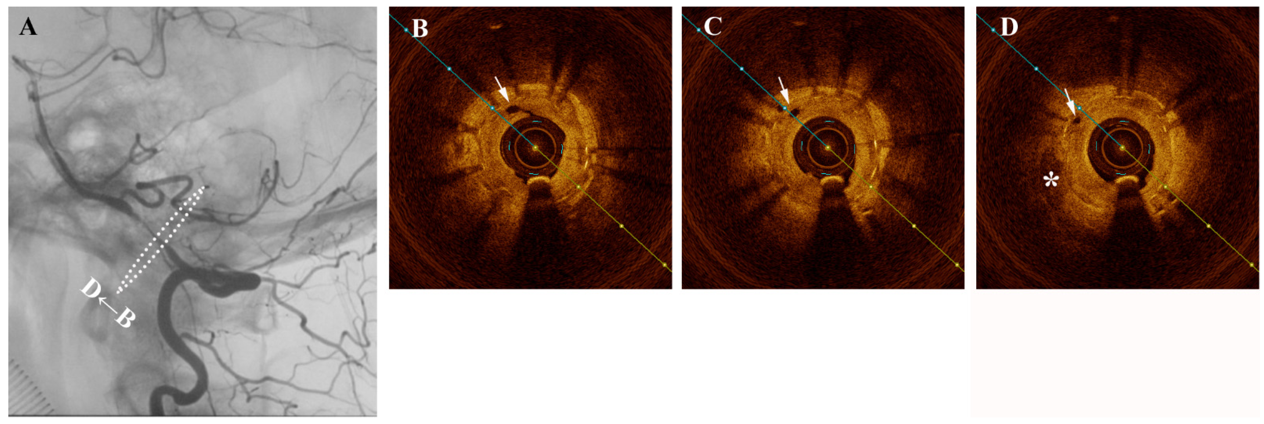
A middle-aged female presented one year after intracranial stenting for severe left intradural segment vertebrobasilar stenosis with recurrent vertigo. Digital subtraction angiography (DSA) confirmed severe in-stent restenosis and same-session optical coherence tomography (OCT) with an Ilumien Optis probe (St. Jude Medical, St. Paul, MN, USA) showed neovascularization-related neointimal hyperplasia (Figure 1). Figure 1 (A) Angiography confirming severe intracranial in-stent restenosis of the left intradural vertebral artery segment. Figure 1 (B–D) Pre-intervention cross-sectional optical coherence tomography (OCT) shows hypertrophic intima and hyporeflective microchannels (white arrow) originating directly from the lumen and penetrating contiguously towards the plaque (*). These are seen as hyporeflectile microchannels originating directly from the lumen, which penetrate contiguously towards irregular plaque and hyperplastic intimomedial complex. Neovascularization plays a role in intracranial atherosclerosis by providing trophic factors to plaque [1]. Such neovasculature is susceptible to intra-plaque hemorrhage and also importantly is a critical factor in the propagation of intimal hyperplasia following stenting [2]. Intracranial neovasculature cannot be resolved in-vivo by traditional imaging methods. In 2011, Mathews et al. performed OCT in the cavernous and the petrous segments of internal carotid artery, which used time-domain OCT assembled in a laboratory rather than a commercial product [3]. Although a small number of cases were enrolled, the result suggested that OCT examination of intracranial arteries was feasible. Until now, there have been numerous preliminary attempts at intracranial OCT use, but visualization of intracranial neovasculature has heretofore not been reported in a living patient. With the approximately 40 micron resolution afforded by OCT, an extremely rare and vivid look at the mechanisms behind plaque nutrition and neointimal hyperplasia of the intracranial vertebral artery is possible [4]. The in-stent stenosis was treated a Sequent Neo paclitaxel-coated balloon (B.Braun, Melsungen, Germany) for intimal hyperplasia inhibition (Figure 2). As plaque and neointimal morphologic characteristics are increasingly recognized as important contributors to disease prognosis and natural history, OCT may stand to inform treatment decisions and assess intimal tearing in the future.
Figure 1.
A middle-aged female presented one year after intracranial stenting for severe left intradural segment vertebrobasilar stenosis with recurrent vertigo. Digital subtraction angiography (DSA) confirmed severe in-stent restenosis and same-session optical coherence tomography (OCT) with an Ilumien Optis probe (St. Jude Medical, St. Paul, MN, USA) showed neovascularization-related neointimal hyperplasia (Figure 1). Figure 1 (A) Angiography confirming severe intracranial in-stent restenosis of the left intradural vertebral artery segment. Figure 1 (B–D) Pre-intervention cross-sectional optical coherence tomography (OCT) shows hypertrophic intima and hyporeflective microchannels (white arrow) originating directly from the lumen and penetrating contiguously towards the plaque (*). These are seen as hyporeflectile microchannels originating directly from the lumen, which penetrate contiguously towards irregular plaque and hyperplastic intimomedial complex. Neovascularization plays a role in intracranial atherosclerosis by providing trophic factors to plaque [1]. Such neovasculature is susceptible to intra-plaque hemorrhage and also importantly is a critical factor in the propagation of intimal hyperplasia following stenting [2]. Intracranial neovasculature cannot be resolved in-vivo by traditional imaging methods. In 2011, Mathews et al. performed OCT in the cavernous and the petrous segments of internal carotid artery, which used time-domain OCT assembled in a laboratory rather than a commercial product [3]. Although a small number of cases were enrolled, the result suggested that OCT examination of intracranial arteries was feasible. Until now, there have been numerous preliminary attempts at intracranial OCT use, but visualization of intracranial neovasculature has heretofore not been reported in a living patient. With the approximately 40 micron resolution afforded by OCT, an extremely rare and vivid look at the mechanisms behind plaque nutrition and neointimal hyperplasia of the intracranial vertebral artery is possible [4]. The in-stent stenosis was treated a Sequent Neo paclitaxel-coated balloon (B.Braun, Melsungen, Germany) for intimal hyperplasia inhibition (Figure 2). As plaque and neointimal morphologic characteristics are increasingly recognized as important contributors to disease prognosis and natural history, OCT may stand to inform treatment decisions and assess intimal tearing in the future.

Figure 2.
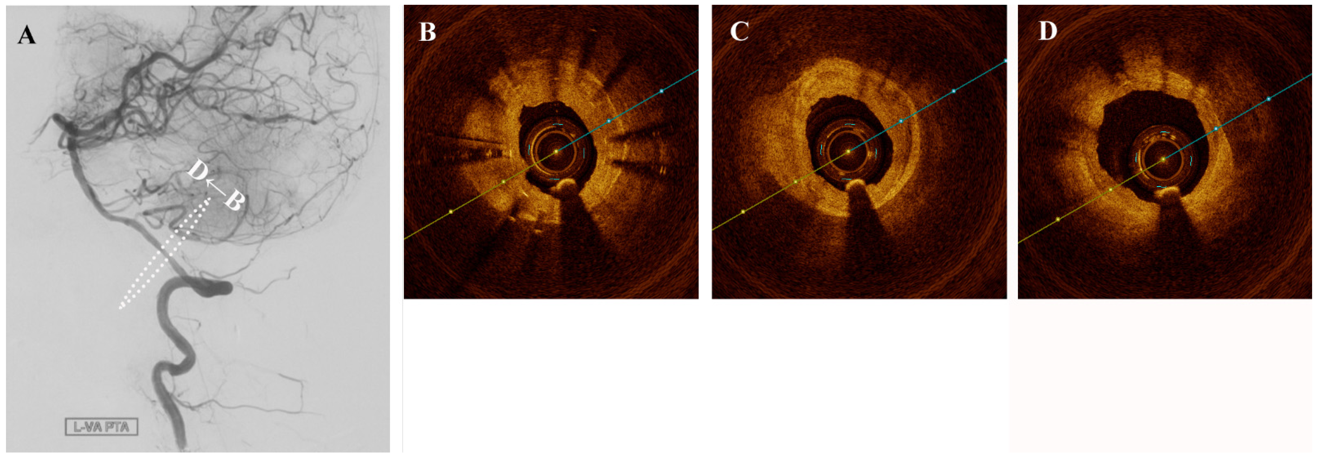
(A) Angioplasty with a drug-coated balloon was performed to recanalize successfully without significant intimal tear (B–D).
Figure 2.
(A) Angioplasty with a drug-coated balloon was performed to recanalize successfully without significant intimal tear (B–D).

Author Contributions
Conception and design, acquisition of data, analysis and interpretation of data: All authors. Drafting the article: Y.F., A.A.D., B.Y. and L.J. Critically revising the article: A.A.D. and L.J. Approved the final version of the manuscript on behalf of all authors: A.A.D. All authors have read and agreed to the published version of the manuscript.
Funding
This research received no external funding.
Institutional Review Board Statement
The study was conducted according to the guidelines of the Declaration of Helsinki, and approved by the Institutional Review Board (or Ethics Committee) of Capital Medical University (protocol code: 20200913, approved date: 3 September 2020).
Informed Consent Statement
Informed consent was obtained from all subjects involved in the study. Written informed consent has been obtained from the patient to publish this paper.
Data Availability Statement
Data are available upon reasonable request.
Acknowledgments
We acknowledge the imaging technologists who make this work possible.
Conflicts of Interest
The authors declare no conflict of interest.
References
- Xu, J.; Lu, X.; Shi, G.-P. Vasa vasorum in atherosclerosis and clinical significance. Int. J. Mol. Sci. 2015, 16, 11574–11608. [Google Scholar] [CrossRef] [PubMed] [Green Version]
- Zhang, M.; Cresswell, N.; Tavora, F.; Mont, E.; Zhao, Z.; Burke, A. In-stent restenosis is associated with neointimal angiogenesis and macrophage infiltrates. Pathol. Res. Pract. 2014, 210, 1026–1030. [Google Scholar] [CrossRef] [PubMed]
- Mathews, M.S.; Su, J.; Heidari, E.; Levy, E.I.; Linskey, M.E.; Chen, Z. Neuroendovascular optical coherence tomography imaging and histological analysis. Neurosurgery 2011, 69, 430–439. [Google Scholar] [CrossRef] [PubMed] [Green Version]
- Vorpahl, M.; Nakano, M.; Virmani, R. Small black holes in optical frequency domain imaging matches intravascular neoangiogenesis formation in histology. Eur. Heart J. 2010, 31, 1889. [Google Scholar] [CrossRef] [PubMed] [Green Version]
Publisher’s Note: MDPI stays neutral with regard to jurisdictional claims in published maps and institutional affiliations. |
© 2021 by the authors. Licensee MDPI, Basel, Switzerland. This article is an open access article distributed under the terms and conditions of the Creative Commons Attribution (CC BY) license (http://creativecommons.org/licenses/by/4.0/).
Share and Cite
MDPI and ACS Style
Feng, Y.; Dmytriw, A.A.; Yang, B.; Jiao, L. Neovascularization in Human Intracranial Atherosclerotic In-Stent Restenosis. Diagnostics 2021, 11, 322. https://doi.org/10.3390/diagnostics11020322
AMA Style
Feng Y, Dmytriw AA, Yang B, Jiao L. Neovascularization in Human Intracranial Atherosclerotic In-Stent Restenosis. Diagnostics. 2021; 11(2):322. https://doi.org/10.3390/diagnostics11020322
Chicago/Turabian StyleFeng, Yiding, Adam A. Dmytriw, Bin Yang, and Liqun Jiao. 2021. "Neovascularization in Human Intracranial Atherosclerotic In-Stent Restenosis" Diagnostics 11, no. 2: 322. https://doi.org/10.3390/diagnostics11020322
Note that from the first issue of 2016, this journal uses article numbers instead of page numbers. See further details here.