HRCT Patterns of Drug-Induced Interstitial Lung Diseases: A Review

 , , ,
, , ,  and
and
Abstract
:1. Introduction
2. Methods
3. Results and Discussion
3.1. Etiopathogenesis and Risk Factors
3.2. Antibiotics
3.3. Anti-Inflammatory and Immunomodulators Drugs
3.4. Anti-Neoplastic Drugs
3.5. Cardiovascular Drugs
3.6. Psychoactive Drug
3.7. Morphological Patterns and Imaging Evaluation
3.8. Diagnosis
3.9. Management
4. Conclusions
Author Contributions
Funding
Conflicts of Interest
References
- Cleverley, J.R.; Screaton, N.J.; Hiorns, M.P.; Flint, J.D.; Müller, N.L. Drug-induced lung disease: High-resolution CT and histological findings. Clin. Radiol. 2002, 57, 292–299. [Google Scholar] [CrossRef] [PubMed]
- Lateef, O.; Shakoor, N.; Balk, R.A. Methotrexate pulmonary toxicity. Expert Opin. Drug Saf. 2005, 4, 723–730. [Google Scholar] [CrossRef] [PubMed]
- Edwards, I.R.; Aronson, J.K. Adverse drug reactions: Definitions, diagnosis, and management. Lancet 2000, 356, 1255–1259. [Google Scholar] [CrossRef]
- Pirmohamed, M.; James, S.; Meakin, S.; Green, C.; Scott, A.K.; Walley, T.J.; Farrar, K.; Park, B.K.; Breckenridge, A.M. Adverse drug reactions as cause of admission to hospital: Prospective analysis of 18,820 patients. BMJ 2004, 329, 15–19. [Google Scholar] [CrossRef] [Green Version]
- Farcas, A.; Sinpetrean, A.; Mogosan, C.; Palage, M.; Vostinaru, O.; Bolita, M.; Domitrascu, D. Adverse drug reactions detected by stimulated spontaneous reporting in an internal medicine department in Romania. Eur. J. Intern. Med. 2010, 21, 453–457. [Google Scholar] [CrossRef]
- Giardina, C.; Cutroneo, P.M.; Mocciaro, E.; Russo, G.T.; Mandraffino, G.; Basile, G.; Rapisarda, F.; Ferrara, R.; Spina, E.; Arcoraci, V. Adverse Drug Reactions in Hospitalized Patients: Results of the FORWARD (Facilitation of Reporting in Hospital Ward) Study. Front. Pharmacol. 2018, 9, 350. [Google Scholar] [CrossRef]
- Thomeer, M.J.; Costabe, U.; Rizzato, G.; Poletti, V.; Demedts, M. Comparison of registries of interstitial lung diseases in three European countries. Eur. Respir. J. Suppl. 2001, 18 (Suppl. 32), 114s–118s. [Google Scholar]
- Kabbara, W.K.; Kordahi, M.C. Nitrofurantoin-induced pulmonary toxicity: A case report and review of the literature. J. Infect Public Health 2015, 8, 309–313. [Google Scholar] [CrossRef]
- Bennoun, L. Drug induced respiratory disorders. Drug Safety 2000, 23, 143–164. [Google Scholar] [CrossRef]
- Dean, P.J.; Groshart, K.D.; Porterfield, J.G. Amiodarone-associated pulmonary toxicity: A clinical and pathologic study of eleven cases. Am. J. Clin. Pathol. 1987, 87, 7–13. [Google Scholar] [CrossRef]
- Jackevicius, C.A.; Tom, A.; Essebag, V.; Eisenberg, M.J.; Rahme, E.; Tu, J.V.; Humphries, K.; Behlouli, H.; Pilote, L. Population-level incidence and risk factors for pulmonary toxicity associated with amiodarone. Am. J. Cardiol. 2011, 108, 705–710. [Google Scholar] [CrossRef] [PubMed]
- Jules-Elysee, K.; White, D. Bleomycin induced pulmonary toxicity. Clin. Chest Med. 1990, 11, 1–20. [Google Scholar] [PubMed]
- Linette, D.C.; McGee, K.H.; McFarland, J.A. Mitomycin-induced pulmonary toxicity: Case report and review of the literature. Ann.Pharmacother. 1992, 26, 481–484. [Google Scholar] [CrossRef] [PubMed]
- Dweik, R.A. Drug-induced pulmonary disease. In Textbook of Pulmonary Diseases, 6th ed.; Baum, G.L., Crapo, J.D., Celli, B.R., Karlinsky, J.B., Eds.; Lippincott Raven: Philadelphia, PA, USA, 1988; pp. 477–490. [Google Scholar]
- Schwaiblmair, M.; Behr, W.; Haeckel, T.; Markl, B.; Foerg, B.; Berghaus, T. Drug induced interstitial lung disease. Open Respir. Med. J. 2012, 6, 63–74. [Google Scholar] [CrossRef] [Green Version]
- Simpson, A.B.; Paul, J.; Graham, J.; Kaye, S.B. Fatal bleomycin pulmonary toxicity inthe west of Scotland 1991–95: A review of patients with germ cell tumours. Br. J. Cancer 1998, 78, 1061–1066. [Google Scholar] [CrossRef] [Green Version]
- Wijnen, P.A.; Drent, M.; Nelemans, P.J.; Kuijipers, P.M.; Koek, G.H.; Neef, C.; Haenen, G.R.; Bekers, O. Role of cytochrome P450 polymorphisms in the development of pulmonary drug toxicity: A case-control study in the Netherlands. Drug Saf. 2008, 31, 1125–1134. [Google Scholar] [CrossRef]
- Park, B.L.; Kim, T.H.; Kim, J.H.; Bae, J.S.; Pasaje, C.F.; Cheong, H.S.; Kim, L.H.; Park, J.S.; Lee, H.S.; Kim, M.S.; et al. Genome-wide association study of aspirin-exacerbated respiratory disease in a Korean population. Hum Genet. 2013, 132, 313–321. [Google Scholar] [CrossRef]
- Roden, A.C.; Camus, P. Iatrogenic pulmonary lesions. Semin. Diagn. Pathol. 2018, 35, 260–271. [Google Scholar] [CrossRef]
- Giridhar, P.; Mallick, S.; Rath, G.K.; Julka, P.K. Radiation induced lung injury: Prediction, assessment and management. Asian Pac. J. Cancer Prev. 2015, 16, 2613–2617. [Google Scholar] [CrossRef] [Green Version]
- Stemmer, S.M.; Cagnoni, P.J.; Shpall, E.J.; Bearman, S.I.; Matthes, S.; Dufton, C.; Day, T.; Taffs, S.; Hami, L.; Martinez, C.; et al. High-dose paclitaxel, cyclophosphamide, and cisplatin with autologous hematopoietic progenitor-cell support: A phase I trial. J. Clin. Oncol. 1996, 14, 1463–1472. [Google Scholar] [CrossRef]
- Yamada, Y.; Shiga, T.; Matsuda, N.; Hagiwara, N.; Kasanuki, H. Incidence and predictors of pulmonary toxicity in Japanese patients receiving low-dose amiodarone. Circ. J. 2007, 71, 1610–1616. [Google Scholar] [CrossRef] [PubMed] [Green Version]
- Kudoh, S.; Kato, H.; Nishiwaki, Y.; Fukuoka, M.; Nakata, K.; Ichinose, Y.; Tsuboi, M.; Yokota, S.; Nakagawa, K.; Suga, M.; et al. Interstitial lung disease in Japanese patients with lung cancer: A cohort and nested case-control study. Am. J. Respir. Crit. Care Med. 2008, 177, 1348–1357. [Google Scholar] [CrossRef] [PubMed]
- Sathi, N.; Chikura, B.; Kaushik, V.V.; Kaushik, V.V.; Wiswell, R.; Dawson, J.K. How common is methotrexate pneumonitis? A large prospective study investigates. Clin. Rheumatol. 2012, 31, 79–83. [Google Scholar] [CrossRef] [PubMed]
- Suissa, S.; Hudson, M.; Ernst, P. Leflunomide use and the risk of interstitial lung disease in rheumatoid arthritis. Arthritis Rheum. 2006, 54, 1435–1439. [Google Scholar] [CrossRef]
- Skeoch, S.; Weatherley, N.; Swift, A.J.; Oldroyd, A.; Johns, C.; Hayton, C.; Giollo, A.; Wild, J.M.; Waterton, J.C.; Buch, M.; et al. Drug-Induced Interstitial Lung Disease: A Systematic Review. J. Clin. Med. 2018, 7, 356. [Google Scholar] [CrossRef] [Green Version]
- Padley, S.P.G.; Adler, B.; Hansell, D.M.; Müller, N.L. High-resolution computed tomography of drug-induced lung disease. Clin. Radiol. 1992, 46, 232–236. [Google Scholar] [CrossRef]
- Wang, K.K.; Bowyer, B.A.; Fleming, C.R.; Schroeder, K.W. Pulmonary infiltrates and eosinophilia associated with sulphasalazine. Mayo Clin. Proc 1984, 59, 343–346. [Google Scholar] [CrossRef]
- Zitnik, R.J.; Cooper, J.A., Jr. Pulmonary disease due to antirheumatic agents. Clin. Chest Med. 1990, 11, 139–150. [Google Scholar]
- Ohbayashi, M.; Suzuki, M.; Yashiro, Y.; Fukuwaka, S.; Yasuda, M.; Kohyama, N.; Kobayashi, Y.; Yamamoto, T. Induction of pulmonary fibrosis by methotrexate treatment in mice lung in vivo and in vitro. J. Toxicol. Sci. 2010, 35, 653–661. [Google Scholar] [CrossRef] [Green Version]
- Prasad, R.; Gupta, P.; Singh, A.; Goel, N. Drug induced pulmonary parenchymal disease. Drug Discov. Ther. 2014, 8, 232–237. [Google Scholar] [CrossRef] [Green Version]
- Peerzada, M.M.; Spiro, T.P.; Daw, H.A. Pulmonary toxicities of biologics: A review. Anticancer Drugs. 2010, 21, 131–139. [Google Scholar] [CrossRef] [PubMed]
- Hadjinicolaou, A.V.; Nisar, M.K.; Bhagat, S.; Parfey, H.; Chilvers, E.R.; Ostor, A.J. Non-infectious pulmonary complications of newer biological agents for rheumatic diseases—A systematic literature review. Rheumatology (Oxford) 2011, 50, 2297–2305. [Google Scholar] [CrossRef] [PubMed] [Green Version]
- Zayen, A.; Rais, H.; Rifi, H.; Ouarda, M.; Afrit, M.; Cherif, A.; Mezline, A. Rituximab-induced interstitial lung disease: Case report and literature review. Pharmacology 2011, 87, 318–320. [Google Scholar] [CrossRef] [PubMed]
- Sekimoto, Y.; Kato, M.; Shukuya, T.; Kouama, R.; Nagaoka, T.; Takahashi, K. Bevacizumab-induced chronic interstitial pneumonia during maintenance therapy in non-small cell lung cancer. Respirol. Case Rep. 2016, 4, e00151. [Google Scholar] [CrossRef]
- Radzikowska, E.; Szczepulska, E.; Chabowski, M.; Bestry, I. Organising pneumonia caused by transtuzumab (Herceptin) therapy for breast cancer. Eur. Respir. J. 2003, 21, 552–555. [Google Scholar] [CrossRef] [Green Version]
- Romond, E.H.; Perez, E.A.; Bryant, J.; Surnan, V.J.; Geyer, C.E.; Davidson, N.E.; Tan-Chiu, E.; Martino, S.; Soonmyung, D.O.; Kaufman, P.A.; et al. Trastuzumab plus adjuvant chemotherapy for operable HER2-positive breast cancer. N. Eng. J. Med. 2005, 353, 1673–1684. [Google Scholar] [CrossRef] [Green Version]
- Hadjinicolaou, A.V.; Nisar, M.K.; Parfrey, H.; Chilvers, E.R.; Ostör, A.J. Non-infectious pulmonary toxicity of rituximab: A systematic review. Rheumatology (Oxford) 2012, 51, 653–662. [Google Scholar] [CrossRef] [Green Version]
- Segura, A.; Yuste, A.; Cercos, A.; López-Tendero, P.; Gironés, R.; Pérez-Fidalgo, J.A.; Herranz, C. Pulmonary fibrosis induced by cyclophosphamide. Ann. Pharmacother. 2001, 35, 894–897. [Google Scholar] [CrossRef]
- Malik, S.W.; Myers, J.L.; DeRemee, R.A.; Specks, U. Lung toxicity associated with cyclophosphamide use. Two distinct patterns. Am. J. Respir. Crit. Care Med. 1996, 154, 1851–1856. [Google Scholar] [CrossRef]
- Martin, W.J.; Rosenow, E.C. Amiodarone pulmonary toxicity (part 1). Chest 1988, 93, 1067–1075. [Google Scholar] [CrossRef]
- Van Cott, T.E.; Yehle, K.S.; DeCrane, S.K.; Thorlton, J.R. Amiodarone-induced pulmonary toxicity: Case study with syndrome analysis. Hear. Lung 2013, 42, 262–266. [Google Scholar] [CrossRef] [PubMed]
- Wolkove, N.; Baltzan, M. Amiodarone pulmonary toxicity. Can. Respir. J. 2009, 16, 43–48. [Google Scholar] [CrossRef] [PubMed]
- Oyama, N.; Oyama, N.; Yokoshiki, H.; Kamishima, T.; Nambu, T.; Tsutsui, H.; Miyasaka, K. Detection of amiodarone-induced pulmonary toxicity in supine and prone positions: High resolution computed tomography study. Circ. J. 2005, 69, 466–470. [Google Scholar] [CrossRef] [Green Version]
- Feinberg, L.; Travis, W.D.; Ferrans, V.; Sato, N. Pulmonary fibrosis associated with tocainide. Am. Rev Respir. Dis. 1990, 141, 505–508. [Google Scholar] [CrossRef] [PubMed]
- Akoun, G.; Cadranel, J.L.; Israel-Biet, D.; Gauthier-Rahman, S. Flecainide-associated pneumonitis. Lancet 1991, 337, 49. [Google Scholar] [CrossRef]
- Dicpinigaitis, P.V. Angiotensin-converting enzyme inhibitor-induced cough: ACCP evidence-based clinical practice guidelines. Chest 2006, 129 (Suppl. 1), 169S–173S. [Google Scholar] [CrossRef]
- Almeida, R.R.; Zanetti, G.; Souza, A.S., Jr.; Zanetti, G.; Souza, A.S. Cocaine-induced pulmonary changes: HRCT findings. J. Bras Pneumol. 2015, 41, 323–330. [Google Scholar] [CrossRef] [Green Version]
- Kleerup, E.C.; Koyal, S.N.; Marques-Magallanes, J.A.; Goldman, M.D.; Tashkin, D.P. Chronic and acute effects of “crack” cocaine on diffusing capacity, membrane diffusion, and pulmonary capillary blood volume in the lung. Chest 2002, 122, 629–638. [Google Scholar] [CrossRef]
- Forrester, J.M.; Steele, A.W.; Waldron, J.A.; Parson, P.E. Crack lung: An acute pulmonary syndrome with a spectrum of clinical and histopathologic findings. Am. Rev. Respir. Dis. 1990, 142, 462–467. [Google Scholar] [CrossRef]
- McCormick, M.; Nelson, T. Cocaine-induced fatal acute eosinophilic pneumonia: A case report. WMJ. 2007, 106, 92–95. [Google Scholar]
- O’Donnell, A.E.; Mappin, F.G.; Sebo, T.J.; Tazelaar, H. Interstitial pneumonitis associated with “crack” cocaine abuse. Chest 1991, 100, 1155–1157. [Google Scholar]
- Vidyasankar, G.; Souza, C.; Lai, C.; Mulpuru, S. A severe complication of crack cocaine use. Can Respir. J. 2015, 22, 77–79. [Google Scholar] [CrossRef] [PubMed]
- Pneumotox Database. Available online: https://www.pneumotox.com/pattern/index/ (accessed on 13 April 2020).
- Lynch, D.A.; Sverzellati, N.; Travis, W.D.; Brown, K.K.; Colby, T.V.; Galvin, J.R.; Goldin, J.G.; Hansell, D.M.; Inoue, Y.; Jonkok, T.; et al. Diagnostic criteria for idiopathic pulmonary fibrosis: A Fleischner Society White Paper. Lancet Respir. Med. 2018, 6, 138–153. [Google Scholar] [CrossRef]
- Pereira, C.A.; Gimenez, A.; Kuranishi, L.; Storrer, K. Chronic hypersensitivity pneumonitis. J. Asthma Allergy 2016, 9, 171–181. [Google Scholar] [CrossRef]
- Mohyuddin, G.R.; Sultan, F.; Zhang, K.; Khaleeg, G. Sulfasalazine induced lung toxicity masquerading as sarcoidosis--case report and review of the literature. Sarcoidosis Vasc. Diffus. Lung Dis. 2013, 30, 226–230. [Google Scholar]
- Schwarz, M.I.; Fontenot, A.P. Drug-induced diffuse alveolar hemorrhage syndromes and vasculitis. Clin. Chest Med. 2004, 25, 133–140. [Google Scholar] [CrossRef]
- Rubin, R.L. Drug-induced lupus. Expert Opin. Drug Saf. 2015, 14, 361–378. [Google Scholar] [CrossRef]
- Janz, D.R.; O’Neal, H.R., Jr.; Ely, E.W. Acute eosinophilic pneumonia: A case report and review of the literature. Crit. Care Med. 2009, 37, 1470–1474. [Google Scholar] [CrossRef]
- American Thoracic Society/European Respiratory Society International Multidisciplinary Consensus Classification of the Idiopathic Interstitial Pneumonias. Am. J. Respir. Crit. Care Med. 2002, 165, 277–304. [CrossRef] [Green Version]


| Drugs | Estimated Incidences | References |
|---|---|---|
| Nitrofurantoina | 1 on 5000 (acute toxicity) | [8] |
| Acetyl-salicylic acid | From 4% (general adult population) to 25% (asthmatic patients) | [9] |
| Amiodarone | 6% | [10] |
| Methotrexate | 7% (chronic toxicity), very rare (acute toxicity) | [11] |
| Bleomycin | 10% | [12] |
| Busulfan | 4% | [9] |
| Mitomycin | 2–38% | [13] |
| Cyclofosphamide | 1% (when used as single agent) | [9] |
| Pattern | Associated Drugs | References |
|---|---|---|
| OP | Amphotericin-B, Amiodarone, Bleomycin, Doxorubicin, Interferon, Metotrexatem, Mitomycin, Nitrofurantonina, Phenytoin, Ticlopidine, Tryptophan, Sulphalazine | [14] |
| HP | Ampicillin, Bupropion, Carbamazepine, Ciprofloxacin, Citarabine, Cephalosporins, interferon-alpha, sulfonamides, ticlopidine, trimethoprim-sulfamethoxazole, sirolimus | [9] |
| Interstitial pneumonia | Adalimumab, Amphotericin B, Amiodarone, Azathioprine, Bleomycin, Busulfan, Chlorambucil, Cyclofosphamide, Etanercept, Flecainide, Interferon alfa, Interferon beta, Infliximab, Melphalan, Methadone, Metotrexate, Nitrofurantoin, Paclitaxel, Penicillamine, Rituximab, Sirolimus, Statine, Sulfasalazine | [14] |
| Loeffler syndorme | Amiodarone, ASA, Bleomycin, Carbamazepine, Captopril, Ibuprofen, Imipramine, Isoniazide, Metotrexate, GM-CSF, Naproxen, Gold salts, Sulfasalazine, Procarbazine, Penicillins, Tryptophans, Zafirleukast | [11] |
| Pulmonary edema | Amlodipine, ASA, Cyclosporine, Citarabine, Chlorothiazide, Clozapine, Heroin, Epinephrine, Gemcitabine, Ketoprofen, Interleukin, Methadone, Metotrexate, Mitomycin, Nitric Oxide, Propanolol, Verapamil | [14] |
| ARDS | Amiodarone, Citarabine, Immunoglobulins, GM-CSF, Nitrofurantoin, Infliximab, Talc, Vinblastine, Vincristine | [14] |
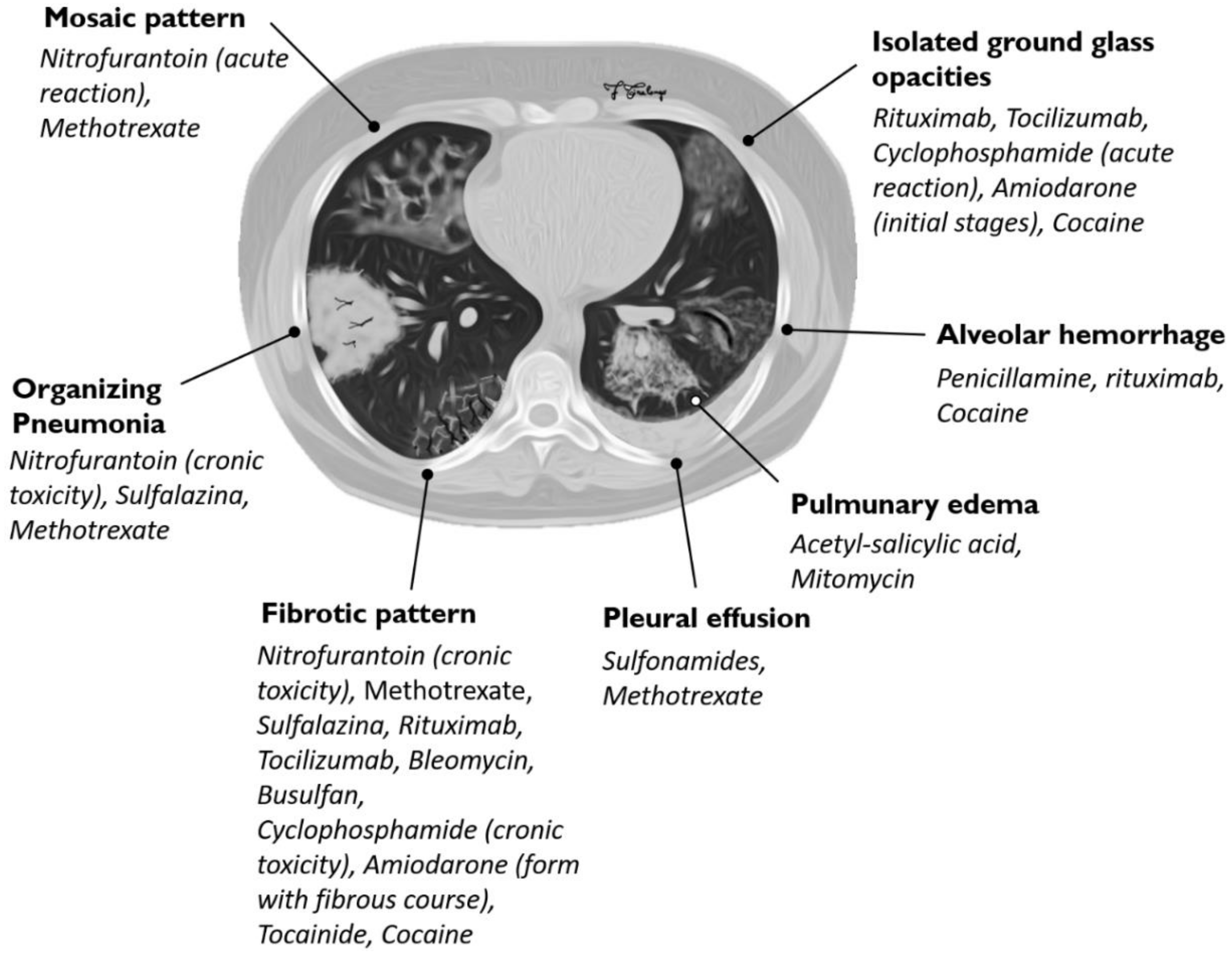
| HRCT Pattern | Associated Drugs |
|---|---|
| Fibrotic pattern | Nitrofurantoin (chronic toxicity), methotrexate, sulfalazina, rituximab, tocilizumab, bleomycin, busulfan, cyclophosphamide (chronic toxicity), amiodarone (form with fibrous course), tocainide, cocaine |
| Organizing pneumonia | Nitrofurantoin (chronic toxicity), methotrexate |
| Mosaic pattern | Nitrofurantoin (acute toxicity), methotrexate, sulfalazina |
| Isolated ground glass | Rituximab, tocilizumab, cyclophosphamide (acute reaction), amiodarone (initial stage), cocaine |
| Alveolar hemorrhage | Penicillamine, rituximab, cocaine |
| Pulmonary edema | Acetyl-salicylic acid, mitomycin |
| Pleural effusion | Sulfonamides, methotrexate |
© 2020 by the authors. Licensee MDPI, Basel, Switzerland. This article is an open access article distributed under the terms and conditions of the Creative Commons Attribution (CC BY) license (http://creativecommons.org/licenses/by/4.0/).
Share and Cite
Distefano, G.; Fanzone, L.; Palermo, M.; Tiralongo, F.; Cosentino, S.; Inì, C.; Galioto, F.; Vancheri, A.; Torrisi, S.E.; Mauro, L.A.; et al. HRCT Patterns of Drug-Induced Interstitial Lung Diseases: A Review. Diagnostics 2020, 10, 244. https://doi.org/10.3390/diagnostics10040244
Distefano G, Fanzone L, Palermo M, Tiralongo F, Cosentino S, Inì C, Galioto F, Vancheri A, Torrisi SE, Mauro LA, et al. HRCT Patterns of Drug-Induced Interstitial Lung Diseases: A Review. Diagnostics. 2020; 10(4):244. https://doi.org/10.3390/diagnostics10040244
Chicago/Turabian StyleDistefano, Giulio, Luigi Fanzone, Monica Palermo, Francesco Tiralongo, Salvatore Cosentino, Corrado Inì, Federica Galioto, Ada Vancheri, Sebastiano E. Torrisi, Letizia A. Mauro, and et al. 2020. "HRCT Patterns of Drug-Induced Interstitial Lung Diseases: A Review" Diagnostics 10, no. 4: 244. https://doi.org/10.3390/diagnostics10040244