Characterization of Nanoparticles Using DSPE-PEG2000 and Soluplus


Laboratory of Drug Safety Management, Faculty of Pharmacy and Pharmaceutical Sciences, Josai University, 1-1 Keyakidai, Sakado, Saitama 350-0295, Japan
*
Author to whom correspondence should be addressed.
Colloids Interfaces 2020, 4(3), 28; https://doi.org/10.3390/colloids4030028
Submission received: 6 June 2020
/
Revised: 11 July 2020
/
Accepted: 13 July 2020
/
Published: 17 July 2020
Abstract
:The aim of this study was to evaluate the characterized hydration method to prepare nanoparticles using Soluplus, a block copolymer with amphipathic properties, and distearoyl phosphatidyl ethanolamine (DSPE)-PEG2000 owing to particle size distribution, zeta potential, particle stability, and transmission electron microscopy (TEM) observed and 31P-NMR spectra. The results showed that, in a suspension of DSPE-PEG2000 and Soluplus at a ratio of 1/1, the prepared microparticles were stable for five days in the dark and at 25 °C. It was also confirmed that the 1/1 suspension of DSPE-PEG2000/Soluplus was stable for five days under the same conditions with the magnesium chloride solution. TEM measurements confirmed the presence of micelle-like particles of 50 to 150 nm in the 1/1 ratio mix of DSPE-PEG2000/Soluplus. 31P-NMR spectral data confirmed that DPSE-PEG2000/Soluplus at mixing ratio of 1/1 has a strong intermolecular with the phosphate group, indicated by the fact that the peak shift and the full width at half maximum were the largest compared with DSPE-PEG2000 with the intermolecular interaction. On the basis of the findings of this study, we conclude that microparticles can be formed using DSPE-PEG2000 and Soluplus via the hydration method, and that the optimum weight ratio of DSPE-PEG2000 to Soluplus is 1/1.
1. Introduction
Nanocarriers are reported to have several benefits in pharmacology, including improved solubility and stability of poorly water-soluble compounds [1], improved pharmacokinetics and biodistribution [2], and delivery to specific sites, allowing for better control of the release of drugs and reduced side effects [3,4]. Furthermore, it is possible to change the application by changing the physiochemical properties of nanocarriers, including composition, shape, surface charge, functional group, and surface properties such as PEGylation [5]. Given these advantages, many kinds of nanocarriers have been developed in the fields of pharmaceuticals, cosmetics, and food science [6,7]. As an example, Suzuki et al. found that encapsulation of oxaliplatin (a known antitumor drug) in a liposome improves the ability to reach the tumor.
Nanocarriers are used as a base for many drug delivery system (DDS) preparations and include polymeric micelles, liposomes, and so on [8,9]. Polymeric micelles are formed from block copolymers with amphipathic properties. The micelle has a hydrophilic structure on the outer shell and a hydrophobic structure on the inner side, which together form a nano-size that protects the poorly water-soluble drug with its own outer shell and disperses the poorly water-soluble compound in water. This allows for the dissolution of poorly water-soluble compounds in an aqueous medium [10,11]. In addition, polymeric micelles have the advantages of low critical micelle concentration, narrow particle size distribution, and low dissociation rate [12], as well as high drug filling amount [13,14]. The application of micellar nanoparticle technology makes it possible to increase the therapeutic effect of a drug and reduce the side effects by extending the time that the drug remains in the blood, thus delivering a larger amount of the drug directly to cancer cells. Nanoparticle applications are also expected to improve convenience and quality of life, while reducing medical costs.
Distearoyl phosphatidyl ethanolamine-polyethylene glycol (DSPE-PEG2000) and Soluplus, which are block copolymers with amphipathic properties, are used as additives in the preparation of polymeric micelles [15,16,17]; for example, micelles prepared with DSPE-PEG2000 solubilize diazepam and micelles prepared with Soluplus solubilize ipriflavone.
DSPE-PEG2000 as a compound is a lipid with a PEG chain grafted in the terminal end on the hydrophobic tail [18,19,20]. DSPE-PEG2000 is used as a PEGylated preparation not only for polymer micelles, but also for many nano platforms such as liposomes, solid lipid nanoparticles, and microemulsions. As an example, it was reported that the formulation of encapsulated doxorubicin in liposome prepared with DSPE-PEG2000 increased tumor uptake by prolonging the circulation time in the body [21].
Soluplus is an amphipathic block copolymer with a polyethylene glycol (PEG) skeleton in the hydrophilic structure and a vinylcaprolactam/vinyl acetate side chain in the lipophilic structure [22]. Reportedly, the water solubility of scopoletin (6-methoxy-7-hydroxycoumarin), which has been used for the treatment of rheumatoid arthritis, swelling, and pain, is enhanced by preparing and encapsulating micelles with Soluplus [23]. Although it has been reported that Soluplus has a low critical micelle concentration and exhibits high dispersibility when diluted (owing to its physical characteristics), it has been infrequently used in the development of nanoparticles [24].
Both DSPE-PEG2000 and Soluplus have a PEG structure within their own structure. By modifying the outer shell of this PEG structure, the nanoparticles are hidden from the host immune system; thus, the mononuclear phagocyte system clearance is reduced, thereby prolonging blood circulation time [25]. The successful preparation of nanoparticles using DSPE-PEG2000 and Soluplus could advance research in the field of polymer micelle formulations. With this goal in mind, we prepared nanoparticles using DSPE-PEG2000 and Soluplus and confirmed the existence and stability of micelle-like particles by measuring particle size and zeta potential and by analyzing transmission electron microscopy (TEM) images of the prepared particles. In addition, the interaction between DSPE-PEG2000 and Soluplus was investigated by measuring 31P-NMR spectra.
2. Materials and Methods
2.1. Materials
DSPE-PEG2000 was purchased from NOF CO., Ltd., Tokyo, Japan. Soluplus was obtained from BASF Japan, Ltd., Kanagawa, Japan (Figure 1). All other reagents were purchased from FUJIFILM Wako Pure Chemical Co., Ltd., Tokyo, Japan.
2.2. Preparation of Fine Particles
Microparticles were prepared by the hydration method, using DSPE-PEG2000 and Soluplus. In the hydration method, a suspension is prepared in which each insoluble substance is dissolved in an organic solvent, the solvent is distilled off by an evaporator, and the prepared lipid thin film is hydrated with an aqueous solvent [26].
DSPE-PEG2000 and Soluplus were dissolved with chloroform to 1 mg/mL. The DSPE-PEG2000 solution and the Soluplus solution were mixed in a pear-shaped flask and sonicated to obtain the following ratios (weight/weight) of DSPE-PEG2000/Soluplus: 10/1, 5/1, 4/1, 3/1, 2/1, 1/1, 1/2, 1/3, 1/4, 1/5, and 1/10. Soluplus is reported to have a critical micelle concentration of 0.82 mg/mL [27]. The solvent was distilled off from these samples using a rotary evaporator (warm bath of 40 °C) and a vacuum distiller to prepare a thin film. The thin film was then prepared with purified water, hydrated, and further ultrasonicated for 6 min to prepare the fine particles. Subsequently, the sample to which magnesium chloride was added was prepared by adding 0.25 × 10⁻³, 0.625 × 10⁻³, and 1.25 × 10⁻³ mmol/L magnesium chloride solution.
2.3. Methods
2.3.1. Particle Diameter, Polydispersity (PDI), and Zeta Potential
The average particle diameter, PDI, and zeta potential of the nanoparticles dispersed in distilled water or owing to coexisting ions various concentration magnesium chloride solution were determined Zetasizer Nano-ZS by dynamic light-scattering method (Malvern Panalytical Instruments, Malvern, UK). Each sample was added to a capillary cell.
2.3.2. Stability Study
To evaluate the stability of the prepared nanoparticles, they were stored for 5 days in the absence of light at 25 °C. The particle sizes and zeta potential values were measured at predetermined time intervals (0, 1, 3, and 5 days).
2.3.3. Transmission Electron Microscopy (TEM) Observation
To evaluate the microstructure of the nanoparticles, cryo-TEM images were collected on a JEM-2100F microscope (JEOL Co., Ltd., Tokyo, Japan). Hydrophilic treatment of a 200-mesh copper grid covered with a perforated polymer film (Nisshin EM Co. Ltd., Tokyo, Japan) was carried out for 60 s with an HDT-400 device (JEOL Co., Ltd.). The liposomal external phase of each suspension was placed in an isotonic 290 mM propylene glycol solution in a size extrusion column. A 2 mL aliquot of each liposome suspension was then applied to the hydrophilized grid. The grid was blotted with a filter paper for 3 s and immediately vitrified by plunging into liquid ethane cooled with liquid nitrogen in a Leica EM CPC cryofixation system (Leica Microsystems GmbH, Wetzar, Germany). Frozen samples were maintained at a temperature of approximately 170 °C, using a Gatan 626 cryo-holder (Gatan, Inc., Pleasanton, CA, USA). The cryo-TEM instrument was operated at 120 kV and a nominal magnification of 50,000. An under-focus of approximately 2 mm was used to enhance image contrast.
2.3.4. Acquisition of 31P-NMR Spectra
The structure of the carrier component of the nanoparticles was determined by 31P-NMR. Measurements of 31P-NMR spectra were carried out on the samples of DSPE-PEG2000 alone and DSPE-PEG2000/Soluplus (weight/weight ratio = 1/1). The concentration of the sample used for measurement was 1 mg/mL for both of the carriers. For 31P-NMR, 85% phosphoric acid, diluted 100 times, was used as an internal standard. Prepared samples were measured with the Varian NMR System 400 MHz (Agilent Technologies, Inc., Santa Clara, California., USA). The resonance frequency was 161.8 MHz, pulse width was 45°, relaxation time was 4.400 s, scan time was 0.600 s, temperature was 25 °C, and the accumulation count was 8192 times.
3. Results and Discussion
3.1. Examination of Mixing Ratio for Preparation of Fine Particles
DSPE-PEG2000 and Soluplus are known to exhibit micelle-like properties by themselves owing to their surfactant activity [15,16,17]. The results of particle size distribution and zeta potential measurement of DSPE-PEG2000 alone and Soluplus alone are shown in Figure 2. The average particle size of DSPE-PEG2000 alone was 52.0 nm (zeta potential approx. −38.0 mV, PDI 0.952), and particles that were considered to be aggregated after 1000 nm and particles approximately 1 to 10 nm were also confirmed. It was speculated that DSPE-PEG alone aggregates as primary particles (around 1 to 10 nm), secondary particles (around 100 nm), and tertiary particles (to be greater than 1000 nm). A single distribution peak was confirmed with Soluplus alone, and the average particle size was 61.8 nm (zeta potential approx. −11.1 mV, PDI 0.095).
In order to prepare DSPE-PEG2000/Soluplus microparticles, samples were prepared at the following mixing ratios (weight/weight) of DSPE-PEG2000 to Soluplus: 10/1, 5/1, 4/1, 3/1, 2/1, 1/1, 1/2, 1/3, 1/4, 1/5, and 1/10, and the samples were evaluated with respect to particle size distribution and zeta potential (Figure 2, Table 1). The average particle size was 36.5 nm (zeta potential −28.5mV, PDI 0.900) at a mixing ratio (DSPE-PEG2000, weight/weight) of 10/1, 80.8 nm (zeta potential −29.2mV, PDI 0.644) at 5/1, and 128.1 nm (zeta potential −28.1 mV, PDI 0.295) at 4/1. The particle size tended to increase as the mixing weight ratio of DSPE-PEG2000 to Soluplus decreased.
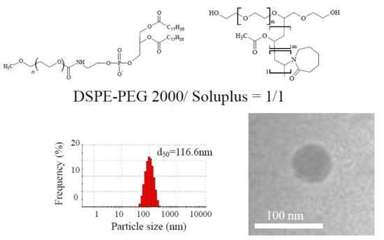
When the mixing weight ratio of DSPE-PEG2000 to Soluplus was 1/1, the average particle diameter was 116.6 nm (zeta potential −13.7 mV, PDI 0.112), and the particle diameter was almost concentrated at approximately 120 nm. On the other hand, when the ratio values are reversed (i.e., Soluplus > DSPE-PEG2000), particle size decreased: 72.0 nm (zeta potential approx. −11.3 mV, PDI 0.103) at a mixing ratio of 1/4 and 54.5 nm (−6.0 mV, PDI 0.057) at a mixing ratio of 1/5. The average particle size at a mixing ratio of 1/10 was 56.1 nm (zeta potential approx. −7.7 mV, PDI 0.101). With respect to particle size distribution, when the proportion of DSPE-PEG2000 was greater than 3/1, small particles that were considered to be dissociated were confirmed around 10 nm. In addition, the particle size distribution of particles considered to be aggregated was confirmed to be above 1000 nm. As these results are similar to the particle size distribution of DSPE-PEG2000 alone, it was inferred that DSPE-PEG2000 was present in excess. In addition, the data confirmed that the zeta potential increased as the proportion of DSPE-PEG2000 increased. It is speculated that this is because the value of zeta potential approaches that of DSPE-PEG2000 alone as the proportion of DSPE-PEG2000 increases. From the above, aggregation of particles was confirmed with DSPE-PEG alone, however, aggregation with suppressor was confirmed owing to being prepared with Soluplus, and a single particle size distribution was confirmed; therefore, between DSPE-PEG and Soluplus, it is speculated that some kind of interaction.
In order to show the relationship between the average particle size and the zeta potential, we plotted average particle size (d50) and zeta potential against the weight ratio of DSPE-PEG2000 to Soluplus (vertical axis and horizontal axis, respectively, in Figure 3). When the zeta potential is high, particles repel each other owing to the surface charge, thus preventing aggregation. Generally, it is reported that the zeta potential provides particle stability at 20 to 30 mV [28]. As the zeta potential changes as the mixing weight ratio of DSPE-PEG2000 to Soluplus changes, it is presumed that the mixing weight ratio affects the stability of the fine particles. Accordingly, we investigated the optimum mixing weight ratio of DSPE-PEG2000 to Soluplus by conducting a stability test of fine particles.
3.2. Particle Stability
In order to examine the stability of the prepared microparticles, the suspension prepared at each weight ratio (DSPE-PEG2000/Soluplus = 2/1, 1/1, 1/2) was evaluated for stability for 5 days at 25 °C in the dark (Figure 4). In the 2/1 suspension, particles that appeared to have aggregated on day 3 were confirmed to be greater than 1000 nm. On day 5, dissociated particles believed to be derived from DSPE-PEG2000 were confirmed in the vicinity of 1 to 10 nm. In the 1/1 suspension, dissociation and aggregation of particles were not confirmed over time, and it was assumed that the particles were stable. In the 1/2 suspension, a single distribution peak was observed up to day 3, but on day 5, a particle size distribution was found to be in excess of 1000 nm.
From the results of particle size distribution and zeta potential alone, the optimum mixing weight ratio of DSPE-PEG2000 to Soluplus cannot be inferred. Although a slight decrease in zeta potential is observed by adding an inorganic salt, it is reported that the critical micelle concentration is decreased, and the formation of fine particles is facilitated, so that stable particles can be formed [29]. It is possible that the addition of magnesium chloride solution elevated the stability of the mixtures of DSPE-PEG2000 and Soluplus at mixing ratios of 2/1, 1/1, and 1/2. In the DSPE-PEG2000/Soluplus suspension with a mixing ratio of 2/1 (i.e., the proportion of DSPE-PEG2000 is high), particle aggregation was confirmed as the magnesium chloride concentration increased (Figure 5A). At a mixing ratio of 1/1, dissociation and aggregation were not confirmed for 5 days in all magnesium chloride concentration groups, and it was confirmed that they exist stably (Figure 5B). However, we found that, as the concentration of magnesium chloride increased, the average particle size increased and the zeta potential decreased. At a mixing ratio of 1/2, particle aggregation was confirmed on day 5 when 0.625 × 10⁻³ mM magnesium chloride solution was added (Figure 5C). From these results, it was inferred that fine particles exist most stably when the mixing ratio of DSPE-PEG 2000 to Soluplus is 1/1. The reason for this is that the addition of salt confirmed the aggregation and dissociation of particles in the 2/1 and 1/2 suspensions, and that the zeta potential was maintained to some extent in the 1/1 suspension. It has been suggested that, owing to intermolecular interactions such as hydrogen bonding between DSPE-PEG2000 and Soluplus, an environment capable of holding a potential on the particle surface is created, and the resulting electrostatic repulsion force contributes to stabilization of the particles in the suspension [30,31]. Therefore, in order to investigate the intermolecular interaction in the suspension, we evaluated the particles with the use of TEM and 31P-NMR.
3.3. Shape Evaluation of DSPE-PEG2000/Soluplus Particles
In order to evaluate the shape of the prepared fine particles, TEM observation of DSPE-PEG2000/Soluplus mixtures at ratios of 1/1 and 1/2 was performed (Figure 6). Particles approximately 50 to 150 nm were observed in the 1/1 and 1/2 mixtures, which was consistent with the result of particle size distribution measurement. From the results of TEM measurement, it is speculated that the particles are properly dispersed in the aqueous solution owing to the average particle size of DSPE-PEG/Soluplus = 1/1 and the surface charge due to the zeta potential. The shape of the particles was circular. It has been reported that DSPE-PEG2000 is characterized by the formation of micelles with lipid membranes and disk structures [32,33]. It was speculated that the DSPE-PEG2000/Soluplus microparticles also formed particles of either structure. Normally, a lamellar structure is observed when forming a bilayer structure such as a liposome [34]. However, as the lamella structure was not observed in the particles prepared in our study, we have reported in previous studies on micelle formation in DSPE-PEG and ascorbic acid derivatives [35]. The lamella structure was not confirmed in the research report, and the TEM image different from that of the liposome was obtained, so it was determined to be a micelle-like structure. In this result as well, the same structure as in the previous study was confirmed, and thus it was inferred that the structure was micellar.
3.4. 31P-NMR Measurement
31P-NMR measurements were performed to investigate intermolecular interactions in suspension (Figure 7). The peak of 31P in DSPE-PEG2000 alone was observed at −11.21 ppm (v1/2 = 7.64) (v1/2 indicates the half width) (Figure 7a). In DSPE-PEG2000/Soluplus at a ratio of 2/1, a peak was observed at −11.17 ppm (v1/2 = 14.64), and it was confirmed that the peak was shifted to the lower position side and broadened (Figure 7b). In DSPE-PEG 2000/Soluplus at 1/1, three broad peaks were observed at −11.07, −11.10, and −11.15 ppm (v1/2 = 24.79) (Figure 7c). A peak was confirmed at −11.17 ppm (v1/2 = 10.90) in the 1/2 ratio sample of DSPE-PEG2000/Soluplus, which was the same as the position in the 2/1 ratio sample. The half-width of the 1/2 ratio sample of DSPE-PEG2000/Soluplus was a narrower peak than that of the 2/1 ratio sample. From these results, it was confirmed that DPSE-PEG2000/Soluplus at a 1/1 mixing ratio has strong intermolecular interaction in phosphorus, as the peak shift and the full width at half maximum were the largest compared with DSPE-PEG2000. In addition, in the case of DSPE-PEG2000/Soluplus at a mixing ratio of 2/1, dissociation and aggregation over time of particle size distribution derived from DSPE-PEG2000 were confirmed, in comparison with the results of the stability test. The peak of the 2/1 ratio mix of DSPE-PEG2000/Soluplus was sharper than that of the 1/1 ratio mix, and a peak similar to that of DSPE-PEG2000 alone was confirmed. We propose that this is because the molecules could not assemble well and there is high mobility in DSPE-PEG2000. In other words, it was speculated that the 31P-mediated interaction contributed to the particles prepared with DSPE-PEG2000 and Soluplus. Our findings suggest that a mixing ratio (weight/weight) of 1/1 DSPE-PEG2000 and Soluplus results in a greater number of molecules assembled, and thus stable particles can be formed.
4. Conclusions
The results of our study suggest that microparticles in DSPE-PEG2000/Soluplus can be formed by the hydration method, and the optimum weight ratio of DSPE-PEG2000 to Soluplus is 1/1. The data also indicate that these fine particles exist stably for 5 days under the condition of shading and 25 °C. It was also confirmed that a 1/1 ratio mix of DSPE-PEG2000/Soluplus is stable for 5 days under the same conditions, even in the group containing the magnesium chloride solution. In mixtures of DSPE-PEG2000/Soluplus at ratios of 2/1 and 3/2, the higher the containing magnesium chloride solution, the faster the particle aggregation rate. In addition, the 31P-NMR measurements showed that the peak of the 1/1 DSPE-PEG2000/Soluplus mix had the largest peak shift compared with DSPE-PEG2000 alone, and a broad peak was obtained. From these data, it was confirmed that this had the strongest intermolecular interaction and formed particles in which the molecules were aggregated. We can conclude that the phosphate group of DSPE-PEG2000 is involved in the intermolecular interaction of the prepared fine particles with respect to the physical properties and structure of the particles. From the results of TEM imaging and measurement, spherical micelle-like particles of 50 to 150 nm were confirmed in both the 1/1 and 1/2 ratio mixes of DSPE-PEG2000/Soluplus. However, the results are not sufficient to determine the detailed formation mechanism and structure, and further investigation is required. In addition, formulation and clinical expectation can be expected by preparing and encapsulating a drug-containing complex and testing its toxicity and kinetics.
Author Contributions
Conceptualization, Y.I.; methodology, Y.I. and R.T.; formal analysis, Y.I., R.T. and I.M.; investigation, Y.I., R.T., I.M., and I.K.; data curation, Y.I., R.T. and I.M.; writing—original draft preparation, Y.I. and R.T.; writing—review and editing, Y.I., R.T., I.M. and I.K.; visualization, Y.I. and R.T.; supervision, Y.I.; project administration, Y.I. All authors have read and agreed to the published version of the manuscript.
Funding
This research received no external funding.
Acknowledgments
We thank Kenjiro Higashi (Chiba University, Japan) for his kind advice and expert TEM measurements and especially for his perspectives in applied science, which encouraged us to undertake this study. We also thank the Graduate School of Pharmaceutical Sciences, Chiba University, Japan, where the TEM work was performed.
Conflicts of Interest
The authors declare that there are no conflicts of interest regarding the publication of this paper.
References
- Maeda, H.; Konno, T. Metamorphosis of Neocarzinostatin to SMANCS; Springer-Verlag GmbH: Heidelberg, Germany, 1997; pp. 227–268. [Google Scholar]
- Daryl, C.; Charles, O.; Mark, E.; John, W.; Dmitri, B. Pharmacokinetics and in vivo drug release rates in liposomal nanocarrier development. Int. J. Pharm. 2008, 97, 4696–4740. [Google Scholar]
- Mishra, B.; Patel, B.B.; Tiwari, S. Colloidal nanocarriers: A review on formulation technology, types and applications toward targeted drug delivery. Int. J. Nanomed. 2010, 6, 9–24. [Google Scholar] [CrossRef] [PubMed]
- How, C.W.; Rasedee, A.; Manickam, S.; Rosli, R. Tamoxifen-loaded nanostructured lipid carrier as a drug delivery system: Characterization, stability assessment and cytotoxicity. Colloids Surf. B Biointerfaces 2013, 112, 393–399. [Google Scholar] [CrossRef] [PubMed] [Green Version]
- Sun, T.; Zhang, Y.S.; Pang, B.; Hyun, D.C.; Yang, M.; Xia, Y. Engineered Nanoparticles for Drug Delivery in Cancer Therapy. Angew. Chem. Int. Ed. 2014, 53, 12320–12364. [Google Scholar] [CrossRef] [PubMed]
- Suzuki, R.; Takizawa, T.; Kuwata, Y.; Mutoh, M.; Ishiguro, N.; Utoguchi, N.; Shinohara, A.; Eriguchi, M.; Yanagie, H.; Maruyama, K. Effective anti-tumor activity of oxaliplatin encapsulated in transferrin–PEG-liposome. Int. J. Pharm. 2008, 346, 143–150. [Google Scholar] [CrossRef]
- Onoue, S.; Yamada, S.; Chan, H.K. Nanodrugs: Pharmacokinetics and safety. Int. J. Nanomed. 2014, 9, 1025–1037. [Google Scholar] [CrossRef] [Green Version]
- Moritz, M.G.; Moritz, M. Solid lipid nanoparticles as attractive drug vehicles: Composition, properties and therapeutic strategies. Mater Sci. Eng. C. 2016, 68, 982–994. [Google Scholar] [CrossRef] [PubMed]
- Suzuki, R.; Takizawa, T.; Negishi, Y.; Utoguchi, N.; Maruyama, K. Effective gene delivery with novel liposomal bubbles and ultrasonic destruction technology. Int. J. Pharm. 2008, 354, 49–55. [Google Scholar] [CrossRef] [PubMed]
- Harris, J.M. Poly(ethylene glycol) Chemistry: Biotechnical and Biomedical Applications; Springer-Verlag GmbH: Heidelberg, Germany, 1992; pp. 1–13. [Google Scholar]
- Nishiyama, N.; Matsumura, Y.; Kataoka, K. Development of polymeric micelles for targeting intractable cancers. Cancer Sci. 2016, 107, 867–874. [Google Scholar] [CrossRef] [PubMed]
- Nagasaki, Y.; Okada, T.; Scholz, C.; Iijima, M.; Kato, M.; Kataoka, K. The Reactive Polymeric Micelle Based on An Aldehyde-Ended Poly(ethylene glycol)/Poly(lactide) Block Copolymer. Macromolecules 1998, 31, 1473–1479. [Google Scholar] [CrossRef]
- Yokoyama, M.; Sugiyama, T.; Okano, T.; Sakurai, Y.; Naito, M.; Kataoka, K. Analysis of Micelle Formation of an Adriamycin-Conjugated Poly(Ethylene Glycol)–Poly(Aspartic Acid) Block Copolymer by Gel Permeation Chromatography. Pharm. Res. 1993, 10, 895–899. [Google Scholar] [CrossRef] [PubMed]
- Yamamoto, Y.; Yasugi, K.; Harada, A.; Nagasaki, Y.; Kataoka, K. Temperature-Related change in the properties relevant to drug delivery of poly(ethylene glycol)-poly(d,l-lactide) block copolymer micelles in aqueous milieu. J. Control. Release 2002, 82, 359–371. [Google Scholar] [CrossRef]
- Ashok, B.; Arleth, L.; Hjelm, R.P.; Rubinstein, I.; Onyuksel, H. In vitro characterization of PEGylated phospholipid micelles for improved drug solubilization: Effects of PEG chain length and PC incorporation. J. Pharm. Sci. 2004, 93, 2476–2487. [Google Scholar] [CrossRef]
- Obata, T.; Suzuki, Y.; Ogawa, N.; Kurimoto, I.; Yamamoto, H.; Furuno, T.; Sasaki, T.; Tanaka, M. Improvement of the Antitumor Activity of Poorly Soluble Sapacitabine (CS-682) by Using Soluplus® as a Surfactant. Boil. Pharm. Bull. 2014, 37, 802–807. [Google Scholar] [CrossRef] [PubMed] [Green Version]
- Takahashi, C.; Saito, S.; Suda, A.; Ogawa, N.; Kawashima, Y.; Yamamoto, H. Antibacterial activities of polymeric poly(dl-lactide-co-glycolide) nanoparticles and Soluplus® micelles against Staphylococcus epidermidis biofilm and their characterization. RSC Adv. 2015, 5, 71709–71717. [Google Scholar] [CrossRef]
- Le, U.M.; Cui, Z. Long-circulating gadolinium-encapsulated liposomes for potential application in tumor neutron capture therapy. Int. J. Pharm. 2006, 312, 105–112. [Google Scholar] [CrossRef]
- Maruyama, K.; Yuda, T.; Okamoto, A.; Kojima, S.; Suginaka, A.; Iwatsuru, M. Prolonged circulation time in vivo of large unilamellar liposomes composed of distearoyl phosphatidylcholine and cholesterol containing amphipathic poly(ethylene glycol). Biochim. Biophys. Acta 1992, 1128, 44–49. [Google Scholar] [CrossRef]
- Moghimi, S.M. Prolonging the circulation time and modifying the body distribution of intravenously injected polystyrene nanospheres by prior intravenous administration of poloxamine-908. A ‘hepatic-blockade’ event or manipulation of nanosphere surface in vivo. Biochim. Biophys. Acta. 1997, 1336, 1–6. [Google Scholar] [CrossRef]
- Matsumura, Y.; Maeda, H. A new concept for macromolecular therapeutics in cancer chemotherapy: Mechanism of tumoritropic accumulation of proteins and the antitumor agent smancs. Am. Assoc. Cancer Res. 1986, 46, 6387–6392. [Google Scholar]
- Kataoka, K.; Harada, A.; Nagasaki, Y. Block copolymer micelles for drug delivery: Design, characterization and biological significance. Adv. Drug Deliv. Rev. 2012, 64, 37–48. [Google Scholar] [CrossRef]
- Zeng, Y.-C.; Li, S.; Liu, C.; Gong, T.; Sun, X.; Fu, Y.; Zhang, Z.-R. Soluplus micelles for improving the oral bioavailability of scopoletin and their hypouricemic effect in vivo. Acta. Pharmacol. Sin. 2017, 38, 424–433. [Google Scholar] [CrossRef] [PubMed] [Green Version]
- Dian, L.; Yu, E.; Chen, X.; Wen, X.; Zhang, Z.; Qin, L.; Wang, Q.; Li, G.; Wu, C. Enhancing oral bioavailability of quercetin using novel soluplus polymeric micelles. Nanoscale Res. Lett. 2014, 9, 684. [Google Scholar] [CrossRef] [PubMed] [Green Version]
- Karlsson, P.C.; Hughes, R.; Rafter, J.J.; Bruce, W.R. Polyethylene glycol reduces inflammation and aberrant crypt foci in carcinogen-initiated rats. Cancer Lett. 2005, 223, 203–209. [Google Scholar] [CrossRef] [PubMed]
- Moribe, K.; Maruyama, S.; Inoue, Y.; Suzuki, T.; Fukami, T.; Tomono, K.; Higashi, K.; Tozuka, Y.; Yamamoto, K. Ascorbyl dipalmitate/PEG-lipid nanoparticles as a novel carrier for hydrophobic drugs. Int. J. Pharm. 2010, 387, 236–243. [Google Scholar] [CrossRef]
- Tanida, S.; Kurokawa, T.; Sato, H.; Kadota, K.; Tozuka, Y. Evaluation of the Micellization Mechanism of an Amphipathic Graft Copolymer with Enhanced Solubility of Ipriflavone. Chem. Pharm. Bull. 2016, 64, 68–72. [Google Scholar] [CrossRef] [Green Version]
- Mishra, P.R.; Al Shaal, L.; Müller, R.H.; Keck, C.M. Production and characterization of Hesperetin nanosuspensions for dermal delivery. Int. J. Pharm. 2009, 371, 182–189. [Google Scholar] [CrossRef]
- Singh, R.; Lillard, J.W. Nanoparticle-based targeted drug delivery. Exp. Mol. Pathol. 2009, 86, 215–223. [Google Scholar] [CrossRef] [Green Version]
- Sarmentocde, B.; Ribeiro, A.J.; Veiga, F.; Ferreira, D. Development and characterization of new insulin containing polysaccharide nanoparticles. Colloids Surf. B 2006, 53, 193–202. [Google Scholar] [CrossRef] [Green Version]
- Terayama, H.; Hirota, K.; Yoshimura, T.; Esumi, K. Effect of dilution on aqueous dispersion of drug particles. Colloids Surf. B 2003, 27, 177–180. [Google Scholar] [CrossRef]
- Sandstrom, M.C.; Johansson, E.; Edwards, K. Influence of preparation path on the formation of discs and threadlike micelles in DSPE-PEG 2000/lipid systems. Biol Chem. 2008, 132, 97–103. [Google Scholar]
- Johnsson, M.; Hansson, P.; Edwards, K. Spherical Micelles and Other Self-Assembled Structures in Dilute Aqueous Mixtures of Poly(Ethylene Glycol) Lipids. J. Phys. Chem. B 2001, 105, 8420–8430. [Google Scholar] [CrossRef]
- Tokudome, Y.; Uchida, R.; Yokote, T.; Todo, H.; Hada, N.; Kon, T.; Yasuda, J.; Hayashi, H.; Hashimoto, F.; Sugibayashi, K. Effect of topically applied sphingomyelin-Based liposomes on the ceramide level in a three-Dimensional cultured human skin model. J. Liposome Res. 2010, 20, 49–54. [Google Scholar] [CrossRef] [PubMed] [Green Version]
- Inoue, Y.; Hibino, M.; Murata, I.; Kanamoto, I. A Nanocarrier Skin-Targeted Drug Delivery System using an Ascorbic Acid Derivative. Pharm. Res. 2017, 35, 1. [Google Scholar] [CrossRef] [PubMed]
Figure 1.
Chemical structure of (a) distearoyl phosphatidyl ethanolamine (DSPE)-PEG2000 and (b) Soluplus.
Figure 1.
Chemical structure of (a) distearoyl phosphatidyl ethanolamine (DSPE)-PEG2000 and (b) Soluplus.

Figure 2.
Relationship between Particle diameter in the difference of mixing weight ratio of DSPE-PEG2000 and Soluplus, (a) DSPE-PEG2000/Soluplus = 10/1, (b) DSPE-PEG2000/Soluplus = 5/1, (c) DSPE-PEG2000/Soluplus = 4/1, (d) DSPE-PEG2000/Soluplus = 3/1, (e) DSPE-PEG2000/Soluplus = 2/1, (f) DSPE-PEG2000/Soluplus = 1/1, (g) DSPE-PEG2000/Soluplus = 1/2, (h) DSPE-PEG2000/Soluplus = 1/3, (i) DSPE-PEG2000/Soluplus = 1/4, (j) DSPE-PEG2000/Soluplus = 1/5, (k) DSPE-PEG2000/Soluplus = 1/10, (l) only DSPE-PEG2000, (m) only Soluplus.

Figure 3.
Relationship between zeta potential (ZP) and particle diameter on the different weight ratios of DPSE-PEG2000 and Soluplus nanoparticles.
Figure 3.
Relationship between zeta potential (ZP) and particle diameter on the different weight ratios of DPSE-PEG2000 and Soluplus nanoparticles.

Figure 4.
Changes in particle size and zeta potential of nanoparticles of DSPE-PEG2000/Soluplus = 2/1, 1/1, 1/2 nanoparticles after the storage at 25 °C day 0 to day 5.
Figure 4.
Changes in particle size and zeta potential of nanoparticles of DSPE-PEG2000/Soluplus = 2/1, 1/1, 1/2 nanoparticles after the storage at 25 °C day 0 to day 5.

Figure 5.
Effect of particle size with magnesium chloride, (A) changes in particle size and zeta potential of nanoparticles of DSPE-PEG2000/Soluplus = 2/1 nanoparticles after the storage at 25 °C day 0 to day 5; (B) changes in particle size and zeta potential of nanoparticles of DSPE-PEG2000/Soluplus = 1/1 nanoparticles after the storage at 25 °C day 0 to day 5; (C) changes in particle size and zeta potential of nanoparticles of DSPE-PEG2000/Soluplus = 1/2 nanoparticles after the storage at 25 °C day 0 to day 5.


Figure 6.
TEM images of nanoparticles, (a) DSPE-PEG2000/Soluplus = 1/1, (b) DSPE-PEG2000/Soluplus = 1/2.
Figure 6.
TEM images of nanoparticles, (a) DSPE-PEG2000/Soluplus = 1/1, (b) DSPE-PEG2000/Soluplus = 1/2.

Figure 7.
31P-NMR spectra of DSPE-PEG 2000 and Soluplus, (a) DSPE-PEG2000, (b) DSPE-PEG2000/Soluplus = 2/1, (c) DSPE-PEG2000/Soluplus = 1/1, (d) DSPE-PEG2000/Soluplus = 1/2.
Figure 7.
31P-NMR spectra of DSPE-PEG 2000 and Soluplus, (a) DSPE-PEG2000, (b) DSPE-PEG2000/Soluplus = 2/1, (c) DSPE-PEG2000/Soluplus = 1/1, (d) DSPE-PEG2000/Soluplus = 1/2.


Table 1.
Particle size and zeta potential (ZP) of nanoparticles.
| Sample | Particle Size (nm) | ZP (mV) | PDI |
|---|---|---|---|
| DSPE-PEG2000 int | 52.0 ± 0.3 | −38.0 ± 1.3 | 0.952 |
| Soluplus int | 61.8 ± 0.4 | −11.1 ± 0.1 | 0.095 |
| DSPE-PEG2000/Soluplus = 10/1 | 36.5 ± 1.1 | −28.5 ± 1.5 | 0.900 |
| DSPE-PEG2000/Soluplus = 5/1 | 80.8 ± 0.9 | −29.2 ± 0.4 | 0.644 |
| DSPE-PEG2000/Soluplus = 4/1 | 128.1 ± 1.8 | −28.1 ± 0.0 | 0.295 |
| DSPE-PEG2000/Soluplus = 3/1 | 128.1 ± 1.1 | −27.1 ± 2.7 | 0.294 |
| DSPE-PEG2000/Soluplus = 2/1 | 135.1 ± 0.4 | −26.3 ± 2.6 | 0.247 |
| DSPE-PEG2000/Soluplus = 1/1 | 116.6 ± 0.0 | −13.7 ± 0.6 | 0.112 |
| DSPE-PEG2000/Soluplus = 1/2 | 107.9 ± 2.2 | −9.6 ± 1.4 | 0.163 |
| DSPE-PEG2000/Soluplus = 1/3 | 68.9 ± 0.8 | −9.9 ± 0.7 | 0.109 |
| DSPE-PEG2000/Soluplus = 1/4 | 72.0 ± 0.4 | −11.3 ± 0.3 | 0.103 |
| DSPE-PEG2000/Soluplus = 1/5 | 54.5 ± 0.1 | −6.0 ± 0.2 | 0.057 |
| DSPE-PEG2000/Soluplus = 1/10 | 56.1 ± 0.1 | −7.7 ± 1.0 | 0.101 |
© 2020 by the authors. Licensee MDPI, Basel, Switzerland. This article is an open access article distributed under the terms and conditions of the Creative Commons Attribution (CC BY) license (http://creativecommons.org/licenses/by/4.0/).
Share and Cite
MDPI and ACS Style
Takayama, R.; Inoue, Y.; Murata, I.; Kanamoto, I. Characterization of Nanoparticles Using DSPE-PEG2000 and Soluplus. Colloids Interfaces 2020, 4, 28. https://doi.org/10.3390/colloids4030028
AMA Style
Takayama R, Inoue Y, Murata I, Kanamoto I. Characterization of Nanoparticles Using DSPE-PEG2000 and Soluplus. Colloids and Interfaces. 2020; 4(3):28. https://doi.org/10.3390/colloids4030028
Chicago/Turabian StyleTakayama, Rina, Yutaka Inoue, Isamu Murata, and Ikuo Kanamoto. 2020. "Characterization of Nanoparticles Using DSPE-PEG2000 and Soluplus" Colloids and Interfaces 4, no. 3: 28. https://doi.org/10.3390/colloids4030028