
Thermoinduced and Photoinduced Sustainable Hydrophilic Surface of Sputtered-TiO2 Thin Film


Department of Electrical Engineering, Gachon University, 1342 Seongnamdaero, Seongnam 13120, Gyeonggi, Korea
*
Authors to whom correspondence should be addressed.
Coatings 2021, 11(11), 1360; https://doi.org/10.3390/coatings11111360
Submission received: 14 October 2021
/
Revised: 29 October 2021
/
Accepted: 2 November 2021
/
Published: 4 November 2021
(This article belongs to the Special Issue Optical Thin Film and Photovoltaic (PV) Related Technologies)
Abstract
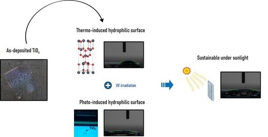
:To achieve self-cleaning at a low maintenance cost, we investigated the possibility of obtaining a sustainable hydrophilic surface of TiO2 thin film. As the hydrophilicity of TiO2 films fabricated by FTS has not yet been studied, we deposited TiOx using FTS, and then TiO2 was formed through additional treatment. Hydrophilic surfaces were obtained by thermoinduced and photoinduced methods. UV irradiation led to the conversion of Ti4+ to Ti3+ in the lattice structure and an increase in the number of OH groups on the surface, and annealing induced the formation of Ti3+ defect sites, as well as organic degradation and changes in the crystal structure. Through the annealing process, the water contact angle of as-deposited film was decreased from 78.7° to 35.7°, and crystallinity changed from amorphous to anatase. These changes contributed to the formation of a hydrophilic surface and reduced the water contact angle by up to 10.8°. After the formation of a hydrophilic surface through annealing and UV irradiation, the sample returned to its original state. We confirmed that the water contact angle of the returned sample was decreased through exposure to sunlight; it reduced the water contact angle of the returned sample by 15.2°. Thus, the results revealed that the crystallinity influences the hydrophilicity and its sustainability for TiO2 films under sunlight.
1. Introduction
Photocatalysts are promising for use in environment- and energy-related applications owing to their sustainability and environmental friendliness. They utilize light energy to cause a chemical reaction such as degradation of organic compounds [1]. In photocatalysis, an electron is excited from the valence band to the conduction band; this leads to the generation of positive electron holes in the valence band when the photon energy of the light source is higher than the bandgap energy. The atmospheric oxygen is reduced to superoxide ions by the excited electrons, and the electron holes oxidize the water or OH− to OH radicals on the surface. The reaction equation for photocatalysis is as follows [2]:
TiO2 + hν → h+VB + e−CB
h+VB + H2O → OH + H+
e−CB + O2 → O2−
Through these reactions, TiO2 can clean the surface by decomposing organic matter and pollutants into CO2 and H2O, as well as produce hydrogen [2,3,4,5]. Since the first report on photocatalysis by Fujishima and Honda in 1972 [6], it has been employed to remove pollutants (e.g., through air and water purification) and convert optical energy into chemical energy, as well as in self-cleaning applications and for hydrogen production [1,2,3,5]. Self-cleaning is an important property for various products, such as wide window glasses, furnishing materials, and solar panels [7,8,9]. There are two types of self-cleaning surfaces, hydrophilic and hydrophobic [10,11]. Water spreads easily on hydrophilic surfaces, and water droplets rapidly flow along the surface to remove impurities. Therefore, films with a hydrophilic surface can be cleaned with flowing water from natural sources, such as rainfall; this considerably reduces the maintenance cost. Consequently, numerous materials have been studied to obtain self-cleaning films. Among them, TiO2 has been extensively investigated owing to its photocatalytic properties, such as a high oxidizing power and hydrophilic surface [12].
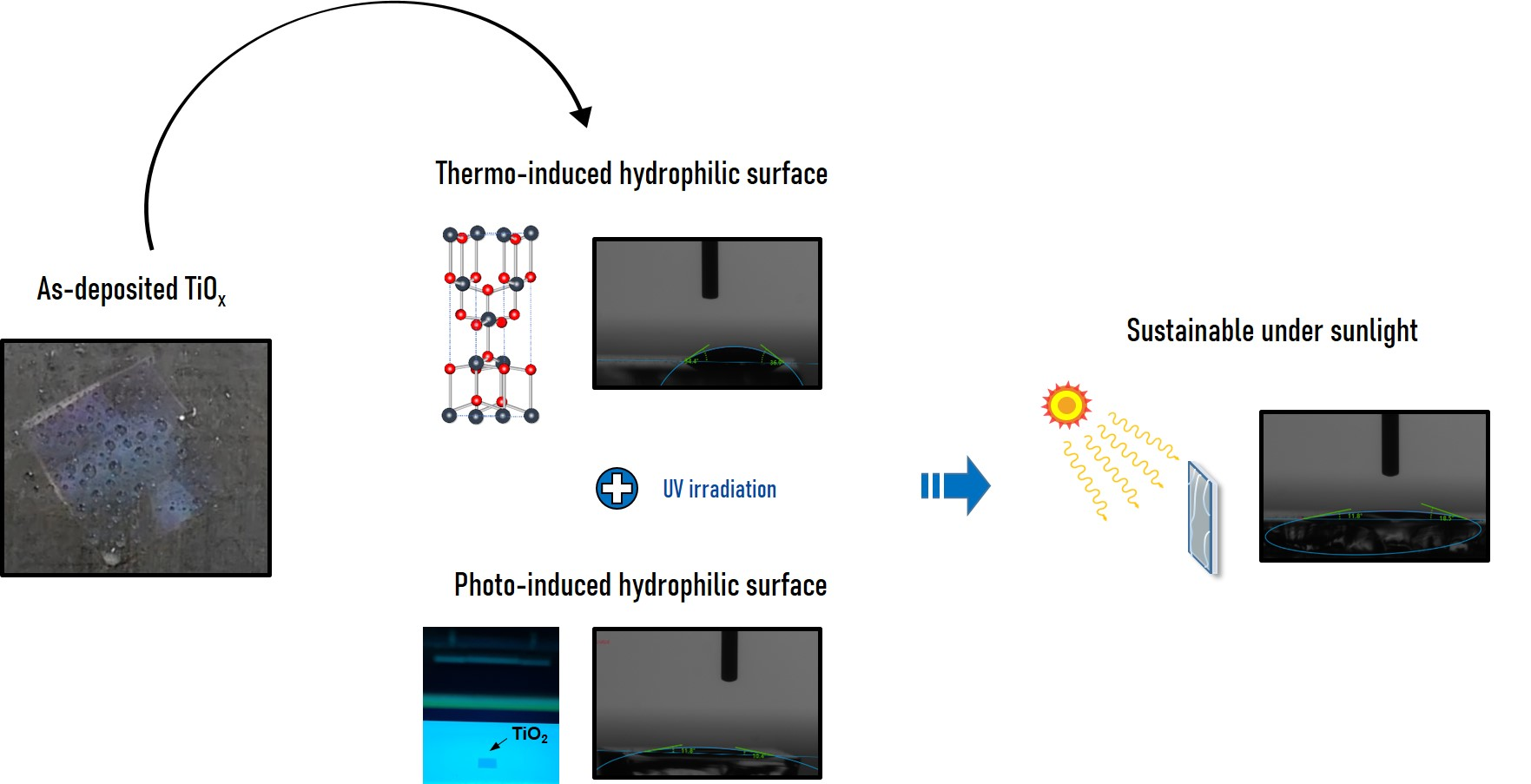
TiO2 thin films have been fabricated by various methods, including solution process such as spin coating, spray pyrolysis, dip coating [13,14,15,16], chemical vapor deposition (CVD) [17], and sputtering [18]. Compared with other methods, DC and RF magnetron sputtering can deposit thin films at a low temperature; further, by adjusting the sputtering conditions, it is easy to control the composition, structure, and thickness of the thin films [15]. However, because the substrate is located within plasma areas, the surface of the films is damaged owing to high-energy-particle collisions in typical sputtering systems. Therefore, in this study, a facing-target sputtering (FTS) system was used for deposition. In an FTS system, the targets face each other within the chamber, plasma is formed between them, and the substrate is separated from the chamber, resulting in less surface damage compared with typical sputtering [19,20]. The high-speed accelerating particles moving between the targets form a magnetic field with a high ionization rate, enabling the fabrication of high-quality thin films [21,22]. Figure 1 shows a schematic diagram of the FTS.
2. Experimental Section
In this study, using an FTS system, we fabricated TiO2 thin films with a hydrophilic surface that are sustainable under sunlight. To investigate the influence of UV irradiation on the formation of a hydrophilic surface, we compared the X-ray photoelectron spectroscopy (XPS) profiles of the films before and after irradiation. The TiO2 films were formed with an anatase or amorphous structure depending on the sputtering conditions. The correlation between the hydrophilicity of the surface and the crystal structure was studied with X-ray diffraction (XRD). Finally, to confirm whether the hydrophilicity of the surface was sustainable under sunlight, the TiO2 films were placed in a dark room and then irradiated with sunlight, following which the sessile drop method was performed for contact angle measurements.
2.1. Fabrication of TiO2 Thin Films
TiO2 thin films were fabricated using a 4-inch FTS system. Soda lime glass substrate with dimensions of 76 × 26 × 1 mm3 (Microspore glass, Marienfeld, Lauda-Königshofen, Germany) was used as the substrates. Ultrasonic cleaning was successively performed with ethyl alcohol (C2H5OH, 94%, extra pure, DUKSAN Pure Chemicals, Ansan, Korea) and acetone (CH3COCH3, ≥99.5%, SAMCHUN Pure Chemicals, Pyeongtaek, Korea) for 10 min; each cleaning procedure was followed by rinsing with distilled water for 5 min. Finally, the substrates were dried using N2 gas and an oven. For sputtering, 4-inch Ti targets were used, the base pressure was 3 × 10⁻⁵ Torr, and the working pressure was set to 2 mTorr. The input power and sputtering time were varied to adjust the crystal structure and thickness. Table 1 lists the specific sputtering conditions, while Table 2 shows the sample parameters.
2.2. Formation of a Sustainable Hydrophilic Surface
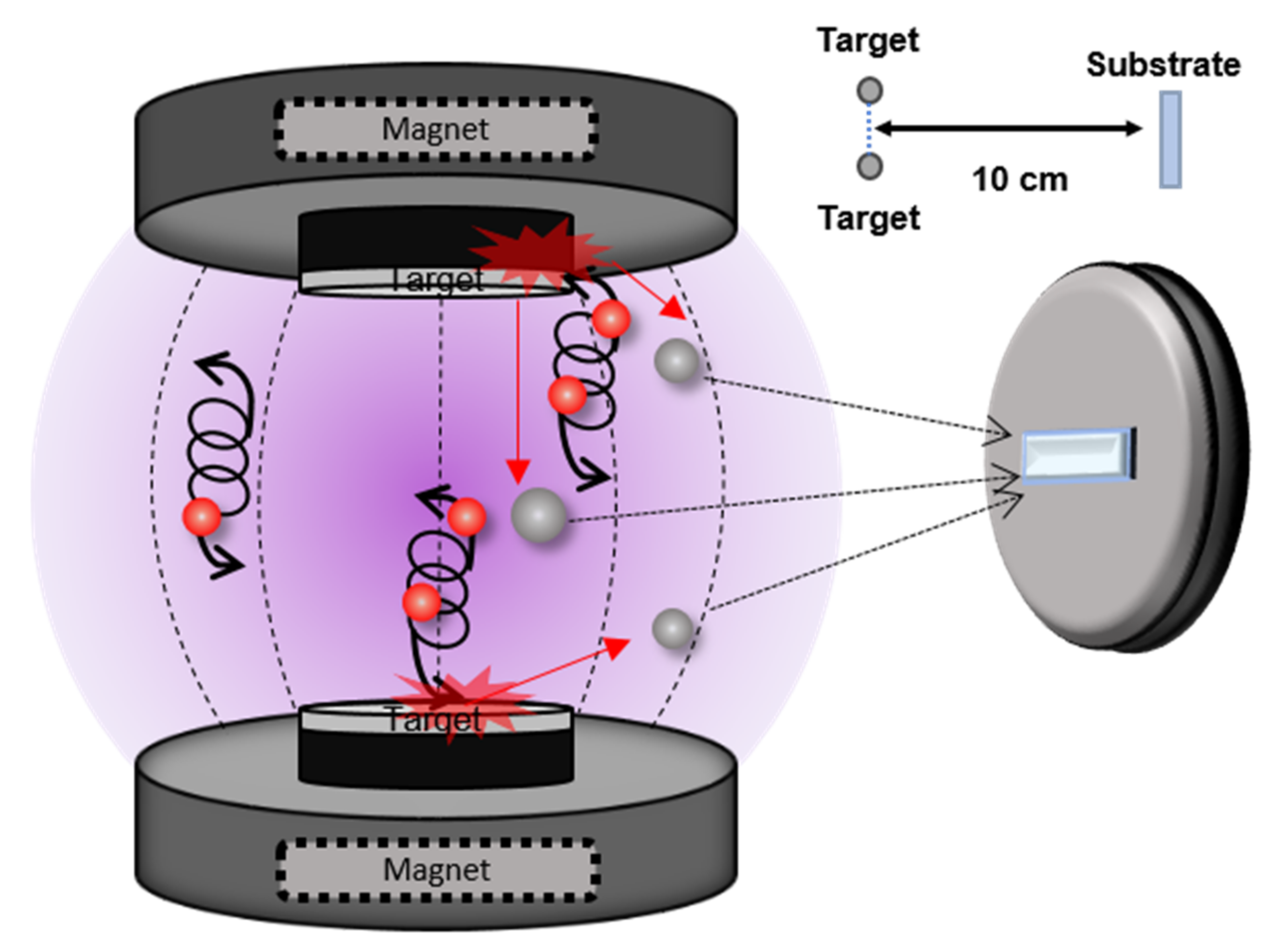
Figure 2 illustrates the method of fabrication of the hydrophilic TiO2 films. The water contact angle was reduced by ultraviolet (UV) irradiation and annealing. After deposition, the samples were cut into two equal parts. One half was placed in an electric muffle furnace and annealed in an air atmosphere and at 500 °C for 3 h to form anatase TiO2. Then, samples were cooled naturally in the furnace to room temperature. Sample H was used to confirm the sustainable hydrophilic properties of the TiO2 film. After 12 h of UV irradiation, the sample was stored in a dark room for a few days, and then exposed to sunlight for 2, 4, 6, and 8 h. The sessile drop method was repeated every 2 h.
2.3. Evaluation of TiO2 Thin Films
A UV-C lamp (G15T8, peak: 253.7 nm, intensity: 1.36 mW/cm2, SANKYO DENKI, Hiratsuka, Japan) was used to irradiate the TiO2 films. The intensity of the UV-C lamp was measured using a UVC light meter (UV C-254SD, Lutron, Coopersburg, PA, USA). The degree of hydrophilicity of the surface was evaluated using a contact angle meter (DSA100, KRUSS, Hamburg, Germany). The thickness of the TiO2 thin films was measured by the Alpha-Step profilometer (Alpha-Step-500 profilers, KLA-Tencor, Milpitas, CA, USA). A UV/Vis spectrometer (Lambda 750 UV/vis/NIR, Perkin Elmer, Waltham, MA, USA) was used to analyze the optical properties. The crystal and surface structures of the films were analyzed by XRD using a high resolution X-ray diffractometer (SmartLab, Rigaku, Tokyo, Japan) in the scanning range of 10°–90° and scanning electron microscopy (SEM, S-4700, Hitachi, Tokyo, Japan), respectively. XPS (K-alpha+, Thermo Fisher Scientific, Waltham, MA, USA) was employed to investigate the difference in the binding energies of Ti and O before and after UV irradiation.
3. Result and Discussion
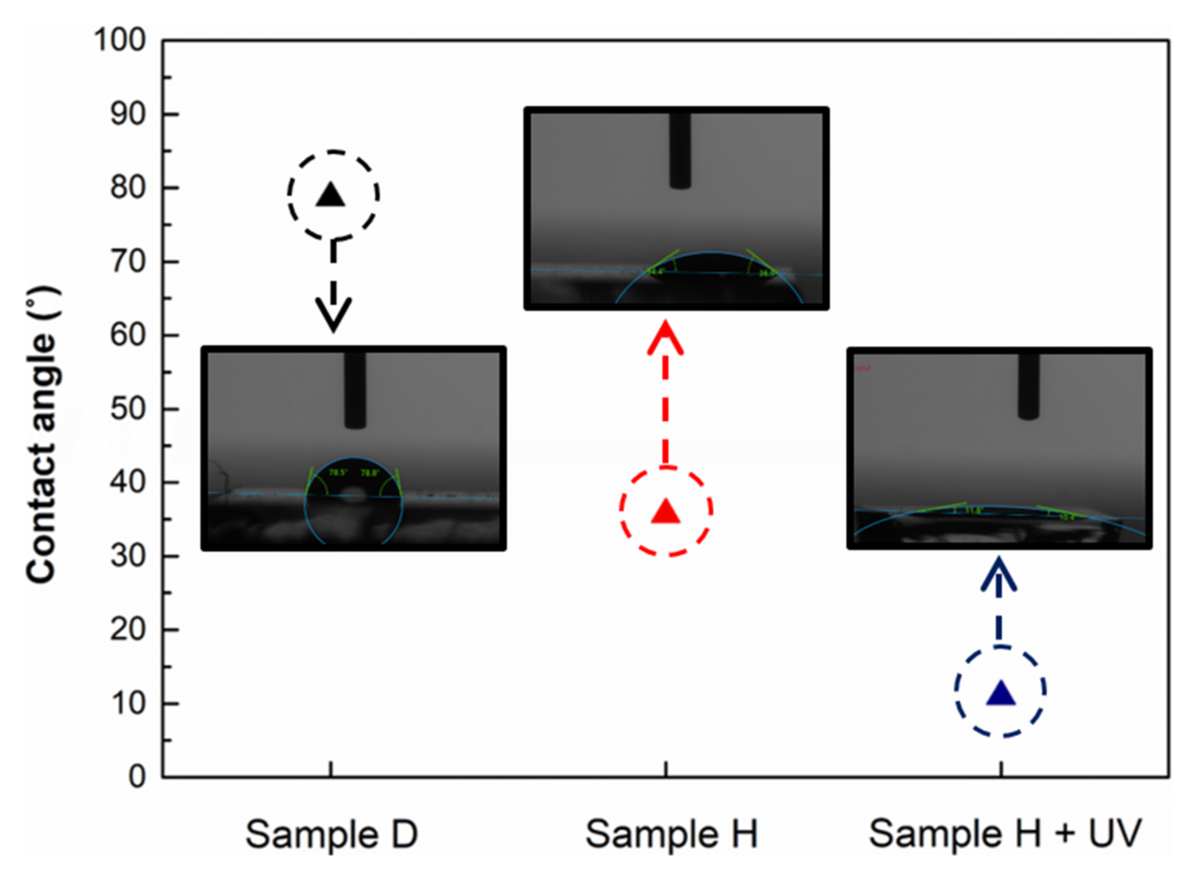
The as-fabricated films possessed a TiOx structure, because the samples were fabricated by DC reactive sputtering. The TiOx films initially exhibited a low hydrophilicity; after thermal treatment, TiOx was converted to anatase TiO2 and the water contact angle drastically decreased. In addition, when the annealed TiO2 thin films were subjected to UV irradiation, the water contact angle decreased to the extent that the surface was almost superhydrophilic. The variation in the water contact angle is illustrated in Figure 3, and indicates that UV irradiation and thermal treatment contributed to the hydrophilicity of the surface of the TiO2 films [18,23]. Therefore, subsequent sections focus on the effects of these methods on the hydrophilicity of the surface. The summaries of analytical data of samples are shown in Table 3. Below, we discuss these results.
3.1. Generation of Light-Induced Surface Oxygen Vacancies
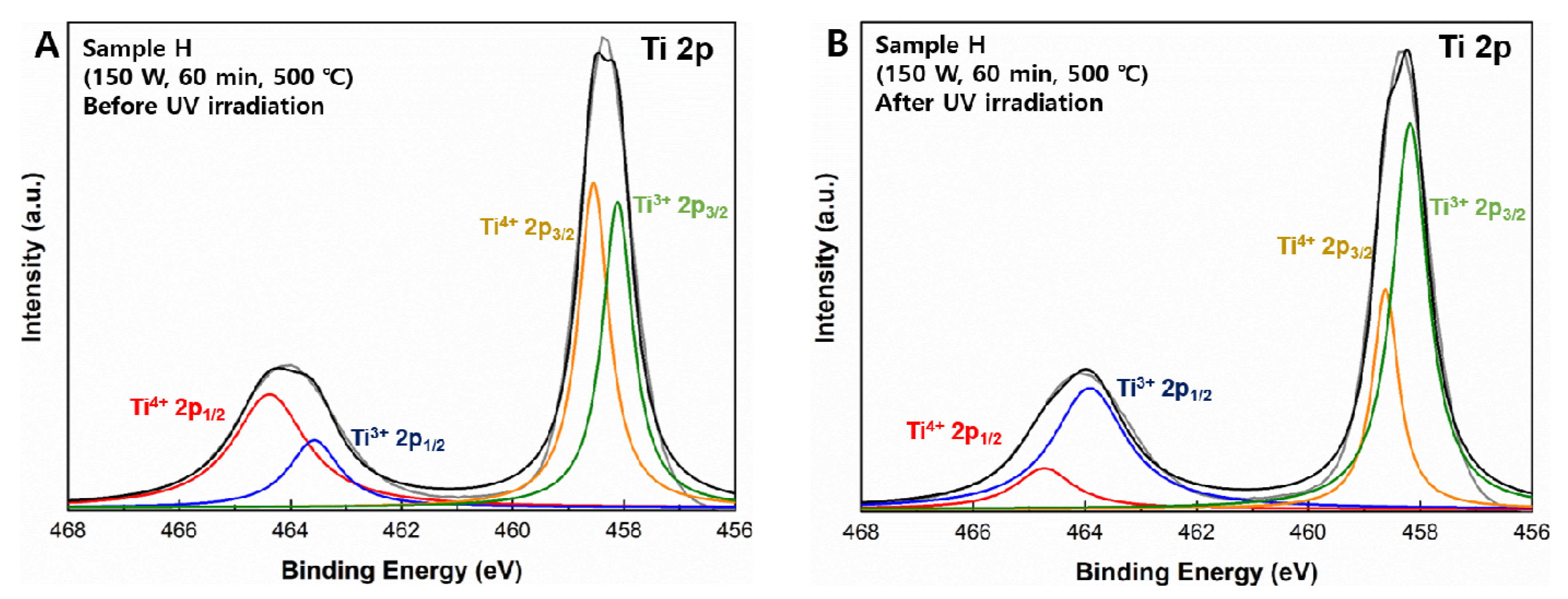
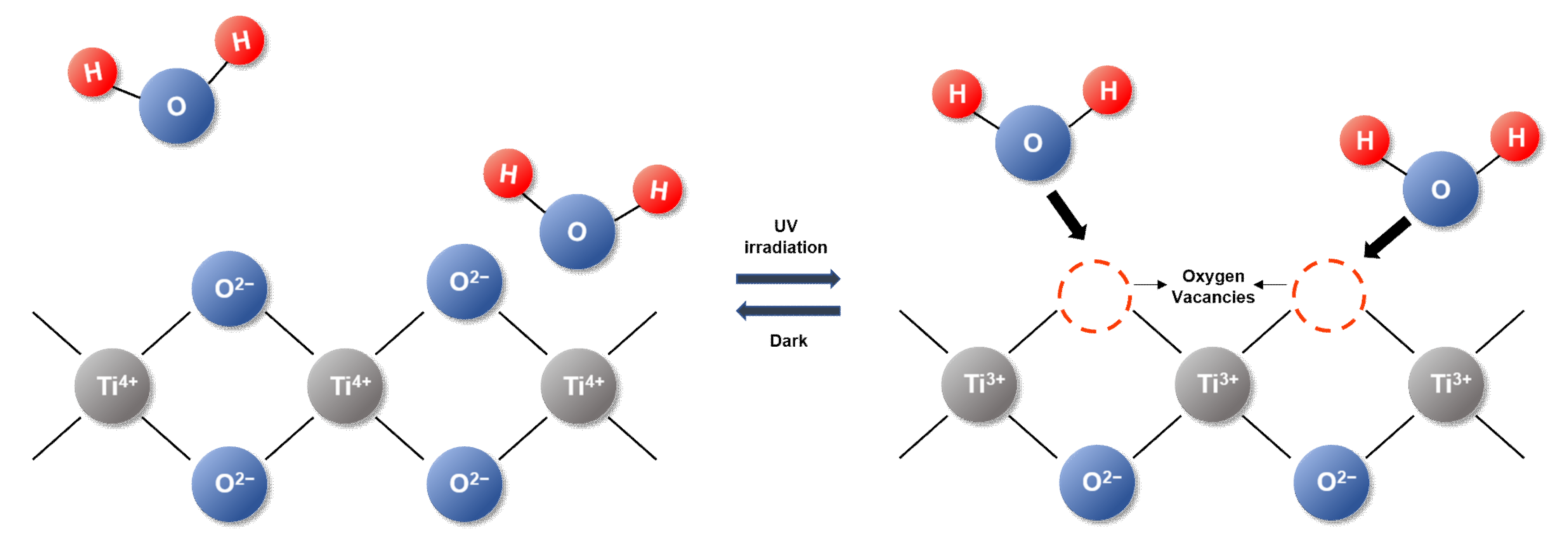
To observe the variation in the lattice structure of TiO2 with UV irradiation, the binding energy of Ti was measured by XPS and is shown in Figure 4. For the measurement, each sample was cut into two pieces (1 × 1 cm2); one piece was stored in a dark room for 12 h and the other was subjected to UV-C irradiation for 12 h, following which both samples were measured simultaneously. Figure 4A shows the XPS profile for the sample stored in the dark, while Figure 4B shows the XPS profile for the UV-irradiated sample. Figure 4 shows two main peaks of Ti 2p, situated at 464.3 and 458.5 eV and assigned to Ti 2p1/2 and Ti 2p3/2, respectively. There are two relatively lower peaks under each main peak; Ti4+ peaks occur at 465.4 eV for Ti 2p1/2 and 458.8 eV for Ti 2p3/2, while Ti3+ peaks occur at 464.5 eV for Ti 2p1/2 and 457.9 eV for Ti 2p3/2 [24]. Several theories have been proposed for the formation of hydrophilic surfaces on TiO2 films via photo-irradiation. Among them, Wang et al. [25] suggested the generation of light-induced surface oxygen vacancies, and reported that the structure of the TiO2 surface changed upon UV irradiation and the contact angle decreased. TiO2 consists of six coordinated Ti atoms and three coordinated O atoms in the bulk. However, on the surface, it comprises five coordinated Ti atoms and two coordinated O atoms. The atomic arrangement is illustrated in Figure 5. When the surface of TiO2 is irradiated with light from a UV-C lamp, oxygen vacancies are possibly generated at the two coordinated bridging sites, converting Ti4+ into Ti3+. Consequently, these defects increase the affinity of the OH− ions and form a hydrophilic surface on the TiO2 films.
Based on the above theory, comparing the profiles in Figure 4A,B, for Ti 2p1/2 and 2p3/2, the Ti3+ content distinctly increased, whereas the Ti4+ content decreased with UV irradiation. The Ti 2p XPS data confirmed that Ti4+ is converted into Ti3+ under UV irradiation; the oxygen vacancies generated by UV-irradiation improve the hydrophilicity of the surface of the TiO2 thin film.
3.2. Photoinduced Reconstruction of Ti–OH
Sakai et al. [26] proposed another theory for the formation of hydrophilic surfaces on TiO2 films, according to which the photoinduced reconstruction of Ti–OH bonds occurs. They suggested that UV irradiation induces the reconstruction of hydroxyl groups on the surface. Positive holes are generated by UV irradiation, which can diffuse to the surface and get trapped at the lattice oxygen sites. Consequently, the binding energies of Ti and the lattice oxygen decrease, owing to which water molecules can be captured by this bond, leading to the formation of new hydroxyl groups. This subsequently improves the degree of hydrophilicity of the surface.
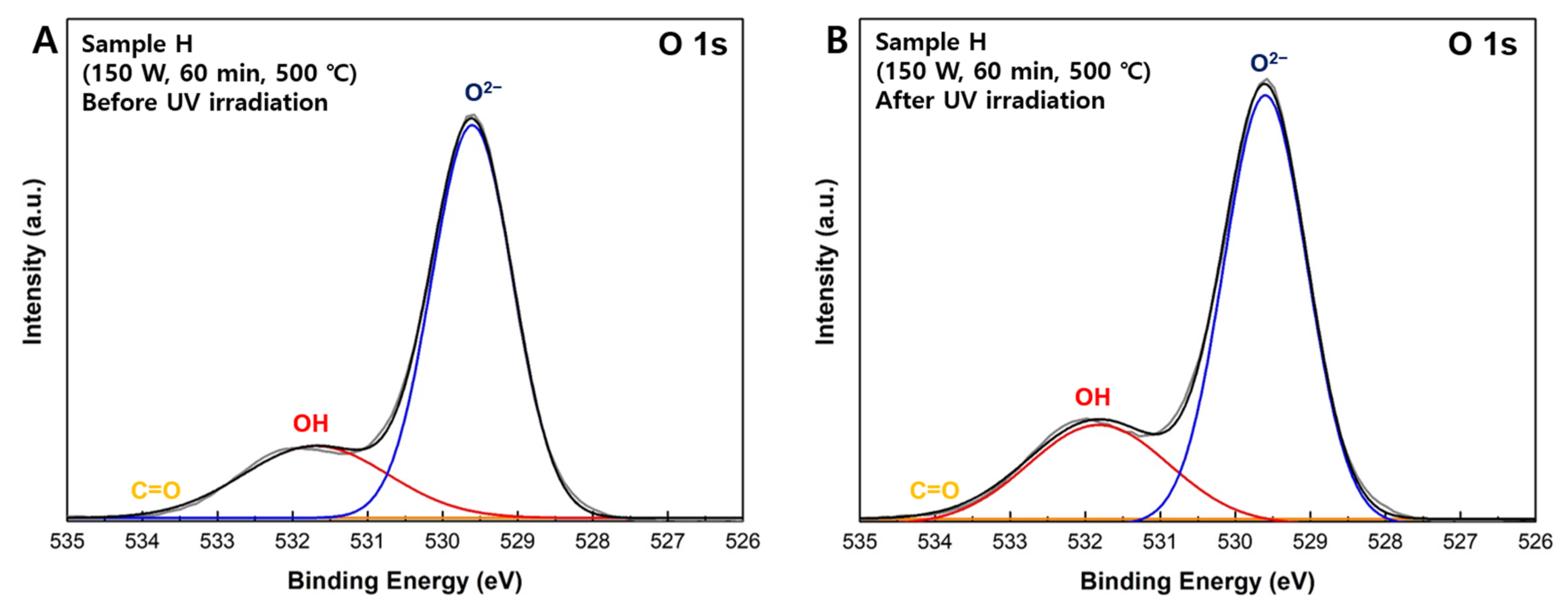
Figure 6 shows the O 1s XPS profiles. To confirm the reconstruction of Ti and OH, we also compared the O 1s spectra for the UV-irradiated and non-UV-irradiated samples. Figure 6A,B show the spectra obtained using the same samples as for Figure 4A,B. In the O 1s spectra, peaks for the O2− species and OH species are observed at binding energies of 529.6 and 532.0 eV, respectively [27]. We compared the intensity ratios of OH to O in Figure 6A,B. The OH/O intensity ratio was 0.337 before UV irradiation and 0.368 after irradiation, indicating that the number of hydroxyl groups increased after UV-C irradiation. The water molecules rupture the bond between Ti and the lattice oxygen under UV irradiation, resulting in the reconstruction of hydroxyl groups. We believed that these additional hydroxyl groups enhanced the affinity for water, resulting in the formation of a hydrophilic surface.
3.3. Influence of Annealing on Water Contact Angle
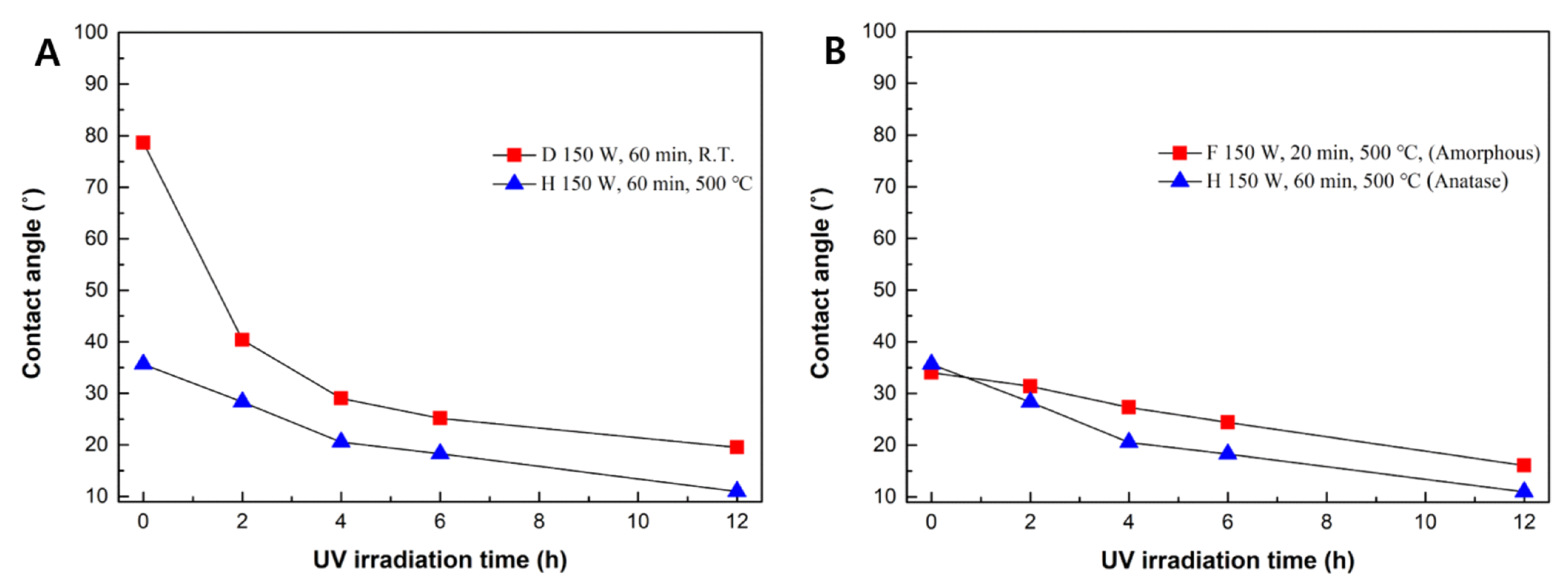
Another method to form a hydrophilic surface on TiO2 films is annealing; the obtained surface is termed a “thermoinduced hydrophilic” surface [28]. To study the influence of annealing on the hydrophilicity of the sample surfaces, the water contact angle and crystallinity of the samples were determined. Figure 7 shows the variation in the water contact angle under UV irradiation, and Figure 8 shows the XRD patterns of the TiO2 thin films.
Samples D and H were fabricated under the same deposition conditions, but sample H was annealed at 500 °C. As shown in Figure 7A, after annealing, the water contact angle decreased from 78.7° to 35.7°, indicating that annealing induced hydrophilicity on the surface. This phenomenon can be attributed to the following, based on previous research: (i) the effect of Ti3+ defect sites, (ii) organic degradation on the surface, and (iii) crystal phase transition [15,25]. Annealing produces Ti3+ defects on the TiO2 surface, generating oxygen vacancies on the surface. The absence of oxygen atoms at the bridging–O site leads to the formation of two subsurface Ti3+ sites. Water molecules occupy these oxygen vacancies and generate OH groups, which adsorb to the surface, resulting in hydrophilicity on the surface of the TiO2 films. Moreover, organic matter on the surface, such as carbon-based or nitrogen-based organic compounds, is largely removed by annealing. Another study confirmed through Fourier-transform infrared (FT-IR) analysis that annealing led to the decomposition of NH3 on the surface of TiO2 [29]. This led to the exposure of the surface of TiO2 to water (the adsorbent), thereby improving the hydrophilicity of the thin films.
3.4. Correlation of Crystallinity and Water Contact Angle
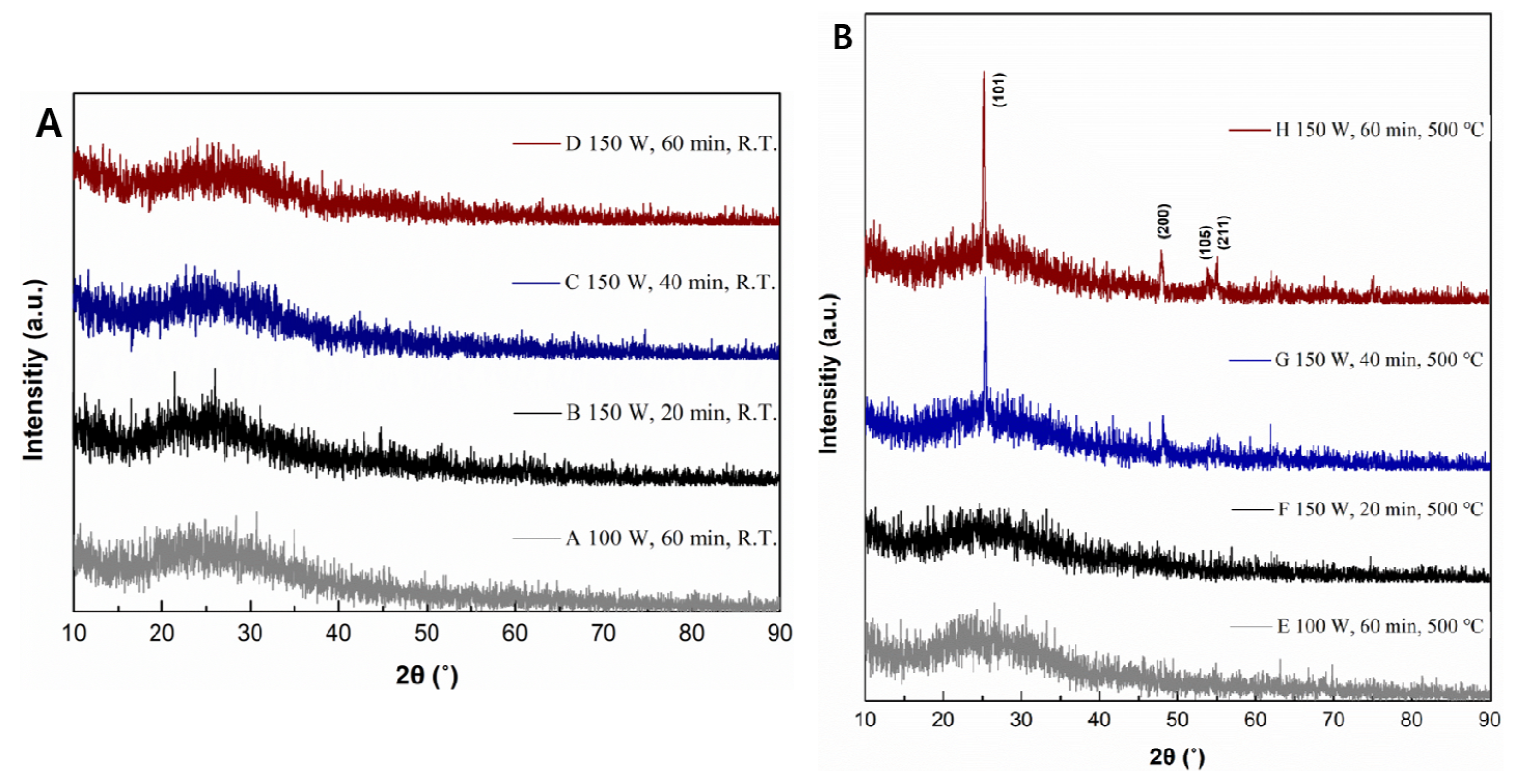
As shown in Figure 8B, annealing not only generated Ti3+ defect sites and decomposed organic matter, but also led to a crystal phase transition from an amorphous to anatase structure at 500 °C. When reactive sputtering Ti and O2 is performed using DC sputtering, the energy of the sputtered target atoms is insufficient for crystallization in the rapidly growing TiO2, and it is fabricated into TiOx structure; initially, TiOx was annealed to form TiO2 at 300 °C. However, although samples were annealed, they just had an amorphous structure. According to another study, although the RF sputtering system, with which it is easier to form crystallinity than when using DC sputtering, was used, samples had an anatase structure at 400 °C and maintained the anatase structure up to 650 °C [18]. Hence, all samples were annealed at 500 °C for 3 h to form the anatase structure [30,31]. Figure 8B confirms the formation of anatase TiO2 through the (101), (200), (105), and (211) peaks. However, samples E and F did not show these peaks in the XRD data, even after annealing. We suggest two reasons as to why sample E did not form an anatase structure. One is the low input power during deposition. When a high sputtering power is supplied, the target atoms possess a high kinetic energy and can migrate to suitable lattice sites [32]. Sample E was prepared with an input power of 100 W; however, an anatase structure was not formed. In the FTS system, the sputtered atoms possess a lower energy than those in other deposition methods, because the atoms that use energy to escape from the plasma region are deposited on the substrate. However, as mentioned earlier, FTS enables the fabrication of high-quality thin films. Therefore, we adjusted the input power from 100 to 150 W to supply enough energy to the target atoms, so that the thin films could easily grow and nucleate.
Although we increased the input power and performed annealing at 500 °C, sample F did not show any peaks in the XRD data. We suggest that this is because of the thickness of the film. In a previous study, Mukherjee et al. studied the intensity of the Raman and integral intensity of the XRD (101) peak, and they proposed that the XRD intensity depends on the thickness [31]. In other words, sample F has insufficient thickness (45.5 nm) to reveal the crystallinity. Consequently, we believe that such samples require a high sputtering power and annealing at approximately 500 °C to form the anatase structure.
We compared the contact angle of sample F, in which crystallinity was not observed, to that of sample H, in which the crystallinity was most pronounced, in order to investigate the effect of crystallinity on the hydrophilicity (Figure 7B). The two samples initially exhibited similar contact angles; however, as the UV irradiation time increased, the anatase TiO2 sample exhibited a lower contact angle than the amorphous TiO2 sample. This is because anatase TiO2 (101) would form the OH species on the surface from H2O gas present in atmosphere under UV irradiation. In other words, the transition in the crystal structure from amorphous to anatase improved the hydrophilicity of the TiO2 films [18,33].
3.5. Crystal Size and Surface Structure of TiO2 Films
The crystallite size of the samples was calculated by the Scherrer equation based on the XRD data in Figure 8B [34]. The Scherrer equation can be written as follows:
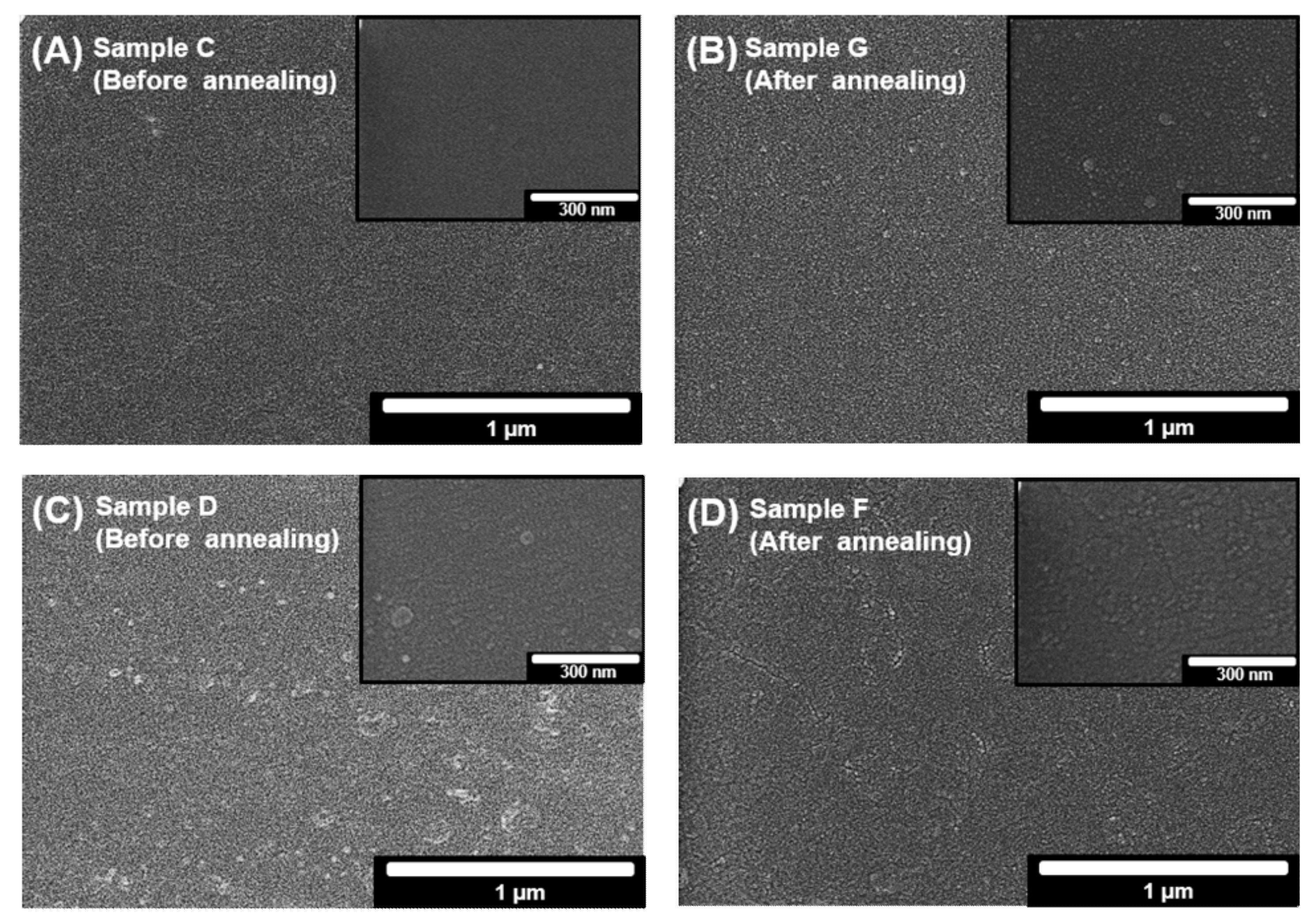
According to this equation, β, the full width at half maximum (FWHM), is inversely related to the crystallite size τ. Therefore, a large FWHM, i.e., a broad XRD pattern, implies a small crystallite size. Based on the average of the (101) and (200) peaks in Figure 8B, it was confirmed that the crystallite sizes depend on the deposition time (sample G: 29.25 nm, sample H: 37.32 nm). We studied the surface structure through SEM images. Figure 9A,B show the morphologies of sample C and sample G, while Figure 9C,D show those of sample D and sample H. Both samples exhibit a smooth and homogeneous surface.
3.6. Optical Properties and Sustainability of TiO2 Films
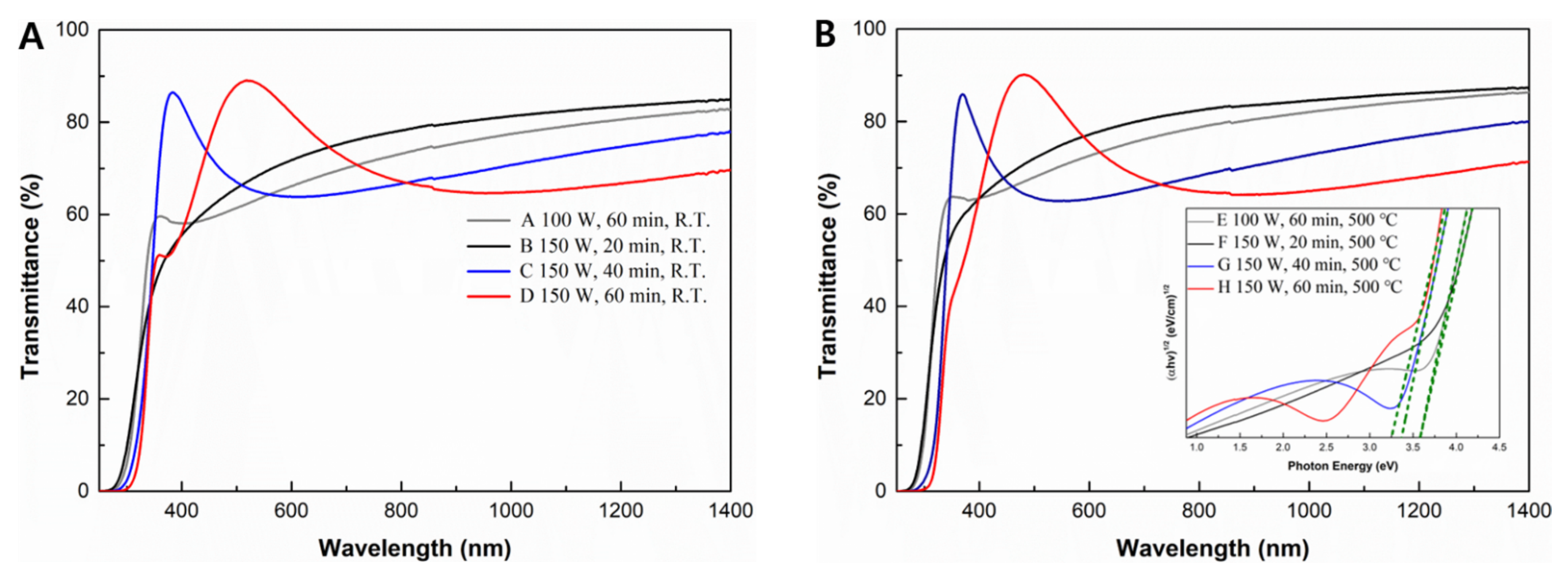
The optical properties of TiO2 were measured using a UV/Vis spectrometer, and the optical bandgap energy was determined. Figure 10A,B show the UV/Vis results before and after annealing, respectively. Sample D shows the highest transmittance in the visible-light region before annealing, while sample H shows the same after annealing. The average transmittances in the visible region (400–700 nm [35]) are 79.1% for sample D and 78.7% for sample H. There is no significant difference in the transmittance before and after annealing. The plot of data reveals interference fringe, indicating that the surface of the films is homogeneous and smooth, as can be seen from the surface SEM image [36]. The optical bandgap energy was also evaluated by the Tauc plot and is shown in Figure 10B, assuming that the absorption product is given the following [37]:
where is a fixed material constant; is the photon energy (= 1240/λ); Eg is the optical bandgap energy; and n is the power coefficient, which can be 3, 2, 3/2, or 1/2 depending on the type of transition (indirect forbidden, indirect allowed, direct forbidden, or direct allowed, respectively). As noted by Zakrzewska, anatase TiO2 possibly permits indirect allowed and direct forbidden transitions [38]. We used the equation for an indirect allowed transition to calculate the Tauc plot. The highest bandgap was 3.6 eV for sample F and sample E and the lowest bandgap was 3.3 eV for sample H. Thus, this optical bandgap energy indicates that a photoreaction of TiO2 occurs under UV wavelength.
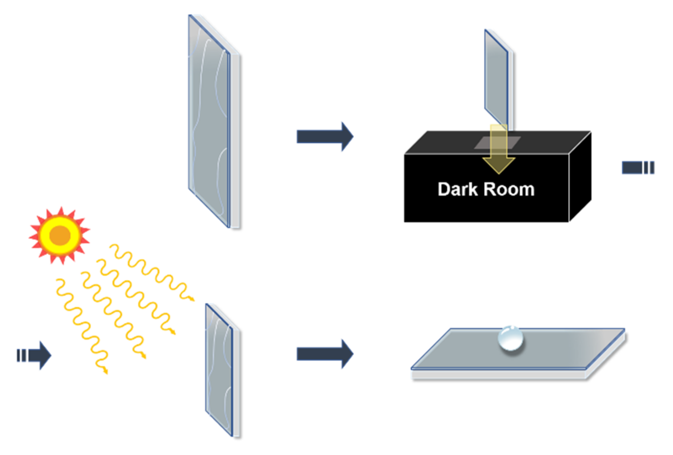
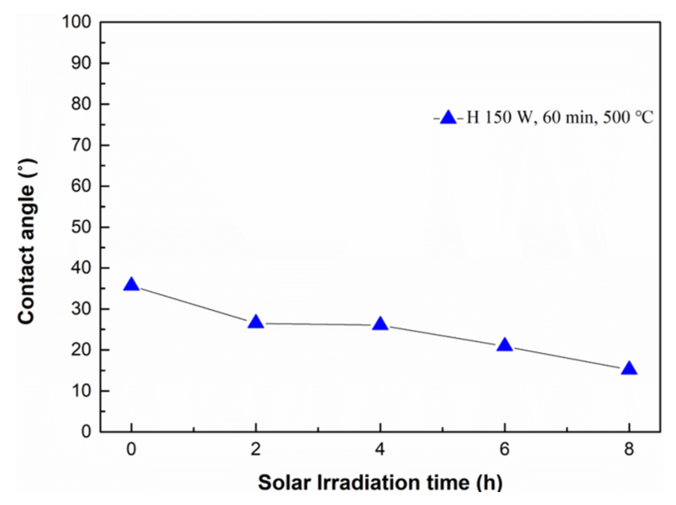
After fabricating a hydrophilic surface on the TiO2 films, we finally verified whether the hydrophilicity would be sustained under sunlight (UV light constitutes 3%–5% of the solar spectrum) [39]. To evaluate the sustainability of the films under sunlight, the changes in the optical properties and water contact angles of the samples after UV irradiation were determined. The experimental process for this evaluation is illustrated in Figure 11. Sample H, on which a hydrophilic surface was formed by annealing and UV irradiation, was used for this evaluation. The sample was stored in a dark room for five weeks, and then irradiated under sunlight when the weather was sunny. The water contact angle was measured every two hours. Figure 12 shows that the water contact angle decreases with increasing sunlight irradiation time, from 35.7° to 15.2° in 8 h. Therefore, we believe that the TiO2 thin films fabricated by FTS can sustain their hydrophilic surfaces under sunlight.
4. Conclusions
TiO2 films were deposited by an FTS system and hydrophilic surfaces were obtained by annealing the films at 500 °C and by UV irradiation. We investigated the influence of annealing and UV irradiation on the water contact angle using XPS and crystallinity data. XPS data reveled that Ti4+ is converted to Ti3+ and the Ti–OH bond is reconstructed under UV irradiation. The annealed TiO2 films exhibited an amorphous or anatase structure depending on the input power and thickness of the sample; further, their hydrophilicity was affected by the crystallinity. Films with an anatase structure exhibited a lower contact angle than those with an amorphous structure as the irradiation time increased. No significant change was observed in the visible-light transmittance before annealing or UV irradiation. Post restoration of its hydrophilic property by placing the sample in a dark room, the contact angle of the hydrophilic surface decreased to 15.2° with 8 h of sunlight exposure. Thus, the results indicate that the hydrophilicity of TiO2 thin films is sustained under natural sunlight. From these results, it is expected that it can be applied to the surface of the sensor or the solar module.
Author Contributions
Conceptualization, J.H., K.K and S.P.; methodology, S.L and T.P.; validation, T.P., S.L. and Y.Y.; formal analysis, S.P. and Y.Y.; investigation, S.P. and J.H.; writing—original draft preparation, S.P.; writing—review and editing, J.H. and K.K.; supervision, J.H. All authors have read and agreed to the published version of the manuscript.
Funding
This research was supported by the Korea Institute of Energy Technology Evaluation and Planning (KETEP) and the Ministry of Trade, Industry, and Energy (MOTIE) of the Republic of Korea (No. 20194030202290) and Basic Science Research Capacity Enhancement Project through Korea Basic Science Institute (National research Facilities and Equipment Center) grant funded by the Ministry of Education (2019R1A6C1010016).
Institutional Review Board Statement
Not applicable.
Informed Consent Statement
Not applicable.
Data Availability Statement
Not applicable.
Conflicts of Interest
The authors declare no competing financial interest.
References
- Pelaez, M.; Nolan, N.T.; Pillai, S.C.; Seery, M.; Falaras, P.; Kontos, A.G.; Dunlop, P.; Hamilton, J.; Byrne, J.; O’Shea, K.; et al. A review on the visible light active titanium dioxide photocatalysts for environmental applications. Appl. Catal. B Environ. 2012, 125, 331–349. [Google Scholar] [CrossRef] [Green Version]
- Banerjee, S.; Dionysiou, D.D.; Pillai, S.C. Self-cleaning applications of TiO2 by photo-induced hydrophilicity and photocatalysis. Appl. Catal. B Environ. 2015, 176, 396–428. [Google Scholar] [CrossRef] [Green Version]
- Ni, M.; Leung, M.K.; Leung, D.Y.; Sumathy, K. A review and recent developments in photocatalytic water-splitting using TiO2 for hydrogen production. Renew. Sustain. Energy Rev. 2007, 11, 401–425. [Google Scholar] [CrossRef]
- Jiang, H.; Katsumata, K.-I.; Hong, J.; Yamaguchi, A.; Nakata, K.; Terashima, C.; Matsushita, N.; Miyauchi, M.; Fujishima, A. Photocatalytic reduction of CO2 on Cu2O-loaded Zn-Cr layered double hydroxides. Appl. Catal. B Environ. 2018, 224, 783–790. [Google Scholar] [CrossRef]
- Hong, J.; Katsumata, K.-I.; Matsushita, N. High-conductivity solution-processed ZnO films realized via UV irradiation and hydrogen treatment. Acta Mater. 2016, 103, 844–849. [Google Scholar] [CrossRef]
- Fujishima, A.; Honda, K. Electrochemical photolysis of water at a semiconductor electrode. Nature 1972, 238, 37–38. [Google Scholar] [CrossRef]
- Parkin, I.P.; Palgrave, R.G. Self-cleaning coatings. J. Mater. Chem. 2005, 15, 1689–1695. [Google Scholar] [CrossRef]
- Takata, Y.; Hidaka, S.; Cao, J.; Nakamura, T.; Yamamoto, H.; Masuda, M.; Ito, T. Effect of surface wettability on boiling and evaporation. Energy 2005, 30, 209–220. [Google Scholar] [CrossRef]
- Fillion, R.; Riahi, A.; Edrisy, A. A review of icing prevention in photovoltaic devices by surface engineering. Renew. Sustain. Energy Rev. 2014, 32, 797–809. [Google Scholar] [CrossRef]
- Zhang, L.; Dillert, R.; Bahnemann, D.; Vormoor, M. Photo-induced hydrophilicity and self-cleaning: Models and reality. Energy Environ. Sci. 2012, 5, 7491–7507. [Google Scholar] [CrossRef]
- Nishimoto, S.; Bhushan, B. Bioinspired self-cleaning surfaces with superhydrophobicity, superoleophobicity, and superhydrophilicity. RSC Adv. 2013, 3, 671–690. [Google Scholar] [CrossRef]
- Nakata, K.; Fujishima, A. TiO2 photocatalysis: Design and applications. J. Photochem. Photobiol. C Photochem. Rev. 2012, 13, 169–189. [Google Scholar] [CrossRef]
- Elfanaoui, A.; Elhamri, E.; Boulkaddat, L.; Ihlal, A.; Bouabid, K.; Laanab, L.; Taleb, A.; Portier, X. Optical and structural properties of TiO2 thin films prepared by sol–gel spin coating. Int. J. Hydrogen Energy 2011, 36, 4130–4133. [Google Scholar] [CrossRef]
- Chanda, A.; Joshi, S.R.; Akshay, V.; Varma, S.; Singh, J.; Vasundhara, M.; Shukla, P. Structural and optical properties of multilayered un-doped and cobalt doped TiO2 thin films. Appl. Surf. Sci. 2021, 536, 147830. [Google Scholar] [CrossRef]
- Dundar, I.; Mere, A.; Mikli, V.; Krunks, M.; Acik, I.O. Thickness effect on photocatalytic activity of TiO2 thin films fabricated by ultrasonic spray pyrolysis. Catalysts 2020, 10, 1058. [Google Scholar] [CrossRef]
- Kayani, Z.N.; Riaz, S.; Naseem, S. Magnetic and antibacterial studies of sol-gel dip coated Ce doped TiO2 thin films: Influence of Ce contents. Ceram. Int. 2020, 46, 381–390. [Google Scholar] [CrossRef]
- Bessergenev, V.; Khmelinskii, I.; Pereira, R.; Krisuk, V.; Turgambaeva, A.; Igumenov, I. Preparation of TiO2 films by CVD method and its electrical, structural and optical properties. Vacuum 2002, 64, 275–279. [Google Scholar] [CrossRef]
- Ye, Q.; Liu, P.; Tang, Z.; Zhai, L. Hydrophilic properties of nano-TiO2 thin films deposited by RF magnetron sputtering. Vacuum 2007, 81, 627–631. [Google Scholar] [CrossRef]
- Lee, M.; Park, Y.; Kim, K.; Hong, J. Influence of sputtering conditions on the properties of aluminum-doped zinc oxide thin film fabricated using a facing target sputtering system. Thin Solid Films 2020, 703, 137980. [Google Scholar] [CrossRef]
- Shin, J.; Kim, K.; Hong, J. Zn-Al layered double hydroxide thin film fabricated by the sputtering method and aqueous solution treatment. Coatings 2020, 10, 669. [Google Scholar] [CrossRef]
- Hong, J.S.; Matsushita, N.; Kim, K.H. Investigation of the effect of oxygen gas on properties of GAZO thin films fabricated by facing targets sputtering system. Semicond. Sci. Technol. 2014, 29, 075007. [Google Scholar] [CrossRef]
- Hong, J.; Kim, K.-H. Characteristic of Al-In-Sn-ZnO Thin film prepared by FTS system with hetero targets. Trans. Electr. Electron. Mater. 2011, 12, 76–79. [Google Scholar] [CrossRef] [Green Version]
- Wang, R.; Hashimoto, K.; Fujishima, A.; Chikuni, M.; Kojima, E.; Kitamura, A.; Shimohigoshi, M.; Watanabe, T. Photo-generation of highly amphiphilic TiO2 surfaces. Adv. Mater. 1998, 10, 135–138. [Google Scholar] [CrossRef]
- Zhang, H.; Shi, X.; Tian, A.; Wang, L.; Liu, C. Electrochemical properties of Ti3+ doped Ag-Ti nanotube arrays coated with hydroxyapatite. Appl. Surf. Sci. 2018, 436, 579–584. [Google Scholar] [CrossRef]
- Wang, R.; Hashimoto, K.; Fujishima, A.; Chikuni, M.; Kojima, E.; Kitamura, A.; Shimohigoshi, M.; Watanabe, T. Light-induced amphiphilic surfaces. Nature 1997, 388, 431–432. [Google Scholar] [CrossRef]
- Sakai, N.; Fujishima, A.; Watanabe, T.; Hashimoto, K. Enhancement of the photoinduced hydrophilic conversion rate of TiO2 film electrode surfaces by anodic polarization. J. Phys. Chem. B 2001, 105, 3023–3026. [Google Scholar] [CrossRef]
- Chi, M.; Sun, X.; Sujan, A.; Davis, Z.; Tatarchuk, B.J. A quantitative XPS examination of UV induced surface modification of TiO2 sorbents for the increased saturation capacity of sulfur heterocycles. Fuel 2019, 238, 454–461. [Google Scholar] [CrossRef]
- Lu, G.; Linsebigler, A.; Yates, J.T., Jr. Ti3+ defect sites on TiO2 (110): Production and chemical detection of active sites. J. Phys. Chem. 1994, 98, 11733–11738. [Google Scholar] [CrossRef]
- Chen, X.; Wang, X.; Hou, Y.; Huang, J.; Wu, L.; Fu, X. The effect of postnitridation annealing on the surface property and photocatalytic performance of N-doped TiO2 under visible light irradiation. J. Catal. 2008, 255, 59–67. [Google Scholar] [CrossRef]
- Mukherjee, S.K.; Mergel, D. Thickness dependence of the growth of magnetron-sputtered TiO2 films studied by Raman and optical transmittance spectroscopy. J. Appl. Phys. 2013, 114, 13501. [Google Scholar] [CrossRef]
- Mukherjee, S.; Nebatti, A.; Mohtascham, F.; Schipporeit, S.; Notthoff, C.; Mergel, D. Influence of thickness on the structural properties of radio-frequency and direct-current magnetron sputtered TiO2 anatase thin films. Thin Solid Films 2014, 558, 443–448. [Google Scholar] [CrossRef]
- Li, S.; Jiao, S.; Wang, D.; Gao, S.; Wang, J. The influence of sputtering power on the structural, morphological and optical properties of β-Ga2O3 thin films. J. Alloys Compd. 2018, 753, 186–191. [Google Scholar] [CrossRef]
- Nadeem, I.M.; Harrison, G.T.; Wilson, A.; Pang, C.L.; Zegenhagen, J.; Thornton, G. Bridging hydroxyls on anatase TiO2(101) by water dissociation in oxygen vacancies. J. Phys. Chem. B 2018, 122, 834–839. [Google Scholar] [CrossRef] [PubMed]
- Scherrer, P. Determination of the size and internal structure of colloidal particles using X-rays. Nachr. Ges. Wiss. Göttingen 1918, 2, 98–100. [Google Scholar]
- Hong, J.S.; Wagata, H.; Ohashi, N.; Katsumata, K.-I.; Okada, K.; Matsushita, N. Transparent ZnO films deposited by aqueous solution process under various pH conditions. J. Electron. Mater. 2015, 44, 2657–2662. [Google Scholar] [CrossRef]
- Nakaruk, A.; Ragazzon, D.; Sorrell, C. Anatase–rutile transformation through high-temperature annealing of titania films produced by ultrasonic spray pyrolysis. Thin Solid Films 2010, 518, 3735–3742. [Google Scholar] [CrossRef]
- Michalow, K.A.; Logvinovich, D.; Weidenkaff, A.; Amberg, M.; Fortunato, G.; Heel, A.; Graule, T.; Rekas, M. Synthesis, characterization and electronic structure of nitrogen-doped TiO2 nanopowder. Catal. Today 2009, 144, 7–12. [Google Scholar] [CrossRef]
- Zakrzewska, K. Titanium Dioxide Thin Films for Gas Sensors and Photonic Applications; AGH Uczelniane Wydawnictwa Naukowo-Dydaktyczne: Krakow, Poland, 2003. [Google Scholar]
- Banerjee, S.; Pillai, S.C.; Falaras, P.; O’Shea, K.E.; Byrne, J.A.; Dionysiou, D.D. New insights into the mechanism of visible light photocatalysis. J. Phys. Chem. Lett. 2014, 5, 2543–2554. [Google Scholar] [CrossRef] [PubMed] [Green Version]
Figure 1.
Schematic of the FTS system.

Figure 2.
Schematic showing the experimental procedure for the formation of TiO2 films with a hydrophilic surface.
Figure 2.
Schematic showing the experimental procedure for the formation of TiO2 films with a hydrophilic surface.

Figure 3.
Variation in water contact angle with thermal treatment and UV irradiation (Sample D: 150 W, 60 min, R.T., Sample H: 150 W, 60 min, 500 °C, Sample H + UV: Sample H and UV irradiation).
Figure 3.
Variation in water contact angle with thermal treatment and UV irradiation (Sample D: 150 W, 60 min, R.T., Sample H: 150 W, 60 min, 500 °C, Sample H + UV: Sample H and UV irradiation).

Figure 4.
Ti 2p XPS profiles (A) before UV irradiation and (B) after UV irradiation.

Figure 5.
Atomic arrangement with the generation of surface oxygen vacancies.

Figure 6.
XPS spectra of O 1s (A) before UV irradiation and (B) after UV irradiation.

Figure 7.
Variation in water contact angles of (A) Sample D (non-annealed) and H (annealed at 500 °C) and (B) Sample F (amorphous structure) and H (anatase structure).
Figure 7.
Variation in water contact angles of (A) Sample D (non-annealed) and H (annealed at 500 °C) and (B) Sample F (amorphous structure) and H (anatase structure).

Figure 8.
XRD pattern of (A) the TiOx thin films (non-annealed) and (B) TiO2 thin films (annealed at 500 °C).
Figure 8.
XRD pattern of (A) the TiOx thin films (non-annealed) and (B) TiO2 thin films (annealed at 500 °C).

Figure 9.
Surface SEM image of samples before and after annealing: (A) sample C (before), (B) sample G (after), (C) sample D (before), and (D) sample H (after).
Figure 9.
Surface SEM image of samples before and after annealing: (A) sample C (before), (B) sample G (after), (C) sample D (before), and (D) sample H (after).

Figure 10.
UV/Vis spectra of (A) TiOx thin films (non-annealed) and (B) TiO2 thin films (annealed at 500 °C), and Tauc plot of TiO2.
Figure 10.
UV/Vis spectra of (A) TiOx thin films (non-annealed) and (B) TiO2 thin films (annealed at 500 °C), and Tauc plot of TiO2.

Figure 11.
Schematic illustrating the procedure of the sustainability evaluation.

Figure 12.
Variation in water contact angle with solar irradiation times.


Table 1.
Sputtering conditions.
| Parameters | Conditions |
|---|---|
| Targets | Ti, 4 inches |
| Substrate | Glass microscope slides |
| Base pressure | 3 × 10⁻⁵ Torr |
| Working pressure | 2 mTorr |
| Gas flow | Ar, 10 sccm; O2, 1 sccm |
| Input power | 100 W, 150 W |
Table 2.
Sample parameters.
| Samples | A | B | C | D | E | F | G | H |
|---|---|---|---|---|---|---|---|---|
| Input power (W) | 100 | 150 | 150 | 150 | 100 | 150 | 150 | 150 |
| Deposition time (min) | 60 | 20 | 40 | 60 | 60 | 20 | 40 | 60 |
| Annealing (500 °C) | X | X | X | X | O | O | O | O |
Table 3.
The summaries of the analytical data of all samples.
| Samples | A | B | C | D | E | F | G | H |
|---|---|---|---|---|---|---|---|---|
| Water contact angle Before UV irradiation (°) | 77.5 | 75.6 | 72.9 | 78.7 | 35.5 | 34.0 | 37.4 | 35.7 |
| Water contact angle After 12 h UV irradiation (°) | 36.9 | 36.7 | 32.0 | 20.0 | 18.4 | 16.1 | 11.0 | 10.8 |
| Thickness (nm) | 90.9 | 45.5 | 91.0 | 136.4 | 90.9 | 45.5 | 91.0 | 136.4 |
| Phase (Amorphous: AP) (Anatase: AT) | AP | AP | AP | AP | AP | AP | AT | AT |
| Band gap (eV) | 3.6 | 3.6 | 3.4 | 3.3 | 3.6 | 3.6 | 3.4 | 3.3 |
Publisher’s Note: MDPI stays neutral with regard to jurisdictional claims in published maps and institutional affiliations. |
© 2021 by the authors. Licensee MDPI, Basel, Switzerland. This article is an open access article distributed under the terms and conditions of the Creative Commons Attribution (CC BY) license (https://creativecommons.org/licenses/by/4.0/).
Share and Cite
MDPI and ACS Style
Park, S.; Yoon, Y.; Lee, S.; Park, T.; Kim, K.; Hong, J. Thermoinduced and Photoinduced Sustainable Hydrophilic Surface of Sputtered-TiO2 Thin Film. Coatings 2021, 11, 1360. https://doi.org/10.3390/coatings11111360
AMA Style
Park S, Yoon Y, Lee S, Park T, Kim K, Hong J. Thermoinduced and Photoinduced Sustainable Hydrophilic Surface of Sputtered-TiO2 Thin Film. Coatings. 2021; 11(11):1360. https://doi.org/10.3390/coatings11111360
Chicago/Turabian StylePark, Sangbin, Younghwa Yoon, Sehyun Lee, Taejun Park, Kyunghwan Kim, and Jeongsoo Hong. 2021. "Thermoinduced and Photoinduced Sustainable Hydrophilic Surface of Sputtered-TiO2 Thin Film" Coatings 11, no. 11: 1360. https://doi.org/10.3390/coatings11111360
Note that from the first issue of 2016, this journal uses article numbers instead of page numbers. See further details here.