Article

On the Biodiversity and Biodeteriogenic Activity of Microbial Communities Present in the Hypogenic Environment of the Escoural Cave, Alentejo, Portugal

1
HERCULES Laboratory, Universidade de Évora, 7000-809 Évora, Portugal
2
City U Macau Chair in Sustainable Heritage, Universidade de Évora, 7000-809 Évora, Portugal
3
Departamento de Química, Universidade de Évora, Escola de Ciências e Tecnologia, 7000-671 Évora, Portugal
4
Geosciences Department, School of Sciences and Technology, Évora University, Rua Romão Ramalho 59, 7000-671 Évora, Portugal
*
Author to whom correspondence should be addressed.
Coatings 2021, 11(2), 209; https://doi.org/10.3390/coatings11020209
Submission received: 6 January 2021 / Revised: 3 February 2021 / Accepted: 8 February 2021 / Published: 11 February 2021
(This article belongs to the Special Issue Biofilms on Cultural Heritage)

Abstract

:
Hypogenic caves represent unique environments for the development of specific microbial communities that need to be studied. Caves with rock art pose an additional challenge due to the fragility of the paintings and engravings and to microbial colonization which may induce chemical, mechanical and aesthetic alterations. Therefore, it is essential to understand the communities that thrive in these environments and to monitor the activity and effects on the host rock in order to better preserve and safeguard these ancestral artforms. This study aims at investigating the Palaeolithic representations found in the Escoural Cave (Alentejo, Portugal) and their decay features. These prehistoric artworks, dating back up to 50,000 B.P., are altered due to environmental conditions and microbial activity inside the cave. Microbial cultivation methods combined with culture-independent techniques, biomarkers’ viability assays and host rock analysis allowed us to better understand the microbial biodiversity and biodeteriogenic activity within the hypogenic environment of this important cave site. This study is part of a long-term monitoring program envisaged to understand the effect of this biocolonisation and to understand the population dynamics that thrive in this hypogean environment.

1. Introduction

Within the cultural heritage research field, cave rock art plays an essential role in understanding the development of art and, more generally, of human cultural development. Communities inhabiting Western Europe in the Upper Palaeolithic (35,000–10,000 BP) left us numerous examples of cave artworks in the form of paintings, drawings and engravings testifying the development of this unique phenomenon. Many of these artworks have suffered and are suffering chemical, mechanical and aesthetic alterations due to physical, chemical and biological weathering processes induced by the colonization of host rock surfaces by microbial communities [1,2,3]. In order to preserve cave artworks for future generations, it is therefore essential to develop analytical tools and protocols enabling fast and reliable monitoring of the microbial activity present on decorated walls in prehistoric caves.
Previous studies have successfully isolated and identified the presence in several cave environments of pathogenic fungi such as Hitoplasma capsulatum, Microsporum gypseum and Trichophyton mentagrophytes [4], or Trichosporon [5,6,7,8]. Beside issues related to a potential threat to human health, fungi and, more generally, micro-organisms are responsible for both aesthetic (formation of red, pink, purple, blue, green, white spots) and/or physico-chemical (cracks, oxidation, solubilisation, and precipitation episodes) alterations on the decorated parietal surfaces of the cave. In particular it has been shown that microbial communities may promote biomediated precipitation [9,10] and/or dissolution of carbonate minerals [11]. Both processes may damage the parietal paintings found inside of caves because the artworks can disappear underneath a biomineralised calcite deposits and/or alter themselves severely due to the weakening of their support caused by microbial acid attack.
Several studies have focused their attention on the formation of biofilms on stone monuments and their alteration induced by microbial communities. Although the large majority of them concentrated on stone building and monuments [8,12,13,14,15,16,17,18,19,20,21], mortars [22,23] and mural paintings [24,25,26,27,28,29,30], the presence of microbial biofilms have also been widely reported on the walls of several hypogean caves [3,31,32,33,34,35,36,37], catacombs [38,39,40,41] and crypts [22,29,42]. Studies carried out on Altamira Cave’s microbial communities, for instance, have revealed the presence of a large diversity of micro-organisms inducing numerous wall’s alterations [2,10,43,44,45,46,47,48,49,50,51]. These studies demonstrated that microbial biofilms in caves are associated with complex and significant interactions between the diverse communities responsible for their growth. A large extent of caves’ speleothems, such as moonmilk, are thought to result from calcification of biofilms.
In the field of cultural heritage and more precisely cave rock art, effective and fast methods are therefore required to identify the communities contaminating these artworks and their influence on the conservation of the archaeological site where they are found. Although several tests exist for assessing biocontamination in samples such as soils and modern building material [52,53,54,55], only few have been done to rapidly assess the extent of microbial contamination in caves displaying rock artworks [36,56,57]. Developing such tests would help to quickly understand the threats facing the artworks and to assist conservators and public authorities in planning effective interventions.
This study reports the use of several complementary techniques to assess the microbial diversity and activity inside a unique Portuguese archaeological site: the Escoural Cave.
The Escoural archaeological site is a natural cave located near Montemor-o-Novo (Alentejo, Portugal, coordinates: 38°32′37.05″ N, 8°8′14.99″ W). The cave was discovered in April 1963 during quarrying after an explosion for extracting-marble-block revealed the existence of a cavity at the site. At present, the visits to Escoural Cave are limited to 2 per day and 10 persons per visit following an advanced-booking agenda. It was occupied by humans from the Middle Palaeolithic until the Early Chalcolithic, with no outside disruption since the Early Chalcolithic until its discovery in 1963. The cave hosts numerous altered paintings and engravings dating back (35,000–10,000 BP) to the Upper Palaeolithic [58]. Most of the representations are covered by layers of re-precipitated calcite minerals due to water leaking on the walls of the cave; on several areas of the cave walls, biological outbreaks associated with the development of microbial communities can be identified. (Figure 1). The present work aims at providing for the first time a first insight on the microbial diversity and activity in Escoural Cave to better understand their influence on the paintings found inside the cavity.

2. Materials and Methods

2.1. Sampling

In situ observations highlighted a large diversity of biological contaminations present within the Escoural cave (Figure 1).
Samples were collected with a sterile scalpel parallel and not touching the walls surface and/or by rubbing the walls’ surface with cotton swabs at different locations of the cave with various potential biological contaminations, and then stored in sterile tubes. The samples collected by cotton swabs were preserved in sterile MRD. The location of each sample was precisely documented through various photographs taken during the sampling procedure.

2.2. Microbial Contamination: Assessment and Diversity
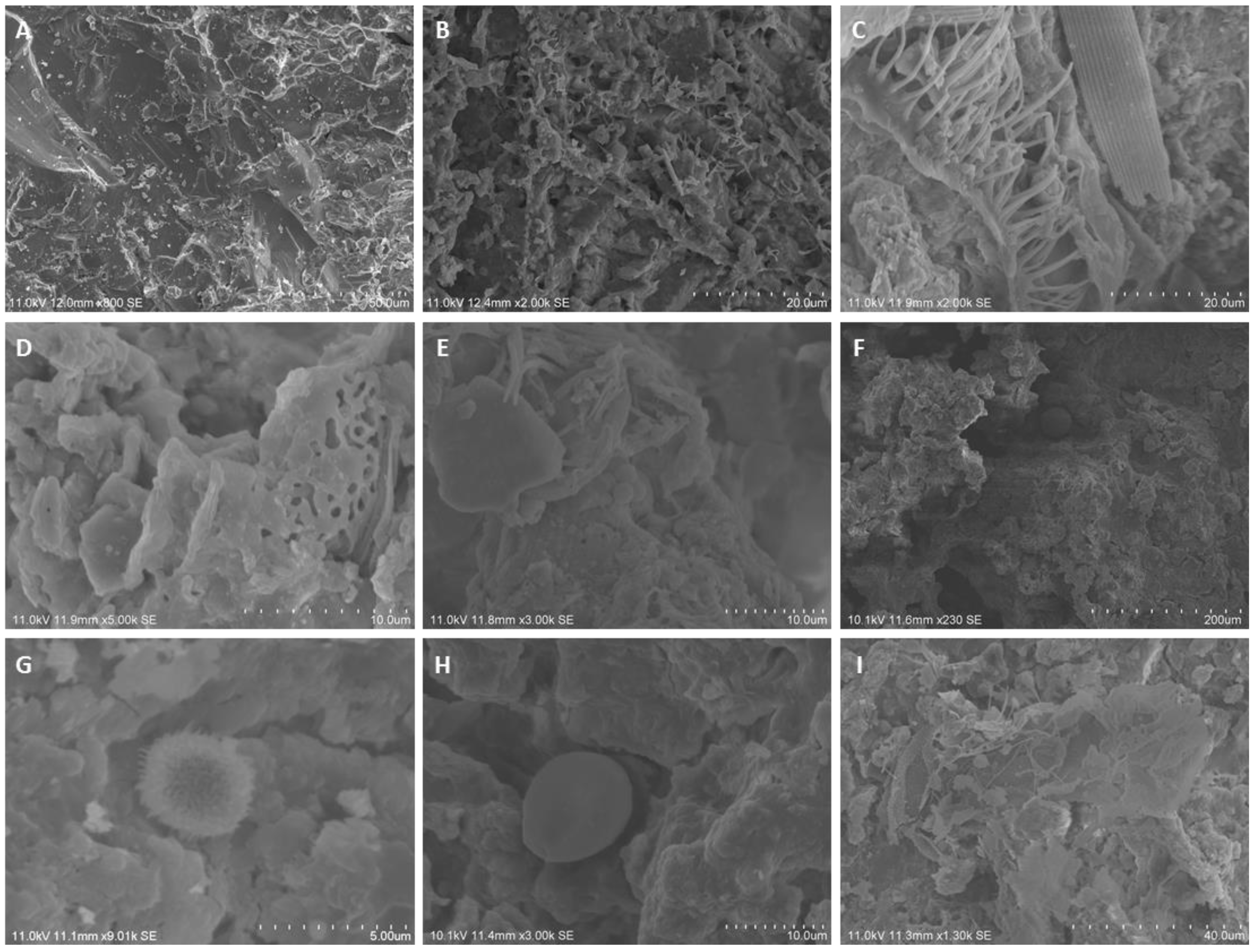
2.2.1. SEM

In order to assess the microbial contamination of the samples and observing the microbiota colonizing the cave walls, its extent and activity on the marble host-rock, microsamples were observed by Scanning Electron Microscopy (SEM).
Microsamples were coated with Au-Pd (Quorum Q150R ES) during 300 s, at 25 mA, and observed in a HITACHI S-3700N variable pressure scanning electron microscope (VP-SEM) with accelerating voltage of 9–10 kV using Backscattered detector mode.

2.2.2. Isolation Procedure

Biological samples were diluted in MRD and inoculated (100 μL) in media such as nutrient agar (NA, HIMEDIA—peptic digest animals of 5 g/L, beef extract 1.5 g/L, yeast extract 1.5 g/L, sodium chloride 5 g/L and agar 15 g/L) for bacteria isolation, malt extract agar (MEA, HIMEDIA—malt extract 30 g/L, peptone mycologic 5 g/L and agar 15 g/L) and Cooke’s Rose Bengal (CRB, Mikrobiologie—peptone 5 g/L, glucose 10 g/L, dipotassium hydrogen phosphate 1 g/L, magnesium sulfate 0.5 g/L, Rose Bengal 0.05 g/L, chloramphenicol 0.1 g/L and agar 15.5 g/L) to fungi development.
The cultures were incubated at 30 °C for 24–48 h for the development of bacteria, and for 4–5 days at 28 °C for fungal growth. To detect slow microbial growth, the inoculated Petri dishes stayed in incubation more time (up to 30 days). Each different colony observed was picked up to obtain pure cultures, stored at 4 °C and periodically peaked to maintain the cultures active.
Microscopic observations of the fungal stained with Lactophenol Blue solution (PanReac AppliChem) and bacterial strains were performed using an optical microscope BA410E Motic coupled to a camera MoticamPRO 282B and the Motic Images Plus 2.0LM software.

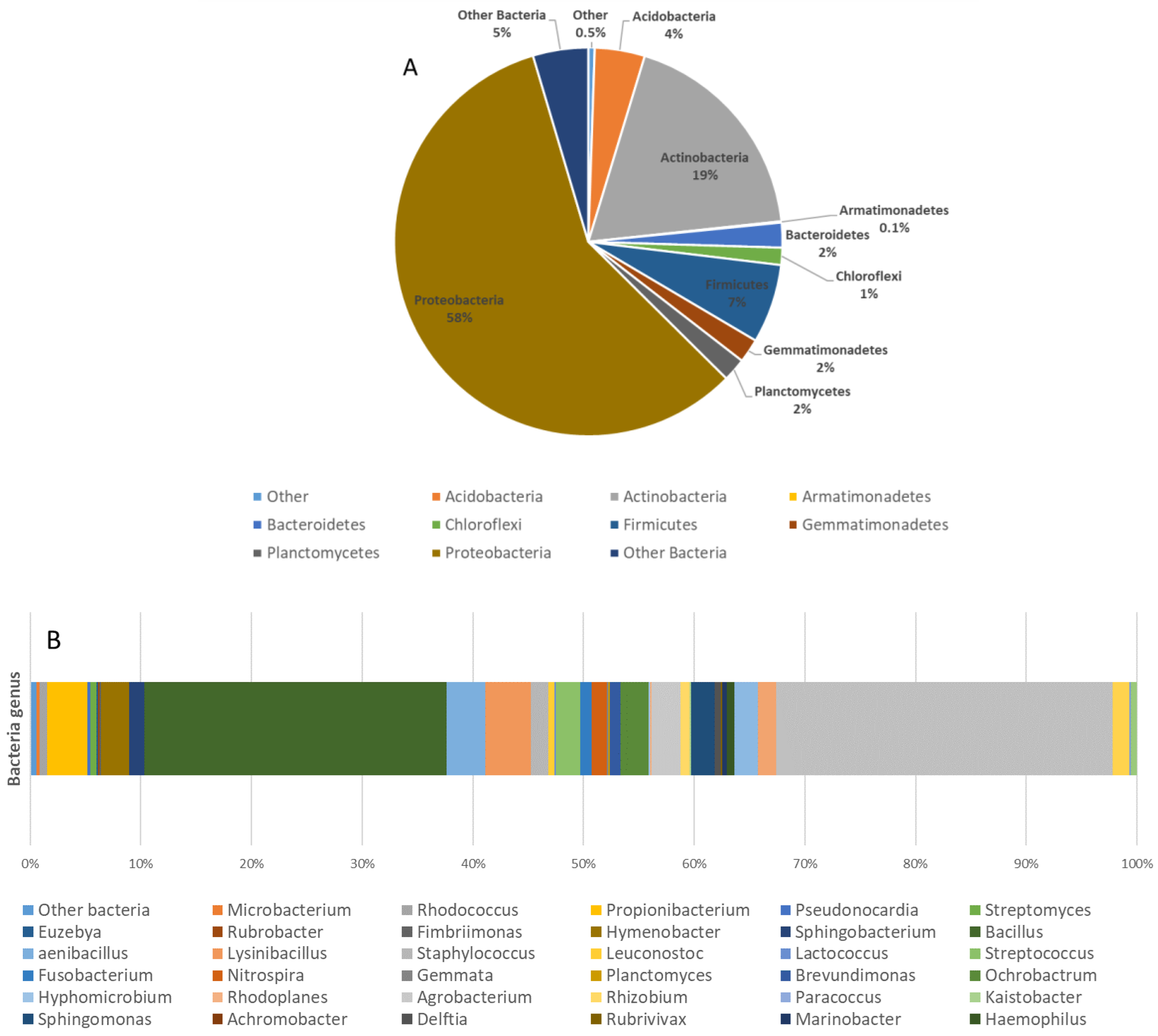
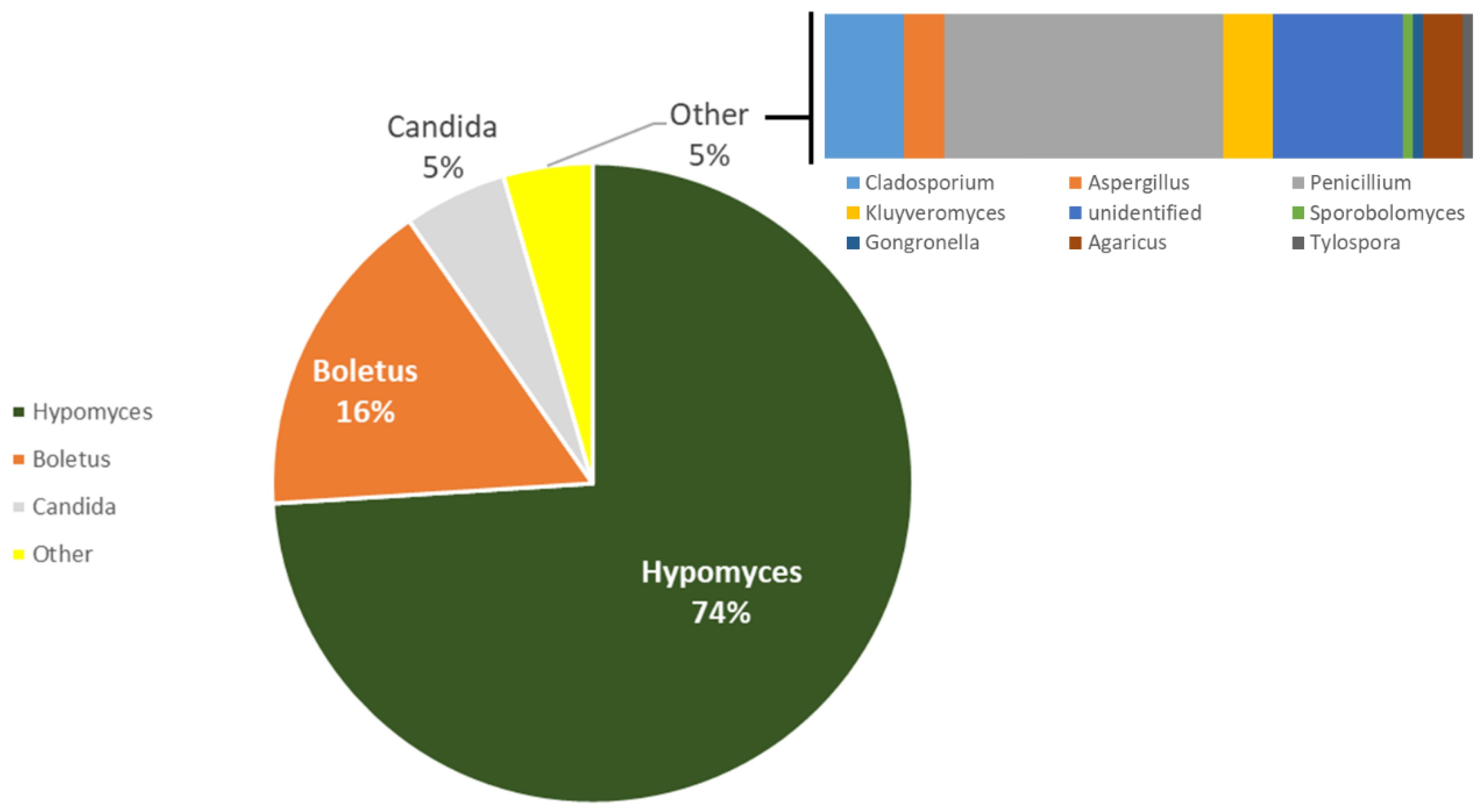
2.2.3. High-Throughput Sequencing

To study the biodiversity and have a full view of the microbial populations thriving inside the Escoural cave, Next Generation Sequencing (NGS) analyses were performed on three different locations of the cavity. The three areas were chosen due to their high potential diversity of microbial contamination.
DNA was extracted from samples using QIAmp DNA Stool Mini Kit (Qiagen, Limburg, Netherlands), with minor modifications to manufacturer’s instructions.
Bacterial and Fungal communities were characterized by Illumina Sequencing for the 16S rRNA V3-V4 region and Internal Transcribed Spacer 2. DNA was amplified for the hypervariable regions with specific primers and further reamplified in a limited-cycle PCR reaction to add sequencing adaptor and dual indexes. First, PCR reactions were performed for each sample using 2X KAPA HiFi HotStart Ready Mix. In a total volume of 25 μL, 12.5 ng of template DNA and 0.2 μM of each PCR primer. For bacteria the following primers were used: forward primer Bakt_341F 5′-CCTACGGGNGGCWGCAG-3′ and reverse primer Bakt_805R 5′-GACTACHVGGGTATCTAATCC-3′ [59,60]. For fungi, a pool of forward primers was used: ITS3NGS1_F 5′-CATCGATGAAGAACGCAG-3′, ITS3NGS2_F 5′-CAACGATGAAGAACGCAG-3′, ITS3NGS3_F 5′-CACCGATGAAGAACGCAG-3′, ITS3NGS4_F 5′-CATCGATGAAGAACGTAG-3′, ITS3NGS5_F 5′-CATCGATGAAGAACGTGG-3′, and ITS3NGS10_F 5′-CATCGATGAAGAACGCTG-3′ with the reverse primer ITS3NGS001_R 5′-TCCTSCGCTTATTGATATGC-3′ [61]. The PCR conditions involved 3 min of denaturation at 95 °C, followed by 25 cycles of 98 °C for 20 s, 55 °C for 30 s and 72 °C for 30 s and a final extension at 72 °C for 5 min. Negative controls were included for all amplification reactions. Electrophoresis of the PCR products was undertaken on a 1% (w/v) agarose gel and the ~490 bp V3-V4 and ~390 bp ITS2 amplified fragments were purified using AMPure XP beads (Agencourt, Beckman Coulter, USA) according to manufacturer’s instructions. Second PCR reactions added indexes and sequencing adaptors to both ends of the amplified target region by the use of 2X KAPA HotStart Ready Mix, 5 μL of each index (i7 and i5) (Nextera XT Index Kit, Illumina, San Diego, CA, USA) and 5 μL of the first PCR product in a total volume of 50 μL. The PCR conditions involved a 3 min denaturation at 95 °C, followed by 8 cycles of 95 °C for 30 s, 55 °C for 30 s and 72 °C for 30 s and a final extension at 72 °C for 5 min. Electrophoresis of the PCR products was undertaken on a 1% (w/v) agarose gel and the amplified fragments were purified using AMPure XP beads (Agencourt, Beckman Coulter, USA) according to manufacturer’s instructions. The amplicons were quantified by fluorimetry with PicoGreen dsDNA quantitation kit (Invitrogen, Life Technologies, Carlsbad, CA, USA), pooled at equimolar concentrations and paired-end sequenced with the V3 chemistry in the MiSeq® according to manufacturer’s instructions (Illumina, San Diego, CA, USA). They were multiplexed automatically by the Miseq® sequencer using the CASAVA package (Illumina, San Diego, CA, USA) and quality-filtered with PRINSEQ software using the following parameters: (1) bases with average quality lower than Q25 in a window of 5 bases were trimmed, and (2) reads with less than 220 bases were discarded for V3-V4 samples and less than 100 bases for ITS samples. The forward and reverse reads were then merged by overlapping paired-end reads using the AdapterRemoval v2.1.5 [62] software with default parameters. The QIIME package v1.8.0 [63] was used for Operational Taxonomic Units (OTU) generation, taxonomic identification and sample diversity and richness indexes calculation. Sample IDs were assigned to the merged reads and converted to fast format (split_libraries_fastq.py, QIIME). Chimeric merged reads were detected and removed using UCHIME [64] against the Greengenes v13.8 database [65] for V3-V4 samples and UNITE/QIIME ITS v12.11 database [66] for ITS samples (script identify_chimeric_seqs.py, QIIME). OTUs were selected at 97% similarity threshold using the open reference strategy. First, merged reads were pre-filtered by removing sequences with a similarity lower than 60% against Greengenes v13.8 database for V3-V4 samples and UNITE/QIIME ITS v12.11 database for ITS samples. The remaining merged reads were then clustered at 97% similarity against the same databases listed above. Merged reads that did not cluster in the previous step were again clustered into OTU at 97% similarity. OTUs with less than two reads were removed from the OTU table. A representative sequence of each OTU was then selected for taxonomy assignment.

2.3. Microbial Activity and Viability Assessment

MTT 3-(4,5-dimethylthiazol-2-yl)-2,5-diphenyltetrazolium bromide assay is a screening method implemented in 1983 [67] to measure cell viability. It relies on the reduction of tetrazolium salt by cells’ respiration’s process (through dehydrogenase enzyme activity) to purple formazan crystals [67,68]. The procedure followed the one developed by Rosado et al. [69]. Weighted microsamples were mixed with 300 µL of MTT, solution prepared with 1 M Tris-HCl buffer (pH 7.5) and 0.5% 2-(p-iodophenyl)-3-(p-nitrophenyl)-5-phenyltetrazolium chloride and incubated 4 h at 37 °C in the dark. The samples were then centrifuged for 10 min at 10,000 rpm and the supernatant phase was removed to leave only the purple crystals, which were dissolved in 350 µL of DMSO/ethanol (1:1). Tubes were then mixed during 30 s with a Vortex mixer to ensure proper dissolution of the crystals. The solutions were then kept for 10 min in the dark at room temperature and centrifuged again during 10 min at 10,000 rpm. The amount of iodonitrotetrazolium formazan (INTF) released was measured spectrophotometrically (U-3010; Hitachi, Tokyo, Japan) with 200 µL of the supernatant in 96-wells plates at 570 nm.
Presto Blue® (PB) is a reagent based on the same process than the almar blue reaction [70]. Cell’s respiration reduces the blue resazurin into red resofurin [71]. Because it is a quite new reagent, only few studies already used it. The few found in the literature focused on comparing its performance with other tests such as Almar Blue an MTT [68,70,72]. Those few studies demonstrated that the PB has comparable results with other methods when used with absorbance but better detection limit and sensitivity when used with fluorescence [70,72].
PB assays were carried out in 96-wells plates, each well containing 90 µL of a prepared solution to be tested and 10 µL of the PB reagent. Plates were then incubated at 37 °C for a specific time, determined after kinetic studies of the reaction of PB with solutions of known number of microbial cells. The change of absorbance at 570 and 600 nm allowed to quantify the viable cells present in the prepared solutions.

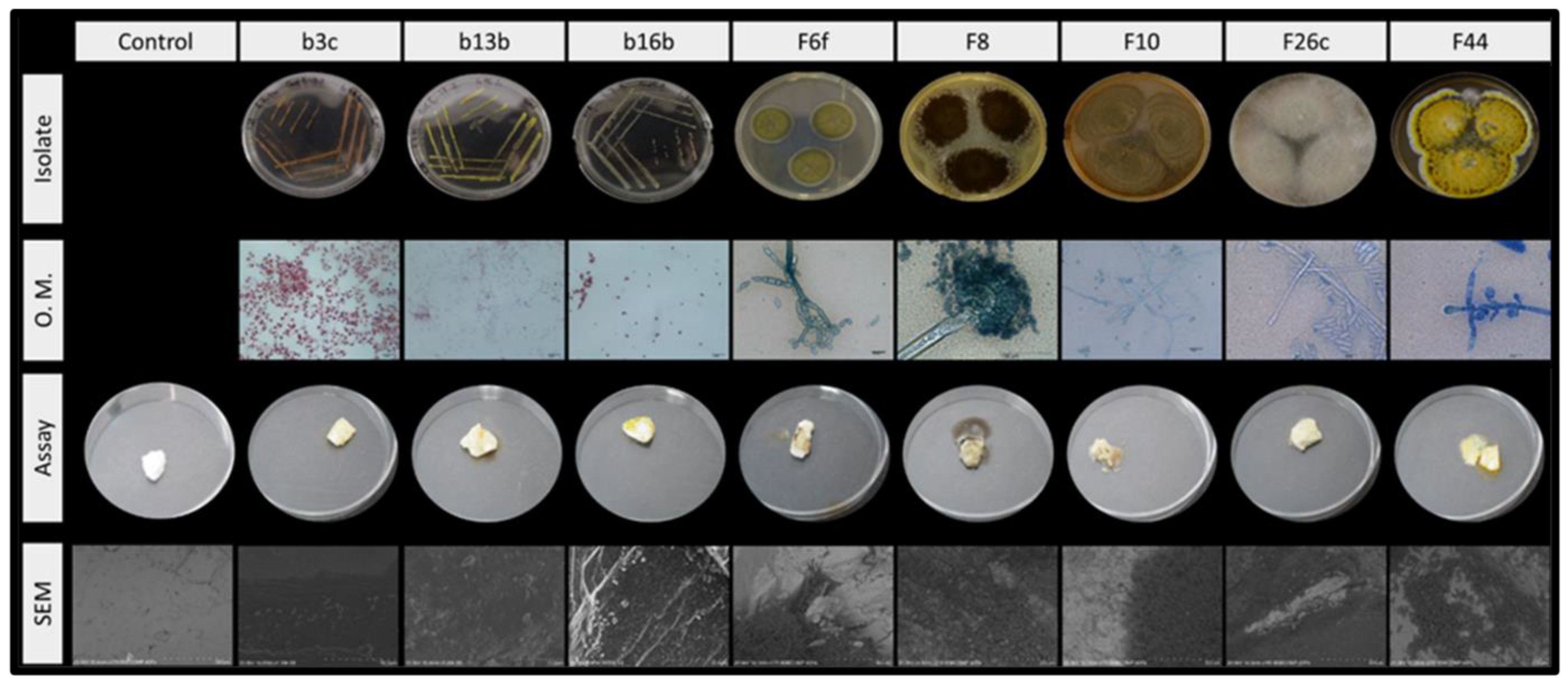
2.4. Biodeteriogenic Activity

2.4.1. Raman Spectroscopy

Raman spectroscopy was used to assess the presence of microbial contamination in the different samples, and to detect some metabolic products derived from their biodeteriogenic activity. Measurements were performed without any preparation of the samples by Raman micro-spectrometry using a HORIBA Xplora Raman microscope (Tokyo, Japan), with capacity increased to one hundred times, and a charge coupled device detector. Analyses were carried out with two different lasers of 638 and 785 nm with a 1–50% filter to avoid samples’ destruction. The spectra were acquired in scanning mode after 5 to 10 scans with an acquisition of 10–20 s and spectral resolution of 5 cm−1.

2.4.2. Simulation Assays

Once the microbial strains were isolated and characterized, experiments were conducted to assess their ability to alter the marble rock substrate and therefore the artwork hosted in Escoural Cave. Rock fragments collected outside the cavity were crushed, cleaned, sterilized and inoculated with different micro-organisms, isolated from culture-dependent techniques.
Eight distinct abundant microbial strains isolated in the cave were used to carry out these microbial colonization and contamination’s laboratory mock-ups: three bacteria (b3c, b13b, b16b) and five fungal strains (Cladosporium sp., Aspergillus niger, Penicilium sp., Mucor sp. and one unknown specie). Each experiment was performed in duplicate with fresh slant culture, 5 days old. Microorganisms were collected by diluting them in 4 mL of Malt Extract (ME). The suspensions were then gently mixed to ensure their relative homogeneity. Thence 100 µL of each suspension was inoculated on a sterile rock. The inoculation and culture were performed in a sterile environment.
Two stone controls were also performed. All the stone were then incubated at controlled temperature for 1 month. During all the period of incubation, samples were carefully monitored and photos were taken to record the development of the microbial communities. At the end of the assays, the rocks were then observed by SEM, following the same procedure as for the real samples (Section 2.2.1).

3. Results

3.1. Assessment and Diversity of Microbial Contamination

The in-situ observations of the biological and microbial contamination of Escoural Cave (Figure 1) allowed us to assess and scale the various biological and microbial contaminations’ state found in the cave. Bats’ feces (guano) were considered to be the most contaminated areas of the cave due to their large organic content and the numerous known fecal micro-organisms present. As microbial growths (Figure 1A,B) were spotted only in some rare occasions, they were considered to be punctual in space and time. Therefore, they only constituted a minor microbial contamination. Presence of roots (Figure 1C,D) coming from the ceiling or the walls of the cave, acted as source of nutrients and represented areas characterized by a high biological contamination hosting a large microbial population. However, numerous spots presented only speleothems that could be attributed to microbial activity with no relation to outdoor nutrient sources. Furthermore, because no particular micro-organism seemed to be active, they were considered as spots of very low contamination (Table 1).
The estimation of the biological outbreaks was later confirmed by SEM observations of the microbial structures present in the samples. It even provided some insight on the microbial populations and revealed the presence of a large diversity of micro-organisms thriving inside the cave. Various structures were spotted inside the cavity ranging from fungal hyphae to bacterial EPS with the occasional of arthropod’s bodies and of unidentified microbial structures (Figure 2).
Analyses of the microbial diversity provided better insights of the population thriving inside the cavity. The culture-dependent methods highlighted the large predominance of bacterial communities (more than 90% of the microbial population) over the fungal ones (around 10%). It was possible to culture different micro-organisms, isolating and distinguishing different strains namely 62 fungal strains, 22 bacterial strains and 5 microalgae or cyanobacterial strains. Among the 62 isolated fungal strains, 13 (representing 7% of the isolated fungal population) could not be yet identified. The commonly encountered genera such as Penicilium, Aspergillus, Cladosporium, Mucor, Trichoderma and Fusarium (Table 2) represent more than 92% of the isolated strains.
NGS analysis complemented these culture-dependent results. In relation to the bacterial communities, it was possible to obtain around 2300 Operational Taxonomic Units (OTU) whose distribution is mostly Proteobacteria (58%), Actinobacteria (19%), Firmicutes (7%), Acidobacteria (4%), Bacteroidetes (2%), Gemmatimonadetes (2%), Planctomycetes (2%) and Chloroflexi (1%) (Figure 3A). The majority were of the genera Pseudomonas and Bacillus but also Lysinibacillus; Staphylococcus; Streptococcus, Sphingobacterium; Nitrospira; Ochrobactrum; Agrobacterium; Sphingomonas; Acinetobacter; Enhydrobacter; Photobacterium and Macromonas (Figure 3B).
In the case of fungi, it was obtained approximately 550 OTU, mostly Ascomycota (80%) and Basidiomycota (11%). The most common were Hypomyces (74%) and Boletus (16%) but also Agaricus, Penicillium, Cladosporium, Candida, Kluyveromyces, Sporobolomyces and Gongronella (Figure 4) were detected.
The large diversity of the microbial populations identified by both culture-dependent and high-throughput sequencing (HTS) did not provide any insight on the activity of the micro-organism and therefore on the potential threat faced by Escoural parietal artworks. They provide information on the microbial population’s diversity without considering its activity or inactivity. Therefore, a large number of the microbial community identified with those two approaches are indeed present in the cave, though only at a dormant stage. As long as they remain in a dormant stage, micro-organisms do not endanger the artwork nor its rock support. This is why analysis of the microbial biofilm’s viability and activity was then required.

3.2. Microbial Activity and Viability

As expected, the MTT exhibited lower signal and therefore sensitivity than the PB (Table 1). Both methods revealed a nondetectable low activity of the microbial populations colonizing the samples collected and analyzed. Only two specific spots inside the cavity appeared to have viable and active populations (detectable signals with MTT and PB). Both of them showed visually obvious macroscopic contaminations with one presenting roots and fungal growths while the other showing contamination by faeces of bats.
SEM observations and Raman analyses (data not shown) confirmed the presence of biocontamination and low activity of the microbial biofilms detection on microsamples from Escoural Cave. Both did not allow to identify and detect any biodeteriogenic activity of the microbial populations such as support’s alterations and precipitation of secondary compounds produce by microbial metabolic activity such as oxalates and carotenoids.

3.3. Assessment of the Biodeteriogenic Ability

The biodeteriogenic activity of the five most common fungal strains isolated and the three most representative bacterial strains were assessed (Figure 5). In presence of nutrients, all of the strains tested could grow on the rock support in less than a month, forming a biofilm over this material.
While bacteria only appeared to alter the aesthetic aspect of the sample, fungal strains alter both the aesthetic and the mechanical structures of the marble rocks they colonized (Figure 5).

4. Discussion

4.1. Biological and Microbial Contamination

During the present work, it was observed the presence of numerous organic nutrients—bat’s faeces, butterflies’ wings, roots—and fungal outbreaks. These numerous outbreaks were favoured by the very specific topographic and geomorphological conditions inside the cave, characterize, in particular, by a high number of cracks in the rock ceiling [73]. Due to these cracks, the Escoural Cave can be regarded as a fragile hypogean site, extremely dependent from the conditions present in the indoor and outdoor nearby environments. Other caves have been reported to have suffered from weathering processes linked to local air pollutants [74], and/or humidity conditions [2,10].
Another crucial aspect has to be taken into account when dealing with microbiology and the conservation of the Palaeolithic parietal representations: touristic visits. Studies have shown that changes in the light entering the cave environment associated with visitors greatly alter the cave’s environmental conditions and ecosystems, jeopardizing the conservation of these unique artworks [49,75,76]. Despite the measures taken to minimize the effects of the visitors on the conservation of the paintings and drawings (at Escoural Cave the number of visits is currently limited to 2 per day and 10 persons per visit), consequences on the cave microbial diversity should be monitored and further studied. The presence of fungal growths (Figure 1B) all over the floor, walls and ceilings of the Escoural Cave and their disappearance one week later (authors observation), highlights the high dynamic of the microbial populations thriving inside the cavity and their relative fragility to nutrients’ outcomes and outbreaks coming from outside. The present study should be used as a first step to understand the dynamic of these populations by providing a first insight of the microbial diversity and its activity in the cave.

4.2. Microbial Diversity

Both the macroscopic diversity of the biological and microbial outbreaks and the microscopic variability of the microbial contaminations highlighted a large diversity of the microbial communities growing inside of Escoural Cave (Figure 1 and Figure 2) as confirmed by culture-dependent (laboratory isolation) and non-culture-dependent (NGS) techniques.
The fungal genus isolated and identified with the culture-dependent method—Penicilium, Aspergillus, Cladosporium, Mucor, Trichoderma, Fusarium—are not specific to hypogean nor karstic environments as their presence have been reported in other caves and cultural heritage artifacts [8,25,33,74,77,78,79,80,81].
As confirmed by these two approaches, the bacterial communities largely predominate over the fungal ones inside the cavity. Only 1% of the bacterial strains and around 10% of the fungal strains spotted with NGS were cultured. These numbers correspond to what is usually considered as being identifiable by culture-dependent methods.
The first use in this karstic environment of HTS, has enabled to confirm the presence of some micro-organisms already identified by culture-dependent methods, though it also detected several genera and species not previously identified in these environments. HTS appeared to be extremely efficient in the detection and identification of known bacterial and fungal species, providing a wider knowledge of the communities thriving in the Escoural Cave.
The Hypomyces and Boletus correspond to the two major genera of fungi contaminating the cave. They are known to be usually found in deep soils. Therefore, their presence in the Escoural Cave is in accordance with the indoor environment. Due to the fact that most of them could not be cultured, they could only be identified at the genus level because their DNA’s sequence was not present in the reference database.
None of the species known for their harmfulness toward cave art, such as Bracteacoccus minor [82], Fusarium solani [3] and Ochroconis lascauxensis [36], have been reported inside Escoural Cave through isolation nor NGS methods. Fungal species isolated and identified in the cavity are known as common species and were already reported in other sites hosting parietal representations [33,50].
Although when compared to the NGS results, the culture-dependent method appears to be somewhat limited, it allows to better understand some of the strains present and, more importantly, to perform simulation assays with these strains in order to understand the activity and biodegradation, and, consequently, find appropriate solutions to preserve the contaminated artworks.

4.3. Microbial Viability and Activity

When dealing with microbial communities in cultural heritage, it is important to assess whether they are viable and active. Indeed, micro-organisms could be present in a passive state and, as such, they would not constitute a threat to the artworks they are contaminating. The assessment of the viability and activity of the microbial communities was performed thanks to chemical assays, SEM observations and Raman analyses.
As expected from the literature, the MTT exhibited lower signal and therefore sensitivity than the PB [70,72]. Such behaviour is expected because studies have shown that MTT’s signal strongly depends on the metabolism of the microbial strain analyzed. Indeed, according to those studies, MTT is reduced by enzymes whose activities rely on the reduced pyridine NADH. These conclusions lead to question the use of MTT due to the high dependence of the cytoplasmic metabolism of different strains. These studies performed on pure microbial cultures also prove that it is necessary to consider that different organisms provide distinct activity results. Because the metabolic activity of a microbial strain changes during the distinct stages it goes through, it is important to consider that according to the development stage, the microbial colony will exhibit distinct activity.
The low signals collected for most of the samples in MTT and PB assays together with the absence in SEM analyses of the characteristic microbial destructive and constructive fabrics typically found in hypogean environments [9] and in Raman analyses of alteration products linkable to microbial metabolism such as oxalates and carotenoid compounds, tend to suggest that most of the species identified with the culture-dependent and the non-culture-dependent techniques are present in low amounts or in a dormant stage, making them a possible future threat to the support and artworks only when and if sufficient nutrients would be available to reactivate them. Only areas showing major biological contamination did in fact present a viable microbial activity, confirming that nutrients’ supply represent a severely limiting factor with respect to the microbial growth inside the cave. Beside this preliminary result, it also implies that the microbial diversity observed in the cave, could easily increase in line with an increase in nutrients supply.

4.4. Microbial Biodeteriogenic Activity

Notwithstanding the aforementioned low activity of the microbial communities, it is also important to assess the biodeteriogenic activity of the most common microbial communities identified on the parietal wall of the Escoural cave. The dynamic of the microbial populations of Escoural Cave is quite important as microbial growths were observed to develop and disappear in less than a month at the site. Microbial colonies could, when active, rapidly alter the artworks. Preventing their growth would therefore represent an essential step in the preservation of the cave artworks.
The fungal species presented the highest threat potential to the rock support’s structure hosting the parietal representations of Escoural Cave. Indeed, all of the five fungal species studied induced cracks and aesthetic alterations on the rock (Figure 5) while the bacterial communities’ effect remained largely only aesthetic, with the development of EPS and biofilms.
The microbial diversity identified during this study by high-throughput sequencing (HTS) should be subsequently monitored to have a better overview of the microbial biocenosis of the Escoural cave. The evolution of microbial communities, associated with the activation of their metabolism, may have a possible influence on support and parietal representations.

5. Conclusions

Escoural Cave, a natural cavity hosting Upper Palaeolithic representations, presents major local biological and microbial contaminations. Although culture-dependent and NGS techniques allowed us to highlight a large microbial diversity, its activity and biodeteriogenic effect seem to be rather limited according to viability assays and biodegradation analyses by SEM-EDS microscopy and Raman spectroscopy. Therefore, the microbial population of the cavity does not seem to constitute a major threat to the conservation of the parietal art found in this important archaeological site. Indeed, water leaking and seepage appear to be of major importance in the alteration of these unique artworks.
Using methods assessing the microbial viability such as MTT and Presto Blue® provided a broad picture allowing us to understand the extent of microbial communities’ activity. The large microbial diversity appears to be at a dormant stage, with low metabolic activity. The dynamic of the microbial populations indicates an important potential threat of the microbial populations on the rock support and the paintings and engravings of the cave. Therefore, a long-term monitoring plan is needed to better understand the colonization and spread of the micro-organisms in this site, taking into account future plans for the full re-opening of the cave to touristic activities.
This work constitutes an important insight on the conservation of the unique Portuguese Palaeolithic cave art site of Escoural. This pioneer study should be completed by further analyses focusing both on identifying the unknown species and understanding their metabolism and effect on the various materials present in this hypogean site.

Author Contributions

Conceptualization, A.T.C., N.S. and A.C.; Formal analysis, G.M., C.S., T.R.; Funding acquisition, A.T.C., A.C., N.S., J.M.; Investigation, A.T.C., N.S., J.M., A.C., G.M., C.S., T.R.; Methodology, A.T.C., C.S., T.R.; Resources, A.T.C., A.C., J.M.; Writing—original draft G.M., T.R., C.S., A.T.C., N.S.; Writing—review and editing, A.T.C., N.S., J.M., A.C. All authors have read and agreed to the published version of the manuscript.

Funding

The authors gratefully acknowledge the following funding sources “PROBIOMA project-Prospection in Subterranean Environments of Microbial Bioactive Compounds with potential use in Medicine, Agriculture and Environment” funded by the Management Committee of the Interreg VA Spain-Portugal Program (POCTEP) 2014-2020, the ERASMUS Mundus Master Programme ARCHMAT for a EACEA scholarship (FPA 2013-0238) and the City University of Macau endowment to the Sustainable Heritage Chair.

Institutional Review Board Statement

Not Applicable.

Informed Consent Statement

Not Applicable.

Data Availability Statement

The data presented in this study are available within this article.

Acknowledgments

The authors are grateful to the Alentejo Regional Directorate for Culture and the archaeologist António Carlos Silva for allowing the study and sampling at the Escoural Cave.

Conflicts of Interest

The authors declare no conflict of interest.

References

  1. Berrouet, F. Les Altérations D’origine Biologique dans L’art Pariétal: Exemple des Relations Structurales et Conceptuelles entre le Mondmilch et les Représentations Paléolithiques: Cas Particulier de la Grotte de Lascaux et Enjeux Conservatoires. Ph.D. Thesis, Université de Bordeaux, Bordeaux, France, 2009. [Google Scholar]
  2. Cuezva, S.; Fernandez-Cortes, A.; Porca, E.; Pašić, L.; Jurado, V.; Hernández, M.; Serrano-Ortiz, P.; Hermosin, B.; Cañaveras, J.C.; Sanchez-Moral, S.; et al. The biogeochemical role of Actinobacteria in Altamira Cave, Spain. FEMS Microbiol. Ecol. 2012, 81, 281–290. [Google Scholar] [CrossRef] [Green Version]
  3. Dupont, J.; Jacquet, C.; Dennetière, B.; Lacoste, S.; Bousta, F.; Orial, G.; Cruaud, C.; Couloux, A.; Roquebert, M.-F. Inva-sion of the French Paleolithic painted cave of Lascaux by members of the Fusarium solani species complex. Mycologia 2007, 99, 526–533. [Google Scholar] [CrossRef]
  4. Saiz-Jimenez, C. Microbiological and environmental issues in show caves. World J. Microbiol. Biotechnol. 2012, 28, 2453–2464. [Google Scholar] [CrossRef]
  5. Jurado, V.; Porca, E.; Cuezva, S.; Fernandez-Cortes, A.; Moral, S.S.; Sáiz-Jiménez, C. Fungal outbreak in a show cave. Sci. Total Environ. 2010, 408, 3632–3638. [Google Scholar] [CrossRef] [PubMed] [Green Version]
  6. Nováková, A. Microscopic fungi isolated from the Domica Cave system (Slovak Karst National Park, Slovakia). A review. Int. J. Speleol. 2009, 38, 71–82. [Google Scholar] [CrossRef] [Green Version]
  7. Sugita, T.; Kikuchi, K.; Makimura, K.; Urata, K.; Someya, T.; Kamei, K.; Niimi, M.; Uehara, Y. Trichosporon Species Isolated from Guano Samples Obtained from Bat-Inhabited Caves in Japan. Appl. Environ. Microbiol. 2005, 71, 7626–7629. [Google Scholar] [CrossRef] [Green Version]
  8. Urzì, C.; Albertano, P. Studying phototrophic and heterotrophic microbial communities on stone monuments. Methods Enzymol. 2001, 336, 340–355. [Google Scholar] [CrossRef] [PubMed]
  9. Cañveras, J.C.; Sanchez-Moral, S.; Saiz-Jimenez, C. Microorganisms and Microbially Induced Fabrics in Cave Walls. Geomicrobiol. J. 2001, 18, 223–240. [Google Scholar] [CrossRef]
  10. Cuezva, S.; Sánchez-Moral, S.; Sáiz-Jiménez, C.; Cañaveras, J.C. Microbial communities and associated mineral fabrics in Altamira Cave, Spain. Int. J. Speleol. 2009, 38, 9. [Google Scholar]
  11. Northup, D.E.; Lavoie, K. Geomicrobiology of Caves: A Review. Geomicrobiol. J. 2001, 18, 199–222. [Google Scholar] [CrossRef]
  12. Garcia-Vallès, M.; Vendrell-Saz, M.; Krumbein, W.E.; Urzì, C. Coloured mineral coatings on monument surfaces as a result of biomineralization: The case of the Tarragona cathedral (Catalonia). Appl. Geochem. 1997, 12, 255–266. [Google Scholar] [CrossRef]
  13. Griffin, P.S.; Indictor, N.; Koestler, R.J. The biodeterioration of stone: A review of deterioration mechanisms, conservation case histories, and treatment. Int. Biodeterior. 1991, 28, 187–207. [Google Scholar] [CrossRef]
  14. Herrera, L.K.; Videla, H.A. Surface analysis and materials characterization for the study of biodeterioration and weathering effects on cultural property. Int. Biodeterior. Biodegrad. 2009, 63, 813–822. [Google Scholar] [CrossRef]
  15. Kusumi, A.; Li, X.; Osuga, Y.; Kawashima, A.; Gu, J.-D.; Nasu, M.; Katayama, Y. Bacterial Communities in Pigmented Biofilms Formed on the Sandstone Bas-Relief Walls of the Bayon Temple, Angkor Thom, Cambodia. Microbes Environ. 2013, 28, 422–431. [Google Scholar] [CrossRef] [PubMed] [Green Version]
  16. Nagai, K.; Suzuki, K.; Okada, G. Studies on the distribution of alkalophilic and alkali-tolerant soil fungi II: Fungal flora in two limestone caves in Japan. Mycoscience 1998, 39, 293–298. [Google Scholar] [CrossRef]
  17. Schiavon, N. Biodeterioration of calcareous and granitic building stones in urban environments. In Natural Stone, Weathering Phenomena, Conservation Strategies and Case Studies; Siegesmund, S., Weiss, T., Vollbrecht, V.A., Eds.; Geological Society of London Special Publication: London, UK, 2002; pp. 195–206. [Google Scholar] [CrossRef]
  18. Schiavon, N.; De Caro, T.; Kiros, A.; Caldeira, A.T.; Parisi, I.E.; Riccucci, C.; Gigante, G.E. A multi-analytical approach to investigate stone biodeterioration at a UNESCO world heritage site: The volcanic rock-hewn churches of Lalibela, Northern Ethiopia. Appl. Phys. Part A 2013, 113, 843–854. [Google Scholar] [CrossRef] [Green Version]
  19. Rosado, T.; Reis, A.; Mirão, J.; Candeias, A.; Vandenabeele, P.; Caldeira, A.T. Pink! Why not? On the unusual colour of Évora Cathedral. Int. Biodeterior. Biodegrad. 2014, 94, 121–127. [Google Scholar] [CrossRef] [Green Version]
  20. Rosado, T.; Dias, L.; Lança, M.; Nogueira, C.; Santos, R.; Martins, M.R.; Candeias, A.; Mirão, J.; Caldeira, A.T. Assessment of microbiota present on a Portuguese historical stone convent using high-throughput sequencing approaches. MicrobiologyOpen 2020, 9, 1067–1084. [Google Scholar] [CrossRef] [PubMed]
  21. Ding, Y.; Salvador, C.; Caldeira, A.T.; Angelini, E.; Schiavon, N. Biodegradation and microbial contamination of lime-stone surfaces: An experimental study from Batalha Monastery, Portugal. Corros. Mater. Degrad. 2021, 2, 31–45. [Google Scholar]
  22. Ariño, X.; Saiz-Jimenez, C. Colonization and deterioration processes in Roman mortars by cyanobacteria, algae and lichens. Aerobiologia 1996, 12, 9–18. [Google Scholar] [CrossRef]
  23. Tran, T.H.; Govin, A.; Guyonnet, R.; Grosseau, P.; Lors, C.; Garcia-Diaz, E.; Damidot, D.; Devès, O.; Ruot, B. Influence of the intrinsic characteristics of mortars on biofouling by Klebsormidium flaccidum. Int. Biodeterior. Biodegrad. 2012, 70, 31–39. [Google Scholar] [CrossRef] [Green Version]
  24. Altenburgera, P.; Kämpferb, P.; Makristathisc, A.; Lubitza, W.; Bussea, H.-J. Classification of bacteria isolated from a medieval wall painting. J. Biotechnol. 1996, 47, 39–52. [Google Scholar] [CrossRef]
  25. Cappitelli, F.; Abbruscato, P.; Foladori, P.; Zanardini, E.; Ranalli, G.; Principi, P.; Villa, F.; Polo, A.; Sorlini, C. Detection and Elimination of Cyanobacteria from Frescoes: The Case of the St. Brizio Chapel (Orvieto Cathedral, Italy). Microb. Ecol. 2009, 57, 633–639. [Google Scholar] [CrossRef]
  26. Gorbushina, A.A.; Heyrman, J.; Dornieden, T.; Gonzalez-Delvalle, M.; Krumbein, W.E.; Laiz, L.; Petersen, K.; Saiz-Jimenez, C.; Swings, J. Bacterial and fungal diversity and biodeterioration problems in mural painting environments of St. Martins church (Greene–Kreiensen, Germany). Int. Biodeterior. Biodegrad. 2004, 53, 13–24. [Google Scholar] [CrossRef]
  27. Gurtner, C.; Heyrman, J.; Piñar, G.; Lubitz, W.; Swings, J.; Rölleke, S. Comparative analyses of the bacterial diversity on two different biodeteriorated wall paintings by DGGE and 16S rDNA sequence analysis. Int. Biodeterior. Biodegrad. 2000, 46, 229–239. [Google Scholar] [CrossRef]
  28. Laiz, L.; Gonzalez, J.M.; Saiz-Jimenez, C. Microbial communities in caves: Ecology, physiology, and effects on paleo-lithic paintings. In Art, Biology, and Conservation: Biodeterioration of Works of Art; The Metropolitan Museum of Art: New York, NY, USA, 2003; pp. 210–225. [Google Scholar]
  29. Nugari, M.; Pietrini, A.; Caneva, G.; Imperi, F.; Visca, P. Biodeterioration of mural paintings in a rocky habitat: The Crypt of the Original Sin (Matera, Italy). Int. Biodeterior. Biodegrad. 2009, 63, 705–711. [Google Scholar] [CrossRef]
  30. Pepe, O.; Sannino, L.; Palomba, S.; Anastasio, M.; Blaiotta, G.; Villani, F.; Moschetti, G. Heterotrophic microorganisms in deteriorated medieval wall paintings in southern Italian churches. Microbiol. Res. 2010, 165, 21–32. [Google Scholar] [CrossRef] [PubMed]
  31. Zucconi, L.; Gagliardi, M.; Isola, D.; Onofri, S.; Andaloro, M.C.; Pelosi, C.; Pogliani, P.; Selbmann, L. Biodeterioration agents dwelling in or on the wall paintings of the Holy Saviour’s cave (Vallerano, Italy). Int. Biodeterior. Biodegrad. 2012, 70, 40–46. [Google Scholar] [CrossRef]
  32. Bastian, F.; Alabouvette, C.; Saiz-Jimenez, C. Bacteria and free-living amoeba in the Lascaux Cave. Res. Microbiol. 2009, 160, 38–40. [Google Scholar] [CrossRef]
  33. Bastian, F.; Jurado, V.; Nováková, A.; Alabouvette, C.; Sáiz-Jiménez, C. The microbiology of Lascaux cave. Microbiology 2010, 156, 644–652. [Google Scholar] [CrossRef] [Green Version]
  34. Cunningham, K.I.; Northup, D.E.; Pollastro, R.M.; Wright, W.G.; LaRock, E.J. Bacteria, fungi and biokarst in Lechu-guilla Cave, Carlsbad Caverns National Park, New Mexico. Environ. Geol. 1995, 25, 2–8. [Google Scholar] [CrossRef]
  35. González, I.; Laiz, L.; Hermosin, B.; Caballero, B.; Incerti, C.; Sáiz-Jiménez, C. Bacteria isolated from rock art paintings: The case of Atlanterra shelter (south Spain). J. Microbiol. Methods 1999, 36, 123–127. [Google Scholar] [CrossRef]
  36. Martin-Sanchez, P.M.; Nováková, A.; Bastian, F.; Alabouvette, C.; Saiz-Jimenez, C. Two new species of the genus Ochroconis, O. lascauxensis and O. anomala isolated from black stains in Lascaux Cave, France. Fungal Biol. 2012, 116, 574–589. [Google Scholar] [CrossRef] [PubMed]
  37. Martin-Sanchez, P.; Jurado, V.; Porca, E.; Bastian, F.; Lacanette, D.; Alabouvette, C.; Saiz-Jimenez, C. Airborne microorganisms in Lascaux Cave (France). Int. J. Speleol. 2014, 43, 295–303. [Google Scholar] [CrossRef]
  38. De Leo, F.; Iero, A.; Zammit, G.; Urzì, C. Chemoorganotrophic bacteria isolated from biodeteriorated surfaces in cave and catacombs. Int. J. Speleol. 2012, 41, 125–136. [Google Scholar] [CrossRef] [Green Version]
  39. Saarela, M.; Alakomi, H.-L.; Suihko, M.-L.; Maunuksela, L.; Raaska, L.; Mattila-Sandholm, T. Heterotrophic microorganisms in air and biofilm samples from Roman catacombs, with special emphasis on actinobacteria and fungi. Int. Biodeterior. Biodegrad. 2004, 54, 27–37. [Google Scholar] [CrossRef]
  40. Sanchez-Moral, S.; Canaveras, J.C.; Laiz, L.; Saiz-Jimenez, C.; Bedoya, J.; Luque, L. Biomediated Precipitation of Calcium Carbonate Metastable Phases in Hypogean Environments: A Short Review. Geomicrobiol. J. 2003, 20, 491–500. [Google Scholar] [CrossRef] [Green Version]
  41. Zammit, G.; Sánchez-Moral, S.; Albertano, P. Bacterially mediated mineralisation processes lead to biodeterioration of artworks in Maltese catacombs. Sci. Total Environ. 2011, 409, 2773–2782. [Google Scholar] [CrossRef] [PubMed]
  42. Cennamo, P.; Montuori, N.; Trojsi, G.; Fatigati, G.; Moretti, A. Biofilms in churches built in grottoes. Sci. Total Environ. 2016, 543, 727–738. [Google Scholar] [CrossRef] [PubMed]
  43. Cañaveras, J.C.; Hoyos, M.; Sanchez-Moral, S.; Sanz-Rubio, E.; Bedoya, J.; Soler, V.; Groth, I.; Schumann, P.; Laiz, L.; Gonza-lez, I.; et al. Microbial Communities Associated with Hydromagnesite and Needle-Fiber Aragonite Deposits in a Karstic Cave (Altamira, Northern Spain). Geomicrobiol. J. 1999, 16, 9–25. [Google Scholar] [CrossRef] [Green Version]
  44. Groth, I.; Vettermann, R.; Schuetze, B.; Schumann, P.; Sáiz-Jiménez, C. Actinomycetes in Karstic caves of northern Spain (Altamira and Tito Bustillo). J. Microbiol. Methods 1999, 36, 115–122. [Google Scholar] [CrossRef]
  45. Laiz, L.; Groth, I.; Gonzalez, I.; Sáiz-Jiménez, C. Microbiological study of the dripping waters in Altamira cave (Santil-lana del Mar, Spain). J. Microbiol. Methods 1999, 36, 129–138. [Google Scholar] [CrossRef] [Green Version]
  46. Portillo, M.C.; Gonzalez, J.M. Comparing bacterial community fingerprints from white colonizations in Altamira Cave (Spain). World J. Microbiol. Biotechnol. 2009, 25, 1347–1352. [Google Scholar] [CrossRef]
  47. Portillo, M.C.; Saiz-Jimenez, C.; Gonzalez, J.M. Molecular characterization of total and metabolically active bacterial communities of “white colonizations” in the Altamira Cave, Spain. Res. Microbiol. 2009, 160, 41–47. [Google Scholar] [CrossRef]
  48. Portillo, M.C.; Gonzalez, J.M. Moonmilk deposits originate from specific bacterial communities in Altamira Cave (Spain). Microb. Ecol. 2011, 61, 182–189. [Google Scholar] [CrossRef] [Green Version]
  49. Sánchez-Moral, S.; Soler, V.; Cañaveras, J.; Sanz-Rubio, E.; Van Grieken, R.; Gysels, K. Inorganic deterioration affecting the Altamira Cave, N Spain: Quantitative approach to wall-corrosion (solutional etching) processes induced by visitors. Sci. Total Environ. 1999, 243–244, 67–84. [Google Scholar] [CrossRef]
  50. Sanchez-Moral, S.; Portillo, M.C.; Janices, I.; Cuezva, S.; Fernández-Cortés, A.; Cañaveras, J.C.; Gonzalez, J.M. The role of microorganisms in the formation of calcitic moonmilk deposits and speleothems in Altamira Cave. Geomorphology 2012, 139–140, 285–292. [Google Scholar] [CrossRef] [Green Version]
  51. Schabereiter-Gurtner, C.; Saiz-Jimenez, C.; Piñar, G.; Lubitz, W.; Rölleke, S. Altamira cave Paleolithic paintings harbor partly unknown bacterial communities. FEMS Microbiol. Lett. 2002, 211, 7–11. [Google Scholar] [CrossRef] [PubMed] [Green Version]
  52. Beni, A.; Soki, E.; Lajtha, K.; Fekete, I. An optimized HPLC method for soil fungal biomass determination and its application to a detritus manipulation study. J. Microbiol. Methods 2014, 103, 124–130. [Google Scholar] [CrossRef]
  53. Saad, D.; Kinsey, G.; Paterson, R.R.M.; Gaylarde, C.C. Ergosterol analysis for the quantification of fungal growth on paint films. Proposal for a standard method. Surf. Coat. Int. Part B Coat. Trans. 2003, 86, 131–134. [Google Scholar] [CrossRef]
  54. Hippelein, M.; Rügamer, M. Ergosterol as an indicator of mould growth on building materials. Int. J. Hyg. Environ. Health 2004, 207, 379–385. [Google Scholar] [CrossRef]
  55. Mille-Lindblom, C.; Von Wachenfeldt, E.; Tranvik, L.J. Ergosterol as a measure of living fungal biomass: Persistence in environmental samples after fungal death. J. Microbiol. Methods 2004, 59, 253–262. [Google Scholar] [CrossRef] [PubMed]
  56. Joblin, Y.; Moularat, S.; Anton, R.; Bousta, F.; Orial, G.; Robine, E.; Picon, O.; Bourouina, T. Detection of moulds by volatile organic compounds: Application to heritage conservation. Int. Biodeterior. Biodegrad. 2010, 64, 210–217. [Google Scholar] [CrossRef]
  57. Porca, E.; Jurado, V.; Martin-Sanchez, P.M.; Hermosin, B.; Bastian, F.; Alabouvette, C.; Saiz-Jimenez, C. Aerobiology: An ecological indicator for early detection and control of fungal outbreaks in caves. Ecol. Indic. 2011, 11, 1594–1598. [Google Scholar] [CrossRef]
  58. Araujo, A.C.; Lejeune, M.; Santos, A.I. Gruta do Escoural: Necropole Neolitica e Arte Rupestre Paleolitica; Instituto Portugues do Patrimonio Arquitectonico e Arqueologico: Lisbon, Portugal, 1995. [Google Scholar]
  59. Herlemann, D.P.R.; Labrenz, M.; Jürgens, K.; Bertilsson, S.; Waniek, J.J.; Andersson, A.F. Transitions in bacterial communities along the 2000 km salinity gradient of the Baltic Sea. ISME J. 2011, 5, 1571–1579. [Google Scholar] [CrossRef] [PubMed] [Green Version]
  60. Klindworth, A.; Pruesse, E.; Schweer, T.; Peplies, J.; Quast, C.; Horn, M.; Glöckner, F.O. Evaluation of general 16S ribosomal RNA gene PCR primers for classical and next-generation sequencing-based diversity studies. Nucleic Acids Res. 2012, 41, e1. [Google Scholar] [CrossRef]
  61. Tedersoo, L.; Bahram, M.; Põlme, S.; Kõljalg, U.; Yorou, N.S.; Wijesundera, R.L.C.; Ruiz, L.V.; Vasco-Palacios, A.M.; Thu, P.Q.; Suija, A.; et al. Global diversity and geography of soil fungi. Science 2014, 346, 1256688. [Google Scholar] [CrossRef] [PubMed] [Green Version]
  62. Schubert, M.; Lindgreen, S.; Orlando, L. AdapterRemoval v2: Rapid adapter trimming, identification, and read merging. BMC Res. Notes 2016, 9, 88. [Google Scholar] [CrossRef] [Green Version]
  63. Caporaso, J.G.; Kuczynski, J.; Stombaugh, J.; Bittinger, K.; Bushman, F.D.; Costello, E.K.; Fierer, N.; Peña, A.G.; Goodrich, J.K.; Gordon, J.I.; et al. QIIME Allows Analysis of High-Throughput Community Sequencing data. Nat. Methods 2010, 7, 335–336. [Google Scholar] [CrossRef] [Green Version]
  64. Edgar, R.C.; Haas, B.J.; Clemente, J.C.; Quince, C.; Knight, R. UCHIME Improves Sensitivity and Speed of Chimera Detection. Bioinformatics 2011, 27, 2194–2200. [Google Scholar] [CrossRef] [Green Version]
  65. DeSantis, T.Z.; Hugenholtz, P.; Larsen, N.; Rojas, M.; Brodie, E.L.; Keller, K.; Huber, T.; Dalevi, D.; Hu, P.; Andersen, G.L. Greengenes, a chimera-checked 16S rRNA gene database and workbench compatible with ARB. Appl. Environ. Microbiol. 2006, 72, 5069–5072. [Google Scholar] [CrossRef] [Green Version]
  66. Abarenkov, K.; Nilsson, R.H.; Larsson, K.-H.; Alexander, I.J.; Eberhardt, U.; Erland, S.; Høiland, K.; Kjøller, R.; Larsson, E.; Pennanen, T.; et al. The UNITE database for molecular identification of fungi—Recent updates and future perspectives. New Phytol. 2010, 186, 281–285. [Google Scholar] [CrossRef]
  67. Mosmann, T. Rapid colorimetric assay for cellular growth and survival: Application to proliferation and cytotoxicity assays. J. Immunol. Methods 1983, 65, 55–63. [Google Scholar] [CrossRef]
  68. Boncler, M.; Różalski, M.; Krajewska, U.; Podsędek, A.; Watala, C. Comparison of PrestoBlue and MTT assays of cellular viability in the assessment of anti-proliferative effects of plant extracts on human endothelial cells. J. Pharmacol. Toxicol. Methods 2014, 69, 9–16. [Google Scholar] [CrossRef] [PubMed]
  69. Rosado, T.; Martins, M.R.; Pires, M.; Mirão, J.; Candeias, A.; Caldeira, A.T. Enzymatic monitorization of mural paintings biodegradation and biodeterioration. Int. J. Conserv. Sci. 2013, 4, 603–612. [Google Scholar]
  70. Xu, M.; McCanna, D.J.; Sivak, J. Use of the viability reagent PrestoBlue in comparison with alamarBlue and MTT to assess the viability of human corneal epithelial cells. J. Pharmacol. Toxicol. Methods 2015, 71, 1–7. [Google Scholar] [CrossRef]
  71. Mariscal, A.; Lopez-Gigosos, R.M.; Carnero-Varo, M.; Fernández-Crehuet, J. Fluorescent assay based on resazurin for detection of activity of disinfectants against bacterial biofilm. Appl. Microbiol. Biotechnol. 2009, 82, 773–783. [Google Scholar] [CrossRef]
  72. Martín-Navarro, C.M.; López-Arencibia, A.; Sifaoui, I.; Reyes-Batlle, M.; Cabello-Vílchez, A.M.; Maciver, S.; Valladares, B.; Piñero, J.E.; Lorenzo-Morales, J. PrestoBlue® and AlamarBlue® are equally useful as agents to determine the viability of Acanthamoeba trophozoites. Exp. Parasitol. 2014, 145, S69–S72. [Google Scholar] [CrossRef]
  73. Malaurent, P.; Huneau, F.; Lastennet, R.; Fabre, R. Etudes Pour la Conservation des Parois de la Grotte d’Escoural-Portugal (Conservation Grottes Ornées No. 2004–24); Centre de Développement des Géosciences Appliquées—CDGA: Bordeaux, France, 2004. [Google Scholar]
  74. Arroyo, G.; Arroyo, I.; Arroyo, E. Microbiological analysis of Maltravieso Cave (Caceres), Spain. Int. Biodeterior. Biodegrad. 1997, 40, 131–139. [Google Scholar] [CrossRef]
  75. Barton, H.A.; Northup, D.E. Geomicrobiology in cave environments: Past, current and future perspectives. J. Cave Karst Stud. 2007, 69, 163–178. [Google Scholar]
  76. Bourges, F.; Genthon, P.; Genty, D.; Lorblanchet, M.; Mauduit, E.; D’Hulst, D. Conservation of prehistoric caves and stability of their inner climate: Lessons from Chauvet and other French caves. Sci. Total Environ. 2014, 493, 79–91. [Google Scholar] [CrossRef]
  77. Barton, H. Microbial life in the underworld: Biogenicity in secondary mineral formations. Geomicrobiol. J. 2001, 18, 359–368. [Google Scholar]
  78. Barton, H.A.; Jurado, V. What’s up down there? Microbial diversity in caves. Microbe 2007, 2, 132–138. [Google Scholar]
  79. Jurado, V.; Sanchez-Moral, S.; Saiz-Jimenez, C. Entomogenous fungi and the conservation of the cultural heritage: A review. Int. Biodeterior. Biodegrad. 2008, 62, 325–330. [Google Scholar] [CrossRef] [Green Version]
  80. Sterflinger, K. Fungi: Their role in deterioration of cultural heritage. Fungal Biol. Rev. 2010, 24, 47–55. [Google Scholar] [CrossRef]
  81. Wollenzien, U.; De Hoog, G.; Krumbein, W.; Urzí, C. On the isolation of microcolonial fungi occurring on and in marble and other calcareous rocks. Sci. Total Environ. 1995, 167, 287–294. [Google Scholar] [CrossRef]
  82. Lefevre, M. La “maladie verte” de Lascaux. Stud. Conserv. 1974, 19, 126–156. [Google Scholar] [CrossRef]
Figure 1. In situ observations of biological and microbial contaminations in the Escoural Cave. (A) Macrofungi growth covered by other microbial communities. (B) Widespread fungal growths on the walls of the cave. (C) Roots and sap developing on the ceiling of the cave. (D) Root coming out of soil covering the walls of the cave. (E) Microbial colonies. (F) Effect of precipitation, with pigmentation and the buildup of calcite crusts. (G) Biological colonization and moonmilk deposition.
Coatings 11 00209 g001
Figure 2. Micrographs from Escoural Cave showing the diversity of the microbial contamination in the samples from Escoural Cave. (A) Rock surface without microbial contamination. (B) Widespread fungal colonization of the rock surface with numerous hyphal structures (C) Hyphal structure developing over a micro-leaf and presence of an unidentified structure. (D) Bacterial EPS developing over the rock surface. (E) Fungal hyphae and bacteria. (F) Spherical unidentified structure of possibly microbial origin. (G) Microbial structure. (H) Fungal spore. (I) Arthropod’s corpse.
Coatings 11 00209 g002
Figure 3. NGS evaluation of the microbial diversity of Escoural Cave. Bacterial phylum (A) and Genus (B) biodiversity.
Coatings 11 00209 g003
Figure 4. NGS evaluation of the microbial diversity of Escoural Cave. Fungal genus biodiversity.
Coatings 11 00209 g004
Figure 5. Simulation assays using rock from Escoural Cave, inoculated with several bacterial (b3c; b13b; b16b) and fungal isolates (F6f—Cladosporium sp., F8—Aspergillus niger, F10—Penicilium sp., F26c—Mucor sp., F44—Unknown specie). Each micro-organism was microscopically characterised (O.M.) and their proliferation ability on the rock was evaluated by scanning electron microscopy (SEM).
Coatings 11 00209 g005
Table 1. Assessment of the biological contaminations inside the Escoural cave by PB and MTT tests.
LocationPB
(au)
MTT
(au)
GdE 31.80 ± 0.020.40 ± 0.04
GdE 86.0 ± 0.50.50 ± 0.07
GdE 230.49 ± 0.050.02 ± 0.005
Table 2. Diversity of the isolated fungal strains present on the Escoural Cave.
Fungi Genus%
Penicillium29.22
Mucor28.84
Aspergillus18.22
Cladosporium10.82
Unknown7.78
Fusarium2.85
Trichoderma1.52
Absidia0.57
Acremonium0.38
Publisher’s Note: MDPI stays neutral with regard to jurisdictional claims in published maps and institutional affiliations.

