
Advances in Antimicrobial Resistance Monitoring Using Sensors and Biosensors: A Review


1
Ciencias Ambientales, Instituto de Ciencias, Benemérita Universidad Autónoma de Puebla, Puebla 72570, Mexico
2
Nanobiosens Join Lab, Università degli Studi di Firenze, Viale Pieraccini 6, 50139 Firenze, Italy
3
Dipartimento di Chimica, Università degli Studi di Firenze, Via della Lastruccia 3, 50019 Sesto Fiorentino, Italy
4
Centro de Quìmica, Benemérita Universidad Autónoma de Puebla, Puebla 72570, Mexico
*
Author to whom correspondence should be addressed.
Chemosensors 2021, 9(8), 232; https://doi.org/10.3390/chemosensors9080232
Submission received: 5 July 2021
/
Revised: 16 August 2021
/
Accepted: 16 August 2021
/
Published: 19 August 2021
(This article belongs to the Special Issue Chemical and Biosensors: A Theme Issue in Honor of Professor Otto S. Wolfbeis)
Abstract
:The indiscriminate use and mismanagement of antibiotics over the last eight decades have led to one of the main challenges humanity will have to face in the next twenty years in terms of public health and economy, i.e., antimicrobial resistance. One of the key approaches to tackling antimicrobial resistance is clinical, livestock, and environmental surveillance applying methods capable of effectively identifying antimicrobial non-susceptibility as well as genes that promote resistance. Current clinical laboratory practices involve conventional culture-based antibiotic susceptibility testing (AST) methods, taking over 24 h to find out which medication should be prescribed to treat the infection. Although there are techniques that provide rapid resistance detection, it is necessary to have new tools that are easy to operate, are robust, sensitive, specific, and inexpensive. Chemical sensors and biosensors are devices that could have the necessary characteristics for the rapid diagnosis of resistant microorganisms and could provide crucial information on the choice of antibiotic (or other antimicrobial medicines) to be administered. This review provides an overview on novel biosensing strategies for the phenotypic and genotypic determination of antimicrobial resistance and a perspective on the use of these tools in modern health-care and environmental surveillance.
1. Antimicrobial Resistance
In recent years, antimicrobial resistance (AMR) has received a great deal of attention because of the impact resistant microorganisms have on public health. Consequently, it has been declared as a global public health concern [1,2,3,4,5,6,7]. According to the World Health Organization (WHO), antimicrobial resistance occurs when bacteria adapt and grow in the presence of an antimicrobial (AM) agent [8]. It can also be described as a phenomenon when microorganisms (bacteria, viruses, fungi, and parasites) are no longer affected by an AM to which it was previously sensitive, as a result of mutation of the microorganism to evade the effect of the antimicrobial or of the acquisition of the resistance gene [9,10]. It is estimated that by 2050, human deaths related to AMR will amount to 10 million, resulting in an economic cost of about 100 billion dollars [11]. Although, there is compelling economic justification for the development of new generations of antibiotics by 2030 [12], the acquisition rate of biological resistance to new drugs is now a major consideration in preventing their introduction [7,13,14].
The WHO has established a list of antimicrobials (limited to antibiotics) that are important in human medicine and in veterinary practice. The top priority antimicrobials are cephalosporins (3rd, 4th, and 5th generation), glycopeptides, macrolides and ketolides, polymyxins and quinolones [4,15]. In 2017, the WHO published a list of priority pathogens resistant to antibiotics; on this list, 12 families of bacteria are selected for the research and development of new antibiotics due to the growing need for innovative treatments against multi-resistant bacteria in hospital environments (Figure 1) [16,17].
One of the leading causes contributing to the global increase in AMR is the empirical use of broad-spectrum AMs in clinical and veterinary practices [18]. For the early detection of microorganisms, for the prevention of diseases and spread of AMR, it is essential to have well-equipped laboratories specialized in the diagnosis of infections [19]. It is difficult to meet this requirement in developing countries because of the lack of funds, shortage of trained personnel, and lack of the required infrastructures. According to the European Committee on Antimicrobial Susceptibility Testing (EUCAST) guidelines, reliable antimicrobial resistance tests require phenotypic evaluation, i.e., experimental procedures to monitor the microbial growth in presence of AM. The routine procedure to determine if a pathogen is resistant is through isolation, including enrichment culture (i.e., urine culture) and plate cultivation, identification and finally antimicrobial susceptibility testing (AST) using different AM loads. The results can be expressed as the lowest concentration of the antibiotic that prevents visible growth, i.e., the minimal inhibitory concentration (MIC) [20]. ASTs based on culturing methods require from 24 to 72 h. This time lapse can be crucial for the patient [21,22]. Another disadvantage of culturing-based techniques is that trained personnel is required [23]. The disk diffusion test, introduced, in the 1960s by Bauer and Kirby to determine the susceptibility to antibiotics in Staphylococcus strains, is still one of the most frequently used phenotypic method for AST [24,25]. Broth microdilution assays is another commonly used technique [26,27], based on visually identifiable growth. Automated commercially available AST instruments have helped in reducing the analysis times to less than a day [28]. Examples of automated instruments include the Vitek systems (BioMèrieux), the BD Phoenix instruments, the Alfred 60AST system (Alifax), and the MicroScan WalkAway system (Beckman Coulter) [18,25,29,30,31].
Other candidates for AST are flow cytometry (FC) and isothermal microcalorimetry (IMC). FC is an optical technique used for cell counting and the detection of biomarkers [32]. Since the mechanism of action of antibiotics induces modifications in some morpho-functional and physiological characteristics of the cell, it is possible to determine the degree of susceptibility with FC [33,34]. The evaluation of AMR by IMC [35] consists of measuring the change in the rate of heat production in the cultural environment in the presence of an AM [36]. One of the advantages of this technique is its non-destructive nature. However, the results can take several hours or even days [37]. Another limitation of IMC is that non-replicating cells in the biofilm or the total biomass are not directly quantified [38].
Recently, matrix-assisted laser desorption/ionization time-of-flight mass spectrometry (MALDI-TOF MS), has been proposed for the identification of pathogens due to its precision. Pathogen identification (ID) is a recommended step of AST. However, one of the disadvantages is that not all hospitals or microbiological laboratories can have this type of instrument, in addition to the fact that cultured clinical samples are always required, which are usually available after 24 h [39,40].
The acquisition of resistance gene can be identified by means of nucleic acid-based methods (genotypic methods) such as the polymerase chain reaction (PCR)-based procedures [41] and other nucleic acid amplification technologies. However, genotypic methods can only find resistances that are searched for, since only selected genes are evaluated. Moreover, DNA extraction is critical so that complex matrices can lead to severe interpretation problems; this means that the potentially found resistance genes are not necessarily from the actual pathogenic organism. PCR-based method can become unspecific if the primers are not well designed or aligned with other regions of the DNA, causing false positives [20,42,43,44]. Whole genome sequencing (WGS) of the pathogen by using next-generation sequencing (NGS), can provide a complete picture of the pathogen’s resistome, such as predicting known resistance phenotypes, but like PCR, this methodology does not express the putative functionality of these genes present in the microorganism. This can be solved with functional metagenomics, but it involves a prolonged time since the genes of interest are inserted into a plasmid and these turn into a host, such as a bacterial strain, continuing with the culturing process and AST, so that the time can be prolonged [9,45]. Another widely used technique for gene resistance evaluation are microarrays technology [46] and fluorescence in situ hybridization (FISH), using synthetic capture probes of different types including peptide nucleic acid (PNA) probes [47,48,49,50]. However, for the development of PNA-FISH, biological samples are commonly previously cultured by automated instruments, thus increasing the analysis time.
The use of chemosensor and biosensor technology is an alternative approach which possesses technical simplicity, low cost, short analysis time, easy operation, and hold exceptional promise for providing critical information for real-time data collection and simultaneous measurements of multiple analytes (multiplexing) within a single device. In general terms, chemosensors are devices that measure the signal produced by a chemical reaction whereas biosensors can be defined as “compact analytical devices incorporating a biological or biologically derived sensing element either integrated within or intimately associated with a physicochemical transducer” [18,19,51,52,53,54,55,56,57,58].
Different examples of sensors and biosensors for antimicrobial susceptibility tests and resistance gene detection have been reported in literature [59,60,61,62,63,64]. Complex real matrices have been analyzed including biological matrices such as saliva, blood, and urine [65,66], environmental matrices such as soil, water (drinking water, rivers, lakes, groundwater, wastewater), plants [67,68,69,70], livestock [71], and food [55,67,68].
The aim of this review is to describe sensor- and biosensor-based methods for detecting AMR, limiting the discussion to the system proposed for phenotypic and genotypic AMR detection. Thus, (bio)sensor-based methods for general pathogen identification are not reviewed here.
2. Sensors and Biosensors for AMR Detection
Methods for AMR detection based on sensor and biosensors are here classified as phenotypic and genotypic tools. The phenotypic techniques consist of detecting the expression of the microorganism’s resistance mechanisms, i.e., the microorganism survival in presence of an antimicrobial. Genotypic techniques allow the detection of genes that express some mechanism of the microorganism resistance to the action of the antimicrobial. Many resistance target genes are known and are listed in specialized databases such as The Comprehensive Antibiotic Resistance Database (CARD, https://card.mcmaster.ca/, accessed on 17 August 2021), ResFinder (https://bitbucket.org/genomicepidemiology/resfinder_db/src/master/, accessed on 17 August 2021) and Gene (https://www.ncbi.nlm.nih.gov/gene/, accessed on 17 August 2021) [9]. Some examples of these genes are reported in Table 1.
2.1. Sensors and Biosensors for Phenotypic AMR Detection
The previous decade has seen a notable increase in the development of chemical sensors and biosensors for phenotypic AMR detection [18,74,75,76,77]. Herein, sensors and biosensors for AMR testing have been classified according to the transducer used, and thus are discussed as magnetic, mass, mechanical, thermal, optical, and electrochemical. Mechanical sensors and biosensors are devices sensitive to physical changes in mechanical properties and capable to convert interactions and processes occurring at their surface into measurable mechanical properties [76]. Optical sensors are based on the measurement of reflectance, chemiluminescence, absorbance, fluorescence, Raman scattering, and surface plasmon resonance (SPR) [78,79,80,81]. SPR is one of the most widely used optical label-free platforms [24,82]. Electrochemical (bio)sensors can measure potentiometric, amperometric, voltametric, or impedimetric signals [51,75,83,84,85,86].
2.1.1. AST Magnetic, Mass, and Mechanical (Bio)Sensors
The magnetic properties of some materials have been explored for AMR monitoring. Asynchronous magnetic bead rotation (AMBR) is one of these. In this technique, the rotational rate of magnetic beads in a rotating magnetic field is monitored. The measurement is based on the changes in the rotation frequency given by the physical properties of the bead itself (shape and volume) and its environment (viscosity and bacterial load) [87]. The number of bacterial cells present in a sample affects the rotation frequency. In presence of an antibiotic, the MIC of the antibiotic can be estimated [88]. Kinnunen et al. measured the bacterial growth of uropathogenic E. coli isolate and its response to the presence of streptomycin and gentamicin by coating magnetic beads with specific antibodies for the bacteria. Bacterial growth alters the drag of the rotating magnetic bead cluster due to the change in viscosity (Figure 2A) [89]. With this device, the MICs of streptomycin (8 µg/mL) and gentamicin (2 µg/mL) were obtained in 2 h.
Other devices that also use beads are those based on the measurement of the Brownian motion. Wang et al. developed a self-powered sensor based on vancomycin-modified microbeads and the use of their Brownian motion for rapid AST and in situ monitoring of microorganisms [90]. The proposed approach is based on the fact that vancomycin is a β-lactam antibiotic that possesses a similar structure to the enzyme dd-transpeptidase that catalyzes the synthesis of bacterial cell walls. Cell walls of almost all bacteria contain d-Ala-d-Ala residues. Gram-negative bacteria are covered with an extra layer of outer membrane, however d-Ala-d-Ala ligases still are exposed to the outside environment through some defects [91]. Thus, the vancomycin-modified microbeads can selectively capture bacteria instead of normal cells or proteins and inhibit their wall synthesis. When the functionalized microbeads capture the bacteria, giving rise to a change in diffusivity, the Brownian motion is weaker; the images of the analysis were then obtained by means a fluorescent microscope. In this study, E. coli and S. aureus strains were used, and gentamicin was added for AST. It was determined that concentrations below 2 µg/mL were insufficient to inhibit the bacteria; the determined MIC was 4 µg/mL with a detection limit of 105 CFU/mL in an analysis time of 2 h and a volume of 5 µL.
Quartz crystal microbalance (QCM) allows piezoelectric/acoustic dynamic measurements. This technique measures the resonant frequency change from mass changes on the transducer surface. The surface of the transducer is a piezoelectric material; quartz is commonly used for its low cost and stability against thermal, chemical, and mechanical stresses [92]. The quartz crystal is coated with a gold electrode; antibodies, DNA or RNA capture probes, enzymes or nanostructures that are specific and highly sensitive to the substance to be detected can be immobilized on the gold surface [93,94]. Reyes et al. reported a rapid and highly sensitive biosensor for monitoring the effects of antibiotics on resistant or susceptible strains of E. coli and S. cerevisiae [95]. A magnesium zinc oxide (MZO) nanostructure-modified quartz crystal microbalance (MZOnano-QCM) was used (Figure 2B). The biosensor was applied to measure the sensitivity of the strains to ampicillin and tetracycline for E. coli, and amphotericin-B and miconazole for S. cerevisiae through the device’s time-dependent frequency shift and motional resistance [95]. An innovative alternative for the determination of AST was proposed by Guliy et al. [96]. The principle of operation of the sensor is based on the fact that the attenuation of an acoustic wave propagating in a piezoelectric plate and its velocity depend on the electrical boundary conditions on the surface of the plate [97]. In the proposed approach the lower part of a lithium niobate plate was excited with interdigital transducers (IDT), the signal generated was determined in the form of an acoustic wave with horizontal polarization; the suspension of E. coli cells, placed on top of the lithium niobate plate, led to the appearance of pronounced resonance peaks. Subsequently, the bacterial suspension was exposed to ampicillin and measurements of the depth and frequency of the resonant peaks were made; at 2 µg/mL of antibiotic, a change in the depth and frequency of the resonance peaks can be observed because ampicillin modifies the properties of the cytoplasmic membrane, favoring the release of species to the suspension by modifying its conductivity; this increase in conductivity decreases the depth and frequency of the resonant peaks of the sensor. This device has the advantage of short analysis times (10–15 min). However, it is affected by temperature changes, causing fluctuations in the resonance frequency.
Mechanical sensors have been proposed for AST [76,98]. The forces produced by the interaction of the target species with the receptors immobilized on the cantilever determine a change in the cantilever resonance frequency that behaves as a harmonic oscillator. For the determination of bacteria and their susceptibility or resistance, the cells adhere to the cantilever, and according to the added mass and position, the change in resonance frequency is monitored [74,99,100]. Currently, the atomic force microscope (AFM) is one of the main nanomechanical techniques used in AST due to its ability to detect displacements in the range of 0.1 Å and a resolution of microseconds [98,101]. Using an AFM system, Stupar et al. developed a methodology capable of detecting nanomotion of E. coli bacteria and observing their behavior when exposed to ciprofloxacin, ampicillin, and ceftriaxone [102]. The bacteria themselves induce a fluctuation in the sensor; when exposing the bacteria to antibiotics, a change in the fluctuations is rapidly recorded (Figure 2C).
Figure 2.
Examples of different magnetic, mass and mechanical (bio)sensors: (A) Schematic of an AMBR biosensor. (a) The droplet lensing effect was used to amplify the rotation signal; the rotation signal can be observed using a photodetector and visualizing a periodic signal that corresponds to the rotational period of the cluster. (b) The rotational period of the cluster is different when (i) the cluster expands, (ii) the bacteria attach to the cluster, or (iii) the viscosity of the surrounding fluid changes. Reprinted from reference [89] with permission from John Wiley and Sons. (B) (a) Scheme of the MZOnano-QCM biosensor; (b) the motional resistance (RLoad) is calculated from the sensors admittance spectrum amplitude modulation for three cases, i.e., ampicillin, tetracycline, and no drug treatment. Reprinted from reference [95] with permission from Elsevier. (C) Representation of nanomechanical sensor based on cantilever. (a) Schematics of nanomotion detector setup; laser beam is focused on the sensor surface, and the movements of the cantilever are monitored by evaluating the reflection. (b) Representation of a nanomotion susceptibility test. The fluctuations are driven only by thermal motion and are low, when bacteria are not attached to the sensor. After attachment of bacteria, fluctuations are linked to their metabolic activity and are high. Finally, after exposure to the antibiotic, bacteria are nonviable, and fluctuations return to low levels. Reprinted from reference [102] with permission from Elsevier.

Table 2 summarizes some of the most recent works for the determination of susceptibility through magnetic, mass, and mechanical sensors. Mainly, these sensors were tested in bacterial culture. Thus, in Table 2, detection times after the isolation and identification of the strains are detailed, as well as the MICs of the antibiotics used for susceptibility or resistance measurement and the detection limits.
2.1.2. AST Optical (Bio)Sensors
Optical (bio)sensors include a wide variety of transducing methods such as fluorescence, chemi- and bioluminescence, colorimetry, surface plasmon resonance (SPR), evanescent optical-planar waveguide, reflectometric interference spectroscopy, etc., [19,116,117].
Colorimetric sensors offer a number of advantages including: simple procedure, low cost, and the possibility of observing color changes with the naked eye or with simple instrumentation in the case of quantitative analysis [70,118]. The use of chemical species that changes color with respect to bacterial growth, such as Resazurin [19,119], or the use of enzymes, such as Horseradish Peroxidase (HRP), that modifies substrates according to the number of microbial cells, can help to measure susceptibility in real-time [70].
In the approach of Sun et al. [120], p-benzoquinone was used to monitor the bacterial concentration and specifically distinguish E. coli from four other common bacteria, namely, Enterococcus faecalis (E. faecalis), Staphylococcus aureus (S. aureus), Streptococcus mutans (S. mutans), and Salmonella pullorum (S. pullorum). A visible color change, evaluated without any complex instruments but with a simple smartphone, is used for the determination of the concentration of the bacteria.
Many (bio)sensors are based on fluorescent labels including nanomaterials [70,121,122]. Nazemi et al. developed a rapid approach to evaluate antibiotic susceptibility of E. coli based on photoluminescence emission of GaAs/AlGaAs quantum well (QW) biochips [123]. Bacterial cells were captured on the surface of the QW, delaying the photoluminescence maximum. The biochips were functionalized with polyclonal antibodies for E. Coli. After 4 h of incubation (LB broth, penicillin, or ciprofloxacin), the position of photoluminescence maximum was measured. The photoluminescence maximum depends on the photocorrosion process due to the immobilized bacteria on the surface of the GaAs/AlGaAs biochips (Figure 3A). When bacterial cells are affected by antibiotic exposure, peak photoluminescence occurs faster than in presence of bacteria in LB.
Tang et al. [124] studied the change in pH due to the rapid accumulation of metabolic products during cell growth. A microfluidic pH sensor manufactured by integrating pH-sensitive chitosan hydrogel with poly(dimethylsiloxane) (PDMS) microfluidic channels was used in their study. To observe the change in pH, Fourier transform reflectometric interference spectroscopy (FT-RIFS) was used. The suspension of the bacterial cells (E. coli, 1 × 10⁶ CFU/mL) with different antibiotics (azithromycin, amikacin, ciprofloxacin, and tetracycline) was injected into the device measuring the reflectance spectra. The increase in EOT was monitored in real-time by determining the possible inhibition of bacterial growth by different antibiotics (Figure 3B). The technique was adequate to monitor microbial growth and MIC of antibiotics in real-time (azithromycin 1 µg/mL, amikacin 16 µg/mL, ciprofloxacin 0.25 µg/mL, and tetracycline 0.5 µg/mL) in 60 min.
Rapid analysis of antibiotics susceptibility (ceftazidime, cefotaxime, ampicillin, amoxicillin, levofloxacin, and doxycycline) was demonstrated in P. aeruginosa and E. coli bacteria by Nag et al. [125] by using localized SPR phenomenon and gold nanoparticles. The analysis consisted of capturing the bacteria (105 CFU/mL) on a gold nanoparticle-coated optical fiber sensor directly from urine (Figure 3C). Subsequently, the bacteria were exposed and induced to cell lysis with different antibiotics for 2 h, observing the change in absorbance with time and degree of susceptibility or resistance of the bacteria to the different antibiotics (Figure 3C). The sensor was also tested in tap water samples [125].
Raman spectroscopy (RS), is commonly used to measure molecular vibrational, rotational, and other low-frequency modes providing characteristic information about the effects of AMs on microbial cells [126]. Intensity and wavenumber of anelastic-scattered light of RS can be used to analyze living cells without labeling or fixation procedures. Surface-enhanced Raman scattering (SERS) can be used to amplify the response [28,74,127,128]. Bi et al. developed an AST procedure based on SERS-active Au@Ag core-shell nanorods (Au@AgNR) [129]. The manufacture of the SERS nanotag system consisted of the functionalization of the Au@AgNR with streptavidin (SA) and their interaction with biotinylated antibodies specific for E. coli to form the complete SERS nanotags (Au@AgNR@SA@Ab) (Figure 3D). Bacterial cells were trapped by the nanotags (Figure 3D) and measured at different CFU concentrations to check the biosensor sensitivity. Finally, the bacterial cells were exposed to ampicillin to observe the susceptibility of the bacteria. The technique showed a detection limit of 102 CFU/mL, an estimated analysis time lower than 4 h and the possibility to analyze complex biological matrices such as blood. Some of the most recent publications related to optical (bio)sensors are summarized in Table 3.
Figure 3.
Examples of different optical (bio)sensors for AST. (A) (a) Schematic illustration of the experimental setup; (b) normalized photoluminescence intensity measured for biochips in E. coli exposed to penicillin and ciprofloxacin. Reprinted from reference [119] with permission from Elsevier. (B) (a) Mechanism for monitoring bacterial metabolism and growth; (b) schematic instrumental setup for the sample detection; (c) time traces of EOT changes during bacterial incubation in the microfluidic channel; (d) EOT changes measured by the sensor loaded with bacteria spiked with different concentrations of antibiotics. Adapted with permission from (Tang et al., 2013). Copyright (2013) American Chemical Society. (C) (a) Optical detection assembly; (b) time-varying response of P. aeruginosa immobilized optical fibers to 10 μg/mL ceftazidime; (c) response of P. aeruginosa; and (d) E. coli immobilized optical fiber to 10 μg/mL of antibiotics ceftazidime (CAZ), cefotaxime (CTX), ampicillin (AMP), amoxicillin (AMX), levofloxacin (LEV), and doxycycline (DOX). DW is the change in absorbance in distilled water only. Reprinted from reference [121] with permission from Elsevier. (D) (a) Scheme of the functionalization process of Au@AgNR as SERS nanotag; (b) attachment of SERS nanotag to E. coli; (c) SERS spectra of E. coli complex at a different concentration ranging from 107 to 102 CFU/mL; (d) growth curve of E. coli from SERS plot for ampicillin (4 μg/mL) exposure and unexposed. Reprinted from reference [125] with permission from Elsevier.

2.1.3. AST Electrochemical (Bio)Sensors
Bacteria can be electroactive and carry out a great variety of redox reactions. Thus, electrochemical sensors can determine the electrical response of microbial cells with great sensitivity, in some cases, eliminating the prolonged isolation and culturing times and allowing the measurement directly in biological and environmental matrices [75,143]. Depending on the interaction of the microbial cell with the recognition element, the response can be measured indirectly or directly [144]. In direct detection, the binding of the cell itself with an antibody or a bacteriophage immobilized on the electrode, produces a reduction or increase in the transfer of electrons due to the presence of membrane enzymes [145]. In the indirect detection, the cell is stimulated to secrete some electroactive species that can be measured [144]. Screen-printed electrodes (SPE) have a significant advantage over traditional electrochemical cell methods since the three electrodes (working, counter, and reference electrode) are integrated into a single chip, facilitating the analysis and reducing manufacturing costs and time [146,147,148]. Hannah et al. implemented an SPE to monitor bacterial growth and select the correct antibiotic to administer to treat infections caused by E. coli at a low-cost diagnosis (Figure 4A) [149]. A gold electrode was modified with an antibiotic-seeded hydrogel to measure the resistance to current flow presented by E. coli strains by electrochemical impedance spectroscopy (EIS). The bacteria were exposed to streptomycin (MIC of 4 µg/mL) for 2.5 h showing a sensitivity of ≥105 CFU/mL.
Furthermore, electroanalytical techniques can be used for pathogen identification significantly reducing the susceptibility determination times [66,150,151]. Altobelli et al. [150] developed an assay for phenotypic antimicrobial susceptibility testing and rapid uropathogen identification. The quantitative determination of bacterial 16S rRNA is based on the use of an electrochemical array functionalized with a panel of complementary DNA capture probes. Pathogen ID was determined using pathogen-specific probes, whereas phenotypic AST with ciprofloxacin MIC was determined using an Enterobacteriaceae probe to measure 16S rRNA levels as a function of bacterial growth. Results are obtained in very short time (≅6 h) in comparison to 2–3 days required for the standard diagnosis.
Mach et al. developed an electrochemical, point of care device for determining the antibiotic susceptibility of pathogens of urinary tract directly in urine samples [152]. In this case, each of the working electrodes of a sensor array was modified with a capture probe specific for the bacterial 16S rRNAs of clinically relevant bacterial urinary pathogens. Detection of the target-probe hybrids was achieved through binding of HRP-conjugated anti-fluorescein antibody to the detector probe. Uropathogenic E. coli was cultured, and samples taken at time intervals of 20 min for the analysis by standard quantitative plating as well as the biosensor assay. Proportional increases in signal strength were observed over a 4-log unit range. A similar correlation between increased cell number and biosensor signal was observed with other common uropathogens including E. faecalis and P. aeruginosa. The specific probes for E. coli were immobilized on the microfabricated sensor with a thin, optical-grade layer of gold electrodes deposited on plastic. Subsequently, the strains were cultured for 2.5 h in different antibiotics (ampicillin, ciprofloxacin, trimethoprim/sulfamethoxazole gentamicin, ceftriaxone, and cefepime). After this time, cell lysis was performed, and amperometric current versus time was measured using a multichannel potentiostat to observe the susceptibility or resistance of the microbiological species [152]. In the analysis of samples directly from urine, it was determined that the identified E. coli bacteria were resistant to ampicillin, ciprofloxacin, trimethoprim/sulfamethoxazole, and gentamicin and susceptible to ceftriaxone and cefepime. A similar approach was reported in [153]. Genefluidics Inc. (Duarte, CA, USA) commercializes a system based on this approach, as also described in the Section 3.
Complementary to oligonucleotide-based capture probes, antibodies are an excellent tool for detecting whole cells due to their high specificity and potential for ID [154]. Recently, Shi et al. implemented a biosensor based on the capture antibody-bacteria-detection antibody sandwich immune complex to demonstrate a culture-free approach and to achieve a rapid diagnosis of bloodstream infections (Figure 4B) [155]. Here, redox electroactive enzymes attached to antibodies are used as sensing elements; the device consists of a three-electrode cell modified with insulated gating electrodes (GE) for applying a gating voltage (VG) between the gating electrode and the working electrode. VG modifies the interfacial charge distribution by inducing an electric field at the solution-enzyme-electrode interface to reduce the tunnel barrier for electrons. A first antibody (specific for E. coli) is immobilized on the surface of the working electrode; the sample is incubated with the biosensor; finally, another antibody marked with HRP is added. Cyclic voltammetry (CV) was used to measure the peak reduction of HRP. The E. coli bacteria were incubated in the presence of ampicillin to measure whether the strains were susceptible or resistant to the antibiotic. The system allows the determination of bacterial cells in the order of 8 CFU/mL in 1–2 h.
Aptamers have also been proposed for bacterial cell determination [156,157,158,159]. To measure, in real-time, the growth and susceptibility of E. coli and S. aureus to the antibiotics gentamicin, ampicillin, and tetracycline, Jo et al. reported an aptamer-functionalized capacitance sensor array [160]. The specific DNA aptamers for each of the different strains were immobilized on the surface of the Au-working electrode. Bacteria can act as capacitors connected between electrodes in parallel, providing valuable information on bacterial activity. When bacteria are treated with antibiotics at inhibitory concentrations, the capacitance decreases. When the bacteria is resistant to the antibiotic, the capacitance increases in the same way as if the strains were not exposed to the antibiotic (Figure 4C); the analysis can be performed in 1 h with a sensitivity of 10 CFU/mL [160].
Figure 4.
Examples of AST electrochemical sensors. (A) (a) Schematic of Au SPE; (b) overview of bacterial growth on the biosensor containing no antibiotic (above) and antibiotic (below) over time; (c) bacterial growth curves of Escherichia coli on gels seeded with and without streptomycin and baseline curve (no bacteria). This article [149] is an open access article distributed under the terms and conditions of the Creative Commons Attribution license (http://creativecommons.org/licenses/by/4.0/, accessed on 17 August 2021). (B) (a) The sandwich immune complex formed on an electrode; (b) a schematic description of the FEED-based detection platform; (c) CV of wild-type E. coli without ampicillin; (d) CV of wild-type E. coli growth in ampicillin; (e) CV of ampicillin-resistant E. coli growth in ampicillin. This article [155] is an open access article distributed under the terms and conditions of the Creative Commons Attribution license (http://creativecommons.org/licenses/by/4.0/, accessed on 17 August 2021). (C) (a) Schematic of aptamer-functionalized capacitance sensor array; (b) real-time capacitance measured for ampicillin-resistant E. coli treated with tetracycline, gentamicin, ampicillin, or medium. Reprinted from reference [160] with permission from Elsevier.

Graphene field effect transistors (G-FETs) have been gaining attention due to their high sensitivity in the detection of biomarkers and DNA, scalability, biocompatibility, and ease of incorporation of many kind of substrates [161,162]. However, the use of G-FET sensors for bacterial detection counts only a small number of papers describing the detection of a lab strain of E. coli, but no reports on the sensing of clinically relevant pathogenic bacteria, nor on antibiotic resistant strains [163,164]. A rapid, selective, and single cell electrical system for the detection of antibiotic-resistant bacteria based on G-FET was recently developed by Kumar at al. [165]. In their approach pyrene-conjugated selected peptides immobilized on G-FETs were used for detecting pathogenic S. aureus. A similar device was also proposed to discriminate between antibiotic resistant and sensitive strains of Acinetobacter baumannii, suggesting that these devices can also be used for detecting antibiotic resistive pathogens. In order to enhance the bacteria attachment, electric-field-assisted binding was used, i.e., bacteria were pushed into the graphene transducer by using electrical pulses. This strategy allowed reducing the detection limit to 104 cells/mL and the detection time to below 5 min. Table 4 shows some electrochemical sensors developed in recent years and different recognition elements used to determine antimicrobial susceptibility.
2.2. Sensors and Biosensors for Genotypic Antimicrobial Resistance Detection
Microorganisms have developed different biological mechanisms to adapt and survive AM attack; these mechanisms are conserved and transmitted to other microorganisms through resistance genes. These genes information can be transferred vertically to the offspring or horizontally through mobile genetic elements (plasmids, prophages, integrons) [176,177].
The continuous mutations in the genome of microorganisms make it challenging to detect resistance genes. Short sequences of oligonucleotides (single-stranded DNA, PNA, LNA) immobilized onto transducer surfaces are the most common techniques in detecting ARGs by using electrochemical platforms, [47,86,178,179], SPR chip [180,181], or thin film transistor [182].
A direct gene-circuit/electrode interface for the electrochemical detection of colistin antibiotic resistance genes has been proposed by Mousavi et al. [183]. The colistin resistance genes (i.e., mcr-1, mcr-2, mcr-3 and mcr-4) have recently been identified in livestock globally. The interesting strategy described in ref. [183] is based on the production of restriction-enzyme-based reporters to catalyze the release of a reporter DNA (single-stranded DNA, ssDNA, labeled with methylene blue), which hybridizes with a DNA capture probe immobilized onto the electrode surface. The approach begins with the synthesis of toehold switches complementary to the 24 top-ranked binding sites within each mcr gene and the ligation of a unique reporter enzyme gene to each set of switches. Restriction enzymes cleave annealed reporter DNA, which is free floating solution, releasing reporter DNA labeled with methylene blue. The capture probes then recruit the redox-active reporter DNA to the surface of nanostructured microelectrodes, generating an electrochemical signal. The approach was validated against the mcr-4 gene. Thus, the mcr-4 gene was expressed in E. coli and then total RNA was collected from the resulting culture. Selected RNA fragments were then amplified by isothermal amplification reaction (1 h) at a concentration of 30 ng/μL, using mcr-4 specific primers. The amplified mcr-4 mix was added without purification to solution containing the specific switch, followed by incubation off- and on-electrochemical chip for 45 min at 37 °C, prior to taking the first electrochemical measurement. The measurement was specific to mcr-4 RNA in the presence of high background off-target RNA sequences and provided a strong, distinct signal against negative controls.
Korri-Youssoufi group [178,179] demonstrated the detection of rpoB gene resistance in Mycobacterium tuberculosis in real samples in a PCR-free approach. The sensitivity of the proposed assay was based on the association of magnetic nanoparticles and polypyrrole on a gold electrode surface. The catalytic properties of iron oxide nanoparticles combined with conducting properties of polypyrrole platform, allowed the detection of 1 fM of DNA target in a 50 μL drop corresponding to 3 × 10⁴ copies of DNA. The naphthoquinone redox signal decreased after the hybridization between a selected probe and the complementary DNA target without cross-hybridization with single nucleotide mismatch DNA target. The biosensors were applied to detect and discriminate wild type DNA from mutated DNA strands of M. tuberculosis, in both PCR amplified samples and samples without PCR amplification.
An electrochemical biosensor based on isothermal strand-displacement polymerization reaction for detection of mecA gene in methicillin resistance-Staphylococcus aureus (MRSA) was proposed in [184]. Methylene blue (MB)-labeled hairpin capture probes were immobilized by self-assembling on a gold electrode, in order to have the MB molecules confined close to the electrode surface for efficient electron transfer. After hybridization with the complementary target DNA, the hairpin probes undergo conformational changes, which led to a decreased electrochemical response. Furthermore, the primers annealed with the opened stems of the hairpin probes were extended by DNA polymerase, which in turn released the target DNA to trigger the next polymerase cycle. Therefore, a significant amplified current suppression for mecA gene detection is generated by the mass of MB molecules moved away from the electrode surface since many rounds can be performed.
Huang et al. developed an electrochemical biosensor based on a specific PNA probe to detect blaNDM, the gene encoding the New Delhi metallo-beta-lactamase, using label-free EIS [185]. The capture probe for the single-stranded DNA of the resistance gene was immobilized on the surface of a gold screen-printed electrode. After extraction and digestion of the plasmatic DNA, hybridization with the biosensor was performed. The detection limit of 100 pM for PCR amplification products of the blaNDM gene was obtained, with excellent results observed for amplification-free from a blaNDM-harboring plasmid.
Rolling-circle amplification (RCA) was coupled to a simple electrochemical impedimetric sensor for the detection of resistance genes of antibiotic belonging to the diverse group of β-lactamases [186].
Using a bimodal waveguide interferometer (BiMW) technique, Maldonado et al. [187] demonstrated a direct detection, without amplification, of blaCTX-M-15 gene and blaNDM-5 gene, two clinically relevant and frequent antimicrobial resistance encoding sequences. Selected DNA capture probes were immobilized on the surface of the BiMW sensor chip. After DNA extraction, fragmentation, and denaturation process, the samples were carried out for alignment with the sensor (Figure 5). The BiMW biosensor showed high sensitivity (20–30 aM) and short analysis times (≤40 min, including sample pretreatment) for resistance gene analysis.
Hu et al. developed a system based on multiple cross displacement amplification (MCDA) assay and label-based lateral flow biosensor (LFB) for the amplification and detection of nucleic acids labeled with different chain displacement biomarkers in the presence of Bst polymerase under isothermal conditions [188]. In this work, the presence of the blaOXA-23-lik (carbapenem resistance) gene was determined in A. baumannii. The reading of the results using the MCDA-LFB technique consists of capturing the amplification products on the test lines based on the combination of antibodies embedded in the LFB test lines and labeled antigens in the amplification products. The method shows a detection limit of 100 fg per reaction with pure culture and high selectivity for the carbapenem resistance gene. Different types of genotypic sensors to determine resistance genes are shown in Table 5.
3. Conclusions and Perspectives
The main challenges in AMR detection is to obtain reliable results in minutes or hours (instead of days as with standard techniques) and with easy-to-use and cost-effective tools. Automated systems have helped in the pursuit to decrease analysis time in the deployment of AST and various methods based on optical imaging and microscopy-based techniques have been developed to measure bacterial growth [204,205,206].
Chemosensors and biosensors are one of the alternative techniques with the most significant projection in the coming years. The use of chemosensors and biosensors will result in the development of fast point-of-care devices in multiplexing formats. Progress in the different transducing techniques will allow AMR monitoring in low resource settings without the need for trained personnel.
However, despite the pressing market demands, commercially available tools are still at the development stage or do not meet validation requirements. There are a number of challenges that have to be surmounted in the validation of the developed methods. Key of which is the detection of low concentrations of analytes in the presence of high concentrations potential interferents. Enrichment and amplification are still necessary for the analysis of real samples due to the need to detect target species at low concentrations (i.e., initial concentration of bacteria in blood is around 1 to 100 CFU/mL) and eventually in the presence of a large excess of other bacterial species.
Integration of many different technologies is needed to solve problems related to low initial pathogen number and the presence of contaminating sample matrices. The problem of contamination can be avoided by the use of magnetic beads or nanoparticles coated with ligands-targeting bacteria. Furthermore, the development of novel isothermal amplification procedures will benefit (bio)sensor field. Among these techniques we can mention the nucleic acid sequence-based amplification (NASBA), the loop-mediated isothermal amplification (LAMP), the recombinase polymerase amplification (RPA), and others. These amplification methods have simplified nucleic acid amplification, often allowing a minimum sample treatment, cut down the costs of instrumentation and are suitable for miniaturization. Moreover, some of these techniques are less sensitive to inhibitors than standard PCR, allowing a minimal treatment of biological fluids. By contrast, multiplexing approaches are less developed for these isothermal amplification techniques than for PCR. Recently, clustered regularly interspaced short palindromic repeats (CRISPR)-associated methods have been studied for the detection of nucleic acids. This and other novel biochemical approaches represent innovative and powerful tools for rapid sensitive and selective monitoring of resistance genes.
Miniaturization of the transducers and the coupling with microfluidic platforms will enable the deployment of multiplexed, compact, and easy-to-use systems that will result in greater acceptance of chemosensors and biosensors by end-users and industrial stakeholders. Indeed, the progress in microfluidics, nucleic acid isothermal amplification, and (bio)sensor technology has recently provided several interesting devices that may eventually be used in AST, especially for low-resource field settings. An example is the Alveo platform (Alveo Technologies Inc, Alameda, CA, USA), an innovative device designed to multiplex 100 or more simultaneous tests from a single sample. The device consists of a fluidic cartridge coupled with electrochemical detection to measure the change in electrical conductivity that occurs during nucleic acid amplification: actually, during biochemical synthesis of DNA from nucleotides, the number and the mobility of electrically charged molecules are altered. Thus, the detection strategy is based on processing the sample by performing RPA and, optionally, a second isothermal amplification reaction. The second isothermal amplification reaction, which includes LAMP, can be carried out with amplification products of the RPA itself. The presence of the amplified target nucleic acid is then evaluated by measuring the impedimetric signal of the amplification reaction solution compared to a control. The analysis time can be of few minutes as well as many hours, depending on the desired degree of amplification that is necessary to achieve.
However, miniaturization requires a high level of standardization of the conditions, as the samples should represent similar growth states and culture densities. Automation can help with accurate reagent dispensing. The UtiMax Lab Automation System of Genefluidics Inc. (Duarte, CA, USA) is an automated diagnostic platform for the identification of uropathogens directly from urine samples, consisting of a robotic liquid handling systems with associated reagent kits and disposable sensor array chips. As already mentioned the chip allows the electrochemical-measurement of species-specific ribosomal 16S rRNA. Each sample is lysed chemically. A built-in multi-channel potentiostat reads the electrical current from the steady-state enzymatic cycling amplification: signal is proportional to the bound 16S rRNA content from lysate and reported in ranges of CFU per milliliter through an established calibration curve.
Research in the field of chemo- and biomimetic receptors, including peptides, imprinted polymers and aptamers, will help in improving selectivity and thus bacterial identification that is another important issue for improving the reliability of the (bio)sensing approach to AMR monitoring. For instance, recently, antimicrobial peptides are considered as interesting biorecognition elements for bacteria ID and detection. These peptides can interact with bacteria, causing their lysis and inhibition of their growth. Some antimicrobial peptides have been described as specific to certain bacterial species, while other peptides can recognize any bacteria. This diversity can be employed both to design highly specific ligands and wide-spectrum probes able to interact with many different bacteria species. In the second case, profiling fingerprints and statistical analysis of data are necessary for the species-specific identification of bacteria.
Thus, in a context of growing global bacterial resistance, the development of better and more rapid detection of pathogenic threats based on biosensors is for sure one of the main interesting targets. This of course implies the development of more efficient sensing devices and also adequate biorecognition elements in the future to allow the specific identification of the various pathogens, particularly in medical settings where the nature of the threat is often not clearly identifiable.
Author Contributions
Conceptualization, E.C.R. and E.T.; writing—original draft preparation, E.C.R., E.T., S.L.; writing—review and editing, I.P. and S.L.; funding acquisition, E.T. All authors have read and agreed to the published version of the manuscript.
Funding
This research project was funded by Consejo Nacional de Ciencia y Tecnología (CONACyT) (Grant Number 754592), by The European Community and the Tuscany Region for their funding within the framework of the SAFE WATER project (European Uninion’s Horizon 2020 Research&Innovation program and the ERA-NET “PhotonicSensing” cofund- G.A. n. 688735), and by Fondazione CR Firenze ID 2020.1662.
Institutional Review Board Statement
Not applicable.
Informed Consent Statement
Not applicable.
Data Availability Statement
Not applicable.
Conflicts of Interest
The authors declare no conflict of interest.
References
- Aslam, B.; Wang, W.; Arshad, M.I.; Khurshid, M.; Muzammil, S.; Rasool, M.H.; Nisar, M.A.; Alvi, R.F.; Aslam, M.A.; Qamar, M.U.; et al. Antibiotic resistance: A rundown of a global crisis. Infect. Drug Resist. 2018, 11, 1645–1658. [Google Scholar] [CrossRef] [Green Version]
- Michael, C.A.; Dominey-Howes, D.; Labbate, M. The Antimicrobial Resistance Crisis: Causes, Consequences, and Management. Front. Public Health 2014, 2, 145. [Google Scholar] [CrossRef] [PubMed]
- OECD. Stemming the Superbug Tide: Just A Few Dollars More. In OECD Health Policy Studies; OECD Health Policy Studies, OECD Publishing: Paris, France, 2018; ISBN 9789264307582. [Google Scholar]
- Sims, N.; Kasprzyk-Hordern, B. Future perspectives of wastewater-based epidemiology: Monitoring infectious disease spread and resistance to the community level. Environ. Int. 2020, 139, 105689. [Google Scholar] [CrossRef] [PubMed]
- Gelband, H.; Laxminarayan, R. Tackling antimicrobial resistance at global and local scales. Trends Microbiol. 2015, 23, 524–526. [Google Scholar] [CrossRef] [PubMed]
- Roope, L.S.J.; Smith, R.D.; Pouwels, K.B.; Buchanan, J.; Abel, L.; Eibich, P.; Butler, C.C.; Tan, P.S.; Sarah Walker, A.; Robotham, J.V.; et al. The challenge of antimicrobial resistance: What economics can contribute. Science 2019, 364, eaau4679. [Google Scholar] [CrossRef]
- Smith, R.; Coast, J. The true cost of antimicrobial resistance. BMJ Br. Med. J. 2013, 346, f1493. [Google Scholar] [CrossRef] [PubMed] [Green Version]
- WHO. Global Action Plan on Antimicrobial Resistance; WHO: Geneva, Switzerland, 2015. [Google Scholar]
- Boolchandani, M.; D’Souza, A.W.; Dantas, G. Sequencing-based methods and resources to study antimicrobial resistance. Nat. Rev. Genet. 2019, 20, 356–370. [Google Scholar] [CrossRef]
- Christaki, E.; Marcou, M.; Tofarides, A. Antimicrobial Resistance in Bacteria: Mechanisms, Evolution, and Persistence. J. Mol. Evol. 2020, 88, 26–40. [Google Scholar] [CrossRef]
- O’Neill, J. Tackling Drug-Resistant Infections Globally: Final Report and Recommendations; The Review on Antimicrobial Resistance: London, UK, 2016; ISBN 9789241564748. [Google Scholar]
- AMR. Action Found the AMR Innovation Challenge. Available online: https://amractionfund.com/amr-innovation-challenge/ (accessed on 17 August 2021).
- Chokshi, A.; Sifri, Z.; Cennimo, D.; Horng, H. Global Contributors to Antibiotic Resistance. J. Glob. Infect. Dis. 2019, 11, 36–42. [Google Scholar] [PubMed]
- Vikesland, P.; Garner, E.; Gupta, S.; Kang, S.; Maile-Moskowitz, A.; Zhu, N. Differential Drivers of Antimicrobial Resistance across the World. Acc. Chem. Res. 2019, 52, 916–924. [Google Scholar] [CrossRef] [PubMed]
- WHO. Critically Important Antimicrobials for Human Medicine; WHO: Geneva, Switzerland, 2019. [Google Scholar]
- Tacconelli, E.; Carrara, E.; Savoldi, A.; Harbarth, S.; Mendelson, M.; Monnet, D.L.; Pulcini, C.; Kahlmeter, G.; Kluytmans, J.; Carmeli, Y.; et al. Discovery, research, and development of new antibiotics: The WHO priority list of antibiotic-resistant bacteria and tuberculosis. Lancet Infect. Dis. 2018, 18, 318–327. [Google Scholar] [CrossRef]
- WHO. WHO Publishes List of Bacteria for Which New Antibiotics Are Urgently Needed. Available online: https://www.ecdc.europa.eu/en/news-events/who-publishes-list-bacteria-which-new-antibiotics-are-urgently-needed (accessed on 17 August 2021).
- Vasala, A.; Hytönen, V.P.; Laitinen, O.H. Modern Tools for Rapid Diagnostics of Antimicrobial Resistance. Front. Cell. Infect. Microbiol. 2020, 10, 308. [Google Scholar] [CrossRef] [PubMed]
- Salimiyan Rizi, K.; Aryan, E.; Meshkat, Z.; Ranjbar, G.; Sankian, M.; Ghazvini, K.; Farsiani, H.; Pourianfar, H.R.; Rezayi, M. The overview and perspectives of biosensors and Mycobacterium tuberculosis: A systematic review. J. Cell. Physiol. 2021, 236, 1730–1750. [Google Scholar] [CrossRef] [PubMed]
- Kahlmeter, G.; Giske, C.G.; Kirn, T.J.; Sharp, S.E. Point-Counterpoint: Differences between the European Committee on Antimicrobial Susceptibility Testing and Clinical and Laboratory Standards Institute Recommendations for Reporting Antimicrobial Susceptibility Results. J. Clin. Microbiol. 2019, 57, e01129-19. [Google Scholar] [CrossRef] [PubMed] [Green Version]
- Davenport, M.; Mach, K.E.; Shortliffe, L.M.D.; Banaei, N.; Wang, T.H.; Liao, J.C. New and developing diagnostic technologies for urinary tract infections. Nat. Rev. Urol. 2017, 14, 298–310. [Google Scholar] [CrossRef] [PubMed] [Green Version]
- McLain, J.E.; Cytryn, E.; Durso, L.M.; Young, S. Culture-based Methods for Detection of Antibiotic Resistance in Agroecosystems: Advantages, Challenges, and Gaps in Knowledge. J. Environ. Qual. 2016, 45, 432–440. [Google Scholar] [CrossRef] [PubMed]
- Lagier, J.-C.; Edouard, S.; Pagnier, I.; Mediannikov, O.; Drancourt, M.; Raoult, D. Current and Past Strategies for Bacterial Culture in Clinical Microbiology. Clin. Microbiol. Rev. 2015, 28, 208–236. [Google Scholar] [CrossRef] [PubMed] [Green Version]
- Bauer, A.W.; Perry, D.M.; Kirby, W.M.M. Single-Disk Antibiotic-Sensitivity Testing of Staphylococci: An Analysis of Technique and Results. AMA Arch. Intern. Med. 1959, 104, 208–216. [Google Scholar] [CrossRef]
- Pitruzzello, G.; Conteduca, D.; Krauss, T.F. Nanophotonics for bacterial detection and antimicrobial susceptibility testing. Nanophotonics 2020, 9, 4447–4472. [Google Scholar] [CrossRef]
- Benkova, M.; Soukup, O.; Marek, J. Antimicrobial susceptibility testing: Currently used methods and devices and the near future in clinical practice. J. Appl. Microbiol. 2020, 129, 806–822. [Google Scholar] [CrossRef]
- Rotilie, C.A.; Fass, R.J.; Prior, R.B.; Perkins, R.L. Microdilution Technique for Antimicrobial Susceptibility Testing of Anaerobic Bacteria. Antimicrob. Agents Chemother. 1975, 7, 311–315. [Google Scholar] [CrossRef] [PubMed] [Green Version]
- Dietvorst, J.; Vilaplana, L.; Uria, N.; Marco, M.-P.; Muñoz-Berbel, X. Current and near-future technologies for antibiotic susceptibility testing and resistant bacteria detection. TrAC Trends Anal. Chem. 2020, 127, 115891. [Google Scholar] [CrossRef]
- Li, Y.; Yang, X.; Zhao, W. Emerging Microtechnologies and Automated Systems for Rapid Bacterial Identification and Antibiotic Susceptibility Testing. SLAS Technol. 2017, 22, 585–608. [Google Scholar] [CrossRef] [PubMed] [Green Version]
- Snyder, J.W.; Munier, G.K.; Johnson, C.L. Direct comparison of the BD phoenix system with the MicroScan WalkAway system for identification and antimicrobial susceptibility testing of Enterobacteriaceae and nonfermentative gram-negative organisms. J. Clin. Microbiol. 2008, 46, 2327–2333. [Google Scholar] [CrossRef] [Green Version]
- Whistler, T.; Sangwichian, O.; Jorakate, P.; Sawatwong, P.; Surin, U.; Piralam, B.; Thamthitiwat, S.; Promkong, C.; Peruski, L. Identification of Gram negative nonfermentative Bacteria: How hard can it be? PLoS Negl. Trop. Dis. 2019, 13, e0007729. [Google Scholar] [CrossRef] [PubMed]
- McKinnon, K.M. Flow Cytometry: An Overview. Curr. Protoc. Immunol. 2018, 120, 5.1.1–5.1.11. [Google Scholar] [CrossRef]
- Adan, A.; Alizada, G.; Kiraz, Y.; Baran, Y.; Nalbant, A. Flow cytometry: Basic principles and applications. Crit. Rev. Biotechnol. 2017, 37, 163–176. [Google Scholar] [CrossRef]
- Schmit, T.; Klomp, M.; Khan, M.N. An Overview of Flow Cytometry: Its Principles and Applications in Allergic Disease Research. Anim. Models Allerg. Dis. 2021, 2223, 169–182. [Google Scholar]
- Entenza, J.M.; Bétrisey, B.; Manuel, O.; Giddey, M.; Sakwinska, O.; Laurent, F.; Bizzini, A. Rapid Detection of Staphylococcus aureus Strains with Reduced Susceptibility to Vancomycin by Isothermal Microcalorimetry. J. Clin. Microbiol. 2014, 52, 180–186. [Google Scholar] [CrossRef] [PubMed] [Green Version]
- Howell, M.; Wirz, D.; Daniels, A.U.; Braissant, O. Application of a Microcalorimetric Method for Determining Drug Susceptibility in Mycobacterium Species. J. Clin. Microbiol. 2012, 50, 16–20. [Google Scholar] [CrossRef] [PubMed] [Green Version]
- Braissant, O.; Wirz, D.; Göpfert, B.; Daniels, A.U. Biomedical Use of Isothermal Microcalorimeters. Sensors 2010, 10, 9369–9383. [Google Scholar] [CrossRef] [PubMed]
- Butini, M.E.; Gonzalez Moreno, M.; Czuban, M.; Koliszak, A.; Tkhilaishvili, T.; Trampuz, A.; Di Luca, M. Real-Time Antimicrobial Susceptibility Assay of Planktonic and Biofilm Bacteria by Isothermal Microcalorimetry. Adv. Exp. Med. Biol. 2019, 1214, 61–77. [Google Scholar]
- Burnham, C.A.D.; Leeds, J.; Nordmann, P.; O’Grady, J.; Patel, J. Diagnosing antimicrobial resistance. Nat. Rev. Microbiol. 2017, 15, 697–703. [Google Scholar] [CrossRef]
- Oviaño, M.; Bou, G. Matrix-Assisted Laser Desorption Ionization–Time of Flight Mass Spectrometry for the Rapid Detection of Antimicrobial Resistance Mechanisms and Beyond. Clin. Microbiol. Rev. 2019, 32, e00037-18. [Google Scholar] [CrossRef] [Green Version]
- Rentschler, S.; Kaiser, L.; Deigner, H.-P. Emerging Options for the Diagnosis of Bacterial Infections and the Characterization of Antimicrobial Resistance. Int. J. Mol. Sci. 2021, 22, 456. [Google Scholar] [CrossRef] [PubMed]
- Anjum, M.F.; Zankari, E.; Hasman, H. Molecular Methods for Detection of Antimicrobial Resistance. In Antimicrobial Resistance in Bacteria from Livestock and Companion Animals; Schwarz, S., Cavaco, L.M., Shen, J., Eds.; ASM Press: Washington, DC, USA, 2018; pp. 35–50. [Google Scholar]
- Leva-Bueno, J.; Peyman, S.A.; Millner, P.A. A review on impedimetric immunosensors for pathogen and biomarker detection. Med. Microbiol. Immunol. 2020, 209, 343–362. [Google Scholar] [CrossRef] [Green Version]
- Luby, E.; Mark Ibekwe, A.; Zilles, J.; Pruden, A. Molecular methods for assessment of antibiotic resistance in agricultural ecosystems: Prospects and challenges. J. Environ. Qual. 2016, 45, 441–453. [Google Scholar] [CrossRef] [PubMed] [Green Version]
- Yee, R.; Simner, P.J. Next-Generation Sequencing Approaches to Predicting Antimicrobial Susceptibility Testing Results. Adv. Mol. Pathol. 2019, 2, 99–110. [Google Scholar] [CrossRef]
- Gillespie, S. Chapter 3—Current status of molecular microbiological techniques for the analysis of drinking water. In Molecular Microbial Diagnostic Methods; Cook, N., D’Agostino, M., Thompson, K.C., Eds.; Academic Press: San Diego, CA, USA, 2016; pp. 39–58. ISBN 978012416999-9. [Google Scholar]
- Laschi, S.; Palchetti, I.; Marrazza, G.; Mascini, M. Enzyme-amplified electrochemical hybridization assay based on PNA, LNA and DNA probe-modified micro-magnetic beads. Bioelectrochemistry 2009, 76, 214–220. [Google Scholar] [CrossRef]
- Nielsen, P.E.; Egholm, M.; Berg, R.H.; Buchardt, O. Sequence-selective recognition of DNA by strand displacement with a thymine-substituted polyamide. Science 1991, 254, 1497–1500. [Google Scholar] [CrossRef]
- Gupta, A.; Mishra, A.; Puri, N. Peptide nucleic acids: Advanced tools for biomedical applications. J. Biotechnol. 2017, 259, 148–159. [Google Scholar] [CrossRef] [PubMed]
- Zhao, X.; Wu, C. Recent Advances in Peptide Nucleic Acids for Rapid Detection of Foodborne Pathogens. Food Anal. Methods 2020, 13, 1956–1972. [Google Scholar] [CrossRef]
- Thévenot, D.R.; Toth, K.; Durst, R.A.; Wilson, G.S. Electrochemical biosensors: Recommended definitions and classification. Anal. Lett. 2001, 34, 635–659. [Google Scholar] [CrossRef] [Green Version]
- Reynoso, E.C.; Torres, E.; Bettazzi, F.; Palchetti, I. Trends and perspectives in immunosensors for determination of currently-used pesticides: The case of glyphosate, organophosphates, and neonicotinoids. Biosensors 2019, 9, 20. [Google Scholar] [CrossRef] [Green Version]
- Palchetti, I.; Mascini, M. Nucleic acid biosensors for environmental pollution monitoring. Analyst 2008, 133, 846–854. [Google Scholar] [CrossRef] [PubMed]
- Palchetti, I.; Mascini, M. Biosensor technology: A brief history. In Sensors and Microsystems; Springer: Dordrecht, The Netherlands, 2010; Volume 54, ISBN 9789048136056. [Google Scholar]
- Leonard, H.; Colodner, R.; Halachmi, S.; Segal, E. Recent Advances in the Race to Design a Rapid Diagnostic Test for Antimicrobial Resistance. ACS Sens. 2018, 3, 2202–2217. [Google Scholar] [CrossRef] [PubMed]
- Van Belkum, A.; Burnham, C.A.D.; Rossen, J.W.A.; Mallard, F.; Rochas, O.; Dunne, W.M. Innovative and rapid antimicrobial susceptibility testing systems. Nat. Rev. Microbiol. 2020, 18, 299–311. [Google Scholar] [CrossRef] [PubMed]
- Bettazzi, F.; Palchetti, I. Nanotoxicity assessment: A challenging application for cutting edge electroanalytical tools. Anal. Chim. Acta 2019, 1072, 61–74. [Google Scholar] [CrossRef]
- Ensafi, A.A. Chapter 1—An introduction to sensors and biosensors. In Electrochemical Biosensors; Ensafi, A.A., Ed.; Elsevier: Amsterdam, The Netherlands, 2019; pp. 1–10. ISBN 9780128164914. [Google Scholar]
- Bonini, A.; Poma, N.; Vivaldi, F.; Kirchhain, A.; Salvo, P.; Bottai, D.; Tavanti, A.; Di Francesco, F. Advances in biosensing: The CRISPR/Cas system as a new powerful tool for the detection of nucleic acids. J. Pharm. Biomed. Anal. 2021, 192, 113645. [Google Scholar] [CrossRef] [PubMed]
- Ceylan Koydemir, H.; Külah, H.; Özgen, C.; Alp, A.; Hasçelik, G. MEMS biosensors for detection of methicillin resistant Staphylococcus aureus. Biosens. Bioelectron. 2011, 29, 1–12. [Google Scholar] [CrossRef] [PubMed]
- Xu, L.; Liang, W.; Wen, Y.; Wang, L.; Yang, X.; Ren, S.; Jia, N.; Zuo, X.; Liu, G. An ultrasensitive electrochemical biosensor for the detection of mecA gene in methicillin-resistant Staphylococcus aureus. Biosens. Bioelectron. 2018, 99, 424–430. [Google Scholar] [CrossRef] [PubMed]
- Bhardwaj, N.; Bhardwaj, S.K.; Mehta, J.; Mohanta, G.C.; Deep, A. Bacteriophage immobilized graphene electrodes for impedimetric sensing of bacteria (Staphylococcus arlettae). Anal. Biochem. 2016, 505, 18–25. [Google Scholar] [CrossRef]
- Gupta, N.; Renugopalakrishnan, V.; Liepmann, D.; Paulmurugan, R.; Malhotra, B.D. Cell-based biosensors: Recent trends, challenges and future perspectives. Biosens. Bioelectron. 2019, 141, 111435. [Google Scholar] [CrossRef]
- Hu, J.; Ghosh, M.; Miller, M.J.; Bohn, P.W. Whole-cell biosensing by siderophore-based molecular recognition and localized surface plasmon resonance. Anal. Methods 2019, 11, 296–302. [Google Scholar] [CrossRef] [PubMed]
- Hoyos-Nogués, M.; Gil, F.J.; Mas-Moruno, C. Antimicrobial Peptides: Powerful Biorecognition Elements to Detect Bacteria in Biosensing Technologies. Molecules 2018, 23, 1683. [Google Scholar] [CrossRef] [PubMed] [Green Version]
- Mach, K.E.; Wong, P.K.; Liao, J.C. Biosensor diagnosis of urinary tract infections: A path to better treatment? Trends Pharmacol. Sci. 2011, 32, 330–336. [Google Scholar] [CrossRef] [PubMed] [Green Version]
- Reder-Christ, K.; Bendas, G. Biosensor Applications in the Field of Antibiotic Research—A Review of Recent Developments. Sensors 2011, 11, 9450–9466. [Google Scholar] [CrossRef] [PubMed] [Green Version]
- Yoo, S.M.; Lee, S.Y. Optical Biosensors for the Detection of Pathogenic Microorganisms. Trends Biotechnol. 2016, 34, 7–25. [Google Scholar] [CrossRef] [PubMed]
- Jung, J.K.; Alam, K.K.; Verosloff, M.S.; Capdevila, D.A.; Desmau, M.; Clauer, P.R.; Lee, J.W.; Nguyen, P.Q.; Pastén, P.A.; Matiasek, S.J.; et al. Cell-free biosensors for rapid detection of water contaminants. Nat. Biotechnol. 2020, 38, 1451–1459. [Google Scholar] [CrossRef] [PubMed]
- Zhou, C.; Pan, Y.; Ge, S.; Coulon, F.; Yang, Z. Rapid methods for antimicrobial resistance diagnosis in contaminated soils for effective remediation strategy. TrAC Trends Anal. Chem. 2021, 137, 116203. [Google Scholar] [CrossRef]
- Li, Z.; Liu, C.; Sarpong, V.; Gu, Z. Multisegment nanowire/nanoparticle hybrid arrays as electrochemical biosensors for simultaneous detection of antibiotics. Biosens. Bioelectron. 2019, 126, 632–639. [Google Scholar] [CrossRef] [PubMed]
- McArthur, A.G.; Waglechner, N.; Nizam, F.; Yan, A.; Azad, M.A.; Baylay, A.J.; Bhullar, K.; Canova, M.J.; De Pascale, G.; Ejim, L.; et al. The Comprehensive Antibiotic Resistance Database. Antimicrob. Agents Chemother. 2013, 57, 3348–3357. [Google Scholar] [CrossRef] [Green Version]
- Argudín, M.A.; Deplano, A.; Meghraoui, A.; Dodémont, M.; Heinrichs, A.; Denis, O.; Nonhoff, C.; Roisin, S. Bacteria from Animals as a Pool of Antimicrobial Resistance Genes. Antibiotics 2017, 6, 12. [Google Scholar] [CrossRef] [PubMed]
- Behera, B.; Anil Vishnu, G.K.; Chatterjee, S.; Sitaramgupta, V.S.N.; Sreekumar, N.; Nagabhushan, A.; Rajendran, N.; Prathik, B.H.; Pandya, H.J. Emerging technologies for antibiotic susceptibility testing. Biosens. Bioelectron. 2019, 142, 111552. [Google Scholar] [CrossRef] [PubMed]
- Ferapontova, E.E. Electrochemical assays for microbial analysis: How far they are from solving microbiota and microbiome challenges. Curr. Opin. Electrochem. 2020, 19, 153–161. [Google Scholar] [CrossRef]
- Pujol-Vila, F.; Villa, R.; Alvarez, M. Nanomechanical Sensors as a Tool for Bacteria Detection and Antibiotic Susceptibility Testing. Front. Mech. Eng. 2020, 6, 44. [Google Scholar] [CrossRef]
- Syal, K.; Mo, M.; Yu, H.; Iriya, R.; Jing, W.; Guodong, S.; Wang, S.; Grys, T.E.; Haydel, S.E.; Tao, N. Current and emerging techniques for antibiotic susceptibility tests. Theranostics 2017, 7, 1795–1805. [Google Scholar] [CrossRef] [PubMed]
- Liu, J.; Xing, Y.; Zhou, X.; Chen, G.Y.; Shi, H. Light-sheet skew rays enhanced U-shaped fiber-optic fluorescent immunosensor for Microcystin-LR. Biosens. Bioelectron. 2021, 176, 112902. [Google Scholar] [CrossRef] [PubMed]
- Cardenosa-Rubio, M.C.; Robison, H.M.; Bailey, R.C. Recent advances in environmental and clinical analysis using microring resonator-based sensors. Curr. Opin. Environ. Sci. Heal. 2019, 10, 38–46. [Google Scholar] [CrossRef]
- Gupta, B.D.; Shrivastav, A.M.; Usha, S.P. Optical Sensors for Biomedical Diagnostics and Environmental Monitoring, 1st ed.; CRC Press: Boca Raton, FL, USA, 2017; ISBN 9781315156033. [Google Scholar]
- Berneschi, S.; Bettazzi, F.; Giannetti, A.; Baldini, F.; Nunzi Conti, G.; Pelli, S.; Palchetti, I. Optical whispering gallery mode resonators for label-free detection of water contaminants. TrAC Trends Anal. Chem. 2020, 126, 115856. [Google Scholar] [CrossRef]
- Miyazaki, C.M.; Shimizu, F.M.; Ferreira, M. 6—Surface Plasmon Resonance (SPR) for Sensors and Biosensors. In Nanocharacterization Techniques; Da Róz, A.L., Ferreira, M., de Lima Leite, F., Oliveira, O.N.J., Eds.; William Andrew Publishing: Norwich, NY, USA, 2017; pp. 183–200. ISBN 9780323497787. [Google Scholar]
- Labuda, J.; Oliveira Brett, A.M.; Evtugyn, G.; Fojta, M.; Mascini, M.; Ozsoz, M.; Palchetti, I.; Paleček, E.; Wang, J. Electrochemical nucleic acid-based biosensors: Concepts, terms, and methodology (IUPAC Technical Report). Pure Appl. Chem. 2010, 82, 1161–1187. [Google Scholar] [CrossRef]
- Ronkainen, N.J.; Halsall, H.B.; Heineman, W.R. Electrochemical biosensors. Chem. Soc. Rev. 2010, 39, 1747–1763. [Google Scholar] [CrossRef]
- Bettazzi, F.; Palchetti, I. Photoelectrochemical genosensors for the determination of nucleic acid cancer biomarkers. Curr. Opin. Electrochem. 2018, 12, 51–59. [Google Scholar] [CrossRef]
- Voccia, D.; Sosnowska, M.; Bettazzi, F.; Roscigno, G.; Fratini, E.; De Franciscis, V.; Condorelli, G.; Chitta, R.; D’Souza, F.; Kutner, W.; et al. Direct determination of small RNAs using a biotinylated polythiophene impedimetric genosensor. Biosens. Bioelectron. 2017, 87, 1012–1019. [Google Scholar] [CrossRef] [Green Version]
- Sinn, I.; Kinnunen, P.; Albertson, T.; McNaughton, B.H.; Newton, D.W.; Burns, M.A.; Kopelman, R. Asynchronous magnetic bead rotation (AMBR) biosensor in microfluidic droplets for rapid bacterial growth and susceptibility measurements. Lab. Chip 2011, 11, 2604–2611. [Google Scholar] [CrossRef]
- Kinnunen, P.; Sinn, I.; McNaughton, B.H.; Kopelman, R. High frequency asynchronous magnetic bead rotation for improved biosensors. Appl. Phys. Lett. 2010, 97, 223701. [Google Scholar] [CrossRef] [PubMed] [Green Version]
- Kinnunen, P.; McNaughton, B.H.; Albertson, T.; Sinn, I.; Mofakham, S.; Elbez, R.; Newton, D.W.; Hunt, A.; Kopelman, R. Self-Assembled Magnetic Bead Biosensor for Measuring Bacterial Growth and Antimicrobial Susceptibility Testing. Small 2012, 8, 2477–2482. [Google Scholar] [CrossRef] [Green Version]
- Wang, J.-C.; Chi, S.-W.; Yang, T.-H.; Chuang, H.-S. Label-Free Monitoring of Microorganisms and Their Responses to Antibiotics Based on Self-Powered Microbead Sensors. ACS Sens. 2018, 3, 2182–2190. [Google Scholar] [CrossRef] [PubMed]
- Gu, H.; Xu, K.; Xu, C.; Xu, B. Biofunctional magnetic nanoparticles for protein separation and pathogen detection. Chem. Commun. 2006, 941–949. [Google Scholar] [CrossRef]
- Jha, S.N. Chapter 5—Biosensor. In Rapid Detection of Food Adulterants and Contaminants; Jha, S.N., Ed.; Academic Press: San Diego, CA, USA, 2016; pp. 125–145. ISBN 9780124200845. [Google Scholar]
- Ragavan, K.V.; Neethirajan, S. Chapter 7—Nanoparticles as Biosensors for Food Quality and Safety Assessment. In Nanomaterials for Food Applications; López Rubio, A., Fabra Rovira, M.J., Martínez Sanz, M., Gómez-Mascaraque, L.G., Eds.; Elsevier: Amsterdam, The Netherlands, 2019; pp. 147–202. ISBN 9780128141304. [Google Scholar]
- Plácido, A.; Amaral, J.S.; Costa, J.; Fernandes, T.J.R.; Oliveira, M.B.P.P.; Delerue-Matos, C.; Mafra, I. Chapter 12—Novel Strategies for Genetically Modified Organism Detection. In Genetically Modified Organisms in Food; Watson, R.R., Preedy, V.R., Eds.; Elsevier: Amsterdam, The Netherlands, 2016; pp. 119–131. ISBN 9780128022597. [Google Scholar]
- Reyes, P.I.; Yang, K.; Zheng, A.; Li, R.; Li, G.; Lu, Y.; Tsang, C.K.; Zheng, S.X.F. Dynamic monitoring of antimicrobial resistance using magnesium zinc oxide nanostructure-modified quartz crystal microbalance. Biosens. Bioelectron. 2017, 93, 189–197. [Google Scholar] [CrossRef] [Green Version]
- Guliy, O.I.; Zaitsev, B.D.; Borodina, I.A. New approach for determination of antimicrobial susceptibility to antibiotics by an acoustic sensor. Appl. Microbiol. Biotechnol. 2020, 104, 1283–1290. [Google Scholar] [CrossRef]
- Jin, Y.; Joshi, S.G. Propagation of a quasi-shear horizontal acoustic wave in Z-X lithium niobate plates [and conductivity sensor application. IEEE Trans. Ultrason. Ferroelectr. Freq. Control. 1996, 43, 491–494. [Google Scholar] [CrossRef]
- Kasas, S.; Malovichko, A.; Villalba, M.I.; Vela, M.E.; Yantorno, O.; Willaert, R.G. Nanomotion Detection-Based Rapid Antibiotic Susceptibility Testing. Antibiotics 2021, 10, 287. [Google Scholar] [CrossRef] [PubMed]
- Dragoman, M.; Dragoman, D. Microelectromechanical Systems. In Encyclopedia of Condensed Matter Physics; Bassani, F., Liedl, G.L., Wyder, P., Eds.; Elsevier: Oxford, UK, 2005; pp. 415–423. ISBN 9780123694010. [Google Scholar]
- Bennett, I.; Pyne, A.L.B.; McKendry, R.A. Cantilever Sensors for Rapid Optical Antimicrobial Sensitivity Testing. ACS Sens. 2020, 5, 3133–3139. [Google Scholar] [CrossRef] [PubMed]
- Sinha Ray, S. 4—Techniques for characterizing the structure and properties of polymer nanocomposites. In Woodhead Publishing Series in Composites Science and Engineering; Sinha Ray, S., Ed.; Woodhead Publishing: Sawston, UK, 2013; pp. 74–88. ISBN 9780857097774. [Google Scholar]
- Stupar, P.; Opota, O.; Longo, G.; Prod’hom, G.; Dietler, G.; Greub, G.; Kasas, S. Nanomechanical sensor applied to blood culture pellets: A fast approach to determine the antibiotic susceptibility against agents of bloodstream infections. Clin. Microbiol. Infect. 2017, 23, 400–405. [Google Scholar] [CrossRef] [Green Version]
- Kinnunen, P.; Sinn, I.; McNaughton, B.H.; Newton, D.W.; Burns, M.A.; Kopelman, R. Monitoring the growth and drug susceptibility of individual bacteria using asynchronous magnetic bead rotation sensors. Biosens. Bioelectron. 2011, 26, 2751–2755. [Google Scholar] [CrossRef] [PubMed] [Green Version]
- Sinn, I.; Albertson, T.; Kinnunen, P.; Breslauer, D.N.; McNaughton, B.H.; Burns, M.A.; Kopelman, R. Asynchronous Magnetic Bead Rotation Microviscometer for Rapid, Sensitive, and Label-Free Studies of Bacterial Growth and Drug Sensitivity. Anal. Chem. 2012, 84, 5250–5256. [Google Scholar] [CrossRef] [Green Version]
- Kinnunen, P.; Carey, M.E.; Craig, E.; Brahmasandra, S.N.; McNaughton, B.H. Rapid bacterial growth and antimicrobial response using self-assembled magnetic bead sensors. Sens. Actuators B Chem. 2014, 190, 265–269. [Google Scholar] [CrossRef]
- Chung, C.-Y.; Wang, J.-C.; Chuang, H.-S. Rapid Bead-Based Antimicrobial Susceptibility Testing by Optical Diffusometry. PLoS ONE 2016, 11, e0148864. [Google Scholar] [CrossRef]
- He, F.; Zhou, J. A new antimicrobial susceptibility testing method of Escherichia coli against ampicillin by MSPQC. J. Microbiol. Methods 2007, 68, 563–567. [Google Scholar] [CrossRef]
- Wu, Y.; Li, G.; Hong, Y.; Zhao, X.; Reyes, P.I.; Lu, Y. Rapid and dynamic detection of antimicrobial treatment response using spectral amplitude modulation in MZO nanostructure-modified quartz crystal microbalance. J. Microbiol. Methods 2020, 178, 106071. [Google Scholar] [CrossRef]
- Ma, F.; Rehman, A.; Sims, M.; Zeng, X. Antimicrobial Susceptibility Assays Based on the Quantification of Bacterial Lipopolysaccharides via a Label Free Lectin Biosensor. Anal. Chem. 2015, 87, 4385–4393. [Google Scholar] [CrossRef]
- Ren, J.; Ma, L.; Li, Z.; lin, Q.; Huang, H.; Yi, S. Simultaneous and early detection of Mycobacterium tuberculosis resistance to antituberculosis drugs using an indirect series piezoelectric system. Biosens. Bioelectron. 2013, 43, 115–119. [Google Scholar] [CrossRef]
- Wan, Y.; Zhang, D.; Hou, B. Determination of sulphate-reducing bacteria based on vancomycin-functionalised magnetic nanoparticles using a modification-free quartz crystal microbalance. Biosens. Bioelectron. 2010, 25, 1847–1850. [Google Scholar] [CrossRef]
- Mustazzolu, A.; Venturelli, L.; Dinarelli, S.; Brown, K.; Floto, R.A.; Dietler, G.; Fattorini, L.; Kasas, S.; Girasole, M.; Longo, G. A Rapid Unraveling of the Activity and Antibiotic Susceptibility of Mycobacteria. Antimicrob. Agents Chemother. 2019, 63, e02194-18. [Google Scholar] [CrossRef] [PubMed] [Green Version]
- Longo, G.; Alonso-Sarduy, L.; Rio, L.M.; Bizzini, A.; Trampuz, A.; Notz, J.; Dietler, G.; Kasas, S. Rapid detection of bacterial resistance to antibiotics using AFM cantilevers as nanomechanical sensors. Nat. Nanotechnol. 2013, 8, 522–526. [Google Scholar] [CrossRef] [PubMed]
- Etayash, H.; Khan, M.F.; Kaur, K.; Thundat, T. Microfluidic cantilever detects bacteria and measures their susceptibility to antibiotics in small confined volumes. Nat. Commun. 2016, 7, 12947. [Google Scholar] [CrossRef] [PubMed]
- Villalba, M.I.; Stupar, P.; Chomicki, W.; Bertacchi, M.; Dietler, G.; Arnal, L.; Vela, M.E.; Yantorno, O.; Kasas, S. Nanomotion Detection Method for Testing Antibiotic Resistance and Susceptibility of Slow-Growing Bacteria. Small 2018, 14, 1702671. [Google Scholar] [CrossRef] [PubMed]
- Qureshi, A.; Niazi, J.H. Biosensors for detecting viral and bacterial infections using host biomarkers: A review. Analyst 2020, 145, 7825–7848. [Google Scholar] [CrossRef]
- Moran, K.L.M.; Fitzgerald, J.; McPartlin, D.A.; Loftus, J.H.; O’Kennedy, R. Chapter 4—Biosensor-Based Technologies for the Detection of Pathogens and Toxins. In Biosensors for Sustainable Food—New Opportunities and Technical Challenges; Scognamiglio, V., Rea, G., Arduini, F., Palleschi, G., Eds.; Elsevier: Amsterdam, The Netherlands, 2016; Volume 74, pp. 93–120. ISBN 0166526X. [Google Scholar]
- Ye, Y.; Wu, T.; Jiang, X.; Cao, J.; Ling, X.; Mei, Q.; Chen, H.; Han, D.; Xu, J.-J.; Shen, Y. Portable Smartphone-Based QDs for the Visual Onsite Monitoring of Fluoroquinolone Antibiotics in Actual Food and Environmental Samples. ACS Appl. Mater. Interfaces 2020, 12, 14552–14562. [Google Scholar] [CrossRef] [PubMed]
- Chen, F.-E.; Kaushik, A.; Hsieh, K.; Chang, E.; Chen, L.; Zhang, P.; Wang, T.-H. Toward Decentralizing Antibiotic Susceptibility Testing via Ready-to-Use Microwell Array and Resazurin-Aided Colorimetric Readout. Anal. Chem. 2021, 93, 1260–1265. [Google Scholar] [CrossRef]
- Sun, J.; Warden, A.R.; Huang, J.; Wang, W.; Ding, X. Colorimetric and Electrochemical Detection of Escherichia coli and Antibiotic Resistance Based on a p-Benzoquinone-Mediated Bioassay. Anal. Chem. 2019, 91, 7524–7530. [Google Scholar] [CrossRef] [PubMed]
- Mocan, T.; Matea, C.T.; Pop, T.; Mosteanu, O.; Buzoianu, A.D.; Puia, C.; Iancu, C.; Mocan, L. Development of nanoparticle-based optical sensors for pathogenic bacterial detection. J. Nanobiotechnol. 2017, 15, 25. [Google Scholar] [CrossRef] [Green Version]
- Kadlec, M.W.; You, D.; Liao, J.C.; Wong, P.K. A Cell Phone-Based Microphotometric System for Rapid Antimicrobial Susceptibility Testing. J. Lab. Autom. 2014, 19, 258–266. [Google Scholar] [CrossRef] [PubMed] [Green Version]
- Nazemi, E.; Hassen, W.M.; Frost, E.H.; Dubowski, J.J. Monitoring growth and antibiotic susceptibility of Escherichia coli with photoluminescence of GaAs/AlGaAs quantum well microstructures. Biosens. Bioelectron. 2017, 93, 234–240. [Google Scholar] [CrossRef] [PubMed]
- Tang, Y.; Zhen, L.; Liu, J.; Wu, J. Rapid antibiotic susceptibility testing in a microfluidic pH sensor. Anal. Chem. 2013, 85, 2787–2794. [Google Scholar] [CrossRef]
- Nag, P.; Sadani, K.; Mukherji, S.; Mukherji, S. Beta-lactam antibiotics induced bacteriolysis on LSPR sensors for assessment of antimicrobial resistance and quantification of antibiotics. Sens. Actuators B Chem. 2020, 311, 127945. [Google Scholar] [CrossRef]
- Adya, A.K.; Canetta, E. Chapter 16—Nanotechnology and its applications to animal biotechnology. In Animal Biotechnology, 2nd ed.; Verma, A.S., Singh, A., Eds.; Academic Press: Boston, MA, USA, 2020; pp. 309–326. ISBN 9780128117101. [Google Scholar]
- Uzunoglu, D.; Altunbek, M.; Kuku, G.; Culha, M. Chapter 8—Single-Cell Omics in Noninvasive Diagnosis and Testing—Surface-Enhanced Raman Spectroscopy-Based Approach. In Single-Cell Omics; Barh, D., Azevedo, V.B., Eds.; Academic Press: Boston, MA, USA, 2019; pp. 153–177. ISBN 9780128149195. [Google Scholar]
- Wood, B.R.; Kochan, K.; Marzec, K.M. Chapter 13—Resonance Raman spectroscopy of hemoglobin in red blood cells. In Vibrational Spectroscopy in Protein Research; Ozaki, Y., Baranska, M., Lednev, I.K., Wood, B.R., Eds.; Academic Press: Boston, MA, USA, 2020; pp. 375–414. ISBN 9780128186107. [Google Scholar]
- Bi, L.; Wang, X.; Cao, X.; Liu, L.; Bai, C.; Zheng, Q.; Choo, J.; Chen, L. SERS-active Au@Ag core-shell nanorod (Au@AgNR) tags for ultrasensitive bacteria detection and antibiotic-susceptibility testing. Talanta 2020, 220, 121397. [Google Scholar] [CrossRef] [PubMed]
- Zhu, F.; Bian, X.; Zhang, H.; Wen, Y.; Chen, Q.; Yan, Y.; Li, L.; Liu, G.; Yan, J. Controllable design of a nano-bio aptasensing interface based on tetrahedral framework nucleic acids in an integrated microfluidic platform. Biosens. Bioelectron. 2021, 176, 112943. [Google Scholar] [CrossRef]
- Thrift, W.J.; Ronaghi, S.; Samad, M.; Wei, H.; Nguyen, D.G.; Cabuslay, A.S.; Groome, C.E.; Santiago, P.J.; Baldi, P.; Hochbaum, A.I.; et al. Deep Learning Analysis of Vibrational Spectra of Bacterial Lysate for Rapid Antimicrobial Susceptibility Testing. ACS Nano 2020, 14, 15336–15348. [Google Scholar] [CrossRef]
- He, P.J.W.; Katis, I.N.; Kumar, A.J.U.; Bryant, C.A.; Keevil, C.W.; Somani, B.K.; Mahobia, N.; Eason, R.W.; Sones, C.L. Laser-patterned paper-based sensors for rapid point-of-care detection and antibiotic-resistance testing of bacterial infections. Biosens. Bioelectron. 2020, 152, 112008. [Google Scholar] [CrossRef] [PubMed]
- Ucak Ozkaya, G.; Durak, M.Z.; Akyar, I.; Karatuna, O. Antimicrobial Susceptibility Test for the Determination of Resistant and Susceptible S. aureus and Enterococcus spp. Using a Multi-Channel Surface Plasmon Resonance Device. Diagnostics 2019, 9, 191. [Google Scholar] [CrossRef] [PubMed] [Green Version]
- Gahlaut, S.K.; Kalyani, N.; Sharan, C.; Mishra, P.; Singh, J.P. Smartphone based dual mode in situ detection of viability of bacteria using Ag nanorods array. Biosens. Bioelectron. 2019, 126, 478–484. [Google Scholar] [CrossRef] [PubMed]
- Kaushik, A.M.; Hsieh, K.; Chen, L.; Shin, D.J.; Liao, J.C.; Wang, T.-H. Accelerating bacterial growth detection and antimicrobial susceptibility assessment in integrated picoliter droplet platform. Biosens. Bioelectron. 2017, 97, 260–266. [Google Scholar] [CrossRef]
- Sabhachandani, P.; Sarkar, S.; Zucchi, P.C.; Whitfield, B.A.; Kirby, J.E.; Hirsch, E.B.; Konry, T. Integrated microfluidic platform for rapid antimicrobial susceptibility testing and bacterial growth analysis using bead-based biosensor via fluorescence imaging. Microchim. Acta 2017, 184, 4619–4628. [Google Scholar] [CrossRef]
- Wang, P.; Pang, S.; Zhang, H.; Fan, M.; He, L. Characterization of Lactococcus lactis response to ampicillin and ciprofloxacin using surface-enhanced Raman spectroscopy. Anal. Bioanal. Chem. 2016, 408, 933–941. [Google Scholar] [CrossRef]
- Mohan, R.; Mukherjee, A.; Sevgen, S.E.; Sanpitakseree, C.; Lee, J.; Schroeder, C.M.; Kenis, P.J.A. A multiplexed microfluidic platform for rapid antibiotic susceptibility testing. Biosens. Bioelectron. 2013, 49, 118–125. [Google Scholar] [CrossRef]
- Chiang, Y.-L.; Lin, C.-H.; Yen, M.-Y.; Su, Y.-D.; Chen, S.-J.; Chen, H. Innovative antimicrobial susceptibility testing method using surface plasmon resonance. Biosens. Bioelectron. 2009, 24, 1905–1910. [Google Scholar] [CrossRef] [PubMed]
- Fu, S.; Wang, X.; Wang, T.; Li, Z.; Han, D.; Yu, C.; Yang, C.; Qu, H.; Chi, H.; Wang, Y.; et al. A sensitive and rapid bacterial antibiotic susceptibility test method by surface enhanced Raman spectroscopy. Braz. J. Microbiol. 2020, 51, 875–881. [Google Scholar] [CrossRef]
- Bernatová, S.; Rebrošová, K.; Pilát, Z.; Šerý, M.; Gjevik, A.; Samek, O.; Ježek, J.; Šiler, M.; Kizovský, M.; Klementová, T.; et al. Rapid detection of antibiotic sensitivity of Staphylococcus aureus by Raman tweezers. Eur. Phys. J. Plus 2021, 136, 233. [Google Scholar] [CrossRef]
- Simoska, O.; Stevenson, K.J. Electrochemical sensors for rapid diagnosis of pathogens in real time. Analyst 2019, 144, 6461–6478. [Google Scholar] [CrossRef] [PubMed]
- Nemr, C.R.; Smith, S.J.; Liu, W.; Mepham, A.H.; Mohamadi, R.M.; Labib, M.; Kelley, S.O. Nanoparticle-Mediated Capture and Electrochemical Detection of Methicillin-Resistant Staphylococcus aureus. Anal. Chem. 2019, 91, 2847–2853. [Google Scholar] [CrossRef] [PubMed]
- Karbelkar, A.A.; Furst, A.L. Electrochemical Diagnostics for Bacterial Infectious Diseases. ACS Infect. Dis. 2020, 6, 1567–1571. [Google Scholar] [CrossRef]
- Liang, B.; Li, L.; Tang, X.; Lang, Q.; Wang, H.; Li, F.; Shi, J.; Shen, W.; Palchetti, I.; Mascini, M.; et al. Microbial surface display of glucose dehydrogenase for amperometric glucose biosensor. Biosens. Bioelectron. 2013, 45, 19–24. [Google Scholar] [CrossRef] [PubMed]
- Farabullini, F.; Lucarelli, F.; Palchetti, I.; Marrazza, G.; Mascini, M. Disposable electrochemical genosensor for the simultaneous analysis of different bacterial food contaminants. Biosens. Bioelectron. 2007, 22, 1544–1549. [Google Scholar] [CrossRef] [PubMed]
- Voccia, D.; Bettazzi, F.; Fratini, E.; Berti, D.; Palchetti, I. Improving impedimetric nucleic acid detection by using enzyme-decorated liposomes and nanostructured screen-printed electrodes. Anal. Bioanal. Chem. 2016, 408, 7271–7281. [Google Scholar] [CrossRef] [PubMed]
- Beitollahi, H.; Mohammadi, S.Z.; Safaei, M.; Tajik, S. Applications of electrochemical sensors and biosensors based on modified screen-printed electrodes: A review. Anal. Methods 2020, 12, 1547–1560. [Google Scholar] [CrossRef]
- Hannah, S.; Dobrea, A.; Lasserre, P.; Blair, E.O.; Alcorn, D.; Hoskisson, P.A.; Corrigan, D.K. Development of a Rapid, Antimicrobial Susceptibility Test for E. coli Based on Low-Cost, Screen-Printed Electrodes. Biosensors 2020, 10, 153. [Google Scholar] [CrossRef] [PubMed]
- Altobelli, E.; Mohan, R.; Mach, K.E.; Sin, M.L.Y.; Anikst, V.; Buscarini, M.; Wong, P.K.; Gau, V.; Banaei, N.; Liao, J.C. Integrated Biosensor Assay for Rapid Uropathogen Identification and Phenotypic Antimicrobial Susceptibility Testing. Eur. Urol. Focus 2017, 3, 293–299. [Google Scholar] [CrossRef] [PubMed] [Green Version]
- Zhu, Y.; Jović, M.; Lesch, A.; Tissières Lovey, L.; Prudent, M.; Pick, H.; Girault, H.H. Immuno-affinity Amperometric Detection of Bacterial Infections. Angew. Chem. Int. Ed. 2018, 57, 14942–14946. [Google Scholar] [CrossRef]
- Mach, K.E.; Ruchika, M.; Jo, B.E.; Mei-Chiung, S.; Vincent, G.; Kin, W.P.; Liao, J.C. A Biosensor Platform for Rapid Antimicrobial Susceptibility Testing Directly From Clinical Samples. J. Urol. 2011, 185, 148–153. [Google Scholar] [CrossRef] [PubMed] [Green Version]
- Liao, J.C.; Mastali, M.; Gau, V.; Suchard, M.A.; Møller, A.K.; Bruckner, D.A.; Babbitt, J.T.; Li, Y.; Gornbein, J.; Landaw, E.M.; et al. Use of Electrochemical DNA Biosensors for Rapid Molecular Identification of Uropathogens in Clinical Urine Specimens. J. Clin. Microbiol. 2006, 44, 561–570. [Google Scholar] [CrossRef] [Green Version]
- Vu, Q.K.; Tran, Q.H.; Vu, N.P.; Anh, T.-L.; Le Dang, T.T.; Matteo, T.; Nguyen, T.H.H. A label-free electrochemical biosensor based on screen-printed electrodes modified with gold nanoparticles for quick detection of bacterial pathogens. Mater. Today Commun. 2021, 26, 101726. [Google Scholar] [CrossRef]
- Shi, X.; Kadiyala, U.; VanEpps, J.S.; Yau, S.-T. Culture-free bacterial detection and identification from blood with rapid, phenotypic, antibiotic susceptibility testing. Sci. Rep. 2018, 8, 3416. [Google Scholar] [CrossRef] [PubMed] [Green Version]
- Mascini, M.; Palchetti, I.; Tombelli, S. Nucleic acid and peptide aptamers: Fundamentals and bioanalytical aspects. Angew. Chem. Int. Ed. 2012, 51, 1316–1332. [Google Scholar] [CrossRef]
- Palchetti, I.; Mascini, M. Electrochemical nanomaterial-based nucleic acid aptasensors. Anal. Bioanal. Chem. 2012, 402, 3103–3114. [Google Scholar] [CrossRef] [PubMed]
- Hasan, M.R.; Pulingam, T.; Appaturi, J.N.; Zifruddin, A.N.; Teh, S.J.; Lim, T.W.; Ibrahim, F.; Leo, B.F.; Thong, K.L. Carbon nanotube-based aptasensor for sensitive electrochemical detection of whole-cell Salmonella. Anal. Biochem. 2018, 554, 34–43. [Google Scholar] [CrossRef]
- Muniandy, S.; Dinshaw, I.J.; Teh, S.J.; Lai, C.W.; Ibrahim, F.; Thong, K.L.; Leo, B.F. Graphene-based label-free electrochemical aptasensor for rapid and sensitive detection of foodborne pathogen. Anal. Bioanal. Chem. 2017, 409, 6893–6905. [Google Scholar] [CrossRef] [PubMed]
- Jo, N.; Kim, B.; Lee, S.-M.; Oh, J.; Park, I.H.; Jin Lim, K.; Shin, J.-S.; Yoo, K.-H. Aptamer-functionalized capacitance sensors for real-time monitoring of bacterial growth and antibiotic susceptibility. Biosens. Bioelectron. 2018, 102, 164–170. [Google Scholar] [CrossRef]
- Fu, W.; Jiang, L.; van Geest, E.P.; Lima, L.M.C.; Schneider, G.F. Sensing at the Surface of Graphene Field-Effect Transistors. Adv. Mater. 2017, 29, 1603610. [Google Scholar] [CrossRef] [PubMed] [Green Version]
- Ping, J.; Vishnubhotla, R.; Vrudhula, A.; Johnson, A.T.C. Scalable Production of High-Sensitivity, Label-Free DNA Biosensors Based on Back-Gated Graphene Field Effect Transistors. ACS Nano 2016, 10, 8700–8704. [Google Scholar] [CrossRef]
- Thakur, B.; Zhou, G.; Chang, J.; Pu, H.; Jin, B.; Sui, X.; Yuan, X.; Yang, C.-H.; Magruder, M.; Chen, J. Rapid detection of single E. coli bacteria using a graphene-based field-effect transistor device. Biosens. Bioelectron. 2018, 110, 16–22. [Google Scholar] [CrossRef] [Green Version]
- Wu, G.; Dai, Z.; Tang, X.; Lin, Z.; Lo, P.K.; Meyyappan, M.; Lai, K.W.C. Graphene Field-Effect Transistors for the Sensitive and Selective Detection of Escherichia coli Using Pyrene-Tagged DNA Aptamer. Adv. Healthc. Mater. 2017, 6, 1700736. [Google Scholar] [CrossRef]
- Kumar, N.; Wang, W.; Ortiz-Marquez, J.C.; Catalano, M.; Gray, M.; Biglari, N.; Hikari, K.; Ling, X.; Gao, J.; van Opijnen, T.; et al. Dielectrophoresis assisted rapid, selective and single cell detection of antibiotic resistant bacteria with G-FETs. Biosens. Bioelectron. 2020, 156, 112123. [Google Scholar] [CrossRef] [PubMed]
- Ren, Y.; Ji, J.; Sun, J.; Pi, F.; Zhang, Y.; Sun, X. Rapid detection of antibiotic resistance in Salmonella with screen printed carbon electrodes. J. Solid State Electrochem. 2020, 24, 1539–1549. [Google Scholar] [CrossRef]
- Lee, K.-S.; Lee, S.-M.; Oh, J.; Park, I.H.; Song, J.H.; Han, M.; Yong, D.; Lim, K.J.; Shin, J.-S.; Yoo, K.-H. Electrical antimicrobial susceptibility testing based on aptamer-functionalized capacitance sensor array for clinical isolates. Sci. Rep. 2020, 10, 13709. [Google Scholar] [CrossRef]
- Hannah, S.; Addington, E.; Alcorn, D.; Shu, W.; Hoskisson, P.A.; Corrigan, D.K. Rapid antibiotic susceptibility testing using low-cost, commercially available screen-printed electrodes. Biosens. Bioelectron. 2019, 145, 111696. [Google Scholar] [CrossRef]
- Mishra, P.; Singh, D.; Mishra, K.P.; Kaur, G.; Dhull, N.; Tomar, M.; Gupta, V.; Kumar, B.; Ganju, L. Rapid antibiotic susceptibility testing by resazurin using thin film platinum as a bio-electrode. J. Microbiol. Methods 2019, 162, 69–76. [Google Scholar] [CrossRef]
- Rueda, D.; Furukawa, R.; Fuentes, P.; Comina, G.; Rey De Castro, N.G.; Requena, D.; Gilman, R.H.; Sheen, P.; Zimic, M. A novel inexpensive electrochemical sensor for pyrazinoic acid as a potential tool for the identification of pyrazinamide-resistant Mycobacterium tuberculosis. Int. J. Mycobacteriol. 2018, 7, 275–281. [Google Scholar]
- Sitkov, N.O.; Zimina, T.M.; Soloviev, A.V. Development of impedimetric sensor for E. coli M-17 antibiotic susceptibility testing. In Proceedings of the 2018 IEEE Conference of Russian Young Researchers in Electrical and Electronic Engineering (EIConRus), Moscow and St. Petersburg, Russia, 29 January–1 February 2018; pp. 1227–1230. [Google Scholar]
- Ibarlucea, B.; Rim, T.; Baek, C.K.; de Visser, J.A.G.M.; Baraban, L.; Cuniberti, G. Nanowire sensors monitor bacterial growth kinetics and response to antibiotics. Lab. Chip 2017, 17, 4283–4293. [Google Scholar] [CrossRef] [PubMed]
- Niyomdecha, S.; Limbut, W.; Numnuam, A.; Asawatreratanakul, P.; Kanatharana, P.; Thavarungkul, P. Capacitive antibacterial susceptibility screening test with a simple renewable sensing surface. Biosens. Bioelectron. 2017, 96, 84–88. [Google Scholar] [CrossRef]
- Safavieh, M.; Pandya, H.J.; Venkataraman, M.; Thirumalaraju, P.; Kanakasabapathy, M.K.; Singh, A.; Prabhakar, D.; Chug, M.K.; Shafiee, H. Rapid Real-Time Antimicrobial Susceptibility Testing with Electrical Sensing on Plastic Microchips with Printed Electrodes. ACS Appl. Mater. Interfaces 2017, 9, 12832–12840. [Google Scholar] [CrossRef] [Green Version]
- Abeyrathne, C.D.; Huynh, D.H.; Mcintire, T.W.; Nguyen, T.C.; Nasr, B.; Zantomio, D.; Chana, G.; Abbott, I.; Choong, P.; Catton, M.; et al. Lab on a chip sensor for rapid detection and antibiotic resistance determination of Staphylococcus aureus. Analyst 2016, 141, 1922–1929. [Google Scholar] [CrossRef]
- Török, M.E.; Cooke, F.J.; Moran, E. Basics of Antimicrobials. In Oxford Handbook of Infectious Diseases and Microbiology; Torok, M.E., Moran, E., Cooke, F., Eds.; Oxford University Press: Oxford, UK, 2016; pp. 3–30. ISBN 9780199671328. [Google Scholar]
- Vikesland, P.J.; Pruden, A.; Alvarez, P.J.J.; Aga, D.; Bu, H.; Li, X.; Manaia, C.M.; Nambi, I.; Wigginton, K.; Zhang, T. Toward a Comprehensive Strategy to Mitigate Dissemination of Environmental Sources of Antibiotic Resistance. Environ. Sci. Technol. 2017, 51, 13061–13069. [Google Scholar] [CrossRef] [Green Version]
- Haddaoui, M.; Sola, C.; Raouafi, N.; Korri-Youssoufi, H. E-DNA detection of rpoB gene resistance in Mycobacterium tuberculosis in real samples using Fe3O4/polypyrrole nanocomposite. Biosens. Bioelectron. 2019, 128, 76–82. [Google Scholar] [CrossRef] [PubMed]
- Bizid, S.; Blili, S.; Mlika, R.; Haj Said, A.; Korri-Youssoufi, H. Direct E-DNA sensor of Mycobacterium tuberculosis mutant strain based on new nanocomposite transducer (Fc-ac-OMPA/MWCNTs). Talanta 2018, 184, 475–483. [Google Scholar] [CrossRef]
- Rachkov, A.; Patskovsky, S.; Soldatkin, A.; Meunier, M. Surface plasmon resonance detection of oligonucleotide sequences of the rpoB genes of Mycobacterium tuberculosis. Talanta 2011, 85, 2094–2099. [Google Scholar] [CrossRef]
- Matsishin, M.; Rachkov, A.; Lopatynskyi, A.; Chegel, V.; Soldatkin, A.; El’skaya, A. Selective Amplification of SPR Biosensor Signal for Recognition of rpoB Gene Fragments by Use of Gold Nanoparticles Modified by Thiolated DNA. Nanoscale Res. Lett. 2017, 12, 252. [Google Scholar] [CrossRef] [Green Version]
- Hu, C.; Kalsi, S.; Zeimpekis, I.; Sun, K.; Ashburn, P.; Turner, C.; Sutton, J.M.; Morgan, H. Ultra-fast electronic detection of antimicrobial resistance genes using isothermal amplification and Thin Film Transistor sensors. Biosens. Bioelectron. 2017, 96, 281–287. [Google Scholar] [CrossRef]
- Sadat Mousavi, P.; Smith, S.J.; Chen, J.B.; Karlikow, M.; Tinafar, A.; Robinson, C.; Liu, W.; Ma, D.; Green, A.A.; Kelley, S.O.; et al. A multiplexed, electrochemical interface for gene-circuit-based sensors. Nat. Chem. 2020, 12, 48–55. [Google Scholar] [CrossRef]
- Wang, T.; Zhang, Z.; Li, Y.; Xie, G. Amplified electrochemical detection of mecA gene in methicillin-resistant Staphylococcus aureus based on target recycling amplification and isothermal strand-displacement polymerization reaction. Sens. Actuators B Chem. 2015, 221, 148–154. [Google Scholar] [CrossRef]
- Huang, J.M.-Y.; Henihan, G.; Macdonald, D.; Michalowski, A.; Templeton, K.; Gibb, A.P.; Schulze, H.; Bachmann, T.T. Rapid Electrochemical Detection of New Delhi Metallo-beta-lactamase Genes To Enable Point-of-Care Testing of Carbapenem-Resistant Enterobacteriaceae. Anal. Chem. 2015, 87, 7738–7745. [Google Scholar] [CrossRef] [PubMed]
- Maier, T.; Kainz, K.; Barisic, I.; Hainberger, R. An Impedimetric Sensor for Real-Time Detection of Antibiotic Resistance Genes Employing Rolling Circle Amplification. Int. J. Electrochem. Sci. 2015, 10, 2026–2034. [Google Scholar]
- Maldonado, J.; González-Guerrero, A.B.; Fernández-Gavela, A.; González-López, J.J.; Lechuga, L.M. Ultrasensitive Label-Free Detection of Unamplified Multidrug-Resistance Bacteria Genes with a Bimodal Waveguide Interferometric Biosensor. Diagnostics 2020, 10, 845. [Google Scholar] [CrossRef]
- Hu, S.; Niu, L.; Zhao, F.; Yan, L.; Nong, J.; Wang, C.; Gao, N.; Zhu, X.; Wu, L.; Bo, T.; et al. Identification of Acinetobacter baumannii and its carbapenem-resistant gene blaOXA-23-like by multiple cross displacement amplification combined with lateral flow biosensor. Sci. Rep. 2019, 9, 17888. [Google Scholar] [CrossRef]
- Lu, S.; Du, J.; Sun, Z.; Jing, C. Hairpin-Structured Magnetic SERS Sensor for Tetracycline Resistance Gene tetA Detection. Anal. Chem. 2020, 92, 16229–16235. [Google Scholar] [CrossRef]
- Meena, G.G.; Wall, T.A.; Stott, M.A.; Brown, O.; Robison, R.; Hawkins, A.R.; Schmidt, H. 7× multiplexed, optofluidic detection of nucleic acids for antibiotic-resistance bacterial screening. Opt. Express 2020, 28, 33019–33027. [Google Scholar] [CrossRef]
- Bizid, S.; Blili, S.; Mlika, R.; Haj Said, A.; Korri-Youssoufi, H. Direct Electrochemical DNA Sensor based on a new redox oligomer modified with ferrocene and carboxylic acid: Application to the detection of Mycobacterium tuberculosis mutant strain. Anal. Chim. Acta 2017, 994, 10–18. [Google Scholar] [CrossRef]
- Bengtson, H.N.; Homolka, S.; Niemann, S.; Reis, A.J.; da Silva, P.E.; Gerasimova, Y.V.; Kolpashchikov, D.M.; Rohde, K.H. Multiplex detection of extensively drug resistant tuberculosis using binary deoxyribozyme sensors. Biosens. Bioelectron. 2017, 94, 176–183. [Google Scholar] [CrossRef]
- Zribi, B.; Roy, E.; Pallandre, A.; Chebil, S.; Koubaa, M.; Mejri, N.; Magdinier Gomez, H.; Sola, C.; Korri-Youssoufi, H.; Haghiri-Gosnet, A.-M. A microfluidic electrochemical biosensor based on multiwall carbon nanotube/ferrocene for genomic DNA detection of Mycobacterium tuberculosis in clinical isolates. Biomicrofluidics 2016, 10, 14115. [Google Scholar] [CrossRef] [Green Version]
- Liu, Y.; Hedström, M.; Chen, D.; Fan, X.; Mattiasson, B. A capacitive DNA sensor-based test for simple and sensitive analysis of antibiotic resistance in field setting. Biosens. Bioelectron. 2015, 64, 255–259. [Google Scholar] [CrossRef]
- Peng, H.-P.; Hu, Y.; Liu, P.; Deng, Y.-N.; Wang, P.; Chen, W.; Liu, A.-L.; Chen, Y.-Z.; Lin, X.-H. Label-free electrochemical DNA biosensor for rapid detection of mutidrug resistance gene based on Au nanoparticles/toluidine blue-graphene oxide nanocomposites. Sens. Actuators B Chem. 2015, 207, 269–276. [Google Scholar] [CrossRef]
- Liong, M.; Hoang, A.N.; Chung, J.; Gural, N.; Ford, C.B.; Min, C.; Shah, R.R.; Ahmad, R.; Fernandez-Suarez, M.; Fortune, S.M.; et al. Magnetic barcode assay for genetic detection of pathogens. Nat. Commun. 2013, 4, 1752. [Google Scholar] [CrossRef] [Green Version]
- Bengtson, H.N.; Kolpashchikov, D.M. A Differential Fluorescent Receptor for Nucleic Acid Analysis. ChemBioChem 2014, 15, 228–231. [Google Scholar] [CrossRef] [Green Version]
- Rachkov, A.; Patskovsky, S.; Soldatkin, A.; Meunier, M. Discrimination of single base mismatched oligonucleotides related to the rpoB gene of Mycobacterium tuberculosis using a surface plasmon resonance biosensor. Biotechnol. Appl. Biochem. 2013, 60, 453–458. [Google Scholar] [CrossRef]
- Lam, B.; Das, J.; Holmes, R.D.; Live, L.; Sage, A.; Sargent, E.H.; Kelley, S.O. Solution-based circuits enable rapid and multiplexed pathogen detection. Nat. Commun. 2013, 4, 2001. [Google Scholar] [CrossRef] [Green Version]
- Strohsahl, C.M.; Miller, B.L.; Krauss, T.D. Detection of methicillin-resistant Staphylococcus aureus (MRSA) using the NanoLantern Biosensor. In Proceedings of the SPIE BiOS, San Jose, CA, USA, 28 February 2008; Volume 7167. [Google Scholar]
- Koets, M.; van der Wijk, T.; van Eemeren, J.T.W.M.; van Amerongen, A.; Prins, M.W.J. Rapid DNA multi-analyte immunoassay on a magneto-resistance biosensor. Biosens. Bioelectron. 2009, 24, 1893–1898. [Google Scholar] [CrossRef]
- Kara, P.; Cavusoglu, C.; Cavdar, S.; Ozsoz, M. Direct electrochemical genosensing for multiple point mutation detection of Mycobacterium tuberculosis during the development of rifampin resistance. Biosens. Bioelectron. 2009, 24, 1796–1800. [Google Scholar] [CrossRef]
- Tombelli, S.; Minunni, M.; Santucci, A.; Spiriti, M.M.; Mascini, M. A DNA-based piezoelectric biosensor: Strategies for coupling nucleic acids to piezoelectric devices. Talanta 2006, 68, 806–812. [Google Scholar] [CrossRef]
- Choi, J.; Yoo, J.; Lee, M.; Kim, E.-G.; Lee, J.S.; Lee, S.; Joo, S.; Song, S.H.; Kim, E.-C.; Lee, J.C.; et al. A rapid antimicrobial susceptibility test based on single-cell morphological analysis. Sci. Transl. Med. 2014, 6, 267ra174. [Google Scholar] [CrossRef]
- Metzger, S.; Frobel, R.A.; Dunne, W.M. Rapid simultaneous identification and quantitation of Staphylococcus aureus and Pseudomonas aeruginosa directly from bronchoalveolar lavage specimens using automated microscopy. Diagn. Microbiol. Infect. Dis. 2014, 79, 160–165. [Google Scholar] [CrossRef] [PubMed] [Green Version]
- Fredborg, M.; Andersen, K.R.; Jørgensen, E.; Droce, A.; Olesen, T.; Jensen, B.B.; Rosenvinge, F.S.; Sondergaard, T.E. Real-time optical antimicrobial susceptibility testing. J. Clin. Microbiol. 2013, 51, 2047–2053. [Google Scholar] [CrossRef] [Green Version]
Figure 1.
List of priority pathogens for research and development of new antibiotics.

Figure 5.
(a) Scheme of the biosensor strategy used for the detection of the genes; (b) signals obtained for the binding of the target (black line) and negative control (blue line) to the blaCTX-M-15; (c) detection of blaCTX-M-15 gene (black line), non-specific Pseudomonas aeruginosa (blue line), and Escherichia coli without blaCTX-M-15 gene (red line). This article [187] is an open access article distributed under the terms and conditions of the Creative Commons Attribution license (http://creativecommons.org/licenses/by/4.0/, accessed on 17 August 2021).


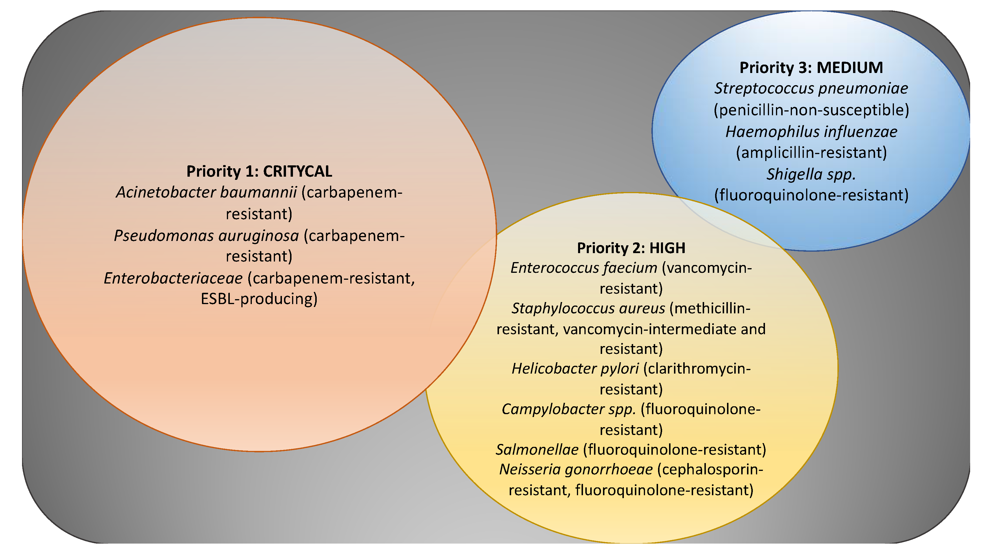
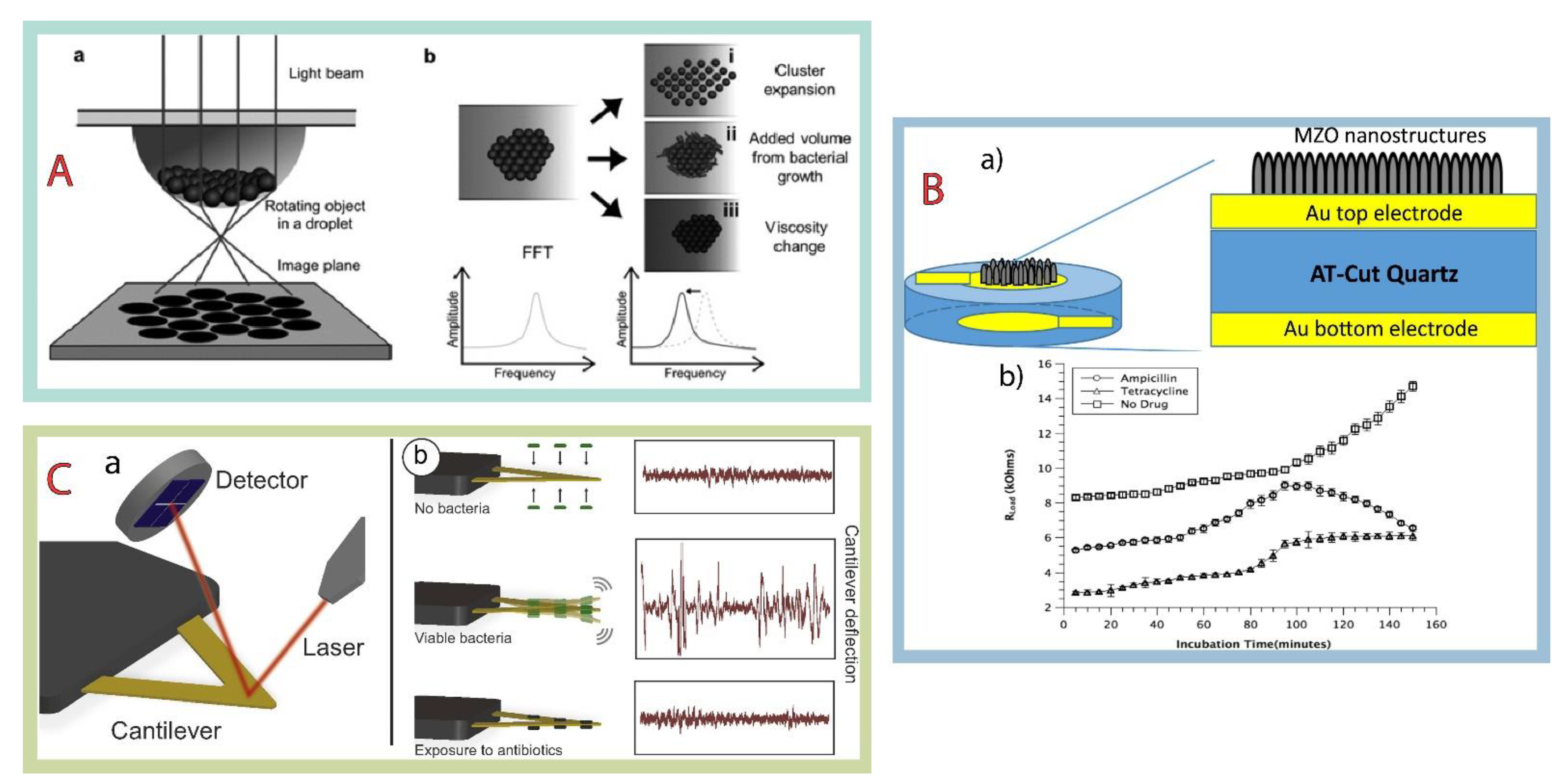
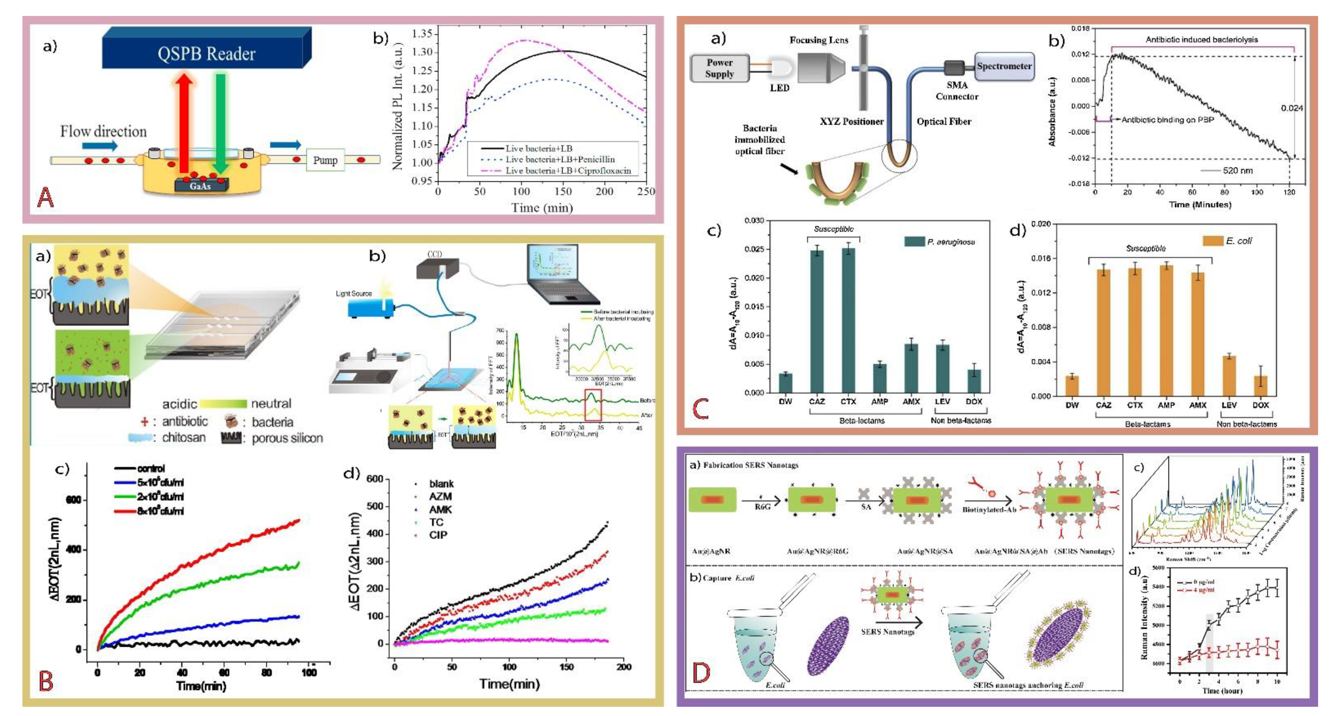
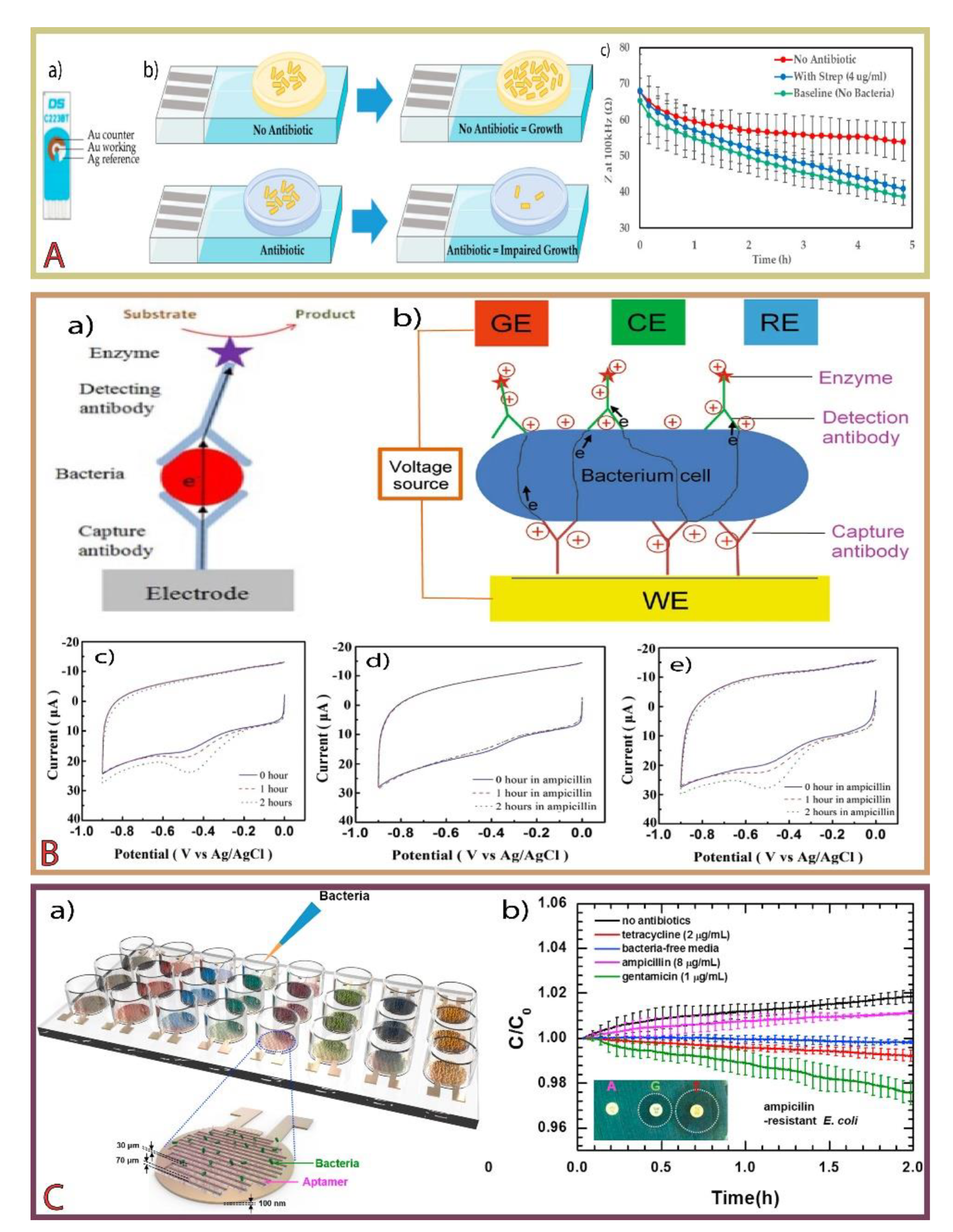
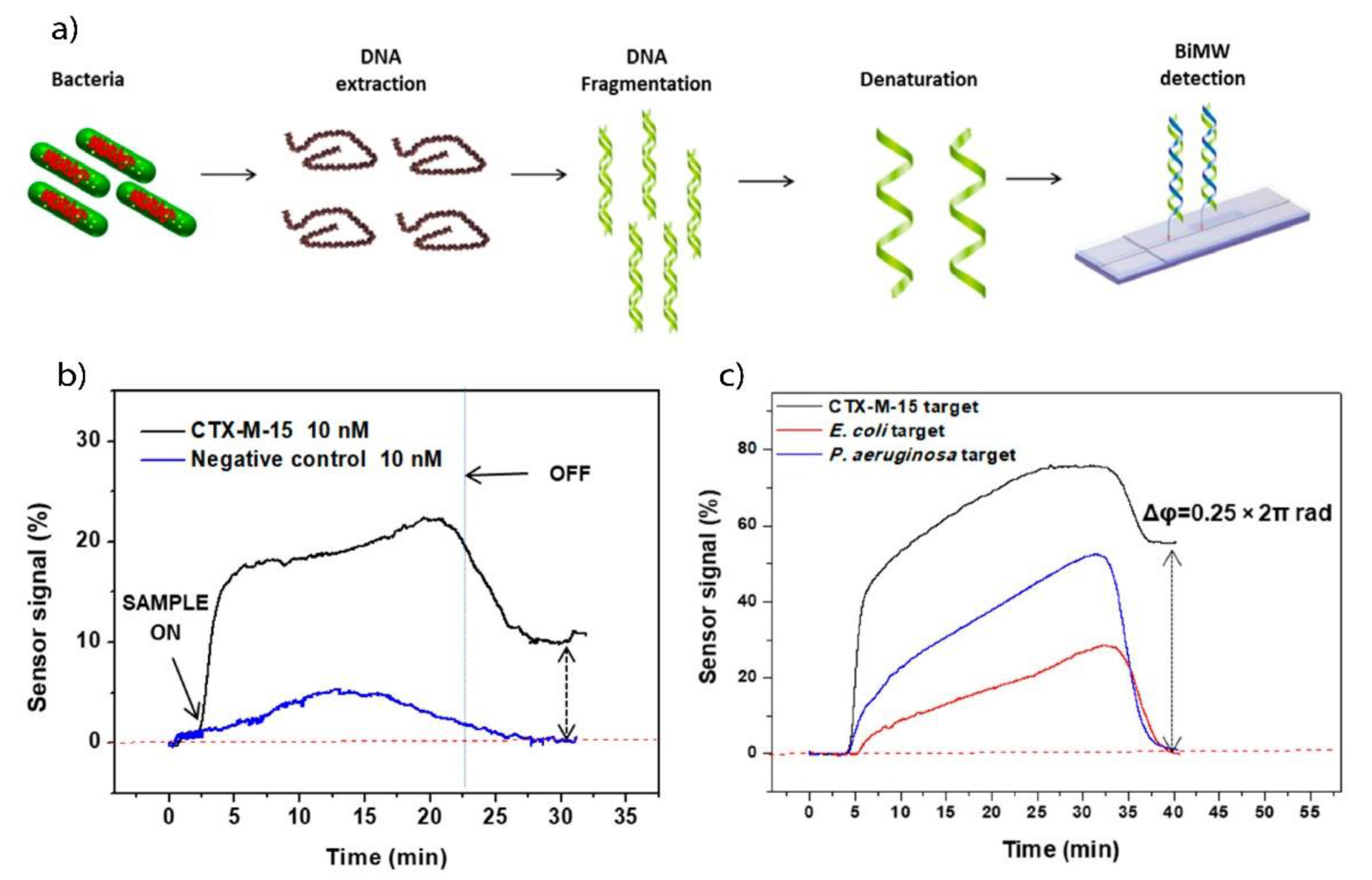
| Antimicrobial Group | Antimicrobial Agent(s) | Some of Resistance Gene(s) | Resistance Mechanism |
|---|---|---|---|
| Aminocoumarins | Novobiocin, Coumermycin, Clorobiocin | gyrB, parE, parY | Target modification |
| Aminoglycosides | Amikacin, Dibekacin, framycetin, gentamicin, kanamycin, neomycin, netilmicin, plazomicin, sisomicin, spectinomycin, streptomycin, tobramycin | aacA-aphD, aadD, aadE, aadY, ant(4′)-Ia, aphA3, armA, rmtA, rmtB, rmtC, str | Reduced permeability, antibiotic efflux, antibiotic modification, target modification |
| Amphenicol | Azidamfenicol, chloramphenicol, florfenicol, thiamphenicol | agmR, catA1, cmlA1, floR, ttgABC | Reduced permeability, antibiotic efflux, antibiotic modification, target modification |
| Ansamycins | Rifampicin, rifamixin | dnaA, rbpA, rpoB | Target modification, antibiotic modification, reduced permeability |
| Carbaphenems | Doripenem, ertapenem, Imipenem | blaOXA-23, blaOXA-58, blaOXA-497, blaVIM-1 | Reduced permeability, antibiotic efflux, antibiotic modification, target modification |
| Cephalosporins | Cefacetrile, cefalexin, cefalotin, cefapyrin, cefazolin, cefiximine, cefotaxime, ceftriaxone, cefuroxime | ampC, bla-genes | Reduced permeability, antibiotic efflux, antibiotic modification, target modification |
| Ethambutol | Ethambutol | embB, embC | Expression changes, target modification |
| Fluoroquinolones | Ciprofloxacin, clinafloxacin, levofloxacin, moxifloxacin, ofloxacin, pazufloxacin, sarafloxacin | gyrA, gyrB, parC, parE | Reduced permeability, antibiotic efflux, antibiotic modification, target modification |
| Fosfomycin | Fosfomycin | fomA, fomB, fosC, fosA, fosB, fosX | Antibiotic modification, target modification |
| Fusidanes | Fusidic acid | fusB, fusC | Reduced permeability, target protection |
| Glycopeptides | Corbomycin, ramoplanin, telavancin, teicoplanin, vancomycin | vanA, vanB, vanD, vanR, vanS | Reduced permeability, target modification |
| Isoniazid | Isoniazid (INH) | ahpC, inhA, katG | Expression changes, target modification |
| Lincosamides | clindamycin, lincomycin, pirlimycin | ermA, ermB, erm(31), ein, lnu(A), lnu(B), lsa(B), sal(A) | Antibiotic efflux, antibiotic modification, target modification |
| Lipopeptides | Bacillomycin, caspofungin, daptomycin, mycosubtilin, surfactin, surotomicyn | cdsA, rpoB | Antibiotic modification, target modification |
| Macrolides | Azithromycin, erythromycin, oleandomycin, josamycin, roxithromycin, spiramycin, | cfr, ermA, ermB, erm(31), ereA, ereB, gimA, mefA, mefE, mel, mgt, ole | Reduced permeability, antibiotic efflux, antibiotic modification, target modification |
| Mupirocin | Mupirocin | mupA, mupB | Target modification |
| Nitroimidazoles | Azanidazole, benzinidazole, dimetridazole, megazol, metronidazole, nimorazole, ornidazole, pretomanid, tinidazole | nimA, nimB, nimC, nimD, nimE | Antibiotic modification |
| Nitrofurans | Furazolidone, nifuroxazide, nifurtimox, nifurtoinol, nitrofural, nitrofurantoin, nifurzide | nfsA, nfsB | Antibiotic efflux, target modification |
| Oxazolidinones | Linezolid, posizolid, radezolid, tedizolid | cfr, optrA, poxtA | Antibiotic efflux, target modification |
| Penicillins | Amoxicillin, ampicillin, benzylpenicillin, cloxacillin, penicillin, phenethicillin | ampC, blaZ, mecA, mecZ | Reduced permeability, antibiotic efflux, antibiotic modification, target modification |
| Polymixins | Bacitracin, colistin | mcr-genes, mgrB, pmrA, pmrB, pmrE | Antibiotic efflux, target modification, expression changes |
| Pyrazinamide | Pyrazinamide | clpC1, pncA, rpsA | Target modification |
| Streptogramins | Quinupristin/dalfopristin, pristinamycin, virginiamycin | cfr, erm-genes, lsa, msrA, vga, vgaB, vatA, vatB, vatC, vatD, vatE | Antibiotic efflux, antibiotic modification, target modification |
| Sulfonamides | Sulfadiazine, sulfadimethoxine, sulfadimidine, sulfafurazole, sulfamerazine, sulfamethoxazole, sulfanilamide, sulfapyridine | sul1, sul2, sul3, sul4 | Reduced permeability, expression changes, target modification |
| Tetracyclines | Chlortetracycline, doxycycline, omadacycline, oxytetracycline, tetracycline | tetA, tetB, tetC, tetM, tetO, tetQ, tetX, tet30, tet31, tet32, tet36 | Reduced permeability, antibiotic efflux, target protection, target modification |
| Trimethoprim | Trimethoprim | dfrA, dfrD, dfrG, dfrK | Target modification |
Table 2.
Different magnetic, mass, and mechanical sensor for AST.
| Technic/Signal/Type | Target | Antibiotic | MIC (µg/L) | Limit of Detection | Time | Reference |
|---|---|---|---|---|---|---|
| Asynchronous magnetic bead rotation | E. coli | Gentamicin | 1 | Single bacterium binding events | 15 min | [87] |
| Atomic force microscope cantilevers | E. coli | Ampicillin | 12.5–50 | NM, 1 × 10⁵ CFU/mL ** | 45 min | [100] |
| Asynchronous magnetic bead rotation | E. coli | Ampicillin | 8 | Single bacterium binding events | 1.5 h | [103] |
| Asynchronous magnetic bead rotation | E. coli | Gentamicin | 1 | 50 cells per drop | 100 min | [104] |
| Asynchronous magnetic bead rotation | E. coli | Gentamicin | 2 | Single bacterium binding events | 91 min | [105] |
| Brownian motion | P. aeruginosa | Gentamicin | 2 | One bacterium | 2 h | [106] |
| Multi-channel series piezoelectric quartz crystal | E. coli | Ampicillin | 32 | 5 × 10⁵ CFU/mL ** | 5–8 h | [107] |
| Spectral amplitude modulation MZO- QCM | S. epidermidis | Ciprofloxacin Oxacillin Ciprofloxacin | 0.5 1 1 | 1 × 10⁵ CFU/mL | 1.5 h | [108] |
| Orthogonal quartz crystal microbalance | E. coli | Ciprofloxacin Ceftriaxone Tetracycline | 12.5 15 150 | 5 × 10⁸ CFU/mL** | 1 h | [109] |
| Indirect series piezoelectric | M. tuberculosis | Isoniazid Rifampin Ethambutol Streptomycin Capreomycin p-Aminosalicylic acid Ethionamide Rifabutin | 0.1 1.0 2.5 2.0 10 2.0 5.0 0.5 | 1 × 10³ CFU/mL ** | >1 day | [110] |
| QCM under an external magnetic field | D. desulfotomaculum | Vancomycin | NR | 1.8 × 10⁴ CFU/mL ** | 30 min | [111] |
| Cantiliver NMS under an external magnetic field | M. bovis, M. abscessus | Amikacin Rifampin Isoniazid | 1.7 0.15 0.17 | 100 bacterial cells | 30 min | [112] |
| Atomic force microscope cantilevers | E. coli, S. aureus | Ampicillin Kanamycin | 2.0 70.0 | 4.6 ± 0.5 bacteria/100 µm2 | 30–40 min | [113] |
| biomaterial microcantilever with an embedded microfluidic channel | E. coli | Ampicillin Kanamycin | NM | 1 × 10⁵ CFU/mL ** | 30 min | [114] |
| Atomic force microscope cantilevers | B. Pertussis | Erythromycin Clarithromycin | 0.06 0.12 | NM | 20–40 min | [115] |
** Initial concentration; CFU, colony-forming unit; MZO, magnesium zinc oxide; NM, not measured; NMS, nanomotion sensor; NR, not reported; QCM, quartz crystal microbalance.
Table 3.
Optical (bio)sensors for determination of susceptibility to antimicrobials.
| Technique | Recognition Probe | Target | Antibiotic | MIC (µg/L) | Limit of Detection | Time | Reference |
|---|---|---|---|---|---|---|---|
| Colorimetric | Tetrazolium salts-8 | E. coli | Ampicillin | 128 | 10 CFU/mL | 2 h | [122] |
| Fluorescence | TDN-aptamer/SYTO 9 Green | E. coli | Kanamycin Streptomycin Ofloxacin Norfloxacin Chloramphenicol | 4.0 8.0 0.5 1.0 2.0 | 10 CFU/mL | 5 h | [130] |
| SPR | 2PAC—Au nanosphere/block copolymer templates (PS-b-PMMA) | E. coli, P. aeruginosa | Carbenicillin Gentamicin Rifampicin | 100 1 Resistant | NR | 30 min | [131] |
| laser-patterned paper-based | Chromogenic agar CHROMagar/photopolymer DeSolite® | E. coli | Amoxicillin | 30 | 2.5 × 10⁹ CFU/mL | 18 h | [132] |
| SPR | Poly-L-lysine/glass slide coated with gold sensor chip | MRSAMSSA | Cefoxitin | 32 to >128 1 to 4 | 5 × 10⁵ CFU/mL * | 3 h | [133] |
| Colorimetric | Endogenous H2S/AgNRs | E. coli | Ampicillin | 100 (MBC) | 102 cell/mL * | 4–6 h | [134] |
| Fluorescence | Resazurin | E. coli | Gentamicin | 4 | Single cell | 1 h | [135] |
| Fluorescence imaging | anti-E.coli antibody/streptavidin-coated polystyrene microsphere | E. coli | Ceftazidime Levofloxacin | 4 32 | Single cell | 30 min | [136] |
| SERS | Gold nanoparticles | L. lactis | Ampicillin Ciprofloxacin | NR | NR | 1.5 h | [137] |
| Fluorescence | PDMS/TLFM | E. coli | Ampicillin Cefalexin Chloramphenicol Tetracycline | 8 12 8 2 | Single cell | 2–4 h | [138] |
| SPR | Poly-L-lysine/Au thin film | E. coli S. epidermidis | Ampicillin Tetracycline | 3 10 | NR | 2 h | [139] |
| SERS | Bacteria-aptamer/AgNPs | E. coli S aureus | Tigecycline Vancomycin | 0.02 0.2 | 5 × 10³ CFU/mL * | 2 h | [140] |
| Raman tweezers | fused-silica microfluidic chip | S. aureus | Oxacillin | 2000 | 1012 cells/L | 4 h | [141] |
* Initial concentration; 2PAC, two-dimensional physically activated chemical assembly method; AgNPs, silver nanoparticles; AgNRs, silver nanorods; MBC, minimal bactericidal concentration; MRSA, methicillin-resistant S. aureus; MSSA, methicillin-susceptible S. aureus; NR, Not reported; PDMS, polidimetilsiloxano; PS-b-PMMA, poly(styrene-block-methyl methacrylate); SERS, surface-enhanced Raman spectroscopy; SPR, surface plasmon resonance; TDN, tetrahedral DNA nanostructure; TLFM, time-lapse fluorescence microscopy.
Table 4.
Electrochemical (bio)sensor for determination of susceptibility to antimicrobials.
| Electrode—Recognizing Element | Respond | Target | Antibiotic | MIC (µg/L) | Limit of Detection | Time | Reference |
|---|---|---|---|---|---|---|---|
| Au SPE—antibody alkaline phosphatase | DPV | S. aureus | MRSA strain | Nm | 845 CFU/mL | 4.5 h | [143] |
| SPE—Thiolated oligonucleotide capture probes | Amperometric | E. coli | Ciprofloxacin | 2 | 103 CFU/mL | 5 h | [150] |
| G-FET—peptide probes | Dirac voltage | S. aureus, A. baumannii | colistin resistant strain | NM | 104 cells/mL | 5 min | [165] |
| SPCE/MWCNTs/AuNPs—reduction of resazurin | DPV | S. gallinarum | Ofloxacin Penicillin | 32 16 | 102 CFU/mL | 1 h | [166] |
| Au—aptamers | Capacitance | E. coli *, A. baumannii, P. aeruginosa, K. pneumoniae, S. aureus, E. faecalis | Amikacin Ampicillin Aztreonam Cefepime Cefotaxime Ceftazidime Gentamicin | ≤2 ≥32 ≤1 ≤1 ≤4 ≤1 ≤1 | 105 CFU/mL ** | 6 h | [167] |
| Au SPE—agarose-based hydrogel deposit | EIS | S. aureus | Amoxicillin Oxacillin | 8 in both | 107 CFU/mL (50,000 CFUs) ** | 45 min | [168] |
| Pt deposited over a glass substrate—reduction of resazurin | DPV | E. coli, K. pneumoniae | Ampicillin Kanamycin Tetracycline | NM | 104 cells/mL | 4 h | [169] |
| Two working electrodes (Au and Pt)—POA detection | CV | M. tuberculosis | Pyrazinamide | NM | 40 µM of POA | NR | [170] |
| Array of interdigital electrodes—FD of the impedance of living and dead microorganisms | Impedance | E. coli | Ceftazidime Ceftriaxone Benzylpenicillin | NR | NR | 2 h | [171] |
| Silicon nanowire FETs | Current caused by varying pH values | E. coli | Kanamycin Cefotaxime Ofloxacin | 1–4 0.1–6 5 | Single cells | 6 h | [172] |
| 3-APBA modified electrode bind with cis-diol groups on the cell wall | Capacitance | E. coli, S. thyphi, P. aeruginosa, S. epidermidis, S. aureus, B. subtilis | Ceftriaxone Q. infectoria Ampicillin Vancomycin Rhodomyrtone | 0.03 20 0.5 1.25 0.5 | 108 CFU/mL | 2.5 h | [173] |
| SPE plastic-based microchips—antibodies | Impedance | E. coli, S. aureus | Erythromycin | 0.1 | 103 CFU/mL ** | 1.5 h | [174] |
| Interdigital electrodes—antibodies | Impedance | S. aureus | Flucloxacillin | 100 | 104 cells/mL ** | 2 h | [175] |
* Antibiotic and MIC correspond to E. coli; ** Initial concentration; 3-APBA, 3-aminophenylboronic acid; AuNPs—gold nanoparticles; CV, cyclic voltammetry; DPV, differential pulse voltammetry; EIS, electrochemical impedance spectroscopy; FD, frequency dependence; G-FET, graphene field effect transistors; MRSA, methicillin-resistant Staphylococcus aureus; MWCNTs, multiwalled carbon nanotubes; Not measured; Not reported; POA, pyrazinoic acid; SPCE, screen-printed carbon electrodes; SPE, screen-printed electrodes.
Table 5.
Different genosensor for determination of antimicrobial resistance genes.
| Technic | Recognition Element | Target | Type of Resistance | Limit of Detection | Previous Amplification | Reference |
|---|---|---|---|---|---|---|
| Electrochemical—EIS and CV | DNA probe | rpoB | Rifampicin resistance | 0.08 fmol/L | Yes | [179] |
| Optic—fluorescence | DNA probe | CTX-MNDM-1 | Cephalosporins resistanceCarbapenems resistance | <10 copies of the gene | Yes | [182] |
| Optic—SERS | Hairpin-structured | tetA | Tetracycline Resistance | 25 copies/μL | No | [189] |
| Optic—fluorescence | Fluorescent nucleic acid probe | VIM NDM IMP KPC | carbapenem antibiotic resistance genes | (1.8 ± 0.7) × 106 beads/mL per target | No | [190] |
| Electrochemical—EIS and CV | DNA probe | rpoB | Rifampicin resistance | 0.2 fM | Yes | [191] |
| Optic—fluorescence | Binary deoxyribozyme | rpoB katG inhA | Rifampin resistanceIsoniazid resistanceFluoroquinolone resistance | 5 fg–15.6 pg | Yes | [192] |
| Electrochemical—EIS and CV | DNA probe | rpoB | Rifampicin resistance | 0.1 fM–1 pM | No | [193] |
| Electrochemical—capacitance | DNA probe | ampR | Ampicillin resistance | 1–4 pM | No | [194] |
| Electrochemical—DPV | DNA probe | MDR1 | Multidrug resistance | 2.95 × 10⁻¹² M | No | [195] |
| Optic—fluorescence | DNA probe | rpoB | Rifampicin resistance | 1 nM ssDNA in 1 mL sample volume | Yes | [196] |
| Optic—fluorescence | DNA probe | rpoB | Rifampicin resistance | 100 nM | Yes | [197] |
| Optic—SPR | DNA probe | rpoB | Rifampicin resistance | NR | No | [198] |
| Electrochemical—DPV | PNA probe | rpoB | Rifampicin resistance | 1 CFU/ml | No | [199] |
| Optic—fluorescence | Fluorescence DNA hairpin | mecR | Methicillin resistance | 1 nM | Yes | [200] |
| Optic—fluorescence | Ab-DNA probe | lamB | Increases resistance to chlortetracycline, ciprofloxacin, balofloxacin and nalidixic acid | 4–250 pM amplicon concentrations | Yes | [201] |
| Electrochemical—DPV | DNA probe | rpoB | Rifampicin resistance | 20 fM | Yes | [202] |
| Mechanic—piezoelectric | DNA probe | mecR | Methicillin resistance | 0.125 µM | Yes | [203] |
Ab, antibody; CV, cyclic voltammetry; DPV, differential pulse voltammetry; EIS, electrochemical impedance spectroscopy; IMP, Imipenemase; KPC, Klebsiella pneumoniae carbapenemase; NDM, New Delhi metallo-β-lactamase; NR, not reported; SERS, surface-enhanced Raman spectroscopy; VIM, Verona integron metallo-β-lactamase.
Publisher’s Note: MDPI stays neutral with regard to jurisdictional claims in published maps and institutional affiliations. |
© 2021 by the authors. Licensee MDPI, Basel, Switzerland. This article is an open access article distributed under the terms and conditions of the Creative Commons Attribution (CC BY) license (https://creativecommons.org/licenses/by/4.0/).
Share and Cite
MDPI and ACS Style
Reynoso, E.C.; Laschi, S.; Palchetti, I.; Torres, E. Advances in Antimicrobial Resistance Monitoring Using Sensors and Biosensors: A Review. Chemosensors 2021, 9, 232. https://doi.org/10.3390/chemosensors9080232
AMA Style
Reynoso EC, Laschi S, Palchetti I, Torres E. Advances in Antimicrobial Resistance Monitoring Using Sensors and Biosensors: A Review. Chemosensors. 2021; 9(8):232. https://doi.org/10.3390/chemosensors9080232
Chicago/Turabian StyleReynoso, Eduardo C., Serena Laschi, Ilaria Palchetti, and Eduardo Torres. 2021. "Advances in Antimicrobial Resistance Monitoring Using Sensors and Biosensors: A Review" Chemosensors 9, no. 8: 232. https://doi.org/10.3390/chemosensors9080232
Note that from the first issue of 2016, this journal uses article numbers instead of page numbers. See further details here.