Article

Vascular Lipidomic Profiling of Potential Endogenous Fatty Acid PPAR Ligands Reveals the Coronary Artery as Major Producer of CYP450-Derived Epoxy Fatty Acids

1
Division of Intramural Research, NIEHS/NIH, Research Triangle Park, NC 27709, USA
2
Department of Entomology and Comprehensive Cancer Center, University of California, Davies, CA 95616-8584, USA
3
Royal Veterinary College, University of London, London N1 0TU, UK
4
North Cornwall Research Institute, Bude, Cornwall EX23 9EE, UK
*
Author to whom correspondence should be addressed.
Cells 2020, 9(5), 1096; https://doi.org/10.3390/cells9051096
Submission received: 13 March 2020 / Revised: 22 April 2020 / Accepted: 27 April 2020 / Published: 29 April 2020
(This article belongs to the Special Issue The Role of PPARs in Disease)

Abstract

:
A number of oxylipins have been described as endogenous PPAR ligands. The very short biological half-lives of oxylipins suggest roles as autocrine or paracrine signaling molecules. While coronary arterial atherosclerosis is the root of myocardial infarction, aortic atherosclerotic plaque formation is a common readout of in vivo atherosclerosis studies in mice. Improved understanding of the compartmentalized sources of oxylipin PPAR ligands will increase our knowledge of the roles of PPAR signaling in diverse vascular tissues. Here, we performed a targeted lipidomic analysis of ex vivo-generated oxylipins from porcine aorta, coronary artery, pulmonary artery and perivascular adipose. Cyclooxygenase (COX)-derived prostanoids were the most abundant detectable oxylipin from all tissues. By contrast, the coronary artery produced significantly higher levels of oxylipins from CYP450 pathways than other tissues. The TLR4 ligand LPS induced prostanoid formation in all vascular tissue tested. The 11-HETE, 15-HETE, and 9-HODE were also induced by LPS from the aorta and pulmonary artery but not coronary artery. Epoxy fatty acid (EpFA) formation was largely unaffected by LPS. The pig CYP2J homologue CYP2J34 was expressed in porcine vascular tissue and primary coronary artery smooth muscle cells (pCASMCs) in culture. Treatment of pCASMCs with LPS induced a robust profile of pro-inflammatory target genes: TNFα, ICAM-1, VCAM-1, MCP-1 and CD40L. The soluble epoxide hydrolase inhibitor TPPU, which prevents the breakdown of endogenous CYP-derived EpFAs, significantly suppressed LPS-induced inflammatory target genes. In conclusion, PPAR-activating oxylipins are produced and regulated in a vascular site-specific manner. The CYP450 pathway is highly active in the coronary artery and capable of providing anti-inflammatory oxylipins that prevent processes of inflammatory vascular disease progression.

1. Introduction

Peroxisome proliferator-activated receptors (PPARs) can be activated by a diverse group of endogenous fatty acid mediators including those produced from cyclooxygenase (COX), lipoxygenase and CYP450 enzymatic pathways [1]. These COX, lipoxygenase and CYP450 enzymes metabolize arachidonic acid and related polyunsaturated fatty acids, linoleic acid (LA), docosahexaenoic acid (DHA) and eicosapentaenoic acid (EPA) into series of biologically active oxylipin mediators [2,3,4]. Cyclooxygenases largely make prostanoids (and some hydroxyeicosatetraenoic acids (HETEs)) [5,6]. Lipoxygenases make hydroperoxy-eicostetraeoic acid HpETE, HETEs, hydroxyoctadecaenoic acids (HODEs), hydroxy-DHAs, and hydroxy-EPAs—some of which are the precursors for leukotrienes [2]. The PUFA-utilizing CYP450s metabolize fatty acids into series of oxylipin mediators through a combination of either epoxidation or lipoxygenase-like or ω- and ω-1-hydroxylation [2,3]. Using arachidonic acid as an example, CYP2J2 can produce both epoxyeicosatrienoic acids (EETs) and 19-HETE by its epoxygenase- and hydroxylase-like activities, respectively [3,7]. The PGD2 metabolite 15 deoxy-D12,14-PGJ2, PGI2, 8-, 12-, and 15-HETE, 9- and 13-HODE [1], and 8-, 9-, 11-, 12-, and 14–15-EET [8] have all been shown to activate PPARs. Soluble (sEH) and microsomal (mEH) epoxide hydrolases (EH; encoded by the gene ephx2 and ephx1 respectively) combine to metabolize nearly all EpFAs in vivo [9]. sEH inhibitors (sEH-I) inhibit the breakdown of EpFAs to their more soluble but less biologically active dihydroxy counterparts and potentiate EET signaling [10,11,12].
While several oxylipins can signal through known or yet-to-be-identified G-protein-coupled receptors, transient increases in oxylipin ligands can also induce PPAR activation toward a variety of downstream signals [1]. PPAR activation induces heterodimerization with other nuclear receptors such as the retinoid X receptor (RXR), which enhances binding to a consensus sequence (direct repeats of ‘AGGTCA’) referred to as PPAR response elements (PPREs). PPAR ligands have diverse roles in the cardiovascular system, from repression of genes encoding pro-inflammatory cytokines to induction (e.g., TNFα, IL1, IL6) of monocytes/macrophages toward foam cell morphology [13].
The roles of oxylipins are of long-standing interest in vascular biology [11,13,14,15,16,17,18,19,20]. COX products have both cardioprotective (prostacyclin; PGI2) and pro-thrombotic (e.g., thromboxane; TXA2) activity [20,21]. CYP450-derived EpFAs are anti-atherosclerotic, vasodilatory and anti-inflammatory [11,22,23,24,25,26,27,28,29,30], with the notable exception of LA-derived dihydroxyoctadecamonoenoic acids (DHOMEs), which regulate cardiac function [31], vascular development [32], and thermal hyperalgesia [33] at low levels, but are toxic at higher levels [34]. CYP450-derived EETs, in particular, were originally described in porcine coronary artery as an endothelium-derived hyperpolarizing factor produced in response to stimulation and stretch sEH-I-treated or sEH-knockout mice show protection to injury induced vascular neointima formation [25], atherosclerosis and aneurysm formation [26], hypertension [35,36], type 2 diabetes [37], and inflammatory cell recruitment [23,30]. Interestingly, in the pulmonary circulation, although sEH inhibitors have been shown to augment hypoxia-induced vasoconstriction, sEH inhibition or overexpression of EpFA-producing enzymes such as CYP2J2 is protective in various acute lung injury models [38,39,40].
We previously showed that PPARs can be activated by CYP2J2 and its products in vitro and in vivo [8]. A number of protective effects of CYP2J2 or EETs have now been shown to be mediated by PPARs, including the protective effects of laminar flow on endothelial cells [24], mediating coronary reactive hyperemia [41,42,43,44] and vascular response in soluble epoxide hydrolase-null mice [45], cytoprotection of cardiomyocytes [46], inhibition of angiotensin II cardiac remodeling [47] and abdominal aortic aneurysm formation [48], inhibition of renal interstitial fibrosis and inflammation [49], improved vascular function and decreased renal injury in hypertensive obese rats [50], and promoting angiogenesis and migration in human endothelial progenitor cells from acute myocardial infarction patients [51].
Pigs have a similar heart and cardiovascular system to humans and undergo spontaneous and diet-induced atherogenesis [52]. Here, we used a lipidomic approach to study endogenous oxylipin PPAR ligand production by the large vessels of the pig: the thoracic aorta compared to the coronary and pulmonary arteries. The vessel releasates were also compared to those of aortic perivascular adipose tissue ex vivo. Perivascular adipose was investigated as it has been shown to release various cytokines that act in an endocrine and paracrine manner to regulate vascular signaling and inflammation which have been implicated in the development of atherosclerosis, hypertension, neointimal formation, aneurysm, arterial formation and vasculitis [53,54].
Using a targeted lipidomic approach, we found coronary artery releases significantly more oxylipins of almost all classes than aorta and pulmonary artery. Perivascular adipose was a particularly rich source of COX-derived PGE2. Coronary artery was the highest source of CYP450-derived EpFAs PPAR ligands. The use of a sEH inhibitor TPPU on pig primary coronary artery vascular smooth muscle cells in culture showed strong anti-inflammatory activity consistent with PPAR activation.

2. Materials and Methods

2.1. Materials

Authentic oxylipins (EETs, DHEQ, and HDPA) were from Cayman Chemical Company (Cambridge Bioscience, Cambridge, UK). SYBR green was from Takara. TPPU (N-[1-(1-oxopropyl)-4-piperidinyl]-N’-[4-(trifluoromethoxy)phenyl)-urea) was synthesized as previously described [55]. Unless stated, all other reagents were from Sigma-Aldrich (Poole, Dorset, UK).

2.2. Vessel Organ Culture

Abattoir pig vessels largely from white X female pigs aged 8–10 weeks old were obtained from the Royal Veterinary College. Fresh tissue was collected and used within 4 h. The 50–500 mg segments of vessel or perivascular aortic adventitia were cultured in serum-free DMEM supplemented with antibiotic/antimycotic mix (Sigma-Aldrich, St. Louis, MO., USA) at 37 °C, 5% CO2 and 95% air, as previously described for rat and human vessels [56,57]. Serum-free media was used, as most sera contain large amounts of oxylipins (unpublished observations). Organ culture was performed for just the first 24 h after explant in order to minimize cell differentiation. In some experiments, lipopolysaccharide (LPS; E. coli, 1 μg/mL) was given to induce an inflammatory response.

2.3. Cell and Tissue Culture

Primary coronary artery smooth muscle cells (pCASMCs) were obtained by explant and grown as previously described for human vascular smooth muscle cells [58]. Briefly, extraneous tissue was removed, coronary arteries were opened along the midline, gently denuded, and chopped into small explants. SMCs were grown in DMEM supplemented with antibiotic/antimycotic mix, and 20% FBS, at 37 °C, 5% CO2 and 95% air. SMCs were identified by a characteristic morphological “hill-and-valley” growth pattern and by smooth muscle α-actin immunostaining. Since FBS interferes with lipid substrate composition and the release and detection of eicosanoids, all experiments were performed with DMEM supplemented with antibiotic/antimycotic mix and without FBS.

2.4. Real-Time qRT-PCR

Pig CYP2J34, sEH, TNFα, VCAM-1, ICAM-1, MCP-1 and CD40 were measured using the SYBR Green ddCT method (see Table S1 for primer pairs). Targets were normalized to 18S expression. RNA was extracted using the ThermoScientific RNA extraction kit and 1 μg of total RNA was used to generate cDNA using Superscript II (Invitrogen) according to the manufacturer’s instructions. SYBR green qPCR was performed using Premix Ex Taq II mastermix (Takara) using a Chromo-4 machine and Opticon software. Genomic sequences were obtained from the UCSC Genome Browser website (http://genome.ucsc.edu/cgi-bin/hgGateway) and primers (see Table S1) were designed from NCBI’s Primer Blast website (http://www.ncbi.nlm.nih.gov/tools/primer-blast/index.cgi?LINK_LOC=BlastHome).

2.5. Oxylipin Measurements

Explants were incubated in serum-free DMEM for 24 h, which allows for detection of both the highly abundant prostaglandins and HETEs and less-abundant CYP-derived oxylipins. LC–MS/MS analysis of oxylipin products in culture supernatants was as previously described [23,59]. LC–MS/MS analytes in samples were quantified against oxylipin standard curves (Cayman Chemical) using TraceFinder v4.1 (Thermo Scientific, Waltham, MA, USA) software.

2.6. Statistical Analyses

Graphical representations, heat maps and statistical analyses between groups (t-tests and paired t-tests) were performed using GraphPad Prism v8.1. When comparing multiple groups, ANOVA was followed by Holm–Sidak correction for multiple comparisons. All distributions appeared and were assumed to be normal.

3. Results

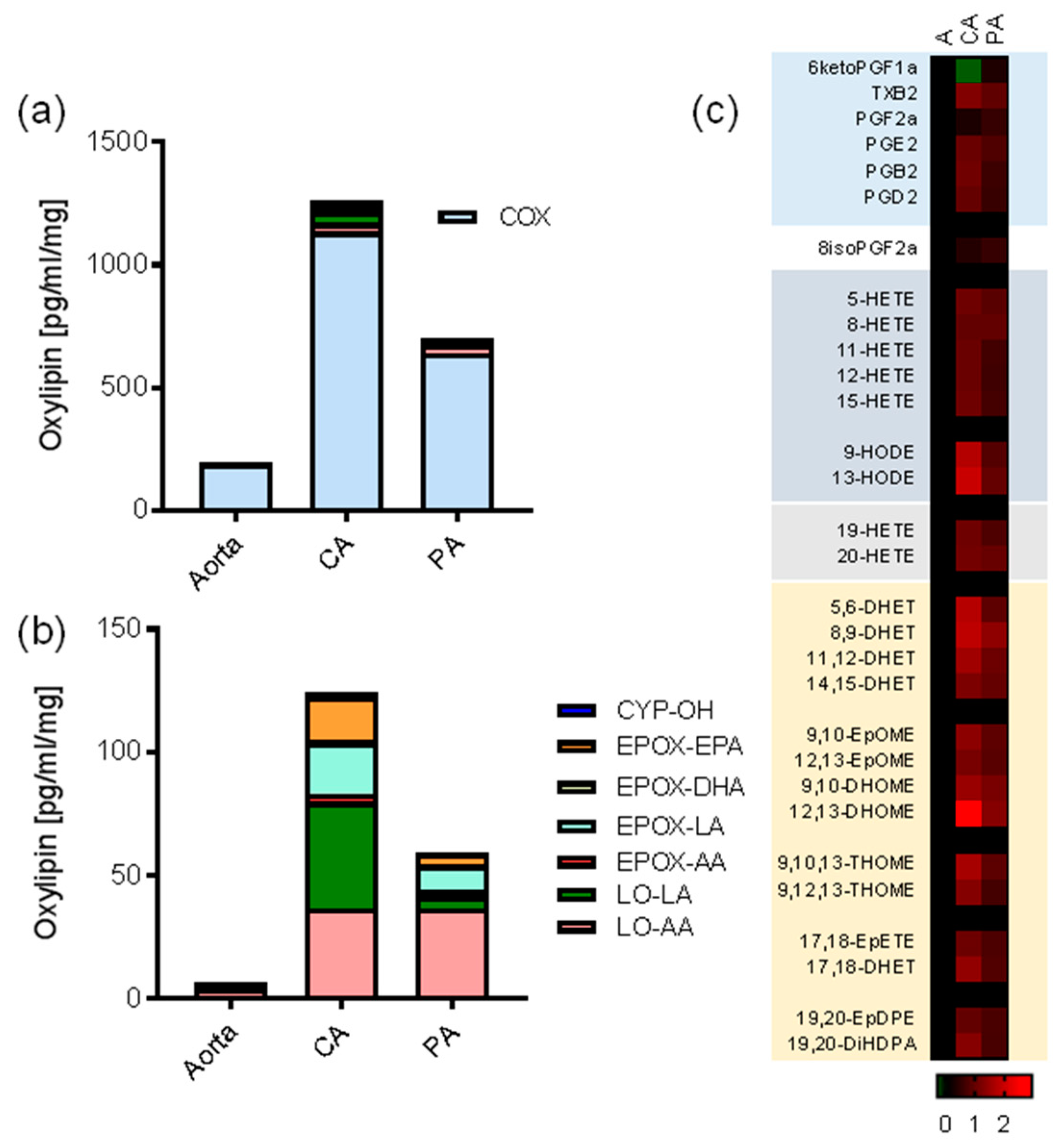
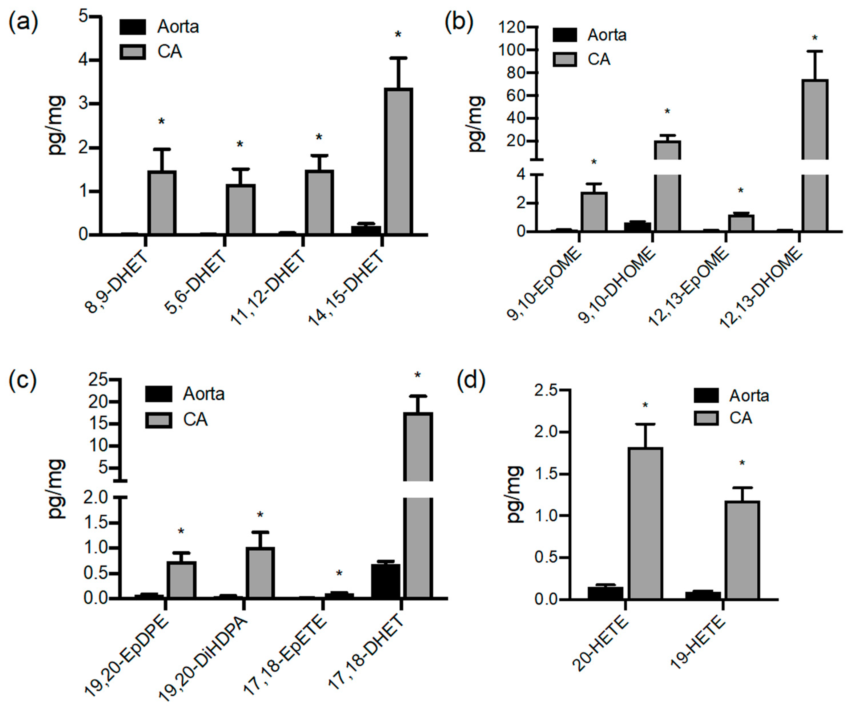
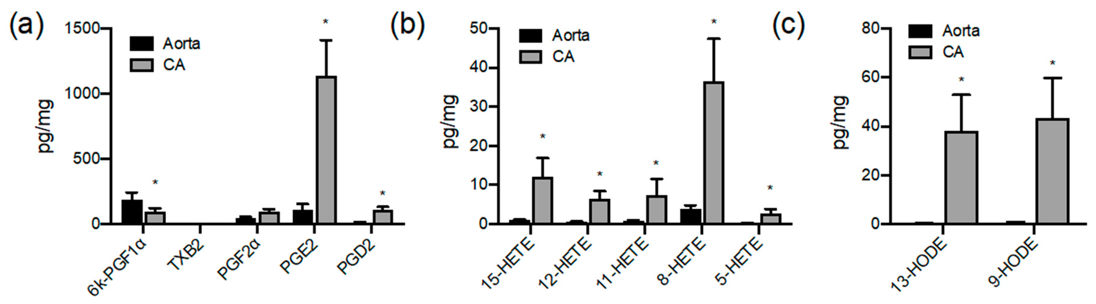
3.1. Oxylipin Lipidomic Profiling of the Large Vessels of the Pig

Young female pigs were selected to be devoid of atherosclerosis and represent non-diseased vascular tissues. Fresh tissue explants were divided into various treatment groups and cultured in serum-free media for 24 h. Serum-free media was used, as most sera contain large amounts of oxylipins (unpublished observations). CYP-, LOX- and COX-derived oxylipins were detectable in organs culture for 24 h after explant. The most abundant oxylipin species represented in 24 h organ culture in all tissues were prostanoids derived from COX (Figure 1a). PGI2 was the major product from aorta (190 pg/mg) and pulmonary artery (640 pg/mg), whereas PGE2 was the major product from coronary artery (1135 pg/mg) and perivascular adipose (1390 pg/mg; Figure 1). The coronary artery generated by far the largest total amounts of measurable oxylipins followed by pulmonary artery, with the aorta producing approximately 1/8 of the prostanoids per unit weight as the coronary artery (Figure 1a). The coronary artery produced significantly more EpFA and hydroxy fatty acids than the aorta or perivascular adipose (Figure 1b; Figure S1), with the pulmonary artery production again intermediate between the aorta and coronary artery (Figure 1 and Figure 2). The perivascular adipose produced similar amounts of PGE2 as the coronary artery, with much lower relative levels of lipoxygenase or CYP450 products formed than any of the vessels (data not shown).
Interestingly, the aorta and coronary artery produced similar levels of COX products, with the notable exceptions of PGI2, which was significantly higher from aorta compared to coronary artery, and PGE2, which was higher in coronary artery compared to aorta (p < 0.05 unpaired t-test; Figure 1 and Figure 2). Lipoxygenase-derived HETEs and HODEs were also produced in significantly higher amounts by coronary artery than the aorta (Figure 3). In particular, LA-derived oxylipin epoxygenase and lipoxygenase products were produced at considerably higher levels (up to 90-fold) by coronary artery than aorta (Figure 1, Figure 2 and Figure 3).

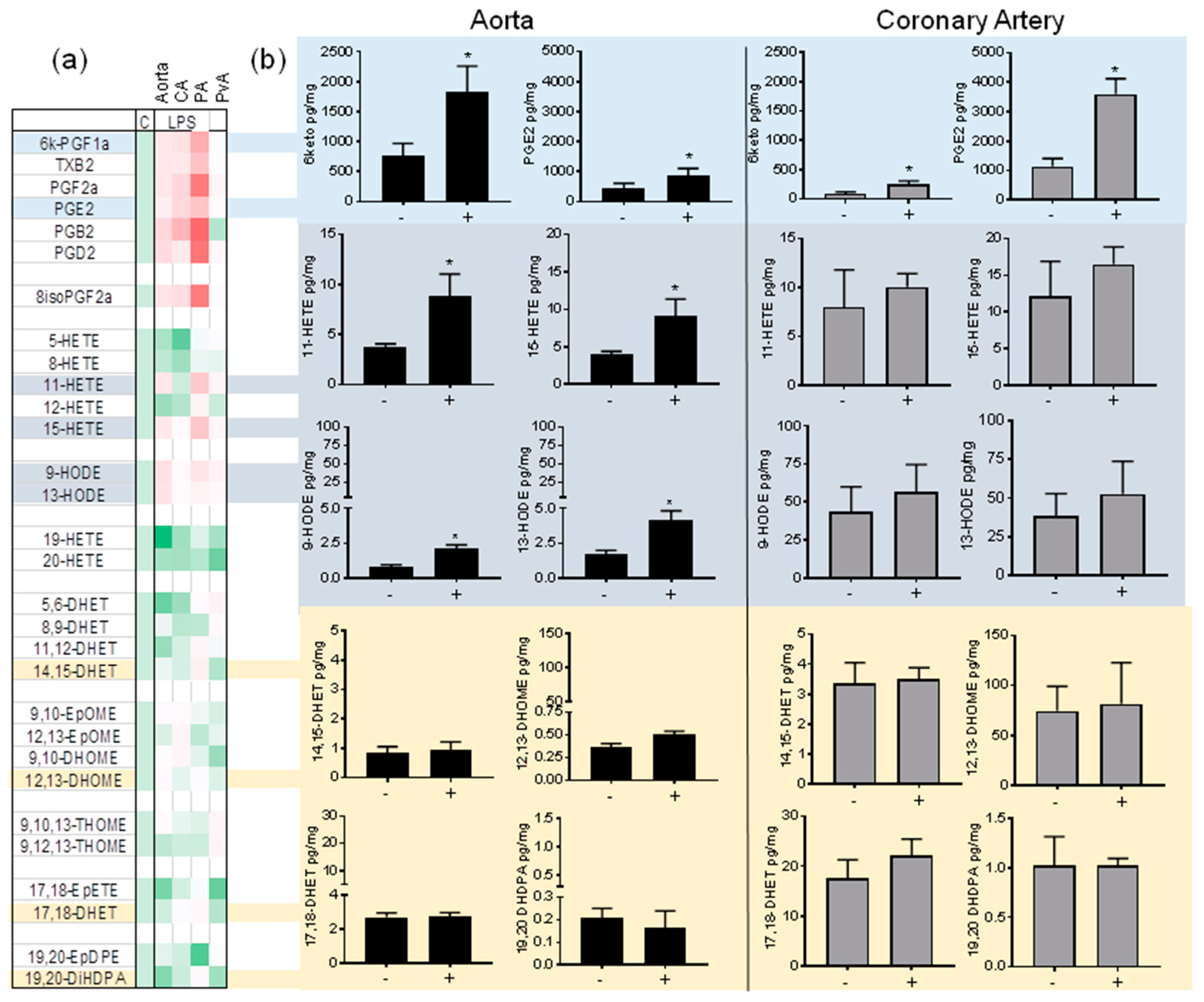
3.2. Regulation of Oxylipin Generation in the Large Vessels of the Pig by Inflammatory Stimuli: LPS/TLR4 Activation

Consistent with the well-established sensitivity of COX-2 induction, LPS elevated prostanoids in aorta, coronary artery, and pulmonary artery. Interestingly, LPS did not induce prostanoids in aortic perivascular adipose tissue (Figure 4a). In particular, the major vascular prostanoids PGI2 and PGE2 were significantly induced by LPS in vascular tissue (Figure 4). The 11-HETE, 15-HETE, 9-HODE and 13-HODE were significantly increased in the aorta and pulmonary artery, but not the coronary artery. With some exceptions, notably 19,20-EpDPE in pulmonary artery and 19-HETE in aorta (Figure 4a; Figure S2), LPS did not consistently alter lipoxygenase or CYP450 product levels (Figure 4a).

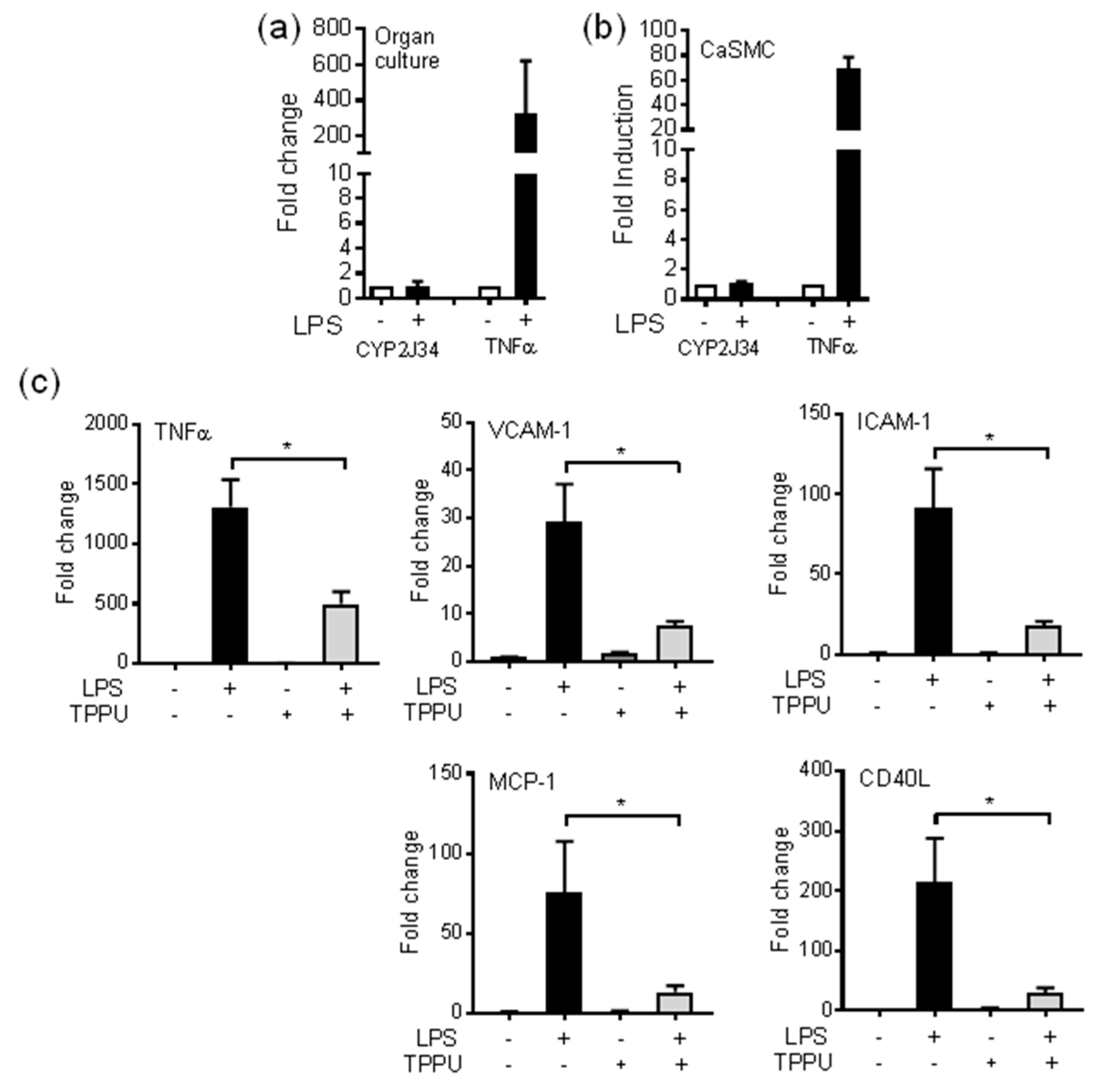
3.3. The sEH Inhibitor TPPU Reduces TLR-4 Induced Inflammation in pCASMCs

LPS did not induce the pig CYP2J homologue CYP2J34 in organ culture tissue (pulmonary artery and coronary artery) at 24 h or in primary pCASMCs (Figure 5a) at 4 h. By contrast, LPS strongly induced TNFα mRNA in both organ culture tissue and pCASMCs (Figure 5a). Although not induced by LPS, the endogenously produced EpFAs were anti-inflammatory in pCASMCs, as co-treatment of pCASMCs with the sEH-I TPPU (1 uM) significantly reduced LPS-induced TNFα, ICAM-1, VCAM-1, MCP-1 (CCL2), and CD40 mRNA (Figure 5b).

4. Discussion

We used a targeted lipidomic approach to identify the profile of oxylipins produced by pig coronary artery, aorta, pulmonary artery and aortic perivascular adipose tissue. In particular, the coronary artery was a major source of epoxygenase-derived oxylipins. Bovine and porcine coronary artery was one of the original sites for the discovery of vasoactive CYP450-derived EETs [60]. Here, we show that the coronary artery also produces CYP450 EpFAs from linoleic acid, EPA and DHA in significantly greater amounts than other large pig vessels. Outside of primates, the pig cardiovascular system is considered the most relevant to human biology. The pig heart is a similar size to human heart, and pigs can spontaneously undergo coronary artery disease [52]. The increased size of the pig compared to rodent models also means that it is also relatively easy to examine specific vascular responses in arteries such as the coronary artery, which would be very difficult in common rodent models.
Using fresh tissue in organ culture comes with certain caveats. Although directly comparative, these results are based upon 24 h accumulation of products. Our previous studies with organ culture indicate that fresh vessels are put under a mild inflammatory stress, which is associated with a low but significant level of COX-2 induction [56,57]. The results here are consistent with these previous studies [56,57]. Additionally, we know relatively little about the long-term stability of a number of oxylipins in media or biological fluids, but clearly there are differences. EETs for example are rapidly metabolized or reincorporated into membranes [3,61], so this 24 h accumulation analysis is likely to underestimate total EET production When we have examined acute oxylipin release (30 min) from rat aorta, prostaglandins and in particular PGI2 are still the most abundant species detected (DBB unpublished observation). Another caveat to this analysis is whether tissue-specific oxylipin metabolism is present. For example, coronary artery endothelial cells are known to metabolize EETs to chain-shortened epoxy-hexadecadienoic acids [62]; additionally, the presence of CYP4A3 may metabolize EETs into 20-OH derivative PPAR ligands [63], which were not included in our analysis.
LPS induces COX-derived prostanoids in rat and human vessels in organ culture in vitro [56,57]. All the pig vessels tested similarly produced prostanoids in response to LPS. The responsiveness of other oxylipin pathways to LPS is less well understood. Since activation of cPLA2 appears to be common to all three pathways, we hypothesized that lipoxygenase and CYP450 pathways would also be activated. Interestingly, the COX-derived eicosanoids were the only species commonly induced by LPS in all vessels. In aorta, but not coronary artery, 11-HETE and 15-HETE were similarly induced. The 11-HETE and 15-HETE are also potentially COX products [5], so it is intriguing why they are induced in the aorta and not coronary artery. Similar findings were previously found in human whole blood treated with LPS for 18 h [63]. Interestingly, HODEs were also induced by LPS in aorta, pulmonary artery and perivascular adipose tissue, which shows a selective induction of linoleic acid- and arachidonic acid-lipoxygenase pathways [64,65]. Unlike HETE induction, this HODE induction was not previously observed in human whole blood treated with LPS [66] but has been observed in the circulation of mice treated with LPS [67]. These lipidomic results clearly show a high compartmentalization between substrate generation and delivery to individual COX, lipoxygenease and CYP450 pathways. This data provides further impetus to look at the actions of these other oxylipin species. EPA and DHA are considered key components of the purported cardiovascular health benefits of oily fish. Supplementation of human or rodent diets with DHA and EPA increases DHA and EPA EpFAs [68,69]. Coronary artery metabolism of EPA and DHA into EpFAs could therefore contribute to these dietary lifestyle modifications in cardiovascular disease. Additionally, further investigations are required to understand the role of PPAR signaling in any effects.
The coronary artery is of particular interest for vascular research, since coronary artery disease and occlusion is the major cause of heart attacks in humans. There has been considerable interest in both the potential cardioprotective effects of PPAR ligands and testing sEH inhibitors in cardiovascular disease [25,26]. As recently reviewed PPAR ligands in experimental animal models have all shown to reduce aortic atherosclerosis [70,71]. There has been considerable interest in whether these findings translate into humans [70,71]. Both PPARα and PPARγ agonists have shown some mild clinical efficacy in reducing cardiovascular event [71]. However, the clinical efficacy of the PPARγ ligand rosiglitazone has been questioned as it appeared to increase cardiovascular events in an early trial [71]. Nonetheless, there has been considerably recent interest in developing selective modulators, and dual- and pan-PPAR agonists, that have increased efficacy and reduced side effects [70,71]. We hypothesize that the potential endogenous oxylipin PPAR ligands are more likely to act as pan/dual or selective modulator-type agonists. sEH inhibitors are anti-hypertensive, anti-diabetic, anti-obesity and reduce the development of aortic atherosclerosis in mouse models [25,26,59,72,73]. Importantly, atherosclerosis is rarely investigated in the coronary circulation of mice. More often, aortic atherosclerotic plaque formation is used as a surrogate for coronary artery disease. Our oxylipin profiling suggests that aorta CYP activity underestimates that found in coronary arteries and likely underpredicts the role of CYP-derived oxylipins in coronary artery atherosclerosis. Preservation of the coronary circulation therefore may be so critical that it has evolved this higher EpFA system to maintain flow and limit inflammation. Originally, these positive benefits of sEH inhibitors were attributed to lipid-lowering actions [26]. Human coronary artery disease, in particular obstructive coronary artery disease, is associated with decreased circulating EETs [74,75]. In the heart and coronary circulation, CYP2J2 or EETs mediate coronary reactive hyperemia [41,42,43,44], cytoprotection of cardiomyocytes [46], and inhibition of angiotensin II cardiac remodeling [47] in part by the activation of PPARs. sEH inhibition acts to maintain higher levels of EETs/EpFAs or may shunt to alternative PPAR ligands such as the 20-OH CYP4A derivatives. Although, DHETs are also PPAR activators [76], the concentrations required are 10–100-fold higher than published for EETs [8,64]; thus, sEH inhibition will act to promote the PPAR agonist activity of CYP-derived epoxides.
The coronary artery was the largest source of epoxygenase products of all the major vessels we tested, suggesting that multiple oxylipin species may have particularly important roles at this site. The pig homologue of human CYP2J2 is CYP2J34 [77]. In human monocytes and endothelial cells, we found that LPS induced CYP2J2 [28,29,78]. By contrast, LPS did not induce CYP2J34 in pig vessels or monocytes (Figure 5a, DBB unpublished observations), indicating at least one difference between human and pig CYP2J enzymes. We previously reported differences in intimal and medial SMC phenotypes isolated from the rat [79,80]. Medial SMCs but not intimal SMCs were sensitive to the anti-inflammatory actions of sEH inhibitors [81]. The sEH inhibitor TPPU inhibited inflammatory mediators induced by TLR-4 activation in primary pCASMCs. This is the first time anti-inflammatory actions have been described in coronary tissue for sEH inhibitors, and this further supports an anti-inflammatory/pro-resolution role for the sEH pathway in mediating cardioprotective actions. The pCASMCs we cultured represent a classical medial SMC phenotype, with a classical spindle shape and hill-and-valley morphology. No epithelial cell types were observed in these primary cultures. We have yet to determine whether distinct porcine ‘intimal’ SMC phenotypes can be identified that share these different properties.
The aorta produced the lowest levels of oxylipins, with the pulmonary artery in between the aorta and coronary artery. The lack of activity in the aorta may just reflect the aorta’s main role as a conduit vessel and one not particularly responsive to vasoactive mediators or a major site for human vascular disease initiation. The pulmonary artery produced the highest levels of basal and LPS-inducible PGI2, consistent with the importance of this eicosanoid in maintaining pulmonary health [79], which may be in part mediated by activation of PPARβ/δ (or PPARα) [1,82]. CYP450-derived eicosanoids contribute to hypoxia-induced pulmonary hypertension [83] and are protective in models of inflammation [38,39,40,84]. The production of these oxylipin mediators from the pulmonary artery suggest that the pig may also be a useful translational model to study oxylipins and PPARs on pulmonary health and disease. Here, we show perivascular adipose is also a large potential source of oxylipins, in particular PGE2, and further suggest a role for oxylipins from alternative cellular sources as potential mediators of vascular health and disease. Similarly, the relative contribution of vascular cell types—endothelial, smooth muscle phenotypes, adventitial fibroblasts and adipose—require further investigation.

5. Conclusions

We have performed a lipidomic analysis on large vessels and perivascular adipose from the pig. Although prostanoids were the dominant detectable species from all tissue, the coronary artery produced considerably more oxylipins in terms of species and amounts when compared to the aorta and pulmonary artery, in particular those from CYP450 pathways. Using porcine pCASMCs, we showed using the sEH inhibitor TPPU that endogenous CYP-derived epoxy-oxylipin PPAR ligands were strongly anti-inflammatory. The CYP450 pathway in the coronary artery not only provides vasodilator tone, but here we propose an anti-inflammatory tone that helps to prevent processes of vascular disease progression. These results also further highlight the potential for sEH inhibitors as therapies for cardiovascular and inflammatory diseases.

Supplementary Materials

The following are available online at https://www.mdpi.com/2073-4409/9/5/1096/s1, Table S1. Primer pairs, Figure S1. Aorta produces low but detectable levels of CYP-derived oxylipins, and Figure S2. Regulation of oxylipin production in large vessels by LPS/TLR4 activation.

Author Contributions

Conceptualization, D.B.-B.; methodology, D.B.-B., M.L.E., F.B.L., and S.T.; formal analysis, D.B.-B., M.L.E., F.B.L., and S.T.; investigation, D.B.-B., M.L.E., and S.T.; resources, D.B.-B., B.D.H., and D.C.Z.; writing—original draft preparation, D.B.-B.; writing—review and editing, D.B.-B., M.L.E., B.D.H., and D.C.Z.; project administration, D.B.-B., B.D.H., and D.C.Z.; funding acquisition, D.B.-B., B.D.H., and D.C.Z. All authors have read and agreed to the published version of the manuscript.

Funding

This work was supported by funding in part from the British Heart Foundation to D.B-B. (PG/11/39/28890), and from the Intramural Research Program of the National Institutes of Health, National Institute of Environmental Health Sciences Z01 ES025034 to D.C.Z. and in part by NIEHS R35 ES030443-01 RIVER Award, NIEHS P42 ES004699 Superfund Program, and NIDDK R01 DK103616 grant NIH – NIEHS (RIVER Award) to B.D.H.

Conflicts of Interest

The authors declare no conflict of interest. The funders had no role in the design of the study; in the collection, analyses, or interpretation of data; in the writing of the manuscript, or in the decision to publish the results.

Abbreviations

AAarachidonic acid
CAcoronary artery
COXcyclooxygenase
CYPcytochrome P450
DHAdocosahexaenoic acid
DHDPAdihydroxydocosapentaenoic acid
DHETdihydroxyeicosatrienoic acid
DHOMEdihydroxyoctadecamonoenoic acid
EETepoxyeicosatrienoic acid
EPAeicosapentaenoic acid
EpDPEepoxydocosapentaenoic acid
EpETEepoxyeicosatetraenoic acid
EpFAepoxy fatty acid
EpOMEepoxyoctadecamonoenoic acid
HETEhydroxyoctadecaenoic acid
HODEhydroxyoctadecaenoic acid
LAlinoleic acid
LOXlipoxygenase
PApulmonary artery
pCASMCprimary coronary artery smooth muscle cell
PPARperoxisome proliferator-activated receptor
PPREPPAR response element
PvAperivascular adipose
sEHsoluble epoxide hydrolase
TPPU(N-[1-(1-oxopropyl)-4-piperidinyl]-N’-[4-(trifluoromethoxy)phenyl)-urea)

References

  1. Bishop-Bailey, D.; Wray, J. Peroxisome proliferator-activated receptors: A critical review on endogenous pathways for ligand generation. Prostaglandins Other Lipid Mediat. 2003, 71, 1–22. [Google Scholar] [CrossRef]
  2. Smilowitz, J.T.; Zivkovic, A.M.; Wan, Y.J.; Watkins, S.M.; Nording, M.L.; Hammock, B.D.; German, J.B. Nutritional lipidomics: Molecular metabolism, analytics, and diagnostics. Mol. Nutr. Food Res. 2013, 57, 1319–1335. [Google Scholar] [CrossRef] [Green Version]
  3. Zeldin, D.C. Epoxygenase pathways of arachidonic acid metabolism. J. Biol. Chem. 2001, 276, 36059–36062. [Google Scholar] [CrossRef] [Green Version]
  4. Bishop-Bailey, D.; Thomson, S.; Askari, A.; Faulkner, A.; Wheeler-Jones, C. Lipid-metabolizing CYPs in the regulation and dysregulation of metabolism. Annu. Rev. Nutr. 2014, 34, 261–279. [Google Scholar] [CrossRef]
  5. Bailey, J.M.; Bryant, R.W.; Whiting, J.; Salata, K. Characterization of 11-HETE and 15-HETE, together with prostacyclin, as major products of the cyclooxygenase pathway in cultured rat aorta smooth muscle cells. J. Lipid Res. 1983, 24, 1419–1428. [Google Scholar]
  6. Thuresson, E.D.; Lakkides, K.M.; Smith, W.L. Different catalytically competent arrangements of arachidonic acid within the cyclooxygenase active site of prostaglandin endoperoxide H synthase-1 lead to the formation of different oxygenated products. J. Biol. Chem. 2000, 275, 8501–8507. [Google Scholar] [CrossRef] [Green Version]
  7. Wu, S.; Moomaw, C.R.; Tomer, K.B.; Falck, J.R.; Zeldin, D.C. Molecular cloning and expression of CYP2J2, a human cytochrome P450 arachidonic acid epoxygenase highly expressed in heart. J. Biol. Chem. 1996, 271, 3460–3468. [Google Scholar] [CrossRef] [Green Version]
  8. Wray, J.A.; Sugden, M.C.; Zeldin, D.C.; Greenwood, G.K.; Samsuddin, S.; Miller-Degraff, L.; Bradbury, J.A.; Holness, M.J.; Warner, T.D.; Bishop-Bailey, D. The epoxygenases CYP2J2 activates the nuclear receptor PPARalpha in vitro and in vivo. PLoS ONE 2009, 4, e7421. [Google Scholar] [CrossRef]
  9. Edin, M.L.; Hamedani, B.G.; Gruzdev, A.; Graves, J.P.; Lih, F.B.; Arbes, S.J., 3rd; Singh, R.; Orjuela Leon, A.C.; Bradbury, J.A.; DeGraff, L.M.; et al. Epoxide hydrolase 1 (EPHX1) hydrolyzes epoxyeicosanoids and impairs cardiac recovery after ischemia. J. Biol. Chem. 2018, 293, 3281–3292. [Google Scholar] [CrossRef] [Green Version]
  10. Hwang, S.H.; Wecksler, A.T.; Wagner, K.; Hammock, B.D. Rationally designed multitarget agents against inflammation and pain. Curr. Med. Chem. 2013, 20, 1783–1799. [Google Scholar] [CrossRef]
  11. Imig, J.D.; Hammock, B.D. Soluble epoxide hydrolase as a therapeutic target for cardiovascular diseases. Nat. Rev. Drug Discov. 2009, 8, 794–805. [Google Scholar] [CrossRef] [Green Version]
  12. Morisseau, C.; Hammock, B.D. Impact of soluble epoxide hydrolase and epoxyeicosanoids on human health. Annu. Rev. Pharmacol. Toxicol. 2013, 53, 37–58. [Google Scholar] [CrossRef] [Green Version]
  13. Wray, J.; Bishop-Bailey, D. Epoxygenases and peroxisome proliferator-activated receptors in mammalian vascular biology. Exp. Physiol. 2008, 93, 148–154. [Google Scholar] [CrossRef]
  14. Bishop-Bailey, D.; Mitchell, J.A.; Warner, T.D. COX-2 in cardiovascular disease. Arter. Thromb. Vasc. Biol. 2006, 26, 956–958. [Google Scholar] [CrossRef] [Green Version]
  15. Baum, S.J.; Kris-Etherton, P.M.; Willett, W.C.; Lichtenstein, A.H.; Rudel, L.L.; Maki, K.C.; Whelan, J.; Ramsden, C.E.; Block, R.C. Fatty acids in cardiovascular health and disease: A comprehensive update. J. Clin. Lipidol. 2012, 6, 216–234. [Google Scholar] [CrossRef]
  16. Spiecker, M.; Liao, J.K. Vascular protective effects of cytochrome p450 epoxygenase-derived eicosanoids. Arch Biochem. Biophys. 2005, 433, 413–420. [Google Scholar] [CrossRef]
  17. Capra, V.; Back, M.; Barbieri, S.S.; Camera, M.; Tremoli, E.; Rovati, G.E. Eicosanoids and their drugs in cardiovascular diseases: Focus on atherosclerosis and stroke. Med. Res. Rev. 2013, 33, 364–438. [Google Scholar]
  18. Hersberger, M. Potential role of the lipoxygenase derived lipid mediators in atherosclerosis: Leukotrienes, lipoxins and resolvins. Clin. Chem. Lab. Med. 2010, 48, 1063–1073. [Google Scholar] [CrossRef] [Green Version]
  19. Fleming, I. The factor in EDHF: Cytochrome P450 derived lipid mediators and vascular signaling. Vasc. Pharm. 2016, 86, 31–40. [Google Scholar] [CrossRef]
  20. Mitchell, J.A.; Kirkby, N.S. Eicosanoids, prostacyclin and cyclooxygenase in the cardiovascular system. Br. J Pharm. 2019, 176, 1038–1050. [Google Scholar] [CrossRef]
  21. Mitchell, J.A.; Warner, T.D. COX isoforms in the cardiovascular system: Understanding the activities of non-steroidal anti-inflammatory drugs. Nat. Rev. Drug Discov. 2006, 5, 75–86. [Google Scholar] [CrossRef]
  22. Chaudhary, K.R.; Zordoky, B.N.; Edin, M.L.; Alsaleh, N.; El-Kadi, A.O.; Zeldin, D.C.; Seubert, J.M. Differential effects of soluble epoxide hydrolase inhibition and CYP2J2 overexpression on postischemic cardiac function in aged mice. Prostaglandins Other Lipid Mediat. 2013, 104–105, 8–17. [Google Scholar] [CrossRef] [Green Version]
  23. Deng, Y.; Edin, M.L.; Theken, K.N.; Schuck, R.N.; Flake, G.P.; Kannon, M.A.; Degraff, L.M.; Lih, F.B.; Foley, J.; Bradbury, J.A.; et al. Endothelial CYP epoxygenase overexpression and soluble epoxide hydrolase disruption attenuate acute vascular inflammatory responses in mice. Faseb. J. 2011, 25, 703–713. [Google Scholar] [CrossRef] [Green Version]
  24. Liu, Y.; Zhang, Y.; Schmelzer, K.; Lee, T.S.; Fang, X.; Zhu, Y.; Spector, A.A.; Gill, S.; Morisseau, C.; Hammock, B.D.; et al. The antiinflammatory effect of laminar flow: The role of PPARgamma, epoxyeicosatrienoic acids, and soluble epoxide hydrolase. Proc. Natl. Acad. Sci. USA 2005, 102, 16747–16752. [Google Scholar] [CrossRef] [Green Version]
  25. Revermann, M.; Schloss, M.; Barbosa-Sicard, E.; Mieth, A.; Liebner, S.; Morisseau, C.; Geisslinger, G.; Schermuly, R.T.; Fleming, I.; Hammock, B.D.; et al. Soluble epoxide hydrolase deficiency attenuates neointima formation in the femoral cuff model of hyperlipidemic mice. Arterioscler. Thromb. Vasc. Biol. 2010, 30, 909–914. [Google Scholar] [CrossRef] [Green Version]
  26. Zhang, L.N.; Vincelette, J.; Cheng, Y.; Mehra, U.; Chen, D.; Anandan, S.K.; Gless, R.; Webb, H.K.; Wang, Y.X. Inhibition of soluble epoxide hydrolase attenuated atherosclerosis, abdominal aortic aneurysm formation, and dyslipidemia. Arter. Thromb. Vasc. Biol. 2009, 29, 1265–1270. [Google Scholar] [CrossRef] [Green Version]
  27. Askari, A.; Thomson, S.J.; Edin, M.L.; Zeldin, D.C.; Bishop-Bailey, D. Roles of the epoxygenase CYP2J2 in the endothelium. Prostaglandins Other Lipid Mediat 2013, 107, 56–63. [Google Scholar] [CrossRef] [Green Version]
  28. Bystrom, J.; Thomson, S.J.; Johansson, J.; Edin, M.L.; Zeldin, D.C.; Gilroy, D.W.; Smith, A.M.; Bishop-Bailey, D. Inducible CYP2J2 and its product 11,12-EET promotes bacterial phagocytosis: A role for CYP2J2 deficiency in the pathogenesis of Crohn’s disease? PLoS ONE 2013, 8, e75107. [Google Scholar] [CrossRef] [Green Version]
  29. Bystrom, J.; Wray, J.A.; Sugden, M.C.; Holness, M.J.; Swales, K.E.; Warner, T.D.; Edin, M.L.; Zeldin, D.C.; Gilroy, D.W.; Bishop-Bailey, D. Endogenous epoxygenases are modulators of monocyte/macrophage activity. PLoS ONE 2011, 6, e26591. [Google Scholar] [CrossRef] [Green Version]
  30. Gilroy, D.W.; Edin, M.L.; De Maeyer, R.P.; Bystrom, J.; Newson, J.; Lih, F.B.; Stables, M.; Zeldin, D.C.; Bishop-Bailey, D. CYP450-derived oxylipins mediate inflammatory resolution. Proc. Natl. Acad. Sci. USA 2016, 113, E3240–E3249. [Google Scholar] [CrossRef] [Green Version]
  31. Edin, M.L.; Wang, Z.; Bradbury, J.A.; Graves, J.P.; Lih, F.B.; DeGraff, L.M.; Foley, J.F.; Torphy, R.; Ronnekleiv, O.K.; Tomer, K.B.; et al. Endothelial expression of human cytochrome P450 epoxygenase CYP2C8 increases susceptibility to ischemia-reperfusion injury in isolated mouse heart. Faseb. J. 2011, 25, 3436–3447. [Google Scholar] [CrossRef] [Green Version]
  32. Fromel, T.; Jungblut, B.; Hu, J.; Trouvain, C.; Barbosa-Sicard, E.; Popp, R.; Liebner, S.; Dimmeler, S.; Hammock, B.D.; Fleming, I. Soluble epoxide hydrolase regulates hematopoietic progenitor cell function via generation of fatty acid diols. Proc. Natl. Acad. Sci. USA 2012, 109, 9995–10000. [Google Scholar] [CrossRef] [Green Version]
  33. Zimmer, B.; Angioni, C.; Osthues, T.; Toewe, A.; Thomas, D.; Pierre, S.C.; Geisslinger, G.; Scholich, K.; Sisignano, M. The oxidized linoleic acid metabolite 12,13-DiHOME mediates thermal hyperalgesia during inflammatory pain. Biochim. Et. Biophys. Acta 2018, 1863, 669–678. [Google Scholar] [CrossRef]
  34. Moghaddam, M.F.; Grant, D.F.; Cheek, J.M.; Greene, J.F.; Williamson, K.C.; Hammock, B.D. Bioactivation of leukotoxins to their toxic diols by epoxide hydrolase. Nat. Med. 1997, 3, 562–566. [Google Scholar] [CrossRef]
  35. Sinal, C.J.; Miyata, M.; Tohkin, M.; Nagata, K.; Bend, J.R.; Gonzalez, F.J. Targeted disruption of soluble epoxide hydrolase reveals a role in blood pressure regulation. J. Biol. Chem. 2000, 275, 40504–40510. [Google Scholar] [CrossRef] [Green Version]
  36. Imig, J.D.; Zhao, X.; Capdevila, J.H.; Morisseau, C.; Hammock, B.D. Soluble epoxide hydrolase inhibition lowers arterial blood pressure in angiotensin II hypertension. Hypertension 2002, 39, 690–694. [Google Scholar] [CrossRef] [Green Version]
  37. Luria, A.; Bettaieb, A.; Xi, Y.; Shieh, G.J.; Liu, H.C.; Inoue, H.; Tsai, H.J.; Imig, J.D.; Haj, F.G.; Hammock, B.D. Soluble epoxide hydrolase deficiency alters pancreatic islet size and improves glucose homeostasis in a model of insulin resistance. Proc. Natl. Acad. Sci. USA 2011, 108, 9038–9043. [Google Scholar] [CrossRef] [Green Version]
  38. Revermann, M.; Barbosa-Sicard, E.; Dony, E.; Schermuly, R.T.; Morisseau, C.; Geisslinger, G.; Fleming, I.; Hammock, B.D.; Brandes, R.P. Inhibition of the soluble epoxide hydrolase attenuates monocrotaline-induced pulmonary hypertension in rats. J. Hypertens. 2009, 27, 322–331. [Google Scholar] [CrossRef] [Green Version]
  39. Wang, L.; Yang, J.; Guo, L.; Uyeminami, D.; Dong, H.; Hammock, B.D.; Pinkerton, K.E. Use of a soluble epoxide hydrolase inhibitor in smoke-induced chronic obstructive pulmonary disease. Am. J. Respir. Cell Mol. Biol. 2012, 46, 614–622. [Google Scholar] [CrossRef] [Green Version]
  40. Podolin, P.L.; Bolognese, B.J.; Foley, J.F.; Long, E., 3rd; Peck, B.; Umbrecht, S.; Zhang, X.; Zhu, P.; Schwartz, B.; Xie, W.; et al. In vitro and in vivo characterization of a novel soluble epoxide hydrolase inhibitor. Prostaglandins Other Lipid Mediat. 2013, 104–105, 25–31. [Google Scholar] [CrossRef]
  41. Hanif, A.; Edin, M.L.; Zeldin, D.C.; Morisseau, C.; Falck, J.R.; Nayeem, M.A. Vascular endothelial overexpression of human CYP2J2 (Tie2-CYP2J2 Tr) modulates cardiac oxylipin profiles and enhances coronary reactive hyperemia in mice. PLoS ONE 2017, 12, e0174137. [Google Scholar] [CrossRef] [Green Version]
  42. Hanif, A.; Edin, M.L.; Zeldin, D.C.; Morisseau, C.; Falck, J.R.; Nayeem, M.A. Vascular Endothelial Over-Expression of Human Soluble Epoxide Hydrolase (Tie2-sEH Tr) Attenuates Coronary Reactive Hyperemia in Mice: Role of Oxylipins and omega-Hydroxylases. PLoS ONE 2017, 12, e0169584. [Google Scholar] [CrossRef]
  43. Hanif, A.; Edin, M.L.; Zeldin, D.C.; Morisseau, C.; Nayeem, M.A. Effect of Soluble Epoxide Hydrolase on the Modulation of Coronary Reactive Hyperemia: Role of Oxylipins and PPARgamma. PLoS ONE 2016, 11, e0162147. [Google Scholar] [CrossRef]
  44. Hanif, A.; Edin, M.L.; Zeldin, D.C.; Morisseau, C.; Nayeem, M.A. Deletion of soluble epoxide hydrolase enhances coronary reactive hyperemia in isolated mouse heart: Role of oxylipins and PPARgamma. Am. J. Physiol. Regul. Integr. Comp. Physiol. 2016, 311, R676–R688. [Google Scholar] [CrossRef] [Green Version]
  45. Nayeem, M.A.; Pradhan, I.; Mustafa, S.J.; Morisseau, C.; Falck, J.R.; Zeldin, D.C. Adenosine A2A receptor modulates vascular response in soluble epoxide hydrolase-null mice through CYP-epoxygenases and PPARgamma. Am. J. Physiol. Regul. Integr. Comp. Physiol. 2012, 304, R23–R32. [Google Scholar] [CrossRef] [Green Version]
  46. Samokhvalov, V.; Vriend, J.; Jamieson, K.L.; Akhnokh, M.K.; Manne, R.; Falck, J.R.; Seubert, J.M. PPAR gamma signaling is required for mediating EETs protective effects in neonatal cardiomyocytes exposed to LPS. Front Pharmacol. 2014, 5, 242. [Google Scholar] [CrossRef]
  47. He, Z.; Zhang, X.; Chen, C.; Wen, Z.; Hoopes, S.L.; Zeldin, D.C.; Wang, D.W. Cardiomyocyte-specific expression of CYP2J2 prevents development of cardiac remodelling induced by angiotensin II. Cardiovasc. Res. 2015, 105, 304–317. [Google Scholar] [CrossRef] [Green Version]
  48. Cai, Z.; Zhao, G.; Yan, J.; Liu, W.; Feng, W.; Ma, B.; Yang, L.; Wang, J.A.; Tu, L.; Wang, D.W. CYP2J2 overexpression increases EETs and protects against angiotensin II-induced abdominal aortic aneurysm in mice. J. Lipid Res. 2013, 54, 1448–1456. [Google Scholar] [CrossRef] [Green Version]
  49. Kim, J.; Imig, J.D.; Yang, J.; Hammock, B.D.; Padanilam, B.J. Inhibition of soluble epoxide hydrolase prevents renal interstitial fibrosis and inflammation. Am. J. Physiol. Renal Physiol. 2014, 307, F971–F980. [Google Scholar] [CrossRef] [Green Version]
  50. Imig, J.D.; Walsh, K.A.; Hye Khan, M.A.; Nagasawa, T.; Cherian-Shaw, M.; Shaw, S.M.; Hammock, B.D. Soluble epoxide hydrolase inhibition and peroxisome proliferator activated receptor gamma agonist improve vascular function and decrease renal injury in hypertensive obese rats. Exp. Biol. Med. (Maywood) 2012, 237, 1402–1412. [Google Scholar] [CrossRef] [Green Version]
  51. Xu, D.Y.; Davis, B.B.; Wang, Z.H.; Zhao, S.P.; Wasti, B.; Liu, Z.L.; Li, N.; Morisseau, C.; Chiamvimonvat, N.; Hammock, B.D. A potent soluble epoxide hydrolase inhibitor, t-AUCB, acts through PPARgamma to modulate the function of endothelial progenitor cells from patients with acute myocardial infarction. Int. J. Cardiol. 2012, 167, 1298–1304. [Google Scholar] [CrossRef] [Green Version]
  52. Shim, J.; Al-Mashhadi, R.H.; Sorensen, C.B.; Bentzon, J.F. Large animal models of atherosclerosis--new tools for persistent problems in cardiovascular medicine. J. Pathol. 2016, 238, 257–266. [Google Scholar] [CrossRef]
  53. Tanaka, K.; Sata, M. Roles of Perivascular Adipose Tissue in the Pathogenesis of Atherosclerosis. Front. Physiol. 2018, 9, 3. [Google Scholar] [CrossRef] [Green Version]
  54. Huang Cao, Z.F.; Stoffel, E.; Cohen, P. Role of Perivascular Adipose Tissue in Vascular Physiology and Pathology. Hypertension 2017, 69, 770–777. [Google Scholar] [CrossRef]
  55. Liu, J.Y.; Lin, Y.P.; Qiu, H.; Morisseau, C.; Rose, T.E.; Hwang, S.H.; Chiamvimonvat, N.; Hammock, B.D. Substituted phenyl groups improve the pharmacokinetic profile and anti-inflammatory effect of urea-based soluble epoxide hydrolase inhibitors in murine models. Eur. J. Pharm. Sci. 2013, 48, 619–627. [Google Scholar] [CrossRef] [Green Version]
  56. Bishop-Bailey, D.; Larkin, S.W.; Warner, T.D.; Chen, G.; Mitchell, J.A. Characterization of the induction of nitric oxide synthase and cyclo-oxygenase in rat aorta in organ culture. Br. J. Pharm. 1997, 121, 125–133. [Google Scholar] [CrossRef] [Green Version]
  57. Bishop-Bailey, D.; Pepper, J.R.; Haddad, E.B.; Newton, R.; Larkin, S.W.; Mitchell, J.A. Induction of cyclooxygenase-2 in human saphenous vein and internal mammary artery. Arter. Thromb. Vasc. Biol. 1997, 17, 1644–1648. [Google Scholar] [CrossRef]
  58. Bishop-Bailey, D.; Pepper, J.R.; Larkin, S.W.; Mitchell, J.A. Differential induction of cyclooxygenase-2 in human arterial and venous smooth muscle: Role of endogenous prostanoids. Arter. Thromb. Vasc. Biol. 1998, 18, 1655–1661. [Google Scholar] [CrossRef] [Green Version]
  59. Lee, C.R.; Imig, J.D.; Edin, M.L.; Foley, J.; DeGraff, L.M.; Bradbury, J.A.; Graves, J.P.; Lih, F.B.; Clark, J.; Myers, P.; et al. Endothelial expression of human cytochrome P450 epoxygenases lowers blood pressure and attenuates hypertension-induced renal injury in mice. Faseb. J. 2010, 24, 3770–3781. [Google Scholar] [CrossRef] [Green Version]
  60. Hecker, M.; Bara, A.T.; Bauersachs, J.; Busse, R. Characterization of endothelium-derived hyperpolarizing factor as a cytochrome P450-derived arachidonic acid metabolite in mammals. J. Physiol. 1994, 481, 407–414. [Google Scholar] [CrossRef]
  61. Fang, X.; Kaduce, T.L.; Weintraub, N.L.; Harmon, S.; Teesch, L.M.; Morisseau, C.; Thompson, D.A.; Hammock, B.D.; Spector, A.A. Pathways of epoxyeicosatrienoic acid metabolism in endothelial cells. Implications for the vascular effects of soluble epoxide hydrolase inhibition. J. Biol. Chem. 2001, 276, 14867–14874. [Google Scholar] [CrossRef] [Green Version]
  62. Fang, X.; Weintraub, N.L.; Oltman, C.L.; Stoll, L.L.; Kaduce, T.L.; Harmon, S.; Dellsperger, K.C.; Morisseau, C.; Hammock, B.D.; Spector, A.A. Human coronary endothelial cells convert 14,15-EET to a biologically active chain-shortened epoxide. Am. J. Physiol. 2002, 283, H2306–H2314. [Google Scholar] [CrossRef] [Green Version]
  63. Cowart, L.A.; Wei, S.; Hsu, M.H.; Johnson, E.F.; Krishna, M.U.; Falck, J.R.; Capdevila, J.H. The CYP4A isoforms hydroxylate epoxyeicosatrienoic acids to form high affinity peroxisome proliferator-activated receptor ligands. J. Biol. Chem. 2002, 277, 35105–35112. [Google Scholar]
  64. Zarbock, A.; Distasi, M.R.; Smith, E.; Sanders, J.M.; Kronke, G.; Harry, B.L.; Von Vietinghoff, S.; Buscher, K.; Nadler, J.L.; Ley, K. Improved survival and reduced vascular permeability by eliminating or blocking 12/15-lipoxygenase in mouse models of acute lung injury (ALI). J. Immunol. 2009, 183, 4715–4722. [Google Scholar]
  65. Serio, K.J.; Reddy, K.V.; Bigby, T.D. Lipopolysaccharide induces 5-lipoxygenase-activating protein gene expression in THP-1 cells via a NF-kappaB and C/EBP-mediated mechanism. Am. J. Physiol. Cell Physiol. 2005, 288, C1125–C1133. [Google Scholar] [CrossRef] [Green Version]
  66. Kirkby, N.S.; Reed, D.M.; Edin, M.L.; Rauzi, F.; Mataragka, S.; Vojnovic, I.; Bishop-Bailey, D.; Milne, G.L.; Longhurst, H.; Zeldin, D.C.; et al. Inherited human group IVA cytosolic phospholipase A2 deficiency abolishes platelet, endothelial, and leucocyte eicosanoid generation. Faseb. J. 2015, 29, 4568–4578. [Google Scholar] [CrossRef] [Green Version]
  67. Apaya, M.K.; Lin, C.Y.; Chiou, C.Y.; Yang, C.C.; Ting, C.Y.; Shyur, L.F. Simvastatin and a plant galactolipid protect animals from septic shock by regulating oxylipin mediator dynamics through the MAPK-cPLA2 signaling pathway. Mol. Med. 2015, 21, 988–1001. [Google Scholar]
  68. Arnold, C.; Markovic, M.; Blossey, K.; Wallukat, G.; Fischer, R.; Dechend, R.; Konkel, A.; Von Schacky, C.; Luft, F.C.; Muller, D.N.; et al. Arachidonic acid-metabolizing cytochrome P450 enzymes are targets of 1-3 fatty acids. J. Biol. Chem. 2010, 285, 32720–32733. [Google Scholar]
  69. Fischer, R.; Konkel, A.; Mehling, H.; Blossey, K.; Gapelyuk, A.; Wessel, N.; Von Schacky, C.; Dechend, R.; Muller, D.N.; Rothe, M.; et al. Dietary omega-3 fatty acids modulate the eicosanoid profile in man primarily via the CYP-epoxygenase pathway. J. Lipid Res. 2014, 55, 1150–1164. [Google Scholar]
  70. Han, L.; Shen, W.J.; Bittner, S.; Kraemer, F.B.; Azhar, S. PPARs: Regulators of metabolism and as therapeutic targets in cardiovascular disease. Part I: PPAR-α. Future Cardiol. 2017, 13, 259–278. [Google Scholar]
  71. Han, L.; Shen, W.J.; Bittner, S.; Kraemer, F.B.; Azhar, S. PPARs: Regulators of metabolism and as therapeutic targets in cardiovascular disease. Part II: PPAR-β/δ and PPAR-γ. Future Cardiol. 2017, 13, 279–296. [Google Scholar]
  72. Luo, P.; Chang, H.H.; Zhou, Y.; Zhang, S.; Hwang, S.H.; Morisseau, C.; Wang, C.Y.; Inscho, E.W.; Hammock, B.D.; Wang, M.H. Inhibition or deletion of soluble epoxide hydrolase prevents hyperglycemia, promotes insulin secretion, and reduces islet apoptosis. J. Phar. Exp. Ther. 2010, 334, 430–438. [Google Scholar]
  73. Xiao, B.; Li, X.; Yan, J.; Yu, X.; Yang, G.; Xiao, X.; Voltz, J.W.; Zeldin, D.C.; Wang, D.W. Overexpression of cytochrome P450 epoxygenases prevents development of hypertension in spontaneously hypertensive rats by enhancing atrial natriuretic peptide. J. Phar. Exp. Ther. 2010, 334, 784–794. [Google Scholar]
  74. Oni-Orisan, A.; Edin, M.L.; Lee, J.A.; Wells, M.A.; Christensen, E.S.; Vendrov, K.C.; Lih, F.B.; Tomer, K.B.; Bai, X.; Taylor, J.M.; et al. Cytochrome P450-derived epoxyeicosatrienoic acids and coronary artery disease in humans: A targeted metabolomics study. J. Lipid Res. 2016, 57, 109–119. [Google Scholar]
  75. Schuck, R.N.; Theken, K.N.; Edin, M.L.; Caughey, M.; Bass, A.; Ellis, K.; Tran, B.; Steele, S.; Simmons, B.P.; Lih, F.B.; et al. Cytochrome P450-derived eicosanoids and vascular dysfunction in coronary artery disease patients. Atherosclerosis 2013, 227, 442–448. [Google Scholar]
  76. Ng, V.Y.; Huang, Y.; Reddy, L.M.; Falck, J.R.; Lin, E.T.; Kroetz, D.L. Cytochrome P450 eicosanoids are activators of peroxisome proliferator-activated receptor alpha. Drug Metab. Dispos. 2007, 35, 1126–1134. [Google Scholar]
  77. Messina, A.; Siniscalco, A.; Puccinelli, E.; Gervasi, P.G.; Longo, V. Cloning and tissues expression of the pig CYP1B1 and CYP2J34. Res. Vet. Sci. 2012, 92, 438–443. [Google Scholar]
  78. Askari, A.A.; Thomson, S.; Edin, M.L.; Lih, F.B.; Zeldin, D.C.; Bishop-Bailey, D. Basal and inducible anti-inflammatory epoxygenase activity in endothelial cells. Biochem. Biophys. Res. Commun. 2014, 446, 633–637. [Google Scholar]
  79. Bishop-Bailey, D.; Hla, T.; Warner, T.D. Intimal smooth muscle cells as a target for peroxisome proliferator-activated receptor-gamma ligand therapy. Circ. Res. 2002, 91, 210–217. [Google Scholar]
  80. Thomson, S.; Edin, M.L.; Lih, F.B.; Davies, M.; Yaqoob, M.M.; Hammock, B.D.; Gilroy, D.; Zeldin, D.C.; Bishop-Bailey, D. Intimal smooth muscle cells are a source but not a sensor of anti-inflammatory CYP450 derived oxylipins. Biochem. Biophys. Res. Commun. 2015, 463, 774–780. [Google Scholar]
  81. Del Pozo, R.; Hernandez Gonzalez, I.; Escribano-Subias, P. The prostacyclin pathway in pulmonary arterial hypertension: A clinical review. Expert. Rev. Respir. Med. 2017, 11, 491–503. [Google Scholar] [CrossRef] [PubMed]
  82. Ali, F.Y.; Egan, K.; FitzGerald, G.A.; Desvergne, B.; Wahli, W.; Bishop-Bailey, D.; Warner, T.D.; Mitchell, J.A. Role of prostacyclin versus peroxisome proliferator-activated receptor beta receptors in prostacyclin sensing by lung fibroblasts. Am. J. Respir. Cell Mol. Biol. 2006, 34, 242–246. [Google Scholar] [CrossRef] [PubMed]
  83. Pokreisz, P.; Fleming, I.; Kiss, L.; Barbosa-Sicard, E.; Fisslthaler, B.; Falck, J.R.; Hammock, B.D.; Kim, I.H.; Szelid, Z.; Vermeersch, P.; et al. Cytochrome P450 epoxygenase gene function in hypoxic pulmonary vasoconstriction and pulmonary vascular remodeling. Hypertension 2006, 47, 762–770. [Google Scholar] [CrossRef] [PubMed] [Green Version]
  84. Zheng, C.; Wang, L.; Li, R.; Ma, B.; Tu, L.; Xu, X.; Dackor, R.T.; Zeldin, D.C.; Wang, D.W. Gene delivery of cytochrome p450 epoxygenase ameliorates monocrotaline-induced pulmonary artery hypertension in rats. Am. J. Respir. Cell Mol. Biol. 2010, 43, 740–749. [Google Scholar] [CrossRef] [PubMed] [Green Version]
Figure 1. Characterization of oxylipin production from aorta, coronary artery, and pulmonary artery. (a,b) Comparative and relative contribution of cyclooxygenase (COX), lipoxygenase-arachidonic acid (LO-AA), lipoxygenase-linoleic acid (LO-LA), CYP-epoxygenase-arachidonic acid (EPOX-AA), CYP-epoxygenase-linoleic acid (EPOX-LA), CYP-epoxygenase-DHA (EPOX-DHA), CYP-epoxygenase-EPA (EPOX-EPA), and CYP-ω-hydroxylase (CYP-OH) products to the oxylipin releasate of aorta, coronary artery (CA), and pulmonary artery (PA) in 24 h organ culture. (a) shows all pathways, whereas (b) shows all pathways minus COX. Bars are based upon the single most oxylipin abundant oxylipin product detected in each pathway which is used as a representative index of oxylipin class. (c) Heatmap showing Log10 fold differences in the mean amount of each oxylipin detected from coronary artery (CA) and pulmonary artery (PA) compared to aorta. The actual fold range in the coronary artery was 0.5-fold for 6-keto PGF to 823-fold for 12,13-DHOME. Data represents organ culture from n = 3–4 separate animals.
Cells 09 01096 g001
Figure 2. Coronary arteries produce high levels of CYP-derived oxylipins. Figures show detectable CYP epoxygenase (a) EPOX-AA, (b) EPOX-LA, (c) EPOX-DHA/EPA and (d) CYP-OH products released by pig aorta (black bars) and coronary artery (grey bars). Oxylipins accumulated in 24 h serum-free organ culture were measured by LC–MS/MS and expressed as pg/mg of wet tissue weight. Data represents organ culture from n = 3–4 separate animals. Data represents organ culture from n = 3–4 separate animals. * indicates p < 0.05 between Aorta and CA.
Cells 09 01096 g002
Figure 3. Comparison of aortic and coronary artery production of cyclooxygenase and ‘lipoxygenase’ oxylipin products. Figures show (a) cyclooxygenase, (b) ‘lipoxygenase’ products of arachidonic acid (HETEs) and (c) linoleic acid (HODEs) products released by pig aorta (black bars) and coronary artery (grey bars). Oxylipins accumulated in 24 h serum-free organ culture were measured by LC–MS/MS and expressed as pg/mg of wet tissue weight. Data represents organ culture from n = 3–4 separate animals. * indicates p < 0.05 between Aorta and CA.
Cells 09 01096 g003
Figure 4. Regulation of oxylipin production in large vessels by LPS/TLR4 activation. (a) Heatmap showing summary of fold differences in the mean oxylipin generation in aorta, coronary artery (CA), pulmonary artery (PA) and perivascular adipose (PvA) untreated tissue (C) compared to tissue treated with LPS (1 μg/mL) ex vivo. The range of fold differences was from 0.5- (19-HETE; Aorta) to 9-fold (PGB2; PVA). (b) Comparison of major oxylipin production: 6-ketoPGF, PGE2, 11-HETE, 15-HETE, 9-HODE, 13-HODE, 14,15-DHET, 12,13-DHOME, 17,18-DHET and 19,20-DHDPA in aorta and coronary artery treated in the absence (-) or presence regulation by LPS (1 μg/mL; +). * indicates p < 0.05 by unpaired t-test between tissue treated in the presence of absence of LPS. Data represents organ culture from n = 3–4 separate animals.
Cells 09 01096 g004
Figure 5. The sEH inhibitor TPPU is anti-inflammatory in coronary artery vascular smooth muscle cells. Expression of TNFα and CYP2J34 mRNA in (a) combined pig coronary and pulmonary artery vessels in organ culture at 24 h (n = 4), and (b) pig primary coronary artery cells at 4 h (CaSMCs) in the presence or absence of LPS (1 μg/mL). mRNA was measured by qRT-PCR and fold levels normalized to 18S. (c) Inflammatory target gene expression of TNFα, VCAM-1, ICAM-1, MCP-1 (CCL2) and CD40 in cultures of pCASMCs in the presence or absence LPS (1 μg/mL; 4 h), and/or sEH inhibitor TPPU (1 μM; given as a 1 h pretreatment before addition of LPS). * indicates p < 0.05 by paired t-test between cells treated with TPPU in the presence of absence of LPS. Data represents mean ± SE from n = 4 cultures from two separate animals.
Cells 09 01096 g005

