
Impact of Glucocorticoid Use in Oncology in the Immunotherapy Era
by
 ,
,
Laura Kalfeist
1,2,†, 
Loïck Galland
1,2,3,†,
Fanny Ledys
1,2,
François Ghiringhelli
1,2,3,4,
Emeric Limagne
1,2,† and
Sylvain Ladoire
1,2,3,4,*,† 1
Platform of Transfer in Cancer Biology, Georges-Francois Leclerc Center, 21000 Dijon, France
2
UMR INSERM 1231 “Lipides Nutrition Cancer”, 21000 Dijon, France
3
Department of Medical Oncology, Georges-François Leclerc Center, 21000 Dijon, France
4
School of Medicine, University of Burgundy Franche-Comté, 21000 Dijon, France
*
Author to whom correspondence should be addressed.
†
These authors contributed equally to this work.
Cells 2022, 11(5), 770; https://doi.org/10.3390/cells11050770
Submission received: 17 January 2022
/
Revised: 18 February 2022
/
Accepted: 18 February 2022
/
Published: 22 February 2022
(This article belongs to the Special Issue Immune Modulations by Glucocorticoids: From Molecular Biology to Clinical Research)
Abstract
:Thanks to their anti-inflammatory, anti-oedema, and anti-allergy properties, glucocorticoids are among the most widely prescribed drugs in patients with cancer. The indications for glucocorticoid use are very wide and varied in the context of cancer and include the symptomatic management of cancer-related symptoms (compression, pain, oedema, altered general state) but also prevention or treatment of common side effects of anti-cancer therapies (nausea, allergies, etc.) or immune-related adverse events (irAE). In this review, we first give an overview of the different clinical situations where glucocorticoids are used in oncology. Next, we describe the current state of knowledge regarding the effects of these molecules on immune response, in particular anti-tumour response, and we summarize available data evaluating how these effects may interfere with the efficacy of immunotherapy using immune checkpoint inhibitors.

1. Background: The Historical Role of Glucocorticoids in Cancer Treatment
Thanks to their anti-inflammatory, anti-oedema, and anti-allergy properties, glucocorticoids (GCs) are among the most widely prescribed drug classes in patients with cancer. However, improvements in our understanding of their effects on immunity have raised the legitimate question of the impact of GCs use on cancer progression, especially since immunotherapy has emerged as a major treatment at the advanced stage in numerous forms of cancer.
In this review, we give an overview of the main current indications for GCs in patients with cancer. Next, we discuss current biological knowledge of the effects of GCs on the main components of the immune response (especially the components involved in anti-tumour immune response). Finally, in light of recently published clinical findings, we review the possible impact of GCs on the efficacy of immunotherapy with immune checkpoint inhibitors (ICI) in patients receiving these two types of treatment.
GCs currently occupy a central position in the therapeutic arsenal for the management of solid or haematological tumours (Figure 1). Schematically, GCs can be used for their direct anti-cancer effects, for their anti-inflammatory effects, or for prophylaxis (or treatment) of certain side effects of anti-cancer therapies. More recently, GCs have even been used to counter adverse, immune-mediated effects in patients treated by immunotherapy.
1.1. Effect of Glucocorticoids on Tumour Cells: Pro- or Anti-Tumour?
Evidence indicates that GCs induce apoptosis in haematological cells, thus supporting their use as chemotherapeutic agents for leukaemias, lymphomas, and myeloma. GCs are a therapeutic component in their own right in chemotherapy protocols for haemopathic malignancies and have been shown to be very efficacious in association with cytotoxic chemotherapy for the treatment of acute lymphoblastic leukaemia, chronic lymphocytic leukaemia, Hodgkin and non-Hodgkin lymphoma, as well as multiple myeloma [1]. The mechanisms by which GCs could induce tumour cell apoptosis are still incompletely understood but appear to be multiple and interrelated.
Indeed, tumour cell death appears to require GC-induced gene regulation. Regarding the potential cytotoxic effects, there have been reports that GCs are implicated in the activation of apoptosis in tumour cells, with activation of the genes capable of inducing pro-apoptotic molecules, such as Bim, and repression of the anti-apoptotic BCL2 molecules [2]. Death receptor and mitochondrial apoptosis signalling as well as caspase activity could be enhanced for example by dexamethasone in lymphoid cells. Because of these pro-apoptotic properties in lymphoid tissues, GCs are frequently used as co-treatment [3]. Conversely, other signalling pathways involving AP-1 or NF-κB and key to cell proliferation processes are inhibited [4]. It is also accepted that GCs inhibit proliferation by suppressing c-MYC [4]. In parallel with this phenomenon, GILZ, a target transcriptional factor, is regulated by GCs and may induce apoptosis in multiple myeloma [5]. Benefits of GCs treatment could also come from the up-regulation of the thioredoxin-interacting protein (TXNIP), resulting in reactive oxygen species accumulation and apoptosis [6]. Lastly, concerning haematological malignancies, it has also been described that GCs can repress miR-17-9, an miRNA that correlates with apoptosis or affects the redox balance, especially in multiple myeloma cells, increasing the susceptibly to cell death.
Nevertheless, it is also important to note that mechanisms of resistance to GCs have been described in haematological malignancies, such as increased secretion of IL-6 in multiple myeloma [7], activating mutations of NOTCH [8], or overexpression of the Akt/mTor signalling pathway in acute leukaemia [9,10].
For the treatment of solid tumours, GCs are also used in association with cytotoxic chemotherapies [11] but mainly for prophylaxis of side effects, without any additional, direct anti-tumour effect having been clearly demonstrated. In in vitro models, inhibition of the process of invasion/migration of tumour cells has been described, involving different mechanisms, such as down-regulation of RhoA [12], MMP2/9, and IL-6, or via induction of E-cadherin [13,14]. Some authors have also underlined a beneficial effect on tumour neo-angiogenesis through the suppression of pro-angiogenic factors, such as VEGF or IL-8 [15]. More specifically in the context of breast or prostate cancer, there have been reports of an interaction between the GC receptor (GR) and oestrogen or androgen receptors that could limit tumour cell proliferation via a “hormone-therapy-like” effect [16,17,18]. GCs treatment was also shown to induce transcription of micro RNA, leading to a reduction of the metastatic process in murine models [19]. However, such biological effects of GCs in clinical conditions remain to be formally demonstrated.
Indeed, in vitro or animal models have yielded conflicting results, with other teams suggesting that GCs induce a reduction in cell adhesion and stimulate cell motility, thereby potentially enhancing the metastatic risk [20,21]. GCs could also be associated with the activation of resistance mechanisms against chemo- or radiotherapy. Accordingly, an increase in IkB-alpha has been described, which inhibits the NF-kB pathway as well as an increase in SGK1 (serine/threonine survival kinase 1) [22,23]. However, these results must be taken with caution, especially because some studies in ovarian cancer did not appear to find any deleterious effect of GCs during chemotherapy on patient survival [24]. Thus, the implication of GCs in pro-tumour processes, notably tumour proliferation, cell adhesion, or epithelial–mesenchymal transition, remains the subject of controversy. It has also been suggested that the use of GCs may render tumours resistant or less susceptible to apoptosis after cancer therapy. Indeed, GC treatment was found to down-regulate basal and chemotherapy-induced expression of apoptosis effectors in human cervical and lung carcinoma cells [25]. Variations in effects have also been observed within tumours, and in breast cancer, variable effects were shown according to the histological subtype or according to the tumour microenvironment [21,26,27,28].
In an attempt to elucidate these discordant findings, summarized in Table 1, some authors have examined the clinical benefit of GCs on top of usual therapy in patients treated in routine practice [11,18]. The conclusions argue in favour of wide variability that depends on the type of cancer. GC treatment has been shown to be very effective in haematological malignancies There is also certain benefit to be yielded in breast or prostate cancer, which is, however, not replicated in digestive tract tumours, while there is even a potentially deleterious effect at high doses in lung cancers.
1.2. Effect of Glucocorticoids on Cancer-Related Symptoms
In oncology, GCs have long been widely used to help manage cancer-related symptoms, especially those related to inflammation and/or oedema caused by the tumour. The majority of patients followed up in oncology will receive GCs at some point during the treatment of their cancer. One of the most commonly encountered symptoms is cancer-related fatigue, especially at advanced stages of the disease. The evidence in favour of the efficacy of corticosteroids for the management of cancer-related fatigue remains weak [29,30]. However, some authors report an improvement in the feeling of physical and mental well-being with the use of this therapeutic option.
Pain is another feared manifestation in cancer patients. Opioid therapy remains the treatment of choice, but corticosteroids are often used as co-analgesics. Thanks to their anti-inflammatory properties, corticosteroids have been reported to have some efficacy against cancer-related pain of inflammatory origin [31]. Corticosteroid therapy is recommended for the management of cancer-related pain, especially in bone metastasis [32]. However, some studies and meta-analyses have highlighted that the efficacy is less than expected and should be considered in perspective with the potential adverse effects of this treatment [29,33,34].
Dyspnoea is one symptom where corticosteroid therapy may be efficacious. Dyspnoea affects up to 70% of cancer patients at the end of life and is often multifactorial in origin. There are currently several arguments in favour of the use of corticosteroids, depending on the origin of the dyspnoea, in particular in conditions that arise during tumour progression, such as bronchospasm, pleural effusion, or superior vena cava syndrome [35,36,37]. GCs have anti-inflammatory activities, and this may explain their ability to attenuate dyspnoea in view of the elevated inflammatory response in patients. As described in chronic obstructive pulmonary diseases (COPD), cancers are characterized by a significant inflammatory component that includes immune airway wall infiltration, increased pro-inflammatory cytokines in lung tumours (such as IL-8, IL-6, C-reactive protein), or increased peripheral neutrophil activation [38,39]. However, as dyspnoea is likely to be multifactorial in the context of cancer, GCs may be more efficient in some cases, such as carcinomatous lymphangitis or airway obstruction by tumour, but less so in other circumstances.
Corticosteroids have become established as a key element in the management of symptomatic cerebral oedema [40] and are recommended to reduce intracranial pressure related to the progression of a primary brain tumour or intracerebral metastasis. Dexamethasone is recognized as the treatment of choice to achieve relief of symptoms of tumour progression, such as signs of intracranial hypertension (nausea, vomiting, headache) or neurological deficits [41,42]. Dexamethasone is often chosen because it also contributes to improving the Karnofsky index in these patients but also for its limited mineralocorticoid effects and the reduced risk of a rebound effect after discontinuation [43]. The management of leptomeningeal metastasis and its symptoms is generally more complex and may require intrathecal administration of GCs [44]. Similarly, corticoid therapy counts among the main efficacious therapeutic options in the management of symptoms related to spinal cord compression, notably during metastasis to the spinal cord occurring in numerous cancers, such as breast, prostate, lung, or kidney cancer [45,46].
GCs are often used with systemic administration to treat occlusive digestive symptoms of tumoral origin in the aim of restoring or maintaining bowel activity [47].
In addition to these specifically intended effects against cancer-related symptoms, GCs may also have favourable effects in advanced cancer patients on the overall state thanks to their orexigenic effects [29,48,49] as well as a positive impact on asthenia. Overall, several randomized trials have shown improved quality of life in patients receiving GCs in advanced cancer compared to placebo [29,50]. However, it should be noted that certain side effects of GCs in the clinical context (oral candidosis, insomnia, severe proximal myopathy) may hamper their use by outweighing any potential benefit [43,51]. Therefore, even in advanced cancer, the rule should be to use the lowest effective dose and for the shortest duration possible.
1.3. Effect of Corticosteroids on Treatment Side Effects
GCs are often prescribed to help manage the side effects of anti-tumour therapies, first among which are nausea and vomiting induced by chemo- or radiotherapy. Although GCs count among the longest-existing drugs used for the prevention of chemotherapy-related nausea and vomiting, they remain nonetheless firmly anchored in current recommendations in association with 5HT3 or NK1 receptor antagonists [52,53,54] according to the emetic response of the chemotherapy protocol. Indeed, chemotherapies induce the production of inflammatory mediators, such as eicosanoids, which may explain the effectiveness of GC treatments for preventing nausea and vomiting [55]. GCs may also reduce pain and opioid use, reducing opioid-related nausea [56]. Several other molecular mechanisms could be involved [57], such as their direct action on the solitary tract nucleus or regulation of the hypothalamic-pituitary-adrenal axis [58,59]. Interactions with certain neurotransmitters have also been suggested (serotonin, adrenaline, etc.) [60].
Certain types of chemotherapy (bleomycin, gemcitabine) [61] or targeted therapies, EGFR (erlotinib, gefitinib) [62], mTOR or MEK inhibitors [63], or even monoclonal antibodies targeting HER2 (especially certain new antibody-drug conjugates such as trastuzumab deruxtecan, known to cause interstitial lung disease (ILD)) can be responsible for lung toxicity [64]. In parallel to discontinuation of the implicated drug, many physicians also prescribe GCs, which can help to resolve the dyspnoeic symptoms and improve the pulmonary radiographic presentation [65]. Radiotherapy may also cause complications that can be alleviated by GCs (brain radionecrosis, radiation pneumonitis, etc.) [42,66].
Allergic reactions occurring during infusion of certain anti-cancer therapies constitute a medical therapeutic emergency. The incidence of hypersensitivity can be quite high for some agents, such as platinum salts or taxanes, but also for certain monoclonal antibodies. The main symptoms coincide with those of a hypersensitivity reaction or even anaphylactic shock and become apparent in the form of a rash or, in more severe cases, laryngeal oedema, dyspnoea, or hypotension. Mast cells and basophils play a central role in mediating an anaphylactic reaction. GCs are anti-allergic compounds that reduce the number, maturation, and activation of mast cells. They rapidly decrease histamine release and up-regulate anti-inflammatory mediators by a non-genomic mechanism, acting through membrane-bound or cytosolic receptors [67]. Pre-medication with GCs before administration of taxanes (docetaxel, paclitaxel) or of etoposide or asparaginase [68] makes it possible to reduce the risks of developing anaphylactic reactions [69,70,71]. In the case of a severe infusion reaction, GCs administered at high doses (1–2 mg/kg prednisolone equivalent) are the cornerstone of treatment along with anti-histamines [67,68].
Clearly, corticosteroids have become well established in the management of patients with cancer at several levels although primarily in the management of cancer-related complications or treatment side effects. Nonetheless, it should be underlined that the use of a number of therapeutic approaches using GCs [72] lack a solid scientific rationale even though the side effects of GCs treatment are well known. This is all the more important to keep in mind since the advent of immunotherapy, which is quickly becoming a leading component of therapy in the majority of cancers.
The effects of corticosteroid therapy on the immune system of patients with cancer deserve close attention but are actually not widely described. In particular, the possibility that corticosteroids might blunt the anti-tumour immune response and actually have a deleterious effect has been raised. The advent of immunotherapy using ICI and their rapid spread into the treatment of many tumour types has yielded marked and lasting response or even cure at the metastatic stage. Furthermore, their introduction at the early stages of disease, including as adjuvant therapy, highlights the need to adequately control the full spectrum of immunological effects of any other treatments being co-administered along with immunotherapy, notably GCs.
2. Effects of Glucocorticoids on the Immune System and the Immune Response in the Setting of Cancer
From a biological point of view, synthetic GCs, as for endogenous hormones, are liposoluble molecules that can cross the plasma membrane and bind their receptor within the cell. The GC receptor (GR, encoded by the NR3C1 gene) is thus activated and translocates to the nucleus in the form of a homodimer, where it regulates gene transcription (stimulation or repression) by directly binding to glucocorticoid response elements (GREs) [73]. In the absence of a ligand, GR localizes in the cytoplasm in a multiprotein complex containing heat shock proteins, immunophilins, and other chaperones, which improve the receptor’s affinity for the ligand and prevent the degradation of the receptors.
On top of these direct genomic effects, the complex ligand-GR also has indirect genomic effects through direct interactions with other proteins (including some transcription factors, such as NF-κB [74,75] or AP1 [76,77], resulting in their inhibition). In this way, GR regulates their transcription via protein-protein interactions (Figure 2).
Overall, the biological effects mediated by the action of the GR comprise an increase in the expression of genes involved in inhibiting inflammatory response [78], an inhibition of expression of genes involved in inflammation (including several pro-inflammatory cytokines). GCs also have various non-genomic effects on cells via regulation of homeostasis of intracellular calcium [79], generation of reactive oxygen species, or regulation of the pathways involved in inflammation or apoptosis [77]. Regarding the effects of GCs on immune response, these can be separated into effects on innate immunity and effects on adaptive immunity, with repercussions for anti-tumour immune response (Figure 3).
2.1. Effect of Glucocorticoids on Innate Immunity
GCs act as powerful modulators of inflammation since they are involved in three essential stages of inflammation, namely alarm, mobilization, and resolution. During the alarm phase, tissue macrophages, mast cells, and stromal cells secrete inflammatory mediators, such as lipid agents, vasoactive amines, and cytokines, after signalling activation via pattern recognition receptors (PRR), which bind danger signals to their receptor. GCs inhibit the downstream signalling pathways of numerous danger signals, thereby supressing the production of inflammatory mediators. For example, GCs can inhibit the signalling of Toll-like receptors (TLRs) at several levels, namely by binding the GCs ligand receptors leading to inhibition of transcription factors, such as NF-κB, activator protein 1 (AP1), or IRF3. GCs also promote expression of inhibitors of TLR signalling, such as Dual specificity protein phosphatase 1 (DUSP1), which attenuates the activity of kinase protein 1 activated by MAP kinases (MAPK1), and of IL-1R associated kinase 3 (IRAK-3) [80]. In addition, GCs also induce the leucine zipper protein (GILZ; also known as TSC22 domain family protein 3), which in turn strongly inhibits NF-κB [81]. The inhibition of certain TLR signalling factors has knock-on effects on other signalling pathways of inflammatory mediators, such as cytokines, which leads to suppression of cytokine production, especially pro-inflammatory cytokines, such as IL-1 beta, TNF alpha, interleukins (IL) 2, 3, 4, 5, 10, and 12, and interferon (IFN)γ [82]. During this phase, GCs can also inhibit the release of histamine by mast cells, thereby making it possible to limit the allergic reaction [83].
During the mobilisation phase, GCs modulate the production of lipid mediators, such as prostaglandins and leukotrienes, thereby preventing vessel dilation and permeability [84,85]. GCs further act on recruitment and adhesion of leukocytes by inhibiting adhesion molecules (integrins and selectins) [86,87].
2.2. Effect of Glucocorticoids on Adaptive Immunity
A key element in cellular immunity is the activation of T-lymphocytes specific to the antigen mediated by the T-cell receptor. Antigen-presenting cells (APC) present antigens as peptides bound to major histocompatibility complex (MHC) class I or class II molecules to active CD8+ or CD4+ T-lymphocytes, respectively. GCs play an important role in adaptive immunity, notably in proliferation, differentiation, and functionality of T cells. In the context of glioblastoma, a decrease in the number of intratumoral immune cells, specifically T cells, was demonstrated after dexamethasone treatment in a murine model [90].
T-cell activation is the result of a chain of events, namely progressive maturation of dendritic cells that can efficiently present the antigen, followed by interaction between the co-stimulatory CD28 molecule located on the CD4+ cell and CD80 located on the dendritic cell (second, co-stimulatory signal). Next, the dendritic cells present antigenic peptides in association with MHC class II to the T-cell receptor on the T lymphocyte. Finally, cytokines are activated, such IL-12 and TNF-α. GCs can modulate the activation of T cells by dendritic cells via their impact on dendritic cell activity, notably by inhibiting their maturation [91] but also by down-regulating expression of MHC class II molecules [92], CD1a lipid-presenting molecules, co-stimulatory molecules (e.g., CD80 and CD86), and pro-inflammatory cytokines (e.g., IL-12 and TNF) while simultaneously promoting expression of anti-inflammatory cytokines (e.g., IL-10) [93].
Furthermore, GCs can also inhibit the activation of T cells via the TCR signalling pathway, which is induced by the interaction between the MHC and the TCR. The mechanisms involved include down-regulation of c-Fos expression and inhibition of activator protein-1 (AP-1), NF-κB, and nuclear factor of activated T cells (NF-AT) [94]. This leads to reductions in the proliferation and secretory capacity of cytokines, such as IL-2 by T-lymphocytes [95].
In parallel, GCs affect the polarisation of naïve T cells by preventing the differentiation of naïve T cells into Th1 cells via inhibition of IL-12 production by APCs, thus promoting polarisation and response by Th2 and regulatory T cells (T-regs) [96]. This phenomenon is associated with a reduction in expression of the Th1-lineage-specific T-bet and strongly enhanced expression of Th2-associated cytokines, such as IL-4, IL-10, and IL-13 [97]. Regarding polarisation of T-regs to the detriment of Th1 response, it was shown in a mouse model that GCs treatment led to a significant increase in transient FOXP3 mRNA expression by CD4 + T cells. Accordingly, in the setting of asthma, the proliferation and circulation of T-regs was augmented in relation to Th1 cells [98]. In addition, GCs inhibit Th17 polarisation via expression of the GILZ protein, which suppresses the factors capable of inducing Th17 lymphocytes (i.e., IL-1, IL-6, IL-23 dendritic cells), and expression of genes implicated in differentiation and activity of Th17 cells (IL-17A, IL-23 receptor, RORγt, STAT3, BATF, and IRF4) [99,100].
2.3. Effects of Glucocorticoids on Anti-Tumoral Immunity
Among the important biological characteristics of tumours is the capacity of tumour cells to escape the immune system [101]. Immunosurveillance seeks to repress the development and proliferation of cells that are recognized as abnormal by the immune system. It involves both innate and adaptive immunity. In view of the predominantly inhibitory action of GCs on immune response (particularly cell response and Th1), it appears of prime importance to evaluate the capacity of GCs to interfere with an efficacious anti-tumour immune response. Paradoxically, outside the context of treatment with immunotherapy (see below), the impact of GCs use on immune response in patients with cancer has not been widely studied. Much of the available biological data comes from preclinical studies. However, it should be underlined that, contrary to other immunosuppressive medical therapies, treatment with GCs has never yet been associated with a significant increase in the occurrence of cancer in humans [102,103]. Conversely, it has been clearly demonstrated that in healthy volunteers, the administration of dexamethasone led to a rapid reduction in the number of circulating T cells to reach a nadir 4 to 8 h after injection, with a rebound in T cells above baseline at 24 h after hydrocortisone infusion [104]. More pronounced lymphopenia has also been observed in patients receiving dexamethasone versus those not receiving it in several series of patients treated for cancer, particularly cerebral tumours [105,106]. Lymphodepletion appears to be more marked in the peripheral compartments compared to intratumoral compartments [91,107,108] and is mainly the result of apoptosis [90] as shown by the expression of late apoptosis markers as early as 1 h after GCs administration. The effect of GCs on lymphocyte apoptosis persisted in these murine models after repeated doses of dexamethasone [90]. Dexamethasone administration was also shown to reduce NK cells and myeloid populations (particularly activated cells), in support of a general inhibitory effect on anti-tumour immune response in tumour-bearing mice [90].
These quantitative effects are also accompanied by qualitative effects on CD4+ and CD8+ T cells, with inhibition of the proliferation and differentiation of naïve T cells but without an equivalent effect on memory T cells [109]. Again in murine models, treatment with dexamethasone was shown to reduce the capacity of CD4+ and CD8+ T cells to mount an effective Th1-mediated response [90].
GCs further appear to play a role in expression of ICIs. Accordingly, the study by Giles et al., showed that dexamethasone treatment led to upregulation of CTLA-4 expression in CD4 and CD8 T cells associated with alteration of CD28 co-stimulation in human T cells and a mouse model. CTLA-4 blockade was shown to partially rescue T cell number [109]. In mouse models, dexamethasone enhanced PD-1 expression during T-cell activation in a dose-dependent manner. This effect is mediated by GR signalling since the addition of the GR antagonist mifepristone blocked induction of the PD-1 receptor [110].
More recently, other preclinical studies have shown in mouse models that there is a gradient of increasing GR expression and signalling from naïve through to terminally dysfunctional CD8+ tumour-infiltrating lymphocytes (TILs) [111]. In this latter study, repeated exposure of CD8+ T cells to dexamethasone not only deeply inhibited pro-inflammatory cytokine production but also rapidly induced multiple checkpoint inhibitors on the cells (including PD-1, Tim-3, Lag-3). All these effects were related to activation of the GC receptor, and the presence of active glucocorticoid signalling was associated with poor response to immune checkpoint blockade [111]. These preclinical data are in line with observations in patients with melanoma, in whom a GCs activation signature was associated with failure of immunotherapy [112]. These biological effects observed in mouse models and partially confirmed in humans likely explain certain clinical findings, such as the fact that patients treated with dexamethasone for cerebral tumours were unable to generate an immune response after anti-tumour neo-antigen vaccination contrary to patients who received no dexamethasone [113].
The deleterious effects of GCs on immune response are clearly well established in preclinical models, and there exists a body of data to support an identical role in humans, especially in patients with cancer, but the magnitude of the effect remains unclear as of yet. The recent advent of new immunotherapies using ICIs has nurtured the debate about the potentially deleterious effects of GCs in this treatment setting.
3. Challenges of Corticosteroid Use in the Immunotherapy Era
Over the last few years, ICIs have revolutionized treatment and become the first-line metastatic treatment in many forms of cancer. New immunotherapies are mainly represented by monoclonal anti-PD1, anti-PD-L1, or anti-CTLA-4 antibodies. They can be used in monotherapy or in combination with each other or with other therapeutic classes, such as chemotherapy [114,115,116]; with other targeted therapies, such as tyrosine kinase inhibitors; or other monoclonal antibodies.
Given the potentially deleterious effects of GCs on the quality of anti-tumour immune response, it is crucial to understand the pharmacological effects of GCs in patients concomitantly receiving immunotherapy. In particular, an improved understanding of the impact of GCs in the setting of immunotherapy would make it possible to better manage their use in certain situations that are frequent in cancer patients, such as to treat immune-mediated adverse events, to manage cancer-related symptoms, or for prophylaxis of adverse effects of chemotherapy associated with immunotherapy (Figure 4).
3.1. GCs to Manage Immune-Related Adverse Events Caused by Immunotherapy
In parallel to the use of GCs to manage or prevent side effects of chemotherapy, the advent of immunotherapy has led to oncologists observing a rise in different side effects, which are immune-mediated. These so-called “immune-related adverse events” (irAEs) arise from the mechanism of action of ICIs, notably by reactivation of exhausted T cells, which can lead to a loss of self-tolerance and thus to the onset of certain auto-immune manifestations [117].
The range of irAEs is wide and varied, and there is a growing body of literature describing them thanks to the numerous randomized trials of ICI [118,119,120,121]. IrAEs occur more frequently with anti-CTLA-4 than with anti-PD-(L)1 in monotherapy [122]. They can affect numerous organs, including the thyroid [123], skin [124], colon, liver, or lungs [125]. The intensity of irAEs also varies: in the least severe cases, they may not require any therapeutic intervention, whereas the most severe cases can lead to hospitalization, intensive care with life-support therapies, or even death [126].
Management of irAEs comprises the temporary or definitive discontinuation of the immunotherapy and introduction of immunomodulatory therapy, namely GCs as first-line choice (recommended for the majority of irAEs of grade 2 or higher), thanks to their multiple anti-inflammatory and immunosuppressant properties as previously described [93,127,128]. The dose and duration of GCs treatment will depend mainly on the affected organ and the severity of symptoms [129]. The use of GCs in this context is now well standardized thanks to guidelines from professional societies in oncology [130,131,132]. In the majority of cases, the initial prescription of GCs should lead to the rapid resolution of the irAE, and then, GCs should be tapered off and ultimately discontinued. In case of irAEs that resist GCs treatment, more powerful immunomodulatory or immunosuppressant therapies may be required [129,133].
Nevertheless, although GC treatment could decrease or abrogate irAEs, it is possible that the immune response will not be entirely inhibited. GCs have major inhibitory effects on naive CD8+ T cells but little impact on the proliferation and activity of activated CD8 T cells. This phenomenon could be explained by the increased production of IL-2 by proliferating CD8 T cells, leading to GC resistance [134]. However, steroid-refractory irAEs are rather uncommon and require a more precise understanding of the physiopathology as underlined previously [135]. To avoid a prolonged duration of GC therapy (especially when given in high doses) and to avoid the risk of GC-resistant mechanisms in case of severe irAEs, some teams have suggested immunosuppressive therapy, such as TNF-alpha inhibitors, as first-line therapy [136]. Nevertheless, nowadays, GC therapy remains the first-line treatment for irAES in current guidelines.
3.2. Influence of Corticosteroids on the Efficacy of Immunotherapy
Due to their effects on innate and adaptive immunity [93], it is legitimate to question the possible consequences on immunotherapy efficacy of prescribing GCs during the management of irAEs.
Accordingly, some studies have reported that corticosteroid use could be associated with lower efficacy of immunotherapy. Firstly, in the setting of metastatic non-small-cell lung cancer (mNSCLC), Arbour et al., investigated the impact of GCs treatment at the time of initiation of immunotherapy with anti-PD-1 or anti-PD-L1 [137]. In independent cohorts, they observed that baseline corticosteroid treatment with ≥10mg of prednisone equivalent daily at the start of PD-(L)1 blockade was associated with reduced survival (overall response rate, progression-free survival, and overall survival). These findings prompted the authors to recommend prudent use of corticosteroids at the time of initiating PD-(L)1 blockade. Similarly, less favourable clinical outcomes and lower overall survival were reported in patients with NSCLC treated with nivolumab and concurrent GCs [138].
In the setting of cerebral tumours, dexamethasone is frequently used, as mentioned above, to treatment symptoms related to cerebral oedema. In this context, Iorgulescu et al., investigated the dose-dependent effect of GCs on the progression of glioblastoma in patients receiving immunotherapy [90]. Baseline dexamethasone in patients with PD-1 blockade was associated with survival. Interestingly, the deleterious effect seemed to be dose-dependent in this study. Indeed, dexamethasone was associated with alterations in both innate and adaptive immunity, with a reduction in the number of myeloid and NK cells, an increase in apoptosis, and a reduction in T-cell function. By multivariate analysis, baseline dexamethasone administration was the strongest predictor of poor survival. Similar results have also been described in metastatic melanoma, where a negative association between baseline corticosteroids and overall survival was reported [139].
However, available data are conflicting, and a recent review of the literature, including 27 studies, concluded that the concomitant administration of corticosteroids and ICIs may not necessarily lead to poorer clinical outcomes although it was not possible to defined a dose or exposure threshold beyond which corticosteroids might be associated with poorer immunotherapy efficacy [140]. In line with this, a pooled analysis of four studies, including two phase 3 trials of immunotherapy in patients with advanced melanoma, failed to find a deleterious effect of GCs on the efficacy of treatment [141]. In metastatic renal cell carcinoma, the largest cohort of patients treated in routine practice with nivolumab (an anti-PD-1) in the GETUG-AFU-26 NIVOREN trial [142] compared patients taking ≥ 10 mg of prednisone equivalent prior to progression under immunotherapy with those not exposed to steroids and failed to find a difference in progression-free or overall survival between the two groups [143].
These results are concordant with the conclusions of Horvat et al., who reviewed the effect of corticosteroids on overall survival and time to treatment failure in patients receiving ipilimumab, outside of clinical trials, for metastatic melanoma [144]. Given the frequency of irAEs under ipilimumab, treated patients often received GCs (35%) for the management of these events, but no difference in survival or time to treatment failure was observed between those who required systemic corticosteroids and those who did not. Additionally, in the setting of metastatic melanoma treated with ipilimumab, Downey et al., reported that the duration of tumour response was not affected by the use of high-dose steroids to abrogate treatment-related toxicities [145].
Taken together, these results nevertheless seems to suggest a difference between exposure to GCs therapy prior to immunotherapy initiation and exposure to GCs for the treatment of irAEs [146]. Exposure to GCs before introduction of ICIs or early after ICI initiation appears to be more often associated with poorer response.
Accordingly, in patients with mNSCLC, several studies have shown a deleterious effect of early steroid use (within 30 days prior to initiation of ICIs) as compared with steroids initiated at a later phase of ICI treatment [138,147]. Early corticosteroid therapy is frequently present in patients with poor general status, more advanced disease, and in those with cerebral metastasis [147]. In a meta-analysis including 16 studies [148], steroids initiated to mitigate irAEs did not affect overall survival, whereas patients taking steroids for cancer-related symptoms had poorer overall and progression-free survival. Similarly, other studies [138,147] have also suggested that the use of GCs (usually for cancer-related symptom control) prior to initiation of immunotherapy is associated with poorer disease control and shorter progression-free and overall survival. Early GCs use is often associated with lower performance status and more extensive disease. Therefore, confounding factors affecting survival exist in these patients that may partially explain the worse outcomes observed with immunotherapy when initiated in patients with previous or early GCs use. This was clearly highlighted in the study by Ricciuti et al., showing that in patients treated with ICIs for NSCLC, poor outcomes were driven mainly by a subgroup of patients receiving corticosteroids for palliative indications to control cancer-related symptoms [149]. In any case, these results, taken together, suggest that the lowest possible dose of GCs should be used at initiation of immunotherapy.
Regarding patients who receive GCs after immunotherapy has been initiated, the majority are patients in whom irAEs emerge. Several studies, notably in lung cancer or melanoma, suggest an association between the occurrence of irAEs and improved survival under immunotherapy [119,150,151]. Indeed, irAEs could be the reflection of a stronger immune response, which is simultaneously associated with greater anti-tumour activity. Accordingly, in patients with renal cell carcinoma, Verzoni et al., observed improved overall survival at one year in patients with irAEs compared to those without adverse immune-induced reactions [152]. Similar findings were observed in patients with mNSCLC [118]. However, these results should be interpreted with caution, as they may simply be the result of immortality bias, whereby patients who live longer are treated longer and thus have a higher probability of eventually presenting irAEs. With regard to the doses of GCs used to mitigate irAEs, literature data are conflicting. Several studies [144,153] suggest that the temporary use of high GCs doses for the management of irAEs may not negatively influence survival.
Conversely, Faje et al., reported that the use of low-dose GCs to treat ipilimumab-induced hypophysitis was associated with longer survival, compared to high-dose GCs [154].
3.3. Role of Corticosteroids in Immunotherapy-Chemotherapy Associations
In numerous cancers, immunotherapy is now widely used on top of other therapies, notably cytotoxic chemotherapy, in the hope of achieving therapeutic synergy in patients who are sensitive to both treatments. There exists a biological rationale to support the idea that some chemotherapies may have favourable immunological effects [155,156]. The positive effects of chemotherapeutic drugs on the immune system are mainly mediated by selective depletion of regulatory and immunosuppressive cells, such as T-regs (with cyclophosphamide) [157] or myeloid-derived suppressor cells (gemcitabine, 5-fluorouracil) [158,159]. Another potential mechanisms is the induction of immunogenic cell death in tumour cells, thereby stimulating an immune response against the tumour (with anthracyclines or oxaliplatin) [160,161,162].
In this context, it remains unclear what the impact on the immune system may be of repeated administration of GCs, notably to mitigate allergies or vomiting, on top of an association combining chemotherapy and immunotherapy. Combinations of chemotherapy plus immunotherapy have become the standard first-line treatment in mNSCLC [116,163], oesophageal-gastric cancer [164], as well as in triple-negative breast cancer both in the neoadjuvant [165] and metastatic settings [166]. The cytotoxic drugs commonly used in these associations are mainly platinum salts, which are highly emetogenic, or taxanes (paclitaxel, docetaxel), which frequently cause allergic reactions and thus theoretically justify pre-treatment regularly provided in the form of GCs.
Regarding the potential impact of GCs on the efficacy of associations combining a taxane and immunotherapy, discordant results have been reported. For example, in metastatic triple-negative breast cancer, the IMpassion 130 study [167] associating the anti-PD-L1 atezolizumab with chemotherapy by nab-paclitaxel (a taxane not requiring anti-allergy premedication by GCs) showed a benefit in terms of progression-free and overall survival. Conversely, the IMpassion 131 study [168] conducted in the same patient population also using atezolizumab but together with paclitaxel (which does require anti-allergy pre-treatment by GCs) found no survival benefit of adding the ICI on top of paclitaxel. The reasons for these discrepancies remain to be elucidated, but the hypothesis of a negative impact of GCs in patients receiving paclitaxel has been advanced. Therefore, these findings should prompt systematic consideration about the appropriate and reasonable use of GCs when immunotherapy is associated with chemotherapy. In this regard, the results of a study by Lansinger et al., are of prime importance: in a chart review of over 3000 patients who received steroid premedication (predominantly dexamethasone) prior to chemotherapy by paclitaxel or docetaxel [169], the authors found no correlation between dexamethasone dose or route and subsequent hypersensitivity reactions. These findings could be determinant for the issuance of guidelines for the use of GCs in the context of chemo-immunotherapy combinations. Furthermore, it should be noted that the conflicting results with atezolizumab between nab-paclitaxel and paclitaxel were not replicated in other studies [170], including in metastatic triple-negative breast cancer [166].
It thus seems clear that pending further data, caution should be advised, and the lowest possible dose of steroids should be used. Lansinger et al., suggested that a dose of 10 mg of dexamethasone is sufficient to limit hypersensitivity reactions occurring with taxanes [169].
4. Conclusions
GCs are one of the most widely used treatments in patients with cancer for the management of numerous cancer-related symptoms or to prevent or mitigate adverse side effects occurring during cancer therapy. The impact of GCs on anti-tumour immune response was long neglected and not taken into consideration. The almost daily use of GCs in oncology is a position that may now need to be revised in light of our improved understanding of their deleterious pleiotropic effects on anti-tumour immune response, particularly in patients treated by ICIs.
The real impact of GCs use on the efficacy of immunotherapy (alone or in association) remains poorly understood but seems to depend not only on the dose but also on the therapeutic indication for GCs and the timing of their introduction with regard to initiation of immunotherapy.
Pending further clinical results, it is mandatory to systematically weigh the potential risks and benefits for every indication for corticosteroid therapy, especially in patients receiving immunotherapy, with a view to prescribing the lowest GCs doses and for the shortest.
Author Contributions
Writing review and editing, L.K., L.G., F.L. and S.L.; supervision, F.G., E.L. and S.L.; L.G. and L.K. prepared all figures under the supervision of S.L. All authors have read and agreed to the published version of the manuscript.
Funding
L.K.: L.G., F.L., F.G., E.L. and S.L. are supported by the “Ligue Nationale contre le Cancer (Labelled teams),” the “Ligue Nationale contre le Cancer Grand-Est,” the “Institut National du Cancer (INCa),” the “Association pour la Recherche sur le Cancer (ARC),” the “Fondation AMGEN,” the “LabEx LipSTIC,” the “Region Bourgogne-Franche-Comté (BFC),” and the “I-Site-BFC program.” This study was also supported by the Georges-François Leclerc Cancer Center.
Institutional Review Board Statement
Not applicable.
Informed Consent Statement
Not applicable.
Data Availability Statement
Not concerned. Data sharing not applicable.
Acknowledgments
We wish to thank Fiona Ecarnot (EA3920, University of Franche-Comté, Besançon, France) for helpful comments.
Conflicts of Interest
S.L. and F.G. received fees for oral communication and travel grants from Lilly, Pfizer, Novartis, Bristol-Myers Squibb, Roche, Ipsen, Janssen Oncology, and Sanofi. Other authors declare no conflict of interest.
References
- Pufall, M.A. Glucocorticoids and Cancer. Adv. Exp. Med. Biol. 2015, 872, 315–333. [Google Scholar] [CrossRef] [PubMed] [Green Version]
- Wang, Z.; Malone, M.H.; He, H.; McColl, K.S.; Distelhorst, C.W. Microarray Analysis Uncovers the Induction of the Proapoptotic BH3-Only Protein Bim in Multiple Models of Glucocorticoid-Induced Apoptosis. J. Biol. Chem. 2003, 278, 23861–23867. [Google Scholar] [CrossRef] [PubMed] [Green Version]
- Planey, S.L.; Litwack, G. Glucocorticoid-Induced Apoptosis in Lymphocytes. Biochem. Biophys. Res. Commun. 2000, 279, 307–312. [Google Scholar] [CrossRef] [PubMed]
- Greenstein, S.; Ghias, K.; Krett, N.L.; Rosen, S.T. Mechanisms of Glucocorticoid-Mediated Apoptosis in Hematological Malignancies. Clin. Cancer Res. Off. J. Am. Assoc. Cancer Res. 2002, 8, 1681–1694. [Google Scholar]
- Grugan, K.D.; Ma, C.; Singhal, S.; Krett, N.L.; Rosen, S.T. Dual Regulation of Glucocorticoid-Induced Leucine Zipper (GILZ) by the Glucocorticoid Receptor and the PI3-Kinase/AKT Pathways in Multiple Myeloma. J. Steroid Biochem. Mol. Biol. 2008, 110, 244–254. [Google Scholar] [CrossRef] [Green Version]
- Tissing, W.J.E.; den Boer, M.L.; Meijerink, J.P.P.; Menezes, R.X.; Swagemakers, S.; van der Spek, P.J.; Sallan, S.E.; Armstrong, S.A.; Pieters, R. Genomewide Identification of Prednisolone-Responsive Genes in Acute Lymphoblastic Leukemia Cells. Blood 2007, 109, 3929–3935. [Google Scholar] [CrossRef] [Green Version]
- Raab, M.S.; Podar, K.; Breitkreutz, I.; Richardson, P.G.; Anderson, K.C. Multiple Myeloma. Lancet 2009, 374, 324–339. [Google Scholar] [CrossRef]
- Real, P.J.; Tosello, V.; Palomero, T.; Castillo, M.; Hernando, E.; de Stanchina, E.; Sulis, M.L.; Barnes, K.; Sawai, C.; Homminga, I.; et al. Gamma-Secretase Inhibitors Reverse Glucocorticoid Resistance in T Cell Acute Lymphoblastic Leukemia. Nat. Med. 2009, 15, 50–58. [Google Scholar] [CrossRef] [Green Version]
- Wei, G.; Twomey, D.; Lamb, J.; Schlis, K.; Agarwal, J.; Stam, R.W.; Opferman, J.T.; Sallan, S.E.; den Boer, M.L.; Pieters, R.; et al. Gene Expression-Based Chemical Genomics Identifies Rapamycin as a Modulator of MCL1 and Glucocorticoid Resistance. Cancer Cell 2006, 10, 331–342. [Google Scholar] [CrossRef] [Green Version]
- Holleman, A.; Cheok, M.H.; den Boer, M.L.; Yang, W.; Veerman, A.J.P.; Kazemier, K.M.; Pei, D.; Cheng, C.; Pui, C.-H.; Relling, M.V.; et al. Gene-Expression Patterns in Drug-Resistant Acute Lymphoblastic Leukemia Cells and Response to Treatment. N. Engl. J. Med. 2004, 351, 533–542. [Google Scholar] [CrossRef]
- Keith, B.D. Systematic Review of the Clinical Effect of Glucocorticoids on Nonhematologic Malignancy. BMC Cancer 2008, 8, 84. [Google Scholar] [CrossRef] [PubMed] [Green Version]
- Rubenstein, N.M.; Guan, Y.; Woo, P.L.; Firestone, G.L. Glucocorticoid Down-Regulation of RhoA Is Required for the Steroid-Induced Organization of the Junctional Complex and Tight Junction Formation in Rat Mammary Epithelial Tumor Cells. J. Biol. Chem. 2003, 278, 10353–10360. [Google Scholar] [CrossRef] [PubMed] [Green Version]
- Zheng, Y.; Izumi, K.; Li, Y.; Ishiguro, H.; Miyamoto, H. Contrary Regulation of Bladder Cancer Cell Proliferation and Invasion by Dexamethasone-Mediated Glucocorticoid Receptor Signals. Mol. Cancer Ther. 2012, 11, 2621–2632. [Google Scholar] [CrossRef] [PubMed] [Green Version]
- Law, M.E.; Corsino, P.E.; Jahn, S.C.; Davis, B.J.; Chen, S.; Patel, B.; Pham, K.; Lu, J.; Sheppard, B.; Nørgaard, P.; et al. Glucocorticoids and Histone Deacetylase Inhibitors Cooperate to Block the Invasiveness of Basal-like Breast Cancer Cells through Novel Mechanisms. Oncogene 2013, 32, 1316–1329. [Google Scholar] [CrossRef] [PubMed] [Green Version]
- Folkman, J.; Langer, R.; Linhardt, R.J.; Haudenschild, C.; Taylor, S. Angiogenesis Inhibition and Tumor Regression Caused by Heparin or a Heparin Fragment in the Presence of Cortisone. Science 1983, 221, 719–725. [Google Scholar] [CrossRef] [PubMed]
- Karmakar, S.; Jin, Y.; Nagaich, A.K. Interaction of Glucocorticoid Receptor (GR) with Estrogen Receptor (ER) α and Activator Protein 1 (AP1) in Dexamethasone-Mediated Interference of ERα Activity. J. Biol. Chem. 2013, 288, 24020–24034. [Google Scholar] [CrossRef] [Green Version]
- Sahu, B.; Laakso, M.; Pihlajamaa, P.; Ovaska, K.; Sinielnikov, I.; Hautaniemi, S.; Jänne, O.A. FoxA1 Specifies Unique Androgen and Glucocorticoid Receptor Binding Events in Prostate Cancer Cells. Cancer Res. 2013, 73, 1570–1580. [Google Scholar] [CrossRef] [Green Version]
- Lin, K.-T.; Wang, L.-H. New Dimension of Glucocorticoids in Cancer Treatment. Steroids 2016, 111, 84–88. [Google Scholar] [CrossRef] [Green Version]
- Lin, K.-T.; Yeh, Y.-M.; Chuang, C.-M.; Yang, S.Y.; Chang, J.-W.; Sun, S.-P.; Wang, Y.-S.; Chao, K.-C.; Wang, L.-H. Glucocorticoids Mediate Induction of MicroRNA-708 to Suppress Ovarian Cancer Metastasis through Targeting Rap1B. Nat. Commun. 2015, 6, 5917. [Google Scholar] [CrossRef] [Green Version]
- Volden, P.A.; Conzen, S.D. The Influence of Glucocorticoid Signaling on Tumor Progression. Brain. Behav. Immun. 2013, 30 (Suppl. S1), S26–S31. [Google Scholar] [CrossRef] [Green Version]
- Obradović, M.M.S.; Hamelin, B.; Manevski, N.; Couto, J.P.; Sethi, A.; Coissieux, M.-M.; Münst, S.; Okamoto, R.; Kohler, H.; Schmidt, A.; et al. Glucocorticoids Promote Breast Cancer Metastasis. Nature 2019, 567, 540–544. [Google Scholar] [CrossRef] [PubMed]
- Mikosz, C.A.; Brickley, D.R.; Sharkey, M.S.; Moran, T.W.; Conzen, S.D. Glucocorticoid Receptor-Mediated Protection from Apoptosis Is Associated with Induction of the Serine/Threonine Survival Kinase Gene, Sgk-1. J. Biol. Chem. 2001, 276, 16649–16654. [Google Scholar] [CrossRef] [PubMed] [Green Version]
- Wu, W.; Chaudhuri, S.; Brickley, D.R.; Pang, D.; Karrison, T.; Conzen, S.D. Microarray Analysis Reveals Glucocorticoid-Regulated Survival Genes That Are Associated with Inhibition of Apoptosis in Breast Epithelial Cells. Cancer Res. 2004, 64, 1757–1764. [Google Scholar] [CrossRef] [PubMed] [Green Version]
- Münstedt, K.; Borces, D.; Bohlmann, M.K.; Zygmunt, M.; von Georgi, R. Glucocorticoid Administration in Antiemetic Therapy: Is It Safe? Cancer 2004, 101, 1696–1702. [Google Scholar] [CrossRef]
- Herr, I.; Ucur, E.; Herzer, K.; Okouoyo, S.; Ridder, R.; Krammer, P.H.; von Knebel Doeberitz, M.; Debatin, K.-M. Glucocorticoid Cotreatment Induces Apoptosis Resistance toward Cancer Therapy in Carcinomas. Cancer Res. 2003, 63, 3112–3120. [Google Scholar]
- Belova, L.; Delgado, B.; Kocherginsky, M.; Melhem, A.; Olopade, O.I.; Conzen, S.D. Glucocorticoid Receptor Expression in Breast Cancer Associates with Older Patient Age. Breast Cancer Res. Treat. 2009, 116, 441–447. [Google Scholar] [CrossRef] [Green Version]
- McNamara, K.M.; Kannai, A.; Sasano, H. Possible Roles for Glucocorticoid Signalling in Breast Cancer. Mol. Cell. Endocrinol. 2018, 466, 38–50. [Google Scholar] [CrossRef]
- Zhidkova, E.M.; Lylova, E.S.; Savinkova, A.V.; Mertsalov, S.A.; Kirsanov, K.I.; Belitsky, G.A.; Yakubovskaya, M.G.; Lesovaya, E.A. A Brief Overview of the Paradoxical Role of Glucocorticoids in Breast Cancer. Breast Cancer Basic Clin. Res. 2020, 14, 1178223420974667. [Google Scholar] [CrossRef]
- Paulsen, O.; Klepstad, P.; Rosland, J.H.; Aass, N.; Albert, E.; Fayers, P.; Kaasa, S. Efficacy of Methylprednisolone on Pain, Fatigue, and Appetite Loss in Patients with Advanced Cancer Using Opioids: A Randomized, Placebo-Controlled, Double-Blind Trial. J. Clin. Oncol. Off. J. Am. Soc. Clin. Oncol. 2014, 32, 3221–3228. [Google Scholar] [CrossRef]
- Yennurajalingam, S.; Frisbee-Hume, S.; Palmer, J.L.; Delgado-Guay, M.O.; Bull, J.; Phan, A.T.; Tannir, N.M.; Litton, J.K.; Reddy, A.; Hui, D.; et al. Reduction of Cancer-Related Fatigue with Dexamethasone: A Double-Blind, Randomized, Placebo-Controlled Trial in Patients with Advanced Cancer. J. Clin. Oncol. Off. J. Am. Soc. Clin. Oncol. 2013, 31, 3076–3082. [Google Scholar] [CrossRef]
- Zimmermann, M.; Drings, P. Guidelines for therapy of pain in cancer patients. In Pain in the Cancer Patient; Recent Results in Cancer Research; Springer: Berlin/Heidelberg, Germany, 1984; Volume 89, pp. 1–12. [Google Scholar] [CrossRef]
- Chouahnia, K.; Luu, M.; Baba-Hamed, N.; Des Guetz, G. Bone metastases pain in the elderly. Rev. Med. Suisse 2009, 5, 1126, 1128, 1130, passim. [Google Scholar] [PubMed]
- Haywood, A.; Good, P.; Khan, S.; Leupp, A.; Jenkins-Marsh, S.; Rickett, K.; Hardy, J.R. Corticosteroids for the Management of Cancer-Related Pain in Adults. Cochrane Database Syst. Rev. 2015, 2015, CD010756. [Google Scholar] [CrossRef] [Green Version]
- Leppert, W.; Buss, T. The Role of Corticosteroids in the Treatment of Pain in Cancer Patients. Curr. Pain Headache Rep. 2012, 16, 307–313. [Google Scholar] [CrossRef] [Green Version]
- Lin, R.J.; Adelman, R.D.; Mehta, S.S. Dyspnea in Palliative Care: Expanding the Role of Corticosteroids. J. Palliat. Med. 2012, 15, 834–837. [Google Scholar] [CrossRef]
- Dudgeon, D.J.; Lertzman, M. Dyspnea in the Advanced Cancer Patient. J. Pain Symptom Manag. 1998, 16, 212–219. [Google Scholar] [CrossRef]
- Haywood, A.; Duc, J.; Good, P.; Khan, S.; Rickett, K.; Vayne-Bossert, P.; Hardy, J.R. Systemic Corticosteroids for the Management of Cancer-Related Breathlessness (Dyspnoea) in Adults. Cochrane Database Syst. Rev. 2019, 2, CD012704. [Google Scholar] [CrossRef] [Green Version]
- Hui, D.; Kilgore, K.; Frisbee-Hume, S.; Park, M.; Tsao, A.; Delgado Guay, M.; Lu, C.; William, W.; Pisters, K.; Eapen, G.; et al. Dexamethasone for Dyspnea in Cancer Patients: A Pilot Double-Blind, Randomized, Controlled Trial. J. Pain Symptom Manag. 2016, 52, 8–16.e1. [Google Scholar] [CrossRef] [Green Version]
- Wang, X.S.; Shi, Q.; Williams, L.A.; Mao, L.; Cleeland, C.S.; Komaki, R.R.; Mobley, G.M.; Liao, Z. Inflammatory Cytokines Are Associated with the Development of Symptom Burden in Patients with NSCLC Undergoing Concurrent Chemoradiation Therapy. Brain. Behav. Immun. 2010, 24, 968–974. [Google Scholar] [CrossRef] [Green Version]
- Ryken, T.C.; McDermott, M.; Robinson, P.D.; Ammirati, M.; Andrews, D.W.; Asher, A.L.; Burri, S.H.; Cobbs, C.S.; Gaspar, L.E.; Kondziolka, D.; et al. The Role of Steroids in the Management of Brain Metastases: A Systematic Review and Evidence-Based Clinical Practice Guideline. J. Neurooncol. 2010, 96, 103–114. [Google Scholar] [CrossRef] [Green Version]
- Bollen, L.; Dijkstra, S.P.D.; Bartels, R.H.M.A.; de Graeff, A.; Poelma, D.L.H.; Brouwer, T.; Algra, P.R.; Kuijlen, J.M.A.; Minnema, M.C.; Nijboer, C.; et al. Clinical Management of Spinal Metastases-The Dutch National Guideline. Eur. J. Cancer 2018, 104, 81–90. [Google Scholar] [CrossRef] [PubMed]
- Roth, P.; Happold, C.; Weller, M. Corticosteroid Use in Neuro-Oncology: An Update. Neuro-Oncol. Pract. 2015, 2, 6–12. [Google Scholar] [CrossRef] [PubMed] [Green Version]
- Hardy, J.R.; Rees, E.; Ling, J.; Burman, R.; Feuer, D.; Broadley, K.; Stone, P. A Prospective Survey of the Use of Dexamethasone on a Palliative Care Unit. Palliat. Med. 2001, 15, 3–8. [Google Scholar] [CrossRef] [PubMed]
- Nagpal, S.; Riess, J.; Wakelee, H. Treatment of Leptomeningeal Spread of NSCLC: A Continuing Challenge. Curr. Treat. Options Oncol. 2012, 13, 491–504. [Google Scholar] [CrossRef] [PubMed]
- Sodji, Q.; Kaminski, J.; Willey, C.; Kim, N.; Mourad, W.; Vender, J.; Dasher, B. Management of Metastatic Spinal Cord Compression. South. Med. J. 2017, 110, 586–593. [Google Scholar] [CrossRef]
- Bach, F.; Larsen, B.H.; Rohde, K.; Børgesen, S.E.; Gjerris, F.; Bøge-Rasmussen, T.; Agerlin, N.; Rasmusson, B.; Stjernholm, P.; Sørensen, P.S. Metastatic Spinal Cord Compression. Occurrence, Symptoms, Clinical Presentations and Prognosis in 398 Patients with Spinal Cord Compression. Acta Neurochir. 1990, 107, 37–43. [Google Scholar] [CrossRef]
- Laval, G.; Marcelin-Benazech, B.; Guirimand, F.; Chauvenet, L.; Copel, L.; Durand, A.; Francois, E.; Gabolde, M.; Mariani, P.; Rebischung, C.; et al. Recommendations for Bowel Obstruction with Peritoneal Carcinomatosis. J. Pain Symptom Manag. 2014, 48, 75–91. [Google Scholar] [CrossRef]
- Desport, J.C.; Gory-Delabaere, G.; Blanc-Vincent, M.P.; Bachmann, P.; Béal, J.; Benamouzig, R.; Colomb, V.; Kere, D.; Melchior, J.C.; Nitenberg, G.; et al. Standards, Options and Recommendations for the Use of Appetite Stimulants in Oncology (2000). Br. J. Cancer 2003, 89 (Suppl. S1), S98–S100. [Google Scholar] [CrossRef] [Green Version]
- Miller, S.; McNutt, L.; McCann, M.-A.; McCorry, N. Use of Corticosteroids for Anorexia in Palliative Medicine: A Systematic Review. J. Palliat. Med. 2014, 17, 482–485. [Google Scholar] [CrossRef] [Green Version]
- Yennurajalingam, S.; Bruera, E. Role of Corticosteroids for Fatigue in Advanced Incurable Cancer: Is It a “wonder Drug” or “Deal with the Devil”. Curr. Opin. Support. Palliat. Care 2014, 8, 346–351. [Google Scholar] [CrossRef]
- Sturdza, A.; Millar, B.-A.; Bana, N.; Laperriere, N.; Pond, G.; Wong, R.K.S.; Bezjak, A. The Use and Toxicity of Steroids in the Management of Patients with Brain Metastases. Support. Care Cancer Off. J. Multinatl. Assoc. Support. Care Cancer 2008, 16, 1041–1048. [Google Scholar] [CrossRef] [PubMed]
- Grunberg, S.M. Antiemetic Activity of Corticosteroids in Patients Receiving Cancer Chemotherapy: Dosing, Efficacy, and Tolerability Analysis. Ann. Oncol. Off. J. Eur. Soc. Med. Oncol. 2007, 18, 233–240. [Google Scholar] [CrossRef] [PubMed]
- Gupta, K.; Walton, R.; Kataria, S.P. Chemotherapy-Induced Nausea and Vomiting: Pathogenesis, Recommendations, and New Trends. Cancer Treat. Res. Commun. 2021, 26, 100278. [Google Scholar] [CrossRef] [PubMed]
- Van Ryckeghem, F. Corticosteroids, the Oldest Agent in the Prevention of Chemotherapy-Induced Nausea and Vomiting: What about the Guidelines? J. Transl. Intern. Med. 2016, 4, 46–51. [Google Scholar] [CrossRef] [PubMed] [Green Version]
- Barnes, P.J. Anti-Inflammatory Actions of Glucocorticoids: Molecular Mechanisms. Clin. Sci. 1998, 94, 557–572. [Google Scholar] [CrossRef] [Green Version]
- Diakos, E.A.; Gallos, I.D.; El-Shunnar, S.; Clarke, M.; Kazi, R.; Mehanna, H. Dexamethasone Reduces Pain, Vomiting and Overall Complications Following Tonsillectomy in Adults: A Systematic Review and Meta-Analysis of Randomised Controlled Trials. Clin. Otolaryngol. Off. J. ENT-UK Off. J. Neth. Soc. Oto-Rhino-Laryngol. Cervico-Facial Surg. 2011, 36, 531–542. [Google Scholar] [CrossRef]
- Chu, C.-C.; Hsing, C.-H.; Shieh, J.-P.; Chien, C.-C.; Ho, C.-M.; Wang, J.-J. The Cellular Mechanisms of the Antiemetic Action of Dexamethasone and Related Glucocorticoids against Vomiting. Eur. J. Pharmacol. 2014, 722, 48–54. [Google Scholar] [CrossRef]
- Sapolsky, R.M.; Romero, L.M.; Munck, A.U. How Do Glucocorticoids Influence Stress Responses? Integrating Permissive, Suppressive, Stimulatory, and Preparative Actions. Endocr. Rev. 2000, 21, 55–89. [Google Scholar] [CrossRef] [Green Version]
- Santini, D.; Vincenzi, B.; Fossati, C.; D’Angelillo, R.M.; Patti, G.; Bianco, V.; Avvisati, G.; Tonini, G. Prevention of Delayed Emesis Induced by Moderately Emetogenic Chemotherapy in Patients with Acute Emesis: A Pilot Study with ACTH-Depot plus Tropisetron. Med. Oncol. 2001, 18, 131–135. [Google Scholar] [CrossRef]
- Mantovani, G.; Maccio, A.; Massa, E.; Lai, P.; Esu, S. Cisplatin Induces Serotonin Release from Human Peripheral Blood Mononuclear Cells of Cancer Patients and Methylprednisolone Inhibits This Effect. Oncol. Rep. 1997, 4, 1051–1053. [Google Scholar] [CrossRef]
- Williamson, J.D.; Sadofsky, L.R.; Hart, S.P. The Pathogenesis of Bleomycin-Induced Lung Injury in Animals and Its Applicability to Human Idiopathic Pulmonary Fibrosis. Exp. Lung Res. 2015, 41, 57–73. [Google Scholar] [CrossRef] [PubMed]
- Ter Heine, R.; van den Bosch, R.T.A.; Schaefer-Prokop, C.M.; Lankheet, N.a.G.; Beijnen, J.H.; Staaks, G.H.A.; van der Westerlaken, M.M.; Malingré, M.M.; van den Brand, J.J.G. Fatal Interstitial Lung Disease Associated with High Erlotinib and Metabolite Levels. A Case Report and a Review of the Literature. Lung Cancer 2012, 75, 391–397. [Google Scholar] [CrossRef] [PubMed]
- Nishino, M.; Hatabu, H.; Hodi, F.S.; Ramaiya, N.H. Drug-Related Pneumonitis in the Era of Precision Cancer Therapy. JCO Precis. Oncol. 2017, 1, PO.17.00026. [Google Scholar] [CrossRef] [PubMed]
- Tarantino, P.; Modi, S.; Tolaney, S.M.; Cortés, J.; Hamilton, E.P.; Kim, S.-B.; Toi, M.; Andrè, F.; Curigliano, G. Interstitial Lung Disease Induced by Anti-ERBB2 Antibody-Drug Conjugates: A Review. JAMA Oncol. 2021, 7, 1873–1881. [Google Scholar] [CrossRef] [PubMed]
- Vahid, B.; Marik, P.E. Pulmonary Complications of Novel Antineoplastic Agents for Solid Tumors. Chest 2008, 133, 528–538. [Google Scholar] [CrossRef] [PubMed] [Green Version]
- Chow, E.; Meyer, R.M.; Ding, K.; Nabid, A.; Chabot, P.; Wong, P.; Ahmed, S.; Kuk, J.; Dar, A.R.; Mahmud, A.; et al. Dexamethasone in the Prophylaxis of Radiation-Induced Pain Flare after Palliative Radiotherapy for Bone Metastases: A Double-Blind, Randomised Placebo-Controlled, Phase 3 Trial. Lancet Oncol. 2015, 16, 1463–1472. [Google Scholar] [CrossRef]
- Liyanage, C.K.; Galappatthy, P.; Seneviratne, S.L. Corticosteroids in Management of Anaphylaxis; a Systematic Review of Evidence. Eur. Ann. Allergy Clin. Immunol. 2017, 49, 196–207. [Google Scholar] [CrossRef] [Green Version]
- Roselló, S.; Blasco, I.; García Fabregat, L.; Cervantes, A.; Jordan, K.; ESMO Guidelines Committee. Management of Infusion Reactions to Systemic Anticancer Therapy: ESMO Clinical Practice Guidelines. Ann. Oncol. Off. J. Eur. Soc. Med. Oncol. 2017, 28, iv100–iv118. [Google Scholar] [CrossRef]
- Syrigou, E.; Triantafyllou, O.; Makrilia, N.; Kaklamanos, I.; Kotanidou, A.; Manolopoulos, L.; Syrigos, K. Acute Hypersensitivity Reactions to Chemotherapy Agents: An Overview. Inflamm. Allergy Drug Targets 2010, 9, 206–213. [Google Scholar] [CrossRef]
- Zanotti, K.M.; Markman, M. Prevention and Management of Antineoplastic-Induced Hypersensitivity Reactions. Drug Saf. 2001, 24, 767–779. [Google Scholar] [CrossRef]
- Gennari, A.; Salvadori, B.; Tognoni, A.; Conte, P.F. Rapid Intravenous Premedication with Dexamethasone Prevents Hypersensitivity Reactions to Paclitaxel. Ann. Oncol. Off. J. Eur. Soc. Med. Oncol. 1996, 7, 978–979. [Google Scholar] [CrossRef] [PubMed]
- Hardy, J.; Haywood, A.; Rickett, K.; Sallnow, L.; Good, P. Practice Review: Evidence-Based Quality Use of Corticosteroids in the Palliative Care of Patients with Advanced Cancer. Palliat. Med. 2021, 35, 461–472. [Google Scholar] [CrossRef] [PubMed]
- Arlt, W.; Stewart, P.M. Adrenal Corticosteroid Biosynthesis, Metabolism, and Action. Endocrinol. Metab. Clin. N. Am. 2005, 34, 293–313, viii. [Google Scholar] [CrossRef] [PubMed]
- Auphan, N.; DiDonato, J.A.; Rosette, C.; Helmberg, A.; Karin, M. Immunosuppression by Glucocorticoids: Inhibition of NF-Kappa B Activity through Induction of I Kappa B Synthesis. Science 1995, 270, 286–290. [Google Scholar] [CrossRef] [PubMed]
- Rhen, T.; Cidlowski, J.A. Antiinflammatory Action of Glucocorticoids--New Mechanisms for Old Drugs. N. Engl. J. Med. 2005, 353, 1711–1723. [Google Scholar] [CrossRef] [PubMed] [Green Version]
- Jonat, C.; Rahmsdorf, H.J.; Park, K.K.; Cato, A.C.; Gebel, S.; Ponta, H.; Herrlich, P. Antitumor Promotion and Antiinflammation: Down-Modulation of AP-1 (Fos/Jun) Activity by Glucocorticoid Hormone. Cell 1990, 62, 1189–1204. [Google Scholar] [CrossRef]
- Oakley, R.H.; Cidlowski, J.A. The Biology of the Glucocorticoid Receptor: New Signaling Mechanisms in Health and Disease. J. Allergy Clin. Immunol. 2013, 132, 1033–1044. [Google Scholar] [CrossRef] [Green Version]
- Barrat, F.J.; Cua, D.J.; Boonstra, A.; Richards, D.F.; Crain, C.; Savelkoul, H.F.; de Waal-Malefyt, R.; Coffman, R.L.; Hawrylowicz, C.M.; O’Garra, A. In Vitro Generation of Interleukin 10-Producing Regulatory CD4(+) T Cells Is Induced by Immunosuppressive Drugs and Inhibited by T Helper Type 1 (Th1)- and Th2-Inducing Cytokines. J. Exp. Med. 2002, 195, 603–616. [Google Scholar] [CrossRef]
- Panettieri, R.A.; Schaafsma, D.; Amrani, Y.; Koziol-White, C.; Ostrom, R.; Tliba, O. Non-Genomic Effects of Glucocorticoids: An Updated View. Trends Pharmacol. Sci. 2019, 40, 38–49. [Google Scholar] [CrossRef]
- Miyata, M.; Lee, J.-Y.; Susuki-Miyata, S.; Wang, W.Y.; Xu, H.; Kai, H.; Kobayashi, K.S.; Flavell, R.A.; Li, J.-D. Glucocorticoids Suppress Inflammation via the Upregulation of Negative Regulator IRAK-M. Nat. Commun. 2015, 6, 6062. [Google Scholar] [CrossRef] [Green Version]
- Beaulieu, E.; Morand, E.F. Role of GILZ in Immune Regulation, Glucocorticoid Actions and Rheumatoid Arthritis. Nat. Rev. Rheumatol. 2011, 7, 340–348. [Google Scholar] [CrossRef] [PubMed]
- Brattsand, R.; Linden, M. Cytokine Modulation by Glucocorticoids: Mechanisms and Actions in Cellular Studies. Aliment. Pharmacol. Ther. 1996, 10 (Suppl. S2), 81–90, discussion 91-92. [Google Scholar] [CrossRef]
- Grosman, N.; Jensen, S.M. Influence of Glucocorticoids on Histamine Release and 45calcium Uptake by Isolated Rat Mast Cells. Agents Actions 1984, 14, 21–30. [Google Scholar] [CrossRef] [PubMed]
- Crocker, I.C.; Zhou, C.Y.; Bewtra, A.K.; Kreutner, W.; Townley, R.G. Glucocorticosteroids Inhibit Leukotriene Production. Ann. Allergy Asthma Immunol. Off. Publ. Am. Coll. Allergy Asthma Immunol. 1997, 78, 497–505. [Google Scholar] [CrossRef]
- Lewis, G.D.; Campbell, W.B.; Johnson, A.R. Inhibition of Prostaglandin Synthesis by Glucocorticoids in Human Endothelial Cells. Endocrinology 1986, 119, 62–69. [Google Scholar] [CrossRef]
- Cronstein, B.N.; Kimmel, S.C.; Levin, R.I.; Martiniuk, F.; Weissmann, G. A Mechanism for the Antiinflammatory Effects of Corticosteroids: The Glucocorticoid Receptor Regulates Leukocyte Adhesion to Endothelial Cells and Expression of Endothelial-Leukocyte Adhesion Molecule 1 and Intercellular Adhesion Molecule 1. Proc. Natl. Acad. Sci. USA 1992, 89, 9991–9995. [Google Scholar] [CrossRef] [Green Version]
- Pitzalis, C.; Pipitone, N.; Perretti, M. Glucocorticoids and Leukocyte Adhesion. In Glucocorticoids; Goulding, N.J., Flower, R.J., Eds.; Milestones in Drug Therapy MDT; Birkhäuser: Basel, Switzerland, 2001; pp. 105–118. ISBN 9783034883481. [Google Scholar]
- Ehrchen, J.M.; Roth, J.; Barczyk-Kahlert, K. More Than Suppression: Glucocorticoid Action on Monocytes and Macrophages. Front. Immunol. 2019, 10, 2028. [Google Scholar] [CrossRef] [Green Version]
- Shapouri-Moghaddam, A.; Mohammadian, S.; Vazini, H.; Taghadosi, M.; Esmaeili, S.-A.; Mardani, F.; Seifi, B.; Mohammadi, A.; Afshari, J.T.; Sahebkar, A. Macrophage Plasticity, Polarization, and Function in Health and Disease. J. Cell. Physiol. 2018, 233, 6425–6440. [Google Scholar] [CrossRef]
- Iorgulescu, J.B.; Gokhale, P.C.; Speranza, M.C.; Eschle, B.K.; Poitras, M.J.; Wilkens, M.K.; Soroko, K.M.; Chhoeu, C.; Knott, A.; Gao, Y.; et al. Concurrent Dexamethasone Limits the Clinical Benefit of Immune Checkpoint Blockade in Glioblastoma. Clin. Cancer Res. 2021, 27, 276–287. [Google Scholar] [CrossRef]
- Larangé, A.; Antonios, D.; Pallardy, M.; Kerdine-Römer, S. Glucocorticoids Inhibit Dendritic Cell Maturation Induced by Toll-like Receptor 7 and Toll-like Receptor 8. J. Leukoc. Biol. 2012, 91, 105–117. [Google Scholar] [CrossRef]
- Scudeletti, M.; Lanza, L.; Contini, P.; Puppo, F.; Indiveri, F. Modulation of MHC Gene Expression by Glucocorticoid Hormones. Int. Arch. Allergy Immunol. 1992, 99, 295–301. [Google Scholar] [CrossRef] [PubMed]
- Cain, D.W.; Cidlowski, J.A. Immune Regulation by Glucocorticoids. Nat. Rev. Immunol. 2017, 17, 233–247. [Google Scholar] [CrossRef] [PubMed]
- Tsitoura, D.C.; Rothman, P.B. Enhancement of MEK/ERK Signaling Promotes Glucocorticoid Resistance in CD4+ T Cells. J. Clin. Investig. 2004, 113, 619–627. [Google Scholar] [CrossRef] [PubMed]
- Gillis, S.; Crabtree, G.R.; Smith, K.A. Glucocorticoid-Induced Inhibition of T Cell Growth Factor Production: I. The Effect on Mitogen-Induced Lymphocyte Proliferation. J. Immunol. 1979, 123, 1624–1631. [Google Scholar] [PubMed]
- Elenkov, I.J. Glucocorticoids and the Th1/Th2 Balance. Ann. N. Y. Acad. Sci. 2004, 1024, 138–146. [Google Scholar] [CrossRef]
- Liberman, A.C.; Budziñski, M.L.; Sokn, C.; Gobbini, R.P.; Steininger, A.; Arzt, E. Regulatory and Mechanistic Actions of Glucocorticoids on T and Inflammatory Cells. Front. Endocrinol. 2018, 9, 235. [Google Scholar] [CrossRef]
- Karagiannidis, C.; Akdis, M.; Holopainen, P.; Woolley, N.J.; Hense, G.; Rückert, B.; Mantel, P.-Y.; Menz, G.; Akdis, C.A.; Blaser, K.; et al. Glucocorticoids Upregulate FOXP3 Expression and Regulatory T Cells in Asthma. J. Allergy Clin. Immunol. 2004, 114, 1425–1433. [Google Scholar] [CrossRef]
- Jones, S.A.; Perera, D.N.; Fan, H.; Russ, B.E.; Harris, J.; Morand, E.F. GILZ Regulates Th17 Responses and Restrains IL-17-Mediated Skin Inflammation. J. Autoimmun. 2015, 61, 73–80. [Google Scholar] [CrossRef]
- Yosef, N.; Shalek, A.K.; Gaublomme, J.T.; Jin, H.; Lee, Y.; Awasthi, A.; Wu, C.; Karwacz, K.; Xiao, S.; Jorgolli, M.; et al. Dynamic Regulatory Network Controlling TH17 Cell Differentiation. Nature 2013, 496, 461–468. [Google Scholar] [CrossRef]
- Dunn, G.P.; Old, L.J.; Schreiber, R.D. The Immunobiology of Cancer Immunosurveillance and Immunoediting. Immunity 2004, 21, 137–148. [Google Scholar] [CrossRef] [Green Version]
- Caplan, L.; Wolfe, F.; Russell, A.S.; Michaud, K. Corticosteroid Use in Rheumatoid Arthritis: Prevalence, Predictors, Correlates, and Outcomes. J. Rheumatol. 2007, 34, 696–705. [Google Scholar] [PubMed]
- Volmer, T.; Effenberger, T.; Trautner, C.; Buhl, R. Consequences of Long-Term Oral Corticosteroid Therapy and Its Side-Effects in Severe Asthma in Adults: A Focused Review of the Impact Data in the Literature. Eur. Respir. J. 2018, 52, 1800703. [Google Scholar] [CrossRef] [PubMed]
- Olnes, M.J.; Kotliarov, Y.; Biancotto, A.; Cheung, F.; Chen, J.; Shi, R.; Zhou, H.; Wang, E.; Tsang, J.S.; Nussenblatt, R.; et al. Effects of Systemically Administered Hydrocortisone on the Human Immunome. Sci. Rep. 2016, 6, 23002. [Google Scholar] [CrossRef] [Green Version]
- Gustafson, M.P.; Lin, Y.; New, K.C.; Bulur, P.A.; O’Neill, B.P.; Gastineau, D.A.; Dietz, A.B. Systemic Immune Suppression in Glioblastoma: The Interplay between CD14+HLA-DRlo/Neg Monocytes, Tumor Factors, and Dexamethasone. Neuro-Oncology 2010, 12, 631–644. [Google Scholar] [CrossRef] [Green Version]
- Chitadze, G.; Flüh, C.; Quabius, E.S.; Freitag-Wolf, S.; Peters, C.; Lettau, M.; Bhat, J.; Wesch, D.; Oberg, H.-H.; Luecke, S.; et al. In-Depth Immunophenotyping of Patients with Glioblastoma Multiforme: Impact of Steroid Treatment. Oncoimmunology 2017, 6, e1358839. [Google Scholar] [CrossRef] [PubMed]
- Aston, W.J.; Hope, D.E.; Cook, A.M.; Boon, L.; Dick, I.; Nowak, A.K.; Lake, R.A.; Lesterhuis, W.J. Dexamethasone Differentially Depletes Tumour and Peripheral Blood Lymphocytes and Can Impact the Efficacy of Chemotherapy/Checkpoint Blockade Combination Treatment. Oncoimmunology 2019, 8, e1641390. [Google Scholar] [CrossRef]
- Maxwell, R.; Luksik, A.; Garzon, T.; Hung, A.; Kim, E.; Wu, A.; Xia, Y.; Belcaid, Z.; Gorelick, N.; Choi, J.; et al. Contrasting Impact of Corticosteroids on Anti-PD-1 Immunotherapy Efficacy for Tumor Histologies Located within or Outside the Central Nervous System. OncoImmunology 2018, 7, 1–14. [Google Scholar] [CrossRef] [Green Version]
- Giles, A.J.; Hutchinson, M.-K.N.D.; Sonnemann, H.M.; Jung, J.; Fecci, P.E.; Ratnam, N.M.; Zhang, W.; Song, H.; Bailey, R.; Davis, D.; et al. Dexamethasone-Induced Immunosuppression: Mechanisms and Implications for Immunotherapy. J. Immunother. Cancer 2018, 6, 51. [Google Scholar] [CrossRef]
- Xing, K.; Gu, B.; Zhang, P.; Wu, X. Dexamethasone Enhances Programmed Cell Death 1 (PD-1) Expression during T Cell Activation: An Insight into the Optimum Application of Glucocorticoids in Anti-Cancer Therapy. BMC Immunol. 2015, 16, 39. [Google Scholar] [CrossRef] [Green Version]
- Acharya, N.; Madi, A.; Zhang, H.; Klapholz, M.; Escobar, G.; Dulberg, S.; Christian, E.; Ferreira, M.; Dixon, K.O.; Fell, G.; et al. Endogenous Glucocorticoid Signaling Regulates CD8+ T Cell Differentiation and Development of Dysfunction in the Tumor Microenvironment. Immunity 2020, 53, 658–671.e6. [Google Scholar] [CrossRef]
- Sade-Feldman, M.; Yizhak, K.; Bjorgaard, S.L.; Ray, J.P.; de Boer, C.G.; Jenkins, R.W.; Lieb, D.J.; Chen, J.H.; Frederick, D.T.; Barzily-Rokni, M.; et al. Defining T Cell States Associated with Response to Checkpoint Immunotherapy in Melanoma. Cell 2018, 175, 998–1013.e20. [Google Scholar] [CrossRef] [PubMed] [Green Version]
- Keskin, D.B.; Anandappa, A.J.; Sun, J.; Tirosh, I.; Mathewson, N.D.; Li, S.; Oliveira, G.; Giobbie-Hurder, A.; Felt, K.; Gjini, E.; et al. Neoantigen Vaccine Generates Intratumoral T Cell Responses in Phase Ib Glioblastoma Trial. Nature 2019, 565, 234–239. [Google Scholar] [CrossRef] [PubMed]
- Larkin, J.; Chiarion-Sileni, V.; Gonzalez, R.; Grob, J.-J.; Rutkowski, P.; Lao, C.D.; Cowey, C.L.; Schadendorf, D.; Wagstaff, J.; Dummer, R.; et al. Five-Year Survival with Combined Nivolumab and Ipilimumab in Advanced Melanoma. N. Engl. J. Med. 2019, 381, 1535–1546. [Google Scholar] [CrossRef] [PubMed] [Green Version]
- Powles, T.; Plimack, E.R.; Soulières, D.; Waddell, T.; Stus, V.; Gafanov, R.; Nosov, D.; Pouliot, F.; Melichar, B.; Vynnychenko, I.; et al. Pembrolizumab plus Axitinib versus Sunitinib Monotherapy as First-Line Treatment of Advanced Renal Cell Carcinoma (KEYNOTE-426): Extended Follow-up from a Randomised, Open-Label, Phase 3 Trial. Lancet Oncol. 2020, 21, 1563–1573. [Google Scholar] [CrossRef]
- Gadgeel, S.; Rodríguez-Abreu, D.; Speranza, G.; Esteban, E.; Felip, E.; Dómine, M.; Hui, R.; Hochmair, M.J.; Clingan, P.; Powell, S.F.; et al. Updated Analysis From KEYNOTE-189: Pembrolizumab or Placebo Plus Pemetrexed and Platinum for Previously Untreated Metastatic Nonsquamous Non-Small-Cell Lung Cancer. J. Clin. Oncol. Off. J. Am. Soc. Clin. Oncol. 2020, 38, 1505–1517. [Google Scholar] [CrossRef] [PubMed]
- Pauken, K.E.; Dougan, M.; Rose, N.R.; Lichtman, A.H.; Sharpe, A.H. Adverse Events Following Cancer Immunotherapy: Obstacles and Opportunities. Trends Immunol. 2019, 40, 511–523. [Google Scholar] [CrossRef]
- Teraoka, S.; Fujimoto, D.; Morimoto, T.; Kawachi, H.; Ito, M.; Sato, Y.; Nagata, K.; Nakagawa, A.; Otsuka, K.; Uehara, K.; et al. Early Immune-Related Adverse Events and Association with Outcome in Advanced Non-Small Cell Lung Cancer Patients Treated with Nivolumab: A Prospective Cohort Study. J. Thorac. Oncol. Off. Publ. Int. Assoc. Study Lung Cancer 2017, 12, 1798–1805. [Google Scholar] [CrossRef] [Green Version]
- Freeman-Keller, M.; Kim, Y.; Cronin, H.; Richards, A.; Gibney, G.; Weber, J.S. Nivolumab in Resected and Unresectable Metastatic Melanoma: Characteristics of Immune-Related Adverse Events and Association with Outcomes. Clin. Cancer Res. Off. J. Am. Assoc. Cancer Res. 2016, 22, 886–894. [Google Scholar] [CrossRef] [Green Version]
- Johnson, D.B.; Friedman, D.L.; Berry, E.; Decker, I.; Ye, F.; Zhao, S.; Morgans, A.K.; Puzanov, I.; Sosman, J.A.; Lovly, C.M. Survivorship in Immune Therapy: Assessing Chronic Immune Toxicities, Health Outcomes, and Functional Status among Long-Term Ipilimumab Survivors at a Single Referral Center. Cancer Immunol. Res. 2015, 3, 464–469. [Google Scholar] [CrossRef] [Green Version]
- Voskens, C.J.; Goldinger, S.M.; Loquai, C.; Robert, C.; Kaehler, K.C.; Berking, C.; Bergmann, T.; Bockmeyer, C.L.; Eigentler, T.; Fluck, M.; et al. The Price of Tumor Control: An Analysis of Rare Side Effects of Anti-CTLA-4 Therapy in Metastatic Melanoma from the Ipilimumab Network. PLoS ONE 2013, 8, e53745. [Google Scholar] [CrossRef] [Green Version]
- Xu, C.; Chen, Y.-P.; Du, X.-J.; Liu, J.-Q.; Huang, C.-L.; Chen, L.; Zhou, G.-Q.; Li, W.-F.; Mao, Y.-P.; Hsu, C.; et al. Comparative Safety of Immune Checkpoint Inhibitors in Cancer: Systematic Review and Network Meta-Analysis. BMJ 2018, 363, k4226. [Google Scholar] [CrossRef] [PubMed]
- Osorio, J.C.; Ni, A.; Chaft, J.E.; Pollina, R.; Kasler, M.K.; Stephens, D.; Rodriguez, C.; Cambridge, L.; Rizvi, H.; Wolchok, J.D.; et al. Antibody-Mediated Thyroid Dysfunction during T-Cell Checkpoint Blockade in Patients with Non-Small-Cell Lung Cancer. Ann. Oncol. Off. J. Eur. Soc. Med. Oncol. 2017, 28, 583–589. [Google Scholar] [CrossRef] [PubMed]
- Hasan Ali, O.; Diem, S.; Markert, E.; Jochum, W.; Kerl, K.; French, L.E.; Speiser, D.E.; Früh, M.; Flatz, L. Characterization of Nivolumab-Associated Skin Reactions in Patients with Metastatic Non-Small Cell Lung Cancer. Oncoimmunology 2016, 5, e1231292. [Google Scholar] [CrossRef] [PubMed] [Green Version]
- Wang, P.-F.; Chen, Y.; Song, S.-Y.; Wang, T.-J.; Ji, W.-J.; Li, S.-W.; Liu, N.; Yan, C.-X. Immune-Related Adverse Events Associated with Anti-PD-1/PD-L1 Treatment for Malignancies: A Meta-Analysis. Front. Pharmacol. 2017, 8, 730. [Google Scholar] [CrossRef]
- Wang, D.Y.; Salem, J.-E.; Cohen, J.V.; Chandra, S.; Menzer, C.; Ye, F.; Zhao, S.; Das, S.; Beckermann, K.E.; Ha, L.; et al. Fatal Toxic Effects Associated With Immune Checkpoint Inhibitors: A Systematic Review and Meta-Analysis. JAMA Oncol. 2018, 4, 1721–1728. [Google Scholar] [CrossRef] [Green Version]
- Galon, J.; Franchimont, D.; Hiroi, N.; Frey, G.; Boettner, A.; Ehrhart-Bornstein, M.; O’Shea, J.J.; Chrousos, G.P.; Bornstein, S.R. Gene Profiling Reveals Unknown Enhancing and Suppressive Actions of Glucocorticoids on Immune Cells. FASEB J. Off. Publ. Fed. Am. Soc. Exp. Biol. 2002, 16, 61–71. [Google Scholar] [CrossRef] [Green Version]
- Strehl, C.; Ehlers, L.; Gaber, T.; Buttgereit, F. Glucocorticoids-All-Rounders Tackling the Versatile Players of the Immune System. Front. Immunol. 2019, 10, 1744. [Google Scholar] [CrossRef] [Green Version]
- Brahmer, J.R.; Lacchetti, C.; Schneider, B.J.; Atkins, M.B.; Brassil, K.J.; Caterino, J.M.; Chau, I.; Ernstoff, M.S.; Gardner, J.M.; Ginex, P.; et al. Management of Immune-Related Adverse Events in Patients Treated with Immune Checkpoint Inhibitor Therapy: American Society of Clinical Oncology Clinical Practice Guideline. J. Clin. Oncol. Off. J. Am. Soc. Clin. Oncol. 2018, 36, 1714–1768. [Google Scholar] [CrossRef]
- Thompson, J.A.; Schneider, B.J.; Brahmer, J.; Andrews, S.; Armand, P.; Bhatia, S.; Budde, L.E.; Costa, L.; Davies, M.; Dunnington, D.; et al. Management of Immunotherapy-Related Toxicities, Version 1.2019. J. Natl. Compr. Cancer Netw. 2019, 17, 255–289. [Google Scholar] [CrossRef] [Green Version]
- Haanen, J.B.a.G.; Carbonnel, F.; Robert, C.; Kerr, K.M.; Peters, S.; Larkin, J.; Jordan, K.; ESMO Guidelines Committee. Management of Toxicities from Immunotherapy: ESMO Clinical Practice Guidelines for Diagnosis, Treatment and Follow-Up. Ann. Oncol. Off. J. Eur. Soc. Med. Oncol. 2017, 28, iv119–iv142. [Google Scholar] [CrossRef]
- Puzanov, I.; Diab, A.; Abdallah, K.; Bingham, C.O.; Brogdon, C.; Dadu, R.; Hamad, L.; Kim, S.; Lacouture, M.E.; LeBoeuf, N.R.; et al. Managing Toxicities Associated with Immune Checkpoint Inhibitors: Consensus Recommendations from the Society for Immunotherapy of Cancer (SITC) Toxicity Management Working Group. J. Immunother. Cancer 2017, 5, 95. [Google Scholar] [CrossRef] [PubMed] [Green Version]
- Michot, J.M.; Bigenwald, C.; Champiat, S.; Collins, M.; Carbonnel, F.; Postel-Vinay, S.; Berdelou, A.; Varga, A.; Bahleda, R.; Hollebecque, A.; et al. Immune-Related Adverse Events with Immune Checkpoint Blockade: A Comprehensive Review. Eur. J. Cancer 2016, 54, 139–148. [Google Scholar] [CrossRef] [PubMed]
- Weber, J.S.; Dummer, R.; de Pril, V.; Lebbé, C.; Hodi, F.S.; MDX010-20 Investigators. Patterns of Onset and Resolution of Immune-Related Adverse Events of Special Interest with Ipilimumab: Detailed Safety Analysis from a Phase 3 Trial in Patients with Advanced Melanoma. Cancer 2013, 119, 1675–1682. [Google Scholar] [CrossRef] [PubMed]
- Luo, J.; Beattie, J.A.; Fuentes, P.; Rizvi, H.; Egger, J.V.; Kern, J.A.; Leung, D.Y.M.; Lacouture, M.E.; Kris, M.G.; Gambarin, M.; et al. Beyond Steroids: Immunosuppressants in Steroid-Refractory or Resistant Immune-Related Adverse Events. J. Thorac. Oncol. Off. Publ. Int. Assoc. Study Lung Cancer 2021, 16, 1759–1764. [Google Scholar] [CrossRef]
- Abu-Sbeih, H.; Ali, F.S.; Wang, X.; Mallepally, N.; Chen, E.; Altan, M.; Bresalier, R.S.; Charabaty, A.; Dadu, R.; Jazaeri, A.; et al. Early Introduction of Selective Immunosuppressive Therapy Associated with Favorable Clinical Outcomes in Patients with Immune Checkpoint Inhibitor-Induced Colitis. J. Immunother. Cancer 2019, 7, 93. [Google Scholar] [CrossRef] [PubMed]
- Arbour, K.C.; Mezquita, L.; Long, N.; Rizvi, H.; Auclin, E.; Ni, A.; Martínez-Bernal, G.; Ferrara, R.; Lai, W.V.; Hendriks, L.E.L.; et al. Impact of Baseline Steroids on Efficacy of Programmed Cell Death-1 and Programmed Death-Ligand 1 Blockade in Patients With Non-Small-Cell Lung Cancer. J. Clin. Oncol. Off. J. Am. Soc. Clin. Oncol. 2018, 36, 2872–2878. [Google Scholar] [CrossRef]
- Scott, S.C.; Pennell, N.A. Early Use of Systemic Corticosteroids in Patients with Advanced NSCLC Treated with Nivolumab. J. Thorac. Oncol. Off. Publ. Int. Assoc. Study Lung Cancer 2018, 13, 1771–1775. [Google Scholar] [CrossRef] [Green Version]
- Chasset, F.; Pages, C.; Biard, L.; Roux, J.; Sidina, I.; Madelaine, I.; Basset-Seguin, N.; Viguier, M.; Madjlessi-EzrA, N.; Schneider, P.; et al. Single-Center Study under a French Temporary Authorization for Use (TAU) Protocol for Ipilimumab in Metastatic Melanoma: Negative Impact of Baseline Corticosteroids. Eur. J. Dermatol. 2015, 25, 36–44. [Google Scholar] [CrossRef]
- Garant, A.; Guilbault, C.; Ekmekjian, T.; Greenwald, Z.; Murgoi, P.; Vuong, T. Concomitant Use of Corticosteroids and Immune Checkpoint Inhibitors in Patients with Hematologic or Solid Neoplasms: A Systematic Review. Crit. Rev. Oncol. Hematol. 2017, 120, 86–92. [Google Scholar] [CrossRef]
- Weber, J.S.; Hodi, F.S.; Wolchok, J.D.; Topalian, S.L.; Schadendorf, D.; Larkin, J.; Sznol, M.; Long, G.V.; Li, H.; Waxman, I.M.; et al. Safety Profile of Nivolumab Monotherapy: A Pooled Analysis of Patients With Advanced Melanoma. J. Clin. Oncol. Off. J. Am. Soc. Clin. Oncol. 2017, 35, 785–792. [Google Scholar] [CrossRef]
- Albiges, L.; Negrier, S.; Dalban, C.; Chevreau, C.; Gravis, G.; Oudard, S.; Laguerre, B.; Barthelemy, P.; Borchiellini, D.; Gross-Goupil, M.; et al. Safety and Efficacy of Nivolumab in Metastatic Renal Cell Carcinoma (MRCC): Final Analysis from the NIVOREN GETUG AFU 26 Study. J. Clin. Oncol. 2019, 37, 542. [Google Scholar] [CrossRef]
- Lefort, F.; Dalban, C.; Gross-Goupil, M.; Laguerre, B.; Barthelemy, P.; Sarradin, V.; Chanez, B.; Negrier, S.; Geoffrois, L.; Gillon, P.; et al. Impact of Corticosteroids on Nivolumab Activity in Metastatic Clear Cell Renal Cell Carcinoma. Ann. Oncol. 2019, 30, v391. [Google Scholar] [CrossRef]
- Horvat, T.Z.; Adel, N.G.; Dang, T.-O.; Momtaz, P.; Postow, M.A.; Callahan, M.K.; Carvajal, R.D.; Dickson, M.A.; D’Angelo, S.P.; Woo, K.M.; et al. Immune-Related Adverse Events, Need for Systemic Immunosuppression, and Effects on Survival and Time to Treatment Failure in Patients With Melanoma Treated With Ipilimumab at Memorial Sloan Kettering Cancer Center. J. Clin. Oncol. Off. J. Am. Soc. Clin. Oncol. 2015, 33, 3193–3198. [Google Scholar] [CrossRef] [PubMed] [Green Version]
- Downey, S.G.; Klapper, J.A.; Smith, F.O.; Yang, J.C.; Sherry, R.M.; Royal, R.E.; Kammula, U.S.; Hughes, M.S.; Allen, T.E.; Levy, C.L.; et al. Prognostic Factors Related to Clinical Response in Patients with Metastatic Melanoma Treated by CTL-Associated Antigen-4 Blockade. Clin. Cancer Res. Off. J. Am. Assoc. Cancer Res. 2007, 13, 6681–6688. [Google Scholar] [CrossRef] [PubMed] [Green Version]
- Aldea, M.; Orillard, E.; Mansi, L.; Marabelle, A.; Scotte, F.; Lambotte, O.; Michot, J.-M. How to Manage Patients with Corticosteroids in Oncology in the Era of Immunotherapy? Eur. J. Cancer 2020, 141, 239–251. [Google Scholar] [CrossRef]
- Fucà, G.; Galli, G.; Poggi, M.; Lo Russo, G.; Proto, C.; Imbimbo, M.; Ferrara, R.; Zilembo, N.; Ganzinelli, M.; Sica, A.; et al. Modulation of Peripheral Blood Immune Cells by Early Use of Steroids and Its Association with Clinical Outcomes in Patients with Metastatic Non-Small Cell Lung Cancer Treated with Immune Checkpoint Inhibitors. ESMO Open 2019, 4, e000457. [Google Scholar] [CrossRef] [Green Version]
- Petrelli, F.; Signorelli, D.; Ghidini, M.; Ghidini, A.; Pizzutilo, E.G.; Ruggieri, L.; Cabiddu, M.; Borgonovo, K.; Dognini, G.; Brighenti, M.; et al. Association of Steroids Use with Survival in Patients Treated with Immune Checkpoint Inhibitors: A Systematic Review and Meta-Analysis. Cancers 2020, 12, 546. [Google Scholar] [CrossRef] [Green Version]
- Ricciuti, B.; Dahlberg, S.E.; Adeni, A.; Sholl, L.M.; Nishino, M.; Awad, M.M. Immune Checkpoint Inhibitor Outcomes for Patients With Non-Small-Cell Lung Cancer Receiving Baseline Corticosteroids for Palliative Versus Nonpalliative Indications. J. Clin. Oncol. Off. J. Am. Soc. Clin. Oncol. 2019, 37, 1927–1934. [Google Scholar] [CrossRef]
- Toi, Y.; Sugawara, S.; Kawashima, Y.; Aiba, T.; Kawana, S.; Saito, R.; Tsurumi, K.; Suzuki, K.; Shimizu, H.; Sugisaka, J.; et al. Association of Immune-Related Adverse Events with Clinical Benefit in Patients with Advanced Non-Small-Cell Lung Cancer Treated with Nivolumab. Oncologist 2018, 23, 1358–1365. [Google Scholar] [CrossRef] [Green Version]
- Attia, P.; Phan, G.Q.; Maker, A.V.; Robinson, M.R.; Quezado, M.M.; Yang, J.C.; Sherry, R.M.; Topalian, S.L.; Kammula, U.S.; Royal, R.E.; et al. Autoimmunity Correlates with Tumor Regression in Patients with Metastatic Melanoma Treated with Anti-Cytotoxic T-Lymphocyte Antigen-4. J. Clin. Oncol. Off. J. Am. Soc. Clin. Oncol. 2005, 23, 6043–6053. [Google Scholar] [CrossRef] [Green Version]
- Verzoni, E.; Cartenì, G.; Cortesi, E.; Giannarelli, D.; De Giglio, A.; Sabbatini, R.; Buti, S.; Rossetti, S.; Cognetti, F.; Rastelli, F.; et al. Real-World Efficacy and Safety of Nivolumab in Previously-Treated Metastatic Renal Cell Carcinoma, and Association between Immune-Related Adverse Events and Survival: The Italian Expanded Access Program. J. Immunother. Cancer 2019, 7, 99. [Google Scholar] [CrossRef] [PubMed] [Green Version]
- De Jong, W.K.; Mulders, A.C.M.; Westendorp, W.; Becker, A. Exceptional Response of Brain Metastases to Short Course Nivolumab While on High-Dose Steroids. Neth. J. Med. 2019, 77, 338–340. [Google Scholar] [PubMed]
- Faje, A.T.; Lawrence, D.; Flaherty, K.; Freedman, C.; Fadden, R.; Rubin, K.; Cohen, J.; Sullivan, R.J. High-Dose Glucocorticoids for the Treatment of Ipilimumab-Induced Hypophysitis Is Associated with Reduced Survival in Patients with Melanoma. Cancer 2018, 124, 3706–3714. [Google Scholar] [CrossRef] [Green Version]
- Opzoomer, J.W.; Sosnowska, D.; Anstee, J.E.; Spicer, J.F.; Arnold, J.N. Cytotoxic Chemotherapy as an Immune Stimulus: A Molecular Perspective on Turning Up the Immunological Heat on Cancer. Front. Immunol. 2019, 10, 1654. [Google Scholar] [CrossRef] [PubMed] [Green Version]
- Galluzzi, L.; Buqué, A.; Kepp, O.; Zitvogel, L.; Kroemer, G. Immunological Effects of Conventional Chemotherapy and Targeted Anticancer Agents. Cancer Cell 2015, 28, 690–714. [Google Scholar] [CrossRef] [PubMed] [Green Version]
- Scurr, M.; Pembroke, T.; Bloom, A.; Roberts, D.; Thomson, A.; Smart, K.; Bridgeman, H.; Adams, R.; Brewster, A.; Jones, R.; et al. Low-Dose Cyclophosphamide Induces Antitumor T-Cell Responses, Which Associate with Survival in Metastatic Colorectal Cancer. Clin. Cancer Res. Off. J. Am. Assoc. Cancer Res. 2017, 23, 6771–6780. [Google Scholar] [CrossRef] [PubMed] [Green Version]
- Vincent, J.; Mignot, G.; Chalmin, F.; Ladoire, S.; Bruchard, M.; Chevriaux, A.; Martin, F.; Apetoh, L.; Rébé, C.; Ghiringhelli, F. 5-Fluorouracil Selectively Kills Tumor-Associated Myeloid-Derived Suppressor Cells Resulting in Enhanced T Cell-Dependent Antitumor Immunity. Cancer Res. 2010, 70, 3052–3061. [Google Scholar] [CrossRef] [Green Version]
- Alizadeh, D.; Trad, M.; Hanke, N.T.; Larmonier, C.B.; Janikashvili, N.; Bonnotte, B.; Katsanis, E.; Larmonier, N. Doxorubicin Eliminates Myeloid-Derived Suppressor Cells and Enhances the Efficacy of Adoptive T-Cell Transfer in Breast Cancer. Cancer Res. 2014, 74, 104–118. [Google Scholar] [CrossRef] [Green Version]
- Voorwerk, L.; Slagter, M.; Horlings, H.M.; Sikorska, K.; van de Vijver, K.K.; de Maaker, M.; Nederlof, I.; Kluin, R.J.C.; Warren, S.; Ong, S.; et al. Immune Induction Strategies in Metastatic Triple-Negative Breast Cancer to Enhance the Sensitivity to PD-1 Blockade: The TONIC Trial. Nat. Med. 2019, 25, 920–928. [Google Scholar] [CrossRef]
- Sistigu, A.; Yamazaki, T.; Vacchelli, E.; Chaba, K.; Enot, D.P.; Adam, J.; Vitale, I.; Goubar, A.; Baracco, E.E.; Remédios, C.; et al. Cancer Cell-Autonomous Contribution of Type I Interferon Signaling to the Efficacy of Chemotherapy. Nat. Med. 2014, 20, 1301–1309. [Google Scholar] [CrossRef]
- Tesniere, A.; Schlemmer, F.; Boige, V.; Kepp, O.; Martins, I.; Ghiringhelli, F.; Aymeric, L.; Michaud, M.; Apetoh, L.; Barault, L.; et al. Immunogenic Death of Colon Cancer Cells Treated with Oxaliplatin. Oncogene 2010, 29, 482–491. [Google Scholar] [CrossRef] [PubMed] [Green Version]
- Paz-Ares, L.; Vicente, D.; Tafreshi, A.; Robinson, A.; Soto Parra, H.; Mazières, J.; Hermes, B.; Cicin, I.; Medgyasszay, B.; Rodríguez-Cid, J.; et al. A Randomized, Placebo-Controlled Trial of Pembrolizumab Plus Chemotherapy in Patients With Metastatic Squamous NSCLC: Protocol-Specified Final Analysis of KEYNOTE-407. J. Thorac. Oncol. Off. Publ. Int. Assoc. Study Lung Cancer 2020, 15, 1657–1669. [Google Scholar] [CrossRef] [PubMed]
- Sun, J.-M.; Shen, L.; Shah, M.A.; Enzinger, P.; Adenis, A.; Doi, T.; Kojima, T.; Metges, J.-P.; Li, Z.; Kim, S.-B.; et al. Pembrolizumab plus Chemotherapy versus Chemotherapy Alone for First-Line Treatment of Advanced Oesophageal Cancer (KEYNOTE-590): A Randomised, Placebo-Controlled, Phase 3 Study. Lancet 2021, 398, 759–771. [Google Scholar] [CrossRef]
- Schmid, P.; Cortes, J.; Pusztai, L.; McArthur, H.; Kümmel, S.; Bergh, J.; Denkert, C.; Park, Y.H.; Hui, R.; Harbeck, N.; et al. Pembrolizumab for Early Triple-Negative Breast Cancer. N. Engl. J. Med. 2020, 382, 810–821. [Google Scholar] [CrossRef] [PubMed]
- Cortes, J.; Cescon, D.W.; Rugo, H.S.; Nowecki, Z.; Im, S.-A.; Yusof, M.M.; Gallardo, C.; Lipatov, O.; Barrios, C.H.; Holgado, E.; et al. Pembrolizumab plus Chemotherapy versus Placebo plus Chemotherapy for Previously Untreated Locally Recurrent Inoperable or Metastatic Triple-Negative Breast Cancer (KEYNOTE-355): A Randomised, Placebo-Controlled, Double-Blind, Phase 3 Clinical Trial. Lancet 2020, 396, 1817–1828. [Google Scholar] [CrossRef]
- Schmid, P.; Rugo, H.S.; Adams, S.; Schneeweiss, A.; Barrios, C.H.; Iwata, H.; Diéras, V.; Henschel, V.; Molinero, L.; Chui, S.Y.; et al. Atezolizumab plus Nab-Paclitaxel as First-Line Treatment for Unresectable, Locally Advanced or Metastatic Triple-Negative Breast Cancer (IMpassion130): Updated Efficacy Results from a Randomised, Double-Blind, Placebo-Controlled, Phase 3 Trial. Lancet Oncol. 2020, 21, 44–59. [Google Scholar] [CrossRef]
- Miles, D.; Gligorov, J.; André, F.; Cameron, D.; Schneeweiss, A.; Barrios, C.; Xu, B.; Wardley, A.; Kaen, D.; Andrade, L.; et al. Primary Results from IMpassion131, a Double-Blind, Placebo-Controlled, Randomised Phase III Trial of First-Line Paclitaxel with or without Atezolizumab for Unresectable Locally Advanced/Metastatic Triple-Negative Breast Cancer. Ann. Oncol. Off. J. Eur. Soc. Med. Oncol. 2021, 32, 994–1004. [Google Scholar] [CrossRef]
- Lansinger, O.M.; Biedermann, S.; He, Z.; Colevas, A.D. Do Steroids Matter? A Retrospective Review of Premedication for Taxane Chemotherapy and Hypersensitivity Reactions. J. Clin. Oncol. Off. J. Am. Soc. Clin. Oncol. 2021, 39, JCO2101200. [Google Scholar] [CrossRef]
- Paz-Ares, L.; Luft, A.; Vicente, D.; Tafreshi, A.; Gümüş, M.; Mazières, J.; Hermes, B.; Çay Şenler, F.; Csőszi, T.; Fülöp, A.; et al. Pembrolizumab plus Chemotherapy for Squamous Non-Small-Cell Lung Cancer. N. Engl. J. Med. 2018, 379, 2040–2051. [Google Scholar] [CrossRef]
Figure 1.
Indications for corticosteroid therapy in oncology (created with BioRender).

Figure 2.
Mechanisms of glucocorticoids activity. Glucocorticoids will penetrate the plasma membrane to the cytoplasm, where they will have genomic and non-genomic effects. Following the binding of the hormone on its receptor (GR), the complex will be translocated into the nucleus and will have multiple mechanisms. (1) The binding of the complex on the response element of glucocorticoids (GRE), which will allow the expression or repression of target genes. (2) The complex binds to a transcription factor (TF) located on its response element (RE) in order to prevent or activate transcription. (3) Binding of the complex to DNA and protein substrates to prevent or activate transcription (created with BioRender).

Figure 3.
Effect of glucocorticoids on the immune system. Glucocorticoids have a role on innate, adaptive, and antitumour immunity. (a) On innate immunity, during the alarm phase, they inhibit toll like receptor (TLR) signalling, which prevents the production of proinflammatory cytokines. In addition, they inhibit the release of histamine from mast cells. The mobilization phase is impacted by a decrease in leukocytes recruitment and adhesion. The resolution phase is characterized by an increased production of TGFβ (Transforming growth factor β) and interleukin 10 (IL-10). (b) On adaptive immunity, glucocorticoids play an important role because they inhibit the co-stimulation of the antigen-presenting cell (APC) to T-CD8+ or T-CD4+ lymphocytes by decreasing the expression of major histocompatibility complex (MHC) type I and II molecules and decreasing the expression of CD28 and CD80 molecules. In addition, T-cell proliferation is decreased by alteration of T-cell receptor (TCR)-initiated signalling and thus decreased IL-2 production. Glucocorticoids also have a role on the polarization and differentiation of naive CD4-T cells by inhibiting T helper 1 (Th1) and 17 (Th17) differentiation and promoting T helper 2 (Th2) and T regulator (T-reg) differentiation. (c) On anti-tumour immunity, glucocorticoids induce the expression of immune checkpoints, such as the cytotoxic T-cell associated protein 4 (CTLA4), programmed death receptor 1 (PD-1), and mucin-3 T-cell immunoglobulin (Tim-3). They also promote an exhausted and therefore dysfunctional phenotype of CD8+ lymphocytes through the co-expression of PD-1 and TIM-3 receptors (created with BioRender).

Figure 4.
Challenge of glucocorticoid therapy in patients receiving immunotherapy (created with BioRender).
Figure 4.
Challenge of glucocorticoid therapy in patients receiving immunotherapy (created with BioRender).


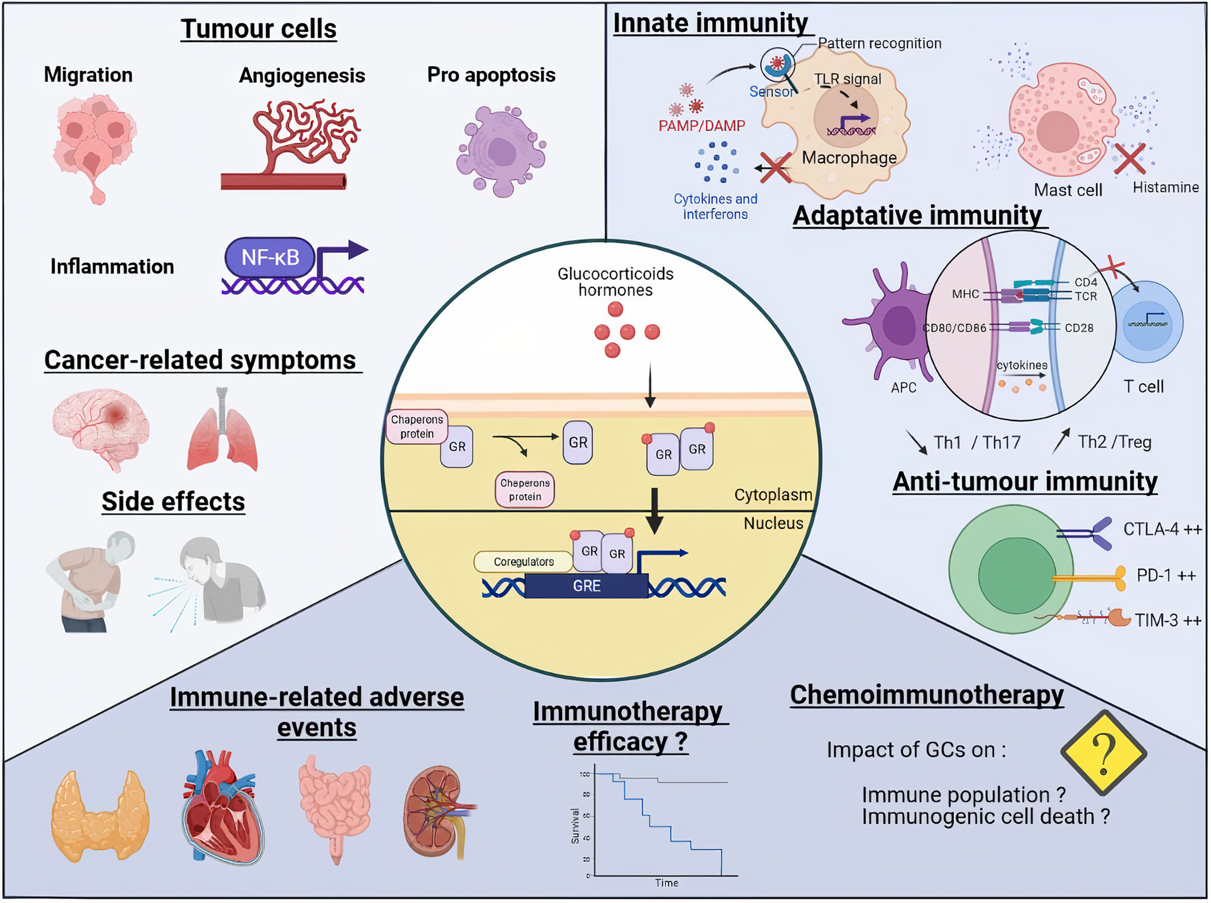
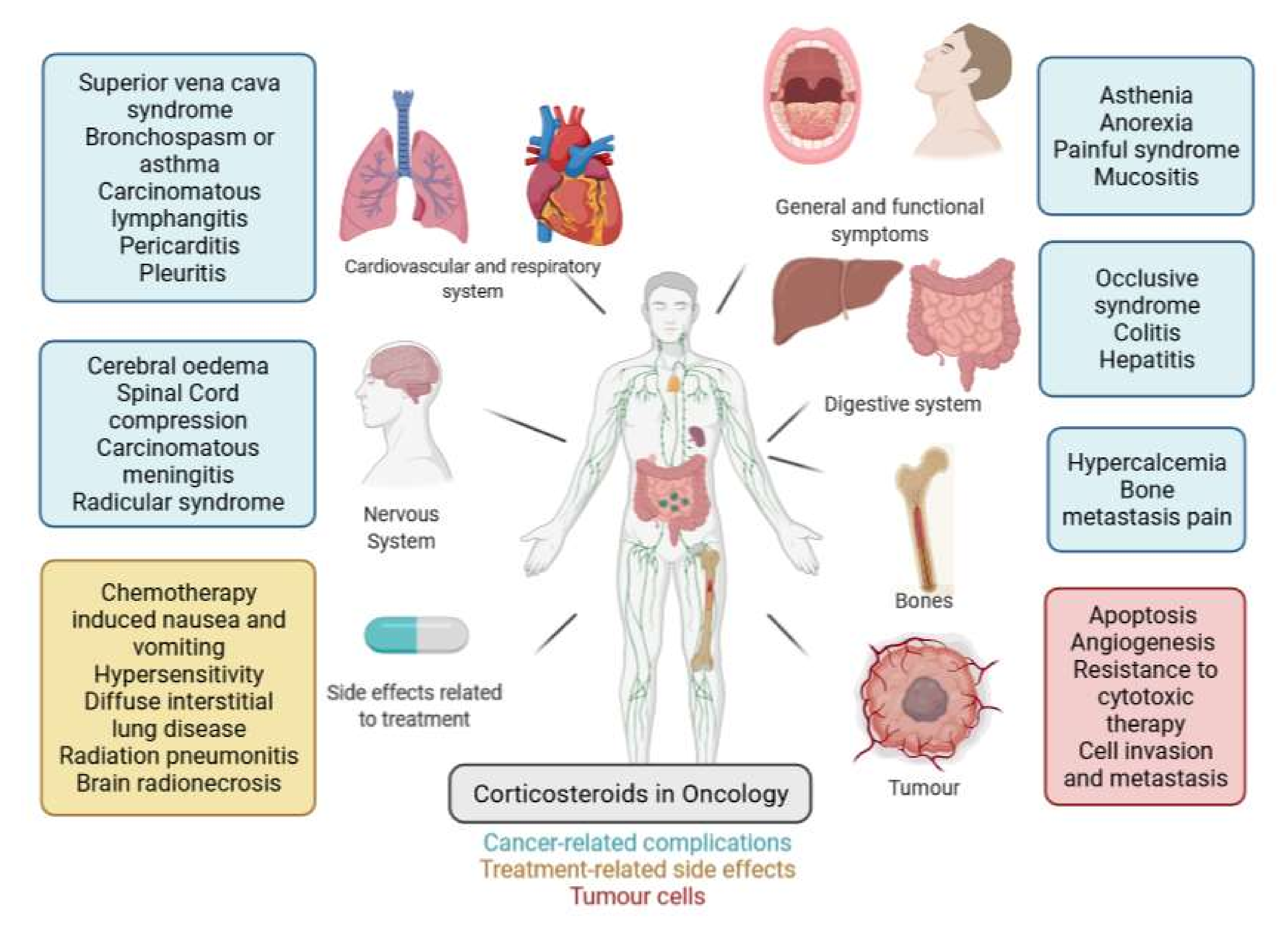
Table 1.
Summarize of effects of glucocorticoids on tumour cells.
| Pathways | Targets | Effects | Tumour Types |
|---|---|---|---|
| Apoptosis | Bcl2 miR-17~9 ROS (reactive oxygen species) | Increased | Haemopathic malignancies |
| BIM, BAX, BAK TXNIP, GILZ miR-708 | Decreased | Haemopathic malignancies and solid tumours | |
| Proliferation | AP1, Nf-κB c-MYC | Decreased | Haemopathic malignancies and solid tumours |
| Invasion/migration | SGK1 Selectins E-cadherin | Decreased | Solid tumours |
| RhoA MMP2/9 IL-6 | Decreased | Solid tumours | |
| Angiogenesis | VEGF/IL-8 | Decreased | Solid tumours |
| Resistance to cytotoxic therapy | SGK1 MKP1 (DUSP1) IκBα | Increased | Solid tumours |
Publisher’s Note: MDPI stays neutral with regard to jurisdictional claims in published maps and institutional affiliations. |
© 2022 by the authors. Licensee MDPI, Basel, Switzerland. This article is an open access article distributed under the terms and conditions of the Creative Commons Attribution (CC BY) license (https://creativecommons.org/licenses/by/4.0/).
Share and Cite
MDPI and ACS Style
Kalfeist, L.; Galland, L.; Ledys, F.; Ghiringhelli, F.; Limagne, E.; Ladoire, S. Impact of Glucocorticoid Use in Oncology in the Immunotherapy Era. Cells 2022, 11, 770. https://doi.org/10.3390/cells11050770
AMA Style
Kalfeist L, Galland L, Ledys F, Ghiringhelli F, Limagne E, Ladoire S. Impact of Glucocorticoid Use in Oncology in the Immunotherapy Era. Cells. 2022; 11(5):770. https://doi.org/10.3390/cells11050770
Chicago/Turabian StyleKalfeist, Laura, Loïck Galland, Fanny Ledys, François Ghiringhelli, Emeric Limagne, and Sylvain Ladoire. 2022. "Impact of Glucocorticoid Use in Oncology in the Immunotherapy Era" Cells 11, no. 5: 770. https://doi.org/10.3390/cells11050770
Note that from the first issue of 2016, this journal uses article numbers instead of page numbers. See further details here.