Article

Gut Microbiome Profiles and Associated Metabolic Pathways in HIV-Infected Treatment-Naïve Patients

by
Wellinton M. do Nascimento
1,2,
Aline Machiavelli
1,2,
Luiz G. E. Ferreira
3,
Luisa Cruz Silveira
1,
Suwellen S. D. de Azevedo
4,
Gonzalo Bello
4,
Daniel P. Smith
5,
Melissa P. Mezzari
5,
Joseph F. Petrosino
5,
Rubens Tadeu Delgado Duarte
6,
Carlos R. Zárate-Bladés
2,*,† and
Aguinaldo R. Pinto
1,†
1
Laboratório de Imunologia Aplicada, Departamento de Microbiologia, Imunologia e Parasitologia, Universidade Federal de Santa Catarina, Campus Universitário da Trindade, Florianópolis, SC 88034-040, Brazil
2
Laboratório de Imunorregulação, iREG, Departamento de Microbiologia, Imunologia e Parasitologia, Universidade Federal de Santa Catarina, Campus Universitário da Trindade, Florianópolis, SC 88034-040, Brazil
3
Hospital Regional Homero de Miranda Gomes, Rua Adolfo Donato da Silva, s/n, São José, SC 88103-901, Brazil
4
Laboratório de AIDS e Imunologia Molecular, Instituto Oswaldo Cruz, FIOCRUZ, Av. Brasil, 4365, Rio de Janeiro, RJ 21045-900, Brazil
5
Alkek Center for Metagenomics and Microbiome Research, Department of Molecular Virology & Microbiology, Baylor College of Medicine, One Baylor Plaza, Houston, TX 77030, USA
6
Laboratório de Ecologia Molecular e Extremófilos, Departamento de Microbiologia, Imunologia e Parasitologia, Universidade Federal de Santa Catarina, Campus Universitário da Trindade, Florianópolis, SC 88034-040, Brazil
*
Author to whom correspondence should be addressed.
These authors contributed equally to this work.
Cells 2021, 10(2), 385; https://doi.org/10.3390/cells10020385
Submission received: 9 December 2020 / Revised: 9 February 2021 / Accepted: 9 February 2021 / Published: 13 February 2021

Abstract

:
The normal composition of the intestinal microbiota is a key factor for maintaining healthy homeostasis, and accordingly, dysbiosis is well known to be present in HIV-1 patients. This article investigates the gut microbiota profile of antiretroviral therapy-naive HIV-1 patients and healthy donors living in Latin America in a cohort of 13 HIV positive patients (six elite controllers, EC, and seven non-controllers, NC) and nine healthy donors (HD). Microbiota compositions in stool samples were determined by sequencing the V3-V4 region of the bacterial 16S rRNA, and functional prediction was inferred using PICRUSt. Several taxa were enriched in EC compared to NC or HD groups, including Acidaminococcus, Clostridium methylpentosum, Barnesiella, Eubacterium coprostanoligenes, and Lachnospiraceae UCG-004. In addition, our data indicate that the route of infection is an important factor associated with changes in gut microbiome composition, and we extend these results by identifying several metabolic pathways associated with each route of infection. Importantly, we observed several bacterial taxa that might be associated with different viral subtypes, such as Succinivibrio, which were more abundant in patients infected by HIV subtype B, and Streptococcus enrichment in patients infected by subtype C. In conclusion, our data brings a significant contribution to the understanding of dysbiosis-associated changes in HIV infection and describes, for the first time, differences in microbiota composition according to HIV subtypes. These results warrant further confirmation in a larger cohort of patients.

Graphical Abstract

1. Introduction

In 2020, according to UNAIDS (The Joint United Nations Programme on HIV/AIDS), around 37 million people were living with the human immunodeficiency virus (HIV), and there were 940,000 deaths from acquired immunodeficiency syndrome (AIDS) [1]. Although a substantial depletion of CD4+ T cells is a hallmark of HIV infection, this is followed by a disruption in the intestinal epithelial barrier, gut microbiota dysbiosis, and chronic activation of the immune system [2]. Various patterns of HIV-1 disease progression are described in clinical practice. Remarkably, a state of apparent durable control of HIV replication occurs in a very small group of individuals, which are designated as “elite controllers’’ (EC). EC represent an extremely rare population of HIV-positive individuals where the viral load is naturally controlled, remaining at low or undetectable levels over a long period in the absence of antiretroviral treatment [3]. This group corresponds to less than 1% of the total HIV infected population [4], and have been extensively studied as a model for the natural control of HIV. Understanding the mechanisms of HIV control in these individuals could contribute to the development of novel therapeutic agents against HIV, providing insights for vaccine development.
In addition, as the gastrointestinal tract is a primary site of HIV replication and persistence, acting as an HIV reservoir, dysbiosis has been well documented in HIV-1 patients [5,6]. In this context, studying the interplay between the microbiome and HIV-1 infection is pivotal in order to better understand the disease [2]. Nonetheless, reports describing the composition of microbiota in HIV-1 infected individuals show great variability and some conflicting results, making it difficult to identify specific bacterial species, genera, or even families associated with specific subgroups of HIV patients in different populations [7,8,9]. This is in part due to the fact that the dysbiosis profile in HIV-1 infection varies substantially among individuals across the globe, which stresses the necessity of microbiome evaluations to be performed in different geographical regions to validate shifts in microbiome composition and/or to identify which bacterial components are equivalent to those observed in HIV-1 patients from other regions [10,11].
In this study, we performed the first microbiota analysis on HIV-1 patients who are born and live in Latin America. Several differences were observed in microbiota composition of HIV-1 infected individuals according to RNA viral load levels and the route of infection, and we describe for the first time that patients display a distinct microbiota profile according to HIV-1 subtype. The results suggest potential lines of further research for the identification of new molecular targets in the development of complementary therapeutics against HIV infection.

2. Methods

2.1. Study Population

This study was conducted with a cohort of 13 individuals with documented HIV-1 infection (HIV+) for over five years and who maintain CD4+ T cell counts >500 cells/mm3 and RNA viral loads <20,000 copies/mL without antiretroviral therapy (ART). These subjects were classified in two groups: (i) Elite controllers (EC) with (100%) plasma viral load (VL) determinations undetectable (<50 copies/mL, n = 6), and (ii) HIV non-controllers (NC) with most (≥70%) VL determinations between 2,000 and 20,000 copies/mL (n = 7). We also included nine healthy donors (HD) as controls.
None of the participants declared any type of gastrointestinal symptoms at the time of stool collection, and none of the HIV+ individuals had ever received ART therapy. Eight participants (2 HD and 6 HIV+) declared occasional use of drugs unrelated to HIV infection, including: Antihistaminic (1 HD and 2 HIV+), analgesic (1 HD and 1 HIV+), antidepressant (1 HIV+), anticonvulsant (1 HIV+), anxiolytic (1 HIV+).
Volunteers were recruited at Hospital Regional Homero de Miranda Gomes, Hospital Nereu Ramos, and at Blood Bank from the Hospital Universitário, Universidade Federal de Santa Catarina (UFSC), all located in the greater Florianópolis area, Santa Catarina, Brazil. The UFSC Ethics Committee (Comitê de Ética em Pesquisa com Seres Humanos, CEPSH-UFSC) approved the study under protocol number 1.622.458, and all volunteers provided written informed consent according to the guidelines of the Brazilian Ministry of Health.

2.2. Sample Collection and Processing

Stool samples were collected in a sterile plastic container either at the volunteers’ residence or at the hospital. DNA was extracted using the commercial kit QIAamp DNA Stool Mini Kit (Qiagen, Hilden, Germany) and stored at −20 °C. As a negative control, a sample of sterile water was used. Data on CD4+ and CD8+ T cells count and plasma HIV-1 VL were accessed via medical records and measured according to the Brazilian Ministry of Health guidelines [12].

2.3. Viral Subtype Analysis

To identify the viral subtype, total DNA was extracted from peripheral blood mononuclear cells (PBMCs), and the HIV-1 env gene was amplified by nested PCR and sequenced as previously described [13]. The chromatograms were assembled using SeqMan 7.0 software (DNASTAR Inc., Madison, WI, USA), and consensus env sequences covering positions 7,008–7,650 relative to the HXB2 reference genome were generated. The env sequences were aligned with subtype reference sequences downloaded from the Los Alamos HIV Sequence Database (http://www.hiv.lanl.gov/ accessed on 11 July 2017) using ClustalW and then manually edited and subtyped by maximum-likelihood (ML) phylogenetic tree reconstructions with the PhyML 3.0 program as described previously [13].

2.4. Sequencing and Bioinformatics Analysis

DNA was quantified using the Qubit dsDNA HS kit (Thermo Fisher Scientific, Waltham, MA, USA). The targeted V3-V4 region of the bacterial 16S rRNA gene from the extracted DNA was PCR-amplified using the Illumina primers S-D-Bact-0341-b-S-17 (forward) and S-D-Bact-0785-a-A-21 (reverse) [14]. The polymerase chain reactions were performed as described by Machiavelli et al. [15]. DNA sequencing was performed using a V2 reagent kit 2 × 250 bp (500 cycles) (MS-102–2003, Illumina Inc, San Diego, CA, USA) at the Illumina MiSeq platform. Raw reads were quality filtered using Trimmomatic v0.38 [16] according to sequence size and Phred score. The three first nucleotides in all sequences presented a Phred score below 20 and were removed. Sequences with less than 200 or more than 259 nucleotides were also excluded. Paired-end joining, determination of amplicon sequence variants (ASVs), and removal of chimeric sequences were performed using the DADA2 [17] R package v1.15.5. Taxonomy was assigned with the DECIPHER R package v2.10.2 IDTAXA [18] algorithm and the SILVA_SSU_r138_2019 [19]. Finally, the assigned taxonomy was organized in a BIOM-formatted ASV table and imported into R and ATIMA (Agile Toolkit for Incisive Microbial Analyses, https://atima.research.bcm.edu/ accessed on 11 June 2020) software for statistical analysis. The functional prediction was performed using PICRUST v2.1.4-b, (http://picrust.github.io/picrust/ accessed on 12 August 2020) [20] based on the Kyoto Encyclopedia of Genes and Genomes (KEGG, https://www.genome.jp/kegg/ accessed on 16 August 2020). Counts were normalized by considering the 16S rRNA gene copy number. The sequence data supporting the results of this study are available in the NCBI repository (Bioproject accession number: PRJNA682416; https://www.ncbi.nlm.nih.gov/bioproject/ accessed on 8 December 2020).

2.5. Statistical Analysis

Statistical analyses were performed using R software v3.6.0 and ATIMA v3.0. Characteristics between groups were compared using a Kruskal–Wallis and Mann–Whitney test. Gut microbiome taxa abundance was assessed using the phyloseq R package, and alpha diversity indices were assessed using ATIMA software. Kruskal–Wallis and Mann–Whitney tests were applied to compare the differences in the number of observed ASVs, alpha diversity richness (Chao1), and diversity (Shannon and Simpson) by groups. Beta diversity was evaluated using the Principal Coordinate Analysis (PCoA) on Bray-Curtis, weighted Unifrac, and unweighted Unifrac distances to determine significant differences between groups. Linear discriminant analysis (LDA) effect size (LefSe) [21] algorithm was used to identify differentially abundant bacterial taxa between the study groups, using the online version of Galaxy. LDA was performed using a one-against-all strategy and a score of >2.0. LefSe uses the Kruskal–Wallis and Wilcoxon rank-sum test to detect features with significantly different abundances between assigned taxa and LDA to estimate the effect size of each feature. The functional prediction was analyzed using ATIMA software applying the Mann–Whitney test to compare the relative abundance between groups. KEGG pathways with P values below 0.05 were considered significant after multiple-comparison correction using the false discovery rate (FDR) method.

3. Results

3.1. Epidemiological Features

This was a cross-sectional study, and detailed information regarding demographic and clinical characteristics of participants (13 HIV+ and nine healthy donors, HD) is displayed in Table 1. HIV+ individuals were separated into two groups: Elite controllers (EC, n = 6) and non-controllers (NC, n = 7) according to the plasma VL. There were no significant differences in age and BMI among HD, EC, and NC groups (Kruskal–Wallis test with Dunn’s as post-hoc test). All EC showed undetectable plasma viral load without blips at all measurements, while the median level of plasma viremia for NC was 4,203 copies/mL. EC and NC had documented HIV-1 infection for a median of 5.6 (IQR 5.0–9.3) years and 6.4 (IQR 5.8–6.9) years, respectively. The median of CD4+ T cell counts in EC and NC was 1190 (IQR 605.5–1370) and 726 (IQR 521–866) cells/µL, respectively. As for CD8+ T cells, the median was 1057 (IQR 810–1356) cells/μL for EC and 1295 (IQR 798–1527) cells/μL for NC. Phylogenetic analysis using env sequences showed that our cohort included two HIV-1 subtypes: B (n = 6, 46%) and C (n = 7, 54%). The distribution according to exposure categories was as follows: Heterosexual (HET; subtype B = 1; subtype C = 5); men who have sex with men (MSM; subtype B = 5; subtype C = 0); injecting drug users (IDU; subtype B = 0; subtype C = 2). None of the participants used antibiotics within the three months preceding enrollment in the study. Four EC, one NC, and one HD, declared the use of antibiotics before this period.

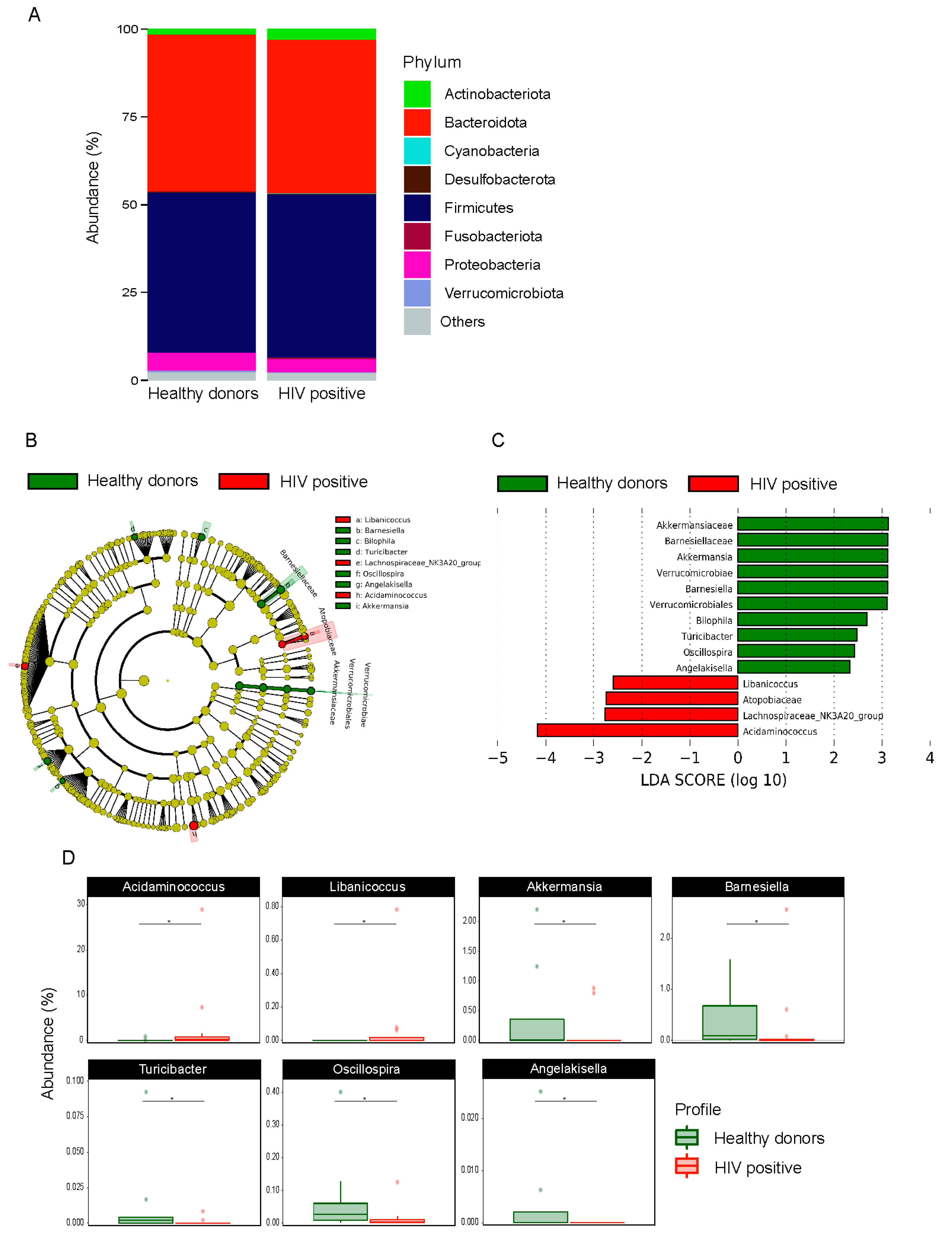
3.2. Comparison of Microbiota Profile between HIV+ and HD

The gut microbiota characterization was analyzed using 16S rRNA sequencing of stool. A total of 2,399,610 reads with an average of 99,983 reads per sample were generated after data processing and quality check, resulting in a final count of 1001 ASVs. In terms of microbial composition, 14 main phyla were found, regardless of the HIV status. The Firmicutes, Bacteroidota, Proteobacteria, and Actinobacteriota phyla were the most abundant (Figure 1A). There was no statistical difference between HIV+ and HD in beta and alpha diversity (Figure S1). To determine and distinguish differentially abundant ASVs associated with HIV-1, we compared the gut microbiota composition between HIV+ and HD using the LefSe algorithm, which showed significant bacterial differences between HIV+ and HD. The family Atopobiaceae, and genera Acidaminococcus, Libanicoccus, Lachnospiraceae NK3A20 group were enriched in HIV+, whereas the order Verrucomicrobiales, class Verrucomicrobiae, families Barnesiellaceae and Akkermansiaceae and genera Angelakissela, Oscillospira, Turicibacter, Bilophila, Barnesiella, Akkermansia were enriched in HD (Figure 1B,C). The relative abundance of taxa identified in LefSe as differentially abundant between HIV+ and HD was compared using the Mann–Whitney test, which revealed statistically significant differences between groups in seven of the nine identified genera (p < 0.05) (Figure 1D).

3.3. Differences in Microbiota Composition between EC, NC, and HD

LefSe analysis was used to compare the gut microbiota of EC, NC, and HD groups. Figure 2A,B show differences in the abundance of taxonomic clades of LDA score >2.0 among EC, NC, and HD individuals. The gut microbiota of the EC group was dominated by taxa Acidaminococcus, Clostridium methylpentosum group, Barnesiella, Eubacterium coprostanoligenes group, and Lachnospiraceae UCG-004, whereas the NC group showed high scores for the family Atopobiaceae, and genera Libanicoccus and Lachnospiraceae NK3A20 group. The features identified by LefSe as being differentially abundant were confirmed by standard statistical analysis using Kruskal–Wallis test (Figure S2). PICRUSt analysis was performed to determine whether the taxonomic differences between the groups’ microbiota corresponded to functional changes, but we found no significant differences between HD and EC groups or between EC and NC groups (data not shown). However, we identified 30 KEGG pathways as significantly different between NC and HD groups (Figure 2C). PICRUSt analyses demonstrated that the NC group’s microbiota exhibits increased S-adenosyl-L-methionine cycle, methylerythritol phosphate pathway, and biosynthesis of threonine, acetylmuramoyl pentapeptide, peptidoglycan, CoA, and CDP-diacylglycerol. Conversely, the microbiota of NC subjects was depleted for sucrose and glycogen degradation, biosynthesis of thiamine, folate, fatty acids, unsaturated fatty acids and lipopolysaccharides, B12 Salvage from cobinamide and sulfur metabolism (p < 0.05).

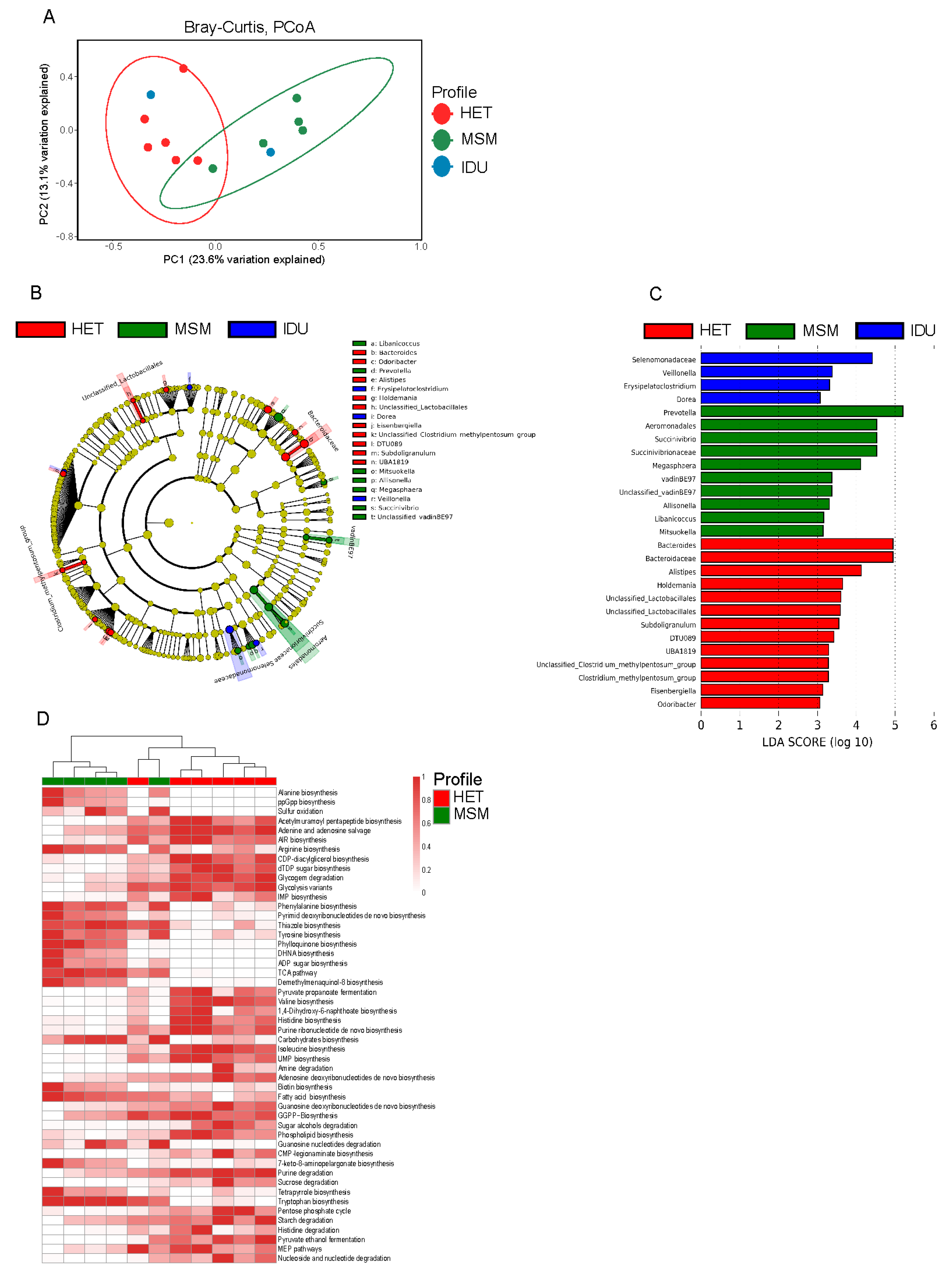
3.4. HIV Transmission Route Influences Composition of Gut Microbiome

HIV+ patients were divided into three groups according to exposure category: HET (n = 6), MSM (n = 5) and IDU (n = 2). Receptive anal intercourse (r.a.i.), was declared by 2 HET, 5 MSM, and 1 IDU patients. Beta diversity with Principal Coordinates Analysis of Bray-Curtis distances indicated differences in the gut microbiota between HET, MSM, and IDU groups, (R = 0.36, p = 0.011) (Figure 3A). We carried out LEfSe analysis to determine the taxa most associated with the route of HIV-1 transmission and found significant differences in the abundance of 27 taxa among HET, MSM, and IDU groups were found. Bacteroidaceae, Bacteroides, Alistipes, Holdemania, Unclassified lactobacillales, Subdoligranulum, DTU089, UBA1819, Unclassified Clostridium methylpentosum group, Clostridium methylpentosum group, Eisenbergiella, and Odoribacter were enriched in feces of HET group, whereas Aeromonodales, Succinivibrionaaceae, VadinBE97, Prevotella, Succinivibrio, Megasphaera, Unclassified vadinBE97, Allisonella, Libanicoccus, and Mitsuokella were enriched in the MSM group (Figure 3B,C). When differences in microbiota functions were assessed, 20 pathways were found to be strengthened, and 30 pathways weakened in MSM in relation to the HET group (Figure 3D). Alanine biosynthesis, metabolic regulators, sulfur oxidation, and biosynthesis of arginine carbohydrates, biotin, fatty acid, and tryptophan were among the enriched pathways in the MSM group compared to the HET group, while biosynthesis of valine, histidine, phospholipid, acetylmuramoyl pentapeptide, as well as purine, glycogen and sucrose degradation, were depleted (p < 0.05).

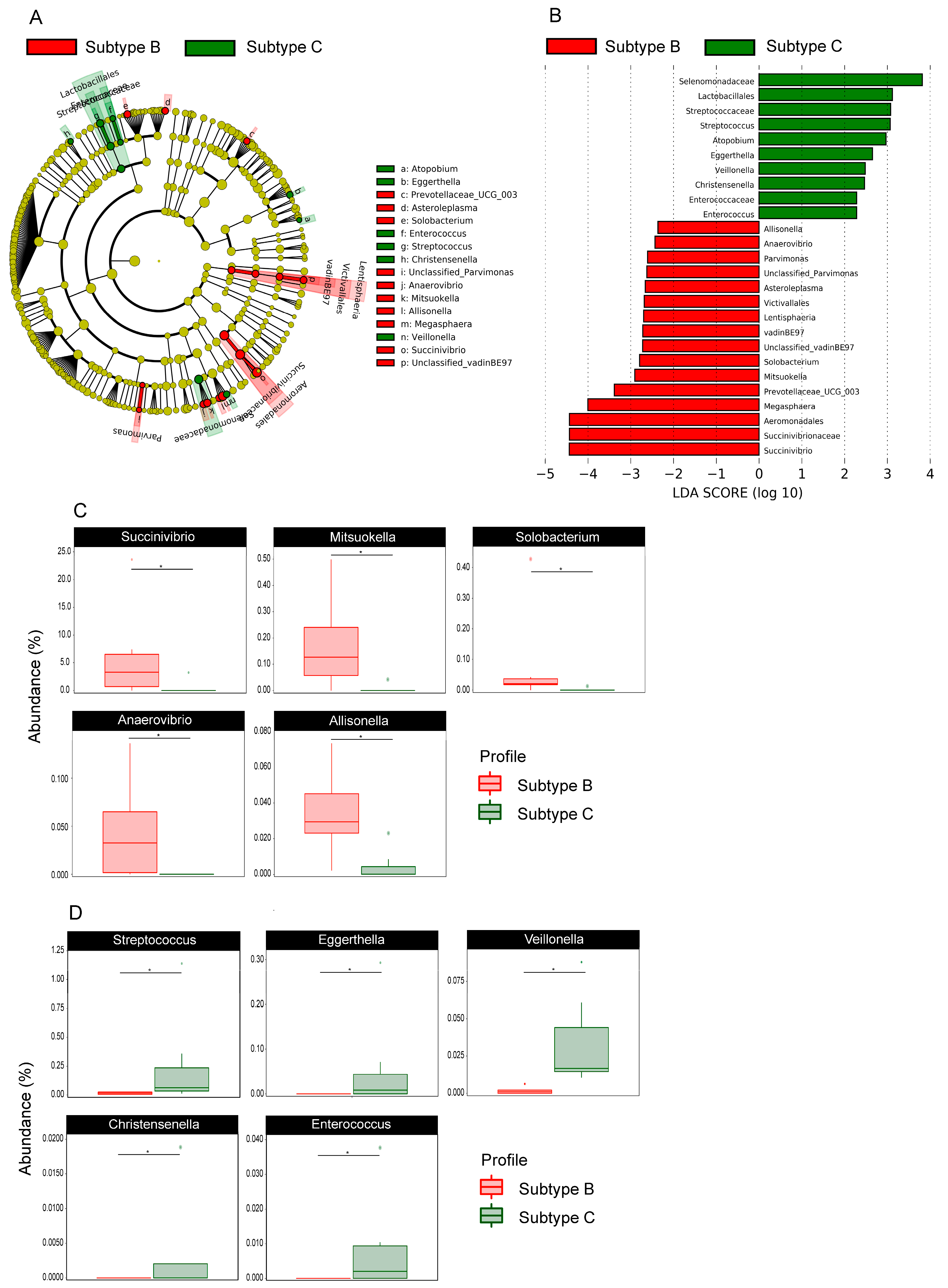
3.5. Gut Bacterial Microbiota Profile Differs Between Patients InfeCTED with HIV-1 Subtype B and C

Next, we explored whether changes in the gut microbiota can be detected between HIV-1 subtypes and found clear differences in the composition of gut microbiomes among patients infected with subtype B (HIV-1B) or C (HIV-1C). Figure 4A,B show increased abundance in HIV-B group for Lentisphaeria class, Aeromonadales, Victivallales order, Succinivibrionaceae and VadinBE97 family, and Succinivibrio, Megasphaera, Prevotellaceae UCG-003, Mitsuokella, Solobacterium, Unclassified vadinBE97, Asteroleplasma, Parvimonas, Unclassified Parvimonas, Anaerovibrio, and Alissonela genus. For the HIV-C group Lactobacillales order, Selenomonadaceae, Streptococcaceae and Enterococcaceae family, Streptococcus, Atopobium, Eggerthella, Veillonella, Christensenella, and Enterococcus genus were more abundant. Differences in bacterial abundance at a genus level between these two groups can be seen in Figure 4C,D.

4. Discussion

Our study is the first to analyze gut microbiota composition in HIV-1 infected patients who are born and live in Latin America (more specifically in Brazil), including a group of EC individuals. Along with other reports [7,8], our data did not reveal differences in microbiota alpha and beta diversity between HIV+ and HD. This finding contrasts with other studies where such differences have been identified [10,11]. Together, these data indicate not only a lack of consensus about differences in alpha and beta diversity in HIV+ in relation to HD, but also that microbiome alterations in HIV+ may vary across populations. This is likely to be due to factors that have not always been considered, such as genetic background, different types of nutritional habits, ART, the clinical differences among HIV+ patients, and even HIV subtype.
The deeper analyses performed in this study revealed several important differences. The HIV+ gut microbiota was enriched with the Acidaminococcus genus, similar to a previous report [22]. Akkermansia genus was also enriched in HIV+ individuals, in agreement with Mutlu et al. [10] findings. Akkermansia is a gram-negative anaerobic genus abundantly found in the human gastrointestinal tract throughout life [23], with several proposed roles, such as mucin degradation [23,24], anti-inflammatory activity [25], immunomodulation properties on the adaptive immune system [26], and protective barrier functions [27]. Some other microorganisms were less abundant in the gut of HIV+ individuals, such as members of the Lachnospiraceae family, which have been implicated in butyric acid production [28] that is associated with the protection of colon cancer [29], influences obesity levels [30], as well as assists maintaining the epithelial barrier [31].
We found that the EC group presented a unique signature composed of Acidaminococcus, Barnesiella, Clostridium methylpentosum group, Eubacterium ventriosum group, and Lachnospiraceae UCG-004. The Barnesiella genus, in particular, has been associated with beneficial effects on the human gastrointestinal tract [32]. Interestingly, although several previous studies have reported members of the Lachnospiraceae family associated with HD [10], in this study, we found an increased abundance of Lachnospiraceae UCG-004 in EC patients instead. It is tempting, based on our findings, to speculate that this genus could be considered as a marker of HIV-1 control in EC patients.
We also identified differences in metabolic pathways that varied between NC and HD groups, such as pathways involved in carbohydrate and lipid metabolism, which were reduced in NC individuals. It is known that specific changes in metabolic pathways are associated with effector functions of the immune system. For example, it has been suggested that the synthesis of fatty acids is involved in the induction of inflammatory responses of macrophages [33]. Vesterbacka et al. [9] also found pathways involved in carbohydrate metabolism that was significantly reduced in HIV+ ART-naive patients compared to HD. This same study also demonstrated that some pathways related to lipid metabolism, including fatty acid metabolism and lipid biosynthesis proteins, were less abundant in HIV+ ART-naive patients, while linoleic acid metabolism was more represented in this same group. We also detected pathways related to the metabolism of cofactors and vitamins, energy metabolism, and glycan biosynthesis that were less represented in NC patients compared to HD subjects.
Similar to what has been observed by Noguera-Julian et al. [11], the microbiome profile of our patients clustered according to the HIV transmission group. Our data revealed HET and MSM microbiomes behave distinctly, whereas the IDU group did not cluster separately, although it displayed some taxa differences compared to the other two groups. Remarkably, Prevotella, and Bacteroides were associated with MSM and HET groups, respectively, as previously reported in individuals from Barcelona and Stockholm [11]. Moreover, we also identified an association between Succinivibrio in the MSM group and between Alistipes and the HET group. In addition to the previously reported anti-inflammatory capacity [34], an increased abundance of the Succinivibrio genus has been found in patients with cervical cancer in comparison to HD [35], whereas the Alistipes genus is an anaerobic bacteria found commonly in the healthy human gastrointestinal tract [36].
Our functional analysis of microbiome associated with transmission route also identified a high number of significantly abundant pathways in HET and MSM, extending the important observations of a previous study [11], and confirming that sexual orientation is a strong factor associated with gut microbiota composition in HIV-1+ patients. For example, pathways involved in amino acid metabolism were differentially distributed between HET and MSM groups. It has been shown that the metabolism of various amino acids can play an important role in mediating the functionality of immune cells [33]. Pathways, such as histidine degradation, isoleucine, valine, and histidine biosynthesis, were enriched in HET groups, while that alanine, phenylalanine tyrosine, and tryptophan biosynthesis were more abundant in the MSM group. Additionally, we identified a lower abundance of pathways involved in carbohydrate metabolism in the HET group. In contrast, some pathways related to the metabolism of cofactors and vitamins were more enriched in the MSM group. Although these results contribute to the understanding of HIV-1 infection, it would be important to identify which of these metabolic connections are consistent factors and truly relevant for the disease history—this would identify whether gut bacterial metabolism is altered as a consequence of sexual orientation. This could potentially target certain molecular pathways as a complementary treatment option to improve patients’ quality of life by reducing the impact of gut microbiota dysbiosis.
Finally, the main finding of our study is that HIV+ patients had a distinct microbiota according to viral subtype B or C. High molecular variability is a striking feature of HIV-1 in the global AIDS pandemic, which has evolved into a multiple number of variants [37]. Although HIV-1B dominates in many countries in Europe and the Americas, more than 50% of the infections worldwide are caused by HIV-1C, which is the most prevalent subtype in southern African countries and India [38]. In the southern region of Brazil, however, HIV-1B and HIV-1C co-circulate, making this region a unique place for epidemiological studies [39]. The importance of subtypes in different clinical parameters of HIV-1 infection is still controversial, but the successful worldwide dissemination of subtype C may be due to this strain being less virulent in comparison with other subtypes while maintaining the same transmission efficiency [40]. In a study conducted by Venner et al. [41] in which women from the same area and infected by subtypes A, C, or D were followed for 9.5 years, and disease progression was evaluated, it was mentioned that it would be important to further evaluate the vaginal microbiota as a way to assess differences in disease progression according to viral subtype within that cohort.
We acknowledge that while our study reveals associations, we can only suggest causality to a limited extent. In addition, the low number of participants is an important limitation in our study. Nonetheless, our cohort included only ART naïve HIV+ patients who have been diagnosed at least five years before the time of recruitment, and we reported for the first time an association of HIV viral subtype B and C with distinct gut microbiota profile. An additional limitation is that our cohort was not matched according to receptive anal intercourse—such information would be important to define if differences in microbiota according to the route of infection are not due to sexual behavior. Although most reports on HIV patients’ gut microbiota have not taken into consideration this feature [7,8,22], it was recently recognized as an important attribute in a remarkable study by Vjuckovic-Cvijin et al. [42]. This should be considered in future studies.
As far as we know, the present report is the first to associate differences in microbiota composition with HIV subtypes. Dang et al. [43] indirectly associated higher colonization of Streptococcus species in oral microbiota of HIV-1 patients on ART when compared to HD and ART-naive HIV+ patients, all infected by HIV-1 subtype B. Our results showed Succinivibrio, Mitsuokella, Solobacterium, Anaerovibrio, and Allisonella associated with HIV-1B, while Streptococcus, Eggerthella, Veillonella, Christensenella, and Enterococcus genus were more abundant in individuals infected by subtype C. Taken together, these data highlight the importance of our findings regarding the difference in the gut microbiota profile according to HIV-1B and HIV-1C infection. Although HIV-1 does not target bacteria, it is worth considering what effects viral subtypes could exert on the immune system and subsequently impact on the gut microbiota. Moreover, our results open the possibility to consider targeting microbiota as a way to improve the response to HIV-1 infection through the effects of viral subtypes on dysbiosis.

Supplementary Materials

The following are available online at https://www.mdpi.com/2073-4409/10/2/385/s1, Figure S1: Comparison of gut microbiota of HIV-1 positive and healthy donors, Figure S2: Comparison of gut microbiota of healthy donors (HD), elite controllers (EC), and Non-controllers (NC).

Author Contributions

W.M.d.N., designed the study, recruited volunteers, processed samples, analyzed the data, and wrote the manuscript. A.M. and L.C.S., analyzed the data. L.G.E.F., designed the study and recruited volunteers. S.S.D.d.A., G.B., D.P.S., M.P.M., J.F.P. and R.T.D.D. helped with data analysis and revised the manuscript. A.R.P. and C.R.Z.-B. designed the study, supervised the work and revised the manuscript. All authors read and approved the final version of the manuscript.

Funding

No direct funding for the execution of this study was received. WM received a CAPES (Coordenação de Aperfeiçoamento de Pessoal de Nível Superior) student fellowship. ARP and GB are CNPq (Conselho Nacional de Desenvolvimento Científico e Tecnológico) scholars. GB is funded by CNPq and FAPERJ (Fundação Carlos Chagas Filho de Amparo à Pesquisa do Estado do Rio de Janeiro). SSDA is funded by Postdoctoral fellowship from the “Pós-Doutorado Nota 10 (PDR 10)” by FAPERJ.

Institutional Review Board Statement

The study was conducted according to the guidelines of the Declaration of Helsinki, and approved by the Institutional Ethics Committee (Comitê de Ética em Pesquisa com Seres Humanos, CEPSH) of the Universidade Federal de Santa Catarina (protocol number 1.622.458, issued on 5 July 2016).

Informed Consent Statement

Informed consent was obtained from all subjects involved in the study.

Data Availability Statement

The data presented in this study are available on request from the corresponding author.

Acknowledgments

The authors would like to thank all the volunteers who participated in this study and the team at the Regional Hospital Homero de Miranda Gomes and Blood Bank from the Hospital Universitário/UFSC who assisted in the recruitment of volunteers and collection of samples. The authors also thank Oscar Bruna-Romero for providing reagents.

Conflicts of Interest

The authors report no conflict of interest.

References

  1. UNAIDS. UNAIDS Data 2020; UNAIDS: Geneva, Switzerland, 2020; Available online: https://www.unaids.org/sites/default/files/media_asset/2020_aids-data-book_en.pdf (accessed on 26 September 2020).
  2. Dillon, S.M.; Frank, D.N.; Wilson, C.C. The gut microbiome and HIV-1 pathogenesis: A two-way street. Aids 2016, 30, 2737–2751. [Google Scholar] [CrossRef] [PubMed] [Green Version]
  3. Okulicz, J.F.; Lambotte, O. Epidemiology and clinical characteristics of elite controllers. Curr. Opin. HIV AIDS 2011, 6, 163–168. [Google Scholar] [CrossRef] [PubMed]
  4. Deeks, S.G.; Walker, B.D. Human Immunodeficiency Virus Controllers: Mechanisms of Durable Virus Control in the Absence of Antiretroviral Therapy. Immunity 2007, 27, 406–416. [Google Scholar] [CrossRef] [PubMed] [Green Version]
  5. Gori, A.; Tincati, C.; Rizzardini, G.; Torti, C.; Quirino, T.; Haarman, M.; Amor, K.B.; Van Schaik, J.; Vriesema, A.; Knol, J.; et al. Early impairment of gut function and gut flora supporting a role for alteration of gastrointestinal mucosa in human immunodeficiency virus pathogenesis. J. Clin. Microbiol. 2008, 46, 757–758. [Google Scholar] [CrossRef] [PubMed] [Green Version]
  6. Dubourg, G.; Surenaud, M.; Lévy, Y.; Hüe, S.; Raoult, D. Microbiome of HIV-infected people. Microb. Pathog. 2017, 106, 85–93. [Google Scholar] [CrossRef] [PubMed]
  7. Wang, Z.; Usyk, M.; Sollecito, C.C.; Qiu, Y.; Williams-Nguyen, J.; Hua, S.; Gradissimo, A.; Wang, T.; Xue, X.; Kurland, I.J.; et al. Altered Gut Microbiota and Host Metabolite Profiles in Women With Human Immunodeficiency Virus. Clin. Infect. Dis. 2019, 10461, 1–9. [Google Scholar] [CrossRef] [PubMed]
  8. Dinh, D.M.; Volpe, G.E.; Duffalo, C.; Bhalchandra, S.; Tai, A.K.; Kane, A.V.; Wanke, C.A.; Ward, H.D. Intestinal Microbiota, microbial translocation, and systemic inflammation in chronic HIV infection. J. Infect. Dis. 2015, 211, 19–27. [Google Scholar] [CrossRef] [PubMed] [Green Version]
  9. Vesterbacka, J.; Rivera, J.; Noyan, K.; Parera, M.; Neogi, U.; Calle, M.; Paredes, R.; Sönnerborg, A.; Noguera-Julian, M.; Nowak, P. Richer gut microbiota with distinct metabolic profile in HIV infected Elite Controllers. Sci. Rep. 2017, 7, 6269. [Google Scholar] [CrossRef] [PubMed]
  10. Mutlu, E.A.; Keshavarzian, A.; Losurdo, J.; Swanson, G.; Siewe, B.; Forsyth, C.; French, A.; DeMarais, P.; Sun, Y.; Koenig, L.; et al. A Compositional Look at the Human Gastrointestinal Microbiome and Immune Activation Parameters in HIV Infected Subjects. PLoS Pathog. 2014, 10. [Google Scholar] [CrossRef] [PubMed] [Green Version]
  11. Noguera-Julian, M.; Rocafort, M.; Guillén, Y.; Rivera, J.; Casadellà, M.; Nowak, P.; Hildebrand, F.; Zeller, G.; Parera, M.; Bellido, R.; et al. Gut Microbiota Linked to Sexual Preference and HIV Infection. EBioMedicine 2016, 5, 135–146. [Google Scholar] [CrossRef] [Green Version]
  12. Ministério da Saúde. Protocolo clínico e diretrizes para manejo da infecção pelo HIV em adultos, 1st ed.; Ministério da Saúde: Brasília, DF, Brazil, 2018. Available online: http://www.aids.gov.br/pt-br/pub/2013/protocolo-clinico-e-diretrizes-terapeuticas-para-manejo-da-infeccao-pelo-hiv-em-adultos (accessed on 12 October 2018).
  13. Azevedo, S.S.D.; Caetano, D.G.; Côrtes, F.H.; Teixeira, S.L.M.; Santos Silva, K.; Hoagland, B.; Grinsztejn, B.; Veloso, V.G.; Morgado, M.G.; Bello, G. Highly divergent patterns of genetic diversity and evolution in proviral quasispecies from HIV controllers. Retrovirology 2017, 14, 1–13. [Google Scholar] [CrossRef]
  14. Klindworth, A.; Pruesse, E.; Schweer, T.; Peplies, J.; Quast, C.; Horn, M.; Glöckner, F.O. Evaluation of general 16S ribosomal RNA gene PCR primers for classical and next-generation sequencing-based diversity studies. Nucleic Acids Res. 2013, 41, 1–11. [Google Scholar] [CrossRef]
  15. Machiavelli, A.; Duarte, R.T.D.; de Souza Pires, M.M.; Zarate-Bladés, C.R.; Pinto, A.R. The impact of in utero HIV exposure on gut microbiota, inflammation, and microbial translocation. Gut Microbes 2019, 10, 599–614. [Google Scholar] [CrossRef]
  16. Bolger, A.M.; Lohse, M.; Usadel, B. Trimmomatic: A flexible trimmer for Illumina sequence data. Bioinformatics 2014, 30, 2114–2120. [Google Scholar] [CrossRef] [PubMed] [Green Version]
  17. Callahan, B.J.; McMurdie, P.J.; Rosen, M.J.; Han, A.W.; Johnson, A.J.A.; Holmes, S.P. DADA2: High-resolution sample inference from Illumina amplicon data. Nat. Methods 2016, 13, 581–583. [Google Scholar] [CrossRef] [PubMed] [Green Version]
  18. Murali, A.; Bhargava, A.; Wright, E.S. IDTAXA: A novel approach for accurate taxonomic classification of microbiome sequences. Microbiome 2018, 6, 1–14. [Google Scholar] [CrossRef] [PubMed]
  19. Quast, C.; Pruesse, E.; Yilmaz, P.; Gerken, J.; Schweer, T.; Yarza, P.; Peplies, J.; Glöckner, F.O. The SILVA ribosomal RNA gene database project: Improved data processing and web-based tools. Nucleic Acids Res. 2013, 41, 590–596. [Google Scholar] [CrossRef] [PubMed]
  20. Langille, M.G.I.; Zaneveld, J.; Caporaso, J.G.; McDonald, D.; Knights, D.; Reyes, J.A.; Clemente, J.C.; Burkepile, D.E.; Vega Thurber, R.L.; Knight, R.; et al. Predictive functional profiling of microbial communities using 16S rRNA marker gene sequences. Nat. Biotechnol. 2013, 31, 814–821. [Google Scholar] [CrossRef]
  21. Segata, N.; Izard, J.; Waldron, L.; Gevers, D.; Miropolsky, L.; Garrett, W.S.; Huttenhower, C. Metagenomic biomarker discovery and explanation. Genome Biol. 2011, 12, R60. [Google Scholar] [CrossRef] [PubMed] [Green Version]
  22. Vázquez-Castellanos, J.F.; Serrano-Villar, S.; Latorre, A.; Artacho, A.; Ferrús, M.L.; Madrid, N.; Vallejo, A.; Sainz, T.; Martínez-Botas, J.; Ferrando-Martínez, S.; et al. Altered metabolism of gut microbiota contributes to chronic immune activation in HIV-infected individuals. Mucosal Immunol. 2015, 8, 760–772. [Google Scholar] [CrossRef] [PubMed] [Green Version]
  23. Derrien, M.; Collado, M.C.; Ben-Amor, K.; Salminen, S.; De Vos, W.M. The mucin degrader Akkermansia muciniphila is an abundant resident of the human intestinal tract. Appl. Environ. Microbiol. 2008, 74, 1646–1648. [Google Scholar] [CrossRef] [Green Version]
  24. Png, C.W.; Lindén, S.K.; Gilshenan, K.S.; Zoetendal, E.G.; McSweeney, C.S.; Sly, L.I.; McGuckin, M.A.; Florin, T.H.J. Mucolytic bacteria with increased prevalence in IBD mucosa augment in vitro utilization of mucin by other bacteria. Am. J. Gastroenterol. 2010, 105, 2420–2428. [Google Scholar] [CrossRef]
  25. Kang, C.S.; Ban, M.; Choi, E.J.; Moon, H.G.; Jeon, J.S.; Kim, D.K.; Park, S.K.; Jeon, S.G.; Roh, T.Y.; Myung, S.J.; et al. Extracellular Vesicles Derived from Gut Microbiota, Especially Akkermansia muciniphila, Protect the Progression of Dextran Sulfate Sodium-Induced Colitis. PLoS ONE 2013, 8, e76520. [Google Scholar] [CrossRef] [PubMed] [Green Version]
  26. Derrien, M.; Van Baarlen, P.; Hooiveld, G.; Norin, E.; Müller, M.; de Vos, W.M. Modulation of mucosal immune response, tolerance, and proliferation in mice colonized by the mucin-degrader Akkermansia muciniphila. Front. Microbiol. 2011, 2, 166. [Google Scholar] [CrossRef] [PubMed] [Green Version]
  27. Ottman, N.; Reunanen, J.; Meijerink, M.; Pietila, T.E.; Kainulainen, V.; Klievink, J.; Huuskonen, L.; Aalvink, S.; Skurnik, M.; Boeren, S.; et al. Pili-like proteins of Akkermansia muciniphila modulate host immune responses and gut barrier function. PLoS ONE 2017, 12, e0173004. [Google Scholar] [CrossRef]
  28. Meehan, C.J.; Beiko, R.G. A phylogenomic view of ecological specialization in the lachnospiraceae, a family of digestive tract-associated bacteria. Genome Biol. Evol. 2014, 6, 703–713. [Google Scholar] [CrossRef] [PubMed] [Green Version]
  29. Han, A.; Bennett, N.; Ahmed, B.; Whelan, J.; Donohoe, D.R. Butyrate decreases its own oxidation in colorectal cancer cells through inhibition of histone deacetylases. Oncotarget 2018, 9, 27280–27292. [Google Scholar] [CrossRef] [PubMed]
  30. Turnbaugh, P.J.; Hamady, M.; Yatsunenko, T.; Cantarel, B.L.; Duncan, A.; Ley, R.E.; Sogin, M.L.; Jones, W.J.; Roe, B.A.; Affourtit, J.P.; et al. A core gut microbiome in obese and lean twins. Nature 2009, 457, 480–484. [Google Scholar] [CrossRef] [PubMed] [Green Version]
  31. Thaiss, C.A.; Zmora, N.; Levy, M.; Elinav, E. The microbiome and innate immunity. Nature 2016, 535, 65–74. [Google Scholar] [CrossRef] [PubMed]
  32. Mancabelli, L.; Milani, C.; Lugli, G.A.; Turroni, F.; Cocconi, D.; van Sinderen, D.; Ventura, M. Identification of universal gut microbial biomarkers of common human intestinal diseases by meta-analysis. FEMS Microbiol. Ecol. 2017, 93, 1–10. [Google Scholar] [CrossRef] [PubMed] [Green Version]
  33. O’Neill, L.A.J.; Kishton, R.J.; Rathmell, J. A guide to immunometabolism for immunologists. Nat. Rev. Immunol. 2016, 16, 553–565. [Google Scholar] [CrossRef] [PubMed] [Green Version]
  34. Serrano-Villar, S.; Rojo, D.; Martínez-Martínez, M.; Deusch, S.; Vázquez-Castellanos, J.F.; Bargiela, R.; Sainz, T.; Vera, M.; Moreno, S.; Estrada, V.; et al. Gut Bacteria Metabolism Impacts Immune Recovery in HIV-infected Individuals. EBioMedicine 2016, 8, 203–216. [Google Scholar] [CrossRef] [PubMed] [Green Version]
  35. Wang, Z.; Wang, Q.; Zhao, J.; Gong, L.; Zhang, Y.; Wang, X.; Yuan, Z. Altered diversity and composition of the gut microbiome in patients with cervical cancer. AMB Express 2019, 9. [Google Scholar] [CrossRef] [PubMed] [Green Version]
  36. Shkoporov, A.N.; Chaplin, A.V.; Khokhlova, E.V.; Shcherbakova, V.A.; Motuzova, O.V.; Bozhenko, V.K.; Kafarskaia, L.I.; Efimov, B.A. Alistipes inops sp. Nov. And coprobacter secundus sp. Nov., isolated from human faeces. Int. J. Syst. Evol. Microbiol. 2015, 65, 4580–4588. [Google Scholar] [CrossRef]
  37. Gartner, M.J.; Roche, M.; Churchill, M.J.; Gorry, P.R.; Flynn, J.K. Understanding the mechanisms driving the spread of subtype C HIV-1. EBioMedicine 2020, 53, 102682. [Google Scholar] [CrossRef] [PubMed]
  38. Wilkinson, E.; Engelbrecht, S.; De Oliveira, T. History and origin of the HIV-1 subtype C epidemic in South Africa and the greater southern African region. Sci. Rep. 2015, 5, 16897. [Google Scholar] [CrossRef] [Green Version]
  39. Gräf, T.; Machado Fritsch, H.; de Medeiros, R.M.; Maletich Junqueira, D.; Esteves de Matos Almeida, S.; Pinto, A.R. Comprehensive Characterization of HIV-1 Molecular Epidemiology and Demographic History in the Brazilian Region Most Heavily Affected by AIDS. J. Virol. 2016, 90, 8160–8168. [Google Scholar] [CrossRef] [Green Version]
  40. Ariën, K.K.; Abraha, A.; Quiñones-mateu, M.E.; Vanham, G.; Arts, E.J.; Arie, K.K.; Quin, M.E.; Kestens, L. The replicative fitness of primary human immunodeficiency virus type 1 ( HIV-1 ) group M, HIV-1 group O, and HIV-2 Isolates. J. Virol. 2005, 79, 8979–8990. [Google Scholar] [CrossRef] [Green Version]
  41. Venner, C.M.; Nankya, I.; Kyeyune, F.; Demers, K.; Kwok, C.; Chen, P.L.; Rwambuya, S.; Munjoma, M.; Chipato, T.; Byamugisha, J.; et al. Infecting HIV-1 Subtype Predicts Disease Progression in Women of Sub-Saharan Africa. EBioMedicine 2016, 13, 305–314. [Google Scholar] [CrossRef] [PubMed] [Green Version]
  42. Vujkovic-Cvijin, I.; Sortino, O.; Verheij, E.; Sklar, J.; Wit, F.W.; Kootstra, N.A.; Sellers, B.; Brenchley, J.M.; Ananworanich, J.; van der Loeff, M.S.; et al. HIV-associated gut dysbiosis is independent of sexual practice and correlates with noncommunicable diseases. Nat. Commun. 2020, 11. [Google Scholar] [CrossRef]
  43. Dang, A.T.; Cotton, S.; Sankaran-Walters, S.; Li, C.S.; Lee, C.Y.M.; Dandekar, S.; Paster, B.J.; George, M.D. Evidence of an increased pathogenic footprint in the lingual microbiome of untreated HIV infected patients. BMC Microbiol. 2012, 12, 153. [Google Scholar] [CrossRef] [PubMed] [Green Version]
Figure 1. Taxonomic differences in fecal microbiota between HIV-1 positive patients and healthy donors. Relative abundance at phylum level (A); Cladogram of LefSe linear discriminant analysis (LDA) scores showing differentially abundant taxonomic clades with an LDA score >2.0 in the gut microbiota of HIV-1 patients and healthy donors (B); LDA scores of differentially abundant taxa in the gut microbiota of HIV-1 patients and healthy donors (C). The relative abundance of genera identified by LefSe as being differentially abundant in HIV-1 patients and healthy donors were compared using a Mann–Whitney test (D). * p < 0.05.
Cells 10 00385 g001
Figure 2. Taxonomic differences and inferred functional content of gut microbiota of healthy donors (HD), elite controllers (EC), and non-controllers (NC). LefSe results for the bacterial taxa that were significantly different between HD, EC, and NC groups. The cladogram showing differentially abundant taxonomic clades (A) and LDA scores showing significant differences between groups (B). Comparison of PICRUSt predicted KEGG function. The heatmap shows the relative abundance of pathways that were significantly different between HD and NC groups (C). Significant differences between groups were tested with Mann–Whitney test (FDR-adjusted p-value < 0.05).
Cells 10 00385 g002
Figure 3. Effects of HIV transmission route on the gut microbiota of HIV-1 positive patients. Principal Coordinate Analysis (PCoA) of Bray Curtis distances among HET, MSM, and IDU groups (A). LEfSe results for the bacterial taxa that were significantly different between HET, MSM, and IDU groups. The cladogram shows differentially abundant taxonomic clades (B), and LDA scores showing significant differences between groups (C). Heatmap of relative abundance pathways that were significantly different between HET and MSM groups (D). Significant differences between the two groups were tested with Mann–Whitney test (FDR-adjusted p-value < 0.05).
Cells 10 00385 g003
Figure 4. Effects on the gut microbiota according to HIV subtype B and C. LEfSe results for the bacterial taxa that were significantly different between HIV-B and HIV-C infected patients. The cladogram shows differentially abundant taxonomic clades (A) and LDA scores showing significant differences between groups (B). The relative abundance of genus identified by LEfSe as being differentially more abundant in HIV positive patients infected by HIV subtype B (C) and subtype C (D) were compared using a Mann–Whitney test. * p values <0.05.
Cells 10 00385 g004
Table 1. Demographic and clinical characteristics of participants.
Elite ControllersNon-ControllersHealthy Donors
Number of individuals060709
Age (years, median (IQR))42 (37–52)37 (32–55)29 (27–39)
Body mass index BMI26.98 (25.4–28.4)23.32 (20.7–25.4)26.03 (22.01–30.6)
Gender, n (%)
Female4 (67%)2 (29%)6 (67%)
Male2 (33%)5 (71%)3 (33%)
Ethnicity, n (%)
Euro-descendant8 (100%)6 (86%)8 (89%)
Afro-descendant01 (14%)1 (11%)
Category Exposure, n (%)
HET4 (67%)2 (28.5%)-
MSM2 (33%)3 (43%)-
IDU02 (28.5%)-
Time since HIV-1 diagnosis years (median (IQR))5.6 (5.0–9.3)6.4 (5.8–6.9)-
CD4+ T-cell count (median (IQR))1190 (605.5–1370)726 (521–866)-
CD8+ T-cell count (median (IQR))1057 (810–1356)1295 (798–1527)-
CD4/CD8+ T-cell ratio (median (IQR)0.99 (0.59–1.49)0.56 (0.4–0.72)-
Viral load (copies/mL) (median (IQR))Undetectable4203 (2316–5510)-
HIV-1 Subtype (%)
B3 (50%)3 (43%)-
C3 (50%)4 (57%)-
Results were presented in median (interquartile range, IQR). Abbreviations: HET, heterosexual; MSM, men who have sex with men; IDU, injecting drug users.
Publisher’s Note: MDPI stays neutral with regard to jurisdictional claims in published maps and institutional affiliations.

