
Flavonoids’ Dual Benefits in Gastrointestinal Cancer and Diabetes: A Potential Treatment on the Horizon?


1
Weill Cornell Medicine-Qatar, Education City, Qatar Foundation, Doha 24144, Qatar
2
Department of Medical Biology, Jessenius Faculty of Medicine, Comenius University in Bratislava, 036 01 Martin, Slovakia
3
Biomedical Centre Martin, Jessenius Faculty of Medicine in Martin, Comenius University in Bratislava, Mala Hora 4D, 036 01 Martin, Slovakia
*
Author to whom correspondence should be addressed.
Cancers 2022, 14(24), 6073; https://doi.org/10.3390/cancers14246073
Submission received: 13 November 2022
/
Revised: 8 December 2022
/
Accepted: 8 December 2022
/
Published: 9 December 2022
(This article belongs to the Special Issue Advances in Cancer Glycobiology)
Abstract
:Simple Summary
The consumption of flavonoids positively influences the same (impaired) metabolic pathways in diabetes and gastrointestinal cancers, leading to the hypothesis that flavonoids could exert dual effects on these diseases, as some flavonoids can target the same pathways in both conditions, such as the apoptosis and AMPK pathways. Here, we identified flavonoids with such interactions and discussed their positive effects on both diseases. Nevertheless, more efforts are required to estimate the appropriate flavonoid dosage and flavonoid–flavonoid interactions, and to identify potential side effects. It is also necessary to assess the possibility of combining multiple flavonoids with the currently used treatment.
Abstract
Diabetes and gastrointestinal cancers (GI) are global health conditions with a massive burden on patients’ lives worldwide. The development of both conditions is influenced by several factors, such as diet, genetics, environment, and infection, which shows a potential link between them. Flavonoids are naturally occurring phenolic compounds present in fruits and vegetables. Once ingested, unabsorbed flavonoids reaching the colon undergo enzymatic modification by the gut microbiome to facilitate absorption and produce ring fission products. The metabolized flavonoids exert antidiabetic and anti-GI cancer properties, targeting major impaired pathways such as apoptosis and cellular proliferation in both conditions, suggesting the potentially dual effects of flavonoids on diabetes and GI cancers. This review summarizes the current knowledge on the impact of flavonoids on diabetes and GI cancers in four significant pathways. It also addresses the synergistic effects of selected flavonoids on both conditions. While this is an intriguing approach, more studies are required to better understand the mechanism of how flavonoids can influence the same impaired pathways with different outcomes depending on the disease.
1. Introduction
1.1. Diabetes
Diabetes is a chronic and complex metabolic disorder with a high prevalence rate worldwide [1]. It is classified into type 1, type 2, and gestational diabetes. These types differ in their diagnostic criteria, genetics, and etiology [2]. Type 2 and gestational diabetes are attributed to unhealthy diet and lifestyle, aging, and urbanization [3]. Diabetes is categorized by hyperglycemia, a continued elevation of blood glucose levels caused either by a defect in insulin secretion or action [4] due to which multiple organs, such as the eyes, heart, and kidneys, become deleterious, leading to organ damage, dysfunction, and, ultimately, organ failure [5,6].
Additionally, complications of diabetes could have profound adverse effects on the social, psychological, and physical health of patients and their families [7]. Symptoms and signs of diabetes include polyuria, polyphagia, polydipsia, blurred vision, and weight loss. Their severity depends on the type and duration of diabetes [8]. Currently, diabetes is treated by administering oral hypoglycemic drugs, antidiabetic drugs, and lifestyle management [9,10]. Further efforts are needed for the development of new therapeutic agents.
1.2. Gastrointestinal Cancer
Gastrointestinal cancer (GI) is one of the major causes of death and disability worldwide [11]. The term “GI cancer” describes cancers that affect the digestive system, including, but not limited to, colorectal cancer and gastric cancer [12]. It is a multifactorial disease and can be influenced by environmental and genetic factors such as obesity, diet, smoking, infection, and low socioeconomic status [13,14]. The symptoms of GI cancers depend on their etiology and may include weight loss, fatigue, anorexia, abdominal pain, and dysphagia [15]. GI cancer progression results from the impairment of major metabolic pathways, such as the intrinsic and extrinsic apoptotic pathways, cellular proliferation pathways, and metastatic pathways [16,17]. The field of cancer treatment is evolving continuously, and the applications of precision medicine and combination therapy provide an alternative approach and result in the long-term survival of patients with GI cancers [18]. Despite this, more efforts are required to estimate GI cancers’ underlying mechanisms and develop new therapeutic approaches for prevention and treatment.
1.3. Flavonoids Influence Diabetes and GI Cancer
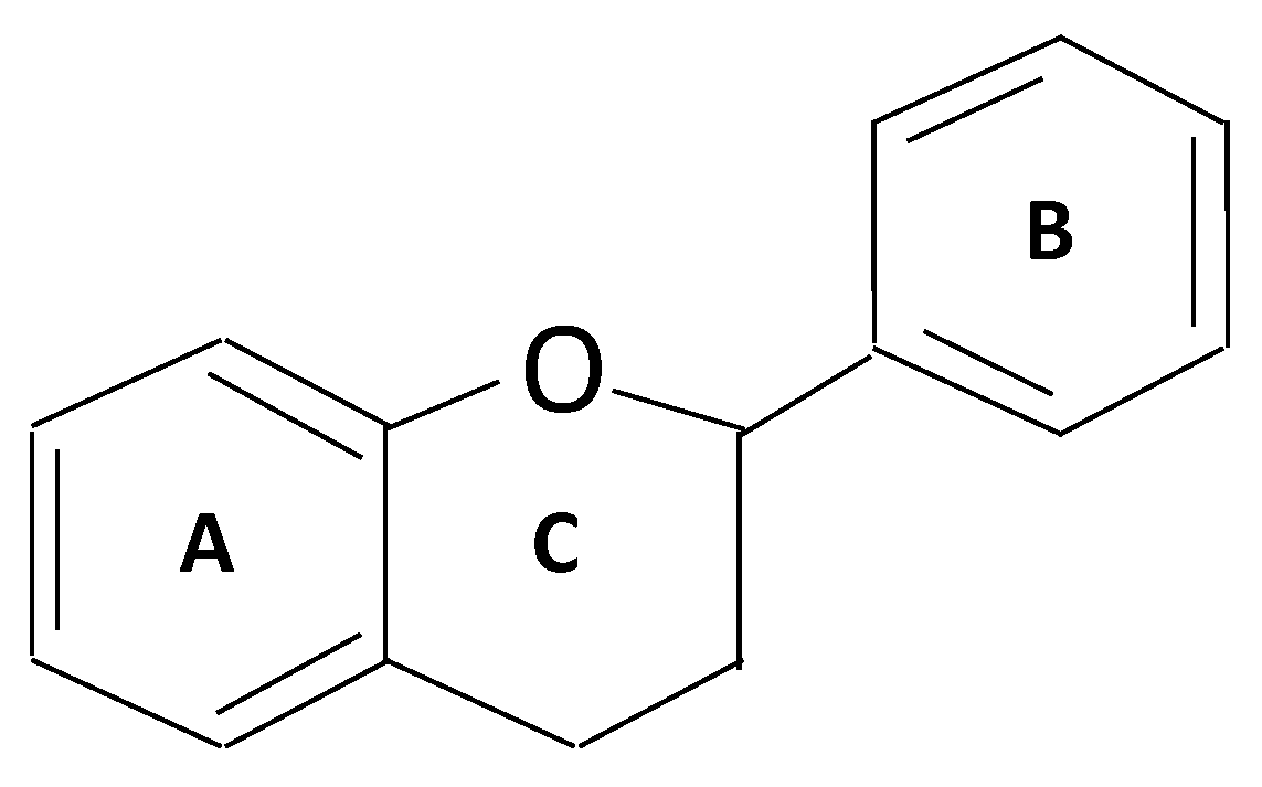
Flavonoids are secondary metabolites derived mainly from plants, fruits, leaves, and vegetables [19]. They represent a large family of compounds consisting of benzopyran rings with polyphenolic groups at different positions. The structure of flavonoids consists of 15 carbons and two rings (A and B) linked by three carbon chains [20] (Figure 1). Flavonoids are classified into multiple types based on their chemical structure, unsaturated linking chain, and degree of oxidation [21]. Each of these groups possesses biological properties, and some are successfully used in therapeutic approaches for treating diabetes and GI cancer [22]. The administration of flavonoids to target diabetes modulated lipid and carbohydrate metabolisms, improved adipose tissue metabolism, attenuated hyperglycemia, and alleviated oxidative stress [23,24]. Moreover, for targeting GI cancer, flavonoids in berries and green tea reduced cellular proliferation and inflammation, inhibited metastasis, and increased apoptosis [25,26]. Additional efforts are necessary to address the mechanism of flavonoids in treating metabolic disorders such as diabetes and cancer.
1.4. Gut Microbiota: The Role in Diabetes and GI Cancer
The human digestive tract harbors several microbial species collectively called the gut microbiome [27]. The human gut microbiome influences health and disease status through its involvement in human nutrition, immunity, and physiology [28]. Recently, we reported the effect of gut microbiome enzymatic modifications on flavonoid metabolism and how these modifications affect the observed biological properties of flavonoids [29].
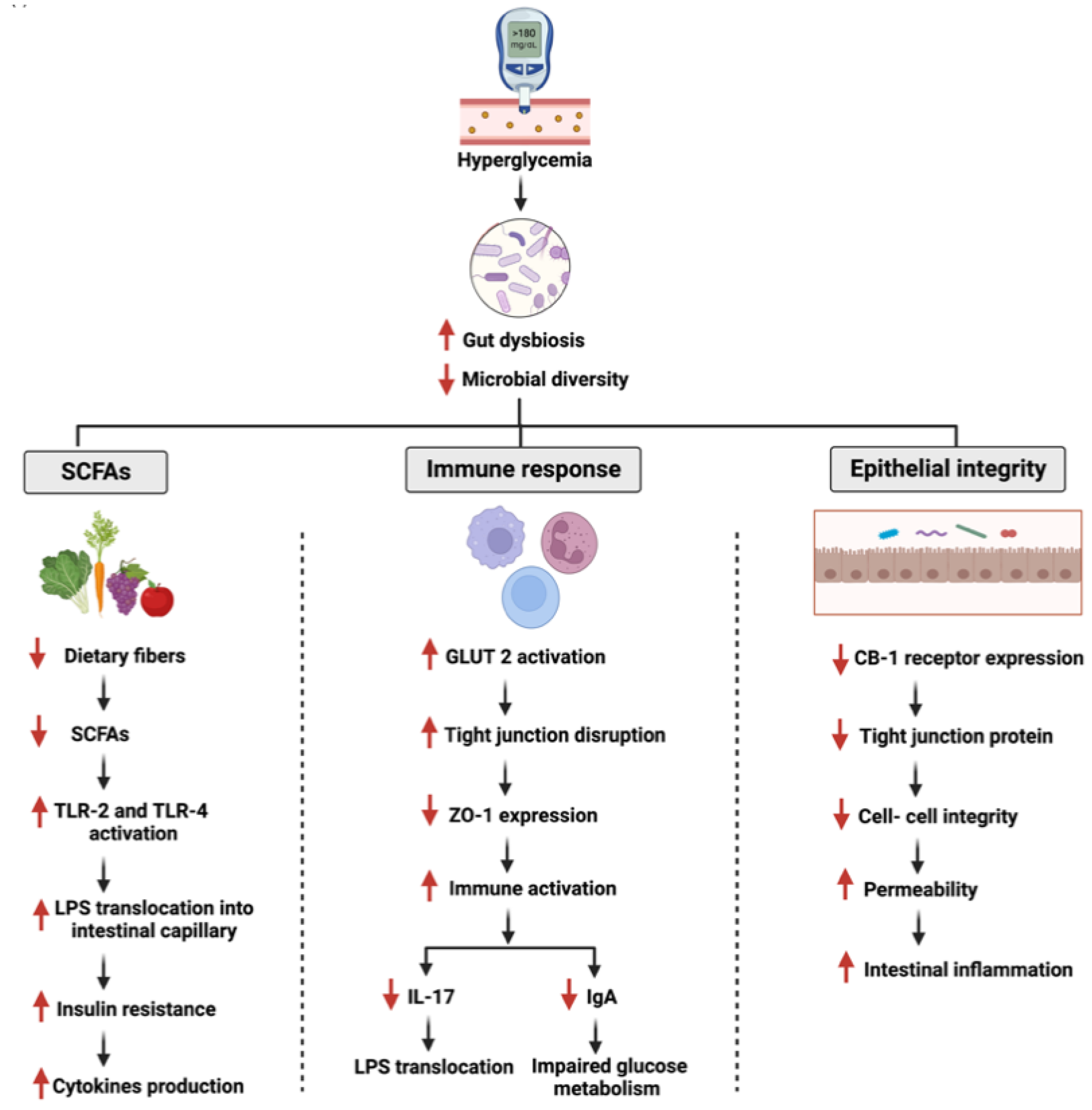
Human gut microbiome diversity disruption is linked to pathological conditions such as inflammatory bowel disease (IBD), obesity, diabetes, and cancers [30,31]. In two population-based studies, the diversity and richness of the gut microbiome were associated directly with the development of type 2 diabetes and insulin resistance [32]. Furthermore, the gut microbiome’s impact on cancer is not only limited to specific pathways such as apoptosis and cellular proliferation, but also enhances the efficacy and reduces the resistance of chemotherapeutic drugs [33]. Figure 2 and Figure 3 summarize the overall interactions between the gut microbiome and specific pathways during diabetes and GI cancer.
In our previous work, we identified the role of different flavonoids on diabetes and GI cancer separately [34,35]. In this review, we evaluate and analyze published studies to report and estimate the possible dual role of flavonoids in targeting diabetes and GI cancers. The literature supports a potential association between both conditions, as hyperglycemia level influences the development of diabetes and GI cancers. Furthermore, we assess the impact of this interaction on specific and common pathways in both metabolic conditions. Finally, we identify gaps in the current research.
2. Search Strategy and Selection Criteria
Medline, Scopus, and PubMed were searched for manuscripts published from 2000 to 2022 using the search terms “flavonoids”, “microbiota”, “flavonoids AND diabetes”, “flavonoids AND cancer”, “flavonoids AND GI cancer”, “microbiome AND GI cancer”, “gut microbiota enzymes”, microbiome AND diabetes”, and “flavonoids AND GI cancer AND diabetes”. In this article, we selected 121 articles and analyzed them in detail. Eligible studies included in vivo, in vitro, and clinical trial papers addressing the influence of flavonoids on both conditions and the possible underlying dual effects. Duplicates and studies with other flavonoid metabolism mechanisms were excluded.
3. Role of Flavonoids on Diabetic and GI Cancer Pathways
As discussed in the previous section, flavonoids exhibit diverse biological properties that target diabetes and GI cancers. In the following paragraphs, we will discuss the influence of specific flavonoids on major impaired pathways in diabetes and GI cancers such as apoptosis, cellular proliferation, enzymatic modifications, and AMPK pathways. Figure 4 highlights an overview and summarizes the impaired pathways in both metabolic conditions and the effects of flavonoids in targeting them.
3.1. Flavonoids and Apoptosis in Diabetes and GI Cancers
Cellular apoptosis is programmed cell death that involves numerous regulatory genes [36]. These regulatory checkpoints are critical to maintaining a homeostatic balance between the newly formed and damaged cells [37]. Loss of this balance may lead to the development of diseases such as diabetes and/or GI cancers [38]. Flavonoids restore the imbalance in these conditions [39].
In diabetes, long-term hyperglycemia in patients may contribute to the accumulation of reactive oxygen species (ROS), which results in endothelial cellular stress [40]. If unresolved, it may lead to apoptosis of microvascular endothelial cells, which, in turn, results in diabetic complications such as diabetic cardiomyopathy and diabetic nephrosis [41]. However, baicalin, a flavone glycoside in tea, fruits, and vegetables, reduces apoptosis in a rat model of diabetes mellitus, thus supporting the neuroprotective efficacy of baicalein against diabetes-associated cognitive deficits [42]. Moreover, a study that stimulated diabetic mice cells with 60.0 M of glucose to induce oxidative stress showed that cells treated with baicalin had significantly alleviated oxidative stress and apoptosis through the regulation of PERK/Nrf2 pathway [43]. Additionally, hesperidin, a glycosidic flavonoid abundant in citrus fruit, also reduces oxidative stress [44]. Normal human hepatocytes stimulated with 33 mM of glucose for 24 h and then treated with hesperidin had improved oxidative stress levels and increased cell viability compared to the untreated cells. These findings demonstrated that hesperidin treatment might reduce apoptosis by regulating miR- 149 expression, a critical apoptotic regulator [45]. Similarly, rutin also alleviated diabetic complications through apoptosis [46]. Myoplast cells stimulated with different glucose concentrations and then treated with rutin showed an improvement in cardiomyocyte injury through the direct inhibition of apoptosis and endoplasmic reticulum stress [47].
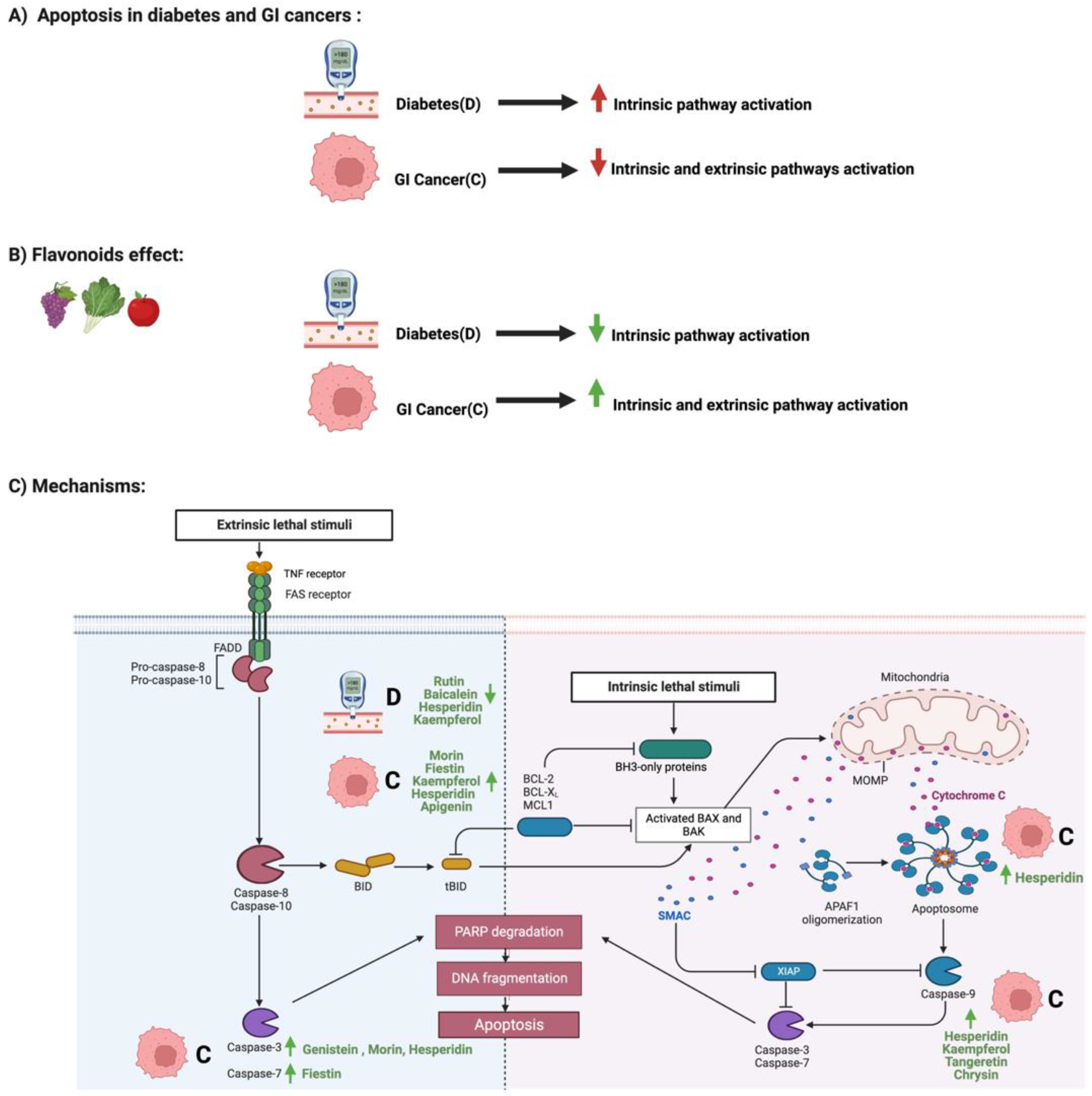
The reduction of apoptotic regulation allows cancerous cells to survive and progress [48]. Flavonoids such as apigenin, a plant flavone, induce cell cycle arrest, disruption, and apoptosis with low toxicity and mutagenicity rates [49]. Additionally, 200 umol/L of genistein for 48 h affected both Notch 1 and epithelial-mesenchymal transition (EMT) pathways and significantly induced apoptosis in colorectal cancer cells [50]. Moreover, the administration of 50 uM of Kaempferol also decreased the Bcl-2 level and increased the cleaved caspase 9 expression, thus inducing apoptosis in gastric cancer cells [51]. Interestingly, the same apoptotic pathway may be targeted by different flavonoid types such as chrysin, morin, and hesperidin. All three flavonoids induced apoptosis separately through the modulation of caspase 3, 9, and BAX expression in colorectal cancer or gastric cancer cells [52,53,54]. Moreover, cotreatment of apigenin and 5-flurouracil increased ROS and mitochondrial membrane potential in colorectal cancer cells, supporting flavonoids’ potential role as a therapeutic agent [55]. Figure 5 summarizes the influence of specific flavonoids on the apoptotic pathways in diabetes and GI cancers.
3.2. Flavonoids and NF-κB Pathway in Diabetes and GI Cancers
Nuclear factor kappa B (NF-κB) transcription factors are critical regulators of the inflammatory and immune responses against infections and injuries [56]. NF-κB dysregulation may lead to the development of cancers, chronic inflammation, and insulin resistance [57]. The activation of NF-κB by oxidative stress in diabetic patients due to the high glucose level plays a crucial role in the vascular complications of diabetes [58]. Additionally, persistent hyperglycemia in diabetes triggers the expression of chemokines, cytokines, and cell adhesion molecules by activating NF-κB expression [59]. Furthermore, activation of the NF-κB pathway correlates with GI cancers’ tumor size, migration, and invasion through the modulation of inflammatory markers, just as in the pathogenesis of GI cancers [60]. Targeting the NF-κB pathway with flavonoids is reported to be a potential therapeutic target for diabetes and GI cancers [61,62].
Different flavonoids exert antidiabetic properties. Baicalin, a bioactive polyphenolic flavonoid, administered to diabetic mice significantly reduced glucose levels and inhibited insulin levels [63]. Additionally, the administered baicalin attenuated steatosis in diabetic mice’s hepatic tissues through the suppression of inflammatory cytokines regulated by the NF-κB pathway. Treatment with 20 mg/kg of apigenin also reduced NF-κB and TNF-a expression and inhibited mouse inflammations [64]. Moreover, luteolin significantly reduced oxidative stress by inhibiting the NF-κB and activating the nuclear factor-erythroid 2-related factor 2 signaling pathway [65]. Interestingly, as reported with quercetin, flavonoids inhibit NF-κB expression and target vascular complications in diabetes [66]. Daily administration of quercetin in rats for six weeks reduced NF-κB expression and protects against diabetic-induced exaggerated vasoconstriction.
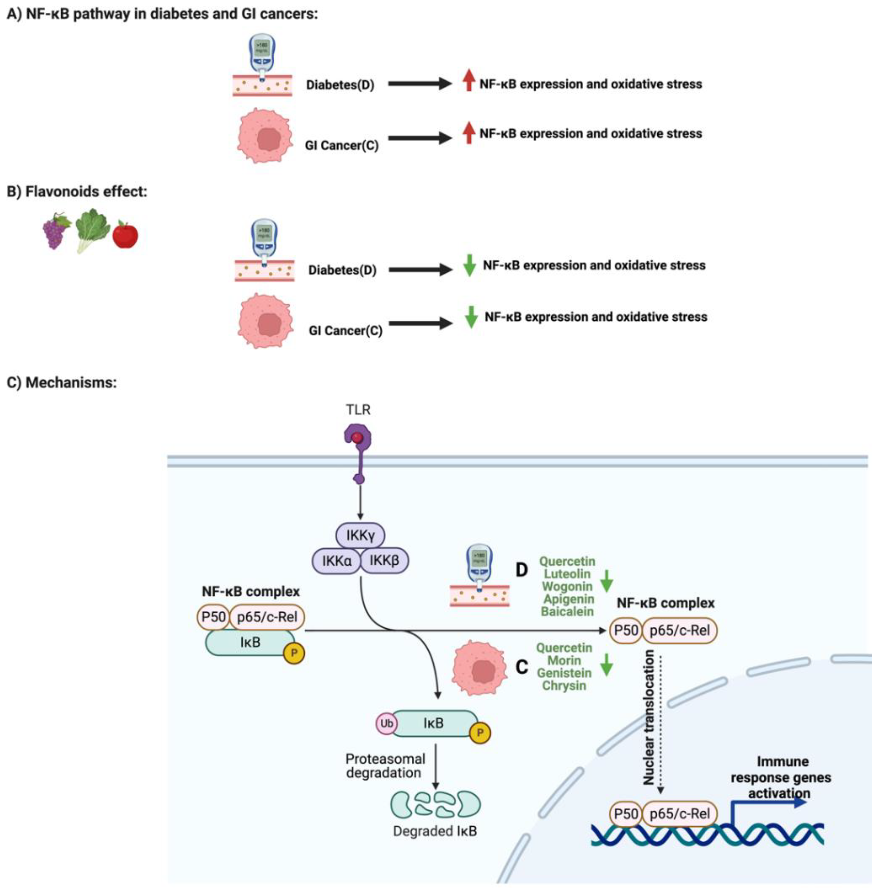
Flavonoids also play a significant role in targeting the NF-κB pathway through different mechanisms in gastric cancer (GC). Chrysin, for example, significantly suppressed NF-κB and Egr-1 expression in gastric cancer cells by inhibiting endogenous recepteur d’órigine nantais (RON) expression and activity, a critical receptor for cancer cell invasion [67]. Additionally, treating colorectal cancer cells with morin suppressed the expression of NF-κB and the production of inflammatory cytokines such IL-8 and IL-6 [68]. Furthermore, genistein administration in gastric cells targeted and significantly reduced the expression of Cyclooxygenase-2 (COX-2), an isoenzyme critical in cancer progression, by targeting and inhibiting its regulator, NF-κB [69]. Quercetin also downregulated the NF-κB pathway, influencing the invasion, migration, and metastasis of gastric cancer cells [70]. Figure 6 highlights the influence of specific flavonoids on the NF-κB pathway and expression in diabetes and GI cancers.
3.3. Flavonoids and AMPK Pathway in Diabetes and GI Cancers
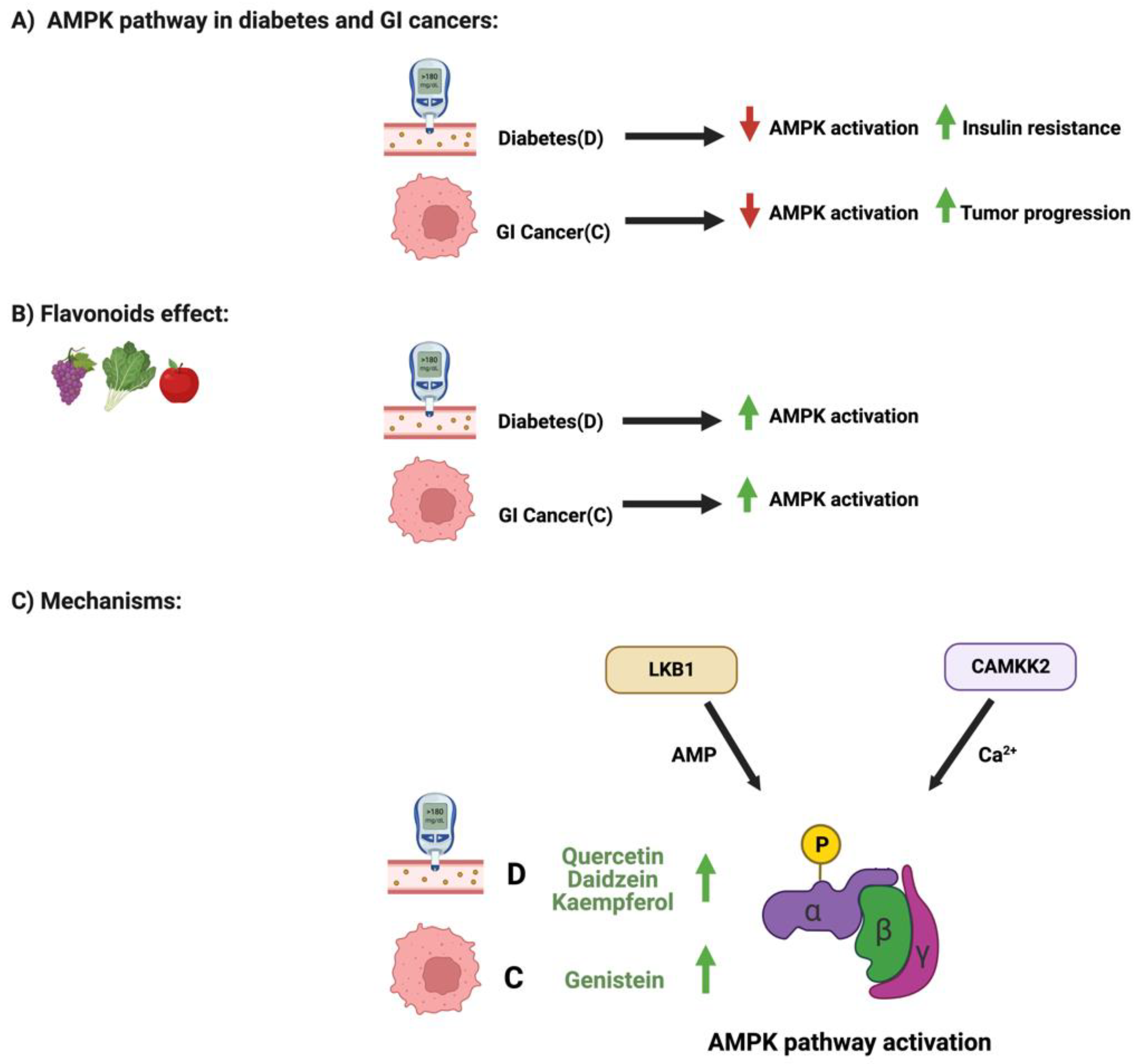
AMPK-activated protein kinase is a highly conserved protein kinase and a central regulator of cellular energy and homeostasis in eukaryotes [71]. It exists in the cell as an obligate heterodimer with a catalytic subunit and two regulatory subunits [72]. Several cellular stressors, such as hypoxia, can activate the AMPK pathway [73]. Impaired regulations of the AMPK pathway play an essential role in developing metabolic conditions such as diabetes, leading to insulin resistance and cancers and tumor progression [74,75]. However, flavonoids could positively influence and regulate AMPK pathways in diabetes and GI cancers.
Three main flavonoids regulate the AMPK pathway in diabetes: quercetin, daidzein, and Kaempferol. Skeletal muscle cells treated with quercetin induced glucose uptake in the cells, changed the mitochondrial membrane potential, and increased intracellular calcium concentration [76]. The observed antidiabetic effects were modulated mainly through the regulation of the AMPK pathway and GLUT4 expression and were similar to the mechanisms of the well-known antidiabetic drug metformin [76]. Additionally, diabetic mice and myotubes cells treated with daidzein showed that this flavonoid promoted glucose uptake through the phosphorylation of AMPK and the translocation of GLUT4 expression [77]. Moreover, the administration of Kaempferol to pancreatic cells upregulated the phosphorylation of the AMPK pathway. When an inhibitor was used, it reduced the pancreatic cells’ survival rate suggesting a potential cytoprotective effect of Kaempferol on diabetes [78].
On the other hand, colorectal cancer cells treated with a soy-derived phytochemical, genistein, demonstrated activated AMPK expression [79]. Additionally, combination therapy of genistein and 5-flurouracil abolished cellular growth, induced cellular apoptosis, and activated AMPK phosphorylation, exerting a potential chemotherapeutic approach against colorectal cancers. Figure 7 illustrates the influence of specific flavonoids on the AMPK pathway and expression in diabetes and GI cancers.
3.4. Flavonoids and Enzymatic Activities in Diabetes and GI Cancers
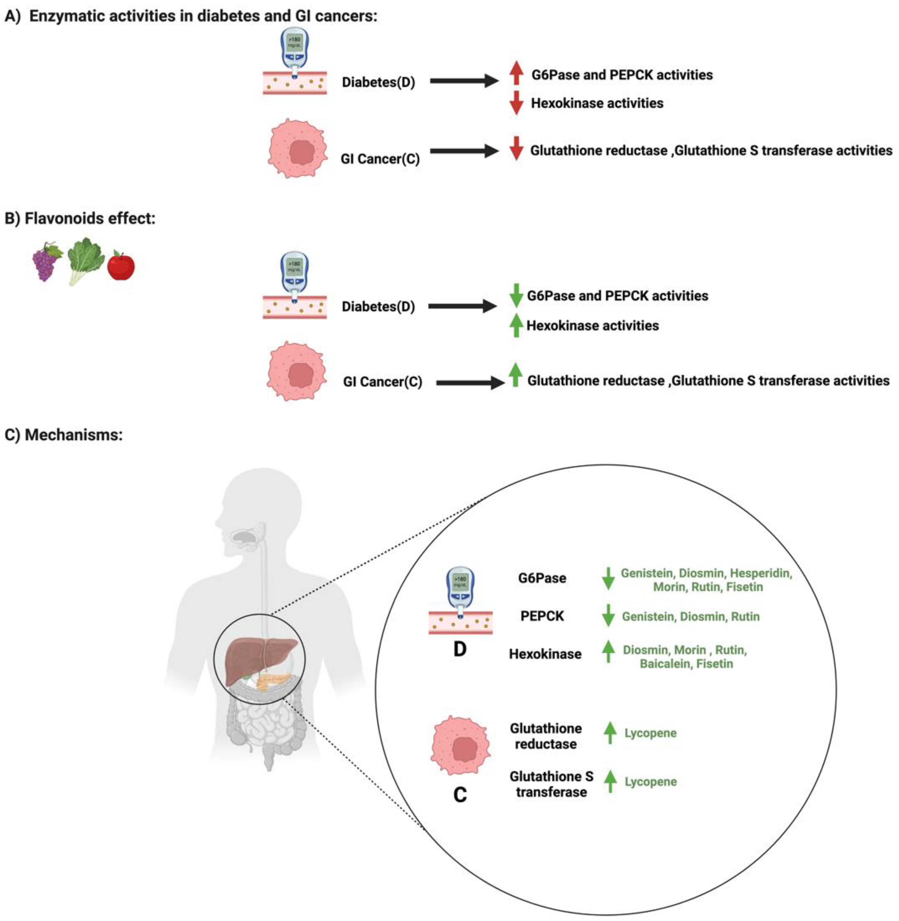
Enzymes are critical modulators in the body, aiding in regulating the metabolic process and chemical reactions [80]. Their influence on these reactions depends on the target tissue and regulatory mechanisms [81]. An imbalance in the regulation or the expression of specific enzymes, such as glucose 6 phosphatase or glutathione reductase, may lead to diabetes and cancers, respectively [82,83].
In diabetes, the level of hepatic enzymes such as glucose 6 phosphatase and phosphoenolpyruvate carboxykinase increases, while the level of hexokinase decreases. These enzymes are critical in maintaining blood glucose regulation in the body [84]. Flavonoids such as genistein, diosmin, morin, rutin, and baicalin improve the activities of these enzymes in diabetic models. The administration of genistein (0.02%) in mice reduced glucose 6 phosphatase and phosphoenolpyruvate carboxykinase activities in the liver compared to the control [85]. Additionally, oral administration of diosmin (100 mg/kg) significantly decreased glucose 6 phosphatase activities and increased hexokinase in diabetic-induced rats [86]. Moreover, rutin administration for 30 days to diabetic rats significantly ameliorated hyperglycemia, decreased glucose 6 phosphatase activities, and increased hexokinase activities [87]. Furthermore, glucose 6 phosphatase and hexokinase activities were restored after the oral administration of morin for 30 days in diabetic rats [88]. These data support the potential role of flavonoids in influencing the hepatic enzymatic activities in the liver, which may be applicable for diabetic management.
In contrast, in GI cancers, enzymes such as glutathione reductase and glutathione S transferase are critical in regulating glutathione utilization, a cellular regulator for the redox state [89]. An imbalance in the utilization of this intracellular antioxidant may impair biological pathways such as apoptosis and cellular proliferation [90]. Limited studies reporting the effect of flavonoids on glutathione utilization enzymes in GI cancers are available. Lycopene administration (2.5 mg/kg) suppressed gastric cancer by increasing glutathione reductase and glutathione S transferase activities [91]. More studies are required to confirm this observation and support the potential role of flavonoids in GI cancer pathogenesis. Figure 8 shows the influence of specific flavonoids on enzymatic activities and expression in diabetes and GI cancers. Table 1 and Table 2 summarize the main findings from the reported studies.
4. Possible Dual Effects of Flavonoids on Diabetes and GI Cancer
As illustrated in the tables above, we identified three main flavonoids targeting metabolic pathways that are impaired in diabetes and GI cancers. These flavonoids are hesperidin, Kaempferol, and quercetin, and they target apoptosis and the NF-κB pathways This could suggest possible dual effects of flavonoids on diabetes and GI cancer. In male diabetic rats, the administration of hesperidin (100 mg/kg) for four weeks significantly reduced blood glucose and increased insulin levels [104]. Additionally, and more importantly, hesperidin effectively regulated the expression of apoptotic proteins, as shown by the upregulation of the antiapoptotic Bcl-xl level and the downregulation of the pro-apoptotic Bax level [104].
Moreover, in three gastric cancer cell lines, the treatment of hesperidin also targeted apoptotic regulatory proteins but with a different biological effect. The administered hesperidin upregulated the pro-apoptotic Bax level and downregulated the antiapoptotic Bcl-2 levels [54]. Despite the differences observed in both studies (ranging from the targeted disease, the model used, the concentration of the hesperidin, the administration route, and the duration of the treatment), hesperidin affected apoptotic proteins and restored their physiological effects in both studies, but in an opposite manner, resulting in reduction in diabetes and induction in GI cancers. Moreover, Kaempferol also targeted the apoptotic pathway in both conditions. In diabetes, Kaempferol significantly improved the expression of antiapoptotic proteins such as Bcl-2, inhibited apoptosis, and reduced caspase 3 activities in INS-1E beta cells [105]. Kaempferol also targeted apoptosis in colorectal cancer cells by blocking ROS production [106]. Although these results may support the potential efficacy of hesperidin and Kaempferol to modulate apoptosis in different metabolic conditions, more efforts are required to test both conditions in alignment using a standardized technique.
As discussed in the previous section, quercetin can affect NF-κB pathways in diabetes and GI cancers. Diabetic mice treated with orally administered quercetin for two weeks showed a suppressed inflammatory response indicated by reduced NF-κB expression [96]. In gastric cancer cells, quercetin also inhibited the dose-dependent NF-κB pathway [107]. However, limited data are available in the literature to support the hypothesis that quercetin may have the same biological effect on the two conditions: diabetes and GI cancers.
Analyzing the available data on three flavonoids, the possible dual effect depends on the targeted pathway, the model used, and the flavonoid dosage, which needs standardization. Additionally, studies evaluating the impact of these flavonoids in combination with the currently used treatment are lacking in the literature; assessing the potential of dual effects of other flavonoids on diabetes and GI cancers is also highly required.
5. Discussion
5.1. Targeting Multiple Pathways with Multiple Flavonoids That Act Synergistically?
As mentioned in the previous sections and the figures, the same flavonoid can trigger and improve diabetes and/or cancer-related pathways, as seen with baicalin and genistein, respectively. Baicalin, for example, exerts its antidiabetic effects by targeting three out of four discussed pathways: apoptosis, NF-κB, and enzymatic modification. Genistein, on the other hand, triggers apoptosis, NF-κB, and AMPK pathways in cancer. Therefore, can the positive effects of flavonoids on physiological pathways be further enhanced when different flavonoids are combined to complement and trigger additional pathways? For instance, with baicalin targeting three diabetic-impaired pathways, combining baicalin with one of the flavonoids that target the AMPK pathway in diabetes (Figure 7) might potentially initiate a positive effect in all four of the discussed pathways due to synergistic effects of both phytochemicals. This hypothesized approach may prevent the over-activation of the same pathway and reduce possible side effects while achieving the antidiabetic effects of these flavonoids. The same approach may also be used with genistein in GI cancer. So far, these thoughts are theoretical and only based on observation from the data cited above. In vitro or in vivo studies are still missing, and extensive studies are required to prove whether they are effective in an expected way. This hypothesis may be tested by first identifying the pathways targeted by the flavonoid in each metabolic condition, then identifying flavonoids that complement each other. Furthermore, estimating the appropriate concentration of each flavonoid is critical to ensure safety and prevent cytotoxicity. Recognizing the potential binding sites of each flavonoid and their bioavailability is essential to ensure their positive effects. Moreover, observing possible side effects from this combination with the currently used treatment is necessary, as the aim is not to replace the available therapeutic tools, but to complement them.
5.2. Gut Microbiome and Flavonoids’ Effects
The relationship between the gut microbiome and flavonoids may be described as a bidirectional interaction. On the one hand, the consumed flavonoids act as a regulator of intestinal hemostasis by either enhancing or suppressing the growth of specific bacterial species. On the other hand, the gut microbiome contributes to the pharmacokinetics of ingested dietary flavonoids to produce aglycones and, subsequently, the ring fission products which facilitate absorption [108,109,110]. It is hypothesized that these enzymatically modified gut microbiome products are responsible for the positive physiological effects of flavonoids observed in diabetes and GI cancer (Figure 2 and Figure 3). This may provide a potential therapeutic target for both metabolic conditions. Patients with type 2 diabetes exhibit gut microbiome dysbiosis characterized by a reduction in butyrate-producing bacterial species [111]. Additionally, the gut microbiome regulates the levels of short-chain fatty acids and lipopolysaccharides, which are critical for developing diabetes [112]. Furthermore, patients with type 2 diabetes exert a low-grade systemic inflammation mediated by the impaired gut barrier [113].
On the other hand, gut microbial species such as Streptococcus bovis, Fusobacterium nucleatum, and Enterococcus faecalis mediate inflammatory response and tumor progression in colorectal cancer [114,115]. The data above show that flavonoids and their metabolites could reduce or restore the observed pathological effects in diabetes and GI cancers. Despite this, more efforts are required to properly study the gut microbiome, including a suitable model and flavonoid interactions. More studies are required to evaluate whether the positive/dual effects observed are due to the flavonoids, their metabolites, or both. In addition, more efforts are needed to address the metabolites from the gut microbiome and the liver, and especially to discuss and understand the substances metabolized in the liver [116]. The development of more omics tools that help analyze data is necessary [117].
5.3. Challenges with Studying the Field
Although flavonoids reduce the pathogenesis of diabetes and GI cancers, further efforts are required to address the main challenges of their utilization in managing cancer and diabetes. First, the recommended dosage of flavonoids for each metabolic condition must be established. The literature shows variations in the estimated dosage for most flavonoids tested based on the population, ethnicity, gender, and geographical locations [118,119,120]. The observed variations may be due to the solubility and molecular weight of the flavonoids used [121]. Second, newly developed tools and techniques are required to overcome flavonoids’ relatively low bioavailability rate.
Additionally, a better understanding of flavonoids’ cellular permeability, excretion, and metabolic alternation is needed to enhance their bioavailability [122]. Third, the potential side effects of using multiple flavonoids to target various metabolic pathways or using flavonoids in combination with currently used drugs as a treatment method must be estimated. This will help to minimize the risk of negative flavonoid–drug interactions. Fourth, the field is in great need of standardization starting from the model used, flavonoid extraction and purification technique, administration route, and duration of the treatment. By overcoming these challenges, using flavonoids as a potential therapeutic target may be implemented with high effectiveness and safety levels.
5.4. GI Cancers and Diabetes: Their Relationship?
The prevalence of diabetes mellitus among patients with GI cancers varies depending on the tumor type. For example, the highest rate of diabetes mellitus was seen in patients with colon cancer (15.5%), rectal cancer (15.3%), and stomach cancer (14%) [123]. On the other hand, diabetes in patients can promote the progression, development, and metastasis of gastric cancers [124]. The observed association may be due to the shared risk factors between both conditions such as diet, smoking, infection, medication, obesity, and hyperglycemia [125]. More data are still needed to identify which condition comes first and whether we can predict the occurrence of one following the other. Despite this, the available data suggest a possible association between both conditions and how they may be targeted using flavonoids as a novel therapeutic agent, which might help improve the quality of the patients’ lives and reduce the burden of both conditions.
6. Conclusions
Flavonoids are naturally occurring products found abundantly in fruits and vegetables. Upon ingestion, flavonoids are further metabolized by the gut microbiome into aglycones that exert antidiabetic and anti-GI cancer effects. Based on the current evidence, flavonoids target impaired pathways such as apoptosis and NF-κB in both conditions [126,127]. Despite this, the mechanisms by which flavonoids target the same pathway with different biological effects depending on the metabolic conditions need further research. Only limited evidence from the literature supports a potential dual effect of flavonoids.
Moreover, a combination of several flavonoids (affecting the pathology synergistically or additively) represents a promising strategy to be further evaluated to target impaired pathways. Still, more efforts are required for protocol standardization to ensure safety and effectiveness. Similarly, vitexin, an apigenin flavone glucoside, exerts beneficial effects in high glucose-induced endothelial cells via inhibition of apoptosis and oxidative stress, thus suggesting the potential effectiveness of vitexin in cardiovascular complications of diabetes. Specifically, vitexin decreased apoptosis via the disruption of Wnt/β-catenin and Nrf2, decreased ROS and malondialdehyde content, and increased superoxide dismutase activity in HG-mediated HUVECs [128]. In addition to isolated flavonoid compounds, extracts rich in flavonoids were also reported to influence metabolic pathways, such as Oldenlandia diffusa, which induced apoptosis via ROS accumulation, mitochondrial membrane potential loss, and activation of mitochondrial pathway in gastric cancer in vitro and in vivo [129]. In this regard, a lack of understanding of flavonoids’ complex mechanisms of action, non-specific selectivity, and low bioavailability in organisms markedly impedes their pharmacological administration in clinical practice. Mentioned obstacles could be overcome using progressive methodological approaches in medicine, i.e., designing nano-engineering systems to achieve the delivery of flavonoids into targeted organs and tissues. Such techniques could improve flavonoids’ efficacy and pharmacokinetic properties in diabetic cancer patients.
Our paper provides a theoretical base for advancing highly effective and low-toxicity flavonoid-based pharmaceutical molecules or their mixtures to target complex signaling pathways associated with diabetes and cancer concurrently. However, only precisely designed and well-controlled clinical evaluations of the context of the antidiabetic and anti-cancer effects of bioactive flavonoids could establish the role of these polyphenolic compounds as beneficial agents in diabetic and GI cancer patients.
Author Contributions
Conceptualization, R.K.A.-I. and D.B.; literature review and resources, R.K.A.-I.; writing—original draft preparation, R.K.A.-I.; writing—review and editing, D.B., P.K. and A.M.; figure preparation and editing, R.K.A.-I. and D.B.; visualization, R.K.A.-I., A.M., P.K. and D.B.; supervision, D.B.; project administration, D.B.; funding acquisition, D.B. All authors have read and agreed to the published version of the manuscript.
Funding
This work was supported by a National Priorities Research Program grant (NPRP14S-0311-210033; awarded to Professor Dr. Dietrich Büsselberg, June 2019-Current) from the Qatar National Research Fund (QNRF, a member of Qatar Foundation). The statements made herein are solely the responsibility of the authors. The APC was covered by the research department of WCM-Q.
Conflicts of Interest
The authors declare no conflict of interest.
Abbreviations
| GI | Gastrointestinal |
| CRC | Colorectal cancer |
| GC | Gastric cancer |
| IBD | Inflammatory bowel disease |
| SCFA | Short chain fatty acid |
| TLR | Toll-like receptor |
| LPS | Lipopolysaccharide |
| GLUT | Glucose transporter |
| AMPK | AMP-activated protein kinase |
| NF-κB | Nuclear factor kappa |
| ROS | Reactive oxygen species |
| EMT | Epithelial-mesenchymal transition |
| FISH | Fluorescence in situ hybridization |
| COX-2 | Cyclooxygenase-2 |
| G6Pase | Glucose 6-phosphatase |
| PEPCK | Phosphoenolpyruvate carboxykinase |
| PCR | Polymerase chain reaction |
| IP | Immunoprecipitation assay |
References
- Patel, D.K.; Kumar, R.; Laloo, D.; Hemalatha, S. Diabetes mellitus: An overview on its pharmacological aspects and reported medicinal plants having antidiabetic activity. Asian Pac. J. Trop. Biomed. 2012, 2, 411–420. [Google Scholar] [CrossRef] [Green Version]
- Banday, M.Z.; Sameer, A.S.; Nissar, S. Pathophysiology of diabetes: An overview. Avicenna J. Med. 2020, 10, 174–188. [Google Scholar] [CrossRef] [PubMed]
- Kharroubi, A.T.; Darwish, H.M. Diabetes mellitus: The epidemic of the century. World J. Diabetes 2015, 6, 850–867. [Google Scholar] [CrossRef]
- Chen, L.; Magliano, D.J.; Zimmet, P.Z. The worldwide epidemiology of type 2 diabetes mellitus-present and future perspectives. Nat. Rev. Endocrinol. 2011, 8, 228–236. [Google Scholar] [CrossRef] [PubMed]
- Reyes, J.; Tripp-Reimer, T.; Parker, E.; Muller, B.; Laroche, H. Factors Influencing Diabetes Self-Management Among Medically Underserved Patients with Type II Diabetes. Glob. Qual. Nurs. Res. 2017, 4, 2333393617713097. [Google Scholar] [CrossRef] [PubMed] [Green Version]
- Chawla, A.; Chawla, R.; Jaggi, S. Microvasular and macrovascular complications in diabetes mellitus: Distinct or continuum? Indian J. Endocrinol. Metab. 2016, 20, 546–551. [Google Scholar] [CrossRef] [PubMed]
- Lin, X.; Xu, Y.; Pan, X.; Xu, J.; Ding, Y.; Sun, X.; Shan, P.F. Global, regional, and national burden and trend of diabetes in 195 countries and territories: An analysis from 1990 to 2025. Sci. Rep. 2020, 10, 14790. [Google Scholar] [CrossRef] [PubMed]
- Ramachandran, A. Know the signs and symptoms of diabetes. Indian J. Med. Res. 2014, 140, 579–581. [Google Scholar]
- Dhatariya, K. Diabetes: The place of new therapies. Adv. Endocrinol. Metab. 2019, 10, 2042018818807599. [Google Scholar] [CrossRef]
- Blaslov, K.; Naranda, F.S.; Kruljac, I.; Renar, I.P. Treatment approach to type 2 diabetes: Past, present and future. World J. Diabetes 2018, 9, 209–219. [Google Scholar] [CrossRef]
- Hassanzade, J.; Molavi, E.V.H.; Farahmand, M.; Rajaiifard, A.R. Incidence and Mortality Rate of Common Gastrointestinal Cancers in South of Iran, a Population Based Study. Iran. J. Cancer Prev. 2011, 4, 163–169. [Google Scholar] [PubMed]
- Bordry, N.; Astaras, C.; Ongaro, M.; Goossens, N.; Frossard, J.L.; Koessler, T. Recent advances in gastrointestinal cancers. World J. Gastroenterol. 2021, 27, 4493–4503. [Google Scholar] [CrossRef] [PubMed]
- Abotaleb, M.; Samuel, S.M.; Varghese, E.; Varghese, S.; Kubatka, P.; Liskova, A.; Busselberg, D. Flavonoids in Cancer and Apoptosis. Cancers 2018, 11, 28. [Google Scholar] [CrossRef] [PubMed] [Green Version]
- Zali, H.; Rezaei-Tavirani, M.; Azodi, M. Gastric cancer: Prevention, risk factors and treatment. Gastroenterol. Hepatol. Bed Bench. 2011, 4, 175–185. [Google Scholar]
- Han, C.J.; Reding, K.; Cooper, B.A.; Paul, S.M.; Conley, Y.P.; Hammer, M.; Miaskowski, C. Symptom Clusters in PatientsWith Gastrointestinal Cancers Using Different Dimensions of the Symptom Experience. J. Pain Symptom Manag. 2019, 58, 224–234. [Google Scholar] [CrossRef] [PubMed]
- Machlowska, J.; Baj, J.; Sitarz, M.; Maciejewski, R.; Sitarz, R. Gastric Cancer: Epidemiology, Risk Factors, Classification, Genomic Characteristics and Treatment Strategies. Int. J. Mol. Sci. 2020, 21, 4012. [Google Scholar] [CrossRef]
- Correa, P. Gastric cancer: Overview. Gastroenterol. Clin. N. Am. 2020, 42, 211–217. [Google Scholar]
- Matsuoka, T.; Yashiro, M. Precision medicine for gastrointestinal cancer: Recent progress and future perspective. World J. Gastrointest Oncol. 2020, 12, 1–20. [Google Scholar] [CrossRef]
- Panche, A.N.; Diwan, A.D.; Chandra, S.R. Flavonoids: An overview. J. Nutr. Sci. 2016, 5, e47. [Google Scholar] [CrossRef] [Green Version]
- Kozlowska, A.; Szostak-Wegierek, D. Flavonoids—food sources and health benefits. Rocz. Panstw. Zakl. Hig. 2014, 65, 79–85. [Google Scholar]
- Ullah, A.; Munir, S.; Badshah, S.L.; Khan, N.; Ghani, L.; Poulson, B.G.; Jaremko, M. Important Flavonoids and Their Role as a Therapeutic Agent. Molecules 2020, 25, 5243. [Google Scholar] [CrossRef] [PubMed]
- Juca, M.M.; Cysne Filho, F.M.S.; de Almeida, J.C.; Mesquita, D.D.S.; Barriga, J.R.M.; Dias, K.C.F.; Vasconcelos, S.M.M. Flavonoids: Biological activities and therapeutic potential. Nat. Prod. Res. 2020, 34, 692–705. [Google Scholar] [CrossRef] [PubMed]
- Sok Yen, F.; Shu Qin, C.; Tan Shi Xuan, S.; Jia Ying, P.; Yi Le, H.; Darmarajan, T.; Salvamani, S. Hypoglycemic Effects of Plant Flavonoids: A Review. Evid Based Complement. Altern. Med. 2021, 2057333. [Google Scholar] [CrossRef]
- Testa, R.; Bonfigli, A.R.; Genovese, S.; De Nigris, V.; Ceriello, A. The Possible Role of Flavonoids in the Prevention of Diabetic Complications. Nutrients 2016, 8, 310. [Google Scholar] [CrossRef] [PubMed]
- Rodriguez-Garcia, C.; Sanchez-Quesada, C.; Gaforio, J.J. Dietary Flavonoids as Cancer Chemopreventive Agents: An Updated Review of Human Studies. Antioxidants 2019, 8, 137. [Google Scholar] [CrossRef] [PubMed] [Green Version]
- Vitelli Storelli, F.; Molina, A.J.; Zamora-Ros, R.; Fernandez-Villa, T.; Roussou, V.; Romaguera, D.; Martin, V. Flavonoids and the Risk of Gastric Cancer: An Exploratory Case-Control Study in the MCC-Spain Study. Nutrients 2019, 11, 967. [Google Scholar] [CrossRef] [Green Version]
- Bull, M.J.; Plummer, N.T. Part 1: The Human Gut Microbiome in Health and Disease. Integr. Med. (Encinitas) 2014, 13, 17–22. [Google Scholar]
- Thursby, E.; Juge, N. Introduction to the human gut microbiota. Biochem. J. 2017, 474, 1823–1836. [Google Scholar] [CrossRef]
- Al-Ishaq, R.K.; Liskova, A.; Kubatka, P.; Busselberg, D. Enzymatic Metabolism of Flavonoids by Gut Microbiota and Its Impact on Gastrointestinal Cancer. Cancers 2021, 13, 3934. [Google Scholar] [CrossRef]
- Forslund, K.; Hildebrand, F.; Nielsen, T.; Falony, G.; Le Chatelier, E.; Sunagawa, S.; Pedersen, O. Disentangling type 2 diabetes and metformin treatment signatures in the human gut microbiota. Nature 2015, 528, 262–266. [Google Scholar] [CrossRef] [Green Version]
- Glassner, K.L.; Abraham, B.P.; Quigley, E.M.M. The microbiome and inflammatory bowel disease. J. Allergy Clin. Immunol. 2020, 145, 16–27. [Google Scholar] [CrossRef] [PubMed] [Green Version]
- Chen, Z.; Radjabzadeh, D.; Chen, L.; Kurilshikov, A.; Kavousi, M.; Ahmadizar, F.; Voortman, T. Association of Insulin Resistance and Type 2 Diabetes With Gut Microbial Diversity: A Microbiome-Wide Analysis From Population Studies. JAMA Netw. Open 2021, 4, e2118811. [Google Scholar] [CrossRef] [PubMed]
- Cheng, W.Y.; Wu, C.Y.; Yu, J. The role of gut microbiota in cancer treatment: Friend or foe? Gut 2020, 69, 1867–1876. [Google Scholar] [CrossRef] [PubMed]
- Al-Ishaq, R.K.; Abotaleb, M.; Kubatka, P.; Kajo, K.; Busselberg, D. Flavonoids and Their Antidiabetic Effects: Cellular Mechanisms and Effects to Improve Blood Sugar Levels. Biomolecules 2019, 9, 430. [Google Scholar]
- Al-Ishaq, R.K.; Overy, A.J.; Busselberg, D. Phytochemicals and Gastrointestinal Cancer: Cellular Mechanisms and Effects to Change Cancer Progression. Biomolecules 2020, 10, 105. [Google Scholar] [CrossRef] [PubMed] [Green Version]
- D’Arcy, M.S. Cell death: A review of the major forms of apoptosis, necrosis and autophagy. Cell Biol. Int. 2019, 43, 582–592. [Google Scholar] [CrossRef]
- Aachoui, Y.; Sagulenko, V.; Miao, E.A.; Stacey, K.J. Inflammasome-mediated pyroptotic and apoptotic cell death, and defense against infection. Curr. Opin. Microbiol. 2013, 16, 319–326. [Google Scholar] [CrossRef] [PubMed] [Green Version]
- Singh, R.; Letai, A.; Sarosiek, K. Regulation of apoptosis in health and disease: The balancing act of BCL-2 family proteins. Nat. Rev. Mol. Cell Biol. 2019, 20, 175–193. [Google Scholar] [CrossRef] [PubMed]
- Slika, H.; Mansour, H.; Wehbe, N.; Nasser, S.A.; Iratni, R.; Nasrallah, G.; Eid, A.H. Therapeutic potential of flavonoids in cancer: ROS-mediated mechanisms. Biomed. Pharm. 2022, 146, 112442. [Google Scholar] [CrossRef]
- Naudi, A.; Jove, M.; Ayala, V.; Cassanye, A.; Serrano, J.; Gonzalo, H.; Pamplona, R. Cellular dysfunction in diabetes as maladaptive response to mitochondrial oxidative stress. Exp. Diabetes Res. 2012, 696215. [Google Scholar] [CrossRef] [Green Version]
- Pitocco, D.; Tesauro, M.; Alessandro, R.; Ghirlanda, G.; Cardillo, C. Oxidative stress in diabetes: Implications for vascular and other complications. Int. J. Mol. Sci. 2013, 14, 21525–21550. [Google Scholar] [CrossRef] [PubMed] [Green Version]
- Ma, P.; Mao, X.Y.; Li, X.L.; Ma, Y.; Qiao, Y.D.; Liu, Z.Q.; Cao, Y.G. Baicalin alleviates diabetes associated cognitive deficits via modulation of mitogen-activated protein kinase signaling, brain derived neurotrophic factor and apoptosis. Mol. Med. Rep. 2015, 12, 6377–6383. [Google Scholar] [CrossRef] [PubMed] [Green Version]
- Dong, Y.; Xing, Y.; Sun, J.; Sun, W.; Xu, Y.; Quan, C. Baicalein Alleviates Liver Oxidative Stress and Apoptosis Induced by High-Level Glucose through the Activation of the PERK/Nrf2 Signaling Pathway. Molecules 2020, 25, 599. [Google Scholar] [CrossRef] [PubMed] [Green Version]
- Parmar, M.S.; Syed, I.; Gray, J.P.; Ray, S.D. Curcumin, Hesperidin, and Rutin Selectively Interfere with Apoptosis Signaling and Attenuate Streptozotocin-Induced Oxidative Stress-Mediated Hyperglycemia. Curr. Neurovasc. Res. 2015, 12, 363–374. [Google Scholar] [CrossRef] [PubMed]
- Tian, M.; Han, Y.B.; Zhao, C.C.; Liu, L.; Zhang, F.L. Hesperidin alleviates insulin resistance by improving HG-induced oxidative stress and mitochondrial dysfunction by restoring miR-149. Diabetol. Metab. Syndr. 2021, 13, 50. [Google Scholar] [CrossRef]
- Ghorbani, A. Mechanisms of antidiabetic effects of flavonoid rutin. Biomed. Pharm. 2017, 96, 305–312. [Google Scholar] [CrossRef]
- Wang, J.; Wang, R.; Li, J.; Yao, Z. Rutin alleviates cardiomyocyte injury induced by high glucose through inhibiting apoptosis and endoplasmic reticulum stress. Exp. Med. 2021, 22, 944. [Google Scholar] [CrossRef]
- Pfeffer, C.M.; Singh, A.T.K. Apoptosis: A Target for Anticancer Therapy. Int. J. Mol. Sci. 2018, 19, 448. [Google Scholar] [CrossRef] [Green Version]
- Maeda, Y.; Takahashi, H.; Nakai, N.; Yanagita, T.; Ando, N.; Okubo, T.; Takiguchi, S. Apigenin induces apoptosis by suppressing Bcl-xl and Mcl-1 simultaneously via signal transducer and activator of transcription 3 signaling in colon cancer. Int. J. Oncol. 2018, 52, 1661–1673. [Google Scholar] [CrossRef] [Green Version]
- Zhou, P.; Wang, C.; Hu, Z.; Chen, W.; Qi, W.; Li, A. Genistein induces apoptosis of colon cancer cells by reversal of epithelial-to-mesenchymal via a Notch1/NF-kappaB/slug/E-cadherin pathway. BMC Cancer 2017, 17, 813. [Google Scholar] [CrossRef]
- Kim, T.W.; Lee, S.Y.; Kim, M.; Cheon, C.; Ko, S.G. Kaempferol induces autophagic cell death via IRE1-JNK-CHOP pathway and inhibition of G9a in gastric cancer cells. Cell Death Dis. 2018, 9, 875. [Google Scholar] [CrossRef] [PubMed] [Green Version]
- Bahadori, M.; Baharara, J.; Amini, E. Anticancer Properties of Chrysin on Colon Cancer Cells, In vitro and In vivo with Modulation of Caspase-3, -9, Bax and Sall4. Iran. J. Biotechnol. 2016, 14, 177–184. [Google Scholar] [CrossRef] [PubMed] [Green Version]
- Sithara, T.; Arun, K.B.; Syama, H.P.; Reshmitha, T.R.; Nisha, P. Morin Inhibits Proliferation of SW480 Colorectal Cancer Cells by Inducing Apoptosis Mediated by Reactive Oxygen Species Formation and Uncoupling of Warburg Effect. Front. Pharm. 2017, 8, 640. [Google Scholar] [CrossRef] [PubMed] [Green Version]
- Zhang, J.; Wu, D.; Vikash, S.J.; Wang, J.; Yi, J.; Dong, W. Hesperetin Induces the Apoptosis of Gastric Cancer Cells via Activating Mitochondrial Pathway by Increasing Reactive Oxygen Species. Dig. Dis Sci. 2015, 60, 2985–2995. [Google Scholar] [CrossRef]
- Yang, C.; Song, J.; Hwang, S.; Choi, J.; Song, G.; Lim, W. Apigenin enhances apoptosis induction by 5-fluorouracil through regulation of thymidylate synthase in colorectal cancer cells. Redox Biol. 2021, 47, 102144. [Google Scholar] [CrossRef]
- Napetschnig, J.; Wu, H. Molecular basis of NF-kappaB signaling. Annu. Rev. Biophys. 2013, 42, 443–468. [Google Scholar] [CrossRef]
- Park, M.H.; Hong, J.T. Roles of NF-kappaB in Cancer and Inflammatory Diseases and Their Therapeutic Approaches. Cells 2016, 5, 15. [Google Scholar] [CrossRef]
- Paneni, F.; Beckman, J.A.; Creager, M.A.; Cosentino, F. Diabetes and vascular disease: Pathophysiology, clinical consequences, and medical therapy: Part I. Eur. Heart J. 2013, 34, 2436–2443. [Google Scholar] [CrossRef]
- Suryavanshi, S.V.; Kulkarni, Y.A. NF-kappabeta: A Potential Target in the Management of Vascular Complications of Diabetes. Front. Pharm. 2017, 8, 798. [Google Scholar] [CrossRef] [Green Version]
- Chaithongyot, S.; Jantaree, P.; Sokolova, O.; Naumann, M. NF-kappaB in Gastric Cancer Development and Therapy. Biomedicines 2021, 9, 870. [Google Scholar]
- Li, G.; Ding, K.; Qiao, Y.; Zhang, L.; Zheng, L.; Pan, T.; Zhang, L. Flavonoids Regulate Inflammation and Oxidative Stress in Cancer. Molecules 2020, 25, 5628. [Google Scholar] [CrossRef] [PubMed]
- Jubaidi, F.F.; Zainalabidin, S.; Taib, I.S.; Hamid, Z.A.; Budin, S.B. The Potential Role of Flavonoids in Ameliorating Diabetic Cardiomyopathy via Alleviation of Cardiac Oxidative Stress, Inflammation and Apoptosis. Int. J. Mol. Sci. 2021, 22, 5094. [Google Scholar] [CrossRef] [PubMed]
- Yin, H.; Huang, L.; Ouyang, T.; Chen, L. Baicalein improves liver inflammation in diabetic db/db mice by regulating HMGB1/TLR4/NF-kappaB signaling pathway. Int. Immunopharmacol. 2018, 55, 55–62. [Google Scholar] [CrossRef] [PubMed]
- Malik, S.; Suchal, K.; Khan, S.I.; Bhatia, J.; Kishore, K.; Dinda, A.K.; Arya, D.S. Apigenin ameliorates streptozotocin-induced diabetic nephropathy in rats via MAPK-NF-kappaB-TNF-alpha and TGF-beta1-MAPK-fibronectin pathways. Am. J. Physiol. Ren. Physiol. 2017, 313, F414–F422. [Google Scholar] [CrossRef] [PubMed] [Green Version]
- Li, L.; Luo, W.; Qian, Y.; Zhu, W.; Qian, J.; Li, J.; Liang, G. Luteolin protects against diabetic cardiomyopathy by inhibiting NF-kappaB-mediated inflammation and activating the Nrf2-mediated antioxidant responses. Phytomedicine 2019, 59, 152774. [Google Scholar] [CrossRef] [PubMed]
- Mahmoud, M.F.; Hassan, N.A.; El Bassossy, H.M.; Fahmy, A. Quercetin protects against diabetes-induced exaggerated vasoconstriction in rats: Effect on low grade inflammation. PLoS ONE 2013, 8, e63784. [Google Scholar] [CrossRef] [PubMed]
- Xia, Y.; Lian, S.; Khoi, P.N.; Yoon, H.J.; Han, J.Y.; Chay, K.O.; Jung, Y.D. Chrysin inhibits cell invasion by inhibition of Recepteur d’origine Nantais via suppressing early growth response-1 and NF-kappaB transcription factor activities in gastric cancer cells. Int. J. Oncol. 2015, 46, 1835–1843. [Google Scholar] [CrossRef]
- Chen, R.; Zhang, L. Morin inhibits colorectal tumor growth through inhibition of NF-kappaB signaling pathway. Immunopharmacol. Immunotoxicol. 2019, 41, 622–629. [Google Scholar] [CrossRef]
- Li, Y.S.; Wu, L.P.; Li, K.H.; Liu, Y.P.; Xiang, R.; Zhang, S.B.; Zhang, L.Y. Involvement of nuclear factor kappaB (NF-kappaB) in the downregulation of cyclooxygenase-2 (COX-2) by genistein in gastric cancer cells. J. Int. Med. Res. 2011, 39, 2141–2150. [Google Scholar] [CrossRef] [Green Version]
- Li, H.; Chen, C. Quercetin Has Antimetastatic Effects on Gastric Cancer Cells via the Interruption of uPA/uPAR Function by Modulating NF-kappab, PKC-delta, ERK1/2, and AMPKalpha. Integr. Cancer 2018, 17, 511–523. [Google Scholar] [CrossRef] [Green Version]
- Mihaylova, M.M.; Shaw, R.J. The AMPK signalling pathway coordinates cell growth, autophagy and metabolism. Nat. Cell Biol. 2011, 13, 1016–1023. [Google Scholar] [CrossRef] [PubMed]
- Hardie, D.G.; Carling, D.; Gamblin, S.J. AMP-activated protein kinase: Also regulated by ADP? Trends Biochem. Sci. 2011, 36, 470–477. [Google Scholar] [CrossRef] [PubMed]
- Wang, S.; Song, P.; Zou, M.H. AMP-activated protein kinase, stress responses and cardiovascular diseases. Clin. Sci. 2012, 122, 555–573. [Google Scholar] [CrossRef] [PubMed] [Green Version]
- Coughlan, K.A.; Valentine, R.J.; Ruderman, N.B.; Saha, A.K. AMPK activation: A therapeutic target for type 2 diabetes? Diabetes Metab. Syndr. Obes. 2014, 7, 241–253. [Google Scholar]
- Li, W.; Saud, S.M.; Young, M.R.; Chen, G.; Hua, B. Targeting AMPK for cancer prevention and treatment. Oncotarget 2015, 6, 7365–7378. [Google Scholar] [CrossRef] [Green Version]
- Dhanya, R.; Arya, A.D.; Nisha, P.; Jayamurthy, P. Quercetin, a Lead Compound against Type 2 Diabetes Ameliorates Glucose Uptake via AMPK Pathway in Skeletal Muscle Cell Line. Front. Pharm. 2017, 8, 336. [Google Scholar] [CrossRef] [Green Version]
- Cheong, S.H.; Furuhashi, K.; Ito, K.; Nagaoka, M.; Yonezawa, T.; Miura, Y.; Yagasaki, K. Daidzein promotes glucose uptake through glucose transporter 4 translocation to plasma membrane in L6 myocytes and improves glucose homeostasis in Type 2 diabetic model mice. J. Nutr. Biochem. 2014, 25, 136–143. [Google Scholar] [CrossRef]
- Varshney, R.; Gupta, S.; Roy, P. Cytoprotective effect of Kaempferol against palmitic acid-induced pancreatic beta-cell death through modulation of autophagy via AMPK/mTOR signaling pathway. Mol. Cell Endocrinol. 2017, 448, 1–20. [Google Scholar] [CrossRef]
- Talib, W.H.; Awajan, D.; Hamed, R.A.; Azzam, A.O.; Mahmod, A.I.; Al-Yasari, I.H. Combination Anticancer Therapies Using Selected Phytochemicals. Molecules 2022, 27, 5452. [Google Scholar] [CrossRef]
- Zallot, R.; Oberg, N.; Gerlt, J.A. Discovery of new enzymatic functions and metabolic pathways using genomic enzymology web tools. Curr. Opin. Biotechnol. 2021, 69, 77–90. [Google Scholar] [CrossRef]
- Robinson, P.K. Enzymes: Principles and biotechnological applications. Essays Biochem. 2015, 59, 1–41. [Google Scholar] [CrossRef] [PubMed]
- Asmat, U.; Abad, K.; Ismail, K. Diabetes mellitus and oxidative stress-A concise review. Saudi Pharm. J. 2016, 24, 547–553. [Google Scholar] [CrossRef] [PubMed] [Green Version]
- Bansal, A.; Simon, M.C. Glutathione metabolism in cancer progression and treatment resistance. J. Cell Biol. 2018, 217, 2291–2298. [Google Scholar] [CrossRef] [PubMed] [Green Version]
- Rabbani, N.; Thornalley, P.J. Hexokinase-2 Glycolytic Overload in Diabetes and Ischemia-Reperfusion Injury. Trends Endocrinol. Metab. 2019, 30, 419–431. [Google Scholar] [CrossRef] [PubMed] [Green Version]
- Choi, M.S.; Jung, U.J.; Yeo, J.; Kim, M.J.; Lee, M.K. Genistein and daidzein prevent diabetes onset by elevating insulin level and altering hepatic gluconeogenic and lipogenic enzyme activities in non-obese diabetic (NOD) mice. Diabetes Metab. Res. Rev. 2008, 24, 74–81. [Google Scholar] [CrossRef]
- Pari, L.; Srinivasan, S. Antihyperglycemic effect of diosmin on hepatic key enzymes of carbohydrate metabolism in streptozotocin-nicotinamide-induced diabetic rats. Biomed. Pharm. 2010, 64, 477–481. [Google Scholar] [CrossRef]
- Patel, S.S.; Shah, R.S.; Goyal, R.K. Antihyperglycemic, antihyperlipidemic and antioxidant effects of Dihar, a polyherbal ayurvedic formulation in streptozotocin induced diabetic rats. Indian J. Exp. Biol. 2009, 47, 564–570. [Google Scholar]
- Vanitha, P.; Uma, C.; Suganya, N.; Bhakkiyalakshmi, E.; Suriyanarayanan, S.; Gunasekaran, P.; Ramkumar, K.M. Modulatory effects of morin on hyperglycemia by attenuating the hepatic key enzymes of carbohydrate metabolism and beta-cell function in streptozotocin-induced diabetic rats. Environ. Toxicol. Pharm. 2014, 37, 326–335. [Google Scholar] [CrossRef]
- Hanschmann, E.M.; Godoy, J.R.; Berndt, C.; Hudemann, C.; Lillig, C.H. Thioredoxins, glutaredoxins, and peroxiredoxins--molecular mechanisms and health significance: From cofactors to antioxidants to redox signaling. Antioxid Redox Signal. 2013, 19, 1539–1605. [Google Scholar] [CrossRef]
- Kennedy, L.; Sandhu, J.K.; Harper, M.E.; Cuperlovic-Culf, M. Role of Glutathione in Cancer: From Mechanisms to Therapies. Biomolecules 2020, 10, 1429. [Google Scholar] [CrossRef]
- Marzocco, S.; Singla, R.K.; Capasso, A. Multifaceted Effects of Lycopene: A Boulevard to the Multitarget-Based Treatment for Cancer. Molecules 2021, 26, 5333. [Google Scholar] [CrossRef] [PubMed]
- JZ, A.L.; BinMowyna, M.N.; AlFaris, N.A.; Alagal, R.I.; El-Kott, A.F.; Al-Farga, A.M. Fisetin protects against streptozotocin-induced diabetic cardiomyopathy in rats by suppressing fatty acid oxidation and inhibiting protein kinase R. Saudi Pharm. J. 2021, 29, 27–42. [Google Scholar]
- Akiyama, S.; Katsumata, S.; Suzuki, K.; Ishimi, Y.; Wu, J.; Uehara, M. Dietary hesperidin exerts hypoglycemic and hypolipidemic effects in streptozotocin-induced marginal type 1 diabetic rats. J. Clin. Biochem. Nutr. 2010, 46, 87–92. [Google Scholar] [CrossRef] [PubMed] [Green Version]
- Li, H.T.; Wu, X.D.; Davey, A.K.; Wang, J. Antihyperglycemic effects of baicalin on streptozotocin-nicotinamide induced diabetic rats. Phytother. Res. 2011, 25, 189–194. [Google Scholar] [CrossRef] [PubMed]
- Zheng, Z.C.; Zhu, W.; Lei, L.; Liu, X.Q.; Wu, Y.G. Wogonin Ameliorates Renal Inflammation and Fibrosis by Inhibiting NF-kappaB and TGF-beta1/Smad3 Signaling Pathways in Diabetic Nephropathy. Drug Des. Devel. 2020, 14, 4135–4148. [Google Scholar] [CrossRef]
- Zhang, F.; Feng, J.; Zhang, J.; Kang, X.; Qian, D. Quercetin modulates AMPK/SIRT1/NF-kappaB signaling to inhibit inflammatory/oxidative stress responses in diabetic high fat diet-induced atherosclerosis in the rat carotid artery. Exp. Med. 2020, 20, 280. [Google Scholar] [CrossRef] [PubMed]
- Cheong, S.H.; Furuhashi, K.; Ito, K.; Nagaoka, M.; Yonezawa, T.; Miura, Y.; Yagasaki, K. Antihyperglycemic effect of equol, a daidzein derivative, in cultured L6 myocytes and ob/ob mice. Mol. Nutr. Food Res. 2014, 58, 267–277. [Google Scholar] [CrossRef] [PubMed]
- Velmurugan, B.; Bhuvaneswari, V.; Nagini, S. Antiperoxidative effects of lycopene during N-methyl-N′-nitro-N-nitrosoguanidine-induced gastric carcinogenesis. Fitoterapia 2002, 73, 604–611. [Google Scholar] [CrossRef]
- Hwang, J.T.; Ha, J.; Park, O.J. Combination of 5-fluorouracil and genistein induces apoptosis synergistically in chemo-resistant cancer cells through the modulation of AMPK and COX-2 signaling pathways. Biochem. Biophys. Res. Commun 2005, 332, 433–440. [Google Scholar] [CrossRef]
- Fan, Q.; Wang, X.; Chinnathambi, A.; Alharbi, S.; Wang, Q. Fisetin suppresses 1,2-dimethylhydrazine-induced colon tumorigenesis in Wistar rats. J. King Saud Univ. Sci. 2020, 32, 1959–1964. [Google Scholar] [CrossRef]
- Park, H.J.; Kim, M.J.; Ha, E.; Chung, J.H. Apoptotic effect of hesperidin through caspase3 activation in human colon cancer cells, SNU-C4. Phytomedicine 2008, 15, 147–151. [Google Scholar] [CrossRef] [PubMed]
- Lee, H.S.; Cho, H.J.; Yu, R.; Lee, K.W.; Chun, H.S.; Park, J.H. Mechanisms underlying apoptosis-inducing effects of Kaempferol in HT-29 human colon cancer cells. Int. J. Mol. Sci. 2014, 15, 2722–2737. [Google Scholar] [CrossRef] [PubMed]
- Dong, Y.; Cao, A.; Shi, J.; Yin, P.; Wang, L.; Ji, G.; Wu, D. Tangeretin, a citrus polymethoxyflavonoid, induces apoptosis of human gastric cancer AGS cells through extrinsic and intrinsic signaling pathways. Oncol. Rep. 2014, 31, 1788–1794. [Google Scholar] [CrossRef] [PubMed] [Green Version]
- Hanchang, W.; Khamchan, A.; Wongmanee, N.; Seedadee, C. Hesperidin ameliorates pancreatic beta-cell dysfunction and apoptosis in streptozotocin-induced diabetic rat model. Life Sci. 2019, 235, 116858. [Google Scholar] [CrossRef] [PubMed]
- Riahi-Chebbi, I.; Souid, S.; Othman, H.; Haoues, M.; Karoui, H.; Morel, A.; Essafi-Benkhadir, K. The Phenolic compound Kaempferol overcomes 5-fluorouracil resistance in human resistant LS174 colon cancer cells. Sci. Rep. 2019, 9, 195. [Google Scholar] [CrossRef] [PubMed] [Green Version]
- Zhang, Y.; Liu, D. Flavonol kaempferol improves chronic hyperglycemia-impaired pancreatic beta-cell viability and insulin secretory function. Eur. J. Pharm. 2011, 670, 325–332. [Google Scholar] [CrossRef] [PubMed]
- Zhang, X.A.; Zhang, S.; Yin, Q.; Zhang, J. Quercetin induces human colon cancer cells apoptosis by inhibiting the nuclear factor-kappa B Pathway. Pharm. Mag. 2015, 11, 404–409. [Google Scholar] [CrossRef] [Green Version]
- Wang, L.; Gao, M.; Kang, G.; Huang, H. The Potential Role of Phytonutrients Flavonoids Influencing Gut Microbiota in the Prophylaxis and Treatment of Inflammatory Bowel Disease. Front. Nutr. 2021, 8, 798038. [Google Scholar] [CrossRef]
- Pei, R.; Liu, X.; Bolling, B. Flavonoids and gut health. Curr. Opin. Biotechnol. 2020, 61, 153–159. [Google Scholar] [CrossRef]
- Murota, K.; Nakamura, Y.; Uehara, M. Flavonoid metabolism: The interaction of metabolites and gut microbiota. Biosci. Biotechnol. Biochem. 2018, 82, 600–610. [Google Scholar] [CrossRef]
- Tilg, H.; Moschen, A.R. Microbiota and diabetes: An evolving relationship. Gut 2014, 63, 1513–1521. [Google Scholar] [CrossRef] [PubMed]
- Li, W.Z.; Stirling, K.; Yang, J.J.; Zhang, L. Gut microbiota and diabetes: From correlation to causality and mechanism. World J. Diabetes 2020, 11, 293–308. [Google Scholar] [CrossRef] [PubMed]
- Karlsson, F.H.; Tremaroli, V.; Nookaew, I. Gut metagenome in European women with normal, impaired and diabetic glucose control. Nature 2013, 498, 99–103. [Google Scholar] [CrossRef] [PubMed]
- Kim, J.; Lee, H.K. Potential Role of the Gut Microbiome In Colorectal Cancer Progression. Front. Immunol. 2021, 12, 807648. [Google Scholar] [CrossRef]
- Liu, Y.; Baba, Y.; Ishimoto, T.; Gu, X.; Zhang, J.; Nomoto, D.; Qiu, P. Gut microbiome in gastrointestinal cancer: A friend or foe? Int. J. Biol Sci. 2022, 18, 4101–4117. [Google Scholar] [CrossRef]
- Tremmel, M.; Paetz, C.; Heilmann, J. In Vitro Liver Metabolism of Six Flavonoid C-Glycosides. Molecules 2021, 26, 6632. [Google Scholar] [CrossRef]
- Wang, L.; Huang, G.; Hou, R.; Qi, D.; Wu, Q.; Nie, Y.; Wei, F. Multi-omics reveals the positive leverage of plant secondary metabolites on the gut microbiota in a non-model mammal. Microbiome 2021, 9, 192. [Google Scholar] [CrossRef]
- Sampson, L.; Rimm, E.; Hollman, P.C.; de Vries, J.H.; Katan, M.B. Flavonol and flavone intakes in US health professionals. J. Am. Diet. Assoc. 2002, 102, 1414–1420. [Google Scholar] [CrossRef]
- Boker, K.L.; Van der Schouw, Y.T.; De Kleijn, M.J.; Jacques, P.F.; Grobbee, D.E.; Peeters, P.H. Intake of Dietary Phytoestrogens by Dutch Women. J. Nutr. 2002, 132, 1319. [Google Scholar] [CrossRef] [Green Version]
- Arts, I.C.; Hollman, P.C.; Feskens, E.J.; Bueno de Mesquita, H.B.; Kromhout, D. Catechin intake and associated dietary and lifestyle factors in a representative sample of Dutch men and women. Eur. J. Clin. Nutr. 2001, 55, 76–81. [Google Scholar] [CrossRef]
- Rodriguez De Luna, S.L.; Ramirez-Garza, R.E.; Serna Saldivar, S.O. Environmentally Friendly Methods for Flavonoid Extraction from Plant Material: Impact of Their Operating Conditions on Yield and Antioxidant Properties. Sci. World J. 2020, 6792069. [Google Scholar] [CrossRef]
- Thilakarathna, S.H.; Rupasinghe, H.P. Flavonoid bioavailability and attempts for bioavailability enhancement. Nutrients 2013, 5, 3367–3387. [Google Scholar] [CrossRef] [PubMed]
- Roderburg, C.; Loosen, S.H.; Hoyer, L.; Luedde, T.; Kostev, K. Prevalence of diabetes mellitus among 80,193 gastrointestinal cancer patients in five European and three Asian countries. J. Cancer Res. Clin. Oncol. 2022, 148, 1057–1062. [Google Scholar] [CrossRef]
- Tseng, C.H. The Relationship between Diabetes Mellitus and Gastric Cancer and the Potential Benefits of Metformin: An Extensive Review of the Literature. Biomolecules 2021, 11, 1022. [Google Scholar] [CrossRef] [PubMed]
- Tseng, C.H.; Tseng, F.H. Diabetes and gastric cancer: The potential links. World J. Gastroenterol. 2014, 20, 1701–1711. [Google Scholar] [CrossRef] [PubMed]
- Samec, M.; Liskova, A.; Koklesova, L.; Mersakova, S.; Strnadel, J.; Kajo, K.; Kubatka, P. Flavonoids Targeting HIF-1: Implications on Cancer Metabolism. Cancers 2021, 13, 559–587. [Google Scholar] [CrossRef]
- Kubatka, P.; Mazurakova, A.; Samec, M.; Koklesova, L.; Zhai, K.; Al-Ishaq, R.; Golubnitschaja, O. Flavonoids against non-physiologic inflammation attributed to cancer initiation, development, and progression-3PM pathways. EPMA J. 2021, 12, 559–587. [Google Scholar] [CrossRef]
- Zhang, S.; Jin, S.; Zhang, S.; Li, Y.Y.; Wang, H.; Chen, Y.; Lu, H. Vitexin protects against high glucose-induced endothelial cell apoptosis and oxidative stress via Wnt/beta-catenin and Nrf2 signalling pathway. Arch. Physiol. Biochem. 2022, 1–10. [Google Scholar] [CrossRef]
- Ling, J.Y.; Wang, Q.L.; Liang, H.N.; Liu, Q.B.; Yin, D.H.; Lin, L. Flavonoid-Rich Extract of Oldenlandia diffusa (Willd.) Roxb. Inhibits Gastric Cancer by Activation of Caspase-Dependent Mitochondrial Apoptosis. Chin. J. Integr. Med. 2022, 1–11. [Google Scholar] [CrossRef]
Figure 1.
Illustration of the General Structure of Flavonoids.

Figure 2.
Illustration of the effects of diabetes on gut microbiome. The figure is divided into three sections highlighting three impaired pathways: short chain fatty acid production, immune response, and epithelial integrity. SCFA: Short Chain Fatty Acids. Created with BioRender.com.
Figure 2.
Illustration of the effects of diabetes on gut microbiome. The figure is divided into three sections highlighting three impaired pathways: short chain fatty acid production, immune response, and epithelial integrity. SCFA: Short Chain Fatty Acids. Created with BioRender.com.

Figure 3.
Illustration of the effects of cancer on gut microbiome. The figure is divided into two sections highlighting two impaired pathways: short chain fatty acid production and immune response. SCFA: Short Chain Fatty Acids. Created with BioRender.com.
Figure 3.
Illustration of the effects of cancer on gut microbiome. The figure is divided into two sections highlighting two impaired pathways: short chain fatty acid production and immune response. SCFA: Short Chain Fatty Acids. Created with BioRender.com.

Figure 4.
The four commonly impaired pathways in diabetes and GI cancer. The figure highlights the main genetic target, pathological changes during the disease, and the reported influence of flavonoids on these changes. Created with BioRender.com.
Figure 4.
The four commonly impaired pathways in diabetes and GI cancer. The figure highlights the main genetic target, pathological changes during the disease, and the reported influence of flavonoids on these changes. Created with BioRender.com.

Figure 5.
Illustrations of the influence of specific flavonoids on apoptotic pathways in diabetes and GI cancers. The figure highlights the pathological changes in apoptosis during both conditions, the flavonoids’ effect, and the mechanisms through which the flavonoids target the pathway. Created with BioRender.com.

Figure 6.
Summary of the influence of specific flavonoids on NF-κB pathways in diabetes and GI cancers. The figure highlights the pathological changes in the pathway with both conditions, the flavonoids’ effect, and the mechanisms through which the flavonoids target the pathway. Created with BioRender.com.

Figure 7.
Schematic illustration of the influence of specific flavonoids on AMPK pathways in diabetes and GI cancers. The figure highlights the pathological changes in the pathway with both conditions, the flavonoids’ effect, and the mechanisms through which the flavonoids target the pathway. Created with BioRender.com.

Figure 8.
The influence of specific flavonoids on the enzymatic modification pathways in diabetes and GI cancers. The figure highlights the pathological changes in the pathway with both conditions, the flavonoids’ effect, and the mechanisms through which the flavonoids target the pathway. Created with BioRender.com.


Table 1.
Representative Flavonoids Mechanisms and their Underlying Antidiabetic Effects.
| Targeted Pathway | Name of Flavonoid | Flavonoid Subclass | Metabolites Produced by Gut Microbiota | Disease(s) | Influence of Flavonoids on Disease | Methods of Testing | Model Used | References | |
|---|---|---|---|---|---|---|---|---|---|
| In vivo | In vitro | ||||||||
| Enzymatic Modification | Rutin | Flavonol | Quercetin-3-O-glucoside Quercetin | Diabetes | * Enhanced the activity of liver hexokinase * Decreased the activities of glucose-6-phosphatase (G6Pase) | Oral glucose tolerance test (OGTT) Histology RNA extraction Reverse transcription PCR | * White male albino rats | * Tissues from the albino rats | [87] |
| Fisetin | Flavonol | No available data | Diabetes | * Inhibited hepatic gluconeogenesis * Decreased the activities of glucose-6-phosphatase (G6Pase) * Decreased the activities of Phosphoenolpyruvate carboxykinase (PEPCK) | Western blot analysis ELISA RNA extraction Reverse transcription PCR | * Male albino Wistar rats | * Liver tissues | [92] | |
| Morin | Flavonol | Morin glucuronides Morin sulfates | Diabetes | * Significantly reduced blood glucose * Morin treatment significantly reduced the G6Pase activities * Increased insulin levels and hexokinase activities | Intraperitoneal glucose tolerance test (IPGTT) Histopathological examination Immunohistochemical staining | * Male Albino Wistar rats | * Pancreatic tissue | [88] | |
| Hesperidin | Flavanones | Hesperetin | Diabetes | * Increased insulin levels * Hesperidin treatment normalized the G6Pase activities | Blood glucose meter Liquid chromatography ELISA | * Male Wistar rats | [93] | ||
| Genistein | Isoflavones | Dihydrogenistein 6-hydroxy-O-desmethylangolensin 2-(4-hydroxyphenyl) propionic acid | Diabetes | * Decreased the activities of glucose-6-phosphatase (G6Pase) * Decreased the activities of Phosphoenolpyruvate carboxykinase (PEPCK) | Intraperitoneal glucose tolerance test (IPGTT) Radioimmunoassay Immunohistochemistry | * Female NOD mice | * Pancreatic tissue *Liver tissues | [85] | |
| Baicalein | Flavones | Baicalein | Diabetes | * Hepatic hexokinase was significantly increased after the administration of 25, 50 or 10 mg/kg of baicalein | Histopathological examination ELISA | * Male Wistar rats | * Pancreatic tissue * Liver tissues | [94] | |
| Diosmin | Flavones | Diosmetin | Diabetes | * Decreased the activities of glucose-6-phosphatase (G6Pase) * Decreased the activities of Phosphoenolpyruvate carboxykinase (PEPCK) * Enhanced hexokinase activities | ELISA | * Male albino Wistar | [86] | ||
| NF-κB Pathway | Baicalein | Flavones | Baicalein | Diabetes | * Baicalein remarkably supressed the inflammatory cascade such as Myd88 and NF-κB thus inhibiting the production of interleukin (IL)-1β and IL-6 | Reverse transcription PCR Flow cytometry Western blot analysis | * C57BL/KsJ-db/db mice | * MPC-5 cells * HepG2 cells | [63] |
| Luteolin | Flavones | No available data | Diabetes | * Luteolin significantly inhibited the activation of nuclear factor-kappa B (NF-κB) pathway | Reverse transcription PCR Tissue histology Western blot analysis Cell viability | * Male C57BL/6 mice | * H9C2 cells | [65] | |
| Apigenin | Flavones | Apigenin 3-(4-hydroxyphenyl) propionic acid | Diabetes | * The intragastric administration of apigenin-SLNP supressed the expression of nuclear factor-kappa B (NF-κB) pathway * Protective effect of apigenin against diabetic nephropathy | Superoxide Dismutase (SOD) Reverse transcription PCR Histopathological Changes Western blot analysis | * Rats | * Kidney tissue | [64] | |
| Wogonin | Flavones | Wogonin | Diabetes | * The intragastric administration of wogonin (10, 20, and 40 mg/kg) for 13 days attenuated histological damages and regulated inflammation through nuclear factor-kappa B (NF-κB) pathway | Immunohistochemistry Assay RNA Isolation Reverse transcription PCR ELISA | * C57/BL male mice | * HK-2 cells | [95] | |
| Quercetin | Flavonol | 3,4-dihydroxyphenylacetic acid 3-(3-hydroxyphenyl) propionic acid 3,4-dihydroxybenzoic 4-hydroxybenzoic acid | Diabetes | * Daily administration of quercetin for six weeks showed inhibitory effects on inflammatory NF-κB signaling pathway | Immunohistochemistry Histopathological Examination | * Male albino rats | [66] | ||
| AMPK Pathway | Kaempferol | Flavonol | Kaempferol -3-O-glucoside p-coumaric acid kaempferol 3-(4 hydroxyphenyl) propionic acid 3-phenylpropionic acid | Diabetes | * Kaempferol illustrated cytoprotective effects during diabetes through the upregulation of AMPK phosphorylation * Kaempferol administration enhanced the metabolism of lipid through the activation of AMPK pathway | Cytotoxicity assays DNA ladder assay Electron microscopy Flow cytometry Reverse transcription PCR | * Female Sprague-Dawley rats | * RIN-5F cells | [78] |
| Quercetin | Flavonol | 3,4-dihydroxyphenylacetic acid 3-(3-hydroxyphenyl) propionic acid 3,4-dihydroxybenzoic 4-hydroxybenzoic acid | Diabetes | * Quercetin administration enhanced the AMPK activation and lipid metabolism | PCR Western blot analysis Immunofluorescence Assay | * Male Wistar rats | * L6 myoblast | [96] | |
| Daidzein | Isoflavones | Dihydrodaidzein O-desmethylangolensin S- equol | Diabetes | * Administration of daidzein markedly improved the AMPK phosphorylation | Protein isolation Western blot analysis PCR ELISA | * Male KK-Ay/Ta Jcl mice * C57BL/6J Jcl mice | * L6 myoblasts | [77,97] | |
| Apoptosis | Baicalein | Flavones | Baicalein | Diabetes | * The administration of baicalein significantly reduced oxidative stress and apoptosis in cells stimulated with glucose | Fluorescence microscopy Flow cytometry Cell viability assays Immunoblotting Quantitative PCR | * Male Wistar rats | * HL-7702 cells | [42,43] |
| Rutin | Flavonol | Quercetin-3-O-glucoside Quercetin | Diabetes | * Rutin administration to myoplastic cells indicated that rutin can, in a dose-dependent manner, reduce the level of endoplasmic reticulum stress and apoptosis | TUNEL assay Cell counting kit-8 assay Western blot analysis | * H9C2 myoblast cells | [47] | ||
| Hesperidin | Flavanones | Hesperetin | Diabetes | * Induced hyperglycemic mice administered with hesperidin downregulated antiapoptotic proteins such as Bcl-2 and Bcl-XL and upregulated the pro-apoptotic proteins | Western blot analysis Glutathione Assay Kit Liver histopathology | * Female ICR mice | [44] | ||
| Kaempferol | Flavonol | Kaempferol-3-O-glucoside p-coumaric acid kaempferol 3-(4 hydroxyphenyl) propionic acid 3-phenylpropionic acid | Diabetes | * Kaempferol treatment enhanced cellular viability and antiapoptotic activitites which suggested a cytoprotective role | Cytotoxicity assays Western blot analysis Flow cytometry | * Female Sprague-Dawley rats | * RIN-5F cells | [78] | |
Table 2.
Representative Flavonoids Mechanisms and their Underlying Anticancer Effects.
| Targeted Pathway | Name of Flavonoid | Flavonoid Subclass | Metabolites Produced by Gut Microbiota | Disease(s) | Influence of Flavonoids on Disease | Methods of Testing | Model Used | References | |
|---|---|---|---|---|---|---|---|---|---|
| In vivo | In vitro | ||||||||
| Enzymatic Modification | Lycopene | Carotenoid | No available data | Cancer | * Lycopene administration could suppress gastric cancer through the reduction of lipid peroxidation and the enhancement of GSH-dependent enzymes such as glutathione reductase and glutathione-s-transferase | Histopathological examination | * Male Wistar rats | [98] | |
| NF-κB Pathway | Genistein | Isoflavones | Dihydrogenistein 6-hydroxy-O-desmethylangolensin 2-(4-hydroxyphenyl) propionic acid | Cancer | * Genistein treatment significantly inhibited the activities of nuclear factor-kappa B (NF-κB) signaling pathway which resulted in a reduced COX-2 protein level | ELISA Flow cytometry Western blot analysis Morphological determination | * Human gastric cancer BGC-823 cells | [69] | |
| Morin | Flavonol | Morin glucuronides Morin sulfates | Cancer | * Morin inhibited the activation of nuclear factor-kappa B (NF-κB) pathway, thus reducing the production of IL-6 and IL-8 | Reverse transcription PCR Apoptosis assay ELISA Western blot analysis Cell viability Immunofluorescent staining assay | * Athymic nude mice | * HT-29 cells * HCT-116 cells | [68] | |
| Chrysin | Flavones | No available data | Cancer | * Chrysin downregulated the activities of nuclear factor-kappa B (NF-κB) which reduced the expression of recepteur d’origine nantais (RON), a c-Met factor receptor critical for gastric cancer invasion and metastasis | Reverse transcription PCR ELISA Western blot analysis Cell viability | * AGS human gastric cancer cells | [67] | ||
| Quercetin | Flavonol | 3,4-dihydroxyphenylacetic acid 3-(3-hydroxyphenyl) propionic acid 3,4-dihydroxybenzoic 4-hydroxybenzoic acid | Cancer | * Administration of quercetin showed an antimetastatic effect against gastric cancer through the inhibition of nuclear factor-kappa B (NF-κB) | uPA activity assay Western blot analysis Cell viability | * MGC803 cells * GC7901 cells * AGS human gastric cancer cells | [70] | ||
| AMPK Pathway | Genistein | Isoflavones | Dihydrogenistein 6-hydroxy-O-desmethylangolensin 2-(4-hydroxyphenyl) propionic acid | Cancer | * Combination of 5-Fluorouracil (5-FU) and genistein in colon cancer cells activated AMPK activities and upregulated p53, p21, and Bax signals | Reverse transcription PCR ELISA Western blot analysis Cell viability | * HT-29 cells | [99] | |
| Apoptosis | Morin | Flavonol | Morin glucuronides Morin sulfates | Cancer | * The administration of morin induced apoptosis through the increased level of ROS | Electron microscopy Flow cytometry Cell viability assays Immunoblotting Quantitative PCR Flow cytometry | * SW480 cells | [53] | |
| Fisetin | Flavonol | No available data | Cancer | * Fisetin treatment significantly induced the expression of pro-apoptotic Caspase-9 and Caspase-3 and inhibited the anti-apoptotic Bcl-2 | Histopathological analysis ELISA Estimation of lipid peroxidation | * Male Wistar rats | [100] | ||
| Hesperidin | Flavanones | Hesperetin | Cancer | * Cells treated with hesperidin showed a decreased expression of B-cell CLL/lymphoma 2 (BCL2) mRNA, and an increased expression of BCL2-associated X protein (BAX). * Hesperidin induced the expression of pro-apoptotic Caspase-3 | Reverse transcription PCR Caspase3 activity assay Western blot analysis Cell viability | * SNU-C4 cells | [101] | ||
| Kaempferol | Flavonol | Kaempferol-3-O-glucoside p-coumaric acid kaempferol 3-(4 hydroxyphenyl) propionic acid 3-phenylpropionic acid | Cancer | * Kaempferol treatment increased the number of early apoptotic cells * Treatment with Kaempferol increased the levels of cleaved caspase-9, caspase-3, and caspase-7 | LDH assays Cell viability Immunoprecipitation (IP) assay Reverse transcription PCR Western blot analysis Caspase-8 assays | * AGS cells * SNU-216 cells * NCI-N87 cells * HT-29 cells | [51,102] | ||
| Apigenin | Flavones | Apigenin 3-(4-hydroxyphenyl) propionic acid | Cancer | * Apigenin treatment reduced cellular proliferation and induced apoptosis through the suppression of the antiapoptotic proteins Bcl-xL and Mcl-1 * Apigenin reported to induce apoptosis through the inhibition of STAT3 phosphorylation | Immunohistochemistry Cell proliferation assay Western blot analysis | * HT29 cells | [49] | ||
| Genistein | Isoflavones | Dihydrogenistein 6-hydroxy-O-desmethylangolensin 2-(4-hydroxyphenyl) propionic acid | Cancer | * Genistein induced apoptosis by promoting the expression of Bax/Bcl-2 and caspase-3 | Quantitative PCR Flow cytometry Immunofluorescence imaging Protein extraction Immunoblot | * HT-29 cells | [50] | ||
| Chrysin | Flavones | No available data | Cancer | * Chrysin reduced cellular proliferation and induced apoptosis in colon cancer cells through caspase-3 and caspase-9 | Caspase-3 and Caspase-9 Asctivity Morphological Assessments Reverse transcription PCR Flow cytometry | * Male BALB/c mice | * CT26 cells | [52] | |
| Tangeretin | Flavones | Tangeretin-O-glucuronides | Cancer | * Tangeretin administration induced apoptosis by upregulating the activities of caspase 3, 8, and 9 * Tangeretin treatment decreased the membrane potential of mitochondria (MMP) | Caspase-3 and Caspase-9 Asctivity Western blot analysis MTT assay for cell viability Flow cytometry | * AGS cells | [103] | ||
Publisher’s Note: MDPI stays neutral with regard to jurisdictional claims in published maps and institutional affiliations. |
© 2022 by the authors. Licensee MDPI, Basel, Switzerland. This article is an open access article distributed under the terms and conditions of the Creative Commons Attribution (CC BY) license (https://creativecommons.org/licenses/by/4.0/).
Share and Cite
MDPI and ACS Style
AL-Ishaq, R.K.; Mazurakova, A.; Kubatka, P.; Büsselberg, D. Flavonoids’ Dual Benefits in Gastrointestinal Cancer and Diabetes: A Potential Treatment on the Horizon? Cancers 2022, 14, 6073. https://doi.org/10.3390/cancers14246073
AMA Style
AL-Ishaq RK, Mazurakova A, Kubatka P, Büsselberg D. Flavonoids’ Dual Benefits in Gastrointestinal Cancer and Diabetes: A Potential Treatment on the Horizon? Cancers. 2022; 14(24):6073. https://doi.org/10.3390/cancers14246073
Chicago/Turabian StyleAL-Ishaq, Raghad Khalid, Alena Mazurakova, Peter Kubatka, and Dietrich Büsselberg. 2022. "Flavonoids’ Dual Benefits in Gastrointestinal Cancer and Diabetes: A Potential Treatment on the Horizon?" Cancers 14, no. 24: 6073. https://doi.org/10.3390/cancers14246073
Note that from the first issue of 2016, this journal uses article numbers instead of page numbers. See further details here.