Larotrectinib versus Prior Therapies in Tropomyosin Receptor Kinase Fusion Cancer: An Intra-Patient Comparative Analysis


 ,
,
Abstract
:Simple Summary
Abstract
1. Introduction
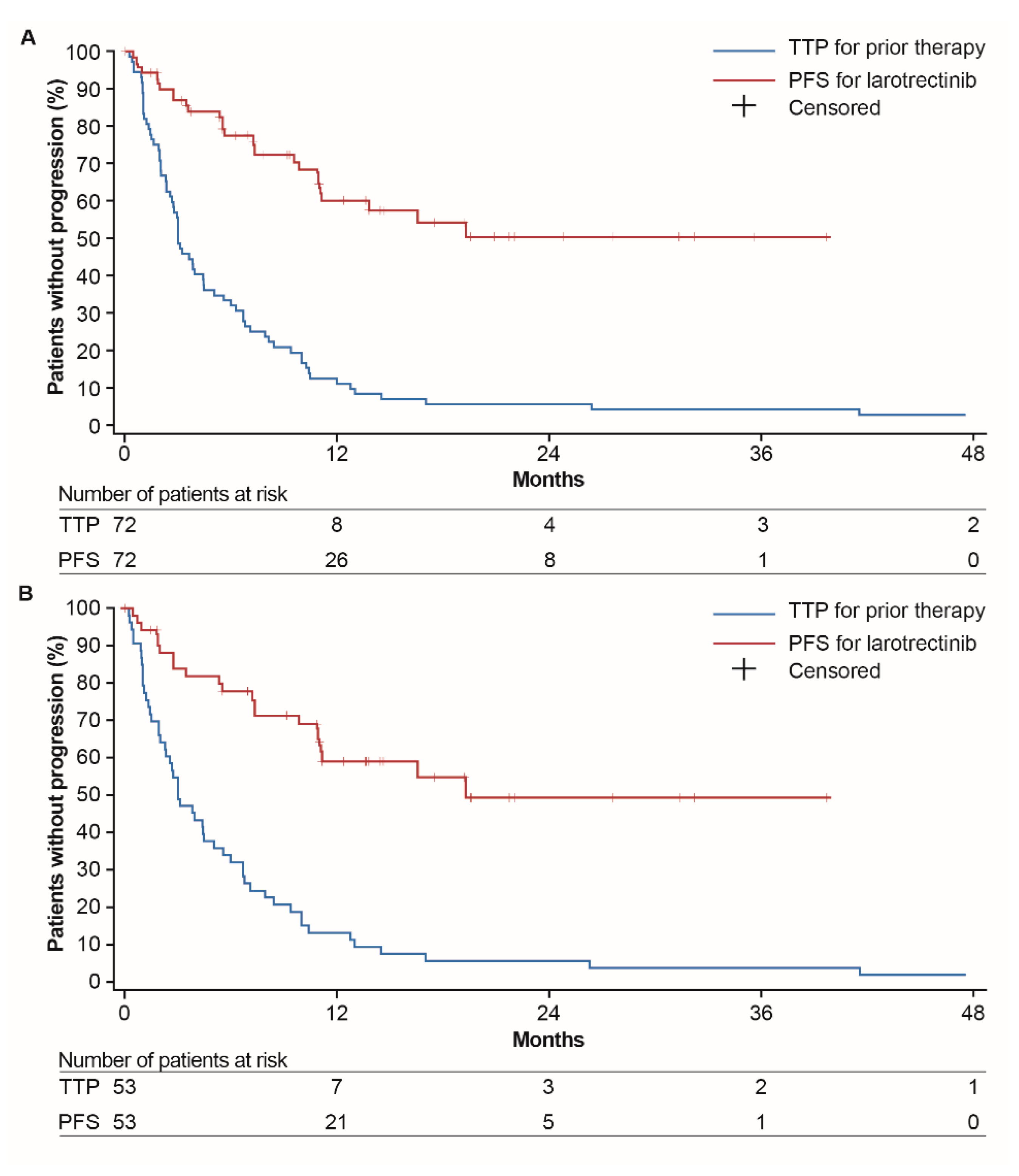
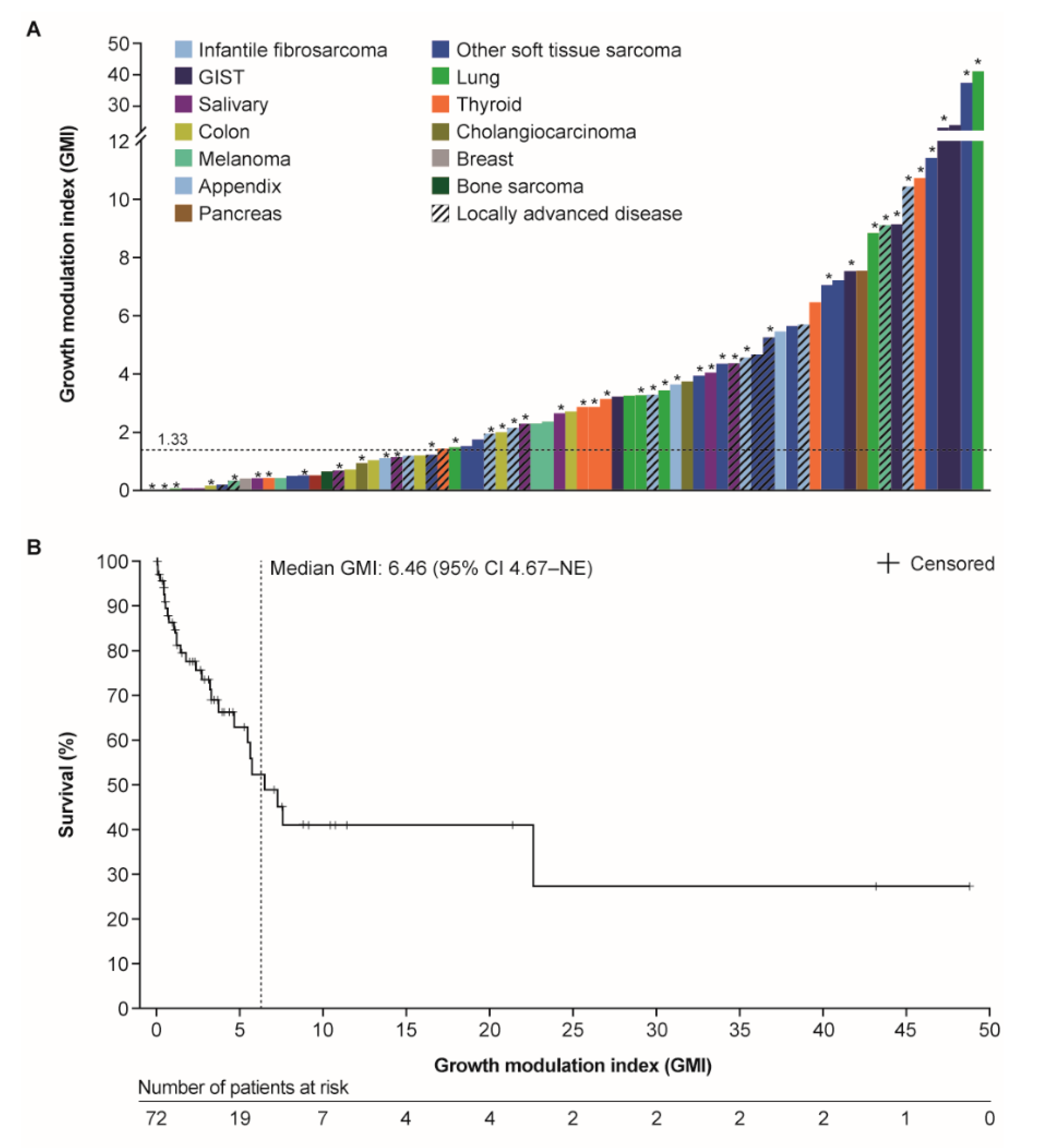
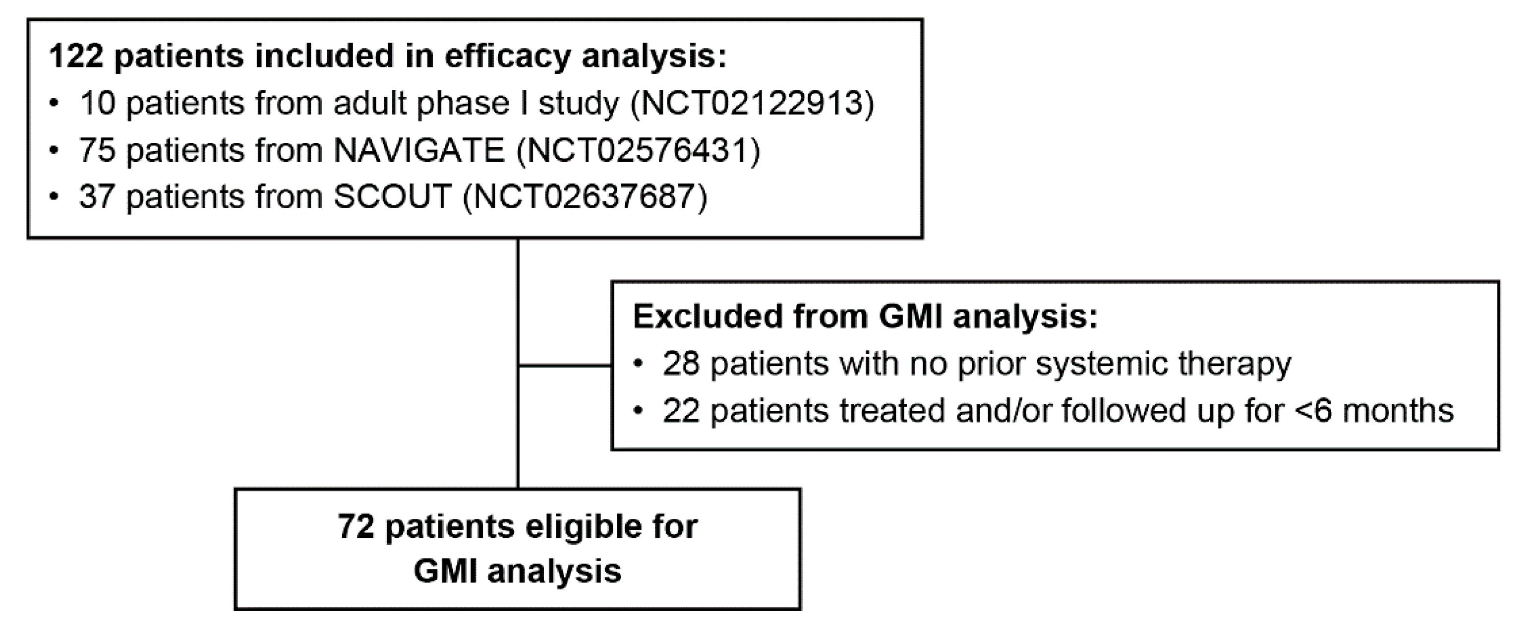
2. Results
3. Discussion
4. Materials and Methods
5. Conclusions
Author Contributions
Funding
Acknowledgments
Conflicts of Interest
References
- Vaishnavi, A.; Le, A.T.; Doebele, R.C. Trking Down an Old Oncogene in a New Era of Targeted Therapy. Cancer Discov. 2015, 5, 25–34. [Google Scholar] [CrossRef] [PubMed] [Green Version]
- Bayer, A.G. Vitrakvi Summary of Product Characteristics. Available online: https://www.ema.europa.eu/en/documents/product-information/vitrakvi-epar-product-information_en.pdf (accessed on 6 August 2020).
- Bayer HealthCare Pharmaceuticals Inc. Vitrakvi Prescribing Information. Available online: http://labeling.bayerhealthcare.com/html/products/pi/vitrakvi_PI.pdf (accessed on 6 August 2020).
- Hong, D.S.; DuBois, S.G.; Kummar, S.; Farago, A.F.; Albert, C.M.; Rohrberg, K.S.; van Tilburg, C.M.; Nagasubramanian, R.; Berlin, J.D.; Federman, N.; et al. Larotrectinib in Patients with Trk Fusion-Positive Solid Tumours: A Pooled Analysis of Three Phase 1/2 Clinical Trials. Lancet Oncol. 2020, 21, 531–540. [Google Scholar] [CrossRef]
- Federman, N.; McDermott, R. Larotrectinib, a Highly Selective Tropomyosin Receptor Kinase (Trk) Inhibitor for the Treatment of Trk Fusion Cancer. Expert Rev. Clin. Pharmacol. 2019, 12, 931–939. [Google Scholar] [CrossRef] [PubMed] [Green Version]
- Mick, R.; Crowley, J.J.; Carroll, R.J. Phase II Clinical Trial Design for Noncytotoxic Anticancer Agents for Which Time to Disease Progression Is the Primary Endpoint. Control. Clin. Trials 2000, 21, 343–359. [Google Scholar] [CrossRef]
- Kovalchik, S.; Mietlowski, W. Statistical Methods for a Phase II Oncology Trial with a Growth Modulation Index (GMI) Endpoint. Contemp. Clin. Trials 2011, 32, 99–107. [Google Scholar] [CrossRef] [PubMed]
- Von Hoff, D.D. There Are No Bad Anticancer Agents, Only Bad Clinical Trial Designs--Twenty-First Richard and Hinda Rosenthal Foundation Award Lecture. Clin. Cancer Res. 1998, 4, 1079–1086. [Google Scholar] [PubMed]
- European Medicines Agency. Guideline on the Evaluation of Anticancer Medicinal Products in Man. Available online: https://www.ema.europa.eu/en/documents/scientific-guideline/guideline-evaluation-anticancer-medicinal-products-man-revision-5_en.pdf (accessed on 9 September 2019).
- Bonetti, A.; Zaninelli, M.; Leone, R.; Franceschi, T.; Fraccon, A.P.; Pasini, F.; Sabbioni, R.; Cetto, G.L.; Sich, D.; Brienza, S.; et al. Use of the Ratio of Time to Progression Following First- and Second-Line Therapy to Document the Activity of the Combination of Oxaliplatin with 5-Fluorouracil in the Treatment of Colorectal Carcinoma. Ann. Oncol. 2001, 12, 187–191. [Google Scholar] [CrossRef] [PubMed]
- Comella, P.; Casaretti, R.; Crucitta, E.; De Vita, F.; Palmeri, S.; Avallone, A.; Orditura, M.; De Lucia, L.; Del Prete, S.; Catalano, G.; et al. Oxaliplatin Plus Raltitrexed and Leucovorin-Modulated 5-Fluorouracil i.v. Bolus: A Salvage Regimen for Colorectal Cancer Patients. Br. J. Cancer 2002, 86, 1871–1875. [Google Scholar] [CrossRef] [PubMed]
- Cousin, S.; Blay, J.Y.; Bertucci, F.; Isambert, N.; Italiano, A.; Bompas, E.; Ray-Coquard, I.; Perrot, D.; Chaix, M.; Bui-Nguyen, B.; et al. Correlation between Overall Survival and Growth Modulation Index in Pre-Treated Sarcoma Patients: A Study from the French Sarcoma Group. Ann. Oncol. 2013, 24, 2681–2685. [Google Scholar] [CrossRef] [PubMed]
- Penel, N.; Demetri, G.D.; Blay, J.Y.; Cousin, S.; Maki, R.G.; Chawla, S.P.; Judson, I.; von Mehren, M.; Schoffski, P.; Verweij, J.; et al. Growth Modulation Index as Metric of Clinical Benefit Assessment among Advanced Soft Tissue Sarcoma Patients Receiving Trabectedin as a Salvage Therapy. Ann. Oncol. 2013, 24, 537–542. [Google Scholar] [CrossRef] [PubMed]
- Bachet, J.B.; Mitry, E.; Lievre, A.; Lepere, C.; Vaillant, J.N.; Declety, G.; Parlier, H.; Emile, J.F.; Julie, C.; Rougier, P. Second- and Third-Line Chemotherapy in Patients with Metastatic Pancreatic Adenocarcinoma: Feasibility and Potential Benefits in a Retrospective Series of 117 Patients. Gastroenterol. Clin. Biol. 2009, 33, 1036–1044. [Google Scholar] [CrossRef] [PubMed]
- Demetri, G.D.; Chawla, S.P.; von Mehren, M.; Ritch, P.; Baker, L.H.; Blay, J.Y.; Hande, K.R.; Keohan, M.L.; Samuels, B.L.; Schuetze, S.; et al. Efficacy and Safety of Trabectedin in Patients with Advanced or Metastatic Liposarcoma or Leiomyosarcoma after Failure of Prior Anthracyclines and Ifosfamide: Results of a Randomized Phase II Study of Two Different Schedules. J. Clin. Oncol. 2009, 27, 4188–4196. [Google Scholar] [CrossRef] [PubMed]
- Austin, P.C. The Use of Propensity Score Methods with Survival or Time-to-Event Outcomes: Reporting Measures of Effect Similar to Those Used in Randomized Experiments. Stat. Med. 2014, 33, 1242–1258. [Google Scholar] [CrossRef] [PubMed] [Green Version]
- Massard, C.; Michiels, S.; Ferte, C.; Le Deley, M.C.; Lacroix, L.; Hollebecque, A.; Verlingue, L.; Ileana, E.; Rosellini, S.; Ammari, S.; et al. High-Throughput Genomics and Clinical Outcome in Hard-to-Treat Advanced Cancers: Results of the Moscato 01 Trial. Cancer Discov. 2017, 7, 586–595. [Google Scholar] [CrossRef] [PubMed] [Green Version]


| Characteristic | All Patients n = 72 | Patients with Metastatic Disease n = 53 |
|---|---|---|
| Sex, n (%) | ||
| Male | 36 (50) | 22 (42) |
| Female | 36 (50) | 31 (58) |
| Age, years, n (%) | ||
| <5 | 10 (14) | 1 (2) |
| 5 to <18 | 11 (15) | 10 (19) |
| ≥18 | 51 (71) | 42 (79) |
| ECOG performance status, n (%) | ||
| 0 | 35 (49) | 22 (42) |
| 1 | 28 (39) | 23 (43) |
| 2 | 9 (13) | 8 (15) |
| Disease setting, n (%) | ||
| Locally advanced | 19 (26) | 0 |
| Metastatic | 53 (74) | 53 (100) |
| Tumor type, n (%) | ||
| STS: Infantile fibrosarcoma | 10 (14) | 3 (6) |
| STS: Other | 16 (22) | 12 (23) |
| Salivary gland | 9 (13) | 4 (8) |
| Lung cancer | 7 (10) | 7 (13) |
| Melanoma | 7 (10) | 5 (9) |
| Thyroid cancer | 7 (10) | 6 (11) |
| Colon cancer | 6 (8) | 6 (11) |
| Gastrointestinal stromal tumor | 4 (6) | 4 (8) |
| Cholangiocarcinoma | 2 (3) | 2 (4) |
| Appendix cancer | 1 (1) | 1 (2) |
| Bone sarcoma | 1 (1) | 1 (2) |
| Breast cancer | 1 (1) | 1 (2) |
| Pancreatic cancer | 1 (1) | 1 (2) |
| Prior lines of treatment for advanced disease, n (%) | ||
| 0 | 0 | 0 |
| 1 | 26 (36) | 13 (25) |
| 2 | 20 (28) | 16 (30) |
| ≥3 | 26 (36) | 24 (45) |
| NTRKgene fusion, n (%) | ||
| NTRK1 | 35 (49) | 29 (55) |
| NTRK2 | 2 (3) | 2 (4) |
| NTRK3 | 35 (49) | 22 (42) |
| Subgroup | GMI, n (% of Each Subgroup) | ||
|---|---|---|---|
| <1 | 1–1.33 | ≥1.33 | |
| Sex | |||
| Male (n = 22) | 4 (18) | 2 (9) | 16 (73) |
| Female (n = 31) | 11 (35) | 1 (3) | 19 (61) |
| Age group | |||
| Adult (≥18 years) (n = 42) | 14 (33) | 2 (5) | 26 (62) |
| Pediatric (<18 years) (n = 11) | 1 (9) | 1 (9) | 9 (82) |
| ECOGperformance status | |||
| 0 (n = 22) | 5 (23) | 2 (9) | 15 (68) |
| 1 (n = 23) | 9 (39) | 1 (4) | 13 (57) |
| 2 (n = 8) | 1 (13) | 0 | 7 (88) |
| NTRKgene | |||
| NTRK1 (n = 29) | 8 (28) | 3 (10) | 18 (62) |
| NTRK2 (n = 2) | 1 (50) | 0 | 1 (50) |
| NTRK3 (n = 22) | 6 (27) | 0 | 16 (73) |
| Lines of prior therapy | |||
| 1 (n = 13) | 2 (15) | 0 | 11 (85) |
| 2 (n = 16) | 7 (44) | 2 (13) | 7 (44) |
| ≥3 (n = 24) | 6 (25) | 1 (4) | 17 (71) |
Publisher’s Note: MDPI stays neutral with regard to jurisdictional claims in published maps and institutional affiliations. |
© 2020 by the authors. Licensee MDPI, Basel, Switzerland. This article is an open access article distributed under the terms and conditions of the Creative Commons Attribution (CC BY) license (http://creativecommons.org/licenses/by/4.0/).
Share and Cite
Italiano, A.; Nanda, S.; Briggs, A.; Garcia-Foncillas, J.; Lassen, U.; Vassal, G.; Kummar, S.; van Tilburg, C.M.; Hong, D.S.; Laetsch, T.W.; et al. Larotrectinib versus Prior Therapies in Tropomyosin Receptor Kinase Fusion Cancer: An Intra-Patient Comparative Analysis. Cancers 2020, 12, 3246. https://doi.org/10.3390/cancers12113246
Italiano A, Nanda S, Briggs A, Garcia-Foncillas J, Lassen U, Vassal G, Kummar S, van Tilburg CM, Hong DS, Laetsch TW, et al. Larotrectinib versus Prior Therapies in Tropomyosin Receptor Kinase Fusion Cancer: An Intra-Patient Comparative Analysis. Cancers. 2020; 12(11):3246. https://doi.org/10.3390/cancers12113246
Chicago/Turabian StyleItaliano, Antoine, Shivani Nanda, Andrew Briggs, Jesus Garcia-Foncillas, Ulrik Lassen, Gilles Vassal, Shivaani Kummar, Cornelis M. van Tilburg, David S. Hong, Theodore W. Laetsch, and et al. 2020. "Larotrectinib versus Prior Therapies in Tropomyosin Receptor Kinase Fusion Cancer: An Intra-Patient Comparative Analysis" Cancers 12, no. 11: 3246. https://doi.org/10.3390/cancers12113246