Next Article in Journal
Conversion Surgery in Metastatic Gastric Cancer and Cancer Dormancy as a Prognostic Biomarker
Previous Article in Journal
Retropharyngeal Lymph Node Involvement in Oropharyngeal Carcinoma: Impact upon Risk of Distant Metastases and Survival Outcomes
 
 
cancers-logo
Article Menu

Article Menu

Article

Real-World Data on Cabozantinib in Previously Treated Patients with Metastatic Renal Cell Carcinoma: Focus on Sequences and Prognostic Factors

by
Matteo Santoni
1,
Daniel Y. Heng
2,
Sergio Bracarda
3,
Giuseppe Procopio
4,
Michele Milella
5,
Camillo Porta
6,
Marc R. Matrana
7,
Giacomo Cartenì
8,
Simon J. Crabb
9,
Ugo De Giorgi
10,
Umberto Basso
11,
Cristina Masini
12,
Fabio Calabrò
13,
Maria Giuseppa Vitale
14,
Daniele Santini
15,
Francesco Massari
16,
Luca Galli
17,
Giuseppe Fornarini
18,
Riccardo Ricotta
19,
Sebastiano Buti
20,
Paolo Zucali
21,
Orazio Caffo
22,
Franco Morelli
23,
Francesco Carrozza
24,
Angelo Martignetti
25,
Alain Gelibter
26,
Roberto Iacovelli
27,
Alessandra Mosca
28,
Francesco Atzori
29,
Nuno Vau
30,
Lorena Incorvaia
31,
Cinzia Ortega
32,
Marina Scarpelli
33,
Antonio Lopez-Beltran
34,
Liang Cheng
35,
Vittorio Paolucci
1,
Jeffrey Graham
2,
Erin Pierce
7,
Sarah Scagliarini
8,
Pierangela Sepe
4,
Elena Verzoni
4,
Sara Merler
5,
Mimma Rizzo
6,
Giulia Sorgentoni
1,
Alessandro Conti
36,
Francesco Piva
37,
Alessia Cimadamore
33,
Rodolfo Montironi
33,* and
Nicola Battelli
1,*
add Show full author list remove Hide full author list
1
Oncology Unit, Macerata Hospital, via Santa Lucia 2, 62100 Macerata, Italy
2
Division of Medical Oncology, Department of Oncology, Tom Baker Cancer Centre, University of Calgary, Calgary, AB T2N 4N2, Canada
3
Medical Oncology, Department of Oncology, AziendaOspedaliera S. Maria, 05100 Terni, Italy
4
Department of Medical Oncology, Istituto Nazionale deiTumori IRCCS, 20133 Milan, Italy
5
U.O.C. Oncology, AziendaOspedalieraUniversitariaIntegrata, University and Hospital Trust of Verona, 37126 Verona, Italy
6
Department of Internal Medicine and Therapeutics, University of Pavia and Division of Translational Oncology, IRCCS Istituti Clinici Scientifici Maugeri, 27100 Pavia, Italy
7
Department of Internal Medicine, Hematology/Oncology, Ochsner Medical Center, New Orleans, LA 70121, USA
8
Department of Medical Oncology, AO “A. Cardarelli”, 80131 Naples, Italy
9
Cancer Sciences Unit, University of Southampton, Southampton SO171BJ, UK
10
Department of Medical Oncology, IstitutoScientifico Romagnolo per lo Studio e la CuradeiTumori (IRST) IRCCS, 47014 Meldola, Italy
11
Department of Medical Oncology, IstitutoOncologico Veneto (IOV) IRCCS, 35128 Padova, Italy
12
Medical Oncology Unit, Arcispedale Santa Maria Nuova, IRCCS Reggio Emilia, 42123 Reggio Emilia, Italy
13
San Camillo-Forlanini Hospital, 00128 Rome, Italy
14
Department of Oncology and Haematology and Respiratory Disease, University Hospital, 41125 Modena, Italy
15
Department of Medical Oncology, Campus Bio-Medico University of Rome, 00128 Rome, Italy
16
Division of Oncology, S. Orsola-Malpighi Hospital, 40138 Bologna, Italy
17
Medical Oncology Unit, Department of Translational Research and New Technologies in Medicine, University of Pisa, 56126 Pisa, Italy
18
Department of Medical Oncology, Ospedale “S. Martino”, 16132 Genova, Italy
19
Niguarda Cancer Center, Grande OspedaleMetropolitano Niguarda, 20162 Milan, Italy
20
Medical Oncology Unit, University Hospital of Parma, 43126 Parma, Italy
21
Humanitas Clinical and Research Center, Humanitas Cancer Center, Rozzano, 20089 Milano, Italy
22
Medical Oncology Department, Santa Chiara Hospital, Largo Medaglied’Oro, 38122 Trento, Italy
23
Medical Oncology Department, Casa SollievodellaSofferenza, VialeCappuccini 1, 71013 San Giovanni Rotondo, Italy
24
Oncology Unit, City Hospital, 48018 Faenza, Italy
25
Dipartimentooncologicouslsud-esttoscana-area senese, LocalitàCampostaggias.n.c., 53036 Poggibonsi, Italy
26
Medical Oncology (B), Policlinico Umberto I, “Sapienza” University of Rome, 00128 Rome, Italy
27
Medical Oncology, Fondazione Policlinico Universitario A. Gemelli IRCCS, 00168 Roma, Italy
28
Medical Oncology Unit, Maggiore dellaCarità University Hospital, University of Eastern Piedmont, 28100 Novara, Italy
29
Medical Oncology Unit, AziendaOspedalieroUniversitaria of Cagliari, 09124 Cagliari, Italy
30
Urologic Oncology, Champalimaud Clinical Center, 1400-038 Lisbon, Portugal
31
Department of Surgical, Oncological and Oral Sciences, Section of Medical Oncology, University of Palermo, 90127 Palermo, Italy
32
Department of Medical Oncology, Ospedale S. Lazzaro ASL CN2 Alba-Bra, 12051 Cuneo, Italy
33
United Hospitals, School of Medicine, Section of Pathological Anatomy, Polytechnic University of the Marche Region, Via Conca 71, I-60126 Ancona, Italy
34
Department of Pathology and Surgery, Faculty of Medicine, 14004 Cordoba, Spain
35
Department of Pathology and Laboratory Medicine, Indiana University School of Medicine, Indianapolis, IN 46202, USA
36
Department of Urology, Bressanone/Brixen hospital, via Dante 51, 39042 Bressanone BZ, Italy
37
Department of Specialistic Clinical and Odontostomatological Sciences, Polytechnic University of Marche, 60126 Ancona, Italy
*
Authors to whom correspondence should be addressed.
Cancers 2020, 12(1), 84; https://doi.org/10.3390/cancers12010084
Submission received: 1 October 2019 / Revised: 7 November 2019 / Accepted: 23 November 2019 / Published: 30 December 2019

Abstract

:
Cabozantinib is approved for the treatment of renal cell carcinoma (RCC). However, prognostic factors are still lacking in this context. The aim of this study was to evaluate prognostic factors in RCC patients treated with second- or third-line cabozantinib. A multicenter retrospective real-world study was conducted, involving 32 worldwide centers. A total of 237 patients with histologically confirmed clear-cell and non-clear-cell RCC who received cabozantinib as second- or third-line therapy for metastatic disease were included. We analyzed overall survival (OS), progression-free survival (PFS) and time-to-strategy failure (TTSF) using Kaplan–Meier curves. Cox proportional models were used at univariate and multivariate analyses.The median PFS and OS of cabozantinib were 7.76 months (95% CI 6.51–10.88) and 11.57 months (95% CI 10.90–not reached (NR)) as second-line and 11.38 months (95% CI 5.79–NR) and NR (95% CI 11.51–NR) as third-line therapy. The median TTSF and OS were 11.57 and 15.52 months with the sequence of cabozantinib–nivolumab and 25.64 months and NR with nivolumab–cabozantinib, respectively. The difference between these two sequences was statistically significant only in good-risk patients. In the second-line setting, hemoglobin (Hb) levels (HR= 2.39; 95% CI 1.24–4.60, p = 0.009) and IMDC (International Metastatic Renal Cell Carcinoma Database Consortium) group (HR = 1.72, 95% CI 1.04–2.87, p = 0.037) were associated with PFS while ECOG-PS (HR = 2.33; 95%CI, 1.16–4.69, p = 0.018) and Hb levels (HR = 3.12; 95%CI 1.18–8.26, p = 0.023) correlated with OS at multivariate analysis, while in the third-line setting, only Hb levels (HR = 2.72; 95%CI 1.04–7.09, p = 0.042) were associated with OS. Results are limited by the retrospective nature of the study.This real-world study provides evidence on the presence of prognostic factors in RCC patients receiving cabozantinib.

1. Introduction

Renal cell carcinoma (RCC), the most common kidney cancer in adults, represents 5% of all cancers in men and 3% in women, with an estimated 65,340 new cases and 14,970 deaths in 2018 in the United States alone [1]. Agents able to target altered pathways promoting neoangiogenesis (e.g., sunitinib, pazopanib, sorafenib, axitinib and tivozanib [2,3,4,5,6,7,8,9]) have demonstrated activity in metastatic RCC (mRCC), as well as immunecheckpoint inhibitors used alone as nivolumab [10] or combined with other immunotherapy (nivolumab plus ipilimumab [11]) or targeted therapies (axitinib plus pembrolizumab or avelumab) [12,13].
Cabozantinib is an orally administered tyrosine kinase inhibitor acting mainly on VEGFR2, MET (mesenchymal epithelial transition receptor) and AXL (anexelekto pathway) [14]. In the randomized phase III METEOR trial comparing cabozantinib to everolimus in pretreated patients, cabozantinib improved overall survival (OS), progression-free survival (PFS) and objective response rate (ORR) [15]. Serious adverse events with cabozantinib occurred in 39% of patients, with the most common toxicities being hypertension, diarrhea and fatigue [16].
Most recently, a randomized phase II clinical trial (CABOSUN) randomized 157 patients with mRCC and intermediate or poorrisk of disease according to IMDC (International Metastatic Renal Cell Carcinoma Database Consortium) criteria (based on the presence of anemia, neutrophilia, thrombocytosis, Karnofskyperformance status <80, hypercalcemia and <1 year from diagnosis to metastatic disease) to receive cabozantinib or sunitinib as first-line therapy [17]. Compared to sunitinib, cabozantinib improved PFS and ORR in this subgroup of patients. Despite the fact that these findings were also confirmed on a subsequent analysis based on independent review [18], the results of this study are still controversial [19].
To date, cabozantinib is indicated for the treatment of patients with advanced RCC in treatment-naïve adults with intermediate poor-risk features (Food and Drug Administration, FDA), and by EMA (European Medical Agency) for adults progressed to prior vascular endothelial growth factor/receptor inhibitors. Here, we report results of a real-world analysis on cabozantinib in previously treated patients with mRCC andaimed to evaluate the presence of prognostic factors and the different therapeutic sequences in this setting.

2. Results

2.1. Overall Population

A total of 237 patients were included in this analysis; 174 (73.42%) were males and 63 (26.58%) females. The median age was 62.56y (range 24.55–85.76). The majority of patients had clear-cell RCC (182 patients, 76.79%), while in 55 patients (23.21%) non-clear-cell RCC (17 papillary type I, 14 papillary type II, 14 clear-cell RCC with sarcomatoid differentiation, 1 with rhabdoid differentiation, 1 chromophobe, 1 with XP11.3 translocation and 7 unclassified RCC tumors) was diagnosed. There were 120 patients (50.63%) who were metastatic at time of diagnosis. At first diagnosis, the Fuhrman or WHO/ISUP grade was G3 in 86 (36.29%) and G4 in 32 (13.50%). The number of metastatic sites was ≥2 in 160 cases (67.51%). The most frequent sites of metastasis were lung (154 patients, 64.98%), lymph nodes (133 patients, 56.12%) and bone (80 patients, 34.04%). According to IMDC criteria, 57 patients (24.05%) were at favorable-risk, 146 (61.60%) at intermediate-risk and 34 (14.35%) had poor-risk features. Patients’ characteristics are reported in Table 1. The distribution of IMDC criteria across the study population is showed in Table 2.
The median follow-up time from diagnosis was 182.79 months (95% CI 131.00 to not reached;NR) and median OS from the start of first-line therapy was 103.23 months (95% CI 63.40–NR). During the follow-up, 73 patients (30.80%) died. A further 112 patients (47.26%) were treated with cabozantinib as second-line therapy, while 125 (52.74%) received cabozantinib in the third-line setting. In 41 patients, second-line cabozantinib was ongoing at the time of data collection. Among the 71 patients who progressed on second-line cabozantinib, 53 (74.65%) received a third-line therapy, which was nivolumab in 29 patients (54.72%). Drug distribution and sequence is reported in Table 1.

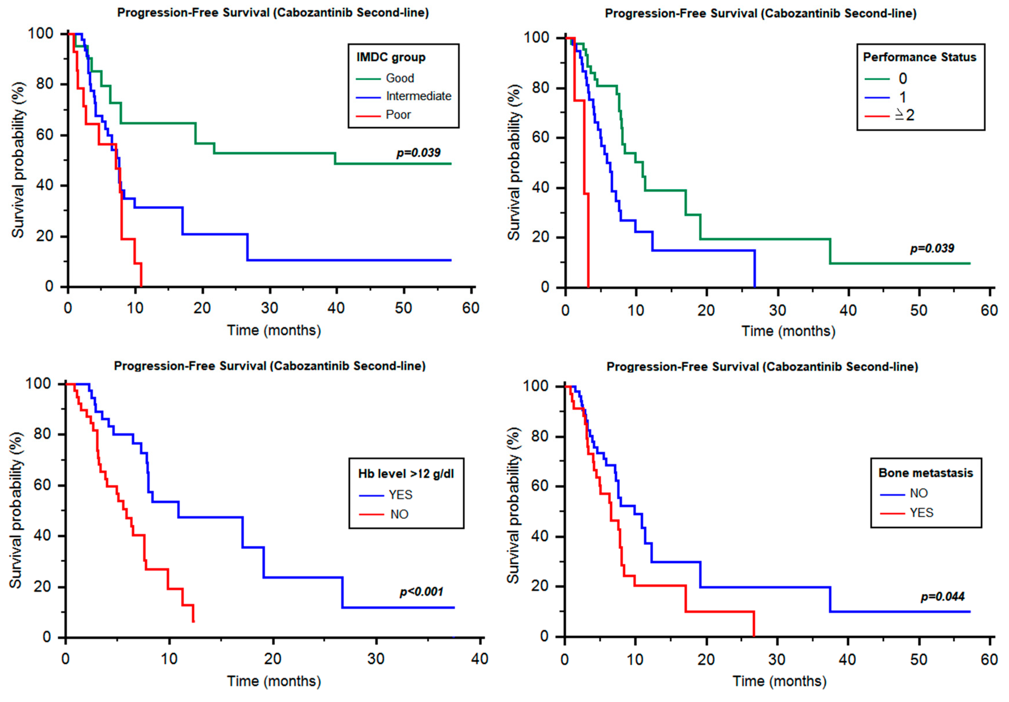
2.2. Progression-Free Survival of Cabozantinib as Second-Line Therapy

The median PFS of cabozantinib as second-line therapy was 7.76 months (95% CI 6.51–10.88, Table 3).
This figure significantly changed when patients were analyzed according to their IMDC status, yielding 11.28 months (95% CI 7.89–NR, Table 3, Figure 1) in good-risk, 7.59 months (95% CI 5.52–NR, Table 3, Figure 1) in intermediate-risk and 7.13 months (95% CI 2.66–NR, Table 3, Figure 1) in poor-risk patients (p = 0.039). Similarly, PFS was different according to ECOG-performance status (PS; 0 vs. 1 vs. ≥2; 10.88 months vs. 5.88 months vs. 2.66 months, p < 0.001, Figure 1) and hemoglobin (Hb) ≥12 g/dL vs. <12 g/dL (10.88 vs. 5.88 months, HR = 0.39, 95% CI 0.18–0.62, p < 0.001, Figure 1). Otherwise, no significant difference was found based on time from diagnosis to systemic therapy (≥1y vs. <1y, 11.28 vs. 7.13 months, HR = 0.62, 95% CI 0. 73–1.14, p = 0.130), neutrophilia (7.76 vs. 4.01 months, HR = 0.48, 95% CI 0.13–1.01, p = 0.051), thrombocytosis (7.89 vs. 6.51 months, HR = 0.50, 95% CI 0.15–1.02, p = 0.055) and hypercalcemia (7.82 vs. 3.06 months, HR = 0.50, 95% CI 0.12–1.22, p = 0.106).
Interestingly, no significant differences were also found between clear-cell and non-clear-cell histology (7.89 vs. 5.06 months, HR = 0.73, 95% CI 0.35–1.40, p = 0.310), age < 70y and ≥70y (7.89 vs. 7.13 months, HR = 0.74, 95% CI 0.37–1.41, p = 0.334), gender (p = 0.678), Fuhrman or WHO/ISUP grade (p = 0.756) or number of metastatic sites (1 site vs. ≥2 sites, 7.59 vs. 7.82 months, HR = 0.99, 95% CI 0.56–1.76, p = 0.987).
By stratifying patients based on the site of metastasis, a significant difference was found between patients with or without bone metastases (6.51 vs. 9.86 months, HR = 0.58, 95% CI 0.31–0.98, p = 0.044, Figure 1), whilst no differences were found between patients with lung (6.05 vs. 6.31 months, HR = 0.88, 95% CI 0.64–1.21, p = 0.446), liver (7.59 vs. 12.3 months, HR = 1.48, 95% CI 0.73–2.81, p = 0.297), lymph node (7.59 vs. 7.89 months, HR = 1.23, 95% CI 0.71–2.16, p = 0.447), or brain metastases (7.76 vs. 7.59 months, HR = 1.24, 95% CI 0.52–2.89, p = 0.638).
Furthermore, we analyzed the eventual prognostic role of the received first-line therapy, with any significant difference between sunitinib and pazopanib (7.89 vs. 7.82 months, HR = 1.25, 95% CI 0.70–2.38, p = 0.418).
Univariate analysis showed that ECOG-PS (HR = 2.47; 95% CI, 1.40–4.36, p = 0.002), Hb levels (HR = 2.90; 95% CI, 1.55–5.42, p < 0.001), IMDC group (HR = 1.77; 95% CI, 1.12–2.80, p = 0.015) and bone metastases (HR = 1.75; 95% CI, 1.10–3.02, p = 0.047) were significantly associated with the PFS of cabozantinib, given as second-line therapy. At multivariate analysis, only Hb levels (HR = 2.39; 95% CI, 1.24–4.60, p = 0.009) and IMDC group (HR = 1.72, 95% CI, 1.04–2.87, p = 0.037) maintained their prognostic significance in this setting.

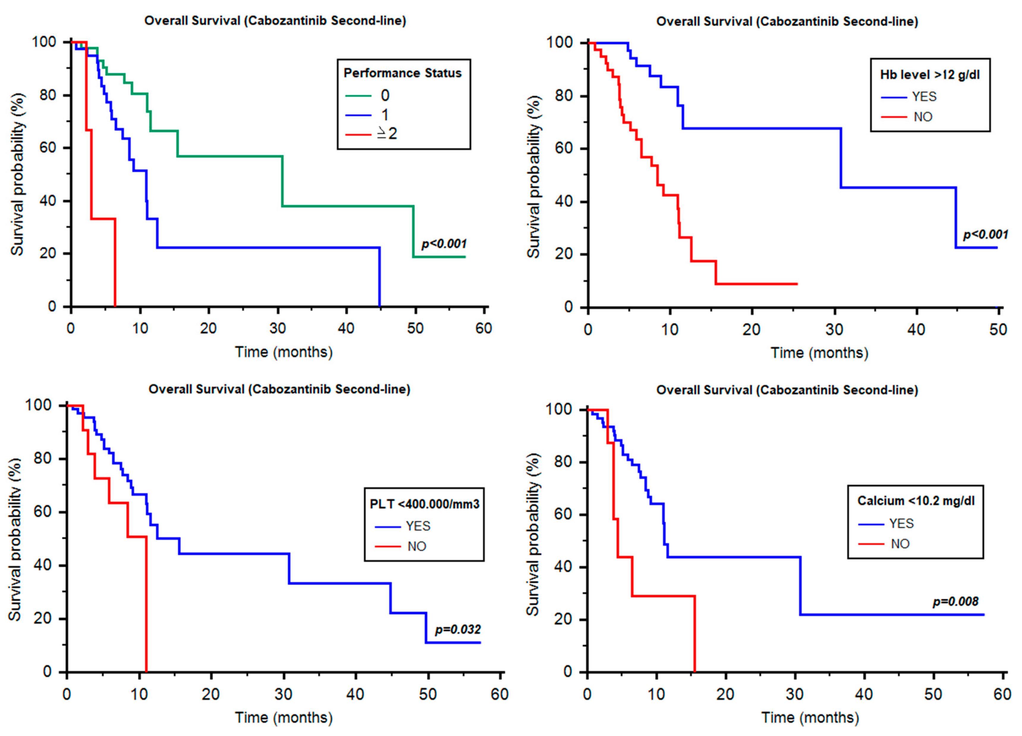
2.3. Overall Survival of Cabozantinib as Second-Line Therapy

The median OS of cabozantinib as second-line therapy was 11.57 months (95% CI 10.90–NR, Table 3). Differently from PFS, IMDC classification was not associated with OS in the three prognostic groups (12.53 vs. 10.95 vs. 11.05 months, p = 0.349, Table 3). Conversely, the median OS was significantly different according to ECOG-PS (0 vs. 1 vs. ≥2; 30.71 months vs. 10.95 months vs. 2.96 months, p < 0.001, Figure 2), Hb ≥12 g/dL vs. <12 g/dL (30.71 vs. 8.42 months, HR = 0.24, 95% CI 0.10–0.44, p < 0.001, Figure 2), thrombocytosis (15.52 vs. 10.95 months, HR = 0.42, 95% CI 0.09–0.90, p = 0.032, Figure 2) and hypercalcemia (11.08 vs. 4.37 months, HR = 0.32, 95% CI 0.04–0.60, p = 0.008, Figure 2). Of note, no significant differences were found for neutrophilia (12.53 vs. 11.57 months, HR = 0.57, 95% CI 0.17–1.48, p = 0.211), time from diagnosis to systemic therapy (≥1y vs. <1y, 11.57 vs. 11.05 months, HR = 1.02, 95% CI 0.51–2.07, p = 0.949), clear-cell and non-clear-cell histology (11.57 months vs. notreached (NR), HR = 0.83, 95% CI 0.33–1.98, p = 0.648), age < 70y and ≥70y (11.57 vs. 11.08 months, HR = 0.93, 95% CI 0.42–2.04, p = 0.856), Fuhrman grade (p = 0.899), choice of first-line therapy (sunitinib vs.pazopanib: 15.52 vs. 11.08 months, HR = 1.44, 95% CI 0.71–3.26, p = 0.281), site of metastasis and number of metastatic sites (1 site vs. ≥2 sites, 15.52 vs. 11.05 months, HR = 0.82, 95% CI 0.41–1. 46, p = 0.573).
At univariate analysis, ECOG-PS (HR = 3.51; 95% CI, 1.86–6.63, p < 0.001), Hb levels (HR = 5.07; 95% CI, 2.18–11.76, p < 0.001), thrombocytosis (HR = 2.52; 95% CI, 1.05–6.01, p = 0.039) and hypercalcemia (11.08 vs. 4.37 months, HR = 3.24, 95% CI 0.31–8.03, p = 0.015) were significant predictors of OS, while at multivariate analysis, only ECOG-PS (HR = 2.33; 95% CI, 1.16–4.69, p = 0.018) and Hb levels (HR = 3.12; 95% CI, 1.18–8.26, p = 0.023) correlated with OS.

2.4. Progression-Free Survival of Cabozantinib as Third-Line Therapy

The median PFS of cabozantinib in the third-line setting was 11.38 months (95% CI 5.79–NR, Table 3). The median PFS was not statistically different among the three IMDC groups (11.38 vs. 7.63 vs. 5.75 months, p = 0.772, Table 3) or according to ECOG-PS (11.38 vs. 5.26 months, HR = 0.54, 95% CI 0.16–1.24, p = 0.120), time from diagnosis to systemic therapy (≥1y vs. <1y, NR vs. NR, HR = 0.57, 95% CI 0.27–1.35, p = 0.217), neutrophilia (7.63 months vs. NR, HR = 1.20, 95% CI 0.55–2.62, p = 0.657) and hypercalcemia (16.34 vs. 6.71 months, HR = 1.23, 95% CI 0.50–3.00, p = 0.652). Otherwise, PFS was statistically different according to Hb levels (17.95 vs. 6.44 months, HR = 0.47, 95% CI 0.24–0.88, p = 0.019, Figure 3) and thrombocytosis (16.34 vs. 3.35 months, HR = 0.39, 95% CI 0.12–0.68, p = 0.005, Figure 3).
Of note, no significant differences were found by stratifying patients by clear-cell vs. non-clear-cell histology (6.71 vs. 11.38 months, HR = 1.26, 95% CI 0.61–2.59, p = 0.539), age < 70y and ≥70y (6.44 vs. 11.38 months, HR = 1.57, 95% CI 0.82–2.80, p = 0.183), Fuhrman grade (p = 0.474) or number or site of metastases.
Hb levels (HR = 2.19; 95% CI, 1.12–4.26, p = 0.022) and thrombocytosis (HR = 2.60; 95% CI, 1.30–5.19, p = 0.007), were significantly correlated with PFS at univariate but not at multivariate analysis.

2.5. Overall Survival of Cabozantinib as Third-Line Therapy

In the 125 patients treated with cabozantinib in third-line setting, the median OS was NR (95% CI 11.51–NR, Table 3). Hb ≥12 g/dL vs. <12 g/dL (NR vs. 7.73 months, HR = 0.33, 95% CI 0.14–0.76, p = 0.009, Figure 3), thrombocytosis (NR vs. 7.40 months, HR = 0.39, 95% CI 0.10–0.86, p = 0.025, Figure 3). Interestingly, no significant differences were found according to IMDC group (p = 0.739, Table 3), ECOG-PS (NR vs. 7.73 months, HR = 0.45, 95% CI 0.10–1.11, p = 0.073), neutrophilia (NR vs. NR months, HR = 0.73, 95% CI 0.26–1.97, p = 0.509), time from diagnosis to systemic therapy (NR vs. NR months, HR = 0.57, 95% CI 0.27–1.35, p = 0.217), hypercalcemia (NR vs. 7.40 months, HR = 0.73, 95% CI 0.21–2.34, p = 0.560), clear-cell vs. non-clear-cell histology (NR vs. NR, HR = 1.06, 95% CI 0.41–2.74, p = 0.906), age < 70y and ≥70y (12.10 months vs. NR, HR = 2.61, 95% CI 0.96–4.77, p = 0.063), Fuhrman or WHO/ISUP grade (p = 0.574) and neither specific sites nor number of metastatic sites (1 site vs. ≥2 sites, NR vs. NR, HR = 0.72, 95% CI 0.34–1.57, p = 0.422).
Fromunivariate analysis, Hb levels (HR = 3.11; 95% CI, 1.27–7.72, p = 0.014) and thrombocytosis (HR = 2.59; 95% CI, 1.10–6.13, p = 0.030) were found to be associated with OS, while for multivariate analysis, only Hb levels (HR = 2.72; 95% CI, 1.04–7.09, p = 0.042) were correlated with OS.

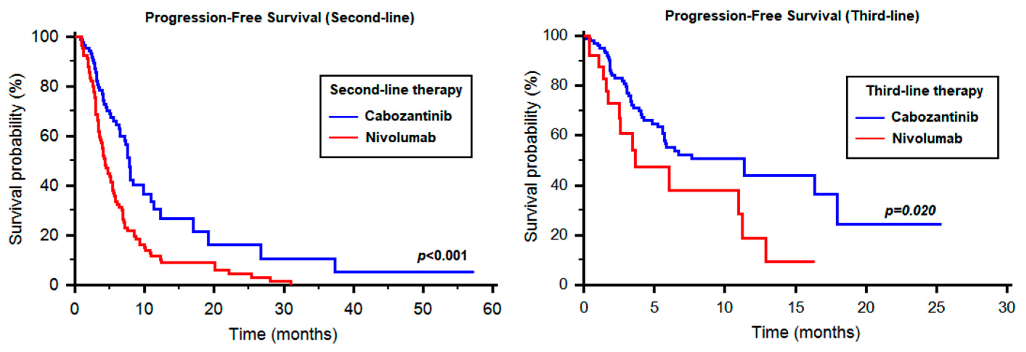
2.6. Time to Strategy Failure and Sequencing: Cabozantinib vs. Nivolumab

In the 89 patients treated with second-line nivolumab, we observed a median PFS of 4.31 months (95% CI 3.65–5.46), which was significantly different from cabozantinib in the same setting (HR 0.50, 95% CI 0.35–0.69, p < 0.001, Figure 4). Similarly, the 29 patients treated with third-line nivolumab had a median PFS of 3.68 months vs. 11.38 months registered by cabozantinib (HR 0.50, 95% CI 0.19–0.87, p = 0.020, Figure 4). The median TTSF from the start of second-line therapy was 18.61 months (95% CI 16.30–21.80).
By stratifying patients according to the therapeutic sequence received, we reported 29 patients treated with cabozantinib followed by nivolumab and 89 patients who received nivolumab followed by cabozantinib. The median TTSF was 11.57 months with cabozantinib–nivolumab (95% CI 9.17–NR, Figure 5) and 25.64 months with nivolumab–cabozantinib (95% CI 23.24–NR, Figure 5). The difference was statistically significant only in the good-risk group (11.57 vs. 25.64 months, HR 8.99, 95% CI 3.91–671.63, p = 0.003).
The median OS from the start of second-line therapy in the cabozantinib–nivolumab and nivolumab–cabozantinib groups were 15.52 months (95% CI 11.10–NR) and NR (95% CI NR–NR), respectively (HR 1.95, 95% CI 0.87–5.83, p = 0.091, Figure 5). Similarly to TTSF, the median OS was significantly longer with nivolumab–cabozantinib only in the good-risk subgroup (13.55 months vs. NR, p = 0.004).

3. Discussion

Cabozantinib represents an effective strategy in untreated and pretreated RCC patients. The lack of validated molecular or clinical predictive and prognostic factors aimed to optimize the efficacy and safety of this agent represents a major challenge for uro-oncologists. In our analysis, only Hb levels were significantly correlated with OS in the second- and third-line settings. The negative prognostic significance of anemia suggests that a prompt management of this condition could have a potential impact on the outcome of RCC patients. Moreover, these data support the necessity of investigating the prognostic significance of anemia in patients treated with nivolumab to understand if this condition may represent a key factor in the decision-making process between these two agents.In particular, reversible causes of anemia need to be addressed, but the data do not indicate attempting erythropoietin(EPO)-induced correction, particularly given the association of EPO with adverse outcomes in cancersin large studies [20].
Interestingly, no difference in terms of efficacy has been found between clear-cell and non-clear-cell histologies. This may be partially explained by the prevalence in the non-clear-cell group of papillary tumors (31/55), in which cabozantinib has demonstrated to be effective [21].
There is a clear statistical difference between the two sequences in favorable-risk RCCpatients (p = 0.003); and for all patients analyzed, there is a trend towards statistical significance with a clear separation of curves for TTSF and OS beyond 12 months. Even though cabozantinibhas better PFSin the second-line setting than nivolumab (Figure 4), there appears to be utility in trying the sequencenivolumab–cabozantinib, rather than cabozantinib–nivolumab, particularly in favorable-risk patients, which needs to be further investigated with a larger sample size. These data also call for studies investigating the biological rationale for differences in outcomes between the sequences. However, the small number of patients in each prognostic group and the retrospective nature of our study do not allow to definitively clarify this issue.

4. Patients and Methods

4.1. Study Population

The study population included adults (>18 years) with clear-cell or non-clear-cell mRCC, treated with cabozantinib as second- or third-line therapy. Patients were treated in 32 worldwide institutions between November 2004 and January 2019. Data were retrospectively collected from patients’ electronic medical records and paper charts. Patients were excluded from this study if they had missing data regarding thesite of metastasis and tumor response to therapy. The research was carried out in accordance with the approval by the ethical committee of the participating institutions. The study has been accepted by the “Comitato Etico Regionale delle Marche”, the accepting number is 2019-403.

4.2. Treatment Regimens and Statistical Analysis

Cabozantinib was administered orally, usually at a starting dose of 60 mg once daily. Treatment was administered until clinical or radiological disease progression, serious adverse events or death. Follow-up commonly consisted of periodic physical examination, laboratory analysis and imaging assessment by computed tomography (CT) or magnetic resonance imaging (MRI) not earlier than 4 weeks, and not later than 6 weeks, according to local regulations. Disease progression was defined by the Response Evaluation Criteria in Solid Tumors, RECIST v.1.1 [22]. Progression-free survival (PFS) was defined as the time from the start of therapy to progression or death from any cause, whichever occurred first. Patients with no tumor progression or death at time of data collection were censored at the last date of evaluation. Time to strategy failure (TTSF) was defined as the interval from the start of first-line therapy to progression on full therapy or death. PFS and OS were estimated using Kaplan–Meier method with Rothman’s 95% confidence intervals (CI) and compared across the groups using the log-rank test. Neutrophilia was defined as ≥7500 neutrophils/mm3; thrombocytosis was defined as ≥400,000 platelets/mm3, while corrected hypercalcemia as ≥10.2 mg/dL.
In order to investigate patients’ characteristics predictors of survival, Cox proportional-hazards models were used at univariate and multivariate analyses. All the significance levels were set at a 0.05 value and all p values were two-sided. The statistical analysis was performed by MedCalc version 11.4.4.0 (MedCalc Software, Broekstraat 52,9030 Mariakerke, Belgium).

5. Conclusions

Our results support the prognostic role of Hb levels in patients treated with cabozantinib and the importance of sequencing immunotherapy and targeted therapy. Further perspective studies should be provided in order to validate these prognostic factors and compare the sequential approaches available in this disease.

Author Contributions

Conceptualization, M.S. (Matteo Santoni) and R.M.; Data curation, V.P., J.G., E.P., S.S., P.S., E.V., S.M., M.R. and G.S.; Formal analysis, A.C. (Alessandro Conti) and F.P.; Funding acquisition, R.M. and N.B.; Investigation, M.S., D.Y.H., S.B. (Sergio Bracarda), G.P., M.M., C.P., M.R.M., G.C., S.J.C., U.D.G., U.B., C.M., F.C. (Fabio Calabrò), M.G.V., D.S., F.M. (Francesco Massari), L.G., G.F., R.R., S.B. (Sebastiano Buti), P.Z., O.C., F.M. (Franco Morelli), F.C. (Francesco Carrozza), A.M. (Angelo Martignetti), A.G., R.I., A.M. (Alessandra Mosca), F.A., N.V., L.I. and C.O.; Methodology, M.S. (Matteo Santoni); Project administration, N.B.; Software, F.P.; Supervision, A.L.-B., L.C., M.S. (Marina Scarpelli) and R.M.; Validation, D.Y.H., S.B. (Sergio Bracarda), G.P., M.M., C.P., M.R.M., C.G., S.J.C., U.D.G., U.B., C.M., F.C. (Fabio Calabrò), M.G.V., D.S., F.M. (Francesco Massari), L.G., G.F., R.R., S.B. (Sebastiano Buti), P.Z., O.C., F.M. (Franco Morelli), F.C. (Francesco Carrozza), A.M. (Angelo Martignetti), A.G., R.I., A.M. (Alessandra Mosca), F.A., N.V., L.I. and C.O.; Writing—original draft, M.S. (Matteo Santoni); Writing—review & editing, M.S. (Matteo Santoni) and A.C. (Alessia Cimadamore). All authors have read and agreed to the published version of the manuscript.

Funding

This research received no external funding.

Conflicts of Interest

The authors declare no conflict of interest.

References

  1. Siegel, R.L.; Miller, K.D.; Jemal, A. Cancer statistics, 2018. CA Cancer J. Clin. 2018, 68, 7–30. [Google Scholar] [CrossRef] [PubMed]
  2. Motzer, R.J.; Hutson, T.E.; Cella, D.; Reeves, J.; Hawkins, R.; Guo, J.; Nathan, P.; Staehler, M.; de Souza, P.; Merchan, J.R.; et al. Pazopanib versus sunitinib in metastatic renal-cell carcinoma. N. Engl. J. Med. 2013, 369, 722–731. [Google Scholar] [CrossRef] [PubMed] [Green Version]
  3. Motzer, R.J.; Escudier, B.; Tomczak, P.; Hutson, T.E.; Michaelson, M.D.; Negrier, S.; Oudard, S.; Gore, M.E.; Tarazi, J.; Hariharan, S.; et al. Axitinib versus sorafenib as second-line treatment for advanced renal cell carcinoma: Overall survival analysis and updated results from a randomized phase 3 trial. Lancet Oncol. 2013, 14, 552–562. [Google Scholar] [CrossRef]
  4. Escudier, B.; Eisen, T.; Stadler, W.M.; Szczylik, C.; Oudard, S.; Siebels, M.; Negrier, S.; Chevreau, C.; Solska, E.; Desai, A.A.; et al. Sorafenib in advanced clear-cell renal-cell carcinoma. N. Engl. J. Med. 2007, 356, 125–134. [Google Scholar] [CrossRef]
  5. Escudier, B.; Pluzanska, A.; Koralewski, P.; Ravaud, A.; Bracarda, S.; Szczylik, C.; Chevreau, C.; Filipek, M.; Melichar, B.; Bajetta, E.; et al. Bevacizumab plus interferon alfa-2a for treatment of metastatic renal cell carcinoma: Arandomised, double-blind phase III trial. Lancet 2007, 370, 2103–2111. [Google Scholar] [CrossRef]
  6. Motzer, R.J.; Hutson, T.E.; Tomczak, P.; Michaelson, M.D.; Bukowski, R.M.; Rixe, O.; Oudard, S.; Negrier, S.; Szczylik, C.; Kim, S.T.; et al. Sunitinib versus interferon alfa in metastatic renal-cell carcinoma. N. Engl. J. Med. 2007, 356, 115–124. [Google Scholar] [CrossRef]
  7. Gore, M.E.; Szczylik, C.; Porta, C.; Bracarda, S.; Bjarnason, G.A.; Oudard, S.; Lee, S.H.; Haanen, J.; Castellano, D.; Vrdoljak, E.; et al. Final results from the large sunitinib global expanded-access trial in metastatic renal cell carcinoma. Br. J. Cancer 2015, 113, 12–19. [Google Scholar] [CrossRef] [Green Version]
  8. Motzer, R.J.; Nosov, D.; Eisen, T.; Bondarenko, I.; Lesovoy, V.; Lipatov, O.; Tomczak, P.; Lyulko, O.; Alyasova, A.; Harza, M.; et al. Tivozanib versus sorafenib as initial targeted therapy for patients with metastatic renal cell carcinoma: Results from a phase III trial. J. Clin. Oncol. 2013, 31, 3791–3799. [Google Scholar] [CrossRef]
  9. Santoni, M.; Massari, F.; Piva, F.; Carrozza, F.; Di Nunno, V.; Cimadamore, A.; Martignetti, A.; Montironi, R.; Battelli, N. Tivozanib for the treatment of renal cell carcinoma. Expert Opin. Pharmacother. 2018, 19, 1021–1025. [Google Scholar] [CrossRef] [PubMed]
  10. Motzer, R.J.; Escudier, B.; McDermott, D.F.; George, S.; Hammers, H.J.; Srinivas, S.; Tykodi, S.S.; Sosman, J.A.; Procopio, G.; Plimack, E.R.; et al. Nivolumab versus Everolimus in Advanced Renal-Cell Carcinoma. N. Engl. J. Med. 2015, 373, 1803–1813. [Google Scholar] [CrossRef]
  11. Motzer, R.J.; Tannir, N.M.; McDermott, D.F.; Arén Frontera, O.; Melichar, B.; Choueiri, T.K.; Plimack, E.R.; Barthélémy, P.; Porta, C.; George, S.; et al. Nivolumab plus Ipilimumab versus Sunitinib in Advanced Renal-Cell Carcinoma. N. Engl. J. Med. 2018, 378, 1277–1290. [Google Scholar] [CrossRef] [PubMed]
  12. Motzer, R.J.; Penkov, K.; Haanen, J.; Rini, B.; Albiges, L.; Campbell, M.T.; Venugopal, B.; Kollmannsberger, C.; Negrier, S.; Uemura, M.; et al. Avelumab plus Axitinib versus Sunitinib for Advanced Renal-Cell Carcinoma. N. Engl. J. Med. 2019, 380, 1103–1115. [Google Scholar] [CrossRef] [PubMed]
  13. Rini, B.I.; Plimack, E.R.; Stus, V.; Gafanov, R.; Hawkins, R.; Nosov, D.; Pouliot, F.; Alekseev, B.; Soulières, D.; Melichar, B.; et al. Pembrolizumab plus Axitinib versus Sunitinib for Advanced Renal-Cell Carcinoma. N. Engl. J. Med. 2019, 380, 1116–1127. [Google Scholar] [CrossRef] [PubMed]
  14. Di Nunno, V.; Cubelli, M.; Massari, F. The role of the MET/AXL pathway as a new target for multikinase inhibitors in renal cell carcinoma. Expert Rev. Precis. Med. Drug Dev. 2017, 2, 169–175. [Google Scholar] [CrossRef]
  15. Choueiri, T.K.; Escudier, B.; Powles, T.; Mainwaring, P.N.; Rini, B.I.; Donskov, F.; Hammers, H.; Hutson, T.E.; Lee, J.L.; Peltola, K.; et al. Cabozantinib versus Everolimus in Advanced Renal-Cell Carcinoma. N. Engl. J. Med. 2015, 373, 1814–1823. [Google Scholar] [CrossRef]
  16. Choueiri, T.K.; Escudier, B.; Powles, T.; Tannir, N.M.; Mainwaring, P.N.; Rini, B.I.; Hammers, H.J.; Donskov, F.; Roth, B.J.; Peltola, K.; et al. Cabozantinib versus everolimus in advanced renal cell carcinoma (METEOR): Final results from a randomised, open-label, phase 3 trial. Lancet Oncol. 2016, 17, 917–927. [Google Scholar] [CrossRef] [Green Version]
  17. Choueiri, T.K.; Halabi, S.; Sanford, B.L.; Hahn, O.; Michaelson, M.D.; Walsh, M.K.; Feldman, D.R.; Olencki, T.; Picus, J.; Small, E.J.; et al. Cabozantinib Versus Sunitinib as initial targeted therapy for patients with metastatic renal cell Carcinoma of poor or intermediate risk: The Alliance A031203 CABOSUN Trial. J. Clin. Oncol. 2017, 35, 591–597. [Google Scholar] [CrossRef]
  18. Choueiri, T.K.; Hessel, C.; Halabi, S.; Sanford, B.; Michaelson, M.D.; Hahn, O.; Walsh, M.; Olencki, T.; Picus, J.; Small, E.J.; et al. Cabozantinib versus sunitinib as initial therapy for metastatic renal cell carcinoma of intermediate or poor risk (Alliance A031203 CABOSUN randomised trial): Progression-free survival by independent review and overall survival update. Eur. J. Cancer 2018, 94, 115–125. [Google Scholar] [CrossRef] [Green Version]
  19. Buti, S.; Bersanelli, M. Is Cabozantinib Really Better Than Sunitinib As First-Line Treatment of Metastatic Renal Cell Carcinoma? J. Clin. Oncol. 2017, 35, 1858–1859. [Google Scholar] [CrossRef]
  20. Debeljak, N.; Solár, P.; Sytkowski, A.J. Erythropoietin and Cancer: The Unintended Consequences of Anemia Correction. Front. Immunol. 2014, 5, 563. [Google Scholar] [CrossRef] [Green Version]
  21. Zhao, H.; Nolley, R.; Chan, A.M.W.; Rankin, E.B.; Peehl, D.M. Cabozantinib inhibits tumor growth and metastasis of a patient-derived xenograft model of papillary renal cell carcinoma with MET mutation. Cancer Biol. Ther. 2017, 18, 863–871. [Google Scholar] [CrossRef] [PubMed] [Green Version]
  22. Eisenhauer, E.A.; Therasse, P.; Bogaerts, J.; Schwartz, L.H.; Sargent, D.; Ford, R.; Dancey, J.; Arbuck, S.; Gwyther, S.; Mooney, M.; et al. New response evaluation criteria in solid tumours: Revised RECIST guideline (version 1.1). Eur. J. Cancer 2009, 45, 228–247. [Google Scholar] [CrossRef] [PubMed]
Figure 1. Progression-free survival of second-line cabozantinib according to different prognostic factors. Hb = hemoglobin; IMDC = International Metastatic Renal Cell Carcinoma Database Consortium.
Cancers 12 00084 g001
Figure 2. Overall survival of second-line cabozantinib according to different prognostic factors. Hb = hemoglobin.
Cancers 12 00084 g002
Figure 3. Progression-free survival and overall survival of third-line cabozantinib according to different prognostic factors. Hb = hemoglobin; PLT = platelets.
Cancers 12 00084 g003
Figure 4. Comparison between cabozantinib and nivolumab in the second- and third-line setting.
Cancers 12 00084 g004
Figure 5. Time to strategy failure and overall survival in patients treated with the sequences cabozantinib–nivolumab and nivolumab–cabozantinib.
Cancers 12 00084 g005
Table 1. Patients’ characteristics. Immunotherapy combinations included axitinib plus pembrolizumab, axitinib plus avelumab and nivolumab plus ipilimumab.IMDC—International Metastatic Renal Cell Carcinoma Database Consortium.
Clinicopathological FeaturesN. of Patients (%)
Age
Median62.56y
Range24.55–85.76y
Gender
Male174 (73.42)
Female63 (26.58)
T-Stage at Diagnosis
T137 (15.61)
T235 (14.77)
T397 (40.93)
T426 (10.97)
Unknown42 (17.72)
Histology
Clear-cell RCC182 (76.79)
Non-clear-cell RCC55 (23.21)
Fuhrman or WHO/ISUP Grade
Grade 14 (1.69)
Grade 262 (26.16)
Grade 386 (36.39)
Grade 432 (13.50)
Unknown59 (22.36)
N. of Metastatic Sites at Recurrence
1 site77 (32.49)
≥2 sites160 (67.51)
Site of Metastasis
Lung154 (64.98)
Lymph nodes133 (56.12)
Bone80 (34.04)
Liver53 (22.36)
Brain20 (8.44)
IMDC Risk Group
Good57 (24.05)
Intermediate146 (61.60)
Poor34 (14.35)
First-Line Therapy
Sunitinib141 (59.49)
Pazopanib81 (34.18)
Immunotherapy combinations9 (3.80)
Other6 (2.53)
Second-Line Therapy237 (100)
Cabozantinib112 (47.26)
Nivolumab89 (37.55)
Axitinib19 (8.01)
Everolimus14 (5.91)
Other3 (1.27)
Third-Line Therapy178 (100)
Cabozantinib125 (70.22)
Nivolumab29 (16.29)
Other24 (13.49)
Table 2. Distribution of risk factors according to IMDC criteria in the study populations. LLN = lower limit of normal; ULN = upper limit of normal.
IMDC CriteriaN of Patients (%)
<1 y from Diagnosis to Systemic Therapy
Yes120 (50.63)
No117 (49.37)
Performance Status < 80% (Karnofsky)
Yes19 (8.02)
No214 (91.98)
Hb Level < LLN
Yes88 (37.13)
No149 (62.87)
Calcium Level > ULN
Yes21 (8.86)
No216 (91.14)
Neutrophil > ULN
Yes29 (12.24)
No208 (87.76)
Platelets > ULN
Yes31 (13.08)
No206 (86.92)
Table 3. Progression-free survival and overall survival obtained by cabozantinib in our study.
GroupsSecond-Line CabozantinibThird-Line Cabozantinib
All PatientsPFS
[Median (95% CI)]
OS
[Median (95% CI)]
PFS
[Median (95% CI)]
OS
[Median (95% CI)]
7.76 (6.51–10.88)11.57 (10.90–NR)11.38 (5.79–NR)NR (11.5–NR)
Second-line Cabozantinib
(29 patients, 25.9%)
Third-line Cabozantinib
(28 patients, 22.4%)
Favourable GroupPFS
[Median (95% CI)]
OS
[Median (95% CI)]
PFS
[Median (95% CI)]
OS
[Median (95% CI)]
11.28 (7.89–NR)12.53 (11.57–NR)11.38 (4.24–NR)NR (7.40–NR)
Second-line Cabozantinib
(64 patients, 57.1%)
Third-line Cabozantinib
(78 patients, 68.4%)
Intermediate
Group
PFS
[Median (95% CI)]
OS
[Median (95% CI)]
PFS
[Median (95% CI)]
OS
[Median (95% CI)]
7.59 (5.52–NR)10.95 (9.11–NR)7.63 (5.56–NR)NR (11.51–NR)
Second-line Cabozantinib
(19 patients, 17.0%)
Third-line Cabozantinib
(19 patients, 9.2%)
Poor-Risk GroupPFS
[Median (95% CI)]
OS
[Median (95% CI)]
PFS
[Median (95% CI)]
OS
[Median (95% CI)]
7.13 (2.66–NR)11.05 (7.46–NR)5.75 (3.19–NR)NR (4.01–NR)

