Article

Win and Loss Responses in the Monetary Incentive Delay Task Mediate the Link between Depression and Problem Drinking

1
Department of Psychiatry, Yale University School of Medicine, New Haven, CT 06519, USA
2
Department of Neuroscience, Yale University School of Medicine, New Haven, CT 06520, USA
3
Interdepartmental Neuroscience Program, Yale University School of Medicine, New Haven, CT 06520, USA
4
Wu Tsai Institute, Yale University, New Haven, CT 06520, USA
*
Author to whom correspondence should be addressed.
Brain Sci. 2022, 12(12), 1689; https://doi.org/10.3390/brainsci12121689
Submission received: 22 November 2022 / Revised: 2 December 2022 / Accepted: 7 December 2022 / Published: 9 December 2022
(This article belongs to the Section Psychiatric Diseases)

Abstract

:
Depression and alcohol misuse, frequently comorbid, are associated with altered reward processing. However, no study has examined whether and how the neural markers of reward processing are shared between depression and alcohol misuse. We studied 43 otherwise-healthy drinking adults in a monetary incentive delay task (MIDT) during fMRI. All participants were evaluated with the Alcohol Use Disorders Identification Test (AUDIT) and Beck’s Depression Inventory (BDI-II) to assess the severity of drinking and depression. We performed whole brain regressions against each AUDIT and BDI-II score to investigate the neural correlates and evaluated the findings at a corrected threshold. We performed mediation analyses to examine the inter-relationships between win/loss responses, alcohol misuse, and depression. AUDIT and BDI-II scores were positively correlated across subjects. Alcohol misuse and depression shared win-related activations in frontoparietal regions and parahippocampal gyri (PHG), and right superior temporal gyri (STG), as well as loss-related activations in the right PHG and STG, and midline cerebellum. These regional activities (β’s) completely mediated the correlations between BDI-II and AUDIT scores. The findings suggest shared neural correlates interlinking depression and problem drinking both during win and loss processing and provide evidence for co-morbid etiological processes of depressive and alcohol use disorders.

1. Introduction

Depression and alcohol misuse are two leading, comorbid, causes of disability [1,2,3,4,5]. Individuals with depression are more likely to drink to cope with negative mood, elevating the risks in developing an alcohol-use disorder (AUD) [3,6]. Drinkers, especially those with AUDs, often experience depression [7], which in turn leads to more drinking [8,9]. Indeed, co-occurring depression and alcohol misuse are known to dispose individuals to greater severity and more frequent relapse of both conditions [10]. In particular, investigators have suggested a causal pathway whereby alcohol dependence increases the risk of major depression rather than vice versa [1]. Specifically, with alcohol use and depression symptom severity quantified at baseline and 1-year follow-up, the structural equation model with AUD leading to major depression showed the best fit [11]. However, it remains unclear whether this is also true of drinkers with mild to moderate alcohol use severity.
Reward delivers pleasure and drives motivated behaviors. Investigators have employed the monetary incentive delay task (MIDT) or card-guessing task to identify the neural responses to win and loss [12,13]. Individuals with depression relative to controls demonstrated altered reward-related activations [14,15,16]. For instance, blunted striatal activity in response to monetary reward and loss has been reported in patients with depression [17,18,19] and anhedonia [20]. Ventral striatal hypoactivity during anticipation of win and loss was associated with the severity of depression [21]. However, the findings seemed less consistent for the orbitofrontal cortex (OFC), anterior cingulate cortex, and middle frontal gyrus, with greater activation reported for depressed vs. non-depressed individuals in some studies [22,23] but the opposite in others [24,25,26,27,28]. A meta-analysis suggested that individuals with depression may be characterized by less ventral striatum (VS) and higher OFC responses to reward and higher amygdala response to punishment [14].
Altered neural activities during reward and punishment processing have also been reported for AUD, although the findings likewise varied. Patients with AUD vs. controls showed reduced VS activation during anticipation of both win and loss in the MIDT [29,30,31] and lower activations in the VS, lateral OFC, medial prefrontal cortex (mPFC), and dorsolateral PFC during win vs. loss in a card-guessing task [32]. In addition, patients with AUD relative to controls showed higher activations in the VS, anterior insula, and mPFC during win outcome and in the insula and lateral frontal cortex during loss outcome in the MIDT [32,33,34], higher VS response to loss outcome in a reward-guessing task [35], and lower superior/middle frontal cortical responses to loss outcome in a risk-taking task [36]. Other studies showed higher activations in the VS, anterior cingulate cortex, and paracentral and postcentral gyri during anticipation of reward in patients with AUD as compared to controls [37] and no differences in activation to reward outcome between patients with AUD and controls [32,37], or between those with and without a family history of alcoholism [38].
In summary, many studies have reported altered regional brain activities during reward/punishment processing in both depression and AUD. However, the findings varied substantially across studies, even for the VS, a hub of the reward circuit. Differences in MIDT paradigms [39] and clinical heterogeneity may account for the discrepancy in findings. Further, it is far from clear, based on these findings, whether alcohol misuse and depression shared neural correlates during reward and punishment processing. Thus, it would seem important to evaluate both depression and drinking severity in the same cohort of individuals.
In the current study, we aimed to investigate the neural correlates of reward and punishment processing shared between problem drinking and depression in a sample of largely nondependent alcohol drinkers. We performed whole-brain regressions to examine regional responses to win and loss outcomes in an MIDT each in association with the severity of problem drinking and with depression. Further, we employed mediation analyses to evaluate the inter-relationships between the shared neural correlates, drinking, and depression severity. We hypothesized that the shared neural correlates would mediate the relationships between problem drinking and depression.

2. Methods

2.1. Subjects

Forty-three adult alcohol drinkers (sixteen women; 23–74 or 45.4 ± 12.9 years of age) participated in this study. All subjects were otherwise healthy with no current medical conditions or use of prescription medications. None reported a history of head injury or neurological illness. Other exclusion criteria included current or past Axis I Disorders, including dependence on a psychoactive substance (except alcohol), according to DSM-IV [40]. The Human Investigation Committee at the Yale University School of Medicine approved the study (protocol code 0906005272). All subjects gave written informed consent prior to participation.

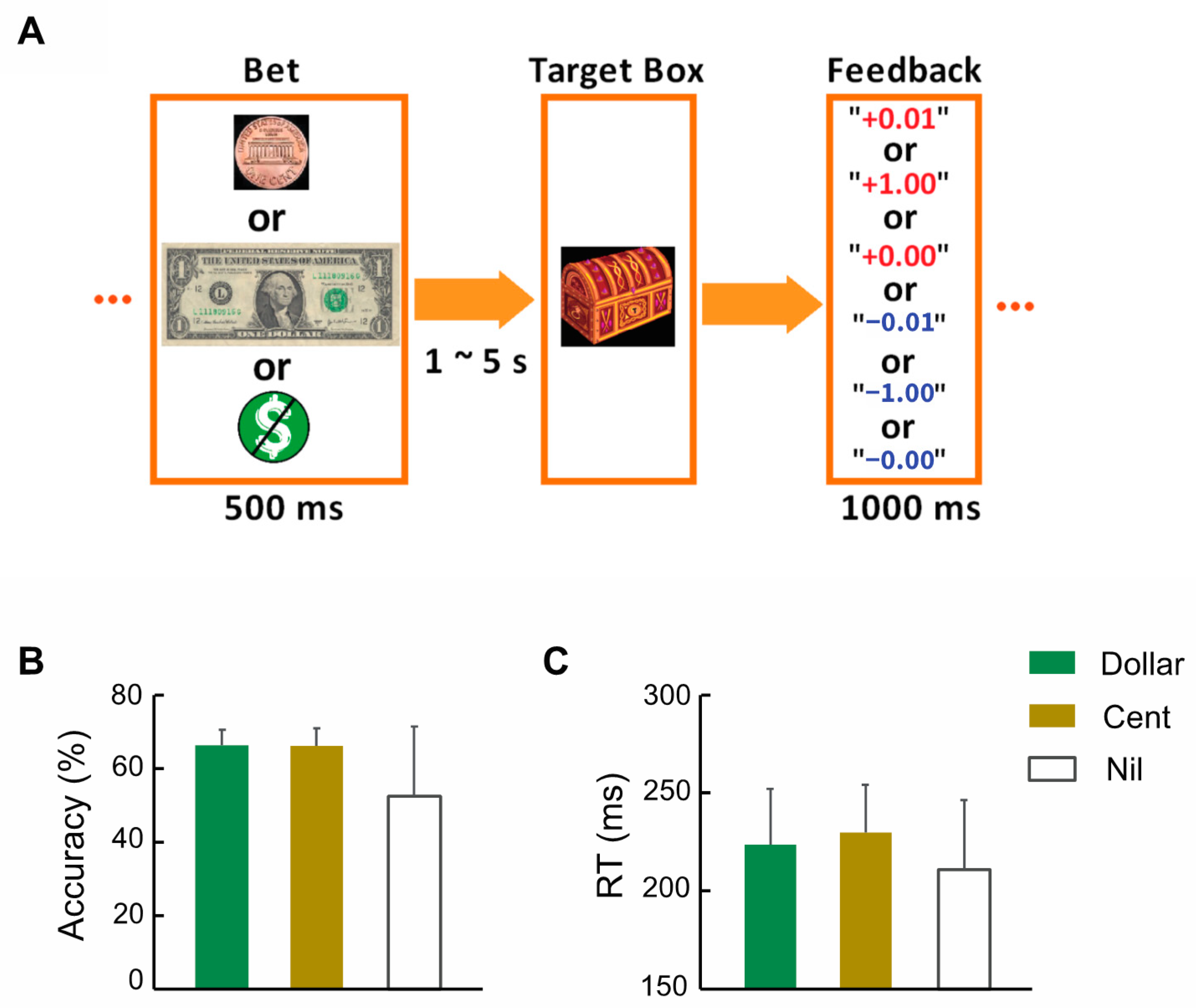
2.2. Assessments, Monetary Incentive Delay Task (MIDT), and Imaging Protocol

All participants were evaluated with the Alcohol Use Disorders Identification Test (AUDIT), Beck’s Depression Inventory (BDI-II), and the Fagerström Test for Nicotine Dependence (FTND). A 10-item instrument to screen for harmful drinking, the AUDIT, assesses the frequency of alcohol consumption, dependence, and associated harm. Each item is scored from 0 to 4 and the total score ranges from 0 to 40, with a higher score indicating greater severity of problematic alcohol use [41,42]. Five of the forty-three participants with the highest AUDIT scores (all > 14) also met criteria for alcohol abuse. The BDI-II is a 21-item assessment of the presence and severity of depression symptoms within the prior 2 weeks, with each item scored 0 to 3. A total score of 0 to 13, 14 to 19, 20 to 28, and 29 to 63 indicates minimal, mild, moderate, and severe depression, respectively [43,44,45]. Three of the forty-three participants scored > 19 and had moderate severity of depression. The FTND assesses the severity of cigarette consumption, compulsion to smoke, and physical dependence on nicotine, with a range of 0–10. A higher FTND score indicates greater severity of nicotine dependence [46]. Each subject completed two 10-min runs of the MIDT (Figure 1A), as described in our previous studies [47,48]. Across subjects, they completed an average of 184 ± 4 (mean ± SD) trials.

2.3. Imaging Data Preprocessing and Group Analyses

Briefly, brain images were collected using multiband imaging (multiband factor = 3) with a three-Tesla MR scanner (Siemens Trio, Erlangen, Germany). Data were analyzed with Statistical Parametric Mapping (SPM8, Wellcome Department of Imaging Neuroscience, University College London, UK), including realignment, slice timing, co-registration, segmentation, normalization, and smoothing, as in our earlier studies [39,48]. We examined event-related BOLD signals in a single model focusing on the feedback or outcome phase of win or loss processing, as described in our previous study [47]. We performed one-sample t tests of win vs. nil and loss vs. nil. To investigate the neural correlates of AUDIT and BDI-II, we conducted whole-brain linear regressions of these contrasts on AUDIT and BDI-II, separately, with age, sex, and FTND scores as covariates. All models were evaluated with a threshold combining voxel p < 0.001, uncorrected, and cluster p < 0.05 family-wise error (FWE), corrected, following current reporting standards. Voxels with peak activity were indicated with Montreal Neurological Institute (MNI) coordinates. We performed inclusive masking to identify the neural correlates shared between AUDIT and BDI-II for win vs. nil and loss vs. nil, respectively.

2.4. Mediation Analyses

We examined how activations of the regions of interest, AUDIT and BDI-II scores were inter-related with mediation analyses [49], as described in our prior work [50]. The mediation test was performed by employing three regression equations [49].
Y = i 1 + c X + e 1
Y = i 2 + c X + b M + e 2
M = i 3 + a X + e 3
where a, b, c′, and c represent path coefficients, and variable M is a mediator of the correlation X → Y. The significant paths a and b, as well as (cc′), indicate that X → Y is mediated by M. Moreover, if the path c′ is not significant, then X → Y is completely mediated by M.

3. Results

3.1. Clinical Characteristics and Behavioral Performance

Table 1 summarizes the demographic and clinical characteristics for men and women separately. The mean FTND score was <1, suggesting a largely non- or light-smoking sample. No sex differences were noted for age, years of education, AUDIT, BDI-II, or FTND score; thus, we combined men and women in data analyses. Figure 1B,C show the accuracy rate and reaction time (RT) of dollar, cent, and nil trials. The accuracy rates were close to 67%, suggesting the success of the staircase procedure. Across subjects, the AUDIT score was significantly and positively correlated with the BDI-II score without (r = 0.555, p < 0.001) or with (r = 0.560, p < 0.001; Figure 2) age, sex, and FTND as covariates. The AUDIT or BDI-II score did not show a significant correlation with either accuracy rate or with RT of dollar, cent, and nil trials (p’s ≥ 0.062 without covariates; p’s ≥ 0.109 with age, sex, and FTND as covariates), or the differences of dollar/cent vs. nil in either accuracy rate or RT (p’s ≥ 0.338 without covariates; p’s ≥ 0.308 with age, sex and FTND as covariates).

3.2. Brain Activations of Win vs. Nil and Loss vs. Nil

In one-sample t-tests, we evaluated regional activations to win vs. nil and loss vs. nil across all subjects. The results are shown in Supplementary Figure S1 and summarized in Supplementary Table S1. Compared to nil, win trials showed higher activations in the bilateral caudate, anterior cingulate cortex, bilateral lingual gyri, and cerebellum; loss trials showed higher activations in the bilateral lingual gyri, anterior cingulate cortex, bilateral insula, bilateral precentral gyri, and cerebellum.

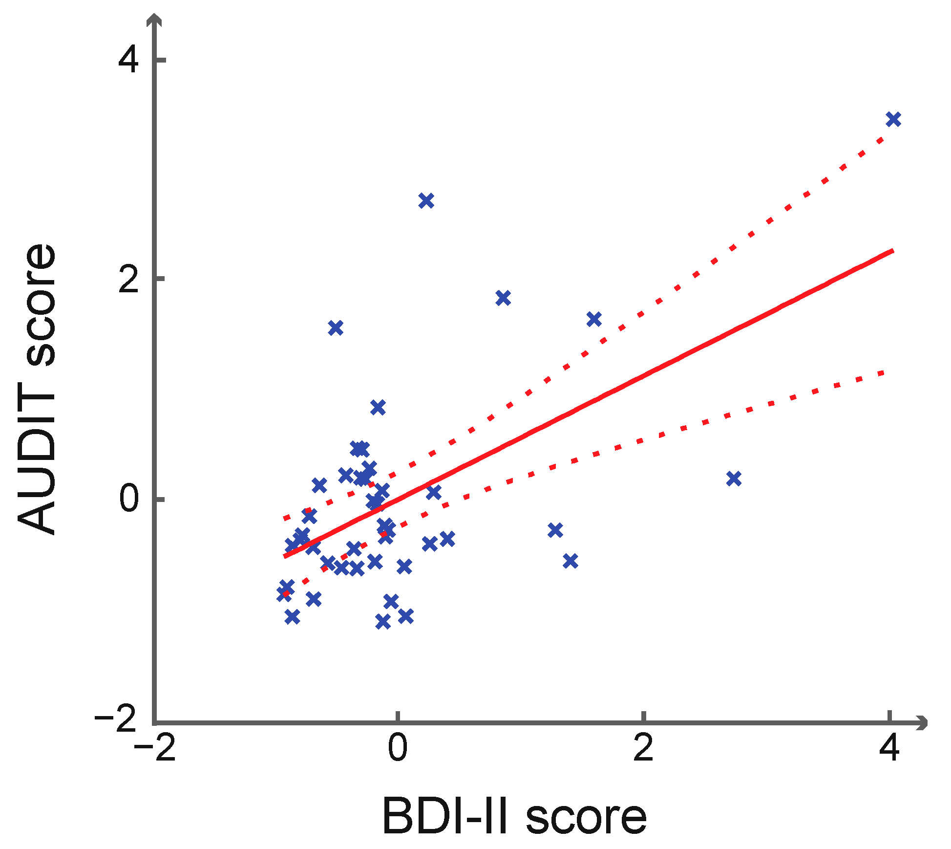
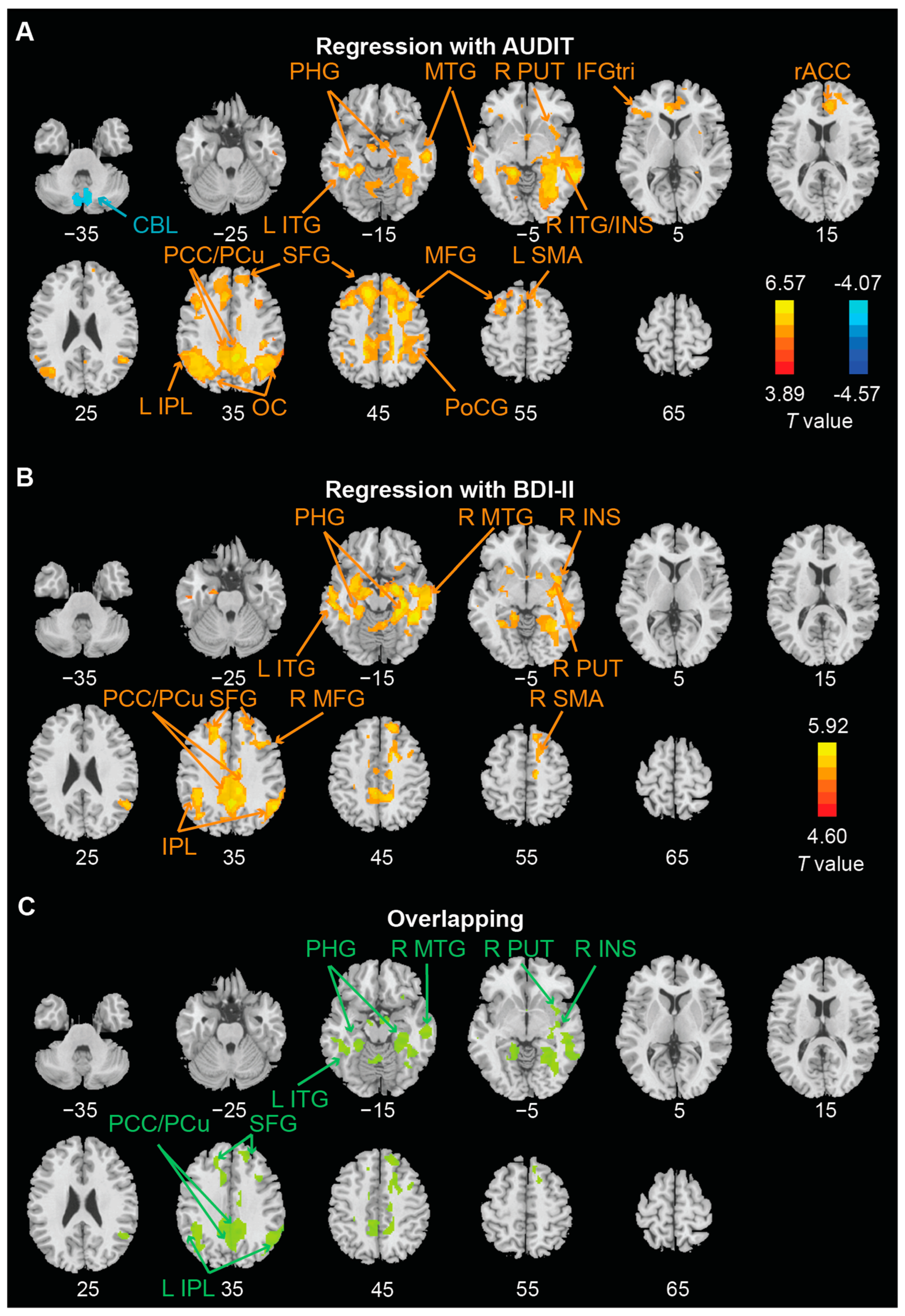
3.3. Whole Brain Regressions on AUDIT and BDI-II Scores

We performed whole-brain regressions of win > nil (Figure 3A,B; Table 2) and loss > nil (Figure 4A,B; Table 2) against AUDIT and BDI-II scores, separately, with age, sex, and FTND score as covariates across subjects. We identified the clusters that overlapped between AUDIT and BDI-II regressions (Figure 3C and Figure 4C). For win > nil, the overlapping clusters included bilateral parahippocampal gyrus, superior frontal gyrus, and posterior cingulate cortex/precuneus, left inferior parietal lobule and inferior temporal gyrus, and right middle temporal gyrus, putamen, and insula. For loss > nil, the overlapping clusters included bilateral hippocampus/parahippocampal gyrus, superior frontal gyrus, mid-cingulate cortex, cerebellum, and right amygdala, middle temporal gyrus, thalamus, and inferior parietal lobule.

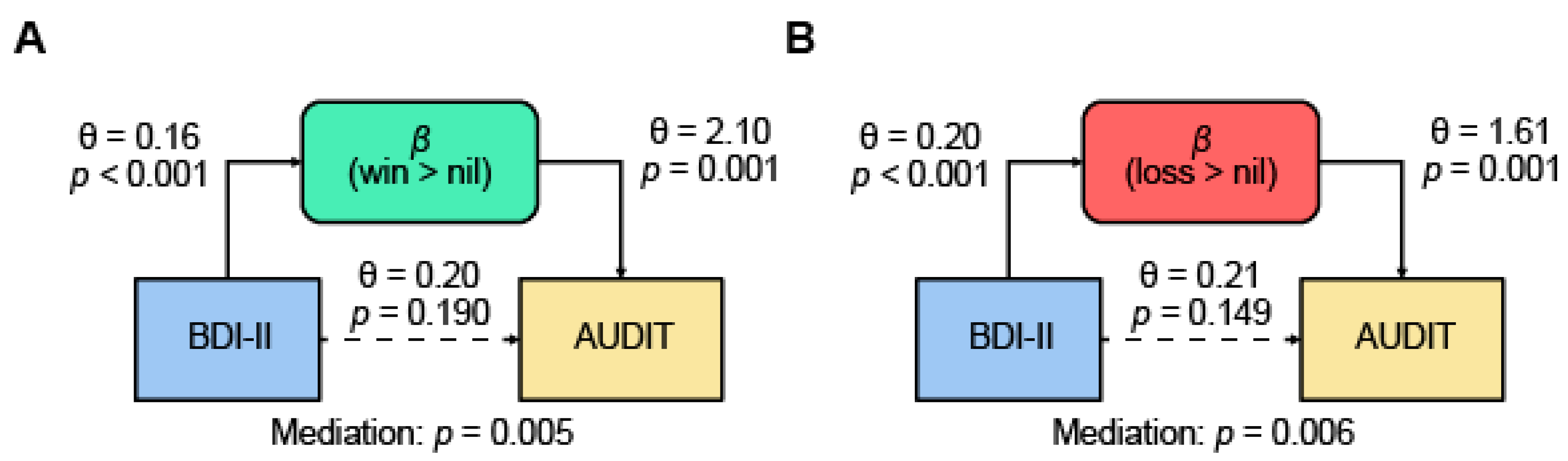
3.4. Mediation Models

The AUDIT and BDI-II scores were positively correlated, as shown earlier. As expected, the activation (β) of overlapping clusters was each positively correlated with the AUDIT score (r = 0.66, p < 0.001 for win > nil and r = 0.65, p < 0.001 for loss > nil) and BDI-II score (r = 0.67, p < 0.001 for win > nil and r = 0.64, p < 0.001 for loss > nil). Thus, we conducted mediation analyses to examine the relationships amongst the βs, AUDIT, and BDI-II scores. We tested all six models for each contrast. The results showed that the β of win > nil and of loss > nil each completely mediated the relationship between BDI-II and AUDIT (Figure 5). None of the other models showed significant mediation at a corrected threshold p < 0.05/6 = 0.0083 (Table 3).

4. Discussion

We identified regional brain responses to monetary win and loss outcomes in correlation with both AUDIT and BDI-II scores in non-dependent drinkers. Individuals with higher AUDIT and BDI-II scores showed greater activation to wins in bilateral fronto-parietal cortex, precuneus/posterior cingulate cortex, and right temporal cortex. Individuals with higher AUDIT and BDI scores also showed higher activation to losses in bilateral (but predominantly right-hemispheric) hippocampus/parahippocampal gyrus, cerebellum, right temporal and inferior parietal cortex, and posterior cingulate cortex. These regional activities completely mediated the relationship between depression and alcohol use severity. The findings highlight shared neural correlates of reward and punishment processing between depression and alcohol misuse and may help research of the etiologies of comorbid depression and AUD. We highlight the major findings for discussion.

4.1. Depression and Alcohol Misuse Shared Neural Responses to Monetary Win and Loss

A wide array of cortical and subcortical regions was involved during win and loss processing in link with depression and problem drinking. Most notable among these regions are bilateral hippocampi/parahippocampal gyri (HC/PHG), which are shared for both contrasts—win vs. nil and loss vs. nil—although the latter also involved the right amygdala in activities shared between depression and alcohol misuse. The HC/PHG is best known for its function in memory encoding and retrieval, and high-arousing, salient events consistently engage the HC/PHG [51,52]. Relative to nil, both win and loss trials are more salient; thus, the current findings suggest elevated HC/PHG responses to saliency both in association with the severity of depression and alcohol misuse. These findings are broadly consistent with previous reports of HC/PHG structural and functional changes in depressive and anxiety [53,54,55] and alcohol use [56,57,58] disorders. Studies of the etiological mechanisms of depression have emphasized ill-adaptive HC/PHG circuit responses to stress [59]. An imaging literature has associated with HC/PHG circuit dysfunction in emotional and reward processing [60,61,62]. Here, we demonstrated that depression and alcohol misuse both implicate the HC/PHG in heightened responses to wins and losses—salient stimuli irrespective of their valence.
The shared correlates that appeared to be specific to win and loss processing are the posterior cingulate cortex and precuneus (PCC/Pcu) and mid-cingulate cortex (MCC), respectively. Although less of a focus in human studies of reward processing, the PCC is implicated in post-decisional reward signaling in neuronal recordings from behaving monkeys [63]. Notably, the PCC/Pcu did not show significantly higher responses to win vs. nil in the one-sample t test (Supplementary Figure S1), suggesting this default mode network region solely as a correlate of individual variation in depression and alcohol misuse. In contrast, a hub of the limbic motor circuit, the MCC responds to learning of aversive consequences and behavioral avoidance [64,65]. The MCC along with other midline brain regions, including the supplementary motor area, showed significantly higher activation to loss vs. nil in the one-sample t test (Supplementary Figure S1). Thus, individuals with more severe depression and alcohol misuse would engage the MCC greater than the average extent, likely to support emotional motor processes of negative reinforcement. Also notable is the rostral anterior cingulate cortex (rACC), which showed higher responses to both win and loss vs. nil across subjects but only higher responses to win vs. nil in correlation with AUDIT but not BDI-II score. The rACC is part of the saliency and executive control circuit [66,67]; rACC responses to reward may potentially represent a unique marker of alcohol misuse. These findings support studies of neuromodulation of the ACC as a treatment of AUD [68,69]. More research is warranted to evaluate sub-regional cingulate cortical responses to reward and punishment and how these responses may be altered in depressive and alcohol use disorders [70].

4.2. Shared Neural Responses Mediated the Link of Depression and Alcohol Misuse

Across individuals, BDI-II and AUDIT scores were positively correlated, supporting the comorbidity of depression and problem drinking even in non-dependent drinkers [7,10,71,72,73,74]. Importantly, we observed shared brain activities in response to reward and to punishment, and these activities completely mediated the relationships between the severity of depression and alcohol misuse. Specifically, with correction for multiple testing, the findings suggest that depression contributes to alcohol misuse through the shared neural responses to monetary win and loss. As discussed in the Introduction, earlier studies have suggested individual differences in reward sensitivity as a risk factor of both depression and alcohol misuse [75,76,77]. The current findings expand this literature by showing evidence that depression contributes to alcohol drinking through brain activities in response to reward and punishment. Notably, although only one of the mediation models—depression → β of win or loss → problem drinking—showed significant, complete mediation, the model problem drinking → β of win or loss → depression showed an incomplete mediation effect with a p < 0.009. Given the small sample size of the study, it remains to be seen whether the shared correlates may mediate the link of depression and problem alcohol use bidirectionally.

4.3. Limitations of the Study and Future Directions

Several limitations need to be noted for the study. Firstly, the study comprised a small sample and, though evaluated at a corrected threshold, the findings and particularly those of regional responses shared between AUDIT and BDI-II regressions, need to be replicated. For the same reason, we did not investigate potential sex differences in the current findings or whether depression and alcohol misuse may be associated with shared reduction in neural responses differentiating reward and punishment [78]. Secondly, reward and punishment come in many forms. Thus, the findings should be considered specific to monetary win and loss. It remains unclear whether or how neural processes of other modalities of reward and punishment (e.g., social interaction and rejection) may be shared in the etiological processes of depression and alcohol misuse. Lastly, although the participants in the current study were largely non- or light smokers (mean FTND score < 1), we cannot entirely rule out the effects of smoking on the current findings. Studies of a larger sample and perhaps of individuals with varying levels of nicotine dependence would be needed to address the effects.
Although the study focused on reward processing, depression and alcohol misuse involve and may share other etiological processes, including fronto-limbic dysfunction in impaired inhibitory control and emotion processing and learning [79,80,81,82,83] and altered mitochondrial bioenergetics [84,85,86]. More studies are needed to evaluate the respective and potentially interactive roles of these neural and biological mechanisms of depression and alcohol misuse. Further, to the extent that these findings are considered specifically for non-dependent drinkers, future studies may incorporate assessment of physical, social, and cultural factors that may influence alcohol intake and the emotional context under which individuals engage in use and misuse of alcohol and “alcohol-like” drinks [87,88].

5. Conclusions

To conclude, we showed a significant correlation between the severity of depression and alcohol misuse in non-dependent drinkers. Neural responses to monetary wins and losses both mediated the relationship between depression and problem drinking. Suggesting shared etiological processes, these findings not only enhance our understanding of the neural mechanisms associated with both psychiatric conditions but also provide evidence for the importance of concurrent treatment of depression and alcohol misuse in clinical populations.

Supplementary Materials

The following supporting information can be downloaded at: https://www.mdpi.com/article/10.3390/brainsci12121689/s1, Figure S1: Regional activations to (A) Win vs. Nil and (B) Loss vs. Nil. Color bars indicate voxel T values. Results were evaluated at voxel p < 0.001 in combination with cluster-level p < 0.05 family-wise error (FWE) corrected. The clusters are summarized in Supplementary Table S1. L: left; R: right; ACC: anterior cingulate cortex; AMY: amygdala; CB: caudate body; CBL: cerebellum; dACC: dorsal ACC; IFGorb: inferior frontal gyrus (pars orbitalis); INS: insula; IPG: inferior parietal gyrus; MCC: mid-cingulate cortex; mOFC: medial orbitofrontal cortex; OC: occipital cortex; PCC: posterior cingulate cortex; PoCG: postcentral gyrus; SMG: supramarginal gyrus; SPG: superior parietal gyrus; STR: striatum; TAL: thalamus; Table S1: Regional activations to Win vs. Nil and Loss vs. Nil in the MIDT (n = 43).

Author Contributions

Conceptualization, Y.C. and C.-S.R.L.; methodology, Y.C., I.D., T.M.L., S.Z. (Simon Zhornitsky), S.Z. (Sheng Zhang), and C.-S.R.L.; software, Y.C.; validation, Y.C., I.D., T.M.L., S.Z. (Simon Zhornitsky), S.Z. (Sheng Zhang), and C.-S.R.L.; formal analysis, Y.C.; investigation, Y.C., I.D., T.M.L., S.Z. (Simon Zhornitsky), S.Z. (Sheng Zhang), and C.-S.R.L.; resources, C.-S.R.L.; data curation, Y.C.; writing—original draft preparation, Y.C., I.D., T.M.L., S.Z. (Simon Zhornitsky), S.Z. (Sheng Zhang), and C.-S.R.L.; writing—review and editing, Y.C., I.D., T.M.L., S.Z. (Simon Zhornitsky), S.Z. (Sheng Zhang), and C.-S.R.L.; visualization, Y.C.; supervision, C.-S.R.L.; project administration, C.-S.R.L.; funding acquisition, C.-S.R.L. All authors have read and agreed to the published version of the manuscript.

Funding

This research was funded by NIH grant number DA051922.

Institutional Review Board Statement

The study was conducted in accordance with the Declaration of Helsinki, and approved by the Institutional Review Board (or Ethics Committee) of Yale University (protocol code 0906005272 and date of initial approval 20 August 2009).

Informed Consent Statement

Informed consent was obtained from all subjects involved in the study.

Data Availability Statement

The data and codes will be shared on request to the corresponding author.

Acknowledgments

This study is supported by NIH grant DA051922. The funding agencies are otherwise not responsible for the design of the study, data collection or analysis, or in the decision to publish these results.

Conflicts of Interest

The authors declare that they have no competing interest in the current work.

References

  1. Boden, J.M.; Fergusson, D.M. Alcohol and depression. Addiction 2011, 106, 906–914. [Google Scholar] [CrossRef] [PubMed]
  2. Whiteford, H.A.; Ferrari, A.J.; Degenhardt, L.; Feigin, V.; Vos, T. The global burden of mental, neurological and substance use disorders: An analysis from the Global Burden of Disease Study 2010. PLoS ONE 2015, 10, e0116820. [Google Scholar] [CrossRef] [PubMed] [Green Version]
  3. Castillo-Carniglia, A.; Keyes, K.M.; Hasin, D.S.; Cerdá, M. Psychiatric comorbidities in alcohol use disorder. Lancet Psychiatry 2019, 6, 1068–1080. [Google Scholar] [CrossRef] [PubMed]
  4. Grant, B.F.; Harford, T.C. Comorbidity between DSM-IV alcohol use disorders and major depression: Results of a national survey. Drug Alcohol Depend. 1995, 39, 197–206. [Google Scholar] [CrossRef] [Green Version]
  5. Swendsen, J.D.; Merikangas, K.R. The comorbidity of depression and substance use disorders. Clin. Psychol. Rev. 2000, 20, 173–189. [Google Scholar] [CrossRef]
  6. Tilki, M. The social contexts of drinking among Irish men in London. Drugs Educ. Prev. Policy 2006, 13, 247–261. [Google Scholar] [CrossRef]
  7. Caldwell, T.M.; Rodgers, B.; Jorm, A.F.; Christensen, H.; Jacomb, P.A.; Korten, A.E.; Lynskey, M.T. Patterns of association between alcohol consumption and symptoms of depression and anxiety in young adults. Addiction 2002, 97, 583–594. [Google Scholar] [CrossRef]
  8. Bell, S.; Britton, A.; Kubinova, R.; Malyutina, S.; Pajak, A.; Nikitin, Y.; Bobak, M. Drinking pattern, abstention and problem drinking as risk factors for depressive symptoms: Evidence from three urban Eastern European populations. PLoS ONE 2014, 9, e104384. [Google Scholar] [CrossRef] [Green Version]
  9. McHugh, R.K.; Weiss, R.D. Alcohol use disorder and depressive disorders. Alcohol Res. Curr. Rev. 2019, 40, arcr.v40.1.01. [Google Scholar] [CrossRef] [Green Version]
  10. Feldstein Ewing, S.W.; Filbey, F.M.; Chandler, L.D.; Hutchison, K.E. Exploring the relationship between depressive and anxiety symptoms and neuronal response to alcohol cues. Alcohol. Clin. Exp. Res. 2010, 34, 396–403. [Google Scholar] [CrossRef]
  11. Fergusson, D.M.; Boden, J.M.; Horwood, L.J. Tests of causal links between alcohol abuse or dependence and major depression. Arch. Gen. Psychiatry 2009, 66, 260–266. [Google Scholar] [CrossRef] [PubMed] [Green Version]
  12. Oldham, S.; Murawski, C.; Fornito, A.; Youssef, G.; Yucel, M.; Lorenzetti, V. The anticipation and outcome phases of reward and loss processing: A neuroimaging meta-analysis of the monetary incentive delay task. Hum. Brain Mapp. 2018, 39, 3398–3418. [Google Scholar] [CrossRef] [PubMed] [Green Version]
  13. Chen, Y.; Chaudhary, S.; Chiang-shan, R.L. Shared and distinct neural activity during anticipation and outcome of win and loss: A meta-analysis of the monetary incentive delay task. Neuroimage 2022, 264, 119764. [Google Scholar] [CrossRef] [PubMed]
  14. Ng, T.H.; Alloy, L.B.; Smith, D.V. Meta-analysis of reward processing in major depressive disorder reveals distinct abnormalities within the reward circuit. Transl. Psychiatry 2019, 9, 293. [Google Scholar] [CrossRef] [Green Version]
  15. Eshel, N.; Roiser, J.P. Reward and punishment processing in depression. Biol. Psychiatry 2010, 68, 118–124. [Google Scholar] [CrossRef]
  16. Admon, R.; Pizzagalli, D.A. Dysfunctional Reward Processing in Depression. Curr. Opin. Psychol. 2015, 4, 114–118. [Google Scholar] [CrossRef] [Green Version]
  17. Pizzagalli, D.A.; Holmes, A.J.; Dillon, D.G.; Goetz, E.L.; Birk, J.L.; Bogdan, R.; Dougherty, D.D.; Iosifescu, D.V.; Rauch, S.L.; Fava, M. Reduced caudate and nucleus accumbens response to rewards in unmedicated individuals with major depressive disorder. Am. J. Psychiatry 2009, 166, 702–710. [Google Scholar] [CrossRef] [PubMed] [Green Version]
  18. O’Callaghan, G.; Stringaris, A. Reward processing in adolescent depression across neuroimaging modalities. Z. Kinder Jugendpsychiatrie Psychother. 2019, 47, 535–541. [Google Scholar] [CrossRef]
  19. Knutson, B.; Greer, S.M. Anticipatory affect: Neural correlates and consequences for choice. Philos. Trans. R. Soc. Lond. B Biol. Sci. 2008, 363, 3771–3786. [Google Scholar] [CrossRef] [Green Version]
  20. Craske, M.G.; Meuret, A.E.; Ritz, T.; Treanor, M.; Dour, H.J. Treatment for anhedonia: A neuroscience driven approach. Depress. Anxiety 2016, 33, 927–938. [Google Scholar] [CrossRef]
  21. Stoy, M.; Schlagenhauf, F.; Sterzer, P.; Bermpohl, F.; Hägele, C.; Suchotzki, K.; Schmack, K.; Wrase, J.; Ricken, R.; Knutson, B. Hyporeactivity of ventral striatum towards incentive stimuli in unmedicated depressed patients normalizes after treatment with escitalopram. J. Psychopharmacol. 2012, 26, 677–688. [Google Scholar] [CrossRef] [PubMed]
  22. Dichter, G.S.; Kozink, R.V.; McClernon, F.J.; Smoski, M.J. Remitted major depression is characterized by reward network hyperactivation during reward anticipation and hypoactivation during reward outcomes. J. Affect. Disord. 2012, 136, 1126–1134. [Google Scholar] [CrossRef] [PubMed] [Green Version]
  23. Gotlib, I.H.; Sivers, H.; Gabrieli, J.D.; Whitfield-Gabrieli, S.; Goldin, P.; Minor, K.L.; Canli, T. Subgenual anterior cingulate activation to valenced emotional stimuli in major depression. Neuroreport 2005, 16, 1731–1734. [Google Scholar] [CrossRef]
  24. Smoski, M.J.; Felder, J.; Bizzell, J.; Green, S.R.; Ernst, M.; Lynch, T.R.; Dichter, G.S. fMRI of alterations in reward selection, anticipation, and feedback in major depressive disorder. J. Affect. Disord. 2009, 118, 69–78. [Google Scholar] [CrossRef] [PubMed] [Green Version]
  25. Bremner, J.D.; Vythilingam, M.; Vermetten, E.; Charney, D.S. Effects of antidepressant treatment on neural correlates of emotional and neutral declarative verbal memory in depression. J. Affect. Disord. 2007, 101, 99–111. [Google Scholar] [CrossRef] [PubMed] [Green Version]
  26. Burger, C.; Redlich, R.; Grotegerd, D.; Meinert, S.; Dohm, K.; Schneider, I.; Zaremba, D.; Forster, K.; Alferink, J.; Bolte, J.; et al. Differential Abnormal Pattern of Anterior Cingulate Gyrus Activation in Unipolar and Bipolar Depression: An fMRI and Pattern Classification Approach. Neuropsychopharmacology 2017, 42, 1399–1408. [Google Scholar] [CrossRef] [Green Version]
  27. Hall, G.B.; Milne, A.M.; Macqueen, G.M. An fMRI study of reward circuitry in patients with minimal or extensive history of major depression. Eur. Arch. Psychiatry Clin. Neurosci. 2014, 264, 187–198. [Google Scholar] [CrossRef]
  28. Smoski, M.J.; Rittenberg, A.; Dichter, G.S. Major depressive disorder is characterized by greater reward network activation to monetary than pleasant image rewards. Psychiatry Res. Neuroimaging 2011, 194, 263–270. [Google Scholar] [CrossRef] [Green Version]
  29. Wrase, J.; Schlagenhauf, F.; Kienast, T.; Wüstenberg, T.; Bermpohl, F.; Kahnt, T.; Beck, A.; Ströhle, A.; Juckel, G.; Knutson, B. Dysfunction of reward processing correlates with alcohol craving in detoxified alcoholics. Neuroimage 2007, 35, 787–794. [Google Scholar] [CrossRef]
  30. Büchel, C.; Peters, J.; Banaschewski, T.; Bokde, A.L.; Bromberg, U.; Conrod, P.J.; Flor, H.; Papadopoulos, D.; Garavan, H.; Gowland, P. Blunted ventral striatal responses to anticipated rewards foreshadow problematic drug use in novelty-seeking adolescents. Nat. Commun. 2017, 8, 14140. [Google Scholar] [CrossRef]
  31. Beck, A.; Schlagenhauf, F.; Wüstenberg, T.; Hein, J.; Kienast, T.; Kahnt, T.; Schmack, K.; Hägele, C.; Knutson, B.; Heinz, A. Ventral striatal activation during reward anticipation correlates with impulsivity in alcoholics. Biol. Psychiatry 2009, 66, 734–742. [Google Scholar] [CrossRef] [PubMed]
  32. Forbes, E.E.; Rodriguez, E.E.; Musselman, S.; Narendran, R. Prefrontal response and frontostriatal functional connectivity to monetary reward in abstinent alcohol-dependent young adults. PLoS ONE 2014, 9, e94640. [Google Scholar] [CrossRef] [Green Version]
  33. Bjork, J.M.; Smith, A.R.; Hommer, D.W. Striatal sensitivity to reward deliveries and omissions in substance dependent patients. Neuroimage 2008, 42, 1609–1621. [Google Scholar] [CrossRef] [PubMed] [Green Version]
  34. Romanczuk-Seiferth, N.; Koehler, S.; Dreesen, C.; Wustenberg, T.; Heinz, A. Pathological gambling and alcohol dependence: Neural disturbances in reward and loss avoidance processing. Addict. Biol. 2015, 20, 557–569. [Google Scholar] [CrossRef] [PubMed]
  35. Crane, N.A.; Gorka, S.M.; Weafer, J.; Langenecker, S.A.; de Wit, H.; Phan, K.L. Preliminary evidence for disrupted nucleus accumbens reactivity and connectivity to reward in binge drinkers. Alcohol Alcohol. 2017, 52, 647–654. [Google Scholar] [CrossRef] [PubMed] [Green Version]
  36. Gilman, J.M.; Smith, A.R.; Bjork, J.M.; Ramchandani, V.A.; Momenan, R.; Hommer, D.W. Cumulative gains enhance striatal response to reward opportunities in alcohol-dependent patients. Addict. Biol. 2015, 20, 580–593. [Google Scholar] [CrossRef] [Green Version]
  37. Becker, A.; Kirsch, M.; Gerchen, M.F.; Kiefer, F.; Kirsch, P. Striatal activation and frontostriatal connectivity during non-drug reward anticipation in alcohol dependence. Addict. Biol. 2017, 22, 833–843. [Google Scholar] [CrossRef]
  38. Andrews, M.M.; Meda, S.A.; Thomas, A.D.; Potenza, M.N.; Krystal, J.H.; Worhunsky, P.; Stevens, M.C.; O’Malley, S.; Book, G.A.; Reynolds, B. Individuals family history positive for alcoholism show functional magnetic resonance imaging differences in reward sensitivity that are related to impulsivity factors. Biol. Psychiatry 2011, 69, 675–683. [Google Scholar] [CrossRef] [Green Version]
  39. Dhingra, I.; Zhang, S.; Zhornitsky, S.; Le, T.M.; Wang, W.; Chao, H.H.; Levy, I.; Li, C.R. The effects of age on reward magnitude processing in the monetary incentive delay task. Neuroimage 2020, 207, 116368. [Google Scholar] [CrossRef]
  40. First, M.B. Structured clinical interview for DSM-IV axis I disorders. Biom. Res. Dep. 1995. Available online: https://cir.nii.ac.jp/crid/1573387451015011328 (accessed on 1 December 2022).
  41. Bohn, M.J.; Babor, T.F.; Kranzler, H.R. The Alcohol Use Disorders Identification Test (AUDIT): Validation of a screening instrument for use in medical settings. J. Stud. Alcohol 1995, 56, 423–432. [Google Scholar] [CrossRef] [PubMed]
  42. Saunders, J.B.; Aasland, O.G.; Babor, T.F.; De la Fuente, J.R.; Grant, M. Development of the alcohol use disorders identification test (AUDIT): WHO collaborative project on early detection of persons with harmful alcohol consumption-II. Addiction 1993, 88, 791–804. [Google Scholar] [CrossRef] [PubMed]
  43. Beck, A.T.; Steer, R.A.; Brown, G.K. Manual for the Beck Depression Inventory-II; Psychological Corporation: San Antonio, TX, USA, 1996. [Google Scholar]
  44. Beck, A.T.; Ward, C.H.; Mendelson, M.; Mock, J.; Erbaugh, J. An inventory for measuring depression. Arch. Gen. Psychiatry 1961, 4, 561–571. [Google Scholar] [CrossRef] [PubMed] [Green Version]
  45. Jackson-Koku, G. Beck depression inventory. Occup. Med. 2016, 66, 174–175. [Google Scholar] [CrossRef] [Green Version]
  46. Heatherton, T.F.; Kozlowski, L.T.; Frecker, R.C.; Fagerstrom, K.O. The Fagerström test for nicotine dependence: A revision of the Fagerstrom Tolerance Questionnaire. Br. J. Addict. 1991, 86, 1119–1127. [Google Scholar] [CrossRef]
  47. Dhingra, I.; Zhang, S.; Zhornitsky, S.; Wang, W.; Le, T.M.; Li, C.R. Sex differences in neural responses to reward and the influences of individual reward and punishment sensitivity. BMC Neurosci. 2021, 22, 12. [Google Scholar] [CrossRef]
  48. Zhornitsky, S.; Dhingra, I.; Le, T.M.; Wang, W.; Li, C.R.; Zhang, S. Reward-Related Responses and Tonic Craving in Cocaine Addiction: An Imaging Study of the Monetary Incentive Delay Task. Int. J. Neuropsychopharmacol. 2021, 24, 634–644. [Google Scholar] [CrossRef]
  49. MacKinnon, D.P.; Fairchild, A.J.; Fritz, M.S. Mediation analysis. Annu. Rev. Psychol. 2007, 58, 593. [Google Scholar] [CrossRef]
  50. Hung, C.C.; Zhang, S.; Chen, C.M.; Duann, J.R.; Lin, C.P.; Lee, T.S.; Li, C.R. Striatal functional connectivity in chronic ketamine users: A pilot study. Am. J. Drug Alcohol Abuse 2020, 46, 31–43. [Google Scholar] [CrossRef]
  51. Osborne, D.M.; Pearson-Leary, J.; McNay, E.C. The neuroenergetics of stress hormones in the hippocampus and implications for memory. Front. Neurosci. 2015, 9, 164. [Google Scholar] [CrossRef] [Green Version]
  52. Frank, D.; Kafkas, A. Expectation-driven novelty effects in episodic memory. Neurobiol. Learn. Mem. 2021, 183, 107466. [Google Scholar] [CrossRef] [PubMed]
  53. Otte, C.; Gold, S.M.; Penninx, B.W.; Pariante, C.M.; Etkin, A.; Fava, M.; Mohr, D.C.; Schatzberg, A.F. Major depressive disorder. Nat. Rev. Dis. Prim. 2016, 2, 16065. [Google Scholar] [CrossRef] [PubMed] [Green Version]
  54. Kunimatsu, A.; Yasaka, K.; Akai, H.; Kunimatsu, N.; Abe, O. MRI findings in posttraumatic stress disorder. J. Magn. Reson. Imaging 2020, 52, 380–396. [Google Scholar] [CrossRef] [PubMed]
  55. Lazarov, A.; Zhu, X.; Suarez-Jimenez, B.; Rutherford, B.R.; Neria, Y. Resting-state functional connectivity of anterior and posterior hippocampus in posttraumatic stress disorder. J. Psychiatr. Res. 2017, 94, 15–22. [Google Scholar] [CrossRef]
  56. Galandra, C.; Basso, G.; Manera, M.; Crespi, C.; Giorgi, I.; Vittadini, G.; Poggi, P.; Canessa, N. Salience network structural integrity predicts executive impairment in alcohol use disorders. Sci. Rep. 2018, 8, 14481. [Google Scholar] [CrossRef] [Green Version]
  57. Yang, X.; Tian, F.; Zhang, H.; Zeng, J.; Chen, T.; Wang, S.; Jia, Z.; Gong, Q. Cortical and subcortical gray matter shrinkage in alcohol-use disorders: A voxel-based meta-analysis. Neurosci. Biobehav. Rev. 2016, 66, 92–103. [Google Scholar] [CrossRef]
  58. Tomasi, D.; Wiers, C.E.; Manza, P.; Shokri-Kojori, E.; Michele-Vera, Y.; Zhang, R.; Kroll, D.; Feldman, D.; McPherson, K.; Biesecker, C. Accelerated aging of the amygdala in alcohol use disorders: Relevance to the dark side of addiction. Cereb. Cortex 2021, 31, 3254–3265. [Google Scholar] [CrossRef]
  59. Campbell, S.; MacQueen, G. The role of the hippocampus in the pathophysiology of major depression. J. Psychiatry Neurosci. 2004, 29, 417–426. [Google Scholar]
  60. Alba-Ferrara, L.; Müller-Oehring, E.; Sullivan, E.; Pfefferbaum, A.; Schulte, T. Brain responses to emotional salience and reward in alcohol use disorder. Brain Imaging Behav. 2016, 10, 136–146. [Google Scholar] [CrossRef] [Green Version]
  61. Walker, C.; Kuhn, C.M.; Risher, M.-L. The effects of peri-adolescent alcohol use on the developing hippocampus. Int. Rev. Neurobiol. 2021, 160, 251–280. [Google Scholar] [CrossRef]
  62. Zorlu, N.; Çapraz, N.; Oztekin, E.; Bagci, B.; Di Biase, M.A.; Zalesky, A.; Gelal, F.; Bora, E.; Durmaz, E.; Beşiroğlu, L. Rich club and reward network connectivity as endophenotypes for alcohol dependence: A diffusion tensor imaging study. Addict. Biol. 2019, 24, 265–274. [Google Scholar] [CrossRef] [PubMed]
  63. McCoy, A.N.; Crowley, J.C.; Haghighian, G.; Dean, H.L.; Platt, M.L. Saccade reward signals in posterior cingulate cortex. Neuron 2003, 40, 1031–1040. [Google Scholar] [CrossRef] [PubMed]
  64. Grupe, D.W.; Nitschke, J.B. Uncertainty and anticipation in anxiety: An integrated neurobiological and psychological perspective. Nat. Rev. Neurosci. 2013, 14, 488–501. [Google Scholar] [CrossRef] [PubMed]
  65. Andrzejewski, J.A.; Greenberg, T.; Carlson, J.M. Neural correlates of aversive anticipation: An activation likelihood estimate meta-analysis across multiple sensory modalities. Cogn. Affect. Behav. Neurosci. 2019, 19, 1379–1390. [Google Scholar] [CrossRef] [PubMed]
  66. Manza, P.; Hu, S.; Chao, H.H.; Zhang, S.; Leung, H.-C.; Chiang-shan, R.L. A dual but asymmetric role of the dorsal anterior cingulate cortex in response inhibition and switching from a non-salient to salient action. Neuroimage 2016, 134, 466–474. [Google Scholar] [CrossRef] [Green Version]
  67. Ide, J.S.; Shenoy, P.; Angela, J.Y.; Chiang-Shan, R.L. Bayesian prediction and evaluation in the anterior cingulate cortex. J. Neurosci. 2013, 33, 2039–2047. [Google Scholar] [CrossRef] [Green Version]
  68. Zhao, Y.; Sallie, S.N.; Cui, H.; Zeng, N.; Du, J.; Yuan, T.; Li, D.; De Ridder, D.; Zhang, C. Anterior cingulate cortex in addiction: New insights for neuromodulation. Neuromodulation Technol. Neural Interface 2021, 24, 187–196. [Google Scholar] [CrossRef]
  69. Harel, M.; Perini, I.; Kämpe, R.; Alyagon, U.; Shalev, H.; Besser, I.; Sommer, W.H.; Heilig, M.; Zangen, A. Repetitive transcranial magnetic stimulation in alcohol dependence: A randomized, double-blind, sham-controlled proof-of-concept trial targeting the medial prefrontal and anterior cingulate cortices. Biol. Psychiatry 2022, 91, 1061–1069. [Google Scholar] [CrossRef]
  70. Fujiwara, J.; Tobler, P.N.; Taira, M.; Iijima, T.; Tsutsui, K.-I. Segregated and integrated coding of reward and punishment in the cingulate cortex. J. Neurophysiol. 2009, 101, 3284–3293. [Google Scholar] [CrossRef] [Green Version]
  71. Huang, Y.; Mohan, A.; De Ridder, D.; Sunaert, S.; Vanneste, S. The neural correlates of the unified percept of alcohol-related craving: A fMRI and EEG study. Sci. Rep. 2018, 8, 923. [Google Scholar] [CrossRef] [Green Version]
  72. Hepner, K.A.; Hunter, S.B.; Edelen, M.O.; Zhou, A.J.; Watkins, K. A comparison of two depressive symptomatology measures in residential substance abuse treatment clients. J. Subst. Abus. Treat. 2009, 37, 318–325. [Google Scholar] [CrossRef] [PubMed] [Green Version]
  73. Dum, M.; Pickren, J.; Sobell, L.C.; Sobell, M.B. Comparing the BDI-II and the PHQ-9 with outpatient substance abusers. Addict. Behav. 2008, 33, 381–387. [Google Scholar] [CrossRef] [PubMed]
  74. Kim, S.-A.; Kim, E.; Morris, R.G.; Park, W.-S. Exploring the non-linear relationship between alcohol consumption and depression in an elderly population in Gangneung: The Gangneung Health Study. Yonsei Med. J. 2015, 56, 418–425. [Google Scholar] [CrossRef] [PubMed] [Green Version]
  75. Baskin-Sommers, A.R.; Foti, D. Abnormal reward functioning across substance use disorders and major depressive disorder: Considering reward as a transdiagnostic mechanism. Int. J. Psychophysiol. 2015, 98, 227–239. [Google Scholar] [CrossRef]
  76. van Hemel-Ruiter, M.E.; de Jong, P.J.; Ostafin, B.D.; Wiers, R.W. Reward sensitivity, attentional bias, and executive control in early adolescent alcohol use. Addict. Behav. 2015, 40, 84–90. [Google Scholar] [CrossRef]
  77. Shankman, S.A.; Klein, D.N.; Tenke, C.E.; Bruder, G.E. Reward sensitivity in depression: A biobehavioral study. J. Abnorm. Psychol. 2007, 116, 95. [Google Scholar] [CrossRef] [Green Version]
  78. Aloi, J.; Blair, K.S.; Crum, K.I.; Bashford-Largo, J.; Zhang, R.; Lukoff, J.; Carollo, E.; White, S.F.; Hwang, S.; Filbey, F.M. Alcohol use disorder, but not cannabis use disorder, symptomatology in adolescents is associated with reduced differential responsiveness to reward versus punishment feedback during instrumental learning. Biol. Psychiatry Cogn. Neurosci. Neuroimaging 2020, 5, 610–618. [Google Scholar] [CrossRef]
  79. Guo, W.; Liu, F.; Yu, M.; Zhang, J.; Zhang, Z.; Liu, J.; Xiao, C.; Zhao, J. Decreased regional activity and network homogeneity of the fronto-limbic network at rest in drug-naive major depressive disorder. Aust. N. Z. J. Psychiatry 2015, 49, 550–556. [Google Scholar] [CrossRef]
  80. Tucker, D.M.; Luu, P.; Frishkoff, G.; Quiring, J.; Poulsen, C. Frontolimbic response to negative feedback in clinical depression. J. Abnorm. Psychol. 2003, 112, 667. [Google Scholar] [CrossRef]
  81. Kamarajan, C.; Porjesz, B.; Jones, K.A.; Choi, K.; Chorlian, D.B.; Padmanabhapillai, A.; Rangaswamy, M.; Stimus, A.T.; Begleiter, H. The role of brain oscillations as functional correlates of cognitive systems: A study of frontal inhibitory control in alcoholism. Int. J. Psychophysiol. 2004, 51, 155–180. [Google Scholar] [CrossRef] [Green Version]
  82. Battaglia, S.; Cardellicchio, P.; Di Fazio, C.; Nazzi, C.; Fracasso, A.; Borgomaneri, S. The Influence of Vicarious Fear-Learning in “Infecting” Reactive Action Inhibition. Front. Behav. Neurosci. 2022, 16, 946263. [Google Scholar] [CrossRef] [PubMed]
  83. Battaglia, S.; Cardellicchio, P.; Di Fazio, C.; Nazzi, C.; Fracasso, A.; Borgomaneri, S. Stopping in (e)motion: Reactive action inhibition when facing valence-independent emotional stimuli. Front. Behav. Neurosci. 2022, 16, 998714. [Google Scholar] [CrossRef] [PubMed]
  84. Tanaka, M.; Spekker, E.; Szabo, A.; Polyak, H.; Vecsei, L. Modelling the neurodevelopmental pathogenesis in neuropsychiatric disorders. Bioactive kynurenines and their analogues as neuroprotective agents-in celebration of 80th birthday of Professor Peter Riederer. J. Neural Transm. 2022, 129, 627–642. [Google Scholar] [CrossRef]
  85. Tanaka, M.; Szabo, A.; Spekker, E.; Polyak, H.; Toth, F.; Vecsei, L. Mitochondrial Impairment: A Common Motif in Neuropsychiatric Presentation? The Link to the Tryptophan-Kynurenine Metabolic System. Cells 2022, 11, 2607. [Google Scholar] [CrossRef] [PubMed]
  86. Klinedinst, N.J.; Regenold, W.T. A mitochondrial bioenergetic basis of depression. J. Bioenerg. Biomembr. 2015, 47, 155–171. [Google Scholar] [CrossRef]
  87. Sikalidis, A.K.; Kelleher, A.H.; Kristo, A.S. Mediterranean Diet. Encyclopedia 2021, 1, 371–387. [Google Scholar] [CrossRef]
  88. Sikalidis, A.K.; Kelleher, A.H.; Maykish, A.; Kristo, A.S. Non-Alcoholic Beverages, Old and Novel, and Their Potential Effects on Human Health, with a Focus on Hydration and Cardiometabolic Health. Medicina 2020, 56, 490. [Google Scholar] [CrossRef]
Figure 1. (A) Monetary incentive delay task: Each trial starts with a bet (a dollar, a cent, or no money). After a randomized interval of 1–5 s, a target box is presented and disappears after a response window. Subjects are requested to make a response as quickly as possible to collect the money (win) before it disappears, following by feedback that indicates the amount of money won (in red) or lost (in blue). (B) Accuracy rate and (C) RT of trials (mean ± SD).
Brainsci 12 01689 g001
Figure 2. Scatter plot of AUDIT and BDI-II scores. Note that the standardized residual scores after controlling for age, sex, and FTND score as covariates are shown here. Dashed lines represent 95% confidence intervals of the mean regression (solid line). Crosses represent individual data points.
Brainsci 12 01689 g002
Figure 3. Regional activations to win vs. nil in correlation with (A) AUDIT score and (B) BDI-II score with age, sex, and FTND score as covariates with threshold of voxel p < 0.001 in combination with cluster-level p < 0.05 FWE corrected; The brain regions are summarized in Table 2. (C) ROIs overlapped of (A,B). Warm/cool colors indicate positive/negative correlations. L: left; R: right; CBL: cerebellum; IFGtri: inferior frontal gyrus pars triangularis; INS: insula; IPL: inferior parietal lobule; ITG: inferior temporal gyrus; MFG: middle frontal gyrus; MTG: middle temporal gyrus; OC: occipital cortex; Pcu: precuneus; PHG: parahippocampal gyrus; PoCG: postcentral gyrus; PUT: putamen; rACC: rostral anterior cingulate cortex; SFG: superior frontal gyrus; SMA: supplementary motor area.
Brainsci 12 01689 g003
Figure 4. Regional activations to loss vs. nil in correlation with (A) AUDIT and (B) BDI-II score with age, sex, and FTND score as covariates at voxel p < 0.001 in combination with cluster-level p < 0.05 FWE corrected. The brain regions are summarized in Table 2. (C) ROIs overlapped of (A,B). L: left; R: right; AMY: amygdala; CBL: cerebellum; IFGorb: inferior frontal gyrus pars orbitalis; HIP: hippocampus; IPL: inferior parietal lobule; ITG: inferior temporal gyrus; MCC: mid-cingulate cortex; MFG: middle frontal gyrus; MTG: middle temporal gyrus; OC: occipital cortex; PHG: parahippocampal gyrus; SFG: superior frontal gyrus; SMA: supplementary motor area; THA: thalamus.
Brainsci 12 01689 g004
Figure 5. Mediation models: brain activation (β) of (A) win > nil (in green) and (B) loss > nil (in red) of the shared ROIs completely mediated the correlation between BDI-II (in blue) and AUDIT (in yellow) scores. All six models were assessed for each contrast and evaluated at a corrected threshold p = 0.05/6 = 0.0083. The p values associated with mediation are for the path “a × b” (see Section 2). The statistics of all models are summarized in Table 3.
Brainsci 12 01689 g005
Table 1. Demographic and clinical characteristics in men and women.
Men (n = 27)Women (n = 16)tp
Age (years)44.9 ± 11.346.4 ± 15.6−0.340.71
Education (years)15.0 ± 3.614.8 ± 30.270.79
AUDIT score7.8 ± 9.34.9 ± 4.81.150.26
BDI-II score5.0 ± 8.77.7 ± 8.8−0.960.34
FTND score0.4 ± 1.40.8 ± 2.2−0.630.53
Note: Values of mean ± SD; AUDIT: Alcohol Use Disorders Identification Test; BDI-II: Beck’s Depression Inventory; FTND: Fagerström Test for Nicotine Dependence.
Table 2. Regional activations to win > nil and loss > nil in correlation with AUDIT and BDI-II scores with age, sex, and FTND score as covariates.
Volume
(mm3)
Peak ZMNI Coordinate (mm)SideIdentified Brain Region
xyz
Regression with AUDIT
Positive with Win > Nil
100,8455.34−152943L/RDLPFC, SFG, Precentral G, SMA
4.8436−3140L/RIPL, PCC, Precuneus
49,8155.27−21−37−11LHippocampus, Amygdala
5.2154−46−5RTemporal G
66694.4864113L/RACC
28624.06−9−7−11L/RHypothalamus
25113.85−33324LIFG
Negative with Win > Nil
42664.066−70−35L/RCBL
Positive with Loss > Nil
68,7965.9754−31−8RTemporal G, Hippocampus, Amygdala, Hypothalamus
5.29−12−58−20LCBL
84,3755.1018−4640L/RPrecuneus, DLPFC, MPFC, MCC
4.9060−5531RIPL, Precentral G,
30784.49−3311−8LInsula
48064.30−33−140LPrecentral gyrus
25384.30959−5RVMPFC
Negative with Loss > Nil
None
Regression with BDI
Positive with Win > Nil
54,3244.9521−7−17RHippocampus, Amygdala, Hypothalamus
4.7645−43−14RTemporal G
33,6964.63−6−4040LPrecuneus, PCC, DLPFC, SFG, SMA
60484.5757−6134RIPL
44014.29−42−4034LIPL
63994.26−122340LSFG
Negative with Win > Nil
None
Positive with Loss > Nil
10,6654.89−18−10−20LHippocampus, Amygdala, Temporal G
25,3264.8124−10−14RHippocampus, Amygdala, Temporal G, CBL
63994.2163−4637RIPL
43204.08215922RSFG
41583.84−9−734LMCC
24033.67−6−3740LMCC
Negative with Loss > Nil
None
Note: Results were evaluated at voxel p < 0.001 and cluster-level p < 0.05, FWE corrected; L: left; R: right; G: gyrus; Cerebellum: CBL; DLPFC: dorsolateral prefrontal cortex; SFG: superior frontal gyrus, IPL: inferior parietal lobule; PCC: posterior cingulate cortex; ACC: anterior cingulate cortex; IFG: inferior frontal gyrus; MPFC: medial prefrontal cortex; MCC: mid-cingulate cortex; VMPFC: ventromedial prefrontal cortex; SMA: supplementary motor area.
Table 3. Mediation models of AUDIT, BDI-II score, and brain activation (β) each for win > nil and loss > nil.
p Values
XMYX → MM → YX → YMediated X → YMediation
Win > Nil
AUDITBDIBrain<0.0010.006<0.001<0.0010.021
BrainBDIAUDIT<0.0010.190<0.001<0.0010.204
AUDITBrainBDI<0.0010.006<0.0010.1900.013
BDIBrainAUDIT<0.001<0.001<0.0010.1900.005 *
BrainAUDITBDI<0.0010.190<0.0010.0060.201
BDIAUDITBrain<0.001<0.001<0.0010.0060.009
Loss > Nil
AUDITBDIBrain<0.0010.010<0.001<0.0010.029
BrainBDIAUDIT<0.0010.149<0.001<0.0010.166
AUDITBrainBDI<0.0010.010<0.0010.1490.020
BDIBrainAUDIT<0.001<0.001<0.0010.1490.006 *
BrainAUDITBDI<0.0010.149<0.0010.0100.162
BDIAUDITBrain<0.001<0.001<0.0010.0100.009
Note: The mediation models were evaluated at a corrected p = 0.05/6 = 0.0083. * p < 0.0083. A significant mediation and non-significant mediated X Y suggest that M completely mediates the correlation of X and Y.
Publisher’s Note: MDPI stays neutral with regard to jurisdictional claims in published maps and institutional affiliations.

