Review

A Systematic Review of Neurofeedback for the Management of Motor Symptoms in Parkinson’s Disease

1
Peninsula Allied Health Centre, School of Health Professions, University of Plymouth, Derriford Road, Plymouth PL6 8BH, UK
2
Brain Research and Imaging Centre, Faculty of Health, University of Plymouth, Research Way, Plymouth PL6 8BU, UK
3
School of Psychology, University of Plymouth, Drake Circus, Plymouth PL4 8AA, UK
4
School of Health Sciences, University of Southampton, Highfield, Southampton SO17 1BJ, UK
*
Author to whom correspondence should be addressed.
Brain Sci. 2021, 11(10), 1292; https://doi.org/10.3390/brainsci11101292
Submission received: 16 September 2021 / Accepted: 27 September 2021 / Published: 29 September 2021
(This article belongs to the Special Issue Innovations in Neurorehabilitation and Neuroplasticity)

Abstract

:
Background: Neurofeedback has been proposed as a treatment for Parkinson’s disease (PD) motor symptoms by changing the neural network activity directly linked with movement. However, the effectiveness of neurofeedback as a treatment for PD motor symptoms is unclear. Aim: To systematically review the literature to identify the effects of neurofeedback in people with idiopathic PD; as defined by measurement of brain activity; motor function; and performance. Design: A systematic review. Included Sources and Articles: PubMed; MEDLINE; Cinhal; PsychoInfo; Prospero; Cochrane; ClinicalTrials.gov; EMBASE; Web of Science; PEDro; OpenGrey; Conference Paper Index; Google Scholar; and eThos; searched using the Population-Intervention-Comparison-Outcome (PICO) framework. Primary studies with the following designs were included: randomized controlled trials (RCTs), non-RCTs; quasi-experimental; pre/post studies; and case studies. Results: This review included 11 studies out of 6197 studies that were identified from the literature search. Neuroimaging methods used were fMRI; scalp EEG; surface brain EEG; and deep brain EEG; where 10–15 Hz and the supplementary motor area were the most commonly targeted signatures for EEG and fMRI, respectively. Success rates for changing one’s brain activity ranged from 47% to 100%; however, both sample sizes and success criteria differed considerably between studies. While six studies included a clinical outcome; a lack of consistent assessments prevented a reliable conclusion on neurofeedback’s effectiveness. Narratively, fMRI neurofeedback has the greatest potential to improve PD motor symptoms. Two main limitations were found in the studies that contributed to the lack of a confident conclusion: (1) insufficient clinical information and perspectives (e.g., no reporting of adverse events), and (2) limitations in numerical data reporting (e.g., lack of explicit statistics) that prevented a meta-analysis. Conclusions: While fMRI neurofeedback was narratively the most effective treatment; the omission of clinical outcome measures in studies using other neurofeedback approaches limits comparison. Therefore, no single neurofeedback type can currently be identified as an optimal treatment for PD motor symptoms. This systematic review highlights the need to improve the inclusion of clinical information and more robust reporting of numerical data in future work. Neurofeedback appears to hold great potential as a treatment for PD motor symptoms. However, this field is still in its infancy and needs high quality RCTs to establish its effectiveness. Review Registration: PROSPERO (ID: CRD42020191097)

1. Introduction

Parkinson’s disease (PD) is a progressive neurological disorder that primarily disrupts normal motor functioning and affects 1–2 in every 1000 individuals in the general population [1]. Motor symptoms such as bradykinesia, rigidity, and tremor have a severe negative impact on quality of life and increase the likelihood of mood disorders such as depression [2,3]. PD motor symptoms are in part caused by degeneration of nigrostriatal neurons that reduces dopamine availability in the basal ganglia. Therefore, the main treatment for PD motor symptoms are pharmaceuticals, such as levodopa, that increase dopaminergic stimulation within the basal ganglia. However, the efficacy of these pharmaceuticals often declines over time and there is an increased occurrence of adverse side effects, such as dyskinesias, which may be severely disabling after prolonged use [4]. Other non-pharmaceutical treatments include high-frequency deep-brain stimulation (DBS) of specific structures of the basal ganglia, such as the subthalamic nuclei or the globus pallidus internus, through implanted electrodes. However, DBS is an invasive method and risks peri-operative complications such as intracranial hemorrhage, infections, and skin erosions [5,6]. Furthermore, DBS is not always effective and still incurs some unwanted side effects such as dysarthria, depression, apathy and executive dysfunction [7,8]. The development of non-pharmaceutical and non-invasive approaches that improve PD motor symptoms and minimize unwanted side effects would be therefore a valuable adjunct to symptom management.
Neurofeedback is a non-pharmaceutical treatment that uses a brain–computer-interface (BCI), allowing individuals to learn voluntary self-regulation of brain activity using an external, real-time representation of that brain activity [9]. Although neurofeedback can be invasive (e.g., recording from DBS electrodes [10]), non-invasive approaches, such as electroencephalography (EEG; [11]) or functional magnetic resonance imaging (fMRI; [12]), are viable alternatives.
The pathological neural activity associated with PD motor symptoms, such as bradykinesia, can be measured using several approaches. For example, non-invasive fMRI studies, have highlighted reductions in blood flow in the supplementary motor area with movement preparation [13] whilst recording of local field potentials from DBS electrodes in the STN have highlighted an increase in bursts of beta (15–30 Hz) oscillation during movement-preparation in PD [14]. Simultaneous recordings of beta oscillations highlight that cortical oscillatory activity phase leads and thus potentially drives that in the basal ganglia [15,16,17,18]. This suggests that neurofeedback of pathological activity using fMRI, EEG or local field potentials measured using DBS electrodes may hold potential for improving associated PD motor symptoms.
Previous neurofeedback research in PD has used a variety of different methodologies and protocols. There is extensive variation in aspects such as training regime, brain signature and feedback approach, which complicates evaluation of neurofeedback efficacy as an intervention for PD. In order to progress in this field, there is a need to establish the current evidence base for neurofeedback application in PD and explore the relative efficacy of different protocols.

Review Question and Objectives

Here, we review the literature to address the following question: what are the effects of neurofeedback in people with idiopathic Parkinson’s disease (PD); as defined by measurement of brain activity, motor function, and performance?
The objectives of the review are to determine:
  • The effectiveness of neurofeedback as a treatment for reducing PD motor symptom severity.
  • The importance of specific protocol parameters for effective and reliable neurofeedback in terms of training regime, targeted brain activity, delivery of brain activity feedback signal, and changes in brain activity.
  • The association between specific neurofeedback protocols and clinical outcomes.

2. Methods

This systematic review was conducted in accordance with the Joanna Briggs Institute (JBI) methodology for systematic review effectiveness and according to the PRISMA statement. This protocol was registered on PROSPERO (ID: CRD42020191097).

2.1. Ethical Considerations

This study is a systematic literature review and did not involve human nor animal data collection. Therefore, ethical approval was not required.

2.2. Inclusion Criteria

The inclusion criteria were developed using the PICO (problem/population, intervention, comparison, and outcome) framework [19].

2.2.1. Population

The current review considered studies that included participants with idiopathic PD (as defined by a stated clinical diagnosis) of any duration or severity, who were at least 18 years of age. Studies that included participants with any atypical parkinsonism (i.e., neurodegenerative parkinsonism other than PD) were excluded. This review also excluded studies involving participants who had any secondary causes of parkinsonism, such as drug-induced parkinsonism or lesions; however, such studies were included if it was possible to individually separate and remove participants with atypical parkinsonism from the analysis.

2.2.2. Intervention

The review considered all studies that examined neurofeedback designed as a treatment for PD motor symptoms. This included studies that examined participants’ success at neurofeedback without any measurement of clinical outcomes. Relevant studies included neurofeedback training using any protocol (i.e., targeted brain activity), duration, frequency, or intensity. Neurofeedback evaluation considered any target brain activity such as EEG, deep brain recording, and other brain imaging (e.g., fMRI/positron emission tomography (PET)), and involved measurements from any brain region.

2.2.3. Comparison

This review considered studies that compared neurofeedback with a comparator intervention or usual care. For studies with no comparator, neurofeedback results were presented narratively and were not included in any meta-analysis.

2.2.4. Outcomes

This review considered studies that included the following outcomes:
  • Immediate and long-term sustained changes in brain activity following neurofeedback.
  • Immediate and long-term sustained changes in motor function or performance as measured by physiology (e.g., electromyography (EMG)) and/or other objective clinical outcome measures such as the Unified Parkinson’s Disease rating scale (UPDRS) or a questionnaire assessment of PD symptoms.
In addition to the above outcomes, this review evaluated the following information for the narrative analysis:
  • Neurofeedback protocol (i.e., targeted brain activity, presentation of brain activity to participants, criteria for “successful” neurofeedback).
  • Neurofeedback training details (i.e., who provides the neurofeedback training, guidance provided to participants, training regime).
  • The relationship between the above neurofeedback details and neurofeedback outcomes (i.e., success at neurofeedback and clinical outcomes).

2.2.5. Types of Studies

This review considered experimental study designs including randomized controlled trials (RCTs), non-RCTs, quasi-experimental, pre/post studies, and case studies. Only studies in English were included. Animal studies, observational studies, and narrative studies were excluded.

2.3. Search Strategy

The search strategy aimed to find both published and unpublished studies. A three-step search strategy, developed in discussion with a data-synthesis specialist, was utilised. An initial limited search of PubMed was undertaken to estimate the volume of relevant literature and to identify key words to assist in developing search terms. A second search using the developed search terms was undertaken and adapted across each included information source (see Supplementary Material “SM1—Search Strategy”); this included searches for published and grey literature. The third strategy involved searching for additional studies within the reference list of all studies that met the inclusion criteria. In cases of ongoing studies, authors were contacted for further information to determine eligibility for inclusion in this review. No limiters were used.

2.4. Information Sources

The databases searched were: PubMed, MEDLINE, Cinhal, PsychoInfo, Prospero, Cochrane, ClinicalTrials.gov, EMBASE, Web of Science, PEDro, OpenGrey, Conference Paper Index, Google Scholar, and eThos.

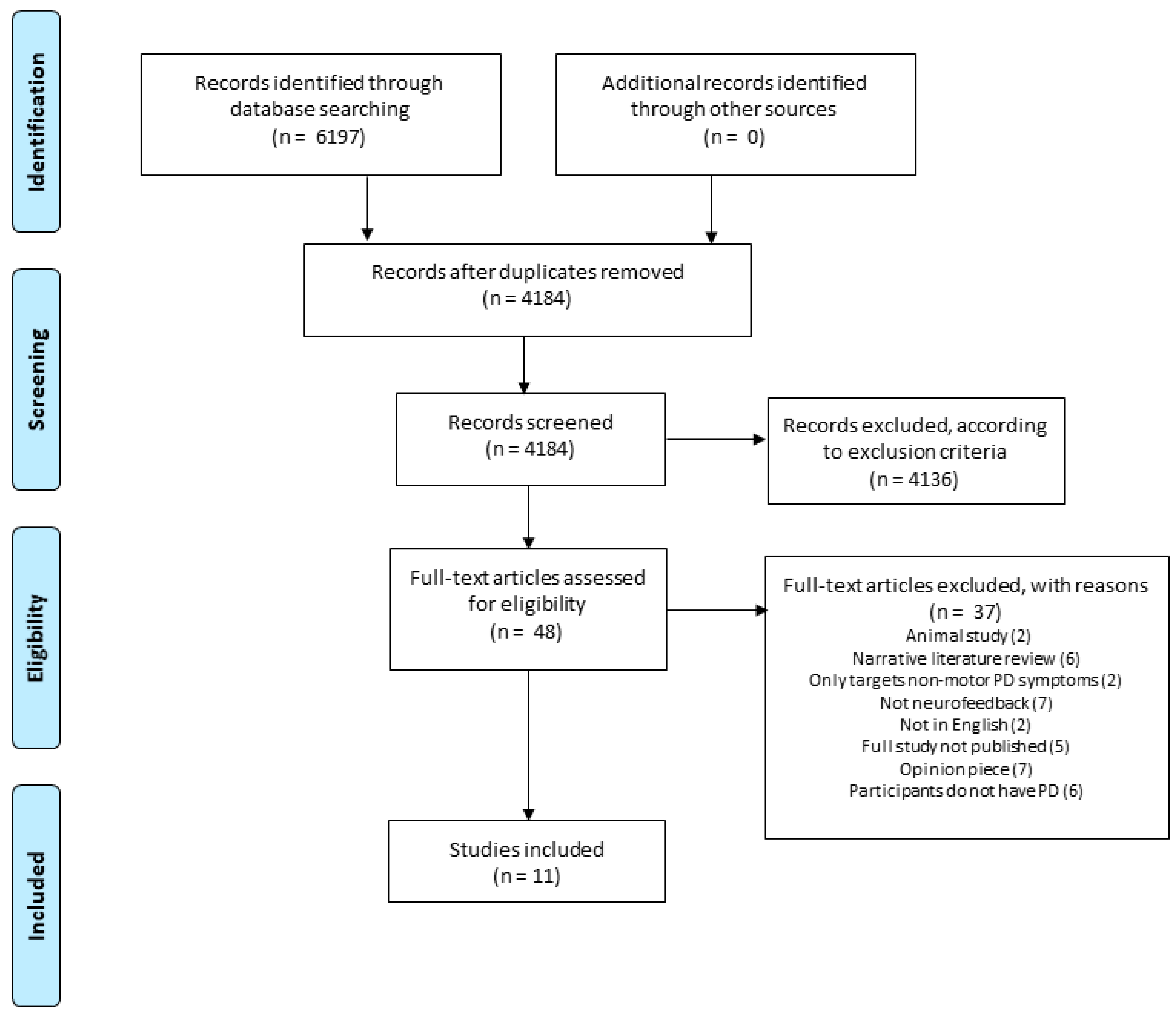
2.5. Study Selection

All identified references were imported into citation software (EndNote, Clarivate Analytics [20]). Duplicates were removed before uploading to the online collaborative systematic review organization tool ‘Raayan’ [21]. Titles and abstracts were screened by two reviewers (KA and JM) independently against the review inclusion criteria. The full-text of potentially eligible studies were retrieved and assessed in detail against the inclusion criteria by the reviewers (See Figure 1 for the PRISMA flowchart. Reviewer discrepancies were resolved through discussion with a third reviewer when necessary.

2.6. Assessment of Methodological Quality

Eligible studies were critically appraised for methodological quality by two independent reviewers using the standardized JBI critical appraisal instruments. Any disagreements were resolved through discussions, and through a third reviewer if necessary. All studies were included regardless of methodological quality. Originally, poor quality studies were planned to be excluded. However, majority of the studies were not of high quality and the decision was made to include all studies that met the eligibility criteria to report on the current status of this field. The results of critical appraisal are reported in Table 1 in the Section 3.

2.7. Data Extraction

Data extraction of included studies was conducted by the same reviewers (KA and JM). Narrative data extraction was conducted using an Excel spreadsheet. The following data were extracted for narrative synthesis: study design, study objectives, attrition details, demographics, PD symptoms, medication details, target brain activity, feedback signal delivery, and training (e.g., who provided the neurofeedback training, guidance provided, training regime), assessment of neurofeedback performance, success rates, clinical measures and outcomes (if any), and follow-up details (if any). Data extraction for a meta-analysis was planned (see protocol on PROSPERO, ID: CRD42020191097); however, the lack of numerical data in the identified articles prevented the meta-analysis.

2.8. Data Synthesis

The narrative synthesis of findings from included studies was structured according to the review objectives. As statistical pooling was not possible due to the poor reporting of numerical data (see the Section 3 and Section 4 for details), the findings were presented narratively aided by appropriate tables and figures. Publication bias investigation was not possible as the meta-analysis could not be conducted.

2.9. Assessing Certainty in Findings

A Summary of Findings (SoF) table was developed and includes the following outcomes: changes in brain activity, neurofeedback success rates, changes in movement and motor function, and adverse events. Further outcomes could not be included as planned due to inadequate reporting of numerical data. These were: absolute risks for the treatment and control, estimates of relative risk and evaluation of bias, directness, heterogeneity, and precision.

3. Results

Eleven studies were included (Figure 1). Table 1 shows the methodological assessments conducted using the relevant JBI critical appraisal instruments, dependent on the study design. Please see Supplementary Material “SM2—JBI Methodological Assessment Items”. Overall, most studies were of low quality with only one study (Fukuma et al. [22]) of high quality, scoring 89%. The main reason for low quality was the lack of appropriate follow-ups. However, several studies did not include various methodological details (e.g., demographic or study condition information), scoring “unclear” on these items. This low quality is reflected in these studies overall lack of numerical data.
Table 2 shows the study characteristics: five examined changes in both brain activity and movement; the remaining six only examined brain activity changes. Three studies included a control condition, of which only one study blinded participants using a sham trial [23]. Participants were partially blinded in one study that used a crossover design only for participants in the control condition, who were subsequently un-blinded when they crossed over to the neurofeedback condition [24]. Assessors were blinded in only one of these three studies [25]. Table 1 shows that only 2 studies specified the PD symptom they were targeting: Buyukturkoglu et al. [26] targeted hand-motor symptoms and akinesia, while Erikson-Davis et al. [24] targeted levodopa-induced dyskinesia. The remaining 9 studies reported general PD symptoms or did not mention their target.
The change in clinical outcomes with NF training are summarised in Table 3, the lack of available data and variability of the contributing studies precluded a meta-analysis resulting in a narrative report of the findings. Table 3 shows that PD severity was defined using the Hoehn and Yahr scale (3 studies [23,26,27]), the United Parkinson’s disease Rating scale (UPDRS) reported in full (2 studies [25,29]), or using the motor subsection of the UPDRS (4 studies [22,24,30,32]). Two studies did not measure PD severity [28,31]. Clinical outcomes included reaction times and finger tapping tests [23,26], EMG amplitude [22], UPDRS [23,25,29], diaries [24], and questionnaires (Modified Abnormal Involuntary Movement Scale [24] and PDQ-39 [25]). The three studies [23,25,29] that used the UPDRS as a clinical outcome reported a reduction in the score in the intervention group (indicating an improvement in clinical outcome) ranging from −0.3 [29] to −5.2 [23]. Mean changes in the primary outcome or measures of outcome variability (e.g., standard deviation) were not reported in 3 studies [22,23,24], while 5 studies did not measure any clinical outcome [27,28,30,31,32]. Adverse events were not reported in any study.
Table 4 displays details of the neurofeedback training protocol and regimes from the included articles. Four types of brain imaging were used for neurofeedback: scalp EEG (Erickson-Davis et al. [24]; Fumuro et al. [27]; Kasahara et al. [30]; Thompson & Thompson [31]), deep brain stimulation EEG using local field potentials (Fukuma et al. [22]; Khanna & Carmena [32]), electrocorticography (ECoG; (He et al. [28])), and fMRI (Buyukturkoglu et al. [26]; Subramanian et al. [23]; Subramanian et al. [25]; Tinaz et al. [29]). The most commonly used was scalp EEG and fMRI. The control direction for targeted brain activity varied amongst the studies; however, 10–15 Hz was the most commonly targeted EEG activity and SMA activation with movement preparation was the most commonly targeted fMRI activity. Success rates (i.e., the rate of participants able to change their brain activity in the desired direction) ranged from 47% to 100%; however, both sample sizes and success criteria differed between studies. Study-defined success criteria were based on brain activity, comparing brain activity power during neurofeedback to baseline or comparing baseline power between pre and post neurofeedback training. One study based its criteria on EEG amplitude threshold (Fumuro et al. [27]), while another identified success by predicting if seemingly successful performances were by chance by comparing it to simulated performances (Khanna & Carmena [32]). Only one study provided audio feedback of brain activity (i.e., a tone sounded when performance was successful) (Erickson-Davis et al. [24]), while the remaining 10 provided visual feedback. The visual feedback was divided into three categories: changes in bar height (Buyukturkoglu et al. [26]; Subramanian et al. [23]; Subramanian et al. [25]; Tinaz et al. [29]), object moves (e.g., up and down) (Fumuro et al. [27]; He et al. [28]; Kasahara et al. [30]; Khanna & Carmena [32]), and object changes in size (i.e., bigger or smaller) (Fukuma et al., 2018 [22]). The bar changing in height was sometimes a solid-colored rectangle, but was commonly a “thermometer” that had height indicators (see figures in Subramanian et al., (Subramanian et al. [23]; Subramanian et al. [25]) for examples). The “object” in the remaining two categories was of various designs, such as a ball or a video game character. No justification was provided for choosing a delivery design in any study. Specific instructions for controlling brain activity were provided in 6 of the 11 studies (Buyukturkoglu et al. [26]; He et al. [28]; Kasahara et al. [30]; Subramanian et al. [23]; Subramanian et al. [25]; Tinaz et al. [29]), all of which involved motor imagery. No specific instructions were provided in 3 of the studies (Erickson-Davis et al. [24]; Fukuma et al. [22]; Fumuro et al. [27]), while the remaining 2 studies did not describe the instructions (Khanna & Carmena [32]; Thompson & Thompson [31]). Neurofeedback runs ranged from 10 s to 12 min, while neurofeedback sessions ranged from 10 min to 50 min. The number of sessions ranged from 1 to 42 sessions, and time between sessions ranged from 1 day to 6 months.
Table 5 provides a summary of the targeted brain activity, success rates, and whether neurofeedback success was accompanied by a change in clinical outcome measures. No studies to date have assessed the long-term follow up of neurofeedback effects. Using fMRI neurofeedback of SMA activity and right insula-dorsomedial frontal cortex functional connectivity were the only approaches that achieved both success in neurofeedback and a change in clinical outcome, as measured by the UPDRS motor scale in all studies (Subramanian et al. [23]; Subramanian et al. [25]; Tinaz et al. [29]), and additionally finger tapping (Subramanian et al. [23]) or the PDQ-39 (Subramanian et al. [25]).

4. Discussion

The 11 studies included in this systematic review highlighted that the development of a neurofeedback intervention for PD motor symptoms is still in its early stages. Neurofeedback studies using fMRI as a measurement approach appear to report the highest success rates compared to other measurement approaches; however, two of these studies were generated by the same research team (i.e., Subramanian et al. [23,25]) and would therefore benefit from external validation. Six of the 11 studies [22,23,24,25,26,29] included a clinical outcome (4 of which used fMRI neurofeedback [23,25,26,29]). The limited number of comparable methodological approaches limits the confidence in any conclusion about the most effective type of neurofeedback for modulation of PD motor symptoms. The inability to draw confident conclusions on the effectiveness of neurofeedback as a modulator of PD motor symptoms, concurs with Esmail and Linden’s [33] systematic review on the therapeutic value of neurofeedback for PD and suggests that this field has seen little advancement in the years since 2014. We identified two principal reasons for this limited progress: (1) insufficient clinical information and perspectives, and (2) limitations in numerical data reporting.

4.1. Insufficient Clinical Information and Perspectives

Six studies assessed clinical outcomes [22,23,24,25,26,29] whilst the remaining studies assessed the feasibility of achieving neurofeedback according to specific success criteria. The primary aim of 5 out of 6 studies investigating clinical outcomes was an improvement in the hypokinetic motor symptoms related to PD. In contrast, Erikson-Davis et al. [24] investigated the effects of neurofeedback on reducing Levodopa-induced dyskinesias. The proposed pathophysiological mechanisms of hypo- and hyper-kinetic deficits in PD vary. Animal and human studies suggest increased bursts of beta oscillations cause hypokinetic deficits [14,34,35,36]. In contrast, dyskinesias resulting from either long term dopamine replacement therapy or DBS are associated with an increase in 4–10 Hz oscillations within the basal ganglia-cortical region [37,38]. Reflecting this, studies using EEG and DBS to target hypokinetic symptoms aimed to reduce beta-band activity through neurofeedback whilst the study targeting dyskinesia aimed to primarily reduce 4–8 Hz activity through neurofeedback [24]. Thus, when using EEG or DBS based neurofeedback, the underlying targeted symptoms should define the spectral band of interest. Although the role of oscillatory activity in the control of normal movements remains under investigation [39,40]. The fact that oscillatory activity is associated with normal movement suggests that a gross reduction in oscillatory power across a certain spectral band in PD may not be the optimal approach. Recent work has highlighted the importance of bursts of beta oscillations in the development of bradykinesia [15,41,42] and future work should target the incidence of these abnormal busts rather than gross changes in oscillatory power. Studies in healthy participants show that this is possible [43].
The optimal target and mechanism for recording brain activity to provide neurofeedback in PD cannot be determined from the review. The strongest evidence provided is for neurofeedback of SMA activity using fMRI. This is supported by a review [13] highlighting the significant reductions in blood flow in the supplementary motor area with movement preparation in PD. However, such changes are not seen in all studies. Although a promising area of research, the use of fMRI may be difficult to implement as an intervention given issues such as expense, availability and technical demands. Whilst fMRI has a good spatial resolution, its temporal resolution is low, resulting in a smearing of information in the time-frequency domain. Therefore, while anatomical resolution is high, it has limited ability to resolve specific phases of the movement process or individual neural signatures involved. In contrast, EEG has poor spatial and good temporal resolution. Therefore, it is capable of resolving discrete phases of movement and neural signatures (e.g., frequency bands), however, spatial smearing means that the target for EEG in the current studies could reflect an amalgamation of activity across primary and secondary motor areas (e.g., SMA). EEG as a neurofeedback device has potential advantages in terms of costs and ease of use with the possibility of home-based training. Simultaneous recordings of beta oscillations at rest highlight that cortical oscillatory activity phase leads and thus potentially drives that in the basal ganglia [15,16,17,18,19]. However, beta power is higher in the STN compared to the cortex, reflecting the fact that beta activity may be amplified within the basal ganglia [44] alongside the low pass filtering effects of the motor cortex on re-entrant beta oscillations [45]. This suggests that targeting activity within the basal ganglia nuclei may be preferable. This, however, requires access to surgically implanted DBS electrodes. Understanding the effects of both increasing and decreasing motor cortical spectral power and oscillatory bursts within motor cortical areas on movement performance in participants with and without PD could inform the role of cortical oscillations in the control of movement, and thus the potential role of EEG as a target for neurofeedback.

4.2. Limitations in Numerical Data Reporting

Examples of limitations in the reporting of numerical data includes a lack of reporting of basic descriptive statistics, such as measures of central tendency and variance required to demonstrate the magnitude of change between time points, study conditions, and neurofeedback parameters. Some studies did not report measures of statistical significance, such as p-values, and others failed to identify the statistical tests applied. This reflects the exploratory nature of some papers in this review. The absence of this information prevents the drawing of meaningful conclusions for individual studies and prevented meta-analysis intended to enrich this review. Additionally, lack of information about statistical power calculations (as well as small sample sizes) further restricted the drawing of meaningful conclusions. Limited reporting of numerical data confines the speed and reliability of progress in the development of neurofeedback for PD motor symptoms. Consequently, in this paper we strongly suggest adherence to CONSORT reporting standards for quantitative data. In particular, we suggest closely following the sections detailed in Table 6 below.
Ros et al. [46] have produced a checklist for reporting experimental neurofeedback studies, which helps authors to clearly outline their study design and analysis. We therefore recommend adhering to both the CONSORT guidance (especially focusing on the guidance in Table 5) and the checklist by Ros et al. [46] to ensure high quality reporting of neurofeedback for PD.

4.3. Study Limitations

There were several limitations to the review. No meta-analysis was undertaken due to limitations in the data presented and heterogeneity in the papers reviewed. Further, there was an English language bias in the papers reviewed. Ongoing studies of neurofeedback in Parkinson’s disease have been identified from clinicaltrials.org (e.g., NCT03837548). These will be published after this paper has been published, and may change this review’s outcome

5. Conclusions

This systematic review highlights the need to improve the reporting of numerical data and neurofeedback parameters in future work. Although based on a few studies there is a suggestion that fMRI-based neurofeedback may be effective and be associated with an improvement in hypokinetic symptoms. It is unclear whether EEG based neurofeedback is effective in PD. The targeted spectral band would vary depending on the symptoms of interest (dyskinesia vs. hypokinesia). Further work should investigate the role of cortical oscillations in driving abnormal oscillatory activity in the Basal Ganglia and whether EEG could be a potential target for neurofeedback.

Supplementary Materials

The following are available online at https://www.mdpi.com/article/10.3390/brainsci11101292/s1.

Author Contributions

K.A. planned and conducted the literature search, organised the review team, and reported the results. K.A. and J.M. conceptualised the study idea, reviewed the articles from the literature search, extracted data, and produced the initial manuscript. All authors (i.e., K.A., S.D.H., S.D., J.A.F., G.G. and J.M.) contributed to data interpretation and critically evaluated the manuscript. All authors have read and agreed to the published version of the manuscript.

Funding

This research did not receive any specific grant from funding agencies in the public, commercial, or not-for-profit sectors.

Institutional Review Board Statement

Not applicable.

Informed Consent Statement

Not applicable.

Data Availability Statement

No data available as this was a systematic review.

Acknowledgments

We would like to sincerely thank Chris Johns from the University of Plymouth, who provided library support during the initial phase of this systematic review, and particularly assisted in developing the search strategy.

Conflicts of Interest

The authors declare no conflict of interest.

References

  1. Tysnes, O.B.; Storstein, A. Epidemiology of Parkinson’s disease. J. Neural Transm. 2017, 124, 901–905. [Google Scholar] [CrossRef]
  2. Schrag, A. Quality of life and depression in Parkinson’s disease. J. Neurol. Sci. 2006, 248, 151–157. [Google Scholar] [CrossRef]
  3. Leroi, I.; Ahearn, D.J.; Andrews, M.; McDonald, K.R.; Bryne, J.E.; Burns, A. Behavioural disorders, disability and quality of life in Parkinson’s disease. Age Ageing 2011, 40, 614–621. [Google Scholar] [CrossRef] [Green Version]
  4. Radad, K.; Gille, G.; Rausch, W.-D. Short review on dopamine agonists: Insight into clinical and research studies relevant to Parkinson’s disease. Pharmacol. Rep. 2005, 57, 701–712. [Google Scholar]
  5. Sixel-Döring, F.; Trenkwalder, C.; Kappus, C.; Hellwig, D. Skin complications in deep brain stimulation for Parkinson’s disease: Frequency, time course, and risk factors. Acta Neurochir. 2010, 152, 195–200. [Google Scholar] [CrossRef]
  6. Xiaowu, H.; Xiufeng, J.; Xiaoping, Z.; Bin, H.; Laixing, W.; Yiqun, C.; Jinchuan, L.; Aiguo, J.; Jianmin, L. Risks of intracranial hemorrhage in patients with Parkinson’s disease receiving deep brain stimulation and ablation. Parkinsonism Relat. Disord. 2010, 16, 96–100. [Google Scholar] [CrossRef] [PubMed]
  7. Bronstein, J.M.; Tagliati, M.; Alterman, R.L.; Lozano, A.M.; Stefani, A.; Horak, F.B.; Okun, M.S.; Foote, K.D.; Krack, P.; Pahwa, R.; et al. Deep brain stimulation for Parkinson disease: An expert consensus and review of key issues. Arch. Neurol. 2011, 68, 165. [Google Scholar] [CrossRef]
  8. Limousin, P.; Foltynie, T. Long-term outcomes of deep brain stimulation in Parkinson disease. Nat. Rev. Neurol. 2019, 15, 234–242. [Google Scholar] [CrossRef] [Green Version]
  9. Marzbani, H.; Marateb, H.R.; Mansourian, M. Methodological Note: Neurofeedback: A Comprehensive Review on System Design, Methodology and Clinical Applications. Basic Clin. Neurosci. J. 2016, 7, 143–158. [Google Scholar] [CrossRef] [Green Version]
  10. Little, S.; Brown, P. What brain signals are suitable for feedback controzl of deep brain stimulation in Parkinson’s disease? Ann. N. Y. Acad. Sci. 2012, 1265, 9–24. [Google Scholar] [CrossRef]
  11. Sitaram, R.; Ros, T.; Stoeckel, L.; Haller, S.; Scharnowski, F.; Lewis-Peacock, J.; Weiskopf, N.; Blefari, M.L.; Rana, R.S.M.; Oblak, E.; et al. Closed-loop brain training: The science of neurofeedback. Nat. Rev. Neurosci. 2016, 18, 86–100. [Google Scholar] [CrossRef] [Green Version]
  12. Eklund, A. Using Real-Time fMRI to Control a Dynamical System by Brain Activity Classification. Med. Image Comput. Comput. Assist. Interv. 2009, 12, 1000–1008. [Google Scholar] [CrossRef]
  13. Spay, C.; Meyer, G.; Welter, M.-L.; Lau, B.; Boulinguez, P.; Ballanger, B. Functional imaging correlates of akinesia in Parkinson’s disease: Still open issues. NeuroImage Clin. 2019, 21, 101644. [Google Scholar] [CrossRef]
  14. Oswal, A.; Beudel, M.; Zrinzo, L.; Limousin, P.; Hariz, M.; Foltynie, T.; Litvak, V.; Brown, P. Deep brain stimulation modulates synchrony within spatially and spectrally distinct resting state networks in Parkinson’s disease. Brain 2016, 139, 1482–1496. [Google Scholar] [CrossRef]
  15. Cagnan, H.; Mallet, N.; Moll, C.K.E.; Gulberti, A.; Holt, A.B.; Westphal, M.; Gerloff, C.; Engel, A.K.; Hamel, W.; Magill, P.; et al. Temporal evolution of beta bursts in the parkinsonian cortical and basal ganglia network. Proc. Natl. Acad. Sci. USA 2019, 116, 16095–16104. [Google Scholar] [CrossRef] [Green Version]
  16. Sharott, A.; Gulberti, A.; Hamel, W.; Koppen, J.A.; Munchau, A.; Buhmann, C.; Potter-Nerger, M.; Westphal, M.; Gerloff, C.; Moll, C.K.E.; et al. Spatio-temporal dynamics of cortical drive to human subthalamic nucleus neurons in Parkinson’s disease. Neurobiol. Dis. 2018, 112, 49–62. [Google Scholar] [CrossRef]
  17. Brown, P. Abnormal oscillatory synchronisation in the motor system leads to impaired movement. Curr. Opin. Neurobiol. 2007, 17, 656–664. [Google Scholar] [CrossRef]
  18. Fogelson, N.; Williams, D.; Tijssen, M.; van Bruggen, G.; Speelman, H.; Brown, P. Different functional loops between cerebral cortex and the subthalmic area in Parkinson’s disease. Cereb. Cortex 2006, 16, 64–75. [Google Scholar] [CrossRef] [Green Version]
  19. Huang, X.; Lin, J.; Demner-Fushman, D. Evaluation of PICO as a knowledge representation for clinical questions. AMIA Annu. Symp. Proc. 2006, 2006, 359–363. [Google Scholar]
  20. Reuter, T. Endnote X9. 2020. Available online: https://endnote.com/product-details (accessed on 9 December 2020).
  21. Ouzzani, M.; Hammady, H.; Fedorowicz, Z.; Elmagarmid, A. Rayyan—A web and mobile app for systematic reviews. Syst. Rev. 2016, 5, 210. [Google Scholar] [CrossRef] [Green Version]
  22. Fukuma, R.; Yanagisawa, T.; Tanaka, M.; Yoshida, F.; Hosomi, K.; Oshino, S.; Tani, N.; Kishima, H. Real-Time Neurofeedback to Modulate β-Band Power in the Subthalamic Nucleus in Parkinson’s Disease Patients. eNeuro 2018, 5. [Google Scholar] [CrossRef]
  23. Subramanian, L.; Hindle, J.V.; Johnston, S.; Roberts, M.V.; Husain, M.; Goebel, R.; Linden, D. Real-time functional magnetic resonance imaging neurofeedback for treatment of Parkinson’s disease. J. Neurosci. 2011, 31, 16309–16317. [Google Scholar] [CrossRef]
  24. Erickson-Davis, C.R.; Anderson, J.S.; Wielinski, C.L.; Richter, S.A.; Parashos, S.A. Evaluation of Neurofeedback Training in the Treatment of Parkinson’s Disease: A Pilot Study. J. Neurother. 2012, 16, 4–11. [Google Scholar] [CrossRef] [Green Version]
  25. Subramanian, L.; Morris, M.B.; Brosnan, M.; Turner, D.L.; Morris, H.R.; Linden, D.E.J. Functional Magnetic Resonance Imaging Neurofeedback-guided Motor Imagery Training and Motor Training for Parkinson’s Disease: Randomized Trial. Front. Behav. Neurosci. 2016, 10, 111. [Google Scholar] [CrossRef] [Green Version]
  26. Buyukturkoglu, K.; Rana, M.; Ruiz, S.; Hackley, S.A.; Soekadar, S.R.; Birbaumer, N.; Sitaram, R. Volitional Regulation of Supplementary Motor Area with FMRI-BCI Neurofeedback in Parkinson’s Diseases: A Pilot Study. J. Neurol. Sci. 2013, 30, 180. [Google Scholar]
  27. Fumuro, T.; Matsuhashi, M.; Mitsueda, T.; Inouchi, M.; Hitomi, T.; Nakagawa, T.; Matsumoto, R.; Kawamata, J.; Inoue, H.; Mima, T.; et al. Bereitschaftspotential augmentation by neuro-feedback training in Parkinson’s disease. Clin. Neurophysiol. 2013, 124, 1398–1405. [Google Scholar] [CrossRef] [Green Version]
  28. He, S.; Syed, E.; Torrecillos, F.; Tinkhauser, G.; Fischer, P.; Pogosyan, A.; Pereira, E.; Ashkan, K.; Hasegawa, H.; Brown, P.; et al. Beta Oscillation-Targeted Neurofeedback Training Based on Subthalamic LFPs in Parkinsonian Patients. In Proceedings of the International IEEE/EMBS Conference on Neural Engineering (NER), San Francisco, CA, USA, 20–23 March 2019; pp. 81–84. [Google Scholar] [CrossRef]
  29. Tinaz, S.; Para, K.; Vives-Rodriguez, A.; Martinez-Kaigi, V.; Nalamada, K.; Sezgin, M.; Scheinost, D.; Hampson, M.; Louis, E.D.; Constable, R. Insula as the Interface Between Body Awareness and Movement: A Neurofeedback-Guided Kinesthetic Motor Imagery Study in Parkinson’s Disease. Front. Hum. Neurosci. 2018, 12, 496. [Google Scholar] [CrossRef]
  30. Kasahara, K.; Hoshino, H.; Furusawa, Y.; DaSalla, C.S.; Honda, M.; Murata, M.; Hanakawa, T. Initial experience with a sensorimotor rhythm-based brain-computer interface in a Parkinson’s disease patient. Brain-Comput. Interfaces 2018, 5, 88–96. [Google Scholar] [CrossRef]
  31. Thompson, M.; Thompson, L. Biofeedback for Movement Disorders (Dystonia with Parkinson’s Disease): Theory and Preliminary Results. J. Neurother. 2002, 6, 51–70. [Google Scholar]
  32. Khanna, P.; Carmena, J.M. Using Brain-Machine Interfaces to Study Motor Cortical Population Activity; University of California: Berkeley, CA, USA, 2017. [Google Scholar]
  33. Esmail, S.; Linden, D. Neural Networks and Neurofeedback in Parkinson’s Disease. Neuroregulation 2014, 1, 240–272. [Google Scholar] [CrossRef] [Green Version]
  34. Devergnas, A.; Caiola, M.; Pittard, D.; Wichmann, T. Cortical Phase–Amplitude Coupling in a Progressive Model of Parkinsonism in Nonhuman Primates. Cereb. Cortex 2017, 29, 167–177. [Google Scholar] [CrossRef]
  35. Sharott, A.; Vinciati, F.; Nakamura, K.C.; Magill, P.J. A Population of Indirect Pathway Striatal Projection Neurons Is Selectively Entrained to Parkinsonian Beta Oscillations. J. Neurosci. 2017, 37, 9977–9998. [Google Scholar] [CrossRef] [Green Version]
  36. West, T.O.; Berthouze, L.; Halliday, D.M.; Litvak, V.; Sharott, A.; Magill, P.; Farmer, S. Propagation of beta/gamma rhythms in the cortico-basal ganglia circuits of the parkinsonian rat. J. Neurophysiol. 2018, 119, 1608–1628. [Google Scholar] [CrossRef] [Green Version]
  37. Alonso-Frech, F.; Zamarbide, I.; Alegre, M.; Rodriguez-Oroz, M.C.; Guridi, J.; Manrique, M.; Valencia, M.; Artieda, J.; Obeso, J.A. Slow oscillatory activity and levodopa-induced dyskinesias in Parkinson’s disease. Brain 2006, 129, 1748–1757. [Google Scholar] [CrossRef] [Green Version]
  38. Foffani, G.; Bianchi, A.M.; Baselli, G.; Priori, A. Movement-related frequency modulation of beta oscillatory activity in the human subthalamic nucleus. J. Physiol. 2005, 568, 699–711. [Google Scholar] [CrossRef]
  39. De Vries, I.E.; Daffertshofer, A.; Stegeman, D.F.; Boonstra, T.W. Functional connectivity in the neuromuscular system underlying bimanual coordination. J. Neurophysiol. 2016, 116, 2576–2585. [Google Scholar] [CrossRef] [Green Version]
  40. Gatev, P.; Darbin, O.; Wichmann, T. Oscillations in the basal ganglia under normal conditions and in movement disorders. Mov. Disord. 2006, 21, 1566–1577. [Google Scholar] [CrossRef]
  41. Lofredi, R.; Tan, H.; Neumann, W.-J.; Yeh, C.-H.; Schneider, G.-H.; Kuhn, A.A.; Brown, P. Beta bursts during continuous movements accompany the velocity decrement in Parkinson’s disease patients. Neurobiol. Dis. 2019, 127, 462–471. [Google Scholar] [CrossRef]
  42. Torrecillos, F.; Tinkhauser, G.; Fischer, P.; Green, A.L.; Aziz, T.Z.; Foltynie, T.; Limousin, P.; Zrinzo, L.; Ashkan, K.; Brown, P.; et al. Modulation of Beta Bursts in the Subthalamic Nucleus Predicts Motor Performance. J. Neurosci. 2018, 38, 8905–8917. [Google Scholar] [CrossRef] [Green Version]
  43. He, S.; Everest-Phillips, C.; Clouter, A.; Brown, P.; Tan, H. Neurofeedback-Linked Suppression of Cortical β Bursts Speeds Up Movement Initiation in Healthy Motor Control: A Double-Blind Sham-Controlled Study. J. Neurosci. 2020, 40, 4021–4032. [Google Scholar] [CrossRef] [Green Version]
  44. Cole, S.R.; van der Meij, R.; Peterson, E.J.; de Hemptinne, C.; Starr, P.A.; Voytek, B. Nonsinusoidal beta oscillations reflect cortical pathophysiology in Parkinson’s disease. J. Neurosci. 2017, 37, 4830–4840. [Google Scholar] [CrossRef]
  45. Rivlin-Etzion, M.; Marmor, O.; Saban, G.; Rosin, B.; Haber, S.N.; Vaadia, E.; Prut, Y.; Bergman, H. Low-Pass Filter Properties of Basal Ganglia Cortical Muscle Loops in the Normal and MPTP Primate Model of Parkinsonism. J. Neurosci. 2008, 28, 633–649. [Google Scholar] [CrossRef]
  46. Ros, T.; Enriquez-Geppert, S.; Zotev, V.; Young, K.D.; Wood, G.; Whitfield-Gabrieli, S.; Wan, F.; Vuilleumier, P.; Vialatte, F.; Van De Ville, D.; et al. Consensus on the reporting and experimental design of clinical and cognitive-behavioural neurofeedback studies (CRED-nf checklist). Brain 2020, 143, 1674–1685. [Google Scholar] [CrossRef] [Green Version]
Figure 1. PRISMA flowchart for the literature search process.
Brainsci 11 01292 g001
Table 1. Methodological assessment of included studies using critical appraisal instruments from the Joanna Briggs Institute.
Quasi Experimental StudiesQ1Q2Q3Q4Q5Q6Q7Q8Q9* Total
Buyukturkoglu et al. [26]YNNNYNNUY33%
Fukama et al. [22]YYYYYNYYY89%
Fumuro et al. [27]YUYNYNYYY67%
He et al. [28]YUUNYYYUY56%
Subramanian et al. [23]YUUYYNYYY67%
Tinaz et al. [29]YYUYUNNYY56%
RCTsQ1Q2Q3Q4Q5Q6Q7Q8Q9Q10Q11Q12Q13* Total
Erikson-Davis et al. [24]UUNNNUNN/AYYYNN23%
Subramanian et al. [25]YNUNNYNNYNYYN38%
Case reportsQ1Q2Q3Q4Q5Q6Q7Q8* Total
Kasahara et al. [30]YNYYYNNN50%
Thompson & Thompson [31]NNYNYYUN38%
Cross-sectional studiesQ1Q2Q3Q4Q5Q6Q7Q8* Total
Khanna & Carmena [32]NNYNNNYY38%
Q = Question; Y = Yes; N = No; U = Unclear; RCT = Randomised controlled trial; N/A = Not applicable; * Total refers to the percentage of “yes” answers for each study, where a higher percentage indicates a higher quality study.
Table 2. Study characteristics.
AuthorAimTargeted PD SymptomCountryDesignIntervention ConditionControl ConditionTotal N
Buyukturkoglu et al., (2013) [26]Examining the effectiveness of real-time fMRI neurofeedback (reinforcement of SMA BOLD signal) on hand motor performanceHand-motor performance/akinesiaUSACase studyfMRI neurofeedback, finger tappingNo control condition1
Erikson-Davis et al., (2012) [24]Testing if scalp EEG neurofeedback (reinforcement of 12–15 Hz, suppression 4–10 Hz and 11–30 Hz) would lead to a decrease in PD motor-symptomsLevodopa-induced dyskinesiaUSARCTScalp EEG neurofeedbackSham trial, followed by scalp EEG neurofeedback9
Fukama et al., (2018) [22]Examining if DBS EEG neurofeedback (reinforcement and suppression of 13–30 Hz from STN) induces plastic changes in the STN activity of individuals with PDGeneral motor symptomsJapanQuasi-experimentalDBS neurofeedbackNo control condition8
Fumuro et al., (2013) [27]Examining whether PD patients could increase BP amplitude with scalp EEG neurofeedbackNo target symptom reportedJapanQuasi-experimentalScalp EEG neurofeedbackNo control condition21
He et al., (2019) [28]Investigating whether DBS EEG neurofeedback (supress beta rhythms in STN) is possible for people with PDGeneral motor symptomsUKObservationalDBS EEG neurofeedbackNo control condition3
Kasahara et al., (2018) [30]Examining scalp EEG neurofeedback (reinforcement and suppression 9.5–12.5 Hz of SMR) in a patient with PDNo target symptom reportedJapanCase studyScalp EEG neurofeedback, motor imagery practiceNo control condition1
Khanna & Carmena (2017) [32]To show that PD patients can control beta activity using DBS EEG neurofeedbackGeneral motor symptomsUSAObservationalDBS neurofeedbackNo control condition3
Subramanian et al., (2011) [23]Assessing whether PD patients are able to alter local brain activity to improve motor functionGeneral motor symptomsUKQuasi-experimentalfMRI neurofeedback, hand movement task, home practice of motor imagerySham trial, hand movement task, home practice of motor imagery10
Subramanian et al., (2016) [25]Determining the effect of neurofeedback and motor training alone on motor and non-motor functions in PDGeneral motor and non-motor symptomsUKRCTfMRI neurofeedback, hand motor task, Wii fit motor training, home practice of motor imageryWii fit motor training30
Thompson & Thompson (2002) [31]To present a theoretical framework for a biofeedback treatment for movement disorders using a case study involving dystonia with PDGeneral motor symptomsCanadaCase studyScalp EEG, RSA trainingNo control condition1
Tinaz et al., (2018) [29]Testing the ability of those with PD to learn to use fMRI neurofeedback (reinforcement of the right insula-dorsomedial frontal cortex functional connectivity)General motor symptomsUSAQuasi-experimentalfMRI neurofeedback, motor imagery practice, heartbeat counting task, home practice of motor imageryNo control condition8
PD = Parkinson’s Disease; RCT = Randomised Controlled Trial; RSA = respiratory sinus arrhythmia; SMA = Supplementary Motor Area; BP = Bereitschaft potential; STN = Subthalamic Nucleus; SMR =Sensorimotor rhythm.
Table 3. Details of clinical outcome measures.
Author(s) (Date)PD Severity MeasureMean (SD/Range)Outcome MeasureChange within the Intervention ConditionChange within the Control ConditionRelative Change a Between Conditions
Buyukturkoglu et al., (2013) [26]Hoehn and Yahr Scale2.5 (SD not reported)Button pressing reaction time in seconds23 (±83)No control conditionN/A
Erikson-Davis et al., (2012) [24]UPDRS-III20 (4–42)Parkinson’s Disease Home Diary0 (SD not reported)2 (SD not reported)Insufficient data for calculation
Modified Abnormal Involuntary Movement Scale−2.5 (SD not reported)−2 (SD not reported)Insufficient data for calculation
Fukama et al., (2018) [22]UPDRS-III31.13 (±20.49)Pre-post EMG resting baselinesNot reported **No control conditionN/A
Fumuro et al., (2013) [27]Hoehn and Yahr ScaleNot reportedNoneN/AN/AN/A
He et al., (2019) [28]NoneN/ANoneN/AN/AN/A
Kasahara et al., (2018) [30]UPDRS-III13 (SD not reported)NoneN/AN/AN/A
Khanna & Carmena (2017) [32]UPDRS-IIINot reportedNoneN/AN/AN/A
Subramanian et al., (2011) [23]Hoehn and Yahr Scale1.3 (±0.64)UPDRS—Motor Scale−5.2 (SD not reported) **−1.6 (SD not reported)4.4 (SD not reported) b
Finger tapping test55.6 (SD not reported) **1.2 (SD not reported)−88 (SD not reported) b
Subramanian et al., (2016) [25]UPDRS25 (±11)UPDRS—Motor Scale−4.5 (±3.3) ***−1.8 (±8.3)Sufficient data not available for calculation
PDQ-39−2.4 (±4.8) *−3.6 (±6.5)Sufficient data not available for calculation
Thompson & Thompson (2002) [31]NoneN/ANoneN/AN/AN/A
Tinaz et al., (2018) [29]UPDRS44.8 (±5.4)UPDRS—Motor Scale−0.3 (±2.1)No control conditionN/A
a Change between final measurements of the control condition and the intervention condition (i.e., condition—intervention = relative change); b Analysis conducted by review authors, not authors of original study; thus, there is no p value for this result; PD = Parkinson’s disease; UPDRS = Unified Parkinson Disease Rating Scale; PDQ = Parkinson’s Disease Questionnaire; ON/OFF = Refers to whether patients were on or off their medication when completing the outcome measure; N/A = Not applicable; * p value < 0.1; ** p value < 0.05; *** p value < 0.01.
Table 4. Details of neurofeedback training, regime, and success.
PaperNF TypeNF Targeted ActivityNF Run LengthNF Session LengthNo. SessionsTime between SessionsDelivery MethodInstructions Given on How to Complete the TaskSuccess Criteria?Success Rates
Buyukturkoglu et al., (2013) [26]fMRISMA
Reinforcement
22.5 s3–4
(Varied between participants)
1–2
(Varied between participants)
5 daysThermometer
(A vertical bar with height targets)
Motor imageryNot reported100%
Erikson-Davis et al., (2012) [24]Scalp EEGC3 & C4
Reinforce 8–15 Hz
Inhibit 4–8 Hz
Inhibit 23–34 Hz
Not reported30 m241–6 daysAudio feedbackNo specific instructionsNot reportedNot reported
Fukama et al., (2018) [22]DBS
EEG
STN
Reinforce or inhibit 13–30 Hz
10 m10 m1N/ACircle whose size changed with 13–30 Hz power changesNo specific instructionsChange in pre post EEG levels as determined by t test75%
Fumuro et al., (2013) [27]Scalp EEGCz
Bereitschaftspotential
10 s8.7 m2–41–6 daysA sunfish moved up or down depending on potential shiftNo specific instructionsAmplitude must have exceeded a defined target level (based on baseline) and remained at that level for at least 2 s in the last 4 s of each trial40% and 45% for PD and control groups, respectively
He et al., (2019) [28]ECoGLeft or Right STN
Inhibit 13–30 Hz
5–8 s30 m1N/AA basketball moved vertically, where the basketball went higher with reduced beta powerMotor imagery of handComparing ball position between neurofeedback training and no neurofeedback training sessions66%
Kasahara et al., (2018) [30]Scalp EEGC3 or C4
Reinforce and inhibit 9.5–12.5 Hz
4 s24 min2
(ON and OFF)
2 daysA falling cursor that moved left or right to hit a target depending on targeted ERDMotor imagery of the left or right handAbility to hit targetOn medication 65%
Off medication 58%
Khanna & Carmena (2017) [32]DBS EEGSTN
Reinforce and inhibit 13–30 Hz
5–15 m25–150 m1NAA video game character (Mario) moved according to 13–30 Hz powerNot reportedComparing actual performance over time to simulated performance over time to determine if actual performance exceeded distribution of chance simulated performance100%
Subramanian et al., (2011) [23]fMRISMA
reinforcement
20 s13 m22–6 monthsThermometer
(A vertical bar with height targets)
Motor imagery suggestedStatistically significant increase in SMA activity compared to baseline100%
Subramanian et al., (2016) [25]fMRISMA
reinforcement
20 s12 min31–4 weeksThermometer
(A vertical bar with height targets)
Motor imagery suggestedPositive “t” or “beta” value for the increase in SMA activity compared to baselineSuccess rate for individuals not reported
Thompson & Thompson (2002) [31]Scalp EEGFCz-CPz or Cz
Reinforce 13–15 Hz
Inhibit 9–10 Hz
Inhibit 25–32 Hz
Not reported50 m421 weekNot reportedNot reportedNot reportedNot reported
Tinaz et al., (2018) [29]fMRIRight insula-dorsomedial frontal cortex functional connectivity reinforcement8 s6.7–8 m21–2 weeksA bar plot, where a blue bar indicated negative brain activity and a red bar indicated positive brain activityMotor ImagerySignificant increase in brain connectivity of pre-post baseline scansSuccess rate for individuals not reported
ON/OFF = Refers to whether patients were on or off their medication when completing the neurofeedback task; NF = Neurofeedback; fMRI = Functional magnetic resonance imaging; EEG = Electroencephalography; DBS = Deep brain stimulation; STN = Subthamalic nucleus; SMA = Supplementary motor area; N/A = Not applicable.
Table 5. Treatment summary table.
Author(s) (Date)NF TypeTargeted ActivityActivity DirectionClinical Outcome Improved?NF Achieved?Indicative * Support for NF Treatment?Follow-Up?
Buyukturkoglu et al., (2013) [26]fMRISMAReinforcementNoYesNoNo
Erikson-Davis et al., (2012) [24]Scalp EEGC3 & C4
8–15 Hz
4–8 Hz
23–34 Hz
Both suppression and reinforcementNoNot reportedNoNo
Fukama et al., (2018) [22]DBS EEGSTN
13–30 Hz
Both suppression and reinforcementNoPartially (75% successful)NoNo
Subramanian et al., (2011) [23]fMRISMA activityReinforcementYesYesYesNo
Subramanian et al., (2016) [25]fMRISMA activityReinforcementYesYesYesNo
Tinaz et al., (2018) [29]fMRIRight insula-dorsomedial frontal cortex functional connectivityReinforcementYesYesYesNo
NF = Neurofeedback; fMRI = Functional magnetic resonance imaging; EEG = Electroencephalography; DBS = Deep brain stimulation; STN = Subthamalic nucleus; SMA = Supplementary motor area; * This support was based on whether there was a “yes” in both the “clinical outcome improve?” and the “NF achieved?” columns. As a meta-analysis could not be conducted, this support is only indicative and not conclusive.
Table 6. Specific sections that should be followed by studies developing neurofeedback for PD motor symptoms.
No.Section NameDescriptionReason for Suggestion
7aSample SizeSample size determination and/or calculationsNo studies included a sample size calculation nor a justification for their recruitment sample size.
12Statistical Methods and Additional AnalysisStatistical methods used for all outcome measures and any additional analysisMany studies did not clearly report (or did not report at all) the statistical tests used nor the justification for these tests. Furthermore, many studies excluded vital information regarding means, standard deviations, error data, or p-values.
13–18These sections all refer to results reportingBeyond reporting outcomes measures, these sections also refer to important information such as participant flow, recruitment, baseline data, and sample size that was analysedThe results section of many studies excluded vital information needed for a meta-analysis and drawing a meaningful conclusion. Specifically, information regarding neurofeedback success rates are needed (e.g., individual success rates and success thresholds).
19HarmsAny adverse events or unintended effectsAny treatment development must monitor side effects. No study reported this monitoring as part of their study process.
Publisher’s Note: MDPI stays neutral with regard to jurisdictional claims in published maps and institutional affiliations.

