
An Overview on Cognitive Function Enhancement through Physical Exercises
by
 and
and
Narayanasamy Sai Srinivas
1,2, 
Vijayaragavan Vimalan
1,2,
Parasuraman Padmanabhan
2,3,* and
Balázs Gulyás
1,2,3,4 1
Lee Kong Chian School of Medicine, Translational Neuroscience, 59 Nanyang Drive, Nanyang Technological University, Singapore 636921, Singapore
2
Cognitive Neuroimaging Centre, 59 Nanyang Drive, Nanyang Technological University, Singapore 636921, Singapore
3
Imaging Probe Development Platform, 59 Nanyang Drive, Nanyang Technological University, Singapore 636921, Singapore
4
Department of Clinical Neuroscience, Karolinska Institute, 17176 Stockholm, Sweden
*
Author to whom correspondence should be addressed.
Brain Sci. 2021, 11(10), 1289; https://doi.org/10.3390/brainsci11101289
Submission received: 16 July 2021
/
Revised: 12 September 2021
/
Accepted: 18 September 2021
/
Published: 29 September 2021
(This article belongs to the Special Issue Relationship between Cortical Activity and Physical Activity)
Abstract
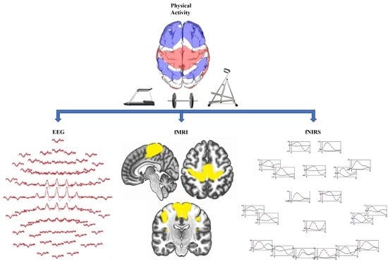
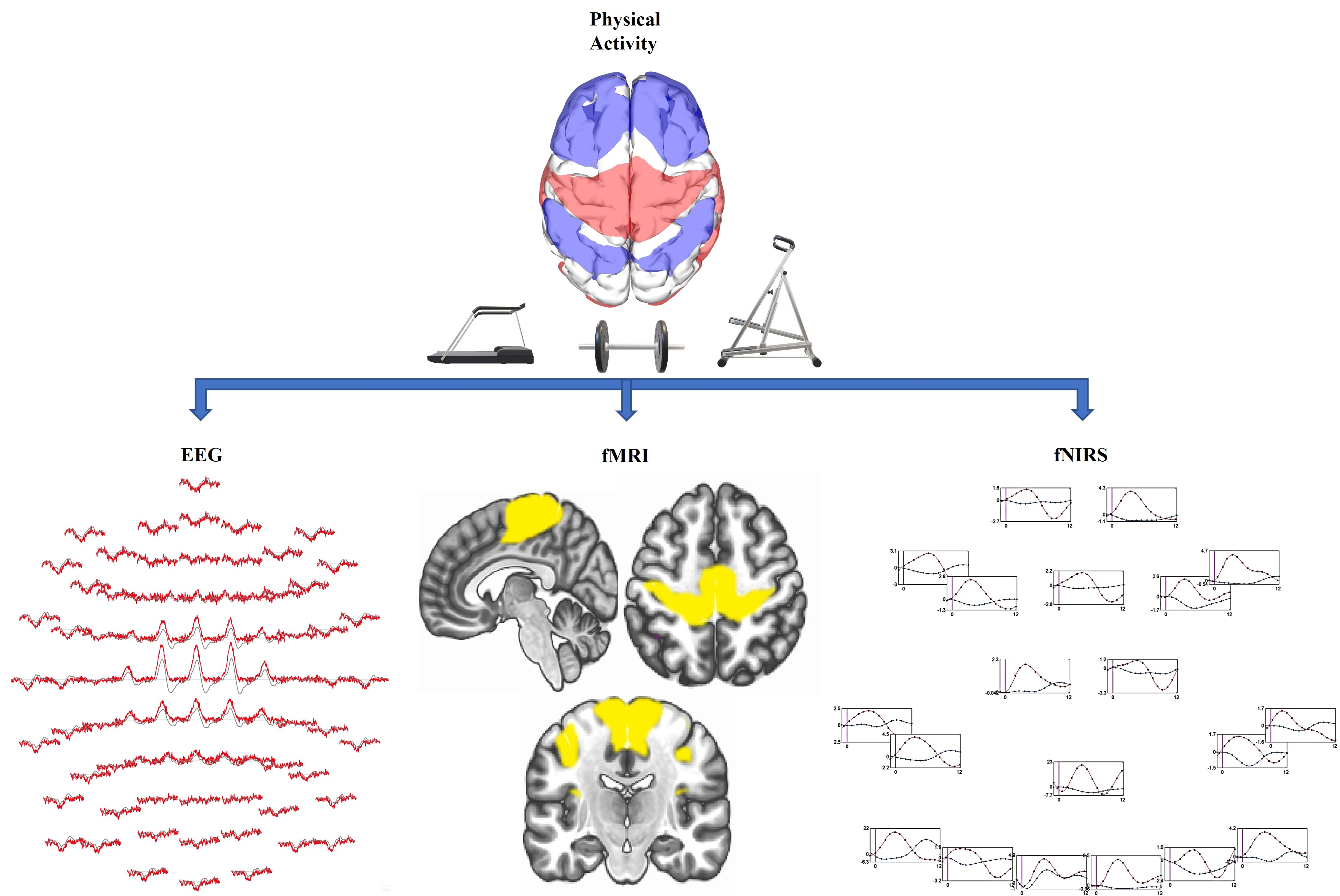
:This review is extensively focused on the enhancement of cognitive functions while performing physical exercises categorized into cardiovascular exercises, resistance training, martial arts, racquet sports, dancing and mind-body exercises. Imaging modalities, viz. functional magnetic resonance imaging (fMRI), functional near-infrared spectroscopy (fNIRS) and electroencephalography (EEG), have been included in this review. This review indicates that differences are present in cognitive functioning while changing the type of physical activity performed. This study concludes that employing fNIRS helps overcome certain limitations of fMRI. Further, the effects of physical activity on a diverse variety of the population, from active children to the old people, are discussed.
1. Introduction
Apart from physical development, cognitive development is an important domain of human growth. Human cognition comprises large-scale networks that are functionally coherent at rest and collectively active during cognitive processes [1]. Cognition alludes to “the psychological activity or process of gaining information and comprehension through idea, experience, and the sense”. It has been branched into nonsocial and social cognition. Nonsocial cognition refers to the mental abilities of an individual, such as his or her attention span, processing speed, problem solving and reasoning skills, as well as working memory. The psychological processes involved in perception, encoding, storage, retrieval and control of knowledge about oneself and others are collectively labeled as social cognition [2]. Cognitive efficiency refers to the complex construct that represents the capacity to achieve learning and problem-solving skills through the optimal usage of mental resources [3]. Many mental health disorders have been observed recently during childhood and adolescence, whereby cognition is highly associated with the quality of life of the individual. At time, the social, mental and biological components decide a person’s emotional well-being, and these can largely influence a person’s quality of life. We also studied the cognitive abilities of stroke and schizophrenia patients in this review. The lifestyles and difficulties of stroke patients were evaluated with simple reaction time tests (cognitive tests) and quality of life assessments. The attention and visuospatial skills are highly associated with the timing of a stroke, where the reaction time tasks act as a marker for quality of life [4]. Cognitive impairment decreases the quality of life over a period of 12 months from the time of diagnosis. Cognitive impairments were also set as a diagnostic criterion for schizophrenia, ranging from moderate to severe during the first outbreak and persisting throughout the period of illness. They have been shown to negatively affect the adaptive life skills of the patients during the later period of their lives [5]. Focusing the four realms of social cognition has been a core intervention in treating schizophrenia, which includes emotional processing, theory of mind (ToM), attributional bias, social perception and social knowledge. Early clinical interventions are required to provide a positive outcome [6]. Recent research on social cognitive processing is used to analyze various disorders such as anxiety, eating and mood disorders among clinical and non-clinical samples [7].
Several scientific approaches have been adapted in enhancing the performance of human cognition [8]. While most of the strategies for enhancing cognition rely on pharmacological, environmental and genetic factors [9], an embodied approach may serve as a better alternative. Such an embodied system for cognition opens the opportunity for various disciplines of science, technology, education and research to incorporate learning tools to upgrade the pedagogy [10]. Such a type of training incorporates perceptual motor activities and cognitive challenges tailored to individual needs as per their respective goals [11]. Among the factors that were examined, stress may act as a facilitating or debilitating element for cognitive function, based on variables such as magnitude, cause (due to external factors or the inherent task) and duration (acute or chronic). Sandi et.al. demonstrated that while mild stress enhances cognitive function in times of easier activities, high stress levels lead to decrements regardless of the cause [12]. Despite sparse studies, observational data has supported that tiredness, lack of attention, easy irritability, loss of sleep and an inability to manage emotions, leading to a decline in executive functioning, may be dictated by the prefrontal cortex [13]. It is noteworthy that a correlation between sleep and cognition has been well documented. In children, loss of sleep results in a decline in cognitive functions.
Reversibility was shown to be dependent on baseline cognitive functioning and involvement in social activities. In analyzing the reversion of mild cognitive impairment, higher chances of recovery were observed midlife (52%) in comparison with late life (26.6%) among a Korean community. Therefore, early interventions are more effective [14] for treating cognitive impairment. Genetics has been shown to be a major determinant for cognitive ability. However, genes encoding, dystrobrevin-binding protein 1 (DTNBP1), brain-derived neurotrophic factor (BDNF), catechol-O-methyl transferase (COMT) and apolipoprotein E (APOE), have demonstrated a repeated association with cognitive decline [15]. Age, education, gender, occupation and lifestyle factors such as smoking also impact cognitive functioning. In addition, the environment at home and around family members plays an important role (Figure 1) [16]. Green environments have also shown enhancement in cognition [17]. Nutritional intake has demonstrated effects on cognition in children and adolescents [18].
Cognitive dysfunction has great clinical significance and longstanding ramifications on a patient’s activities. Pharmacological (e.g., antidepressants) and non-pharmacological (e.g., cognitive-oriented treatments and cognitive remediation therapy (CRT)) interventions focusing predominantly on learning and memory, which potentially improve the cognitive depression of the brain, are being advanced. Non-pharmacological interventions employ cognition remediation therapy and cognitive-oriented treatments, with increased cognition in patients with depression [20]. Particular strategies have been used to promote memory, problem-solving and decision-making abilities [21]. In one study, cognitive training was employed as an intervention to enhance driving skills in elderly people. Five weeks of training was provided to enhance aspects such as attention control along with memory and processing speed. This was shown to be more relevant in the elderly population, with a major contribution to improvement of the working memory [22], since working memory forms a vital component of cognition that is effective during inhibition and switching processes, which are dependent on the maturation of the prefrontal lobes. Performing interventions in the earlier stages of childhood as described earlier in the text has shown better impacts in enhancing the functions of the prefrontal lobes, as they are not yet fully developed in children. Major techniques involve using language as a mediator for enhancement in cognition control [23]. Different categories of cognitive interventions, namely cognitive stimulation (CS), focusing on social abilities and cognition; cognitive training (CT), or the repeated practice of tasks performed in daily life; cognitive rehabilitation (CR), a personalized approach toward impaired functions; and a hybrid of CT and CS (MCTS) were employed in 33 studies for dementia patients. Assessments were made using mini-mental state examination (MMSE) and the Alzheimer’s Disease Assessment Scale-Cognition (ASAS-cog). While CS showed positive enhancements in cognition, CR, CT and MCTS did not demonstrate any significant changes [24].
Physical exercise has also been found to play a pivotal role in enhancing cognitive function for all age groups, where it elevates the levels of cerebral blood flow and growth factors, including brain-derived neurotrophic factor and neurotransmitters (e.g., dopamine and norepinephrine) [25]. Furthermore, physical exercise has served as an intervention for the aging population to enhance cognitive functioning and prevent cognitive decline [26]. Exercise has been shown to be effective in making notable lifestyle changes in the elderly population and minimizing the risk of neurological disorders such as Alzheimer’s disease [27]. While physical activity has shown increments in academic performance, it occurred mainly in females. This study highlights the need to conduct further experiments to understand the role of sex and the intensity of exercise along with the psychological variables [28]. An integration of physical activity and cognitive tasks has been established in multiple studies. Additionally, there has been an analysis of the variations of physical activities and how it impacts the cognitive state. A 20-week physical exercise program for an elderly population with moderate cognitive impairment has resulted in post–intervention improvements [29], whereas a 12-week physical activity program led to an enhancement in cognitive function and brain activation among the elderly population when combined with cognitive exercises. Clinical research has demonstrated a abundant hippocampal and basal ganglia volume, greater white matter integrity, efficient brain activity along with superior cognitive performance and academic achievements for physically active and fit children and preadolescents. Physical fitness and regular exercise have been shown to positively impact the prefrontal cortex, functional brain connectivity and executive and memory functions [30]. The impact of physical exercises has been explored in detail in the following section.
2. Types of Physical Activities That Enhance Cognitive Functions
2.1. Aerobic Exercise
Performing aerobic exercise for a period of 20 min demonstrated enhanced cognitive function among preadolescents and young adults. In this study, acute exercise was performed in a single bout at a moderate intensity for 20 min on a treadmill, with a 65–75% heart rate reserve along with a five minutes warm up and cool down [31]. A separate study also demonstrated an increase in cognitive functioning and working memory due to better cortical activity after acute aerobic exercise for 20 min with a cycling ergometer among female college students [32]. Twenty sedentary young adults performed 30 min of aerobic exercise on a stationary bike. This demonstrated an increase in activation of the left dorsolateral prefrontal cortex and left orbital frontal cortex. The control group, which did not perform exercise, did not see any benefits, but an enhanced performance in cognitive tasks was seen in the exercising group [33]. Better enhancements in cognition were seen when aerobic training was combined with CRT. BDNF, being a pivotal element for brain development and growth, has shown increased levels after aerobic exercise. However, further studies are required to identify other factors [34].
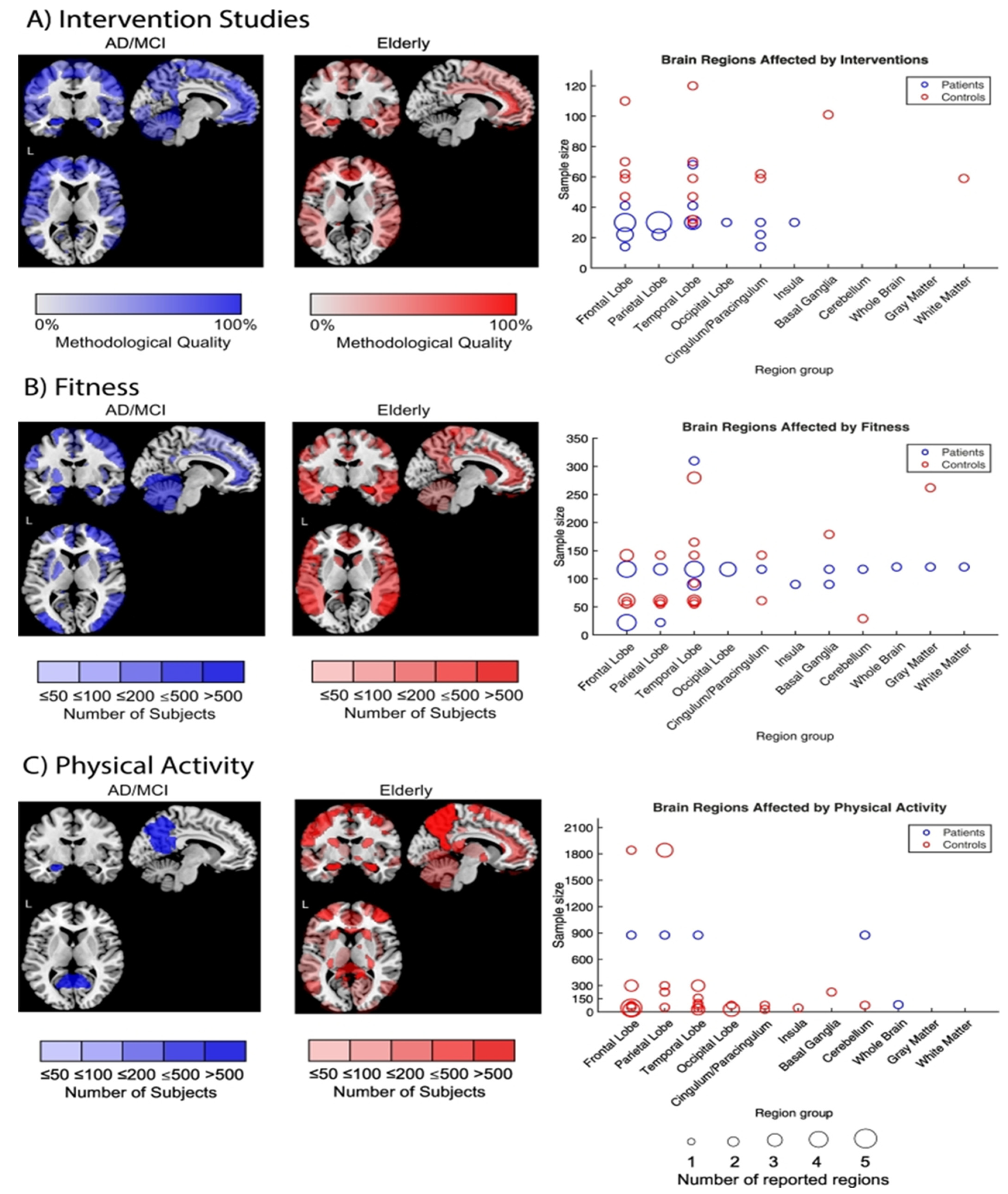
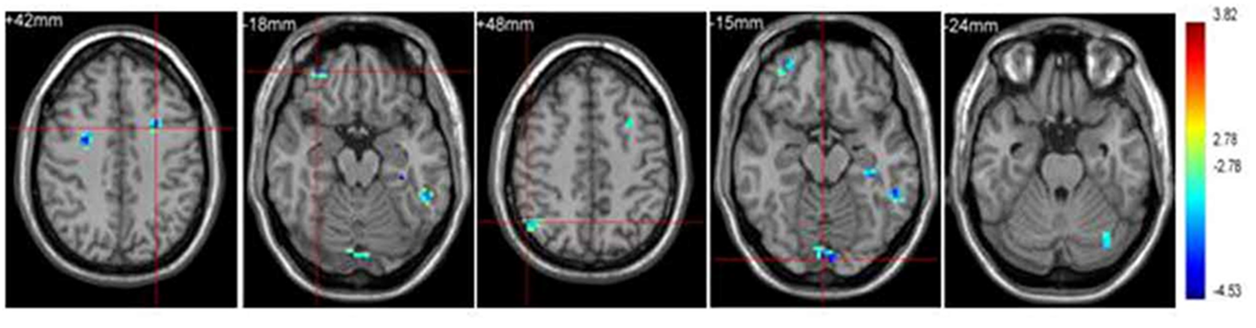
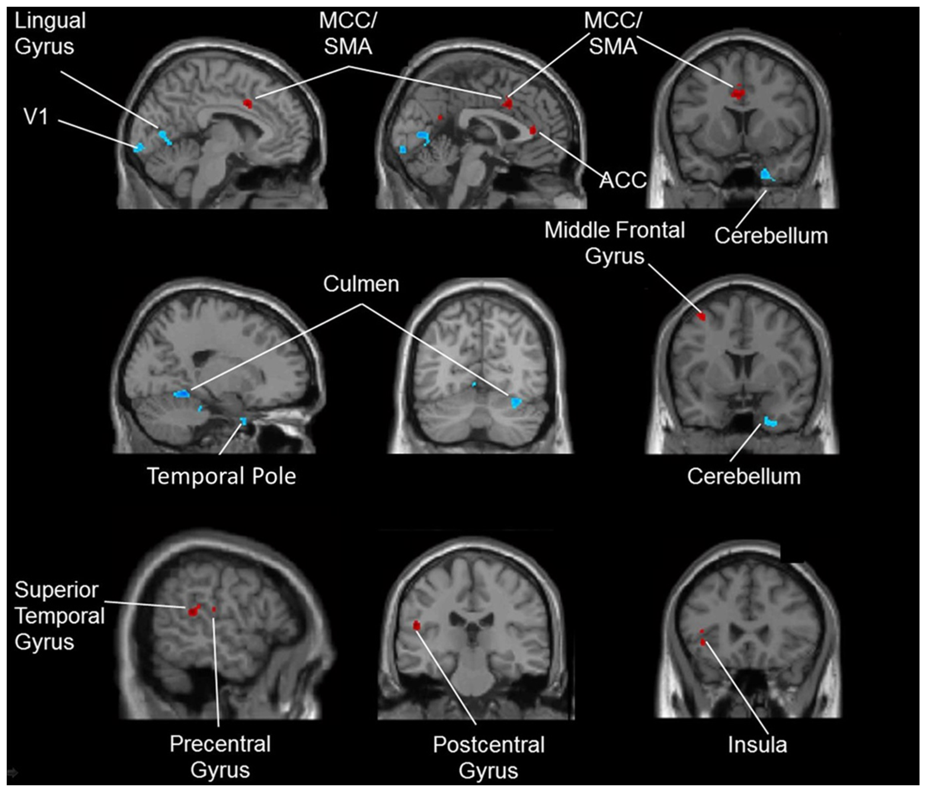
Functional MRI assessment demonstrated better cognitive control among children through aerobic fitness enhancement [35]. Regular aerobic exercise has been shown to reduce neuronal decline in the elderly population. Furthermore, it acts as an efficient mechanism for the restoration of normal functions due to changes in the brain’s structure [36]. As discussed earlier in the text, performing aerobic exercises demonstrated to be beneficial in enhancing the cognitive capabilities of the elderly population, especially in the regions of driving memory, inhibition of redundant information and switching between tasks. This was achieved predominantly by the frontal lobes of the brain. Capillary development in the brain and an increase in the length and number of dendritic synapses between neurons were among the processes involved [37]. It has been discovered that the impacts of aerobic exercises on cognition and executive functioning are due to the influence of the temporal, frontal and parietal brain regions. The frontal lobe performs a significant part in maintaining executive functioning in aging. Meanwhile, performing regular aerobic exercise showed an impact on the hippocampal region (Figure 2) [38]. Enhancements in neural efficiency were observed in seventeen volunteers with mild cognitive impairment following a twelve week exercise intervention of supervised treadmill walking. This led to an increase in cognition mainly due to the activation of 11 brain regions—especially in older adults—and has been proven effective for delaying Alzheimer’s disease [39].
2.2. Resistance Training
Resistance training has gained popularity in recent years. While it was popular only among bodybuilders and powerlifters earlier, lately it has found fresh interest among athletes in other fields, such as soccer and basketball. Recently, it has also been followed closely by several fitness enthusiasts who aim to achieve benefits such as muscular mass growth and strength. Moreover, the contribution of such training to cognitive development should not be dismissed. A study comparing the effects of 30 min of aerobics and resistance training in a group of untrained youths reported similar increases in cognition in comparison with the control group [40]. Positive correlation has been established between cortical hemodynamics of the prefrontal cortex and the handgrip strength of the individual. This was analyzed among 39 young adults aged 18–30 who used a handgrip dynamometer to assess strength and fNIRS for identifying the cortical hemodynamics of the prefrontal cortex [41].
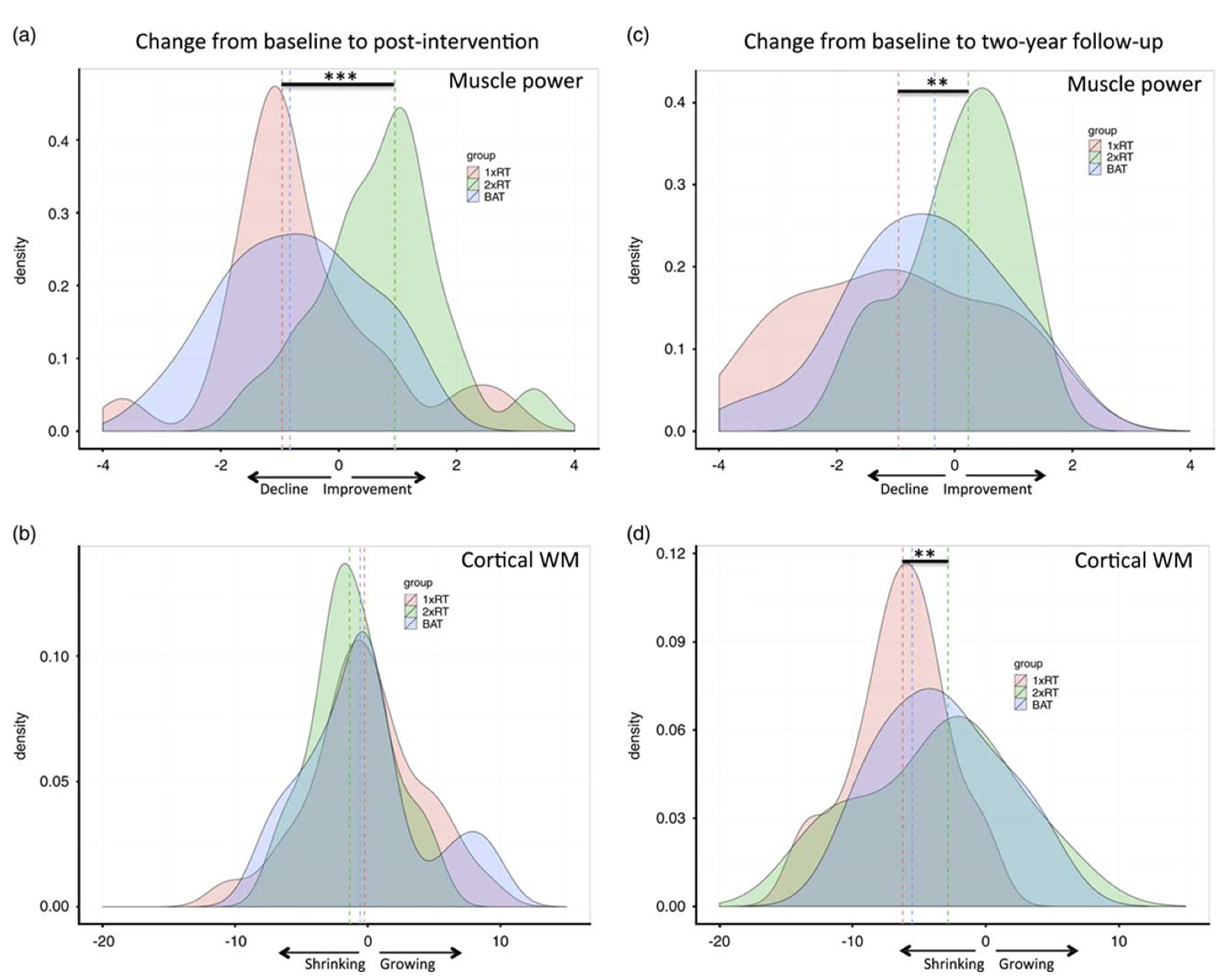
Incorporating resistance training at a frequency of twice a week in addition to aerobic exercise and balance training for a period of 52 weeks for a group of 155 elderly women showed enhancements in memory and a drop in cortical white matter size (Figure 3) [42]. Upon reviewing multiple studies of resistance training in healthy elders, cognitive differences were observed. Resistance training protocols mixed with chess and gambling can limit cognitive decline and enhance the QoL, including attention, calculation, recall and language. Resistance training potentially elevates the level of insulin-like growth factor 1 (IGF-1). IGF-1 raises the synthesis of BDNF1 and vascular endothelial growth factor (VEGF), which improves cognitive functioning [43]. A 6-month resistance training intervention was performed on aged persons with type-2 diabetes who were also at increased risk of cognitive deterioration. Resistance training’s effects on cognition have also been compared with water-based exercise among older adults. A mixture of free weights and HUR equipment operated by pressurized air weights were used to target all the primary muscle groups, such as quadriceps, pectoralis major and latissimus dorsi. Assessments of maximal muscular strength were performed after the third week. The control group solely performed basic exercises like stretching and bodyweight exercises. However, the group that performed resistance training demonstrated better increments in cognition and brain health, hence illustrating this as a cost-effective intervention for the aging population [44]. An elderly group of 100 (aged 70 years) was subjected to a six month intervention and random assignment to cognitive training or a mix of both of cognitive and resistance exercise. Resistance training was proven to be superior for improving global cognition and expanding gray matter volume in the posterior cingulate rather than cognitive training alone. Cognitive training was beneficial to memory due to greater functional communication between the hippocampus and superior frontal cortex [45]. Similar results of cognitive enhancements were found in both groups in comparison to the control group, except for the reaction time. The water-based exercise group had better results in that domain [46].
2.3. Sports
Martial arts and combat sports have brought about many benefits to cognitive health. While most forms of such sports originated in East Asia geographically, it has been popularized worldwide. In modern times, many forms of martial arts are viewed as a sport and are attributed to many health benefits including strength, power, endurance and balance [47]. Martial arts have also been studied as a means to develop cognitive functioning, given its predisposition to mind-body nature. The complex movements have a greater cortical requirement in comparison with aerobic exercises or resistance training. Certain elements of cognition, such as concentration and mindfulness, which may not be adequately developed in aerobic or resistance training sessions are enhanced in martial arts sessions. An increased regional cerebral blood flow has also been observed following more complex moves, enhancing executive functioning, which serves as a pivotal component in cognition, while aerobic exercises could only improve attention and the processing speed [48]. Magnetic resonance imaging (MRI) assessments were performed to compare 14 professional karateka and healthy controls. All the professionals demonstrated higher cognitive performances alongside improved motor performances due to the demands of the sport [49]. In another study, karate instruction was imparted to 39 sedentary children to analyze its effects on motor and cognitive development. A wide range of cognitive functions saw improvement in those who had practiced karate, which was assessed by the visual selective attention task, tasks measuring working memory function and the Tower of London test [50]. Three months of karate training had significantly benefited the cognitive functions of healthy older adults, mainly pertaining to visual tasks, executive functions, motion tasks and memory. The main components of karate are basic techniques, kata (forms) and combat practice. Assessments were performed through various questionnaires that included a memory-complaint battery of tests and MMSE [51].
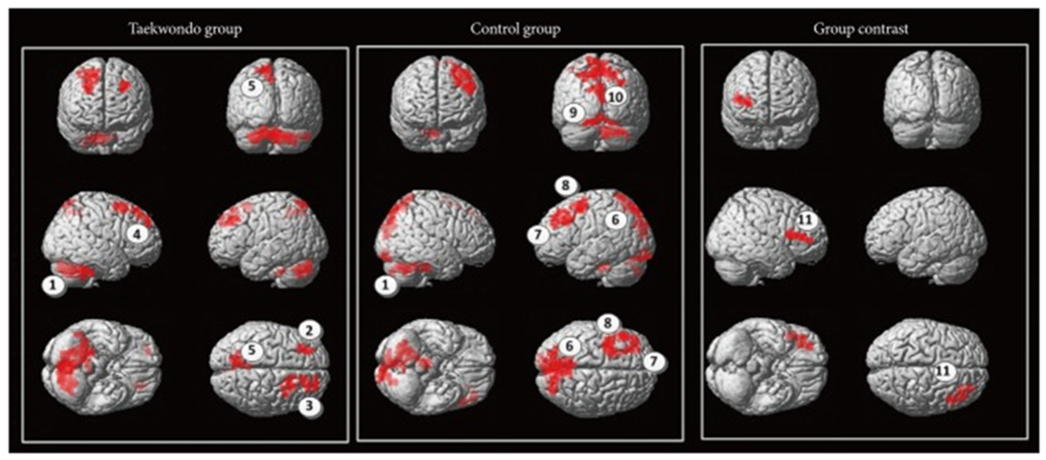
In another study comprising aged subjects, an enhancement of cognition was observed in the aspects of attention, resilience and motor reaction time following a 5-month intervention with 89 older adults aged above 70 years. The main reason was attributed to the combination of aerobic, balance and coordination functions found in this East Asian martial art [52]. In another study, 1 h of weekly taekwondo training was provided to a group of 24 healthy volunteers aged above 40 years. The intervention lasted for a period of 4 months. It comprised the components of a warm-up, muscle strengthening and stretching, followed by taekwondo-based techniques. These components were aimed at improving stances, blocking, kicking, punching and the usage of kicking pads. All of them had shown improvement in reaction time and motor timing [53]. Taekwondo was analyzed for its efficiency in implementation in a public school setting. The executive functioning skills of the students who took part in taekwondo were higher than the control group. Computer administration was employed to assess executive functioning [54]. Around 30 healthy men volunteered to analyze the effects of the Japanese martial art kendo. Kendo practitioners demonstrated higher functional connectivity, mainly between the left intraparietal sulcus and left precentral gyrus [55]. The attention network test was performed on 48 participants of 2 groups, in which one had martial arts experience and the other did not. It was seen that all the martial artists had higher working memory, attention and behavioral inhibition. While this was more common among children, adults had improved corticospinal excitability resulting from karate and better motor cortex function from taekwondo. It was also seen that most benefits, especially among older adults, were due to chronic adaptations and not acute ones [56]. When analyzing the brain connectivity in children, taekwondo was proven to be effective in enhancing functional interconnection from the cerebellum to the inferior frontal gyrus. They also had enhanced minimal frequency oscillations in the right frontal precentral gyrus and right parietal precuneus. This resulted in enhanced intelligence [19].
Higher inhibitory control (the capacity to suppress planned but unsuitable preparatory activities in the current environment) have been vital in human performance. Sports requiring open skills such as tennis have been more advantageous in enhancing this component of cognition in humans in comparison with closed skill sports such as swimming. Tennis players also demonstrate faster reactions with better accuracy during cognitive tasks. This type of skill training has been shown to benefit inhibitory control when combined with aerobic training instead of aerobic training alone [57]. It was observed that tennis was effective in enhancing action observation, anticipation and motor control through increased activation of the cerebellum and action observation network (AON) [58]. Another experiment analyzed tennis with fNIRS and observed an increase in the left premotor regions, which led to enhanced cognition, especially in unpredictable conditions [59]. To differentiate elite table tennis players and non-players, fMRI was employed. While the players had around 8 years of experience, the non-players had no prior experience. Diminished cortical activity was observed in the athletic group in the context of sports-related and unrelated visual-spatial tasks. Shorter reaction times were also seen among the players, leading to faster responses, which are indicative of higher neural efficiency. The areas studied included the right supplementary motor area, right angular gyrus, left supramarginal gyrus and right paracentral lobule. However, it must be noted that this adaptation is an effect of long-term training (Figure 4) [60]. Similar to tennis, recreational badminton players demonstrated higher anticipation due to the open skill nature of the sport, which involved the cortical areas pertaining to observation and interpretation of others’ actions. Enhancements were mainly noticed in the medial, dorsolateral and ventrolateral frontal cortices [61].
Higher anticipation rates were observed among 15 basketball players in comparison with the novices. The athletic group also displayed more accuracy for anticipation along with gaze behavior. The attribution was mainly due to the increased activity in the inferior parietal lobe and inferior frontal gyrus (Figure 5) [62]. In a study of 21 basketball players, higher portion of gray matter, mainly in the left anterior insula, inferior frontal gyrus, inferior parietal lobule and right anterior cingulate cortex, and increased functional connectivity in the resting state were observed in comparison with the novices. These adaptations enhance the executive control network, leading to better cognition (Table 1) [63].
2.4. Dance
Apart from musical experience, which has been shown to provide a positive influence on the health of the participant, the effects of dance have also been studied. When analyzing 475 amateur dancers, benefits were seen in the physical as well as social dimensions. This has raised the importance of dance as a medium in health benefits [65]. Introducing dance as an exercise among older adults showed greater improvements in brain volumes in comparison with the studies discussed in the previous section, which focused on aerobic exercises, anaerobic strength and stretching exercises. Enhanced brain volumes from the 6-month intervention caused higher cognitive functions, such as working memory and attention hence attenuating the age-related cognitive decline. This could be attributed to the multi-faceted nature of dance, encompassing spatial orientation, movement coordination, balance, endurance, interaction and communication (Figure 6) [64].
Activation of the medial superior parietal lobule and improvement in the proprioceptive and somatosensory aspects were caused by the spatial navigation of leg movements during dance and controlled muscle contractions [67]. Six months of dance intervention was performed for a cohort of seniors. There were noticeable changes in terms of attention and cognition between the intervention and control groups. Improvements were seen in the corticospinal tracts in the group that underwent intervention [68].
2.5. Yoga
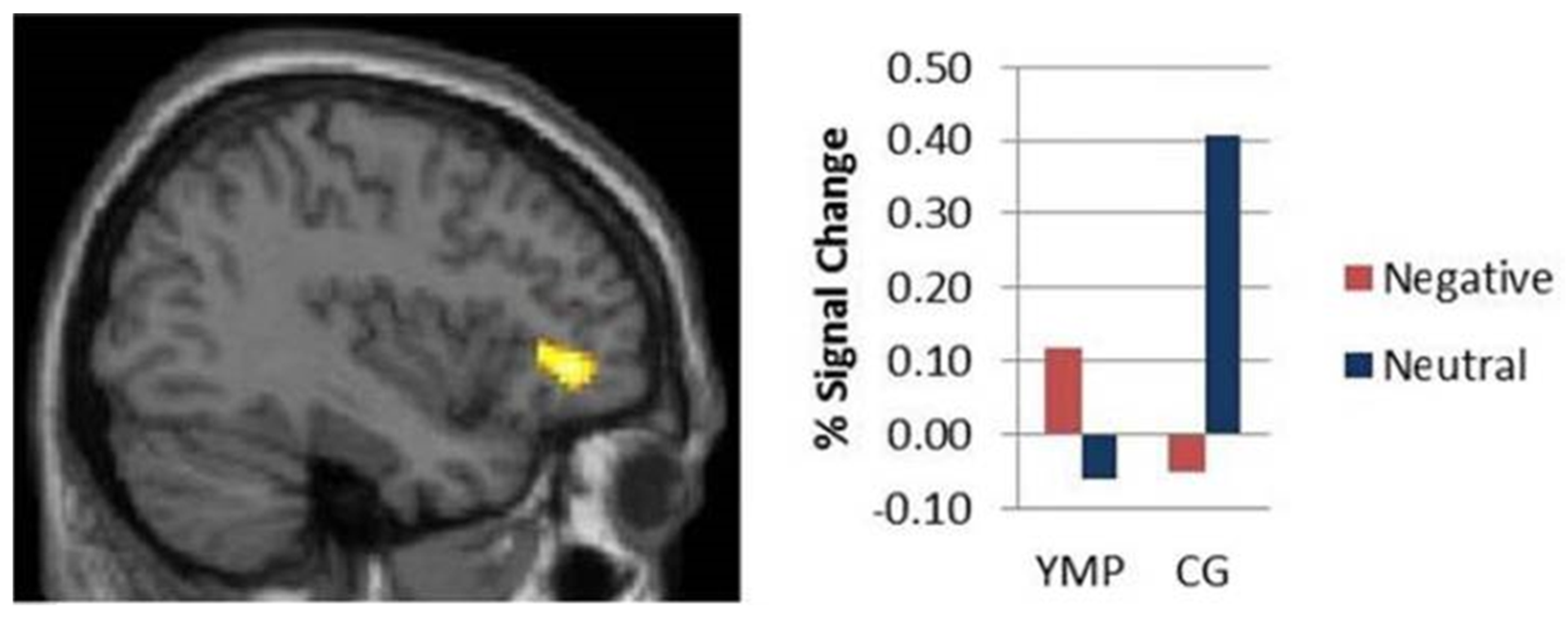
Yoga is a popular kind of mind-body workout which consists of meditation, breathing exercises and posture control. Systematic investigations have been made on its physical and psychological health benefits, and it serves as an intervention for circumstances like as arthritis, pain and musculoskeletal conditions. Certain psychological conditions such as depression and anxiety have been reverted through yoga. Over the years, its effects on enhancing cognition have been an area of active research to identify the underlying neurological correlates. The gray matter volume was higher in the left hippocampus of experienced yoga practitioners, and the reduced activity in the dorsolateral prefrontal cortex compared with the controls. Reaction times were similar in both groups [69]. Yoga has demonstrated greater activation of the prefrontal cortex, and practitioners exhibit higher executive functions independent of negative emotional stimuli [66]. Performing yoga has been displayed as a successful strategy for enhancing mindfulness, leading to prevention of age-related decline in fluid intelligence along with enhanced brain functioning [70].
3. Neuroimaging Modalities
Functional neuroimaging techniques have aided the examination of human brain function and created a revolution in neuroscience studies. Applications of these modalities have helped to assess the brain damage and to understand how the surviving brain networks are altered, which aids potentially to improve the treatment [71]. These techniques have demonstrated a wide range of diverse applications, including understanding neuropsychiatric illnesses such as obsessive-compulsive disorder [72], the neural substrates of parent–infant attachment [73] and gait disorders [74]. Neuroimaging has also been employed in analyzing the advantages of physical activity on brain health and cognitive functioning. It has shown to be an effective tool in understanding both the structural and functional elements of cognitive processes [75]. It has been shown to be effective in measuring many dimensions such as the gray matter volumes [76], cortical regions which perform an important role in episodic, spatial memory and impacted cognitive performance [77] and hippocampal volume [36]. A few key neuroimaging modalities used in examining brain function are explored below.
3.1. Functional Magnetic Resonance Imaging (fMRI)
fMRI is a popular neuroimaging technique which has facilitated several developments pertaining to medical applications, mainly for the human brain. Its advantages include the non-invasive nature of the machine and noise reduction. Applications include both the diagnosis and treatment of brain lesions [78]. In cognitive neuroscience, fMRI is important since it is employed to generate pictures of brain functions. These are important in many aspects of neuroscience, such as understanding language, social interactions of the individual, behavior and cognition [79]. It was also shown to be effective in assessing pain elicited by noxious heat in 14 healthy participants [80]. Recent improvements in the fMRI provided the ability to present high-definition images of the brain while performing various motor tasks. This has aided in comprehending various structures of the human brain, which can be affected while performing physical activity. However, constraints have existed due to the low magnetic properties and limitations in head movement while performing exercises. Due to this, predominantly only small muscle groups, such as those for performing handgrip strength, have worked in this modality, but studies have been made on cycle ergometers [81]. This method also solved a research gap of identifying the neurobiological pathways implicated in exercise-induced alterations during cognitive processing. It demonstrated that pathway variation between the elderly and younger individuals. While in the elderly, alterations were limited to the left parietal lobe, widespread changes were observed in the right hemisphere for the younger age group [82]. This method was also used to investigate and demonstrate concurrent and independent connections between different dimensions of physical fitness, working memory, and brain function in over 1000 healthy people. [83]. The major advantage of fMRI is the spatial resolution and whole-brain coverage. Considering its limited temporal resolution, correlations between multiple brain regions and temporal delays of the brain might be performed [84].
3.2. Functional near Infrared Spectroscopy (fNIRS)
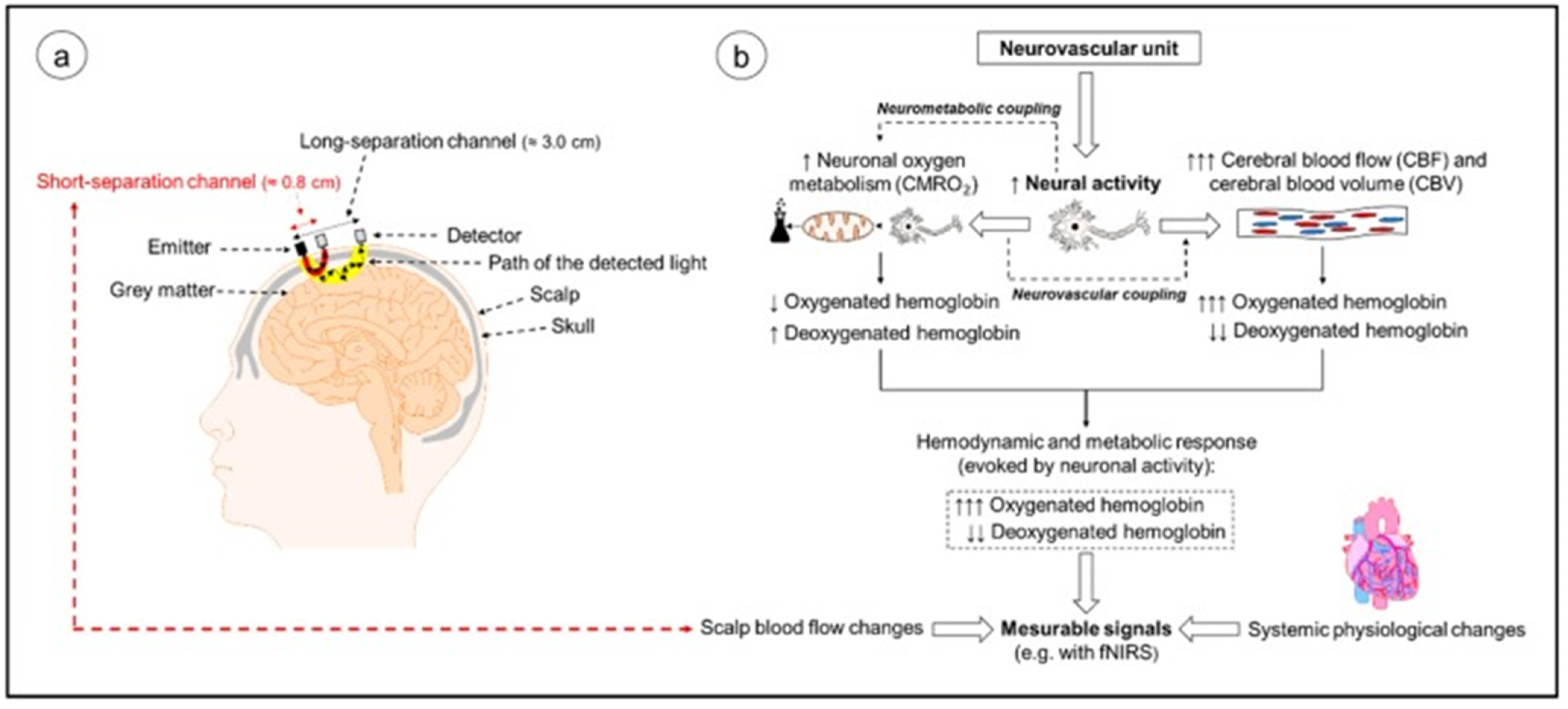
Functional near infrared spectroscopy (fNIRS) has gained popularity and rapidly advanced over the past decade in the cognitive neuroscience field. This can primarily be attributed to its advantages over other neuroimaging devices such as fMRI and EEG in terms of its safety, tolerance to bodily movement, portability and suitability for all age groups, ranging from newborns to the elderly. It has aided in the comprehension of different aspects of cognition which were not adequately demonstrated in other modalities. Those areas include functional specialization in the visual and sensorimotor systems, face and language processing and in assessing tasks performed in everyday life [85]. fNIRS has been shown to be an effective neuroimaging modality for assessing the effects of exercise on cerebral oxygenation and hemodynamics due to its capability in quantifying changes (Figure 7) [86].
It has helped for comparing the effectiveness of high-intensity aerobic exercises with moderate-intensity exercises in enhancing working memory and neural activity in patients dependent on methamphetamine (MA). The former was proven to be more effective and has been established as a therapeutic tool [87]. The neural bases of cognitive contributions in the gait were also investigated with fNIRS to record brain activation. Simple and complex walking could be assessed in adults of different age groups and those with balance disorders. Greater prefrontal cortical activation was seen in those who performed secondary tasks such as arithmetic along with walking [88]. Application of this modality was extended to understanding the cognition and attentional demands required by lower limb amputees. The amputees demonstrated similar cognitive strategies in challenging situations but had increased attention demands in regular walking [89]. Tai Chi Chuan (TCC), a popular mind body exercise involving cognitive training and movement meditations, was also examined. The effect of TCC on enhancing the brain’s prefrontal structure function and memory was observed via fMRI. However, fNIRS would be a suitable imaging modality, which measures the change in the HbO2 and HHb levels for the tasks involving body movements compared with other noninvasive neuroimaging techniques [90]. The major disadvantage of this modality for clinical use is its lack of accuracy and precision. The employment of high-density whole head optode arrays, precise sensor locations relative to the head, short-distance channels, anatomical co-registration and multi-dimensional signal processing could be performed to enhance the sensitivity of fNIRS. This could make it a widespread clinical tool for assessing brain function [91].
3.3. Electroencephalography (EEG)
Electroencephalography (EEG) measures the electric potentials of the brain and helps for investigating the cortical activity in lab and field settings. Its cost-effectiveness and compact size provide an advantage over other modalities such as functional magnetic resonance imaging (fMRI). Its high temporal resolution offers precise means for examining the cognitive processes [92]. EEG was employed in identifying the relationship between skipping and cognitive learning. Three-minute bouts of skipping had shown increments in both repetitive and variable learning [93]. Weight training was also an interest for identifying its effects on the brain and cognition. A common exercise, the bench press, was performed, and EEG served as an efficient tool for recording brain amplitudes. Significant increments were observed in the beta and gamma frequency bands. However, further studies are required to assess the changes with respect to the intensity and its effects on cognition and perception [94]. A 12-week resistance training program was performed among elderly subjects with mild cognitive impairment with elastic bands. Fifteen repetitions were employed with 65% 1RM for all the exercises. Increments in cognitive functioning were observed, mainly for the alpha and theta power [95]. The effects of yoga on brain waves were also analyzed with EEG. Positive effects from performing this mind-body exercise for enhancing cognition, along with alleviation of blood pressure and anxiety, were seen while using EEG [96]. Despite the importance in the application of representing the neuronal activity of the brain, many applications fail to take use of all of the information accessible from the brain’s dynamic sources. Localization of the sources of the brain’s signals is pivotal for any analysis [97].
4. Conclusions
This study looked at the effect of physical activity on enhancing cognitive performance in a variety of individuals, ranging from children to the elderly. The mechanism of the increase in cognitive function could be attributed to the increased hippocampal and basal ganglia volume and greater white matter integrity. Enhanced cerebral flow along with alterations in neurotransmitter release and structural changes in the central nervous system have also shown they can serve as physiological mechanisms to explain its effect. Comparisons in improvements have been made between aerobic exercises, resistance training, mind-body exercises such as yoga, racket sports such as tennis and basketball, combat sports and dance. While performing regular aerobic exercises like cycling has shown benefits, resistance training was proven to be more effective in improving certain dimensions of cognition to a greater degree than aerobic exercises. These exercises have helped via enhanced functioning of the BDNF, where it has been able to stop the neuronal decline caused by age and aid in the growth of capillaries in the brain. Major benefits included enhancements in memory and changes in brain volume. Involvement in open skill sports such as martial arts and tennis has demonstrated greater corticospinal excitability, motor cortex function, faster reaction times with better accuracy and better inhibitory control in comparison with performing aerobic exercises or resistance training. The modalities employed were fMRI and fNIRS. Even though the former was helpful for identifying brain images and functioning, there were certain limitations, such as low magnetic properties and limitation in head movement while performing the exercises. fNIRS helped to overcome these limitations though its safety, portability and suitability. It could be understood that variations in physical activity have shown different effects on cognitive functioning. More studies are required to understand the best choice of exercise for improving cognition.
Author Contributions
N.S.S., literature search and review and manuscript writing; V.V., content planning and contributing to the writing of the manuscript; P.P., content reviewing and editing; B.G., content planning and editing. All authors have read and agreed to the published version of the manuscript.
Funding
This research received no external funding.
Acknowledgments
V.V., P.P. and B.G. acknowledge support from Lee Kong Chian School of Medicine and Data Science and AI Research (DSAIR) Centre of NTU (project number ADH-11/2017-DSAIR) and the support from the Cognitive Neuro Imaging Centre (CONIC) at NTU.
Conflicts of Interest
The authors declare no conflict of interest.
References
- Dosenbach, N.U.; Fair, D.A.; Miezin, F.M.; Cohen, A.L.; Wenger, K.K.; Dosenbach, R.A.; Fox, M.D.; Snyder, A.Z.; Vincent, J.L.; Raichle, M.E. Distinct brain networks for adaptive and stable task control in humans. Proc. Natl. Acad. Sci. USA 2007, 104, 11073–11078. [Google Scholar] [CrossRef] [Green Version]
- Green, M.F.; Horan, W.P.; Lee, J. Nonsocial and social cognition in schizophrenia: Current evidence and future directions. World Psychiatry 2019, 18, 146–161. [Google Scholar] [CrossRef] [Green Version]
- Lv, M.; Liu, H.; Zhou, W.; Zheng, C. Efficiency model of micro-course study based on cognitive psychology in the college. Comput. Hum. Behav. 2020, 107, 106027. [Google Scholar] [CrossRef]
- Cumming, T.B.; Brodtmann, A.; Darby, D.; Bernhardt, J. The importance of cognition to quality of life after stroke. J. Psychosom. Res. 2014, 77, 374–379. [Google Scholar] [CrossRef] [PubMed]
- Bowie, C.R.; Harvey, P.D. Cognition in schizophrenia: Impairments, determinants, and functional importance. Psychiatr. Clin. 2005, 28, 613–633. [Google Scholar] [CrossRef]
- Javed, A.; Charles, A. The importance of social cognition in improving functional outcomes in schizophrenia. Front. Psychiatry 2018, 9, 157. [Google Scholar] [CrossRef] [PubMed]
- Teachman, B.A.; Cody, M.W.; Clerkin, E.M. Clinical Applications of Implicit Social Cognition Theories and Methods; The Guilford Press: New York, NY, USA, 2010. [Google Scholar]
- Zona, C.; Raab, M.; Fischer, M.H. Embodied perspectives on behavioral cognitive enhancement. J. Cogn. Enhanc. 2019, 3, 144–160. [Google Scholar] [CrossRef] [Green Version]
- Dubljević, V.; Venero, C.; Knafo, S. What is cognitive enhancement? In Cognitive Enhancement; Elsevier: Amsterdam, The Netherlands, 2015; pp. 1–9. [Google Scholar]
- Weisberg, S.M.; Newcombe, N.S. Embodied cognition and STEM learning: Overview of a topical collection in CR: PI. Cogn. Res. Princ. Implic. 2017, 2, 38. [Google Scholar] [CrossRef] [PubMed] [Green Version]
- Carlson, R.A.; Avraamides, M.N.; Cary, M.; Strasberg, S. What do the hands externalize in simple arithmetic? J. Exp. Psychol. Learn. Mem. Cogn. 2007, 33, 747. [Google Scholar] [CrossRef]
- Sandi, C. Stress and cognition. Wiley Interdiscip. Rev. Cogn. Sci. 2013, 4, 245–261. [Google Scholar] [CrossRef]
- Dahl, R.E. The Impact of Inadequate Sleep On Children’s Daytime Cognitive Function. Semin. Pediatr. Neurol. 1996, 3, 44–50. [Google Scholar] [CrossRef]
- Jang, A.R.; Yoon, J.Y. Factors affecting reversion from mild cognitive impairment to normal cognition in midlife to later life in Korea: A national population study. Geriatr. Gerontol. Int. 2019, 19, 1129–1135. [Google Scholar] [CrossRef]
- Harris, S.E.; Deary, I.J. The genetics of cognitive ability and cognitive ageing in healthy older people. Trends Cogn. Sci. 2011, 15, 388–394. [Google Scholar] [CrossRef]
- Kim, M.; Park, J.-M. Factors affecting cognitive function according to gender in community-dwelling elderly individuals. Epidemiol. Health 2017, 39, e2017054. [Google Scholar] [CrossRef] [Green Version]
- Wells, N.M. At home with nature: Effects of “greenness” on children’s cognitive functioning. Environ. Behav. 2000, 32, 775–795. [Google Scholar] [CrossRef] [Green Version]
- Bellisle, F. Effects of diet on behaviour and cognition in children. Br. J. Nutr. 2004, 92, S227–S232. [Google Scholar] [CrossRef] [Green Version]
- Kim, Y.J.; Cha, E.J.; Kim, S.M.; Kang, K.D.; Han, D.H. The effects of taekwondo training on brain connectivity and body intelligence. Psychiatry Investig. 2015, 12, 335. [Google Scholar] [CrossRef] [PubMed] [Green Version]
- Baune, B.T.; Renger, L. Pharmacological and non-pharmacological interventions to improve cognitive dysfunction and functional ability in clinical depression–a systematic review. Psychiatry Res. 2014, 219, 25–50. [Google Scholar] [CrossRef] [PubMed]
- Clark, G.J.F.; Schlabach, T.L. Systematic review of occupational therapy interventions to improve cognitive development in children ages birth–5 years. Am. J. Occup. Ther. 2013, 67, 425–430. [Google Scholar] [CrossRef] [PubMed] [Green Version]
- Seidler, R.D.; Bernard, J.A.; Buschkuehl, M.; Jaeggi, S.; Jonides, J.; Humfleet, J. Cognitive Training as an Intervention to Improve Driving Ability in the Older Adult; Michigan Center for Advancing Safe Transportation Throughout the Lifespan: Ann Arbor, MI, USA, 2010.
- Kray, J.; Ferdinand, N.K. How to improve cognitive control in development during childhood: Potentials and limits of cognitive interventions. Child Dev. Perspect. 2013, 7, 121–125. [Google Scholar] [CrossRef]
- Huntley, J.; Gould, R.; Liu, K.; Smith, M.; Howard, R. Do cognitive interventions improve general cognition in dementia? A meta-analysis and meta-regression. BMJ Open 2015, 5, e005247. [Google Scholar] [CrossRef] [Green Version]
- Dietz, P. The influence of sports on cognitive task performance—A critical overview. Cogn. Enhanc. 2013, 67–72. [Google Scholar]
- Lautenschlager, N.T.; Almeida, O.P. Physical activity and cognition in old age. Curr. Opin. Psychiatry 2006, 19, 190–193. [Google Scholar] [CrossRef] [PubMed]
- Kramer, A.F.; Erickson, K.I. Capitalizing on cortical plasticity: Influence of physical activity on cognition and brain function. Trends Cogn. Sci. 2007, 11, 342–348. [Google Scholar] [CrossRef]
- Esteban-Cornejo, I.; Tejero-Gonzalez, C.M.; Sallis, J.F.; Veiga, O.L. Physical activity and cognition in adolescents: A systematic review. J. Sci. Med. Sport 2015, 18, 534–539. [Google Scholar] [CrossRef] [PubMed]
- Bademli, K.; Lok, N.; Canbaz, M.; Lok, S. Effects of Physical Activity Program on cognitive function and sleep quality in elderly with mild cognitive impairment: A randomized controlled trial. Perspect. Psychiatr. Care 2019, 55, 401–408. [Google Scholar] [CrossRef] [PubMed]
- Erickson, K.I.; Hillman, C.H.; Kramer, A.F. Physical activity, brain, and cognition. Curr. Opin. Behav. Sci. 2015, 4, 27–32. [Google Scholar] [CrossRef]
- Chu, C.-H.; Kramer, A.F.; Song, T.-F.; Wu, C.-H.; Hung, T.-M.; Chang, Y.-K. Acute exercise and neurocognitive development in preadolescents and young adults: An ERP study. Neural Plast. 2017, 2017, 2631909. [Google Scholar] [CrossRef] [Green Version]
- Li, L.; Men, W.-W.; Chang, Y.-K.; Fan, M.-X.; Ji, L.; Wei, G.-X. Acute aerobic exercise increases cortical activity during working memory: A functional MRI study in female college students. PLoS ONE 2014, 9, e99222. [Google Scholar] [CrossRef]
- Zhaang, Y.; Shi, W.; Wang, H.; Liu, M.; Tang, D. The impact of acute exercise on implicit cognitive reappraisal in association with left dorsolateral prefronta activation: A fNIRS study. Behav. Brain Res. 2021, 406, 113233. [Google Scholar] [CrossRef]
- Firth, J.; Stubbs, B.; Vancampfort, D.; Schuch, F.; Lagopoulos, J.; Rosenbaum, S.; Ward, P.B. Effect of aerobic exercise on hippocampal volume in humans: A systematic review and meta-analysis. NeuroImage 2018, 166, 230–238. [Google Scholar] [CrossRef] [PubMed]
- Chaddock-Heyman, L.; Erickson, K.I.; Voss, M.; Knecht, A.; Pontifex, M.B.; Castelli, D.; Hillman, C.; Kramer, A. The effects of physical activity on functional MRI activation associated with cognitive control in children: A randomized controlled intervention. Front. Hum. Neurosci. 2013, 7, 72. [Google Scholar] [CrossRef] [PubMed] [Green Version]
- Gligoroska, J.P.; Manchevska, S. The effect of physical activity on cognition–physiological mechanisms. Mater. Socio-Med. 2012, 24, 198. [Google Scholar] [CrossRef] [PubMed] [Green Version]
- Colcombe, S.J.; Erickson, K.I.; Scalf, P.E.; Kim, J.S.; Prakash, R.; McAuley, E.; Elavsky, S.; Marquez, D.X.; Hu, L.; Kramer, A.F. Aerobic exercise training increases brain volume in aging humans. J. Gerontol. Ser. A Biol. Sci. Med. Sci. 2006, 61, 1166–1170. [Google Scholar] [CrossRef] [PubMed] [Green Version]
- Haeger, A.; Costa, A.S.; Schulz, J.B.; Reetz, K. Cerebral changes improved by physical activity during cognitive decline: A systematic review on MRI studies. NeuroImage Clin. 2019, 23, 101933. [Google Scholar] [CrossRef] [PubMed]
- Smith, J.C.; Nielson, K.A.; Antuono, P.; Lyons, J.-A.; Hanson, R.J.; Butts, A.M.; Hantke, N.C.; Verber, M.D. Semantic memory functional MRI and cognitive function after exercise intervention in mild cognitive impairment. J. Alzheimer’s Dis. 2013, 37, 197–215. [Google Scholar] [CrossRef] [PubMed] [Green Version]
- Harveson, A.T.; Hannon, J.C.; Brusseau, T.A.; Podlog, L.; Papadopoulos, C.; Durrant, L.H.; Hall, M.S.; Kang, K.-d. Acute effects of 30 minutes resistance and aerobic exercise on cognition in a high school sample. Res. Q. Exerc. Sport 2016, 87, 214–220. [Google Scholar] [CrossRef] [PubMed]
- Herold, F.; Behrendt, T.; Torpel, A.; Hamacher, D.; Muller, N.G.; Schega, L. Cortical hemodynamics as a function of handgrip strength and cognitive performance: A cross-sectional fNIRS study in younger adults. BMC Neurosci. 2021, 22, 10. [Google Scholar] [CrossRef]
- Best, J.R.; Chiu, B.K.; Hsu, C.L.; Nagamatsu, L.S.; Liu-Ambrose, T. Long-term effects of resistance exercise training on cognition and brain volume in older women: Results from a randomized controlled trial. J. Int. Neuropsychol. Soc. 2015, 21, 745–756. [Google Scholar] [CrossRef]
- Chang, Y.-K.; Pan, C.-Y.; Chen, F.-T.; Tsai, C.-L.; Huang, C.-C. Effect of resistance-exercise training on cognitive function in healthy older adults: A review. J. Aging Phys. Act. 2012, 20, 497–517. [Google Scholar] [CrossRef] [Green Version]
- Furlano, J.A.; Nagamatsu, L.S. Feasibility of a 6-month pilot randomised controlled trial of resistance training on cognition and brain health in Canadian older adults at-risk for diabetes: Study protocol. BMJ Open 2019, 9, e032047. [Google Scholar] [CrossRef]
- Suo, C.; Singh, M.F.; Gates, N.; Wen, W.; Sachdev, P.; Brodaty, H.; Saigal, N.; Wilson, G.C.; Meiklejohn, J.; Singh, N. Therapeutically relevant structural and functional mechanisms triggered by physical and cognitive exercise. Mol. Psychiatry 2016, 21, 1633–1642. [Google Scholar] [CrossRef] [Green Version]
- Bento-Torres, N.V.O.; Bento-Torres, J.; Tomás, A.M.; Souza, L.G.T.d.; Freitas, J.O.d.; Pantoja, J.A.d.S.; Picanço-Diniz, C.W. Water-based exercise and resistance training improve cognition in older adults. Rev. Bras. De Med. Do Esporte 2019, 25, 71–75. [Google Scholar] [CrossRef]
- Burke, D.; Al-Adawi, S.; Lee, Y.; Audette, J. Martial arts as sport and therapy. J. Sports Med. Phys. Fit. 2007, 47, 96. [Google Scholar]
- Douris, P.; Douris, C.; Balder, N.; LaCasse, M.; Rand, A.; Tarapore, F.; Zhuchkan, A.; Handrakis, J. Martial art training and cognitive performance in middle-aged adults. J. Hum. Kinet. 2015, 47, 277. [Google Scholar] [CrossRef] [PubMed] [Green Version]
- Berti, B.; Momi, D.; Sprugnoli, G.; Neri, F.; Bonifazi, M.; Rossi, A.; Muscettola, M.M.; Benocci, R.; Santarnecchi, E.; Rossi, S. Peculiarities of Functional Connectivity—Including Cross-Modal Patterns—In Professional Karate Athletes: Correlations with Cognitive and Motor Performances. Neural Plast. 2019, 2019, 6807978. [Google Scholar] [CrossRef]
- Alesi, M.; Bianco, A.; Padulo, J.; Vella, F.P.; Petrucci, M.; Paoli, A.; Palma, A.; Pepi, A. Motor and cognitive development: The role of karate. Muscles Ligaments Tendons J. 2014, 4, 114. [Google Scholar] [CrossRef] [PubMed]
- Lopes Filho, B.J.P.; Oliveira, C.R.d.; Gottlieb, M.G.V. Effects of karate-dŌ training in older adults cognition: Randomized controlled trial. J. Phys. Educ. 2019, 30. [Google Scholar] [CrossRef] [Green Version]
- Witte, K.; Kropf, S.; Darius, S.; Emmermacher, P.; Böckelmann, I. Comparing the effectiveness of karate and fitness training on cognitive functioning in older adults—A randomized controlled trial. J. Sport Health Sci. 2016, 5, 484–490. [Google Scholar] [CrossRef] [Green Version]
- Pons Van Dijk, G.; Lenssen, A.; Leffers, P.; Kingma, H.; Lodder, J. Taekwondo training improves balance in volunteers over 40. Front. Aging Neurosci. 2013, 5, 10. [Google Scholar] [CrossRef] [Green Version]
- Lakes, K.D.; Bryars, T.; Sirisinahal, S.; Salim, N.; Arastoo, S.; Emmerson, N.; Kang, D.; Shim, L.; Wong, D.; Kang, C.J. The healthy for life taekwondo pilot study: A preliminary evaluation of effects on executive function and BMI, feasibility, and acceptability. Ment. Health Phys. Act. 2013, 6, 181–188. [Google Scholar] [CrossRef] [Green Version]
- Fujiwara, H.; Ueno, T.; Yoshimura, S.; Kobayashi, K.; Miyagi, T.; Oishi, N.; Murai, T. Martial arts “Kendo” and the motivation network during attention processing: An fMRI study. Front. Hum. Neurosci. 2019, 13, 170. [Google Scholar] [CrossRef] [Green Version]
- Johnstone, A.; Marí-Beffa, P. The effects of martial arts training on attentional networks in typical adults. Front. Psychol. 2018, 9, 80. [Google Scholar] [CrossRef]
- Wang, C.-H.; Chang, C.-C.; Liang, Y.-M.; Shih, C.-M.; Chiu, W.-S.; Tseng, P.; Hung, D.L.; Tzeng, O.J.; Muggleton, N.G.; Juan, C.-H. Open vs. closed skill sports and the modulation of inhibitory control. PLoS ONE 2013, 8, e55773. [Google Scholar] [CrossRef] [Green Version]
- Balser, N.; Lorey, B.; Pilgramm, S.; Naumann, T.; Kindermann, S.; Stark, R.; Zentgraf, K.; Williams, A.M.; Munzert, J. The influence of expertise on brain activation of the action observation network during anticipation of tennis and volleyball serves. Front. Hum. Neurosci. 2014, 8, 568. [Google Scholar] [CrossRef] [Green Version]
- Balardin, J.B.; Zimeo Morais, G.A.; Furucho, R.A.; Trambaiolli, L.; Vanzella, P.; Biazoli, C., Jr.; Sato, J.R. Imaging brain function with functional near-infrared spectroscopy in unconstrained environments. Front. Hum. Neurosci. 2017, 11, 258. [Google Scholar] [CrossRef] [PubMed]
- Guo, Z.; Li, A.; Yu, L. “Neural efficiency” of athletes’ brain during visuo-spatial task: An fMRI study on table tennis players. Front. Behav. Neurosci. 2017, 11, 72. [Google Scholar] [CrossRef] [PubMed] [Green Version]
- Wright, M.J.; Bishop, D.T.; Jackson, R.C.; Abernethy, B. Functional MRI reveals expert-novice differences during sport-related anticipation. Neuroreport 2010, 21, 94–98. [Google Scholar] [CrossRef] [Green Version]
- Wu, Y.; Zeng, Y.; Zhang, L.; Wang, S.; Wang, D.; Tan, X.; Zhu, X.; Zhang, J. The role of visual perception in action anticipation in basketball athletes. Neuroscience 2013, 237, 29–41. [Google Scholar] [CrossRef]
- Tan, X.-Y.; Pi, Y.-L.; Wang, J.; Li, X.-P.; Zhang, L.-L.; Dai, W.; Zhu, H.; Ni, Z.; Zhang, J.; Wu, Y. Morphological and functional differences between athletes and novices in cortical neuronal networks. Front. Hum. Neurosci. 2017, 10, 660. [Google Scholar] [CrossRef] [PubMed] [Green Version]
- Rehfeld, K.; Lüders, A.; Hökelmann, A.; Lessmann, V.; Kaufmann, J.; Brigadski, T.; Müller, P.; Müller, N.G. Dance training is superior to repetitive physical exercise in inducing brain plasticity in the elderly. PLoS ONE 2018, 13, e0196636. [Google Scholar] [CrossRef] [Green Version]
- Quiroga Murcia, C.; Kreutz, G.; Clift, S.; Bongard, S. Shall we dance? An exploration of the perceived benefits of dancing on well-being. Arts Health 2010, 2, 149–163. [Google Scholar] [CrossRef]
- Froeliger, B.; Garland, E.L.; Modlin, L.A.; McClernon, F.J. Neurocognitive correlates of the effects of yoga meditation practice on emotion and cognition: A pilot study. Front. Integr. Neurosci. 2012, 6, 48. [Google Scholar] [CrossRef] [Green Version]
- Brown, S.; Martinez, M.J.; Parsons, L.M. The neural basis of human dance. Cereb. Cortex 2006, 16, 1157–1167. [Google Scholar] [CrossRef]
- Sejnoha Minsterova, A.; Klobusiakova, P.; Kropacova, S.; Novakova, L.; Brabenec, L.; Balazova, Z.; Grmela, R.; Skotakova, A.; Svobodova, L.; Rektorova, I. Multishell Diffusion MRI Reflects Improved Physical Fitness Induced by Dance Intervention. Neural Plast. 2020, 2020, 8836925. [Google Scholar] [CrossRef]
- Gothe, N.P.; Hayes, J.M.; Temali, C.; Damoiseaux, J.S. Differences in brain structure and function among yoga practitioners and controls. Front. Integr. Neurosci. 2018, 12, 26. [Google Scholar] [CrossRef] [PubMed]
- Gard, T.; Taquet, M.; Dixit, R.; Hölzel, B.K.; de Montjoye, Y.-A.; Brach, N.; Salat, D.H.; Dickerson, B.C.; Gray, J.R.; Lazar, S.W. Fluid intelligence and brain functional organization in aging yoga and meditation practitioners. Front. Aging Neurosci. 2014, 6, 76. [Google Scholar] [CrossRef] [PubMed]
- Ward, N.S. Chapter 11: Neurological Rehabilitation. In Functional Neuroimaging; Elsevier Inc.: Amsterdam, The Netherlands, 2013; Volume 110. [Google Scholar]
- Saxena, S.; Rauch, S.L. Functional neuroimaging and the neuroanatomy of obsessive-compulsive disorder. Psychiatr. Clin. N. Am. 2000, 23, 563–586. [Google Scholar] [CrossRef]
- Swain, J.E. Baby stimuli and the parent brain: Functional neuroimaging of the neural substrates of parent-infant attachment. Psychiatry (Edgmont) 2008, 5, 28. [Google Scholar] [PubMed]
- Bakker, M.; Verstappen, C.; Bloem, B.; Toni, I. Recent advances in functional neuroimaging of gait. J. Neural Transm. 2007, 114, 1323–1331. [Google Scholar] [CrossRef] [Green Version]
- Nowrangi, M.A.; Lyketsos, C.; Rao, V.; Munro, C.A. Systematic review of neuroimaging correlates of executive functioning: Converging evidence from different clinical populations. J. Neuropsychiatry Clin. Neurosci. 2014, 26, 114–125. [Google Scholar] [CrossRef] [PubMed] [Green Version]
- Sexton, C.E.; Betts, J.F.; Demnitz, N.; Dawes, H.; Ebmeier, K.P.; Johansen-Berg, H. A systematic review of MRI studies examining the relationship between physical fitness and activity and the white matter of the ageing brain. Neuroimage 2016, 131, 81–90. [Google Scholar] [CrossRef] [PubMed] [Green Version]
- Shimada, H.; Ishii, K.; Makizako, H.; Ishiwata, K.; Oda, K.; Suzukawa, M. Effects of exercise on brain activity during walking in older adults: A randomized controlled trial. J. Neuroeng. Rehabil. 2017, 14, 1–9. [Google Scholar] [CrossRef] [PubMed] [Green Version]
- Tabelow, K.; Polzehl, J. Statistical parametric maps for functional MRI experiments in R: The package fmri. J. Stat. Softw. 2011, 44, 6355. [Google Scholar] [CrossRef] [Green Version]
- Alač, M.; Hutchins, E. I see what you are saying: Action as cognition in fMRI brain mapping practice. J. Cogn. Cult. 2004, 4, 629–661. [Google Scholar] [CrossRef] [Green Version]
- Wager, T.D.; Atlas, L.Y.; Lindquist, M.A.; Roy, M.; Woo, C.-W.; Kross, E. An fMRI-based neurologic signature of physical pain. N. Engl. J. Med. 2013, 368, 1388–1397. [Google Scholar] [CrossRef] [Green Version]
- Fontes, E.B.; Okano, A.H.; De Guio, F.; Schabort, E.J.; Min, L.L.; Basset, F.A.; Stein, D.J.; Noakes, T.D. Brain activity and perceived exertion during cycling exercise: An fMRI study. Br. J. Sports Med. 2015, 49, 556–560. [Google Scholar] [CrossRef]
- Yu, Q.; Herold, F.; Becker, B.; Klugah-Brown, B.; Zhang, Y.; Perrey, S.; Veronese, N.; Müller, N.G.; Kramer, A.F.; Zou, L. Cognitive benefits of exercise interventions: An fMRI activation likelihood estimation meta-analysis. Brain Struct. Funct. 2021, 226, 601–619. [Google Scholar] [CrossRef]
- Ishihara, T.; Miyazaki, A.; Tanaka, H.; Matsuda, T. Identification of the brain networks that contribute to the interaction between physical function and working memory: An fMRI investigation with over 1,000 healthy adults. NeuroImage 2020, 221, 117152. [Google Scholar] [CrossRef]
- Misaki, M.; Kerr, K.L.; Ratliff, E.L.; Cosgrove, K.T.; Simmons, W.K.; Morris, A.S.; Bodurka, J. Beyond synchrony: The capacity of fMRI hyperscanning for the study of human social interaction. Soc. Cogn. Affect. Neurosci. 2021, 16, 84–92. [Google Scholar] [CrossRef]
- Pinti, P.; Tachtsidis, I.; Hamilton, A.; Hirsch, J.; Aichelburg, C.; Gilbert, S.; Burgess, P.W. The present and future use of functional near-infrared spectroscopy (fNIRS) for cognitive neuroscience. Ann. N. Y. Acad. Sci. 2020, 1464, 5. [Google Scholar] [CrossRef] [PubMed]
- Herold, F.; Wiegel, P.; Scholkmann, F.; Müller, N.G. Applications of functional near-infrared spectroscopy (fNIRS) neuroimaging in exercise–cognition science: A systematic, methodology-focused review. J. Clin. Med. 2018, 7, 466. [Google Scholar] [CrossRef] [Green Version]
- Chen, Y.; Lu, Y.; Zhou, C.; Wang, X. The effects of aerobic exercise on working memory in methamphetamine-dependent patients: Evidence from combined fNIRS and ERP. Psychol. Sport Exerc. 2020, 49, 101685. [Google Scholar] [CrossRef]
- Pelicioni, P.H.; Tijsma, M.; Lord, S.R.; Menant, J. Prefrontal cortical activation measured by fNIRS during walking: Effects of age, disease and secondary task. PeerJ 2019, 7, e6833. [Google Scholar] [CrossRef] [Green Version]
- Schack, J.; Pripp, A.H.; Mirtaheri, P.; Steen, H.; Güler, E.; Gjøvaag, T. Increased prefrontal cortical activation during challenging walking conditions in persons with lower limb amputation–an fNIRS observational study. Physiother. Theory Pract. 2020, 5, 1–11. [Google Scholar] [CrossRef]
- Xie, H.; Zhang, M.; Huo, C.; Xu, G.; Li, Z.; Fan, Y. Tai Chi Chuan exercise related change in brain function as assessed by functional near–infrared spectroscopy. Sci. Rep. 2019, 9, 13198. [Google Scholar] [CrossRef]
- Chen, W.L.; Wagner, J.; Heugel, N.; Suagr, J.; Lee, Y.W.; Conant, L.; Malloy, M.; Heffernan, J.; Quirk, B.; Zinos, A.; et al. Functional Near-Infrared Spectroscopy and Its Clinical Application in the Field of Neuroscience: Advances and Future Directions. Front. Neurosci. 2020, 14, 724. [Google Scholar] [CrossRef]
- Wang, C.-H.; Moreau, D.; Kao, S.-C. From the lab to the field: Potential applications of dry EEG systems to understand the brain-behavior relationship in sports. Front. Neurosci. 2019, 13, 893. [Google Scholar] [CrossRef] [Green Version]
- John, A.; Schöllhorn, W.I. Acute effects of instructed and self-created variable rope skipping on EEG brain activity and heart rate variability. Front. Behav. Neurosci. 2018, 12, 311. [Google Scholar] [CrossRef]
- Engchuan, P.; Wongsuphasawat, K.; Sittiprapaporn, P. Changes of EEG Power Spectra in Bench Press Weight Training Exercise. In Proceedings of the 14th International Conference on Electrical Engineering/Electronics, Computer, Telecommunications and Information Technology (ECTI-CON), Phuket, Thailand, 27–30 June 2017; pp. 13–16. [Google Scholar]
- Hong, S.-G.; Kim, J.-H.; Jun, T.-W. Effects of 12-week resistance exercise on electroencephalogram patterns and cognitive function in the elderly with mild cognitive impairment: A randomized controlled trial. Clin. J. Sport Med. 2018, 28, 500–508. [Google Scholar] [CrossRef]
- Gaur, S.; Panjwani, U.; Kumar, B. EEG Brain Wave Dynamics: A Systematic Review and Meta Analysis on Eff ect of Yoga on Mind Relaxation. J. Biomed. Res. Environ. Sci. 2020, 1, 353–362. [Google Scholar] [CrossRef]
- Nara, S.; Sheoran, P. Advancements in EEG source localisation methods. Int. J. Med. Eng. Inform. 2018, 10, 30–48. [Google Scholar] [CrossRef]
Figure 1.
Taekwondo group demonstrating enhanced functional connectivity (Reprinted with permission [19]).
Figure 1.
Taekwondo group demonstrating enhanced functional connectivity (Reprinted with permission [19]).

Figure 2.
Overview of the brain regions affected by the intervention studies (A) intervention studies, (B) Fitness, (C) Physical activity. Blue figures indicate healthy adults, and red figures refer to the elderly. Comparisons were made between the volume and cortical thickness (Reprinted with permission [38]).

Figure 3.
Impact of treatment conditions on change in peak muscle power and white matter volume from baseline to post-intervention (a,b) and from baseline to 2-year follow-up (c,d). Peak muscle power (in watts) values have been divided by 100. Cortical white matter volumes are expressed in cm3, and the dashed vertical lines represent the median score for each distribution. ** p < 0.01. *** p < 0.001. (Used with permission [42]).

Figure 4.
The brain regions (right supplementary motor area, right paracentral lobule, left supramarginal gyrus and right angular gyrus) that were activated for the table tennis players (Used with permission [60]).
Figure 4.
The brain regions (right supplementary motor area, right paracentral lobule, left supramarginal gyrus and right angular gyrus) that were activated for the table tennis players (Used with permission [60]).

Figure 5.
Higher activity was observed in the inferior parietal lobule and frontal gyrus in athletes compared with novices (Used with permission [64]).
Figure 5.
Higher activity was observed in the inferior parietal lobule and frontal gyrus in athletes compared with novices (Used with permission [64]).

Figure 6.
The dance group demonstrated higher volumes in the frontal and temporal cortical areas compared with the sports group. Red-colored regions provide an overview of the regions ([66] with permission).
Figure 6.
The dance group demonstrated higher volumes in the frontal and temporal cortical areas compared with the sports group. Red-colored regions provide an overview of the regions ([66] with permission).

Figure 7.
(a) Neural activity-induced oxygenation, changes in neurovascular units and changes in cerebral hemodynamics. (b) An illustration of NIRS on the human head and assumed shape of light and “long separation channels” (Used with permission [86] ).
Figure 7.
(a) Neural activity-induced oxygenation, changes in neurovascular units and changes in cerebral hemodynamics. (b) An illustration of NIRS on the human head and assumed shape of light and “long separation channels” (Used with permission [86] ).


Table 1.
Larger gray matter volumes were observed in basketball players in multiple areas, including the right precuneus, left anterior insula, right anterior cingulate cortex, left inferior frontal gyrus and left inferior parietal lobule (Used with permission [63]).
Table 1.
Larger gray matter volumes were observed in basketball players in multiple areas, including the right precuneus, left anterior insula, right anterior cingulate cortex, left inferior frontal gyrus and left inferior parietal lobule (Used with permission [63]).
| Brain Region | Side | x | y | z | Voxels | t-Value |
|---|---|---|---|---|---|---|
| Precuneus (BA 31) | R | 11 | −74 | 23 | 52 | 8.52 |
| Anterior insula (BA 13) | L | −42 | −6 | −0 | 136 | 7.88 |
| Anterior cingulate cortex (BA 32) | R | 2 | 32 | 6 | 35 | 6.32 |
| Inferior frontal gyrus (BA 9) | L | −45 | 11 | 33 | 48 | 6.75 |
| Inferior parietal lobule (BA 3) | L | −45 | −24 | 44 | 75 | 7.50 |
BA: Brodmann’s area; L: left; R: right. Coordinates refer to Talairach space. Brain areas with corrected p < 0.05 were listed.
Publisher’s Note: MDPI stays neutral with regard to jurisdictional claims in published maps and institutional affiliations. |
© 2021 by the authors. Licensee MDPI, Basel, Switzerland. This article is an open access article distributed under the terms and conditions of the Creative Commons Attribution (CC BY) license (https://creativecommons.org/licenses/by/4.0/).
Share and Cite
MDPI and ACS Style
Srinivas, N.S.; Vimalan, V.; Padmanabhan, P.; Gulyás, B. An Overview on Cognitive Function Enhancement through Physical Exercises. Brain Sci. 2021, 11, 1289. https://doi.org/10.3390/brainsci11101289
AMA Style
Srinivas NS, Vimalan V, Padmanabhan P, Gulyás B. An Overview on Cognitive Function Enhancement through Physical Exercises. Brain Sciences. 2021; 11(10):1289. https://doi.org/10.3390/brainsci11101289
Chicago/Turabian StyleSrinivas, Narayanasamy Sai, Vijayaragavan Vimalan, Parasuraman Padmanabhan, and Balázs Gulyás. 2021. "An Overview on Cognitive Function Enhancement through Physical Exercises" Brain Sciences 11, no. 10: 1289. https://doi.org/10.3390/brainsci11101289
Note that from the first issue of 2016, this journal uses article numbers instead of page numbers. See further details here.