
Small Noncoding RNAs in Reproduction and Infertility


1
State Key Laboratory of Molecular Biology, Shanghai Key Laboratory of Molecular Andrology, Shanghai Institute of Biochemistry and Cell Biology, Center for Excellence in Molecular Cell Science, Chinese Academy of Sciences, Shanghai 200031, China
2
Department of Cancer Biology, Dana-Farber Cancer Institute, Boston, MA 02215, USA
3
Department of Genetics, Blavatnik Institute, Harvard Medical School, Boston, MA 02115, USA
*
Authors to whom correspondence should be addressed.
Biomedicines 2021, 9(12), 1884; https://doi.org/10.3390/biomedicines9121884
Submission received: 31 August 2021
/
Revised: 16 November 2021
/
Accepted: 17 November 2021
/
Published: 12 December 2021
(This article belongs to the Special Issue Non-coding RNAs in Health and Disease)
Abstract
:Infertility has been reported as one of the most common reproductive impairments, affecting nearly one in six couples worldwide. A large proportion of infertility cases are diagnosed as idiopathic, signifying a deficit in information surrounding the pathology of infertility and necessity of medical intervention such as assisted reproductive therapy. Small noncoding RNAs (sncRNAs) are well-established regulators of mammalian reproduction. Advanced technologies have revealed the dynamic expression and diverse functions of sncRNAs during mammalian germ cell development. Mounting evidence indicates sncRNAs in sperm, especially microRNAs (miRNAs) and transfer RNA (tRNA)-derived small RNAs (tsRNAs), are sensitive to environmental changes and mediate the inheritance of paternally acquired metabolic and mental traits. Here, we review the critical roles of sncRNAs in mammalian germ cell development. Furthermore, we highlight the functions of sperm-borne sncRNAs in epigenetic inheritance. We also discuss evidence supporting sncRNAs as promising biomarkers for fertility and embryo quality in addition to the present limitations of using sncRNAs for infertility diagnosis and treatment.
1. Introduction
Noncoding RNAs (ncRNAs) are transcripts that are not designed to be translated. Since their discovery in C. elegans [1,2], ncRNAs are identified as important regulators in various biological processes and disease states in animals. ncRNAs cover diverse RNA species that can be classified based on their functions, biogenesis, and size. ncRNAs with size smaller than200-nucleotides (nt) are classified as small noncoding RNAs (sncRNAs) that are comprised of housekeeping ncRNAs, including small nuclear RNAs (snRNAs), small nucleolar RNAs (snoRNAs) [3], as well as others with regulatory roles in gene expression, i.e., regulatory sncRNAs. Regulatory sncRNAs such as microRNAs (miRNAs), PIWI-interacting RNAs (piRNAs), and endogenous small interfering RNAs (endo-siRNAs), have been discovered for more than two decades. Their regulation and functions are further elucidated and better understood. Multiple platforms have been developed for the detection and measurement of sncRNAs, including real-time quantitative PCR (qPCR), microarray-based methods, and, most recently, high-throughput next generation sequencing (NGS) [4,5,6,7]. Novel categories of sncRNAs, such as transfer RNA (tRNA)-derived small RNAs (tsRNAs) (or tRNA-derived fragments, tRFs) [8,9] and ribosomal RNA (rRNA)-derived small RNAs (rsRNAs) (or ribosomal RNA-derived fragments, rRFs) [10], were identified with high-throughput small RNA sequencing. The mechanisms of their biogenesis and regulatory roles have only recently begun to emerge. Recent studies employing advanced technologies and bioinformatic analyses further expand the ncRNA field and shed light on the complexity of RNA modification, RNA structure, and dynamic interaction between RNA, DNA, and protein [11].
Gametes are the only cell type that transmit information across generations of sexually reproducing organisms. Studies have shown that miRNAs, endo-siRNAs, and piRNAs are critical regulators of germ cell development, as depletion of enzymes essential for their biogenesis leads to infertility [12,13]. sncRNAs regulate germ cell development with mechanisms at epigenetic, transcriptional, and post-transcriptional levels [14,15], performing distinct, sexually dimorphic functions in male versus female germlines. For example, piRNAs, although essential to male germ cell development, appear to be dispensable to murine oogenesis [16,17,18,19,20]. Further studies suggest that extracellular sncRNAs operate as mediators of somatic-germ cell communication and can be used as biomarkers for sperm and oocyte quality [21,22,23,24]. sncRNAs also play important roles during early embryonic development [25,26,27,28]. Increasing evidence indicates that sncRNAs, especially miRNAs and tsRNAs, may mediate the transmission of paternally acquired metabolic and mental disorders [29,30,31], presenting an important mechanism of disease predisposition in the developmental origins of health and disease (DOHaD) field.
Overall, sncRNAs demonstrate increasingly clear importance in mammalian reproduction. In this review, we discuss the regulation and significance of five classes of sncRNAs, miRNA, piRNA, endo-siRNA, tsRNA, and rsRNA, in the context of germ cell development and disease predisposition. We focus on these sncRNA subtypes as carriers of paternal epigenetic information across generations, as well as the discovery of novel sncRNA species such as tsRNA by NGS technologies. The potential utilization of sncRNAs as biomarkers for infertility diagnostics and the improvement of assisted reproduction will also be discussed in this review. Reproduction is a complex process involving not only germ cells but also gonadal somatic cells and multiple organs. The roles of sncRNAs in other reproductive diseases, such as reproductive cancer [32,33,34], ovarian and endocrinal disorders [35], and implantation failure [36,37], have been comprehensively reviewed and are not discussed in detail here.
2. Methods
We searched databases of PubMed, Scopus, and Web of Science for studies relevant to the role of sncRNAs in mammalian reproduction and infertility, with a focus on mammalian germ cell and epigenetic inheritance. The search was conducted before the end of September 2021. The following text words were used in the searching syntax: “sncRNA” or “small non-coding RNA” or “ncRNA” or “non-coding RNA” or “small RNA” or “miRNA” or “microRNA” or “piRNA” or “piwi-interacting RNA” or “endo-siRNA” or “tsRNA” or “tRNA-derived small RNA” or “tRF” or “tRNA-derived fragment” or “rsRNA” or “rRNA-derived small RNA” or “rRF” or “rRNA-derived fragment” or “sperm-borne RNA” or “extracellular RNA”, in combination with “mammalian” and “germ cell” or “primordial germ cell” or “PGC” or “oogenesis” or “oocyte” or “spermatogenesis” or “spermiogenesis” or “spermatogonia” or “spermatocyte” or “spermatid” or “epididymis” or “epididymosome” or “extracellular vesicles” or “early embryo” or “zygote” or “embryo culture” or “inheritance”. For example, the searching strategy in Scopus was listed as the following: TITLE-ABS-KEY (“sncRNA” OR “small non-coding RNA” OR “ncRNA” OR “non-coding RNA” OR “small RNA” OR “miRNA” OR “microRNA” OR “piRNA” OR “piwi-interacting RNA” OR “endo-siRNA” OR “tsRNA” OR “tRNA-derived small RNA” OR “tRF” OR “tRNA-derived fragment” OR “rsRNA” OR “rRNA-derived small RNA” OR “rRF” OR “rRNA-derived fragment” OR “sperm-borne RNA” OR “extracellular RNA”) AND ALL (“mammalian”) AND TITLE-ABS-KEY (“germ cell” OR “primordial germ cell” OR “PGC” OR “oogenesis” OR “oocyte” OR “spermatogenesis” OR “spermiogenesis” OR “spermatogonia” OR “spermatocyte” OR “spermatid” OR “epididymis” OR “epididymosome” OR “extracellular vesicles” OR “early embryo” OR “zygote” OR “embryo culture” OR “inheritance”). All studies associated with the discovery, regulation, and function of sncRNAs in mammalian germ cell and epigenetic inheritance were then manually selected based on their publication year and the number of their citing articles, as we aim to include the most recent studies with high impact and novelty. The references and cited articles of included studies were also manually searched. The quality of these studies was assessed manually as the included studies, depending on their hypotheses and purposes, varied largely in methods and analyses.
3. Biogenesis and Functions of sncRNAs
miRNAs, endo-siRNAs, and piRNAs are the most well-studied classes of sncRNAs. They are mainly distinguished by their biogenesis mechanisms and interacting partners. In animals, the canonical pathway of miRNA processing begins with the transcription of primary miRNA (pri-miRNA) by RNA polymerase II. Processed by the Drosha-DGCR8 complex, the hairpin-shaped pri-miRNAs are trimmed to ~65 nt precursor miRNAs (pre-miRNAs) in the nucleus [38]. Transported into the cytoplasm by Exportin 5-Ran GTPase, pre-miRNAs are further cleaved by DICER into ~22 nt miRNA duplexes, which are loaded onto AGO proteins by DICER, TRBP, and/or PACT [39]. The mature (or major) miRNA remains on AGO proteins, forming the miRNA-induced silencing complex (miRISC). Within miRISC, miRNA functions as the guide molecule targeting the 3′ untranslated region (3′ UTR) of mRNAs via imperfect base pairing, leading to mRNA decay or repression of translation. The other strand (minor) is mostly degraded [40]. Individual miRNAs can generate isoforms (isomiRs) that vary in length and/or sequence. Consequently, these isomiRs may differ from each other in structural stability, how they are loaded into the miRISC, and their interaction with targets [28,41,42]. A recent study further suggests that miRNA may also regulate the RNA modification of mRNA transcripts. It was reported that miRNAs modulate the binding of N6-methyladenosine (m6a) writer, METTL3, to mRNA via a base-pairing mechanism and, in turn, regulate m6a abundance [43].
Endo-siRNAs, which are slightly shorter than miRNAs (~21 nt), were ubiquitously found in higher eukaryotic models [44,45,46]. The generation of endo-siRNA precursors is less conserved among species; in worms and plants this process is frequently dependent on RNA-dependent RNA polymerases (RdRPs) [47,48,49], while RdRPs are in general not required for endo-siRNA generation in flies and mammals. Distinct from exogenous siRNAs (exo-siRNAs) arising from ectopically introduced transcripts such as viral RNA, endo-siRNA precursors are often sense–antisense transcript pairs or long stem-loop structures that originated from repetitive elements and pseudogenes [13,44,45]. Endo-siRNAs also demonstrate differences from miRNAs as endo-siRNA precursors are long double-strand RNAs (dsRNAs) and their processing is independent of Drosha and DGCR8 [50,51]. Endo-siRNA precursors are cleaved by DICER in cytoplasm. Similar to miRNA, endo-siRNA are loaded into AGO-family protein such as AGO2, forming the RISC complex that regulates target transcripts by inducing RNA cleavage [13] and also translational repression [52].
piRNAs, as their name indicates, are associated with PIWI proteins, a germline-specific and highly conserved group of the AGO family. These small RNAs are around 24–32 nt in length. The 5’ end of piRNAs maintain a strong preference for uridine [53,54] while the 3’ end often show 2′-O-methylation mediated by Hen1 (Henmt1 in mice) [55,56,57]. Biogenesis of piRNAs is most commonly investigated in flies and mice. Distinct from miRNAs and endo-siRNAs, the generation of piRNAs from single-stranded precursors does not depend on DICER and Drosha. These single-stranded precursors are produced by RNA polymerase II from repetitive elements and loci called piRNA clusters, which are then exported into cytoplasm via interaction with UAP56 and Vasa [58]. The processing of piRNA precursors involves two mechanisms, i.e., the primary processing and the ping-pong cycle that post-transcriptionally amplifies piRNAs [59,60]. In brief, during the primary processing, long single-stranded precursors are converted into small RNAs with a clear preference for 5′ uridine and 2′-O-methylation at the 3′ end. Activity of several nucleases, as well as their targeting and recruiting mechanisms, are key to this process. This includes the endonuclease Zucchini (also known as MitoPLD or PLD6) [61,62,63,64] and TDRKH-mediated recruitment of 3′ exonuclease Trimmer (PNLDC1 in mice) for trimming [65,66,67]. TDRKH is a Tudor-domain containing (TDRD) protein. Multiple members of the TDRD protein family show interaction with PIWI proteins and are essential for piRNA biogenesis and retrotransposon repression [68,69]. Functional piRNAs generated via primary processing form complexes with PIWI proteins and initiate the ping-pong cycle, during which the endonuclease activity of PIWI proteins is crucial [70,71]. The most documented role of piRNAs is to repress transposable elements (TE) in both post-transcriptional and epigenetic ways [72]. Multiple studies reveal that piRNAs, by associating with PIWI proteins, also form a piRNA-induced silencing complex (piRISC) and mediate mRNA degradation similar to miRNA and endo-siRNA [73,74,75]. Recent studies further indicate a translation-activating role of piRNA/PIWI in mouse spermiogenesis [15] and Drosophila embryogenesis [76].
A portion of miRNA and piRNA subtypes are suggested to be derived from tRNA- and/or rRNA-encoding regions, supporting the concept that tsRNAs and rsRNAs are functional sncRNAs [77,78,79,80]. Evidence surrounding the biogenesis and functions of tsRNAs and rsRNAs has been growing rapidly in the past decade, yet far less is known about them compared to the three aforementioned sncRNA species. Mature tRNAs or tRNA precursors can be cleaved at different sites to generate tsRNAs. The preferred sites and cleavage events are determined by the structure of tRNA and regulated by RNA modifications and RNases, including DICER and Angiogenin (ANG) [81,82,83]. For example, DNMT2 and NSUN2-mediated 5-methylcytosine (m5C) modification of tRNAs increases the stability of some tRNAs, making them less likely to be cleaved into tsRNAs [84,85]. TET2, on the other hand, promotes the conversion of m5C on tRNA to 5-hydroxymethylcytosine (hm5C) on tRNA, displaying an opposite effect to NSUN2 on regulating the tsRNA profile [86]. As a result of different cleavage sites, tsRNAs can be mapped to different regions of tRNAs and are classified into several types, including tRNA halves (also called tiRNAs or tiRs) (31–40 nt) and smaller (14–30 nt) 5′ tRF, 3′ tRF, tRF-1, tRF-2, and internal tRFs (i-tRFs) [8,11,87,88]. rsRNAs, which are derived from both nucleus- and mitochondria-encoded rRNAs, have been mostly overlooked during small RNA analysis. Recent studies indicate the biogenesis of rsRNAs are non-random and regimented, as different types of rRNAs exhibit preferred cleavage sites and generate rsRNAs with favored lengths [89]. rsRNA profiles also appear to be highly context specific [90,91]. The regulation of rsRNAs, although largely undiscovered, may show some similarity with tsRNAs, as both tRNA and rRNA are ancient RNA classes with highly conserved secondary structures, sensitive to environmental changes. RNases, such as yeast Rny1p and mammalian SLFN13, are suggested to have nuclease activity towards both tRNA and rRNA [92,93]. As with the sncRNAs mentioned above, tsRNAs and rsRNAs may also be associated with AGO proteins, mediating post-transcriptional regulation for retrotransposons and protein-coding genes [10,90,94]. tsRNAs and rsRNAs may also function using more diverse mechanisms, including targeting, mimicking, or replacing tRNAs and rRNAs, respectively, based on the sequence and structure similarity [11,95,96,97].
4. Functions of sncRNAs during Mammalian Germ Cell Development
During spermatogenesis in mouse testes, piRNA is the dominant species of the small RNA population. piRNA-related factors often locate to a germline-specific, electron-dense structure known as nuage [61]. Mutations of piRNA pathway components including PIWI proteins lead to male sterility, indicating that piRNAs are essential for male fertility [18,19,61,70]. Based on the stages they appear and their functions, piRNAs detected during mice spermatogenesis can be divided into three sets: the first set of piRNAs (26–28 nt) emerges in fetal testis and initiates transposon silencing via de novo DNA methylation [71,98]; the second set, pre-pachytene piRNAs (26–27 nt), derives mainly from the 3′UTR of mRNA in neonatal testis, of which their functions remain unknown [99,100,101]; and the third set, pachytene piRNAs (26–30 nt), accumulating from the pachytene phase of meiosis, is highly diverse and abundant. The pachytene piRNAs are generated from ~100 well-defined autosomal clusters that are relatively depleted of transposon sequences [100] and are marked by a first exon with unusual length (≥10 kb) [102]. Transcription factor A-MYB (also known as MYBL1) is suggested to initiate the transcription of pachytene piRNA clusters [100,103]. Transcriptional elongation factor BTBD18 is also required for the generation of piRNAs from ~50 of those clusters [104]. Mutation of those two factors also results in male infertility [100,104]. Recent studies have proposed several roles of pachytene piRNAs including (1) regulating meiotic progression by cleaving meiotic mRNA such as TDRD1 [74,105]; (2) eliminating bulk mRNAs in spermatids via miRNA-like mechanism [73]; and (3) activating translation of a subset of spermiogenic mRNAs, which contain AU-rich elements in the 3′ UTRs, via imperfect base pairing in spermatids. This piRNA-activating translation of mRNAs, such as Agfg1 and Tbpl1, is implied to be critical for acrosome formation during spermiogenesis [15]. Later studies generated mice with disruption in the promoter of specific pachytene piRNA clusters, revealing detailed roles of pachytene piRNAs in regulating spermiogenesis. Deletion of the promoter of a conserved pachytene piRNA locus on Chr6 (termed pi6) induces defective sperm acrosome function in male mice, manifesting as impaired capacitation and defects in egg fertilization. These sub-fertile male mice also produce embryos with reduced viability in utero [106]. Similarly, male mice lacking another pachytene piRNA cluster on Chr18, which is one of the clusters targeted by BTBD18, are sterile due to sperm acrosome overgrowth. Acrosome overgrowth is likely to be related to the increased abundance of GOLGA2 transcript, leading to severe sperm head deformation and failure in egg fertilization [107].
Unlike in Drosophila and zebrafish where the piRNA pathway is essential for both spermatogenesis and oogenesis [108,109], mutation of key components of the piRNA machinery, such as MIWI (PIWIL1) and MILI (PIWIL2), in mouse oocyte development does not affect female fertility. However, almost complete loss of piRNAs, which is observed in Mili mutant females, does lead to derepression of some retrotransposons in mouse oocytes, suggesting an uncoupling between retrotransposon expression and fertility [16,20,110]. Another key PIWI protein, PIWIL4 (MIWI2 in mice and HIWI2 in humans), which is essential for spermatogenesis [19], is negligibly expressed in mammalian oocytes [110,111]. Notably, mice and rats do not encode a fourth PIWI protein, PIWIL3 (HIWI3 in humans), which is oocyte specific and detected during oogenesis of many other mammalian species [111,112]. A recent small RNA sequencing of single oocytes revealed a specific class of sncRNAs (~20 nt) associated with PIWIL3 during human and monkey oogenesis. This particular sncRNA class, termed oocyte short piRNAs (os-piRNAs), exhibits feature similar to both piRNAs and endo-siRNAs [113]. Similarly, in hamsters, a class of short piRNAs (~19 nt) are found to be uniquely associated with phosphorylated PIWIL3 in MII oocytes [111]. Although both studies suggest that the PIWIL3-associated piRNAs may target TEs, the function of PIWIL3 and its associated piRNAs in oogenesis remain largely unexplored [111,113].
miRNAs are also necessary for mammalian primordial germ cell (PGC) development and different stages of spermatogenesis. Depletion of miRNA processing components (i.e., DICER1, DROSHA, or DGCR8) in mouse germ cells leads to massive loss of miRNAs and male infertility [12,114,115,116,117,118]. Various miRNAs have been predicted in silico and/or experimentally proven to target protein-coding genes that are important for gametogenesis. A single miRNA may have multiple targets and, vice versa, a singular mRNA may be targeted by several miRNAs. This complex miRNA–mRNA regulatory network is stage- and cell type-specific. Aberrant expression of miRNAs at each stage of spermatogenesis leads to distinctive defects [119]. For example, during the differentiation of PGCs to spermatogonia cells, several miRNAs potentially target and regulate the abundance of Pten transcripts which are suppressors of proliferation [77]. During male meiosis in spermatocytes, miRNAs are also extensively involved in events such as meiotic sex chromosome inactivation (MSCI) and apoptosis [120,121]. miRNAs in mature spermatozoa also show association with male fertility [122]. Although general biogenesis of miRNAs is well-understood, fine-tuning of specific miRNAs in a spatiotemporal manner remains relatively unexplored. RNA modification, in specific m6a, has been proposed to regulate the recognition and processing of pri-miRNA [123,124]. Mutation of multiple m6a writers and readers results in spermatogenic defects [125,126,127,128]. Intriguingly, increased level of m6a in human mature spermatozoa may be indicative of spermatogenic impairments [27]. Still, whether m6a modification occurs on miRNAs and other sncRNAs during spermatogenesis is largely unknown [125,126,127,128]. Another possible regulator of miRNAs, circular RNA (circRNA), is detected in human testis, spermatozoa, and seminal plasma [129,130] as well as mouse spermatogenic cells [131]. circRNAs are widely recognized as miRNA sponges, i.e., competing transcripts that sequester and inhibit miRNA activity. A testis-specific circRNA derived from male sex-determining gene Sry was shown to contain 16 sites for miR-138, acting as its sponge [132]. Furthermore, circRNAs were shown to respond to toxicity and stress in the germline and may play protective roles in germline development by sponging miRNAs [133,134,135].
While miRNAs are required for both male and female PGC development [12,118] and detectable throughout oogenesis, their functions in developing oocytes are predominantly suppressed. DICER1 and AGO2 are required for both miRNA and endo-siRNA pathways, and depletion of either in mouse oocytes leads to meiotic spindle defects. However, Dgcr8 mutant female mice, which display defects in the canonical miRNA pathway while retaining a fully functional endo-siRNA pathway, exhibit no meiotic defect and are fertile. Although DGCR8-depleted oocytes show profound loss of miRNAs such as DICER1-depleted oocytes, they are relatively similar to wild-type (WT) oocytes at transcriptomic level [136,137]. On the other hand, a decrease in endo-siRNAs, observed in Dicer1 and Ago2 mutant but not Dgcr8 mutant oocytes, increases levels of both transposon and protein-coding transcripts that are complementary to those endo-siRNAs [13]. These observations suggest that endo-siRNAs, rather than miRNAs, are critical for female meiosis. The optimal biogenesis of endo-siRNA in mouse and rat oocytes is further ensured by an N-terminally truncated isoform of DICER (DICERO), with higher cleavage activity compared to somatic DICER (DICERs). Expression of this mouse- and rat-specific DICERO is driven by an oocyte-specific promoter derived from retrotransposition [138]. Using a low-input sequencing technique (LACE-seq) that identifies RNA networks associated with specific proteins, a recent study reveals the direct targets of AGO2 in mouse MII oocytes. Surprisingly, many AGO2 targets are unaltered at the transcriptional level in AGO2- or DICERO -deficient oocytes, suggesting endo-siRNAs and AGO2 may predominantly mediate translational regulation. Further proteomics analysis confirmed this translation regulatory function of the endo-siRNA-AGO2 complex in mouse oocytes [52].
Numerous endo-siRNAs are also detected in mouse spermatogenic cells [50]. Using a similar strategy, i.e., comparing the phenotypes of DICER1- or DGCR8-depleted male germlines, Zimmermann et al. suggest that endo-siRNAs have some effect on mouse spermatogenesis. However, endo-siRNAs are insufficient for male meiosis, as Dgcr8 mutant male mice with a defected miRNA pathway but an intact endo-siRNA pathway are infertile, albeit with slightly less severe phenotypic outcome as compared to Dicer1 mutant mice [115]. dsRNAs, which are processed by DICER into endo-siRNA, are particularly abundant in pachytene spermatocytes. The expression profile of dsRNAs in testis shows a significant association with endo-siRNAs and antisense transcripts and is, in general, greatly altered from those in somatic tissues [139]. Over 80% of endo-siRNAs in mouse MII oocytes were mapped to repetitive elements [140], whereas the majority of the dsRNAs in testis were potentially derived from protein-coding regions, implying a potentially different endo-siRNA pathway between males and females. Nevertheless, the function of endo-siRNAs in spermatogenesis remains largely unexplored [139].
Taken together, recent studies employing advanced technologies have revealed the dynamics and complexities of sncRNA pathways during mammalian germline development and further emphasized their extensive influence on reproduction (Figure 1). Different sncRNAs seem to carry out unequal contributions to germline development between genders and among species. piRNAs, miRNAs, and, to a lesser extent, endo-siRNAs contribute to spermatogenesis, while only endo-siRNAs and possibly noncanonical miRNAs are required for mouse oogenesis. Comprehensive knowledge of the sncRNA network during spermatogenesis and oogenesis may provide new clues for the pathogenesis and potential treatment of idiopathic infertility, which is proposed to be the most common diagnosis of infertility [141,142,143].
5. Contribution of Epididymosome-Encapsulated sncRNAs to Sperm-Borne sncRNAs
As the sperm leaves the testis and transits through the epididymis, the process of sperm maturation occurs, during which spermatozoa acquire full fertility capacity. The epididymis is a long, highly coiled tubule linking the testis and vas deferens. Conventionally, mammalian epididymides are comprised of three main regions (caput, corpus, and cauda), and each region can be subdivided into a number of morphologically and transcriptionally distinct segments [144,145]. Epididymosomes, the extracellular vesicles (EV) secreted by epididymal epithelial cells, are composed of lipids, proteins, and ncRNAs. Epididymosomes contribute to the sperm maturation process by mediating the crosstalk between the epididymis and the passing spermatozoa, including the transfer of the environmental information from epididymal epithelial cells to sperm [146,147,148,149]. This soma-germline crosstalk provides an additional mechanism affecting male fertility and embryo development [21,150,151]. The content of epididymosomes, including ncRNAs, exhibits segment to segment variations [21,152]. Although fusion of epididymosomes with spermatozoa has been observed in vitro [21], the underlying mechanisms regulating such epididymosome-sperm crosstalk remain largely unknown. As sperm transits from the testis to epididymis, tsRNAs become the dominant sncRNA (~80%) present in mouse sperm [21]. Similarly, ~56% of sncRNAs in mature human spermatozoa are annotated to tsRNAs, of which ~75% are 5′-tRNA halves, i.e., tsRNAs derived from the 5′half of mature tRNAs [23]. In addition, both human and mouse mature spermatozoa contain abundant rsRNAs, which were initially overlooked and filtered out during analysis [153]. The majority of these rsRNAs in mouse and human spermatozoa are derived from 28S rRNA [23,153]. As spermatozoa are generally believed to be transcriptionally silent when leaving the testis, the remodeling of sncRNA repertoire in maturing sperm may be causally linked to the uptake of epididymosome-encapsulated tsRNAs and potentially rsRNAs. Maturing sperm also absorb miRNAs that are shown to be abundant in the cauda epididymosome [21,154]. Notably, a substantial increase in piRNAs is detected in cauda spermatozoa independent of the epididymosome-sperm trafficking, as piRNAs are generally scarce in epididymosomes [152]. Overall, this complex sncRNA profile of mature spermatozoa and the information it carries is a major part of the “sperm RNA code” [155].
Although regulatory mechanisms of tsRNA and rsRNA in testicular sperm or epididymis remain largely unknown, several proteins may be involved in tsRNA biogenesis, including RNA modifiers DNMT2 and NSUN2 [84,85]. Given that RNA modifications are rich in tRNAs and rRNAs and contribute to the generation of tsRNAs and rsRNAs [84,85], the resulting tsRNAs and rsRNAs are likely to inherit those modifications. Indeed, multiple RNA modifications such as m5C and N2-methylguanosine (m2G) have been detected in sperm tsRNAs [29,84]. A recent study further reports previously undetected sncRNAs in multiple tissues when sequencing-aborting RNA modifications are removed before small RNA sequencing (PANDORA-seq). Most of the newly detected sncRNAs are tsRNAs and rsRNAs [156]. RNA modifications have been known to affect RNA stability and functions and therefore may expand the complexity of the sperm RNA code. Apart from the regulation of sncRNA composition and associated RNA modification, the compartmentalization of sperm sncRNAs may also be relevant to sperm RNA code as it is correlated with the origins and functions of sncRNAs. A greater proportion of rsRNAs is observed in mouse mature sperm compared to mature sperm head, suggesting that a portion of rsRNAs may be associated with the sperm tail which is rich in mitochondria. Interestingly, the rsRNA-generating positions on mitochondria-encoded 12S and 16S rRNA also appear to be different between the mature sperm and mature sperm head [156]. In addition, a relatively greater amount of piRNAs are detected in the sperm tail than in the sperm head [21]. It is tempting to speculate that mitochondria in the sperm tail may retain some transcriptional activities and therefore provide an additional source of sncRNAs in mature sperm [80].
6. Sperm-Borne sncRNAs and Their Potential Roles in Developmental Origins of Health and Disease (DOHaD)
Immediately following fertilization, the early embryo remains transcriptionally quiescent. Transcriptional activity is first detected in mid to late one-cell mouse zygote and at 4- to 8-cell stage in human zygote. This resumption of transcription from the zygotic genome is termed maternal to zygotic transition (MZT). Embryonic development before MZT is therefore dependent on maternally stored transcripts and sperm-borne RNAs. In recent years, increasing evidence in mouse and rat models has established the existence of epigenetic inheritance in mammals, where the impact of parental exposure to environmental changes is transmitted to the offspring via epigenetic information in the germline [157]. Sperm-borne sncRNAs, which are sensitive to environmental stress, are proven to be one of the carriers of paternally acquired characteristics and impact the health of future progeny (Figure 2). These environmental stresses primarily include metabolic stress, psychological trauma, and exposure to toxicants [157,158,159]. These features of sperm-borne sncRNAs show potential interrelatedness with the DOHaD concept, a theory focusing on the effects of environmental exposures during early development and the susceptibility (or developmental origins) of non-communicable diseases during adulthood, such as type 2 diabetes and depression [160,161] (Table 1). Although mechanistic evidence remains unelucidated, research on the epigenetic inheritance of acquired traits and the roles of sperm-borne RNA may contribute to our understanding of the epidemiology of obesity and type 2 diabetes, which currently cannot be fully explained by genetic factors [162,163,164].
6.1. Sperm-Borne sncRNAs Are Carried into Oocytes and Contribute to Early Development
In general, evidence in multiple species and with various methods indicates that sperm-borne sncRNAs are most likely to be carried into oocytes during fertilization and can impact early development. miRNA- and endo-siRNA-deficient spermatozoa generated by germline-specific Dicer1 and Drosha conditional knockout (cKO) mice can fertilize WT eggs but display moderate reduction in developmental potential from the pronuclei stage and abnormal gene expression during preimplantation development. This diminished developmental potential is enhanced by injecting WT sperm-borne small RNAs or total RNA, suggesting that paternal miRNAs and endo-siRNAs may contribute to the maternal transcript degradation and MZT [190]. Using a highly sensitive sequencing method, five miRNAs that were prominently expressed in sperm were found to be upregulated in the one-cell zygote compared to parthenogenetic one cell, suggesting those miRNAs are carried into oocytes by sperm during fertilization [140]. In bovine, injecting sperm-borne sncRNAs in somatic cell nuclear transfer (SCNT) embryos showed decreased H3K9me3 level and increased acetylation of α-tubulin K40, which subsequently lead to postponedfirst embryo cleavage and enhance developmental competence of SCNT embryos [191]. Further experiments indicate that a sperm-enriched miRNA in bovine, miR-202, may be involved in regulating the timing of the first cleavage by targeting SEPT7, a cytoskeletal GTPase required for mitosis [192]. In porcine, depletion of a specific functional tsRNA group derived from tRNAGln-TTG (termed Gln-TTG) in IVF oocytes by injecting the antisense strand led to aberrant first cleavage. The depletion of Gln-TTGs in porcine IVF oocytes also resulted in downregulation of a LINE1 family retrotransposon, L1M3b [193].
It is worth noting that “readouts” of the impact of sperm-borne sncRNAs on early development, such as fertilization rate and blastocyst rate, are largely affected by the type of experimental procedures used in studies, including the intracytoplasmic sperm injection (ICSI) procedure and methods used to “neutralize” sperm-borne sncRNAs. Consequently, discrepancies have been reported between studies. For example, the sperm-borne miRNA miR-34c-5p was initially reported as essential for the first cleavage in mouse embryos [194] but was later determined to be dispensable during early development in another study [195]. Liu et al. used antisense strands for miRNA depletion, while Yuan et al. used a miRNA knockout (KO) model. It was suggested that the antisense strands, if overdosed, may have off-target effects where developmental arrest is observable. Therefore, precise targeting of sncRNAs of interest is crucial to avoid technological bias and to appropriately interpret the resulting phenotype [195]. Another discrepancy concerns the necessity of sncRNAs from cauda epididymosome for early development [21,196,197,198,199]. Conine et al. initially showed that embryos generated via ICSI with sperm extracted from caput epididymis had abnormal expression and were arrested shortly after implantation. This embryo lethality can be rescued by microinjection of small RNAs from cauda epididymosome, suggesting that those small RNAs can be transferred into an embryo by sperm and maintain essential roles in embryonic development [196]. However, studies from different groups report that both caput and cauda sperm can generate full-term pups and embryo lethality observed by Conine et al. is mainly due to the “needle shearing” procedure used in their study to prepare the sperm head for ICSI. Therefore, the acquisition of sncRNAs from cauda epididymosomes by sperm is not necessary for their competence to fertilize and support embryonic development [197,199]. Still, it cannot be ruled out that small RNAs from cauda epididymosomes may have a functional role in embryonic development, given that they enhance embryo survival rates in the specific condition that Connie et al. used [198].
The above studies focus on the existence and functional mechanisms of sperm-borne sncRNAs in early embryos. In the meantime, other studies begin to investigate sperm-borne sncRNAs with a slightly different perspective, i.e., whether they can act as carriers of paternally acquired information that leads to phenotypic changes in offspring. The first pieces of evidence that outline the role of sncRNAs as hereditary epigenetic modifiers are presented in paramutation mouse models, where injection of miRNAs, which may not be sperm-borne naturally, into fertilized eggs was used as a signal to induce the hereditary transfer of the paramutated state [200,201,202]. There is a growing body of evidence in support of sperm-borne sncRNAs exhibiting sensitivity to various environmental factors. Abnormal changes in the sperm-borne sncRNA profile due to environmental stress were shown to mediate the inheritance of paternally acquired adverse phenotypes in mammals. In several studies, this inheritance of acquired phenotypes appears to be transgenerational. However, mechanistic evidence, i.e., detailed roles of sperm-borne sncRNAs, has mostly been investigated in an intergenerational manner.
6.2. Sperm-Borne sncRNAs Mediate Transmission of Diet-Induced Phenotypes into Offspring
Since 2015, a growing number of studies have demonstrated inheritance patterns of diet-induced metabolic disorders via sperm-borne sncRNAs. Grandjean et al. injected testis and sperm RNA of mice kept on a Western-like diet (WD, i.e., high fat and high sugar) into a normal zygote, both of which resulted in progenies developing WD-induced metabolic phenotype. Injection of a high amount of one specific miRNA that is increased in the testis of WD-fed mice into normal zygote similarly induced metabolic abnormalities in the offspring which were subsequently transmitted to the next generation [172]. Strikingly, a more recent study reported that when male mice were fed with WD for five consecutive generations, the successive four generations of male descendants, when fed with normal diet, can develop a ‘healthy’ overweight phenotype, i.e., overweight but with normal glucose metabolism and without fatty liver. The sperm sncRNA signatures observed in the first generation of WD-fed mice, including an increased proportion of rsRNAs, tend to disappear in the fifth generation of WD-fed mice. These results suggest that a coping mechanism in response to metabolic stress can evolve within five generations [173].
Chen et al. observed that in sperm of male mice kept on a high-fat diet (HFD), a subset of 30–40 nt 5′-tRNA halves exhibit changed expression profiles as well as RNA modifications (m5C and m2G). When injected with 30–40 nt small RNAs from the sperm of HFD-mice, normal zygotes develop into adults with HFD-induced metabolic disorders. Moreover, RNA modifications may also be informative as injection of synthetic small RNAs did not reproduce the phenotypes in offspring [29]. A recent study suggests that some of those 30–40 nt small RNAs are rsRNAs, in addition to tsRNAs [156]. The same group reported that DNMT2-mediated modification is required for the generation of tsRNAs and rsRNAs in spermatozoa, thus emphasizing their importance in the transmission of diet-induced metabolic disorders. However, progenies of the Dnmt2 KO parents with the fathers fed on HFD still display some metabolic defects including insulin resistance, calling attention to the existence of an uncharted intergenerational mechanism [84]. HFD has also been used to investigate the effect of maternal overnutrition throughout preconception, gestation, and lactation. This maternal HFD is shown to affect three generations of offspring via the paternal lineage. Intriguingly, the maternal HFD induces both metabolic and mental phenotypes, with clear sexual dimorphism observed in the third generation. The third generational males with maternal HFD ancestors exhibit metabolic defects including obesity and insulin resistance, while the females exhibit addictive-like behaviors [175]. Microinjection of sperm total or tsRNAs from F1 with maternal HFD ancestors into normal zygotes generate offspring with both metabolic and mental phenotypes, although the phenotypes present in the tsRNA-injected group are subtler compared to those of the total RNA-injected group. This finding indicates potential contributions by other ncRNAs, such as long noncoding RNAs (lncRNAs). Surprisingly, some sperm tsRNAs are predicted to target genes involved in addiction pathology, yet whether and how they contribute to the observed phenotypes is unknown [174].
Sharma et al. used a mouse model consuming low-protein diet (LPD), which they report induced altered liver gene expression in F1 offspring [203]. Altered sncRNA profile was also observed in the sperm of LPD-mice, presenting decreased levels of miRNA let-7 and increased 5′ tRFs from glycine tRNAs (28–34 nt). A specific type of 5′ tRF derived from tRNAGly-GCC was found to supress genes associated with MERVL, an endogenous retroelement. Those 5′ tRFs were supposedly derived from the epididymis rather than the testis, as tRFs were primarily detected in the epididymis [21,30]. A recent study further indicated that LPD led to an increased level of reactive oxygen species (ROS) in testicular germ cells (TGCs) which indirectly enhanced expression of some tRNAs in TGCs. However, higher level of tRNA in testis does not appear to be correlated with the increased level of tsRNA in spermatozoa of LPD mice, alluding to involvement of additional sources such as epididymosomes. Whether and how the increased level of tRNAs in TGCs contributes to the sncRNA profile in spermatozoa and if increased ROS levels also occur and contribute to tRNA cleavage in the epididymis remains to be explored [171,204].
Similar to the aforementioned observations in mice, the sperm sncRNA profile in humans is sensitive to changes in metabolic health [177] as well as environmental changes, including diet intervention. In one study, participants were introduced to one week of a healthy diet followed by one week of a high-sugar diet (HSD). Upon conclusion of this study, Nätt et al. observed that mitochondrial-derived sperm tsRNAs and rsRNAs were upregulated significantly after the one-week of HSD. A specific type of tsRNA derived from the internal T-loop was also sensitively elevated after HSD. Interestingly, sperm motility was generally enhanced after the two-week diet intervention, gesturing towards a novel and intriguing theory regarding shared regulatory factors between sperm motility and sncRNAs [180,205]. A most recent study investigated the effects of a 6-week diet intervention, which was enriched in vitamin D and omega-3 fatty acids, on the sncRNA profile of human spermatozoa. Expression of a few sncRNAs, especially piRNAs, was altered post-intervention. Some of those altered sncRNAs are predicted to target genes involved in fatty acid metabolism and vitamin D response [179].
6.3. Sperm-Borne sncRNAs Mediate Transmission of Psychological Condition-Induced Phenotypes into Offspring
Psychological condition, including early traumatic experience, stress, and depression, is another transgenerationally influential factor that has been extensively linked to alteration in sperm sncRNA profiles. Using mice experiencing unpredictable maternal separation combined with unpredictable maternal stress (MSUS), Gapp et al. have investigated the transgenerational effects of early trauma. Abnormal behavioral and metabolic traits induced by early traumatic experience, such as depressive-like behavior and insulin hypersensitivity, can be transmitted to the third generation. Microinjection of total RNA from sperm of MSUS-exposed mouse (F1 MSUS) into normal zygote generated offspring that recapitulate these traits. Several miRNAs have been shown to be upregulated in F1 MSUS sperm, while some piRNAs were downregulated. However, the miRNA signatures observed in F1 MSUS sperm disappeared in F2 and F3, suggesting that additional mechanisms are involved to maintain and transmit the traits after F2 [167]. Interestingly, the following study has indicated that the profiles of long RNAs, including mRNA and lncRNA, are also altered in F1 MSUS sperm. Microinjection of long RNA (>200 nt) and small RNA (<200 nt) fractions from F1 MSUS sperm reproduce different traits, with long RNA fraction reproducing traits related to food intake, risk-taking, and glucose response to insulin while sRNA fraction leading to increase in body weight and increased trends of behavioral despair [168].
In a mouse model of paternal chronic stress prior to conception, the offspring often exhibited a reduced hypothalamic–pituitary–adrenal (HPA) stress axis response and transcriptional changes in the hypothalamus cells [165]. Expression of nine miRNAs increased in the sperm of chronic stress-exposed mice. Remarkably, microinjection of this set of nine miRNAs reproduced the blunted HPA stress response in adult offspring, which was not observed in a single-miRNA injection with the same total concentration. These findings suggest a specific miRNA network may be required for transmission of stress-induced phenotypes [31]. A second mouse model mimics paternal stress via intake of additional corticosterone (CORT), the stress hormone. The male founder mice were administered water supplemented with CORT for 4 weeks prior to mating. Interestingly in this model, the F1 offspring displayed gender segregation between phenotypes, with F2 offspring exhibiting distinct phenotypes compared to that of F1. Similarly, the sperm sncRNA profile in this paternal stress model also changed, displaying elevated levels of three miRNAs that are predicted to target multiple growth factors [166]. Another recent study has explored the potential source of the altered sperm sncRNA profile in chronic stress-exposed mice. Chan et al. have shown that sperm incubated with EVs produced by CORT-treated epididymal epithelial cells, which emulate stress exposure, produce offspring with altered neurodevelopment and stress reactivity during adulthood. Long-lasting changes have been discovered in both protein and miRNA profiles of these EVs, revealing a somatic mechanism of how the paternal environment communicates with germ cells during intergenerational transmission [151].
A more recent study has suggested that sperm miRNA, and potentially rsRNAs, also mediates depression susceptibility of offspring in an intergenerational but not transgenerational manner. F1, but not F2, offspring of the male depression-like model developed depressive-like behaviors when exposed to slight chronic variable stress lasting for 2 weeks. Both the depressive F0 and F1 showed aberrant neuronal gene activation, enhanced HPA axis activity, and impaired synaptic transmission. Altered expression profiles of miRNAs as well as rsRNAs were detected in sperm of the F0 depression-like model. Again, microinjection of the small RNAs (<200 nt) from those sperm into normal oocytes reproduced the depressive-like phenotype in offspring. Remarkably, this injection of small RNAs induced dysregulation of several genes with known neurological functions detected in early embryos. These dysregulated genes were also predicted to be direct targets of differentially expressed miRNAs in sperm of the F0 depression-like model, conveying the possible existence of an altered transcriptional cascade induced by sperm-borne sncRNAs [169].
6.4. Paternally Acquired Cognitive Benefit May Also Be Transmitted to Offspring via Sperm-Borne sncRNAs
Although the majority of studies have focused on uncovering inheritance patterns of adverse phenotypes via sperm-borne sncRNAs, a few studies have also shown that paternally acquired cognitive benefit may also be transmitted to offspring. A mouse model of voluntary wheel running was used to investigate the transgenerational effect of paternal exercise. The male, but not female, offspring of the runners exhibited anxiolytic behaviors. Surprisingly, only a few miRNAs and tsRNAs showed alteration in the sperm of runners [188]. In humans, endurance training in healthy individuals was found to affect the expression of eight piRNAs in sperm [189]. Another study exposed adult male mice to an environmental enrichment (EE) paradigm for 10 weeks, which mimicked a combination of physical exercise and cognitive training. This long duration of EE exposure led to enhanced hippocampal synaptic plasticity and memory function, which is transmittable in an intergenerational manner. Microinjection of RNA from sperm of EE mice into normal zygotes also reproduced this cognitive benefit in the resulting offspring. Two miRNAs, miR-132 and miR-212, were upregulated both in the sperm and hippocampus of mice with 10 weeks of EE exposure [170].
7. sncRNAs in Sperm, Seminal Plasma, Follicular Fluid, and Embryo Culture Medium as Biomarkers for Fertility and Embryo Development Potential
sncRNAs in biological fluid hold great potential for use as novel biomarkers in various diseases, such as ovarian cancer [206,207], and are therefore of great interest to the development of liquid biopsy. Liquid biopsy is a promising diagnostic surrogate for conventional solid biopsy in the clinic as it is less or non-invasive compared to solid biopsy, yet still provides equally comprehensive and representative information reflecting the state of the tissue. In biological fluid, extracellular sncRNAs are normally encapsulated in exosomes or bound with carrier proteins [207,208,209]. These associations enhance the stability of extracellular sncRNAs, allowing for their detection and use as biomarkers. Previous studies have profiled the sncRNA population in spermatozoa, seminal plasma, follicular fluid, and embryo culture medium, with an emphasis on identifying specific sets of sncRNAs whose relative abundance show strong correlation with reproductive diseases, infertility, or embryo developmental potential [119,210,211,212,213].
Given the abundance of spermatozoa and existence of noninvasive retrieval methods, sperm analysis is standard during fertility evaluation. It is widely agreed upon that the miRNA profile of mature spermatozoa can be used as biomarkers for impaired spermatogenesis [119,210]. Differential expression of several miRNAs was detectable in asthenozoospermic and oligoasthenozoospermic males and compared to normozoospermic males [122]. Another study revealed an aberrant miRNA profile in oligozoospermia males [214]. The miRNA profile of a sperm cell may also impact early embryonic quality. High hsa-mir-191-5p level in human sperm is associated with better early embryo quality, and is therefore considered a potential marker to screen high-quality sperm for enhancing success rates of in vitro fertilization (IVF) [215]. A recent study further indicates that the sncRNA profile of normal sperm selected by classical semen parameters can still be divergent. A panel of miRNA, tsRNA, and rsRNA in sperm has been identified as being significantly associated with embryo quality and they are considered viable biomarkers for screening sperm for IVF [23]. Furthermore, expression profiles of sperm sncRNAs are altered following exposure to environmental stress and toxicants, including cigarettes [185,216], alcohol [183,184], endocrine disruptors [186,187], and air pollution [181]. Alterations in sperm RNA code due to environmental exposure may not affect sperm morphology, but will nonetheless transmit adverse information to subsequent generations. Overall, these studies reinforce the necessity of involving additional epigenetic biomarkers such as sncRNAs for assessing sperm quality.
Seminal plasma, which is partly derived from epididymal fluid, generates sncRNA profiles that implicate seminal plasma quality in and male fertility. miRNA profiles in seminal plasma are correlated with specific spermatogenic defects and, more broadly, in male infertility [217,218,219]. Effects of environmental exposures on spermatogenesis, such as heat stress, may also be reflected by the miRNA profile of seminal plasma [220]. tsRNAs were found to be abundant in human semen-derived exosomes [221]. A recent study focusing on the tsRNA profile in seminal plasma has identified several 5′ tRFs that are elevated in individuals with repeated failure in ICSI [222]. A panel of piRNAs displays differences between fertile and infertile males and five of those piRNAs may potentially distinguish asthenozoospermic and azoospermic from normozoospermic individuals [223].
Components of follicular fluid, including sncRNAs, are likely critical determinants of oocyte quality [211]. Multiple miRNAs in follicular fluid have been reported for their correlations with reproductive diseases such as polycystic ovarian syndrome [224,225,226,227] and premature ovarian dysfunction [228]. Interestingly, miRNAs, specifically miR-320, in human and mouse follicular fluid appear to be another determinant of embryo quality [229], although the underlying mechanism of this correlation remains unknown. Embryos cultured in vitro also produce extracellular vesicles that contain sncRNAs [230]. With constantly improving technologies, various miRNAs, piRNAs, and tRNA halves (tiRNAs) are detectable in spent culture media and suggest indicative potential for embryo quality, such as ploidy and pregnancy outcomes [212,213,230,231,232,233,234,235].
The major challenge of using sncRNA profiles from the aforementioned biological fluids for clinical diagnosis is that sncRNA profiling can be extensively affected by technical differences in materials (e.g., culture media and supplements) [230,233], sample handling procedures (e.g., contaminations and degradation), and detection methods (e.g., qRT-PCR, microarray, or NGS) [236]. Furthermore, a recent study, using both microarray and NGS, indicated that miRNAs were either not altered or not reliably detected in the spent culture medium of a single human blastocyst. This suggests miRNAs in spent culture medium may not be suitable as biomarkers for embryo quality [235]. In general, sncRNA signatures currently reported by different studies tend to be divergent, thus necessitating thorough cross-validation to determine their clinical utility. Culturing techniques, handling, and detection procedures to yield these signatures will also need to be standardized. If cross-validated, those sncRNA signatures in biological fluid can be valuable biomarkers beyond morphological criteria for sperm, oocyte, and embryo selection during the IVF cycle. As mentioned in the previous section, the sncRNA profiles of mature spermatozoa reflect paternal exposures. It is possible that the sncRNA profiles in seminal plasma and follicular fluid also respond to parental exposures and are, therefore, potentially indicative of the effect of certain exposures on fertility and offspring wellbeing.
8. Conclusions and Future Perspectives
Across the past few decades, infertility has become a prevailing impairment to human health, affecting nearly one in six couples worldwide [141,142]. A significant portion of infertility cases are idiopathic and fertility continues to decline globally [141]. It is of utmost importance to identify the pathologies of idiopathic infertility and the molecular underpinnings of deteriorating fertility, as well as to improve the assisted reproduction technology (ART). Multiple risk factors for infertility or subfertility have been reported, including unhealthy lifestyles and exposure to toxicants [142,237,238,239]. Recent studies have further indicated that some of those risk factors have intergenerational and even transgenerational effects, emphasizing the urgency to understand the true hereditary mechanisms of infertility [157,158,159].
In this review, we have discussed the relevance of sncRNAs in the pathology of infertility. As biomedical technology continuously advances, novel species of sncRNAs, as well as novel functions and regulation of known sncRNAs, have been uncovered during mammalian germ cell development. Those novel findings imply the existence of a highly complex and dynamic network of sncRNAs in the mammalian germline, where the functions and regulations of sncRNAs correlate to RNA modifications [156], RNA binding proteins [52], interaction with other ncRNAs [132], and potentially RNA structures [11]. Knowledge about this network will offer new perspectives for the pathogenesis and treatment of idiopathic infertility.
The roles of paternal sncRNAs in epigenetic inheritance have been relatively well-established, yet far less is known about how the environmental exposures are translated into the sperm RNA code, as well as how this sncRNA-encoded signal is maintained and amplified into the phenotypes observed in adults. A few studies have suggested the involvement of ROS and oxidative stress in sncRNA regulation in sperm, but detailed mechanisms remain to be explored [204,205,240]. Another study has demonstrated that circulating factors such as metabolites may be altered in response to paternal experience and convey information to the germline [241]. With regard to the functions of sperm-borne sncRNAs in early embryos, it is speculated that a “butterfly effect” occurs [169]. In this “butterfly effect” scenario, the paternally inherited sncRNAs in early embryos directly affect the transcriptional network and/or translation activity. In the meantime, this RNA code may somehow be translated into more stable epigenetic modifications. Those events form a cascade that amplifies the paternally inherited signal and, in the end, affects the whole organism in adulthood [155]. Still, evidence that unequivocally links sncRNA, transcriptional or epigenetic alteration in the embryo, and the acquired phenotypes is yet to exist. It is worth noting that maternal sncRNAs stored in oocytes are likely to be equally, if not more, important than sperm-borne ncRNAs [190]. However, due to confounding factors such as in utero variations, much less is known about the effect of altered expression of maternal ncRNAs.
Finally, we have highlighted the possibility of using sncRNAs in biological fluid as biomarkers for diagnosis of fertility as well as improvement of ART. Although the idea of sncRNAs as biomarkers for fertility and embryo quality is inspiring and promising, extensive work remains to be undertaken in validating their clinical utilization, standardizing clinical procedures, and improving the accuracy and sensitivity of their detection. For example, as RNA modifications have been shown to abort NGS, removal of RNA modifications must be employed when NGS is used as the detection method. Additionally, as bioinformatics analysis is vital to NGS data interpretation, a reliable pipeline is required for procedural standardization [6]. Databases curating reproduction-related sncRNAs, as demonstrated by the SpermBase [242] and Mammalian ncRNA-Disease Repository (MNDR) [243], will also be highly beneficial for the utilization of sncRNAs as fertility biomarkers. Overall, sncRNAs are critical for mammalian reproduction. Various classes of sncRNAs participate in the development of the mammalian germline, with some mediating the transmission of paternal life experience. The family of discovered sncRNAs has been expanding in recent years, with the biogenesis and functions of the newest members being largely unelucidated. Novel functions and regulations were also reported for the relatively well-studied miRNAs, endo-siRNAs, and piRNAs. Further discoveries within this complex realm of sncRNAs will unequivocally benefit the diagnosis and treatment of infertility, as well as understanding of DOHaD.
Author Contributions
Conceptualization: L.-T.G., C.C. and Q.Z.; writing—original draft preparation, Q.Z.; writing—review and editing, L.-T.G., C.C. and J.A.K.; supervision, L.-T.G. and C.C. All authors have read and agreed to the published version of the manuscript.
Funding
This research was supported by National Key Research and Development Project (2021YFC2700202), Programs of National Natural Science Foundation (NSFC) of China (32170815 and 31701119), Natural Science Foundation of Shanghai, Science and Technology Innovation Program (21ZR1470200); L.-T.G. was supported by Shanghai Pujiang Program (21PJ1413800); Q.Z. was supported by China Postdoctoral Science Foundation (2021M703212).
Data Availability Statement
Not applicable.
Conflicts of Interest
The authors declare no conflict of interest.
References
- Lee, R.C.; Feinbaum, R.L.; Ambros, V. The C. elegans heterochronic gene lin-4 encodes small RNAs with antisense complementarity to lin-14. Cell 1993, 75, 843–854. [Google Scholar] [CrossRef]
- Wightman, B.; Ha, I.; Ruvkun, G. Posttranscriptional regulation of the heterochronic gene lin-14 by lin-4 mediates temporal pattern formation in C. elegans. Cell 1993, 75, 855–862. [Google Scholar] [CrossRef]
- Matera, A.G.; Terns, R.M.; Terns, M.P. Non-coding RNAs: Lessons from the small nuclear and small nucleolar RNAs. Nature Rev. Mol. Cell Biol. 2007, 8, 209–220. [Google Scholar] [CrossRef] [PubMed]
- Git, A.; Dvinge, H.; Salmon-Divon, M.; Osborne, M.; Kutter, C.; Hadfield, J.; Bertone, P.; Caldas, C. Systematic comparison of microarray profiling, real-time PCR, and next-generation sequencing technologies for measuring differential microRNA expression. RNA 2010, 16, 991–1006. [Google Scholar] [CrossRef] [Green Version]
- Mestdagh, P.; Hartmann, N.; Baeriswyl, L.; Andreasen, D.; Bernard, N.; Chen, C.; Cheo, D.; D’Andrade, P.; DeMayo, M.; Dennis, L.; et al. Evaluation of quantitative miRNA expression platforms in the microRNA quality control (miRQC) study. Nat. Methods 2014, 11, 809–815. [Google Scholar] [CrossRef]
- Benesova, S.; Kubista, M.; Valihrach, L. Small RNA-Sequencing: Approaches and Considerations for miRNA Analysis. Diagnostics 2021, 11, 964. [Google Scholar] [CrossRef]
- Dard-Dascot, C.; Naquin, D.; d’Aubenton-Carafa, Y.; Alix, K.; Thermes, C.; van Dijk, E. Systematic comparison of small RNA library preparation protocols for next-generation sequencing. BMC Genom. 2018, 19, 118–118. [Google Scholar] [CrossRef] [Green Version]
- Lee, Y.S.; Shibata, Y.; Malhotra, A.; Dutta, A. A novel class of small RNAs: tRNA-derived RNA fragments (tRFs). Genes Dev. 2009, 23, 2639–2649. [Google Scholar] [CrossRef] [Green Version]
- Thompson, D.M.; Lu, C.; Green, P.J.; Parker, R. tRNA cleavage is a conserved response to oxidative stress in eukaryotes. RNA 2008, 14, 2095–2103. [Google Scholar] [CrossRef] [Green Version]
- Wei, H.; Zhou, B.; Zhang, F.; Tu, Y.; Hu, Y.; Zhang, B.; Zhai, Q. Profiling and Identification of Small rDNA-Derived RNAs and Their Potential Biological Functions. PLoS ONE 2013, 8, e56842. [Google Scholar] [CrossRef] [Green Version]
- Chen, Q.; Zhang, X.; Shi, J.; Yan, M.; Zhou, T. Origins and evolving functionalities of tRNA-derived small RNAs. Trends Biochem. Sci. 2021, 46, 790–804. [Google Scholar] [CrossRef]
- Hayashi, K.; Chuva de Sousa Lopes, S.M.; Kaneda, M.; Tang, F.; Hajkova, P.; Lao, K.; O’Carroll, D.; Das, P.P.; Tarakhovsky, A.; Miska, E.A.; et al. MicroRNA biogenesis is required for mouse primordial germ cell development and spermatogenesis. PLoS ONE 2008, 3, e1738. [Google Scholar] [CrossRef] [Green Version]
- Watanabe, T.; Totoki, Y.; Toyoda, A.; Kaneda, M.; Kuramochi-Miyagawa, S.; Obata, Y.; Chiba, H.; Kohara, Y.; Kono, T.; Nakano, T.; et al. Endogenous siRNAs from naturally formed dsRNAs regulate transcripts in mouse oocytes. Nature 2008, 453, 539–543. [Google Scholar] [CrossRef]
- Gou, L.T.; Dai, P.; Liu, M.F. Small noncoding RNAs and male infertility. Wiley Interdiscip. Rev. RNA 2014, 5, 733–745. [Google Scholar] [CrossRef]
- Dai, P.; Wang, X.; Gou, L.T.; Li, Z.T.; Wen, Z.; Chen, Z.G.; Hua, M.M.; Zhong, A.; Wang, L.; Su, H.; et al. A Translation-Activating Function of MIWI/piRNA during Mouse Spermiogenesis. Cell 2019, 179, 1566–1581. [Google Scholar] [CrossRef]
- Lim, A.K.; Lorthongpanich, C.; Chew, T.G.; Tan, C.W.G.; Shue, Y.T.; Balu, S.; Gounko, N.; Kuramochi-Miyagawa, S.; Matzuk, M.M.; Chuma, S.; et al. The nuage mediates retrotransposon silencing in mouse primordial ovarian follicles. Development 2013, 140, 3819–3825. [Google Scholar] [CrossRef] [Green Version]
- Kuramochi-Miyagawa, S.; Kimura, T.; Ijiri, T.W.; Isobe, T.; Asada, N.; Fujita, Y.; Ikawa, M.; Iwai, N.; Okabe, M.; Deng, W.; et al. Mili, a mammalian member of piwi family gene, is essential for spermatogenesis. Development 2004, 131, 839–849. [Google Scholar] [CrossRef] [Green Version]
- Deng, W.; Lin, H. Miwi, a Murine Homolog of piwi, Encodes a Cytoplasmic Protein Essential for Spermatogenesis. Dev. Cell 2002, 2, 819–830. [Google Scholar] [CrossRef] [Green Version]
- Carmell, M.A.; Girard, A.; van de Kant, H.J.G.; Bourc’his, D.; Bestor, T.H.; de Rooij, D.G.; Hannon, G.J. MIWI2 Is Essential for Spermatogenesis and Repression of Transposons in the Mouse Male Germline. Dev. Cell 2007, 12, 503–514. [Google Scholar] [CrossRef] [Green Version]
- Taborska, E.; Pasulka, J.; Malik, R.; Horvat, F.; Jenickova, I.; Matošević, Z.J.; Svoboda, P. Restricted and non-essential redundancy of RNAi and piRNA pathways in mouse oocytes. PLoS Genet. 2019, 15, e1008261. [Google Scholar] [CrossRef] [Green Version]
- Sharma, U.; Sun, F.; Conine, C.C.; Reichholf, B.; Kukreja, S.; Herzog, V.A.; Ameres, S.L.; Rando, O.J. Small RNAs Are Trafficked from the Epididymis to Developing Mammalian Sperm. Dev. Cell 2018, 46, 481–494. [Google Scholar] [CrossRef]
- Chu, C.; Zhang, Y.L.; Yu, L.; Sharma, S.; Fei, Z.L.; Drevet, J.R. Epididymal small non-coding RNA studies: Progress over the past decade. Andrology 2019, 7, 681–689. [Google Scholar] [CrossRef] [Green Version]
- Hua, M.; Liu, W.; Chen, Y.; Zhang, F.; Xu, B.; Liu, S.; Chen, G.; Shi, H.; Wu, L. Identification of small non-coding RNAs as sperm quality biomarkers for in vitro fertilization. Cell Discov. 2019, 5, 20. [Google Scholar] [CrossRef] [Green Version]
- Martinez, R.M.; Liang, L.; Racowsky, C.; Dioni, L.; Mansur, A.; Adir, M.; Bollati, V.; Baccarelli, A.A.; Hauser, R.; Machtinger, R. Extracellular microRNAs profile in human follicular fluid and IVF outcomes. Sci. Rep. 2018, 8, 17036. [Google Scholar] [CrossRef]
- Bernstein, E.; Kim, S.Y.; Carmell, M.A.; Murchison, E.P.; Alcorn, H.; Li, M.Z.; Mills, A.A.; Elledge, S.J.; Anderson, K.V.; Hannon, G.J. Dicer is essential for mouse development. Nat. Genet. 2003, 35, 215–217. [Google Scholar] [CrossRef]
- Tang, F.; Kaneda, M.; O’Carroll, D.; Hajkova, P.; Barton, S.C.; Sun, Y.A.; Lee, C.; Tarakhovsky, A.; Lao, K.; Surani, M.A. Maternal microRNAs are essential for mouse zygotic development. Genes Dev. 2007, 21, 644–648. [Google Scholar] [CrossRef] [Green Version]
- Yang, Y.; Huang, W.; Huang, J.-T.; Shen, F.; Xiong, J.; Yuan, E.-F.; Qin, S.-S.; Zhang, M.; Feng, Y.-Q.; Yuan, B.-F.; et al. Increased N6-methyladenosine in Human Sperm RNA as a Risk Factor for Asthenozoospermia. Sci. Rep. 2016, 6, 24345. [Google Scholar] [CrossRef] [Green Version]
- Paloviita, P.; Hydén-Granskog, C.; Yohannes, D.A.; Paluoja, P.; Kere, J.; Tapanainen, J.S.; Krjutškov, K.; Tuuri, T.; Võsa, U.; Vuoristo, S. Small RNA expression and miRNA modification dynamics in human oocytes and early embryos. Genome Res. 2021, 31, 1474–1485. [Google Scholar] [CrossRef] [PubMed]
- Chen, Q.; Yan, M.; Cao, Z.; Li, X.; Zhang, Y.; Shi, J.; Feng, G.H.; Peng, H.; Zhang, X.; Zhang, Y.; et al. Sperm tsRNAs contribute to intergenerational inheritance of an acquired metabolic disorder. Science 2016, 351, 397–400. [Google Scholar] [CrossRef] [PubMed] [Green Version]
- Sharma, U.; Conine, C.C.; Shea, J.M.; Boskovic, A.; Derr, A.G.; Bing, X.Y.; Belleannee, C.; Kucukural, A.; Serra, R.W.; Sun, F.; et al. Biogenesis and function of tRNA fragments during sperm maturation and fertilization in mammals. Science 2016, 351, 391–396. [Google Scholar] [CrossRef] [PubMed] [Green Version]
- Rodgers, A.B.; Morgan, C.P.; Leu, N.A.; Bale, T.L. Transgenerational epigenetic programming via sperm microRNA recapitulates effects of paternal stress. Proc. Natl. Acad. Sci. USA 2015, 112, 13699–13704. [Google Scholar] [CrossRef] [Green Version]
- Javadi, M.; Rad, J.S.; Farashah, M.S.G.; Roshangar, L. An Insight on the Role of Altered Function and Expression of Exosomes and MicroRNAs in Female Reproductive Diseases. Reprod. Sci. 2021, 1–13. [Google Scholar] [CrossRef]
- Dwivedi, S.K.D.; Rao, G.; Dey, A.; Mukherjee, P.; Wren, J.D.; Bhattacharya, R. Small Non-Coding-RNA in Gynecological Malignancies. Cancers 2021, 13, 1085. [Google Scholar] [CrossRef]
- Zeuschner, P.; Linxweiler, J.; Junker, K. Non-coding RNAs as biomarkers in liquid biopsies with a special emphasis on extracellular vesicles in urological malignancies. Expert Rev. Mol. Diagn. 2020, 20, 151–167. [Google Scholar] [CrossRef]
- Zhang, R.; Wesevich, V.; Chen, Z.; Zhang, D.; Kallen, A.N. Emerging roles for noncoding RNAs in female sex steroids and reproductive disease. Mol. Cell. Endocrinol. 2020, 518, 110875. [Google Scholar] [CrossRef]
- Paul, A.B.M.; Sadek, S.T.; Mahesan, A.M. The role of microRNAs in human embryo implantation: A review. J. Assist. Reprod. Genet. 2019, 36, 179–187. [Google Scholar] [CrossRef]
- Jiang, N.X.; Li, X.L. The Complicated Effects of Extracellular Vesicles and Their Cargos on Embryo Implantation. Front. Endocrinol. 2021, 12, 681266. [Google Scholar] [CrossRef]
- Han, J.; Lee, Y.; Yeom, K.-H.; Kim, Y.-K.; Jin, H.; Kim, V.N. The Drosha-DGCR8 complex in primary microRNA processing. Genes Dev. 2004, 18, 3016–3027. [Google Scholar] [CrossRef] [Green Version]
- Lee, H.Y.; Zhou, K.; Smith, A.M.; Noland, C.L.; Doudna, J.A. Differential roles of human Dicer-binding proteins TRBP and PACT in small RNA processing. Nucleic Acids Res. 2013, 41, 6568–6576. [Google Scholar] [CrossRef] [Green Version]
- Kim, V.N.; Han, J.; Siomi, M.C. Biogenesis of small RNAs in animals. Nat. Rev. Mol. Cell Biol. 2009, 10, 126–139. [Google Scholar] [CrossRef]
- Gebert, L.F.R.; MacRae, I.J. Regulation of microRNA function in animals. Nat. Rev. Mol. Cell Biol. 2019, 20, 21–37. [Google Scholar] [CrossRef]
- Neilsen, C.T.; Goodall, G.J.; Bracken, C.P. IsomiRs—the overlooked repertoire in the dynamic microRNAome. Trends Genet. 2012, 28, 544–549. [Google Scholar] [CrossRef]
- Chen, T.; Hao, Y.-J.; Zhang, Y.; Li, M.-M.; Wang, M.; Han, W.; Wu, Y.; Lv, Y.; Hao, J.; Wang, L.; et al. m6A RNA Methylation Is Regulated by MicroRNAs and Promotes Reprogramming to Pluripotency. Cell Stem. Cell 2015, 16, 289–301. [Google Scholar] [CrossRef] [Green Version]
- Babiarz, J.E.; Ruby, J.G.; Wang, Y.; Bartel, D.P.; Blelloch, R. Mouse ES cells express endogenous shRNAs, siRNAs, and other Microprocessor-independent, Dicer-dependent small RNAs. Genes Dev. 2008, 22, 2773–2785. [Google Scholar] [CrossRef] [Green Version]
- Tam, O.H.; Aravin, A.A.; Stein, P.; Girard, A.; Murchison, E.P.; Cheloufi, S.; Hodges, E.; Anger, M.; Sachidanandam, R.; Schultz, R.M.; et al. Pseudogene-derived small interfering RNAs regulate gene expression in mouse oocytes. Nature 2008, 453, 534–538. [Google Scholar] [CrossRef] [Green Version]
- Ghildiyal, M.; Seitz, H.; Horwich, M.D.; Li, C.; Du, T.; Lee, S.; Xu, J.; Kittler, E.L.; Zapp, M.L.; Weng, Z.; et al. Endogenous siRNAs derived from transposons and mRNAs in Drosophila somatic cells. Science 2008, 320, 1077–1081. [Google Scholar] [CrossRef] [Green Version]
- Peragine, A.; Yoshikawa, M.; Wu, G.; Albrecht, H.L.; Poethig, R.S. SGS3 and SGS2/SDE1/RDR6 are required for juvenile development and the production of trans-acting siRNAs in Arabidopsis. Genes Dev. 2004, 18, 2368–2379. [Google Scholar] [CrossRef] [Green Version]
- Sijen, T.; Fleenor, J.; Simmer, F.; Thijssen, K.L.; Parrish, S.; Timmons, L.; Plasterk, R.H.A.; Fire, A. On the Role of RNA Amplification in dsRNA-Triggered Gene Silencing. Cell 2001, 107, 465–476. [Google Scholar] [CrossRef] [Green Version]
- Pak, J.; Fire, A. Distinct Populations of Primary and Secondary Effectors During RNAi in C. elegans. Science 2007, 315, 241–244. [Google Scholar] [CrossRef] [Green Version]
- Song, R.; Hennig, G.W.; Wu, Q.; Jose, C.; Zheng, H.; Yan, W. Male germ cells express abundant endogenous siRNAs. Proc. Natl. Acad. Sci. USA 2011, 108, 13159–13164. [Google Scholar] [CrossRef] [Green Version]
- Czech, B.; Malone, C.D.; Zhou, R.; Stark, A.; Schlingeheyde, C.; Dus, M.; Perrimon, N.; Kellis, M.; Wohlschlegel, J.A.; Sachidanandam, R.; et al. An endogenous small interfering RNA pathway in Drosophila. Nature 2008, 453, 798–802. [Google Scholar] [CrossRef] [PubMed] [Green Version]
- Su, R.; Fan, L.-H.; Cao, C.; Wang, L.; Du, Z.; Cai, Z.; Ouyang, Y.-C.; Wang, Y.; Zhou, Q.; Wu, L.; et al. Global profiling of RNA-binding protein target sites by LACE-seq. Nat. Cell Biol. 2021, 23, 664–675. [Google Scholar] [CrossRef] [PubMed]
- Girard, A.; Sachidanandam, R.; Hannon, G.J.; Carmell, M.A. A germline-specific class of small RNAs binds mammalian Piwi proteins. Nature 2006, 442, 199–202. [Google Scholar] [CrossRef] [PubMed]
- Aravin, A.; Gaidatzis, D.; Pfeffer, S.; Lagos-Quintana, M.; Landgraf, P.; Iovino, N.; Morris, P.; Brownstein, M.J.; Kuramochi-Miyagawa, S.; Nakano, T.; et al. A novel class of small RNAs bind to MILI protein in mouse testes. Nature 2006, 442, 203–207. [Google Scholar] [CrossRef] [PubMed]
- Kirino, Y.; Mourelatos, Z. 2′-O-methyl modification in mouse piRNAs and its methylase. Nucleic Acids Symp. Ser. 2007, 51, 417–418. [Google Scholar] [CrossRef] [PubMed]
- Saito, K.; Sakaguchi, Y.; Suzuki, T.; Suzuki, T.; Siomi, H.; Siomi, M.C. Pimet, the Drosophila homolog of HEN1, mediates 2’-O-methylation of Piwi- interacting RNAs at their 3’ ends. Genes Dev. 2007, 21, 1603–1608. [Google Scholar] [CrossRef] [Green Version]
- Lim, S.L.; Qu, Z.P.; Kortschak, R.D.; Lawrence, D.M.; Geoghegan, J.; Hempfling, A.L.; Bergmann, M.; Goodnow, C.C.; Ormandy, C.J.; Wong, L.; et al. HENMT1 and piRNA Stability Are Required for Adult Male Germ Cell Transposon Repression and to Define the Spermatogenic Program in the Mouse. PLoS Genetics 2015, 11, e1005620. [Google Scholar] [CrossRef]
- Zhang, F.; Wang, J.; Xu, J.; Zhang, Z.; Koppetsch, B.S.; Schultz, N.; Vreven, T.; Meignin, C.; Davis, I.; Zamore, P.D.; et al. UAP56 Couples piRNA Clusters to the Perinuclear Transposon Silencing Machinery. Cell 2012, 151, 871–884. [Google Scholar] [CrossRef] [Green Version]
- Gunawardane, L.S.; Saito, K.; Nishida, K.M.; Miyoshi, K.; Kawamura, Y.; Nagami, T.; Siomi, H.; Siomi, M.C. A slicer-mediated mechanism for repeat-associated siRNA 5’ end formation in Drosophila. Science 2007, 315, 1587–1590. [Google Scholar] [CrossRef] [Green Version]
- Brennecke, J.; Aravin, A.A.; Stark, A.; Dus, M.; Kellis, M.; Sachidanandam, R.; Hannon, G.J. Discrete Small RNA-Generating Loci as Master Regulators of Transposon Activity in Drosophila. Cell 2007, 128, 1089–1103. [Google Scholar] [CrossRef] [Green Version]
- Watanabe, T.; Chuma, S.; Yamamoto, Y.; Kuramochi-Miyagawa, S.; Totoki, Y.; Toyoda, A.; Hoki, Y.; Fujiyama, A.; Shibata, T.; Sado, T.; et al. MITOPLD Is a Mitochondrial Protein Essential for Nuage Formation and piRNA Biogenesis in the Mouse Germline. Dev. Cell 2011, 20, 364–375. [Google Scholar] [CrossRef] [Green Version]
- Mohn, F.; Handler, D.; Brennecke, J. piRNA-guided slicing specifies transcripts for Zucchini-dependent, phased piRNA biogenesis. Science 2015, 348, 812–817. [Google Scholar] [CrossRef] [Green Version]
- Han, B.W.; Wang, W.; Li, C.; Weng, Z.; Zamore, P.D. piRNA-guided transposon cleavage initiates Zucchini-dependent, phased piRNA production. Science 2015, 348, 817–821. [Google Scholar] [CrossRef] [Green Version]
- Izumi, N.; Shoji, K.; Suzuki, Y.; Katsuma, S.; Tomari, Y. Zucchini consensus motifs determine the mechanism of pre-piRNA production. Nature 2020, 578, 311–316. [Google Scholar] [CrossRef]
- Saxe, J.P.; Chen, M.; Zhao, H.; Lin, H. Tdrkh is essential for spermatogenesis and participates in primary piRNA biogenesis in the germline. EMBO J. 2013, 32, 1869–1885. [Google Scholar] [CrossRef]
- Ding, D.; Liu, J.; Dong, K.; Midic, U.; Hess, R.A.; Xie, H.; Demireva, E.Y.; Chen, C. PNLDC1 is essential for piRNA 3′ end trimming and transposon silencing during spermatogenesis in mice. Nat. Commun. 2017, 8, 819. [Google Scholar] [CrossRef] [Green Version]
- Nishimura, T.; Nagamori, I.; Nakatani, T.; Izumi, N.; Tomari, Y.; Kuramochi-Miyagawa, S.; Nakano, T. PNLDC1, mouse pre-piRNA Trimmer, is required for meiotic and post-meiotic male germ cell development. EMBO Rep. 2018, 19, e44957. [Google Scholar] [CrossRef]
- Pandey, R.R.; Tokuzawa, Y.; Yang, Z.; Hayashi, E.; Ichisaka, T.; Kajita, S.; Asano, Y.; Kunieda, T.; Sachidanandam, R.; Chuma, S.; et al. Tudor domain containing 12 (TDRD12) is essential for secondary PIWI interacting RNA biogenesis in mice. Proc. Natl. Acad. Sci. USA 2013, 110, 16492–16497. [Google Scholar] [CrossRef] [Green Version]
- Reuter, M.; Chuma, S.; Tanaka, T.; Franz, T.; Stark, A.; Pillai, R.S. Loss of the Mili-interacting Tudor domain-containing protein-1 activates transposons and alters the Mili-associated small RNA profile. Nat. Struct. Mol. Biol. 2009, 16, 639–646. [Google Scholar] [CrossRef]
- Reuter, M.; Berninger, P.; Chuma, S.; Shah, H.; Hosokawa, M.; Funaya, C.; Antony, C.; Sachidanandam, R.; Pillai, R.S. Miwi catalysis is required for piRNA amplification-independent LINE1 transposon silencing. Nature 2011, 480, 264–267. [Google Scholar] [CrossRef]
- De Fazio, S.; Bartonicek, N.; Di Giacomo, M.; Abreu-Goodger, C.; Sankar, A.; Funaya, C.; Antony, C.; Moreira, P.N.; Enright, A.J.; O’Carroll, D. The endonuclease activity of Mili fuels piRNA amplification that silences LINE1 elements. Nature 2011, 480, 259–263. [Google Scholar] [CrossRef] [PubMed]
- Iwasaki, Y.W.; Siomi, M.C.; Siomi, H. PIWI-Interacting RNA: Its Biogenesis and Functions. Annu. Rev. Biochem. 2015, 84, 405–433. [Google Scholar] [CrossRef] [PubMed]
- Gou, L.-T.; Dai, P.; Yang, J.-H.; Xue, Y.; Hu, Y.-P.; Zhou, Y.; Kang, J.-Y.; Wang, X.; Li, H.; Hua, M.-M.; et al. Pachytene piRNAs instruct massive mRNA elimination during late spermiogenesis. Cell Res. 2014, 24, 680–700. [Google Scholar] [CrossRef] [PubMed]
- Zhang, P.; Kang, J.-Y.; Gou, L.-T.; Wang, J.; Xue, Y.; Skogerboe, G.; Dai, P.; Huang, D.-W.; Chen, R.; Fu, X.-D.; et al. MIWI and piRNA-mediated cleavage of messenger RNAs in mouse testes. Cell Res. 2015, 25, 193–207. [Google Scholar] [CrossRef]
- Watanabe, T.; Cheng, E.-c.; Zhong, M.; Lin, H. Retrotransposons and pseudogenes regulate mRNAs and lncRNAs via the piRNA pathway in the germline. Genome Res. 2015, 25, 368–380. [Google Scholar] [CrossRef]
- Ramat, A.; Garcia-Silva, M.R.; Jahan, C.; Naït-Saïdi, R.; Dufourt, J.; Garret, C.; Chartier, A.; Cremaschi, J.; Patel, V.; Decourcelle, M.; et al. The PIWI protein Aubergine recruits eIF3 to activate translation in the germ plasm. Cell Res. 2020, 30, 421–435. [Google Scholar] [CrossRef] [Green Version]
- García-López, J.; Alonso, L.; Cárdenas, D.B.; Artaza-Alvarez, H.; Hourcade, J.d.D.; Martínez, S.; Brieño-Enríquez, M.A.; del Mazo, J. Diversity and functional convergence of small noncoding RNAs in male germ cell differentiation and fertilization. RNA 2015, 21, 946–962. [Google Scholar] [CrossRef] [Green Version]
- Chak, L.L.; Mohammed, J.; Lai, E.C.; Tucker-Kellogg, G.; Okamura, K. A deeply conserved, noncanonical miRNA hosted by ribosomal DNA. RNA 2015, 21, 375–384. [Google Scholar] [CrossRef] [Green Version]
- Honda, S.; Kawamura, T.; Loher, P.; Morichika, K.; Rigoutsos, I.; Kirino, Y. The biogenesis pathway of tRNA-derived piRNAs in Bombyx germ cells. Nucleic Acids Res. 2017, 45, 9108–9120. [Google Scholar] [CrossRef] [Green Version]
- Larriba, E.; Rial, E.; del Mazo, J. The landscape of mitochondrial small non-coding RNAs in the PGCs of male mice, spermatogonia, gametes and in zygotes. BMC Genom. 2018, 19, 634. [Google Scholar] [CrossRef]
- Haussecker, D.; Huang, Y.; Lau, A.; Parameswaran, P.; Fire, A.Z.; Kay, M.A. Human tRNA-derived small RNAs in the global regulation of RNA silencing. RNA 2010, 16, 673–695. [Google Scholar] [CrossRef] [Green Version]
- Cole, C.; Sobala, A.; Lu, C.; Thatcher, S.R.; Bowman, A.; Brown, J.W.S.; Green, P.J.; Barton, G.J.; Hutvagner, G. Filtering of deep sequencing data reveals the existence of abundant Dicer-dependent small RNAs derived from tRNAs. RNA 2009, 15, 2147–2160. [Google Scholar] [CrossRef] [Green Version]
- Yamasaki, S.; Ivanov, P.; Hu, G.F.; Anderson, P. Angiogenin cleaves tRNA and promotes stress-induced translational repression. J. Cell Biol. 2009, 185, 35–42. [Google Scholar] [CrossRef] [Green Version]
- Zhang, Y.; Zhang, X.; Shi, J.; Tuorto, F.; Li, X.; Liu, Y.; Liebers, R.; Zhang, L.; Qu, Y.; Qian, J.; et al. Dnmt2 mediates intergenerational transmission of paternally acquired metabolic disorders through sperm small non-coding RNAs. Nat. Cell Biol. 2018, 20, 535–540. [Google Scholar] [CrossRef] [Green Version]
- Tuorto, F.; Liebers, R.; Musch, T.; Schaefer, M.; Hofmann, S.; Kellner, S.; Frye, M.; Helm, M.; Stoecklin, G.; Lyko, F. RNA cytosine methylation by Dnmt2 and NSun2 promotes tRNA stability and protein synthesis. Nat. Struct. Mol. Biol. 2012, 19, 900–905. [Google Scholar] [CrossRef]
- He, C.; Bozler, J.; Janssen, K.A.; Wilusz, J.E.; Garcia, B.A.; Schorn, A.J.; Bonasio, R. TET2 chemically modifies tRNAs and regulates tRNA fragment levels. Nat. Struct. Mol. Biol. 2021, 28, 62–70. [Google Scholar] [CrossRef]
- Kumar, P.; Kuscu, C.; Dutta, A. Biogenesis and Function of Transfer RNA-Related Fragments (tRFs). Trends Biochem. Sci. 2016, 41, 679–689. [Google Scholar] [CrossRef] [Green Version]
- Telonis, A.G.; Loher, P.; Honda, S.; Jing, Y.; Palazzo, J.; Kirino, Y.; Rigoutsos, I. Dissecting tRNA-derived fragment complexities using personalized transcriptomes reveals novel fragment classes and unexpected dependencies. Oncotarget 2015, 6, 24797–24822. [Google Scholar] [CrossRef] [Green Version]
- Lambert, M.; Benmoussa, A.; Provost, P. Small Non-Coding RNAs Derived from Eukaryotic Ribosomal RNA. Non-Coding RNA 2019, 5, 16. [Google Scholar] [CrossRef] [Green Version]
- Guan, L.; Grigoriev, A. Computational meta-analysis of ribosomal RNA fragments: Potential targets and interaction mechanisms. Nucleic Acids Res. 2021, 49, 4085–4103. [Google Scholar] [CrossRef]
- Cherlin, T.; Magee, R.; Jing, Y.; Pliatsika, V.; Loher, P.; Rigoutsos, I. Ribosomal RNA fragmentation into short RNAs (rRFs) is modulated in a sex- and population of origin-specific manner. BMC Biol. 2020, 18, 38. [Google Scholar] [CrossRef]
- Thompson, D.M.; Parker, R. The RNase Rny1p cleaves tRNAs and promotes cell death during oxidative stress in Saccharomyces cerevisiae. J. Cell Biol. 2009, 185, 43–50. [Google Scholar] [CrossRef]
- Yang, J.-Y.; Deng, X.-Y.; Li, Y.-S.; Ma, X.-C.; Feng, J.-X.; Yu, B.; Chen, Y.; Luo, Y.-L.; Wang, X.; Chen, M.-L.; et al. Structure of Schlafen13 reveals a new class of tRNA/rRNA- targeting RNase engaged in translational control. Nat. Commun. 2018, 9, 1165. [Google Scholar] [CrossRef] [Green Version]
- Kumar, P.; Anaya, J.; Mudunuri, S.B.; Dutta, A. Meta-analysis of tRNA derived RNA fragments reveals that they are evolutionarily conserved and associate with AGO proteins to recognize specific RNA targets. BMC Biol. 2014, 12, 78. [Google Scholar] [CrossRef]
- Zhou, X.; Feng, X.; Mao, H.; Li, M.; Xu, F.; Hu, K.; Guang, S. RdRP-synthesized antisense ribosomal siRNAs silence pre-rRNA via the nuclear RNAi pathway. Nat. Struct. Mol. Biol. 2017, 24, 258–269. [Google Scholar] [CrossRef]
- Chen, L.; Xu, W.; Liu, K.; Jiang, Z.; Han, Y.; Jin, H.; Zhang, L.; Shen, W.; Jia, S.; Sun, Q. tRNA-Derived 5’tiRNAs are Essential for Embryonic Development by Facilitating tRNA Gene Transcription. Soc. Sci. Electron. Publ. 2020. [Google Scholar] [CrossRef]
- Schorn, A.J.; Gutbrod, M.J.; LeBlanc, C.; Martienssen, R. LTR-Retrotransposon Control by tRNA-Derived Small RNAs. Cell 2017, 170, 61–71. [Google Scholar] [CrossRef] [Green Version]
- Kuramochi-Miyagawa, S.; Watanabe, T.; Gotoh, K.; Totoki, Y.; Toyoda, A.; Ikawa, M.; Asada, N.; Kojima, K.; Yamaguchi, Y.; Ijiri, T.W.; et al. DNA methylation of retrotransposon genes is regulated by Piwi family members MILI and MIWI2 in murine fetal testes. Genes Dev. 2008, 22, 908–917. [Google Scholar] [CrossRef] [Green Version]
- Ozata, D.M.; Gainetdinov, I.; Zoch, A.; O’Carroll, D.; Zamore, P.D. PIWI-interacting RNAs: Small RNAs with big functions. Nat. Rev. Genet. 2019, 20, 89–108. [Google Scholar] [CrossRef] [PubMed] [Green Version]
- Li, X.Z.; Roy, C.K.; Dong, X.; Bolcun-Filas, E.; Wang, J.; Han, B.W.; Xu, J.; Moore, M.J.; Schimenti, J.C.; Weng, Z.; et al. An ancient transcription factor initiates the burst of piRNA production during early meiosis in mouse testes. Mol. Cell 2013, 50, 67–81. [Google Scholar] [CrossRef] [PubMed] [Green Version]
- Robine, N.; Lau, N.C.; Balla, S.; Jin, Z.; Okamura, K.; Kuramochi-Miyagawa, S.; Blower, M.D.; Lai, E.C. A broadly conserved pathway generates 3’UTR-directed primary piRNAs. Curr. Biol. CB 2009, 19, 2066–2076. [Google Scholar] [CrossRef] [PubMed] [Green Version]
- Yu, T.; Fan, K.; Özata, D.M.; Zhang, G.; Fu, Y.; Theurkauf, W.E.; Zamore, P.D.; Weng, Z. Long first exons and epigenetic marks distinguish conserved pachytene piRNA clusters from other mammalian genes. Nat. Commun. 2021, 12, 1–17. [Google Scholar] [CrossRef] [PubMed]
- Bolcun-Filas, E.; Bannister, L.A.; Barash, A.; Schimenti, K.J.; Hartford, S.A.; Eppig, J.J.; Handel, M.A.; Shen, L.; Schimenti, J.C. A-MYB (MYBL1) transcription factor is a master regulator of male meiosis. Development 2011, 138, 3319–3330. [Google Scholar] [CrossRef] [PubMed] [Green Version]
- Zhou, L.; Canagarajah, B.; Zhao, Y.; Baibakov, B.; Tokuhiro, K.; Maric, D.; Dean, J. BTBD18 Regulates a Subset of piRNA-Generating Loci through Transcription Elongation in Mice. Dev. Cell 2017, 40, 453–466. [Google Scholar] [CrossRef] [PubMed]
- Goh, W.S.; Falciatori, I.; Tam, O.H.; Burgess, R.; Meikar, O.; Kotaja, N.; Hammell, M.; Hannon, G.J. piRNA-directed cleavage of meiotic transcripts regulates spermatogenesis. Genes Dev. 2015, 29, 1032–1044. [Google Scholar] [CrossRef] [Green Version]
- Wu, P.-H.; Fu, Y.; Cecchini, K.; Özata, D.M.; Arif, A.; Yu, T.; Colpan, C.; Gainetdinov, I.; Weng, Z.; Zamore, P.D. The evolutionarily conserved piRNA-producing locus pi6 is required for male mouse fertility. Nat. Genet. 2020, 52, 728–739. [Google Scholar] [CrossRef]
- Choi, H.; Wang, Z.; Dean, J. Sperm acrosome overgrowth and infertility in mice lacking chromosome 18 pachytene piRNA. PLoS Genet. 2021, 17, e1009485. [Google Scholar] [CrossRef]
- Ketting, R.F. The Many Faces of RNAi. Dev. Cell 2011, 20, 148–161. [Google Scholar] [CrossRef] [Green Version]
- Houwing, S.; Kamminga, L.M.; Berezikov, E.; Cronembold, D.; Girard, A.; van den Elst, H.; Filippov, D.V.; Blaser, H.; Raz, E.; Moens, C.B.; et al. A Role for Piwi and piRNAs in Germ Cell Maintenance and Transposon Silencing in Zebrafish. Cell 2007, 129, 69–82. [Google Scholar] [CrossRef] [Green Version]
- Kabayama, Y.; Toh, H.; Katanaya, A.; Sakurai, T.; Chuma, S.; Kuramochi-Miyagawa, S.; Saga, Y.; Nakano, T.; Sasaki, H. Roles of MIWI, MILI and PLD6 in small RNA regulation in mouse growing oocytes. Nucleic Acids Res. 2017, 45, 5387–5398. [Google Scholar] [CrossRef] [Green Version]
- Ishino, K.; Hasuwa, H.; Yoshimura, J.; Iwasaki, Y.W.; Nishihara, H.; Seki, N.M.; Hirano, T.; Tsuchiya, M.; Ishizaki, H.; Masuda, H.; et al. Hamster PIWI proteins bind to piRNAs with stage-specific size variations during oocyte maturation. Nucleic Acids Res. 2021, 49, 2700–2720. [Google Scholar] [CrossRef]
- Roovers, E.F.; Rosenkranz, D.; Mahdipour, M.; Han, C.-T.; He, N.; de Chuva Sousa Lopes, S.M.; van der Westerlaken, L.A.J.; Zischler, H.; Butter, F.; Roelen, B.A.J.; et al. Piwi Proteins and piRNAs in Mammalian Oocytes and Early Embryos. Cell Rep. 2015, 10, 2069–2082. [Google Scholar] [CrossRef] [Green Version]
- Yang, Q.; Li, R.; Lyu, Q.; Hou, L.; Liu, Z.; Sun, Q.; Liu, M.; Shi, H.; Xu, B.; Yin, M.; et al. Single-cell CAS-seq reveals a class of short PIWI-interacting RNAs in human oocytes. Nat. Commun. 2019, 10, 3389. [Google Scholar] [CrossRef]
- Wu, Q.; Song, R.; Ortogero, N.; Zheng, H.; Evanoff, R.; Small, C.L.; Griswold, M.D.; Namekawa, S.H.; Royo, H.; Turner, J.M.; et al. The RNase III Enzyme DROSHA Is Essential for MicroRNA Production and Spermatogenesis*. J. Biol. Chem. 2012, 287, 25173–25190. [Google Scholar] [CrossRef] [Green Version]
- Zimmermann, C.; Romero, Y.; Warnefors, M.; Bilican, A.; Borel, C.; Smith, L.B.; Kotaja, N.; Kaessmann, H.; Nef, S. Germ Cell-Specific Targeting of DICER or DGCR8 Reveals a Novel Role for Endo-siRNAs in the Progression of Mammalian Spermatogenesis and Male Fertility. PLoS ONE 2014, 9, e107023. [Google Scholar] [CrossRef]
- Kotaja, N. MicroRNAs and spermatogenesis. Fertil. Steril. 2014, 101, 1552–1562. [Google Scholar] [CrossRef]
- Medeiros, L.A.; Dennis, L.M.; Gill, M.E.; Houbaviy, H.; Markoulaki, S.; Fu, D.; White, A.C.; Kirak, O.; Sharp, P.A.; Page, D.C.; et al. Mir-290-295 deficiency in mice results in partially penetrant embryonic lethality and germ cell defects. Proc. Natl. Acad. Sci. USA 2011, 108, 14163–14168. [Google Scholar] [CrossRef] [Green Version]
- Fernández-Pérez, D.; Brieño-Enríquez, M.A.; Isoler-Alcaraz, J.; Larriba, E.; Del Mazo, J. MicroRNA dynamics at the onset of primordial germ and somatic cell sex differentiation during mouse embryonic gonad development. RNA 2018, 24, 287–303. [Google Scholar] [CrossRef] [Green Version]
- Vashisht, A.; Gahlay, G.K. Using miRNAs as diagnostic biomarkers for male infertility: Opportunities and challenges. Mol. Hum. Reprod. 2020, 26, 199–214. [Google Scholar] [CrossRef]
- Modzelewski, A.J.; Hilz, S.; Crate, E.A.; Schweidenback, C.T.; Fogarty, E.A.; Grenier, J.K.; Freire, R.; Cohen, P.E.; Grimson, A. Dgcr8 and Dicer are essential for sex chromosome integrity during meiosis in males. J. Cell Sci. 2015, 128, 2314–2327. [Google Scholar] [CrossRef] [Green Version]
- Greenlee, A.R.; Shiao, M.S.; Snyder, E.; Buaas, F.W.; Gu, T.J.; Stearns, T.M.; Sharma, M.; Murchison, E.P.; Puente, G.C.; Braun, R.E. Deregulated Sex Chromosome Gene Expression with Male Germ Cell-Specific Loss of Dicer1. PLoS ONE 2012, 7, e46359. [Google Scholar] [CrossRef] [Green Version]
- Abu-Halima, M.; Hammadeh, M.; Schmitt, J.; Leidinger, P.; Keller, A.; Meese, E.; Backes, C. Altered microRNA expression profiles of human spermatozoa in patients with different spermatogenic impairments. Fertil. Steril. 2013, 99, 1249–1255. [Google Scholar] [CrossRef] [PubMed]
- Alarcón, C.R.; Lee, H.; Goodarzi, H.; Halberg, N.; Tavazoie, S.F. N6-methyladenosine marks primary microRNAs for processing. Nature 2015, 519, 482–485. [Google Scholar] [CrossRef] [PubMed]
- Alarcón, C.R.; Goodarzi, H.; Lee, H.; Liu, X.; Tavazoie, S.; Tavazoie, S.F. HNRNPA2B1 Is a Mediator of m6A-Dependent Nuclear RNA Processing Events. Cell 2015, 162, 1299–1308. [Google Scholar] [CrossRef] [PubMed] [Green Version]
- Lin, Z.; Tong, M.-H. m6A mRNA modification regulates mammalian spermatogenesis. Biochim. Biophys. Acta-Gene Regul. Mech. 2019, 1862, 403–411. [Google Scholar] [CrossRef] [PubMed]
- Xu, K.; Yang, Y.; Feng, G.H.; Sun, B.F.; Chen, J.Q.; Li, Y.F.; Chen, Y.S.; Zhang, X.X.; Wang, C.X.; Jiang, L.Y.; et al. Mettl3-mediated m(6)A regulates spermatogonial differentiation and meiosis initiation. Cell Res. 2017, 27, 1100–1114. [Google Scholar] [CrossRef] [PubMed] [Green Version]
- Lin, Z.; Hsu, P.J.; Xing, X.; Fang, J.; Lu, Z.; Zou, Q.; Zhang, K.-J.; Zhang, X.; Zhou, Y.; Zhang, T.; et al. Mettl3-/Mettl14-mediated mRNA N6-methyladenosine modulates murine spermatogenesis. Cell Res. 2017, 27, 1216–1230. [Google Scholar] [CrossRef]
- Tang, C.; Klukovich, R.; Peng, H.; Wang, Z.; Yu, T.; Zhang, Y.; Zheng, H.; Klungland, A.; Yan, W. ALKBH5-dependent m6A demethylation controls splicing and stability of long 3’-UTR mRNAs in male germ cells. Proc. Natl. Acad. Sci. USA 2018, 115, E325–E333. [Google Scholar] [CrossRef] [Green Version]
- Dong, W.-W.; Li, H.-M.; Qing, X.-R.; Huang, D.-H.; Li, H.-G. Identification and characterization of human testis derived circular RNAs and their existence in seminal plasma. Sci. Rep. 2016, 6, 39080. [Google Scholar] [CrossRef] [Green Version]
- Manfrevola, F.; Chioccarelli, T.; Cobellis, G.; Fasano, S.; Ferraro, B.; Sellitto, C.; Marella, G.; Pierantoni, R.; Chianese, R. CircRNA Role and circRNA-Dependent Network (ceRNET) in Asthenozoospermia. Front. Endocrinol 2020, 11, 395. [Google Scholar] [CrossRef]
- Lin, X.; Han, M.; Cheng, L.; Chen, J.; Zhang, Z.; Shen, T.; Wang, M.; Wen, B.; Ni, T.; Han, C. Expression dynamics, relationships, and transcriptional regulations of diverse transcripts in mouse spermatogenic cells. RNA Biol. 2016, 13, 1011–1024. [Google Scholar] [CrossRef] [Green Version]
- Hansen, T.B.; Jensen, T.I.; Clausen, B.H.; Bramsen, J.B.; Finsen, B.; Damgaard, C.K.; Kjems, J. Natural RNA circles function as efficient microRNA sponges. Nature 2013, 495, 384–388. [Google Scholar] [CrossRef]
- Li, H.M.; Dai, Y.W.; Yu, J.Y.; Duan, P.; Ma, X.L.; Dong, W.W.; Li, N.; Li, H.G. Comprehensive circRNA/miRNA/mRNA analysis reveals circRNAs protect against toxicity induced by BPA in GC-2 cells. Epigenomics 2019, 11, 935–949. [Google Scholar] [CrossRef]
- Chioccarelli, T.; Pierantoni, R.; Manfrevola, F.; Porreca, V.; Fasano, S.; Chianese, R.; Cobellis, G. Histone Post-Translational Modifications and CircRNAs in Mouse and Human Spermatozoa: Potential Epigenetic Marks to Assess Human Sperm Quality. J. Clin. Med. 2020, 9, 640. [Google Scholar] [CrossRef] [Green Version]
- Gao, L.; Chang, S.; Xia, W.; Wang, X.; Zhang, C.; Cheng, L.; Liu, X.; Chen, L.; Shi, Q.; Huang, J.; et al. Circular RNAs from BOULE play conserved roles in protection against stress-induced fertility decline. Sci. Adv. 2020, 6, eabb7426. [Google Scholar] [CrossRef]
- Suh, N.; Baehner, L.; Moltzahn, F.; Melton, C.; Shenoy, A.; Chen, J.; Blelloch, R. MicroRNA function is globally suppressed in mouse oocytes and early embryos. Curr. Biol. 2010, 20, 271–277. [Google Scholar] [CrossRef] [Green Version]
- Ma, J.; Flemr, M.; Stein, P.; Berninger, P.; Malik, R.; Zavolan, M.; Svoboda, P.; Schultz, R.M. MicroRNA Activity Is Suppressed in Mouse Oocytes. Curr. Biol. 2010, 20, 265–270. [Google Scholar] [CrossRef] [Green Version]
- Flemr, M.; Malik, R.; Franke, V.; Nejepinska, J.; Sedlacek, R.; Vlahovicek, K.; Svoboda, P. A Retrotransposon-Driven Dicer Isoform Directs Endogenous Small Interfering RNA Production in Mouse Oocytes. Cell 2013, 155, 807–816. [Google Scholar] [CrossRef] [Green Version]
- Werner, A.; Clark, J.E.; Samaranayake, C.; Casement, J.; Zinad, H.S.; Sadeq, S.; Al-Hashimi, S.; Smith, M.; Kotaja, N.; Mattick, J.S. Widespread formation of double-stranded RNAs in testis. Genome Res. 2021, 31, 1174–1186. [Google Scholar] [CrossRef]
- Yang, Q.; Lin, J.; Liu, M.; Li, R.; Tian, B.; Zhang, X.; Xu, B.; Liu, M.; Zhang, X.; Li, Y.; et al. Highly sensitive sequencing reveals dynamic modifications and activities of small RNAs in mouse oocytes and early embryos. Sci. Adv. 2016, 2, e1501482. [Google Scholar] [CrossRef] [Green Version]
- Agarwal, A.; Baskaran, S.; Parekh, N.; Cho, C.-L.; Henkel, R.; Vij, S.; Arafa, M.; Panner Selvam, M.K.; Shah, R. Male infertility. Lancet 2021, 397, 319–333. [Google Scholar] [CrossRef]
- Vos, T.; Allen, C.; Arora, M.; Barber, R.M.; Bhutta, Z.A.; Brown, A.; Carter, A.; Casey, D.C.; Charlson, F.J.; Chen, A.Z.; et al. Global, regional, and national incidence, prevalence, and years lived with disability for 310 diseases and injuries, 1990–2015: A systematic analysis for the Global Burden of Disease Study 2015. Lancet 2016, 388, 1545–1602. [Google Scholar] [CrossRef] [Green Version]
- Gelbaya, T.A.; Potdar, N.; Jeve, Y.B.; Nardo, L.G. Definition and epidemiology of unexplained infertility. Obstet. Gynecol. Surv. 2014, 69, 109–115. [Google Scholar] [CrossRef]
- Johnston, D.S.; Jelinsky, S.A.; Bang, H.J.; DiCandeloro, P.; Wilson, E.; Kopf, G.S.; Turner, T.T. The Mouse Epididymal Transcriptome: Transcriptional Profiling of Segmental Gene Expression in the Epididymis1. Biol. Reprod. 2005, 73, 404–413. [Google Scholar] [CrossRef] [PubMed]
- Jelinsky, S.A.; Turner, T.T.; Bang, H.J.; Finger, J.N.; Solarz, M.K.; Wilson, E.; Brown, E.L.; Kopf, G.S.; Johnston, D.S. The Rat Epididymal Transcriptome: Comparison of Segmental Gene Expression in the Rat and Mouse Epididymides. Biol. Reprod. 2007, 76, 561–570. [Google Scholar] [CrossRef] [PubMed]
- Sullivan, R.; Saez, F. Epididymosomes, prostasomes, and liposomes: Their roles in mammalian male reproductive physiology. J. Reprod. 2013, 146, R21–R35. [Google Scholar] [CrossRef] [PubMed] [Green Version]
- Nixon, B.; De Iuliis, G.N.; Hart, H.M.; Zhou, W.; Mathe, A.; Bernstein, I.R.; Anderson, A.L.; Stanger, S.J.; Skerrett-Byrne, D.A.; Jamaluddin, M.F.B.; et al. Proteomic Profiling of Mouse Epididymosomes Reveals their Contributions to Post-testicular Sperm Maturation. Mol. Cell Proteom. 2019, 18, S91–S108. [Google Scholar] [CrossRef] [Green Version]
- James, E.R.; Carrell, D.T.; Aston, K.I.; Jenkins, T.G.; Yeste, M.; Salas-Huetos, A. The Role of the Epididymis and the Contribution of Epididymosomes to Mammalian Reproduction. Int. J. Mol. Sci. 2020, 21, 5377. [Google Scholar] [CrossRef]
- Ayaz, A.; Houle, E.; Pilsner, J.R. Extracellular vesicle cargo of the male reproductive tract and the paternal preconception environment. Syst. Biol. Reprod. Med. 2021, 67, 103–111. [Google Scholar] [CrossRef]
- Belleannee, C. Extracellular microRNAs from the epididymis as potential mediators of cell-to-cell communication. Asian J. Androl. 2015, 17, 730–736. [Google Scholar] [CrossRef]
- Chan, J.C.; Morgan, C.P.; Adrian Leu, N.; Shetty, A.; Cisse, Y.M.; Nugent, B.M.; Morrison, K.E.; Jašarević, E.; Huang, W.; Kanyuch, N.; et al. Reproductive tract extracellular vesicles are sufficient to transmit intergenerational stress and program neurodevelopment. Nat. Commun. 2020, 11, 1499. [Google Scholar] [CrossRef] [Green Version]
- Hutcheon, K.; McLaughlin, E.A.; Stanger, S.J.; Bernstein, I.R.; Dun, M.D.; Eamens, A.L.; Nixon, B. Analysis of the small non-protein-coding RNA profile of mouse spermatozoa reveals specific enrichment of piRNAs within mature spermatozoa. RNA Biol. 2017, 14, 1776–1790. [Google Scholar] [CrossRef]
- Chu, C.; Yu, L.; Wu, B.; Ma, L.; Gou, L.-T.; He, M.; Guo, Y.; Li, Z.-T.; Gao, W.; Shi, H.; et al. A sequence of 28S rRNA-derived small RNAs is enriched in mature sperm and various somatic tissues and possibly associates with inflammation. J. Mol. Cell Biol. 2017, 9, 256–259. [Google Scholar] [CrossRef]
- Zhou, W.; De Iuliis, G.N.; Dun, M.D.; Nixon, B. Characteristics of the Epididymal Luminal Environment Responsible for Sperm Maturation and Storage. Front. Endocrinol. 2018, 9, 59. [Google Scholar] [CrossRef] [Green Version]
- Zhang, Y.; Shi, J.; Rassoulzadegan, M.; Tuorto, F.; Chen, Q. Sperm RNA code programmes the metabolic health of offspring. Nat. Rev. Endocrinol. 2019, 15, 489–498. [Google Scholar] [CrossRef] [Green Version]
- Shi, J.; Zhang, Y.; Tan, D.; Zhang, X.; Yan, M.; Zhang, Y.; Franklin, R.; Shahbazi, M.; Mackinlay, K.; Liu, S.; et al. PANDORA-seq expands the repertoire of regulatory small RNAs by overcoming RNA modifications. Nat. Cell Biol. 2021, 23, 424–436. [Google Scholar] [CrossRef]
- Heard, E.; Martienssen, R.A. Transgenerational epigenetic inheritance: Myths and mechanisms. Cell 2014, 157, 95–109. [Google Scholar] [CrossRef] [Green Version]
- Jawaid, A.; Jehle, K.L.; Mansuy, I.M. Impact of Parental Exposure on Offspring Health in Humans. Trends Genet. 2021, 37, 373–388. [Google Scholar] [CrossRef]
- King, S.E.; Skinner, M.K. Epigenetic Transgenerational Inheritance of Obesity Susceptibility. Trends Endocrinol. Metab. 2020, 31, 478–494. [Google Scholar] [CrossRef]
- Haugen, A.C.; Schug, T.T.; Collman, G.; Heindel, J.J. Evolution of DOHaD: The impact of environmental health sciences. J. Dev. Orig. Health Dis. 2015, 6, 55–64. [Google Scholar] [CrossRef] [Green Version]
- Barouki, R.; Gluckman, P.D.; Grandjean, P.; Hanson, M.; Heindel, J.J. Developmental origins of non-communicable disease: Implications for research and public health. Environ. Health 2012, 11, 42. [Google Scholar] [CrossRef] [PubMed] [Green Version]
- Ling, C.; Rönn, T. Epigenetics in Human Obesity and Type 2 Diabetes. Cell Metab. 2019, 29, 1028–1044. [Google Scholar] [CrossRef] [PubMed] [Green Version]
- Chooi, Y.C.; Ding, C.; Magkos, F. The epidemiology of obesity. Metab. Clin. Exp. 2019, 92, 6–10. [Google Scholar] [CrossRef] [PubMed] [Green Version]
- Forouhi, N.G.; Wareham, N.J. Epidemiology of diabetes. Medicine 2014, 42, 698–702. [Google Scholar] [CrossRef] [Green Version]
- Rodgers, A.B.; Morgan, C.P.; Bronson, S.L.; Revello, S.; Bale, T.L. Paternal stress exposure alters sperm microRNA content and reprograms offspring HPA stress axis regulation. J. Neurosci. Off. J. Soc. Neurosci. 2013, 33, 9003–9012. [Google Scholar] [CrossRef] [Green Version]
- Short, A.K.; Fennell, K.A.; Perreau, V.M.; Fox, A.; O’Bryan, M.K.; Kim, J.H.; Bredy, T.W.; Pang, T.Y.; Hannan, A.J. Elevated paternal glucocorticoid exposure alters the small noncoding RNA profile in sperm and modifies anxiety and depressive phenotypes in the offspring. Transl. Psychiatry 2016, 6, e837. [Google Scholar] [CrossRef]
- Gapp, K.; Jawaid, A.; Sarkies, P.; Bohacek, J.; Pelczar, P.; Prados, J.; Farinelli, L.; Miska, E.; Mansuy, I.M. Implication of sperm RNAs in transgenerational inheritance of the effects of early trauma in mice. Nat. Neurosci. 2014, 17, 667–669. [Google Scholar] [CrossRef] [Green Version]
- Gapp, K.; van Steenwyk, G.; Germain, P.L.; Matsushima, W.; Rudolph, K.L.M.; Manuella, F.; Roszkowski, M.; Vernaz, G.; Ghosh, T.; Pelczar, P.; et al. Alterations in sperm long RNA contribute to the epigenetic inheritance of the effects of postnatal trauma. Mol. Psychiatry 2020, 25, 2162–2174. [Google Scholar] [CrossRef] [Green Version]
- Wang, Y.; Chen, Z.-P.; Hu, H.; Lei, J.; Zhou, Z.; Yao, B.; Chen, L.; Liang, G.; Zhan, S.; Zhu, X.; et al. Sperm microRNAs confer depression susceptibility to offspring. Sci. Adv. 2021, 7, eabd7605. [Google Scholar] [CrossRef]
- Benito, E.; Kerimoglu, C.; Ramachandran, B.; Pena-Centeno, T.; Jain, G.; Stilling, R.M.; Islam, M.R.; Capece, V.; Zhou, Q.; Edbauer, D.; et al. RNA-Dependent Intergenerational Inheritance of Enhanced Synaptic Plasticity after Environmental Enrichment. Cell Rep. 2018, 23, 546–554. [Google Scholar] [CrossRef] [Green Version]
- Yoshida, K.; Maekawa, T.; Ly, N.H.; Fujita, S.-i.; Muratani, M.; Ando, M.; Katou, Y.; Araki, H.; Miura, F.; Shirahige, K.; et al. ATF7-Dependent Epigenetic Changes Are Required for the Intergenerational Effect of a Paternal Low-Protein Diet. Mol. Cell 2020, 78, 445–458. [Google Scholar] [CrossRef]
- Grandjean, V.; Fourré, S.; De Abreu, D.A.F.; Derieppe, M.-A.; Remy, J.-J.; Rassoulzadegan, M. RNA-mediated paternal heredity of diet-induced obesity and metabolic disorders. Sci. Rep. 2015, 5, 18193. [Google Scholar] [CrossRef] [Green Version]
- Raad, G.; Serra, F.; Martin, L.; Derieppe, M.-A.; Gilleron, J.; Costa, V.L.; Pisani, D.F.; Amri, E.-Z.; Trabucchi, M.; Grandjean, V. Paternal multigenerational exposure to an obesogenic diet drives epigenetic predisposition to metabolic diseases in mice. eLife 2021, 10, e61736. [Google Scholar] [CrossRef]
- Sarker, G.; Sun, W.; Rosenkranz, D.; Pelczar, P.; Opitz, L.; Efthymiou, V.; Wolfrum, C.; Peleg-Raibstein, D. Maternal overnutrition programs hedonic and metabolic phenotypes across generations through sperm tsRNAs. Proc. Natl. Acad. Sci. USA 2019, 116, 10547–10556. [Google Scholar] [CrossRef] [Green Version]
- Sarker, G.; Berrens, R.; von Arx, J.; Pelczar, P.; Reik, W.; Wolfrum, C.; Peleg-Raibstein, D. Transgenerational transmission of hedonic behaviors and metabolic phenotypes induced by maternal overnutrition. Transl. Psychiatry 2018, 8, 195. [Google Scholar] [CrossRef]
- Gong, P.; Bailbé, D.; Bianchi, L.; Pommier, G.; Liu, J.; Tolu, S.; Stathopoulou, M.G.; Portha, B.; Grandjean, V.; Movassat, J. Paternal High-Protein Diet Programs Offspring Insulin Sensitivity in a Sex-Specific Manner. Biomolecules 2021, 11, 751. [Google Scholar] [CrossRef]
- Donkin, I.; Versteyhe, S.; Ingerslev, L.R.; Qian, K.; Mechta, M.; Nordkap, L.; Mortensen, B.; Appel, E.V.; Jorgensen, N.; Kristiansen, V.B.; et al. Obesity and Bariatric Surgery Drive Epigenetic Variation of Spermatozoa in Humans. Cell Metab. 2016, 23, 369–378. [Google Scholar] [CrossRef] [Green Version]
- López, P.; Castro, A.; Flórez, M.; Miranda, K.; Aranda, P.; Sánchez-González, C.; Llopis, J.; Arredondo, M. miR-155 and miR-122 expression of spermatozoa in obese subjects. Front. Genet. 2018, 9, 175. [Google Scholar] [CrossRef] [Green Version]
- Vaz, C.; Kermack, A.J.; Burton, M.; Tan, P.F.; Huan, J.; Yoo, T.P.X.; Donnelly, K.; Wellstead, S.J.; Fisk, H.L.; Houghton, F.D.; et al. Short-term diet intervention alters the small non-coding RNA (sncRNA) landscape of human sperm. bioRxiv 2021. [Google Scholar] [CrossRef]
- Nätt, D.; Kugelberg, U.; Casas, E.; Nedstrand, E.; Zalavary, S.; Henriksson, P.; Nijm, C.; Jäderquist, J.; Sandborg, J.; Flinke, E.; et al. Human sperm displays rapid responses to diet. PLoS Biol. 2019, 17, e3000559. [Google Scholar] [CrossRef] [Green Version]
- Chen, M.; Xu, Y.; Wang, W.; Wang, X.; Qiu, L.; Chen, S.; Kan, H.; Ying, Z. Paternal Exposure to PM2.5 Programs Offspring’s Energy Homeostasis. Environ. Sci. Technol. 2021, 55, 6097–6106. [Google Scholar] [CrossRef]
- Finegersh, A.; Rompala, G.R.; Martin, D.I.K.; Homanics, G.E. Drinking beyond a lifetime: New and emerging insights into paternal alcohol exposure on subsequent generations. Alcohol 2015, 49, 461–470. [Google Scholar] [CrossRef] [Green Version]
- Rompala, G.R.; Mounier, A.; Wolfe, C.M.; Lin, Q.; Lefterov, I.; Homanics, G.E. Heavy Chronic Intermittent Ethanol Exposure Alters Small Noncoding RNAs in Mouse Sperm and Epididymosomes. Front. Genet. 2018, 9, 32. [Google Scholar] [CrossRef] [Green Version]
- Bedi, Y.; Chang, R.C.; Gibbs, R.; Clement, T.M.; Golding, M.C. Alterations in sperm-inherited noncoding RNAs associate with late-term fetal growth restriction induced by preconception paternal alcohol use. Reprod. Toxicol. 2019, 87, 11–20. [Google Scholar] [CrossRef] [PubMed]
- Hammer, B.; Kadalayil, L.; Boateng, E.; Buschmann, D.; Rezwan, F.I.; Wolff, M.; Reuter, S.; Bartel, S.; Knudsen, T.M.; Svanes, C.; et al. Preconceptional smoking alters spermatozoal miRNAs of murine fathers and affects offspring’s body weight. Int. J. Obes. 2021, 45, 1623–1627. [Google Scholar] [CrossRef] [PubMed]
- Beck, D.; Ben Maamar, M.; Skinner, M.K. Integration of sperm ncRNA-directed DNA methylation and DNA methylation-directed histone retention in epigenetic transgenerational inheritance. Epigenetics Chromatin 2021, 14, 1–14. [Google Scholar] [CrossRef] [PubMed]
- Ben Maamar, M.; Sadler-Riggleman, I.; Beck, D.; McBirney, M.; Nilsson, E.; Klukovich, R.; Xie, Y.; Tang, C.; Yan, W.; Skinner, M.K. Alterations in sperm DNA methylation, non-coding RNA expression, and histone retention mediate vinclozolin-induced epigenetic transgenerational inheritance of disease. Environ. Epigenetics 2018, 4, dvy010. [Google Scholar] [CrossRef] [PubMed]
- Short, A.K.; Yeshurun, S.; Powell, R.; Perreau, V.M.; Fox, A.; Kim, J.H.; Pang, T.Y.; Hannan, A.J. Exercise alters mouse sperm small noncoding RNAs and induces a transgenerational modification of male offspring conditioned fear and anxiety. Transl. Psychiatry 2017, 7, e1114. [Google Scholar] [CrossRef] [Green Version]
- Ingerslev, L.R.; Donkin, I.; Fabre, O.; Versteyhe, S.; Mechta, M.; Pattamaprapanont, P.; Mortensen, B.; Krarup, N.T.; Barres, R. Endurance training remodels sperm-borne small RNA expression and methylation at neurological gene hotspots. Clin. Epigenetics 2018, 10, 12. [Google Scholar] [CrossRef] [Green Version]
- Yuan, S.Q.; Schuster, A.; Tang, C.; Yu, T.; Ortogero, N.; Bao, J.Q.; Zheng, H.L.; Yan, W. Sperm-borne miRNAs and endo-siRNAs are important for fertilization and preimplantation embryonic development. Development 2016, 143, 635–647. [Google Scholar] [CrossRef] [Green Version]
- Qu, P.; Zuo, Z.; Liu, Z.; Niu, Z.; Zhang, Y.; Du, Y.; Ma, X.; Qiao, F.; Wang, M.; Zhang, Y.; et al. Sperm-borne small RNAs regulate α-tubulin acetylation and epigenetic modification of early bovine somatic cell nuclear transfer embryos. Mol. Hum. Reprod. 2019, 25, 471–482. [Google Scholar] [CrossRef]
- Wang, M.; Du, Y.; Gao, S.; Wang, Z.; Qu, P.; Gao, Y.; Wang, J.; Liu, Z.; Zhang, J.; Zhang, Y.; et al. Sperm-borne miR-202 targets SEPT7 and regulates first cleavage of bovine embryos via cytoskeletal remodeling. Development 2021, 148, dev189670. [Google Scholar] [CrossRef]
- Chen, X.; Zheng, Y.; Lei, A.; Zhang, H.; Niu, H.; Li, X.; Zhang, P.; Liao, M.; Lv, Y.; Zhu, Z.; et al. Early cleavage of preimplantation embryos is regulated by tRNAGln-TTG–derived small RNAs present in mature spermatozoa. J. Biol. Chem. 2020, 295, 10885–10900. [Google Scholar] [CrossRef]
- Liu, W.-M.; Pang, R.T.K.; Chiu, P.C.N.; Wong, B.P.C.; Lao, K.; Lee, K.-F.; Yeung, W.S.B. Sperm-borne microRNA-34c is required for the first cleavage division in mouse. Proc. Natl. Acad. Sci. USA 2012, 109, 490–494. [Google Scholar] [CrossRef] [Green Version]
- Yuan, S.; Tang, C.; Zhang, Y.; Wu, J.; Bao, J.; Zheng, H.; Xu, C.; Yan, W. mir-34b/c and mir-449a/b/c are required for spermatogenesis, but not for the first cleavage division in mice. Biol. Open 2015, 4, 212–223. [Google Scholar] [CrossRef] [Green Version]
- Conine, C.C.; Sun, F.; Song, L.; Rivera-Pérez, J.A.; Rando, O.J. Small RNAs Gained during Epididymal Transit of Sperm Are Essential for Embryonic Development in Mice. Dev. Cell 2018, 46, 470–480. [Google Scholar] [CrossRef] [Green Version]
- Zhou, D.; Suzuki, T.; Asami, M.; Perry, A.C.F. Caput Epididymidal Mouse Sperm Support Full Development. Dev. Cell 2019, 50, 5–6. [Google Scholar] [CrossRef]
- Conine, C.C.; Sun, F.; Song, L.; Rivera-Pérez, J.A.; Rando, O.J. Sperm Head Preparation and Genetic Background Affect Caput Sperm ICSI Embryo Viability: Cauda-Enriched miRNAs Only Essential in Specific Conditions. Dev. Cell 2020, 55, 677–678. [Google Scholar] [CrossRef]
- Wang, Y.; Yamauchi, Y.; Wang, Z.; Zheng, H.; Yanagimachi, R.; Ward, M.A.; Yan, W. Both Cauda and Caput Epididymal Sperm Are Capable of Supporting Full-Term Development in FVB and CD-1 Mice. Dev. Cell 2020, 55, 675–676. [Google Scholar] [CrossRef]
- Rassoulzadegan, M.; Grandjean, V.; Gounon, P.; Vincent, S.; Gillot, I.; Cuzin, F. RNA-mediated non-mendelian inheritance of an epigenetic change in the mouse. Nature 2006, 441, 469–474. [Google Scholar] [CrossRef]
- Wagner, K.D.; Wagner, N.; Ghanbarian, H.; Grandjean, V.; Gounon, P.; Cuzin, F.; Rassoulzadegan, M. RNA Induction and Inheritance of Epigenetic Cardiac Hypertrophy in the Mouse. Dev. Cell 2008, 14, 962–969. [Google Scholar] [CrossRef] [Green Version]
- Grandjean, V.r.; Gounon, P.; Wagner, N.; Martin, L.; Wagner, K.D.; Bernex, F.; Cuzin, F.O.; Rassoulzadegan, M. The miR-124-Sox9 paramutation: RNA-mediated epigenetic control of embryonic and adult growth. Development 2009, 136, 3647–3655. [Google Scholar] [CrossRef] [Green Version]
- Carone, B.R.; Fauquier, L.; Habib, N.; Shea, J.M.; Hart, C.E.; Li, R.; Bock, C.; Li, C.; Gu, H.; Zamore, P.D.; et al. Paternally Induced Transgenerational Environmental Reprogramming of Metabolic Gene Expression in Mammals. Cell 2010, 143, 1084–1096. [Google Scholar] [CrossRef] [Green Version]
- Zhang, X.; Chen, Q. A Twist between ROS and Sperm-Mediated Intergenerational Epigenetic Inheritance. Mol. Cell 2020, 78, 371–373. [Google Scholar] [CrossRef]
- Zhang, Y.; Chen, Q. Human sperm RNA code senses dietary sugar. Nat. Rev. Endocrinol. 2020, 16, 200–201. [Google Scholar] [CrossRef]
- Giannopoulou, L.; Zavridou, M.; Kasimir-Bauer, S.; Lianidou, E.S. Liquid biopsy in ovarian cancer: The potential of circulating miRNAs and exosomes. Transl. Res. 2019, 205, 77–91. [Google Scholar] [CrossRef]
- Izzotti, A.; Carozzo, S.; Pulliero, A.; Zhabayeva, D.; Ravetti, J.L.; Bersimbaev, R. Extracellular MicroRNA in liquid biopsy: Applicability in cancer diagnosis and prevention. Am. J. Cancer Res. 2016, 6, 1461–1493. [Google Scholar]
- Turchinovich, A.; Weiz, L.; Langheinz, A.; Burwinkel, B. Characterization of extracellular circulating microRNA. Nucleic Acids Res. 2011, 39, 7223–7233. [Google Scholar] [CrossRef]
- Wang, K.; Zhang, S.; Weber, J.; Baxter, D.; Galas, D.J. Export of microRNAs and microRNA-protective protein by mammalian cells. Nucleic Acids Res. 2010, 38, 7248–7259. [Google Scholar] [CrossRef] [Green Version]
- Alves, M.B.R.; Celeghini, E.C.C.; Belleannée, C. From Sperm Motility to Sperm-Borne microRNA Signatures: New Approaches to Predict Male Fertility Potential. Front. Cell Dev. Biol. 2020, 8, 791. [Google Scholar] [CrossRef]
- Da Broi, M.G.; Giorgi, V.S.I.; Wang, F.; Keefe, D.L.; Albertini, D.; Navarro, P.A. Influence of follicular fluid and cumulus cells on oocyte quality: Clinical implications. J. Assist. Reprod. Genet. 2018, 35, 735–751. [Google Scholar] [CrossRef] [PubMed]
- Rosenbluth, E.M.; Shelton, D.N.; Sparks, A.E.T.; Devor, E.; Christenson, L.; Van Voorhis, B.J. MicroRNA expression in the human blastocyst. Fertil. Steril. 2013, 99, 855–861. [Google Scholar] [CrossRef] [PubMed]
- Hawke, D.C.; Watson, A.J.; Betts, D.H. Extracellular vesicles, microRNA and the preimplantation embryo: Non-invasive clues of embryo well-being. Reprod. Biomed. Online 2021, 42, 39–54. [Google Scholar] [CrossRef] [PubMed]
- McCubbin, N.I.; McCallie, B.R.; Parks, J.C.; Schoolcraft, W.B.; Katz-Jaffe, M. Disrupted sperm mirna expression profiles revealed a fingerprint of impaired spermatogenesis in oligozoospermia males. Fertil. Steril. 2017, 108, e139. [Google Scholar] [CrossRef]
- Xu, H.; Wang, X.; Wang, Z.; Li, J.; Xu, Z.; Miao, M.; Chen, G.; Lei, X.; Wu, J.; Shi, H.; et al. MicroRNA expression profile analysis in sperm reveals hsa-mir-191 as an auspicious omen of in vitro fertilization. BMC Genom. 2020, 21, 165. [Google Scholar] [CrossRef] [Green Version]
- Marczylo, E.L.; Amoako, A.A.; Konje, J.C.; Gant, T.W.; Marczylo, T.H. Smoking induces differential miRNA expression in human spermatozoa: A potential transgenerational epigenetic concern? Epigenetics 2012, 7, 432–439. [Google Scholar] [CrossRef]
- Wang, C.; Yang, C.; Chen, X.; Yao, B.; Yang, C.; Zhu, C.; Li, L.; Wang, J.; Li, X.; Shao, Y.; et al. Altered Profile of Seminal Plasma MicroRNAs in the Molecular Diagnosis of Male Infertility. Clin. Chem. 2011, 57, 1722–1731. [Google Scholar] [CrossRef] [Green Version]
- Belleannée, C.; Légaré, C.; Calvo, É.; Thimon, V.; Sullivan, R. microRNA signature is altered in both human epididymis and seminal microvesicles following vasectomy. Hum. Reprod. 2013, 28, 1455–1467. [Google Scholar] [CrossRef]
- Zhang, W.; Zhang, Y.; Zhao, M.; Ding, N.; Yan, L.; Chen, J.; Gao, L.; Zhang, G.; Sun, X.; Gu, Y.; et al. MicroRNA expression profiles in the seminal plasma of nonobstructive azoospermia patients with different histopathologic patterns. Fertil. Steril. 2021, 115, 1197–1211. [Google Scholar] [CrossRef]
- Alves, M.B.R.; Arruda, R.P.d.; Batissaco, L.; Garcia-Oliveros, L.N.; Gonzaga, V.H.G.; Nogueira, V.J.M.; Almeida, F.d.S.; Pinto, S.C.C.; Andrade, G.M.; Perecin, F.; et al. Changes in miRNA levels of sperm and small extracellular vesicles of seminal plasma are associated with transient scrotal heat stress in bulls. Theriogenology 2021, 161, 26–40. [Google Scholar] [CrossRef]
- Vojtech, L.; Woo, S.; Hughes, S.; Levy, C.; Ballweber, L.; Sauteraud, R.P.; Strobl, J.; Westerberg, K.; Gottardo, R.; Tewari, M.; et al. Exosomes in human semen carry a distinctive repertoire of small non-coding RNAs with potential regulatory functions. Nucleic Acids Res. 2014, 42, 7290–7304. [Google Scholar] [CrossRef] [Green Version]
- Grosso, J.B.; Zoff, L.; Calvo, K.L.; Maraval, M.B.; Perez, M.; Carbonaro, M.; Brignardello, C.; Morente, C.; Spinelli, S.V. Levels of seminal tRNA-derived fragments from normozoospermic men correlate with the success rate of ART. Mol. Hum. Reprod. 2021, 27, gaab017. [Google Scholar] [CrossRef]
- Hong, Y.T.; Wang, C.; Fu, Z.; Liang, H.W.; Zhang, S.Y.; Lu, M.L.; Sun, W.; Ye, C.; Zhang, C.Y.; Zen, K.; et al. Systematic characterization of seminal plasma piRNAs as molecular biomarkers for male infertility. Sci. Rep. 2016, 6, 1–10. [Google Scholar] [CrossRef] [Green Version]
- Sang, Q.; Yao, Z.; Wang, H.; Feng, R.; Wang, H.; Zhao, X.; Xing, Q.; Jin, L.; He, L.; Wu, L.; et al. Identification of MicroRNAs in Human Follicular Fluid: Characterization of MicroRNAs That Govern Steroidogenesis in Vitro and Are Associated with Polycystic Ovary Syndrome in Vivo. J. Clin. Endocrinol. Metab. 2013, 98, 3068–3079. [Google Scholar] [CrossRef] [Green Version]
- Chen, Z.; Ou, H.; Wu, H.; Wu, P.; Mo, Z. Role of microRNA in the Pathogenesis of Polycystic Ovary Syndrome. DNA Cell Biol. 2019, 38, 754–762. [Google Scholar] [CrossRef]
- Roth, L.W.; McCallie, B.; Alvero, R.; Schoolcraft, W.B.; Minjarez, D.; Katz-Jaffe, M.G. Altered microRNA and gene expression in the follicular fluid of women with polycystic ovary syndrome. J. Assist. Reprod. Genet. 2014, 31, 355–362. [Google Scholar] [CrossRef]
- Scalici, E.; Traver, S.; Mullet, T.; Molinari, N.; Ferrieres, A.; Brunet, C.; Belloc, S.; Hamamah, S. Circulating microRNAs in follicular fluid, powerful tools to explore in vitro fertilization process. Sci. Rep. 2016, 6, 24976. [Google Scholar] [CrossRef] [Green Version]
- Thabet, E.; Yusuf, A.; Abdelmonsif, D.A.; Nabil, I.; Mourad, G.; Mehanna, R.A. Extracellular vesicles miRNA-21: A potential therapeutic tool in premature ovarian dysfunction. Mol. Hum. Reprod. 2020, 26, 906–919. [Google Scholar] [CrossRef]
- Feng, R.; Sang, Q.; Zhu, Y.; Fu, W.; Liu, M.; Xu, Y.; Shi, H.; Xu, Y.; Qu, R.; Chai, R.; et al. MiRNA-320 in the human follicular fluid is associated with embryo quality in vivo and affects mouse embryonic development in vitro. Sci. Rep. 2015, 5. [Google Scholar] [CrossRef]
- Rosenbluth, E.M.; Shelton, D.N.; Wells, L.M.; Sparks, A.E.T.; Van Voorhis, B.J. Human embryos secrete microRNAs into culture media—a potential biomarker for implantation. Fertil. Steril. 2014, 101, 1493–1500. [Google Scholar] [CrossRef]
- McCallie, B.; Schoolcraft, W.B.; Katz-Jaffe, M.G. Aberration of blastocyst microRNA expression is associated with human infertility. Fertil. Steril. 2010, 93, 2374–2382. [Google Scholar] [CrossRef]
- Abu-Halima, M.; Khaizaran, Z.A.; Ayesh, B.M.; Fischer, U.; Khaizaran, S.A.; Al-Battah, F.; Hammadeh, M.; Keller, A.; Meese, E. MicroRNAs in combined spent culture media and sperm are associated with embryo quality and pregnancy outcome. Fertil. Steril. 2020, 113, 970–980. [Google Scholar] [CrossRef]
- Capalbo, A.; Ubaldi, F.M.; Cimadomo, D.; Noli, L.; Khalaf, Y.; Farcomeni, A.; Ilic, D.; Rienzi, L. MicroRNAs in spent blastocyst culture medium are derived from trophectoderm cells and can be explored for human embryo reproductive competence assessment. Fertil. Steril. 2016, 105, 225–235. [Google Scholar] [CrossRef] [Green Version]
- Timofeeva, A.; Drapkina, Y.; Fedorov, I.; Chagovets, V.; Makarova, N.; Shamina, M.; Kalinina, E.; Sukhikh, G. Small Noncoding RNA Signatures for Determining the Developmental Potential of an Embryo at the Morula Stage. Int. J. Mol. Sci. 2020, 21, 9399. [Google Scholar] [CrossRef]
- Kirkegaard, K.; Yan, Y.; Sørensen, B.S.; Hardarson, T.; Hanson, C.; Ingerslev, H.J.; Knudsen, U.B.; Kjems, J.; Lundin, K.; Ahlström, A. Comprehensive analysis of soluble RNAs in human embryo culture media and blastocoel fluid. J. Assist. Reprod. Genet. 2020, 37, 2199–2209. [Google Scholar] [CrossRef]
- Abu-Halima, M.; Häusler, S.; Backes, C.; Fehlmann, T.; Staib, C.; Nestel, S.; Nazarenko, I.; Meese, E.; Keller, A. Micro-ribonucleic acids and extracellular vesicles repertoire in the spent culture media is altered in women undergoing In Vitro Fertilization. Sci. Rep. 2017, 7, 1–11. [Google Scholar] [CrossRef] [Green Version]
- Broughton, D.E.; Moley, K.H. Obesity and female infertility: Potential mediators of obesity’s impact. Fertil. Steril. 2017, 107, 840–847. [Google Scholar] [CrossRef] [Green Version]
- Craig, J.R.; Jenkins, T.G.; Carrell, D.T.; Hotaling, J.M. Obesity, male infertility, and the sperm epigenome. Fertil. Steril. 2017, 107, 848–859. [Google Scholar] [CrossRef] [Green Version]
- Carpenter, B.L.; Remba, T.K.; Thomas, S.L.; Madaj, Z.; Brink, L.; Tiedemann, R.L.; Odendaal, H.J.; Jones, P.A. Oocyte age and preconceptual alcohol use are highly correlated with epigenetic imprinting of a noncoding RNA (nc886). Proc. Natl. Acad. Sci. USA 2021, 118, e2026580118. [Google Scholar] [CrossRef]
- Chu, C.; Yu, L.; Henry-Berger, J.; Ru, Y.F.; Kocer, A.; Champroux, A.; Li, Z.T.; He, M.; Xie, S.S.; Ma, W.B.; et al. Knockout of glutathione peroxidase 5 down-regulates the piRNAs in the caput epididymidis of aged mice. Asian J. Androl. 2020, 22, 590–601. [Google Scholar] [CrossRef] [PubMed]
- Van Steenwyk, G.; Gapp, K.; Jawaid, A.; Germain, P.-L.; Manuella, F.; Tanwar, D.K.; Zamboni, N.; Gaur, N.; Efimova, A.; Thumfart, K.M.; et al. Involvement of circulating factors in the transmission of paternal experiences through the germline. EMBO J. 2020, 39, e104579. [Google Scholar] [CrossRef] [PubMed]
- Schuster, A.; Tang, C.; Xie, Y.; Ortogero, N.; Yuan, S.; Yan, W. SpermBase: A Database for Sperm-Borne RNA Contents1. Biol. Reprod. 2016, 95, 99. [Google Scholar] [CrossRef] [PubMed] [Green Version]
- Ning, L.; Cui, T.; Zheng, B.; Wang, N.; Luo, J.; Yang, B.; Du, M.; Cheng, J.; Dou, Y.; Wang, D. MNDR v3.0: Mammal ncRNA-disease repository with increased coverage and annotation. Nucleic Acids Res. 2021, 49, D160–D164. [Google Scholar] [CrossRef]
Figure 1.
sncRNA profiles during mammalian gametogenesis. Spatiotemporal profiles of sncRNA pathways have been investigated during mammalian germ cell development. In male, piRNAs and miRNAs are essential to spermatogenesis and spermiogenesis. Endo-siRNA pathway is observed during spermatogenesis, yet its function remains largely unknown (shown as dashed line). Spermatozoa also gain sncRNAs during their transit through the epididymis. In female, only endo-siRNAs and potentially noncanonical miRNAs are crucial for mouse oogenesis. As knockout of key components of piRNA and canonical miRNA pathways does not affect female fertility, the activities of those pathways are suggested to be suppressed during mouse oogenesis (shown as grey lines). However, recent studies suggest that piRNA pathway is critical for female fertility in golden hamster and potentially primates, with PIWIL3-interacting oocyte short piRNAs (os-piRNAs) detected in those non-rodent mammals, raising the possibility that piRNAs and PIWI genes may also be required for human fertility in women.

Figure 2.
Environmental factors, including psychological trauma, diet, lifestyle choices, and exposure to toxicants, not only affect the exposed individuals but also elicit multigenerational effects. These environmental exposures induce alterations in the sperm sncRNA profile of humans. The roles of sperm sncRNA in transmitting acquired traits are mainly investigated in mouse models. Phenotypic changes are induced in those mouse models, mimicking various environmental exposures in humans. Subsequently, total RNA or the small RNA fraction extracted from mature spermatozoa of the phenotypically-changed mouse (F0) is injected into normal zygotes (i.e., generated by unexposed parents). Those RNA-injected zygotes are implanted in unexposed surrogates for further development. The resulting pups or adult mice (F1) are evaluated for phenotypes.


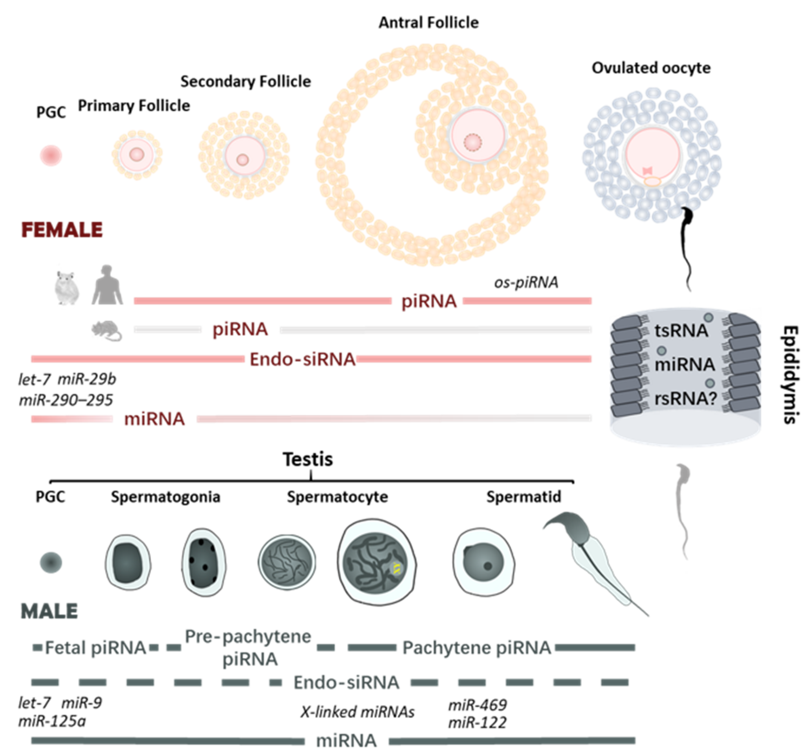
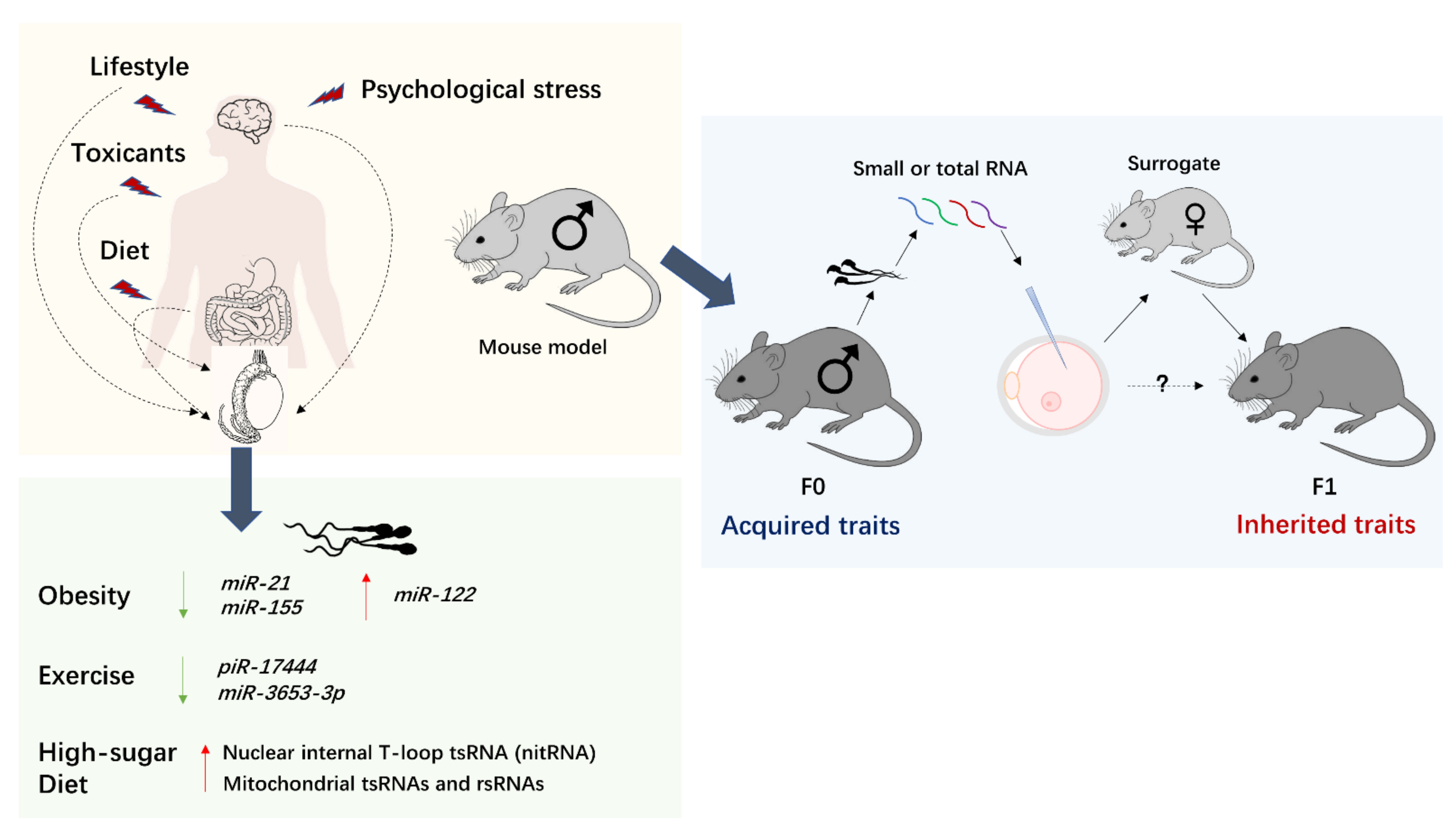
Table 1.
Summary information of environmental exposure, altered sperm sncRNA profiles, and the influence on offspring. “ND” indicates transgenerational effects are not evaluated.
Table 1.
Summary information of environmental exposure, altered sperm sncRNA profiles, and the influence on offspring. “ND” indicates transgenerational effects are not evaluated.
| Species | Environmental Exposures/Paternal Phenotype | Offspring Phenotype | Intergenerational or Transgenerational Phenotype | Epigenetic Alteration in Sperm | Reference |
|---|---|---|---|---|---|
| Psychological effects | |||||
| Mouse | Chronic variable stress | Reduced HPA stress axis responsivity | ND | Upregulation of nine miRNAs | [31,165] |
| Mouse | Elevated paternal glucocorticoid exposure | Various behavioral changes, including hyperanxiety-like and depressive-like behavior | Transgenerational | Elevated levels of three microRNAs, miR-98, miR-144, and miR-190b | [166] |
| Mouse | Early traumatic stress (MSUS) | Multiple behavioral and metabolic changes, including food intake, insulin hypersensitivity, increased body weight, increased risk-taking, and behavioral despair | Transgenerational | Alteration in multiple sncRNAs and lncRNAs | [167,168] |
| Mouse | Depression-like model (chronic mild stress induced) | Increased susceptibility to depression | Intergenerational | Total of 19 miRNAs, 24 piRNAs, and 45 rsRNAs show altered expressions | [169] |
| Mouse | Environmental enrichment paradigm | Enhancement of synaptic plasticity and cognition | Intergenerational | Upregulation of miR 212/132 | [170] |
| Metabolic effects | |||||
| Mouse | High-fat diet | Obesity; metabolic disorders in the F1 offspring including glucose intolerance and insulin resistance; altered gene expression of metabolic pathways in early embryos and islets of F1 offspring | ND | Altered expression and RNA modification of tsRNAs, mainly 5′ tRNA halves | [29] |
| Mouse | Low-protein diet | Altered hepatic cholesterol biosynthesis | ND | Decreased miRNA let-7 levels; increased level of 5′ tRFs derived from glycine tRNAs | [30,171] |
| Mouse | Western-like diet | Obesity and metabolic pathologies including insulin resistance | Transgenerational | mir-19b | [172] |
| Mouse | Multigenerational exposure to Western-like diet (WD) | Offspring of the fifth generation of WD-fed male are overweight but with normal glucose metabolism and without fatty liver | Transgenerational | sncRNA signature in the first generation of WD-fed male, such as Increase in rsRNAs, tends to disappear in the fifth generation of WD-fed male | [173] |
| Mouse | Maternal overnutrition transmitted via paternal lineage | Hedonic behaviors and metabolic defects with gender segregation in F3, i.e., F3 males exhibiting metabolic defects, while females exhibiting addictive-like behaviors | Transgenerational | Elevated level of tsRNAs, predominantly 5′ tRNA halves | [174,175] |
| Rat | High-protein diet | Increased insulin sensitivity in male but not female F1 | ND | Decrease in miRNAs; increase in tsRNAs; increase of a 42 nt mitochondrial rsRNA | [176] |
| Human | Obesity and bariatric surgery | / | ND | Altered miRNA content in sperm | [177] |
| Human | Obesity | / | ND | Decreased miR-21 and miR-155; increased miR-122 | [178] |
| Human | 6-week diet intervention enriched in vitamin D and omega-3 fatty acids | / | ND | Alteration of 3 tRFs, 15 miRNAs, and 112 piRNAs | [179] |
| Human | Heathly diet followed by high-sugar diet in two weeks | / | ND | Increase of nitRNA and mitochondrial tsRNA and rsRNA | [180] |
| Others effects | |||||
| Mouse | PM2.5 | Male-specific hypophagia, weight loss in general accompanied with decreased liver and kidney massed but increased adipose mass increase | Transgenerational | mmu-mir6909-5p | [181] |
| Mouse | Alcohol | Diverse metabolic and behavioral changes in adulthood; late-term fetal growth restriction and a loss of placental efficiency | ND | Altered expression of multiple sncRNAs; reduced epididymal expression of a tRNA methyltransferase, Nsun2 | [182,183,184] |
| Mouse | Cigarette | Mild increase in body weight of F1 | ND | Altered miRNA profile | [185] |
| Rat | Maternal (gestation) exposure to endocrine disruptors transmitted via paternal lineages | Increased incidence of male infertility in vinclozolin-exposure studies; increased susceptibility to obesity in DDT-exposure studies; susceptibility to diseases in multiple organs and immune system in both cases | Transgenerational | Concurrent alterations in DNA methylation, ncRNA, and histone retention | [186,187] |
| Mouse | Exercise | Male-specific anxiolytic behaviors | ND | Increase in miR-19b; decrease in miR-455 and miR-133a; increased tRNA-Gly- and decreased tRNA-Pro-derived RNAs | [188] |
| Human | Endurance training | / | ND | Altered expression of 8 piRNAs | [189] |
Publisher’s Note: MDPI stays neutral with regard to jurisdictional claims in published maps and institutional affiliations. |
© 2021 by the authors. Licensee MDPI, Basel, Switzerland. This article is an open access article distributed under the terms and conditions of the Creative Commons Attribution (CC BY) license (https://creativecommons.org/licenses/by/4.0/).
Share and Cite
MDPI and ACS Style
Zhu, Q.; Kirby, J.A.; Chu, C.; Gou, L.-T. Small Noncoding RNAs in Reproduction and Infertility. Biomedicines 2021, 9, 1884. https://doi.org/10.3390/biomedicines9121884
AMA Style
Zhu Q, Kirby JA, Chu C, Gou L-T. Small Noncoding RNAs in Reproduction and Infertility. Biomedicines. 2021; 9(12):1884. https://doi.org/10.3390/biomedicines9121884
Chicago/Turabian StyleZhu, Qifan, Jane Allyn Kirby, Chen Chu, and Lan-Tao Gou. 2021. "Small Noncoding RNAs in Reproduction and Infertility" Biomedicines 9, no. 12: 1884. https://doi.org/10.3390/biomedicines9121884
Note that from the first issue of 2016, this journal uses article numbers instead of page numbers. See further details here.