
Synthesis, Structure, and Antiproliferative Action of 2-Pyridyl Urea-Based Cu(II) Complexes

 , , , , , and
Abstract
:1. Introduction
2. Materials and Methods
2.1. General
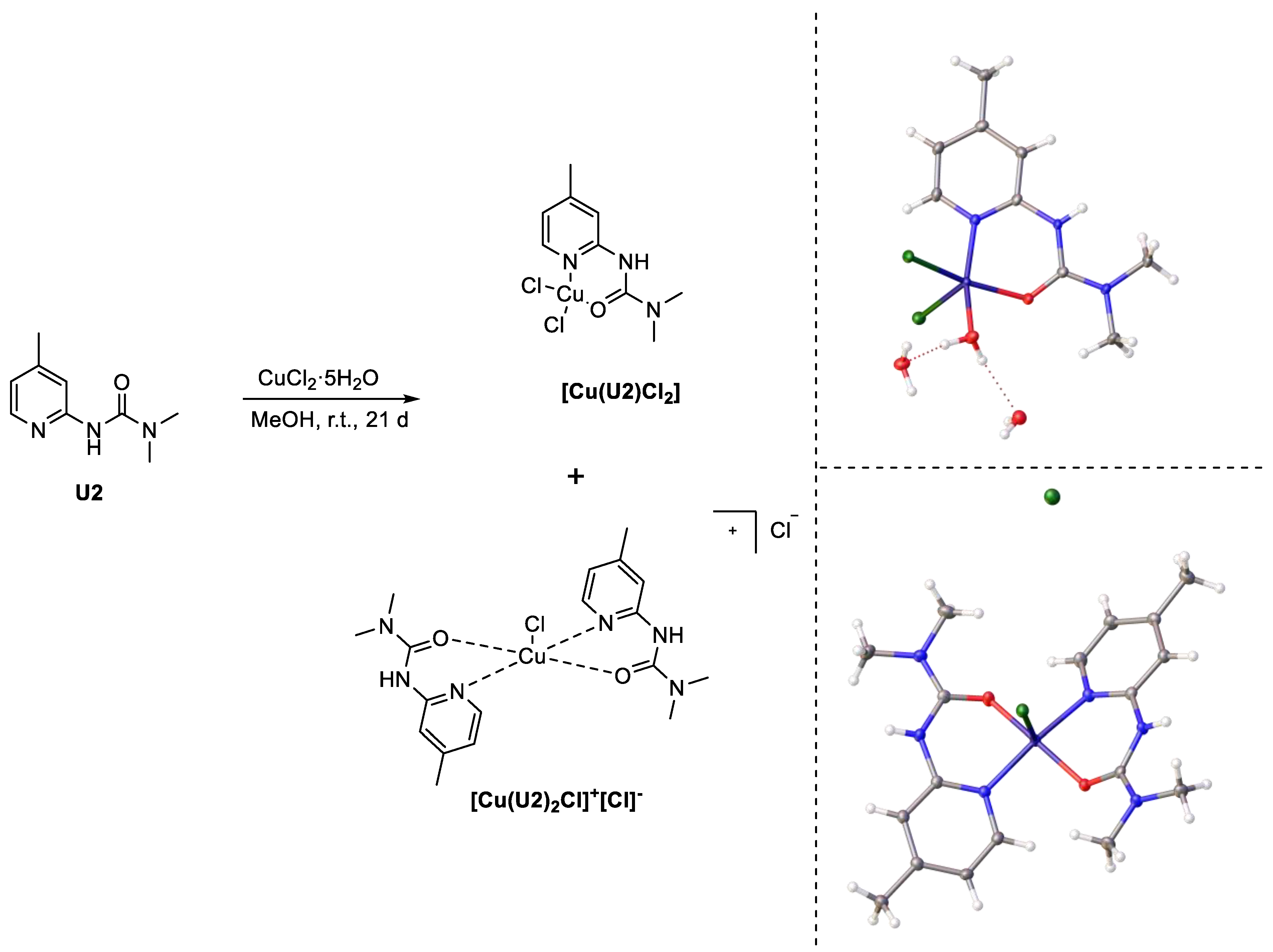
2.2. Synthesis and Characterization of Complexes [CuL2Cl]+[Cl]− and [CuL2Cl2]
2.3. Crystallography
2.4. Cell Culture
2.5. MTT Assay
2.6. Apoptosis Assay
2.7. Cell Cycle Assay
3. Results
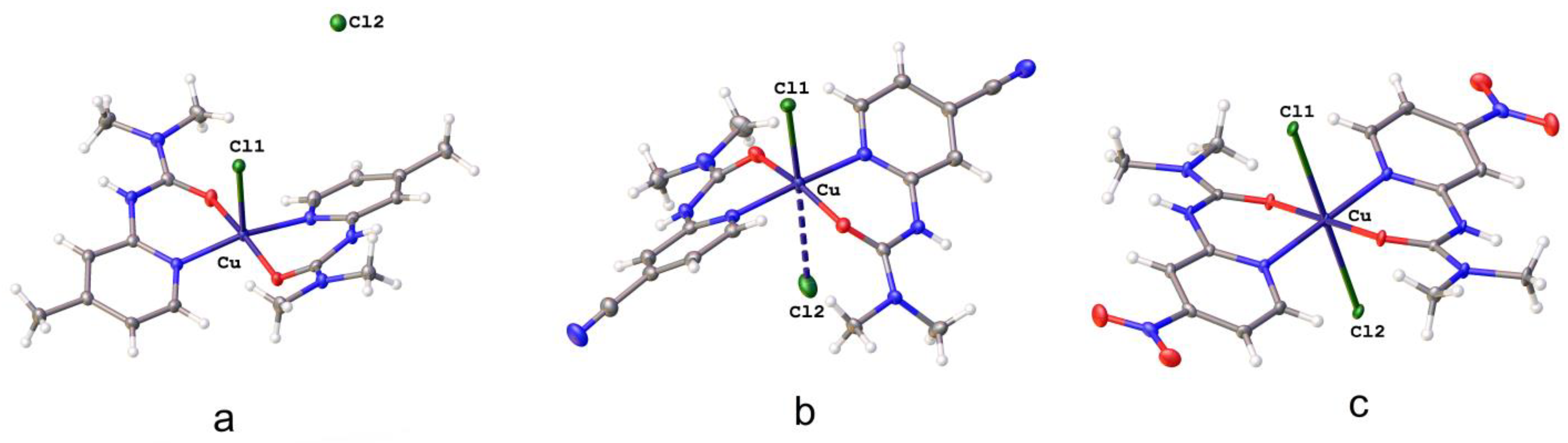
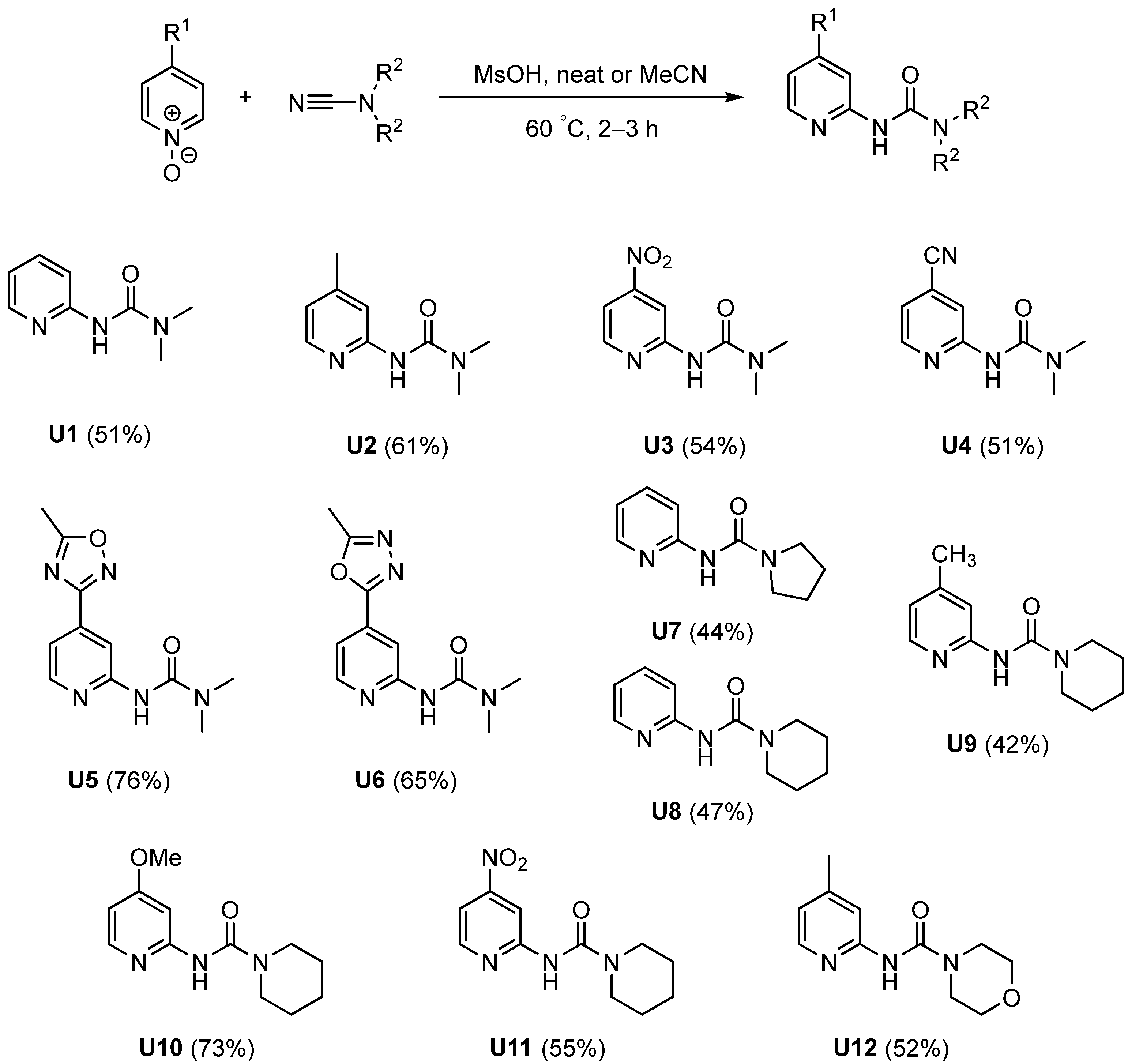
3.1. Chemistry

| Complex | Cu–Cl1, Å | Cu–Cl2, Å 1 | Cu–N1, Å | Cu–N4, Å | Cu–O1, Å | Cu–O2, Å |
|---|---|---|---|---|---|---|
| [Cu(U1)2Cl]+[Cl]− | 2.3055(6) | 6.1200(4) | 1.9807(13) | 1.9807(13) | 2.0177(11) | 2.0177(11) |
| [Cu(U2)2Cl]+[Cl]− | 2.5608(5) | 6.7765(6) | 2.0048(15) | 2.0061(15) | 1.9300(13) | 1.9270(13) |
| [Cu(U3)2Cl2] | 2.6502(4) | 2.6502(4) | 2.0047(14) | 2.0047(14) | 1.9665(13) | 1.9665(13) |
| [Cu(U4)2Cl]+[Cl]− | 2.5614(4) | 3.0220(6) 1 | 1.9907(13) | 1.9860(14) | 1.9549(12) | 1.9518(12) |
| [Cu(U7)2Cl]+[Cl]− | 2.2749(5) | 6.2742(5) | 1.9884(18) | 1.9919(18) | 2.1184(14) | 2.0106(14) |
| [Cu(U9)2Cl]+[Cl]− | 2.2827(5) | 6.3767(7) | 1.9715(15) | 1.9768(15) | 1.9850(12) | 2.1131(12) |
| [Cu(U12)2Cl]+[Cl]− | 2.2971(7) | 6.5641(8) | 1.990(2) | 1.986(2) | 2.0780(18) | 1.9833(18) |
3.2. Cell Experiments
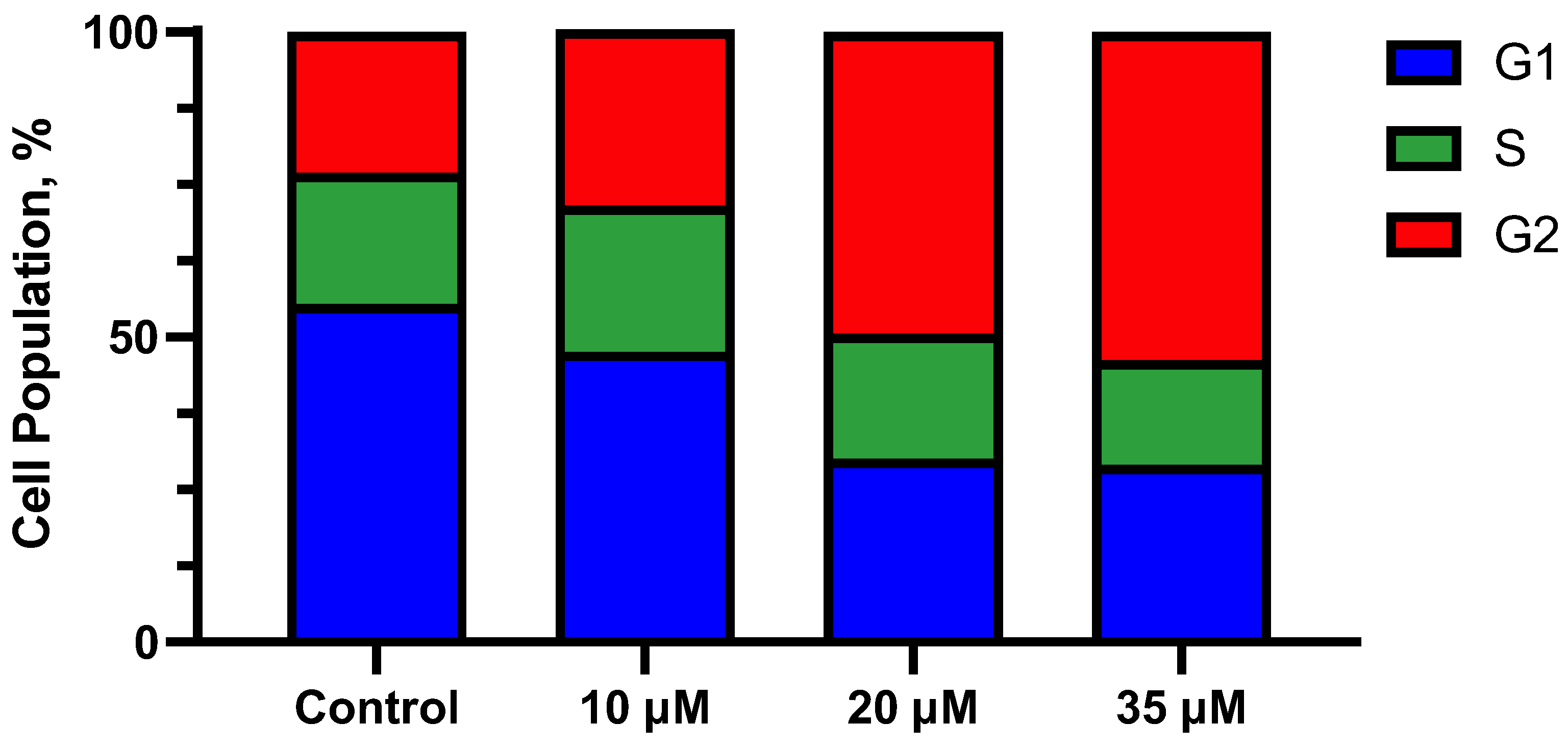
3.3. Cell Cycle Analysis
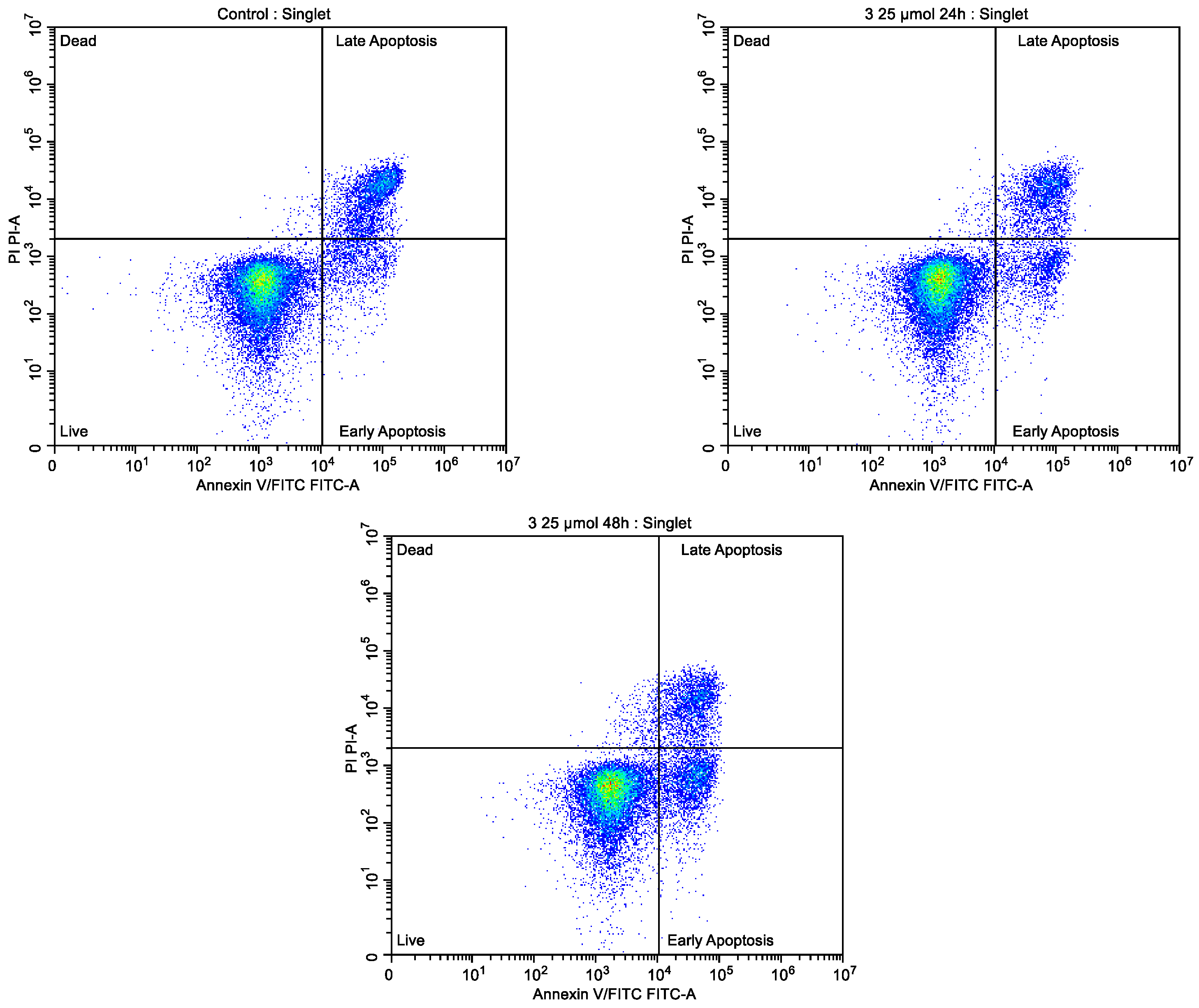
3.4. Proapoptotic Activity
4. Conclusions
Supplementary Materials
Author Contributions
Funding
Institutional Review Board Statement
Informed Consent Statement
Data Availability Statement
Acknowledgments
Conflicts of Interest
References
- Olgen, S. Overview on Anticancer Drug Design and Development. Curr. Med. Chem. 2018, 25, 1704–1719. [Google Scholar] [CrossRef] [PubMed]
- Holohan, C.; Van Schaeybroeck, S.; Longley, D.B.; Johnston, P.G. Cancer drug resistance: An evolving paradigm. Nat. Rev. Cancer 2013, 13, 714–726. [Google Scholar] [CrossRef] [PubMed]
- Cavalcanti, I.D.L.; Soares, J.C.S. Conventional Chemotherapy Versus Targeted Therapy. In Advances in Cancer Treatment; Springer International Publishing: Cham, Switzerland, 2021; pp. 79–89. [Google Scholar]
- Ndagi, U.; Mhlongo, N.; Soliman, M. Metal complexes in cancer therapy—An update from drug design perspective. Drug Des. Devel. Ther. 2017, 11, 599–616. [Google Scholar] [CrossRef] [PubMed]
- Lazarević, T.; Rilak, A.; Bugarčić, Ž.D. Platinum, palladium, gold and ruthenium complexes as anticancer agents: Current clinical uses, cytotoxicity studies and future perspectives. Eur. J. Med. Chem. 2017, 142, 8–31. [Google Scholar] [CrossRef] [PubMed]
- Kenny, R.G.; Marmion, C.J. Toward Multi-Targeted Platinum and Ruthenium Drugs—A New Paradigm in Cancer Drug Treatment Regimens? Chem. Rev. 2019, 119, 1058–1137. [Google Scholar] [CrossRef]
- Johnstone, T.C.; Suntharalingam, K.; Lippard, S.J. The Next Generation of Platinum Drugs: Targeted Pt(II) Agents, Nanoparticle Delivery, and Pt(IV) Prodrugs. Chem. Rev. 2016, 116, 3436–3486. [Google Scholar] [CrossRef]
- Malik, M.A.; Dar, O.A.; Gull, P.; Wani, M.Y.; Hashmi, A.A. Heterocyclic Schiff base transition metal complexes in antimicrobial and anticancer chemotherapy. Med. Chem. Comm. 2018, 9, 409–436. [Google Scholar] [CrossRef]
- Zou, T.; Lok, C.-N.; Wan, P.-K.; Zhang, Z.-F.; Fung, S.-K.; Che, C.-M. Anticancer metal-N-heterocyclic carbene complexes of gold, platinum and palladium. Curr. Opin. Chem. Biol. 2018, 43, 30–36. [Google Scholar] [CrossRef]
- Englinger, B.; Pirker, C.; Heffeter, P.; Terenzi, A.; Kowol, C.R.; Keppler, B.K.; Berger, W. Metal Drugs and the Anticancer Immune Response. Chem. Rev. 2019, 119, 1519–1624. [Google Scholar] [CrossRef]
- Hannon, M.J. Metal-based anticancer drugs: From a past anchored in platinum chemistry to a post-genomic future of diverse chemistry and biology. Pure Appl. Chem. 2007, 79, 2243–2261. [Google Scholar] [CrossRef]
- Molinaro, C.; Martoriati, A.; Pelinski, L.; Cailliau, K. Copper Complexes as Anticancer Agents Targeting Topoisomerases I and II. Cancers 2020, 12, 2863. [Google Scholar] [CrossRef] [PubMed]
- Krasnovskaya, O.; Naumov, A.; Guk, D.; Gorelkin, P.; Erofeev, A.; Beloglazkina, E.; Majouga, A. Copper Coordination Compounds as Biologically Active Agents. Int. J. Mol. Sci. 2020, 21, 3965. [Google Scholar] [CrossRef]
- Santini, C.; Pellei, M.; Gandin, V.; Porchia, M.; Tisato, F.; Marzano, C. Advances in Copper Complexes as Anticancer Agents. Chem. Rev. 2014, 114, 815–862. [Google Scholar] [CrossRef] [PubMed]
- Balewski, Ł.; Sączewski, F.; Bednarski, P.; Gdaniec, M.; Borys, E.; Makowska, A. Structural Diversity of Copper(II) Complexes with N-(2-Pyridyl)Imidazolidin-2-Ones(Thiones) and Their in Vitro Antitumor Activity. Molecules 2014, 19, 17026–17051. [Google Scholar] [CrossRef]
- Shobha Devi, C.; Thulasiram, B.; Aerva, R.R.; Nagababu, P. Recent Advances in Copper Intercalators as Anticancer Agents. J. Fluoresc. 2018, 28, 1195–1205. [Google Scholar] [CrossRef] [PubMed]
- AlAjmi, M.; Hussain, A.; Rehman, M.; Khan, A.; Shaikh, P.; Khan, R. Design, Synthesis, and Biological Evaluation of Benzimidazole-Derived Biocompatible Copper(II) and Zinc(II) Complexes as Anticancer Chemotherapeutics. Int. J. Mol. Sci. 2018, 19, 1492. [Google Scholar] [CrossRef] [PubMed]
- Pitucha, M.; Korga-Plewko, A.; Czylkowska, A.; Rogalewicz, B.; Drozd, M.; Iwan, M.; Kubik, J.; Humeniuk, E.; Adamczuk, G.; Karczmarzyk, Z.; et al. Influence of Complexation of Thiosemicarbazone Derivatives with Cu (II) Ions on Their Antitumor Activity against Melanoma Cells. Int. J. Mol. Sci. 2021, 22, 3104. [Google Scholar] [CrossRef]
- Hindo, S.S.; Frezza, M.; Tomco, D.; Heeg, M.J.; Hryhorczuk, L.; McGarvey, B.R.; Dou, Q.P.; Verani, C.N. Metals in anticancer therapy: Copper(II) complexes as inhibitors of the 20S proteasome. Eur. J. Med. Chem. 2009, 44, 4353–4361. [Google Scholar] [CrossRef]
- Moyaert, T.; Schroeder, Z.W.; Dawe, L.N. Synthesis, Coordination Chemistry and Anion Binding by a Cyanophenyl-Substituted 2-Pyridinylurea. Eur. J. Inorg. Chem. 2018, 2018, 167–172. [Google Scholar] [CrossRef]
- Shatrava, I.; Ovchynnikov, V.; Gubina, K.; Shishkina, S.; Shishkin, O.; Amirkhanov, V. Varieties in structures of Co(II), Ni(II) and Cu(II) coordination compounds based on dimethyl pyridin-2-ylcarbamoylphosphoramidate. Struct. Chem. 2016, 27, 1413–1425. [Google Scholar] [CrossRef]
- Sun, Y.; Zhang, Z.; Wang, X.; Li, X.; Weng, L.; Zhou, X. Isocyanate diinsertion into the N–H bond of the 2-pyridylamino ligand of organolanthanides. Dalt. Trans. 2010, 39, 221–226. [Google Scholar] [CrossRef] [PubMed]
- Guisado-Barrios, G.; Slawin, A.M.Z.; Richens, D.T. Iron complexes of new hydrophobic derivatives of tris (2-pyridylmethyl)amine: Synthesis, characterization, and catalysis of alkane oxygenation by H2O2. J. Coord. Chem. 2010, 63, 2642–2658. [Google Scholar] [CrossRef]
- Tiliakos, M.; Cordopatis, P.; Terzis, A.; Raptopoulou, C.; Perlepes, S.P.; Manessi-Zoupa, E. Reactions of 3d-metal nitrates with N,N′-bis(2-pyridyl)urea (LH2): Preparation, X-ray crystal structures and spectroscopic studies of the products trans-[M(II)(ONO2)2(LH2)2] (M = Mn, Fe, Co, Ni, Cu, Zn) and mer-[Co(III)(LH)2](NO3)·MeOH. Polyhedron 2001, 20, 2203–2214. [Google Scholar] [CrossRef]
- Orysyk, S.I.; Bon, V.V.; Pekhnyo, V.I.; Zborovskii, Y.L.; Orysyk, V.V.; Vovk, M.V. Synthesis, structure and spectral characteristics of Ni(II), Pd(II) and Zn(II) complexes with N-(2-pyridinyl)morpholine-4-carbothioamide. Polyhedron 2012, 38, 15–25. [Google Scholar] [CrossRef]
- Qureshi, N.; Yufit, D.S.; Steed, K.M.; Howard, J.A.K.; Steed, J.W. Anion hydrogen bonding from a ‘revealed’ urea ligand. Cryst. Eng. Comm. 2016, 18, 5333–5337. [Google Scholar] [CrossRef]
- German, E.A.; Ross, J.E.; Knipe, P.C.; Don, M.F.; Thompson, S.; Hamilton, A.D. β-Strand Mimetic Foldamers Rigidified through Dipolar Repulsion. Angew. Chem. Int. Ed. 2015, 54, 2649–2652. [Google Scholar] [CrossRef]
- Papesch, V.; Schroeder, E.F. Synthesis of 1-Mono- and 1,3-Di-Substituted 6-Aminouracils. Diuretic Activity. J. Org. Chem. 1951, 16, 1879–1890. [Google Scholar] [CrossRef]
- Cioffi, C.L.; Dobri, N.; Freeman, E.E.; Conlon, M.P.; Chen, P.; Stafford, D.G.; Schwarz, D.M.C.; Golden, K.C.; Zhu, L.; Kitchen, D.B.; et al. Design, Synthesis, and Evaluation of Nonretinoid Retinol Binding Protein 4 Antagonists for the Potential Treatment of Atrophic Age-Related Macular Degeneration and Stargardt Disease. J. Med. Chem. 2014, 57, 7731–7757. [Google Scholar] [CrossRef] [PubMed]
- Kurita, K.; Matsumura, T.; Iwakura, Y. Trichloromethyl chloroformate. Reaction with amines, amino acids, and amino alcohols. J. Org. Chem. 1976, 41, 2070–2071. [Google Scholar] [CrossRef]
- Eckert, H.; Forster, B. Triphosgene, a Crystalline Phosgene Substitute. Angew. Chem. Int. Ed. Engl. 1987, 26, 894–895. [Google Scholar] [CrossRef]
- Rassadin, V.A.; Boyarskiy, V.P.; Kukushkin, V.Y. Facile Gold-Catalyzed Heterocyclization of Terminal Alkynes and Cyanamides Leading to Substituted 2-Amino-1,3-Oxazoles. Org. Lett. 2015, 17, 3502–3505. [Google Scholar] [CrossRef]
- Rassadin, V.A.; Zimin, D.P.; Raskil’dina, G.Z.; Ivanov, A.Y.; Boyarskiy, V.P.; Zlotskii, S.S.; Kukushkin, V.Y. Solvent- and halide-free synthesis of pyridine-2-yl substituted ureas through facile C–H functionalization of pyridine N-oxides. Green Chem. 2016, 18, 6630–6636. [Google Scholar] [CrossRef]
- Geyl, K.; Baykov, S.; Tarasenko, M.; Zelenkov, L.E.; Matveevskaya, V.; Boyarskiy, V.P. Convenient entry to N-pyridinylureas with pharmaceutically privileged oxadiazole substituents via the acid-catalyzed C H activation of N-oxides. Tetrahedron Lett. 2019, 60, 151108. [Google Scholar] [CrossRef]
- Baykov, S.V.; Boyarskiy, V.P. Metal-Free Functionalization of Azine N-Oxides with Electrophilic Reagents. Chem. Heterocycl. Compd. 2020, 56, 814–823. [Google Scholar] [CrossRef]
- Baykov, S.; Mikherdov, A.; Novikov, A.; Geyl, K.; Tarasenko, M.; Gureev, M.; Boyarskiy, V. π–π Noncovalent Interaction Involving 1,2,4- and 1,3,4-Oxadiazole Systems: The Combined Experimental, Theoretical, and Database Study. Molecules 2021, 26, 5672. [Google Scholar] [CrossRef]
- Kasatkina, S.O.; Geyl, K.K.; Baykov, S.V.; Boyarskaya, I.A.; Boyarskiy, V.P. Catalyst-free synthesis of substituted pyridin-2-yl, quinolin-2-yl, and isoquinolin-1-yl carbamates from the corresponding hetaryl ureas and alcohols. Org. Biomol. Chem. 2021, 19, 6059–6065. [Google Scholar] [CrossRef] [PubMed]
- Dobrynin, M.V.; Kasatkina, S.O.; Baykov, S.V.; Savko, P.Y.; Antonov, N.S.; Mikherdov, A.S.; Boyarskiy, V.P.; Islamova, R.M. Deprotonated diaminocarbene platinum complexes for thermoresponsive luminescent silicone materials: Both catalysts and luminophores. Dalt. Trans. 2021, 50, 14994–14999. [Google Scholar] [CrossRef]
- Sheldrick, G.M. SHELXT—Integrated space-group and crystal-structure determination. Acta Crystallogr. Sect. A Found. Adv. 2015, 71, 3–8. [Google Scholar] [CrossRef] [PubMed]
- Sheldrick, G.M. Crystal structure refinement with SHELXL. Acta Crystallogr. Sect. C Struct. Chem. 2015, 71, 3–8. [Google Scholar] [CrossRef]
- Dolomanov, O.V.; Bourhis, L.J.; Gildea, R.J.; Howard, J.A.K.; Puschmann, H. OLEX2: A complete structure solution, refinement and analysis program. J. Appl. Crystallogr. 2009, 42, 339–341. [Google Scholar] [CrossRef]
- CrysAlis Pro. Data Collection and Processing Software for Agilent X-ray Diffractometers; Aglient Technologies: Yarnton, UK, 2013. [Google Scholar]
- Mink, J.; Hajba, L.; Pápai, I.; Mihály, J.; Neméth, C.; Skripkin, M.Y.; Sandström, M. Vibrational Spectroscopic and Theoretical Studies of Urea Derivatives with Biochemical Interest: N,N′-Dimethylurea, N,N,N′,N′-Tetramethylurea, and N,N′-Dimethylpropyleneurea. Appl. Spectrosc. Rev. 2010, 45, 274–326. [Google Scholar] [CrossRef]
- Sundaraganesan, N.; Ilakiamani, S.; Saleem, H.; Wojciechowski, P.M.; Michalska, D. FT-Raman and FT-IR spectra, vibrational assignments and density functional studies of 5-bromo-2-nitropyridine. Spectrochim. Acta Part A Mol. Biomol. Spectrosc. 2005, 61, 2995–3001. [Google Scholar] [CrossRef] [PubMed]
- Szafran, M.; Koput, J. Ab initio and DFT calculations of structure and vibrational spectra of pyridine and its isotopomers. J. Mol. Struct. 2001, 565–566, 439–448. [Google Scholar] [CrossRef]
- Green, J.H.S.; Harrison, D.J. Vibrational spectra of cyano-, formyl- and halogeno-pyridines. Spectrochim. Acta Part A Mol. Spectrosc. 1977, 33, 75–79. [Google Scholar] [CrossRef]
- Crama, W.J. The Jahn–Teller distorted structure of caesium copper(II) trichloride. Acta Crystallogr. Sect. B Struct. Crystallogr. Cryst. Chem. 1981, 37, 2133–2136. [Google Scholar] [CrossRef]
- Wells, A.F. 333. The crystal structure of anhydrous cupric chloride, and the stereochemistry of the cupric atom. J. Chem. Soc. 1947, 11, 1670. [Google Scholar] [CrossRef]
- Willett, R.D.; Dwiggins, C.; Kruh, R.F.; Rundle, R.E. Crystal Structures of KCuCl3 and NH4 CuCl3. J. Chem. Phys. 1963, 38, 2429–2436. [Google Scholar] [CrossRef]
- Persson, I.; Lundberg, D.; Bajnóczi, É.G.; Klementiev, K.; Just, J.; Sigfridsson Clauss, K.G.V. EXAFS Study on the Coordination Chemistry of the Solvated Copper(II) Ion in a Series of Oxygen Donor Solvents. Inorg. Chem. 2020, 59, 9538–9550. [Google Scholar] [CrossRef]
- Addison, A.W.; Rao, T.N.; Reedijk, J.; van Rijn, J.; Verschoor, G.C. Synthesis, structure, and spectroscopic properties of copper(II) compounds containing nitrogen–sulphur donor ligands; the crystal and molecular structure of aqua[1,7-bis(N-methylbenzimidazol-2′-yl)-2,6-dithiaheptane]copper(II) pe. J. Chem. Soc. Dalt. Trans. 1984, 1349–1356. [Google Scholar] [CrossRef]
- Smolentsev, A.I.; Lider, E.V.; Lavrenova, L.G.; Sheludyakova, L.A.; Bogomyakov, A.S.; Vasilevsky, S.F. Steric influence of the 6-methyl group on the molecular and crystal structures of copper(II) chloride complexes with 2-(N-acetylamino)-6-methylpyridine. Polyhedron 2014, 77, 81–88. [Google Scholar] [CrossRef]
- Małecki, J.G.; Machura, B.; Świtlicka, A.; Kusz, J. X-ray studies, spectroscopic characterization and DFT calculations for Mn(II), Ni(II) and Cu(II) complexes with 2-benzoylpyridine. Polyhedron 2011, 30, 410–418. [Google Scholar] [CrossRef]
- Gusev, V.Y.; Radushev, A.V. Crystal structure of the CuCl2 complex with two molecules of N′,N′-dimethyl-para-tert-butylbenzohydrazide [Cu(p-(t-Bu)C6H4CONHN(Me)2)2]Cl2 1.34 H2O. Russ. J. Coord. Chem. 2016, 42, 763–767. [Google Scholar] [CrossRef]
- Smirnov, A.S.; Martins, L.M.D.R.S.; Nikolaev, D.N.; Manzhos, R.A.; Gurzhiy, V.V.; Krivenko, A.G.; Nikolaenko, K.O.; Belyakov, A.V.; Garabadzhiu, A.V.; Davidovich, P.B. Structure and catalytic properties of novel copper isatin Schiff base complexes. New J. Chem. 2019, 43, 188–198. [Google Scholar] [CrossRef]
- Bondi, A. van der Waals Volumes and Radii of Metals in Covalent Compounds. J. Phys. Chem. 1966, 70, 3006–3007. [Google Scholar] [CrossRef]
- Ali, I.; Wani, W.A.; Saleem, K. Empirical Formulae to Molecular Structures of Metal Complexes by Molar Conductance. Synth. React. Inorg. Met. Nano-Metal Chem. 2013, 43, 1162–1170. [Google Scholar] [CrossRef]
- Barta, J.A.; Powell, C.A.; Wisnivesky, J.P. Global Epidemiology of Lung Cancer. Ann. Glob. Heal 2019, 85, 2419. [Google Scholar] [CrossRef]
- Tumbrink, H.L.; Heimsoeth, A.; Sos, M.L. The next tier of EGFR resistance mutations in lung cancer. Oncogene 2021, 40, 1–11. [Google Scholar] [CrossRef] [PubMed]
- Mosmann, T. Rapid colorimetric assay for cellular growth and survival: Application to proliferation and cytotoxicity assays. J. Immunol. Methods 1983, 65, 55–63. [Google Scholar] [CrossRef]
- Khan, K.H.; Blanco-Codesido, M.; Molife, L.R. Cancer therapeutics: Targeting the apoptotic pathway. Crit. Rev. Oncol. Hematol. 2014, 90, 200–219. [Google Scholar] [CrossRef]
- Call, J.A.; Eckhardt, S.G.; Camidge, D.R. Targeted manipulation of apoptosis in cancer treatment. Lancet Oncol. 2008, 9, 1002–1011. [Google Scholar] [CrossRef]


| Entry | Complex | Structure | Yield, % | CCDC Number 1 |
|---|---|---|---|---|
| 1 | [Cu(U1)2Cl]+[Cl]− |  | 46 | 2104013 |
| 2 | [Cu(U2)2Cl]+[Cl]− |  | 54 | 2104015 |
| 3 | [Cu(U3)2Cl2] |  | 72 | 2104016 |
| 4 | [Cu(U4)2Cl]+[Cl]− |  | 57 | 2104017 |
| 5 | [Cu(U5)2Cl]+[Cl]− |  | 65 | |
| 6 | [Cu(U6)2Cl]+[Cl]− |  | 58 | |
| 7 | [Cu(U7)2Cl]+[Cl]− |  | 52 | 2104021 |
| 8 | [Cu(U8)2Cl]+[Cl]− |  | 39 | |
| 9 | [Cu(U9)2Cl]+[Cl]− |  | 64 | 2104018 |
| 10 | [Cu(U10)2Cl]+[Cl]− |  | 52 | |
| 11 | [Cu(U11)2Cl2] |  | 61 | |
| 12 | [Cu(U12)2Cl]+[Cl]− |  | 63 | 2104019 |
| Complex | [Cu(U2)2Cl]+[Cl]− | [Cu(U3)2Cl2] | [Cu(U4)2Cl]+[Cl]− | [Cu(U11)2Cl2] |
|---|---|---|---|---|
| Molar conductivity, Ω−1·cm2·mol−1 | 24.8 | 24.1 | 25.2 | 21.7 |
| Compound | Cell Viability, % | |
|---|---|---|
| 10 µM | 100 µM | |
| U1 | 100.5 ± 18.0 | 86.5 ± 7.5 |
| U2 | 101.5 ± 2.7 | 98.7 ± 3.8 |
| U3 | 95.5 ± 3.3 | 91.6 ± 4.3 |
| U4 | 94.2 ± 6.7 | 89.0 ± 5.1 |
| U5 | 101.2 ± 5.5 | 90.2 ± 8.7 |
| U6 | 94.5 ± 7.1 | 89.9 ± 2.4 |
| U7 | 103.3 ± 2.8 | 99.8 ± 5.7 |
| U8 | 100.3 ± 9.4 | 97.25 ± 6.1 |
| U9 | 103.2 ± 6.3 | 98.3 ± 8.1 |
| U10 | 101.8 ± 5.2 | 99.5 ± 8.1 |
| U11 | 104.8 ± 6.7 | 94.3 ± 9.0 |
| U12 | 97.4 ± 5.5 | 85.4 ± 7.8 |
| control | 100.0 ± 3.7 | |
| Compound | IC50, μM | |||
|---|---|---|---|---|
| A549 | NCI-H460 | NCI-H1975 | WI-26 VA4 | |
| [Cu(U1)2Cl]+[Cl]− | 106.8 ± 15.1 | 120.3 ± 18.4 | 65.3 ± 10.9 | 108.3 ± 27.5 |
| [Cu(U2)2Cl]+[Cl]− | 94.5 ± 14.1 | 121.6 ± 15.3 | 65.4 ± 8.6 | 100.8 ± 15.1 |
| [Cu(U3)2Cl2] | 61.4 ± 6.52 | 77.5 ± 6.4 | 39.6 ± 4.5 | 81.6 ± 10.1 |
| [Cu(U4)2Cl]+[Cl]− | 106.5 ± 12.2 | 118.0 ± 13.4 | 68.3 ± 8.5 | 108.8 ± 16.4 |
| [Cu(U5)2Cl]+[Cl]− | 114.5 ± 14.5 | 136.3 ± 12.5 | 71.2 ± 6.9 | 117.5 ± 24.9 |
| [Cu(U6)2Cl]+[Cl]− | 88.7 ± 12.7 | 115.4 ± 26.3 | 63.8 ± 8.7 | 104.7 ± 14.1 |
| [Cu(U7)2Cl]+[Cl]− | 102.0 ± 15.8 | 102.8 ± 13.1 | 66.7 ± 7.6 | 100.2 ± 16.6 |
| [Cu(U8)2Cl]+[Cl]− | 98.4 ± 11.1 | 101.6 ± 17.3 | 64.4 ± 10.5 | 111.2 ± 19.3 |
| [Cu(U9)2Cl]+[Cl]− | 76.1 ± 8.86 | 66.4 ± 5.7 | 61.6 ± 6.0 | 112.8 ± 23.5 |
| [Cu(U10)2Cl]+[Cl]− | 86.7 ± 9.72 | 64.0 ± 5.7 | 62.3 ± 7.0 | 104.2 ± 17.4 |
| [Cu(U11)2Cl2] | 46.01 ± 4.98 | 53.6 ± 4.7 | 33.4 ± 3.8 | 62.4 ± 5.6 |
| [Cu(U12)2Cl]+[Cl]− | 89.3 ± 6.67 | 128.0 ± 19.5 | 70.2 ± 4.9 | 109.3 ± 13.7 |
Publisher’s Note: MDPI stays neutral with regard to jurisdictional claims in published maps and institutional affiliations. |
© 2022 by the authors. Licensee MDPI, Basel, Switzerland. This article is an open access article distributed under the terms and conditions of the Creative Commons Attribution (CC BY) license (https://creativecommons.org/licenses/by/4.0/).
Share and Cite
Geyl, K.K.; Baykov, S.V.; Kalinin, S.A.; Bunev, A.S.; Troshina, M.A.; Sharonova, T.V.; Skripkin, M.Y.; Kasatkina, S.O.; Presnukhina, S.I.; Shetnev, A.A.; et al. Synthesis, Structure, and Antiproliferative Action of 2-Pyridyl Urea-Based Cu(II) Complexes. Biomedicines 2022, 10, 461. https://doi.org/10.3390/biomedicines10020461
Geyl KK, Baykov SV, Kalinin SA, Bunev AS, Troshina MA, Sharonova TV, Skripkin MY, Kasatkina SO, Presnukhina SI, Shetnev AA, et al. Synthesis, Structure, and Antiproliferative Action of 2-Pyridyl Urea-Based Cu(II) Complexes. Biomedicines. 2022; 10(2):461. https://doi.org/10.3390/biomedicines10020461
Chicago/Turabian StyleGeyl, Kirill K., Sergey V. Baykov, Stanislav A. Kalinin, Alexandr S. Bunev, Marina A. Troshina, Tatiana V. Sharonova, Mikhail Yu. Skripkin, Svetlana O. Kasatkina, Sofia I. Presnukhina, Anton A. Shetnev, and et al. 2022. "Synthesis, Structure, and Antiproliferative Action of 2-Pyridyl Urea-Based Cu(II) Complexes" Biomedicines 10, no. 2: 461. https://doi.org/10.3390/biomedicines10020461