
Navigating the ERK1/2 MAPK Cascade

1
Department of Pharmacology, UT Southwestern Medical Center, 6001 Forest Park Rd., Dallas, TX 75390, USA
2
Simmons Comprehensive Cancer Center, UT Southwestern Medical Center, 6001 Forest Park Rd., Dallas, TX 75390, USA
*
Author to whom correspondence should be addressed.
Biomolecules 2023, 13(10), 1555; https://doi.org/10.3390/biom13101555
Submission received: 30 September 2023
/
Revised: 16 October 2023
/
Accepted: 18 October 2023
/
Published: 20 October 2023
(This article belongs to the Special Issue MAP Kinases: Functions in Signal Transduction and Disease)
Abstract
:The RAS-ERK pathway is a fundamental signaling cascade crucial for many biological processes including proliferation, cell cycle control, growth, and survival; common across all cell types. Notably, ERK1/2 are implicated in specific processes in a context-dependent manner as in stem cells and pancreatic β-cells. Alterations in the different components of this cascade result in dysregulation of the effector kinases ERK1/2 which communicate with hundreds of substrates. Aberrant activation of the pathway contributes to a range of disorders, including cancer. This review provides an overview of the structure, activation, regulation, and mutational frequency of the different tiers of the cascade; with a particular focus on ERK1/2. We highlight the importance of scaffold proteins that contribute to kinase localization and coordinate interaction dynamics of the kinases with substrates, activators, and inhibitors. Additionally, we explore innovative therapeutic approaches emphasizing promising avenues in this field.
1. Introduction: MAPK Pathways
MAPK (Mitogen-Activated Protein Kinase) signaling pathways are among the best studied ubiquitous regulatory cascades that respond to a wide range of extracellular hormones, growth factors, neurotransmitters and other cellular cues to control rapid cellular responses, decision making and cell fate. MAPK cascades are comprised of three or more kinases acting in series, beginning with MAPK kinase kinases (MAP3Ks) that activate MAPK kinases (MAP2Ks or MAP/ERK kinases (MEKs)), that then with high selectivity activate MAPKs, the effector kinases of the cascades. Prior to its purification or recognition of its enzymatic activity, the MAPK ERK2 (extracellular signal-regulated kinase 2) had been observed as a 42 kDa tyrosine-phosphorylated protein in lysates from growth factor-stimulated and virally transformed cells [1]. Subsequently, what turned out to be the same protein was found as a growth factor-activated protein kinase [2] that phosphorylated microtubule-associated protein 2 (MAP-2 Protein Kinase) and could activate ribosomal protein S6 kinase [3]. Purification and molecular cloning identified two closely related but distinct enzymes ERK1 (p44, MAPK3) and ERK2 (MAPK1), often the more abundant [4,5,6]. Other MAPKs include Stress-Activated Protein Kinases (SAPKs)/Jun-N-terminal Kinases (JNKs), p38 family kinases and ERK5/Big MAPKs (BMK), in addition to atypical MAPK pathways ERK3/4, ERK7/8 and NLK (Nemo-like kinase) [7,8,9]. These MAPKs belong to the CMGC group of highly conserved serine–threonine kinases [10]. A hallmark of the typical MAPKs is their activation by dual phosphorylation on nearby tyrosine and threonine residues in their activation loop, in what is known as the TXY motif [11], where the amino acid X is distinct for subsets of MAPKs. In the case of ERK1/2 and ERK5, the motif is Thr-Glu-Tyr (TEY), JNKs contain Pro as the intermediate residue (TPY), and Gly for p38s (TGY) [9] (reviewed in more detail in [12]).
While all MAPK pathways handle an array of functions in cellular physiology and pathology, this review focuses on the ERK1/2 MAPK pathway. This pathway is crucial in maintaining normal functions in most if not all cells, and abnormalities in this pathway disturb many cellular processes and contribute to pathologies ranging from birth defects to cancer. As a consequence, significant efforts have been dedicated to understanding and manipulating this pathway. Here, we provide an overview of pathway components and functions and highlight the broad array of scaffolds as well as novel interventional strategies.
2. ERK Pathway Components
2.1. RAS
RAS (RAt Sarcoma) refers to small GTPase (G)-proteins encoded by three genes yielding proteins with molecular weights of ~21 kDa. RAS proteins contain a GTP-binding domain and a hypervariable region (HVR) which marks the following differences between them [13]: HRAS (Harvey sarcoma viral oncogene), NRAS (neuroblastoma oncogene) and KRAS (Kirsten sarcoma viral oncogene). KRAS has two alternatively spliced forms named KRAS4A and KRAS4B. To be active, RAS proteins need to be localized at the plasma membrane or endomembranes. Lipid post-translational modifications (PTMs), farnesylation and palmitoylation, are essential for RAS association on the membrane [14,15]. The membrane sub-domain and subcellular localization from which the RAS signal originates determines the downstream effectors that are activated and, consequently, the cellular outcome [16]. As GTPases, RAS proteins cycle from the GDP-loaded OFF configurations to GTP-loaded ON conformations assisted by RAS–guanine nucleotide exchange factors (RAS-GEFs) and return to OFF states facilitated by RAS–GTPase-activating proteins (RAS-GAPs) [17]. The RAS-GEFs SOS (Son of Sevenless)1 and SOS2 are activated by receptor tyrosine kinases coordinated by the adaptor protein Grb2 [18]. Once activated, RAS can interact with the RAS binding domain (RBD) in RAS effectors. In addition to RAF proteins, MAP3Ks in the ERK1/2 pathway, there are over fifty other RAS effectors including RALGDS, RALBP1, RIN1, TIAM1, PLCε, REPAC, RASSF1/5, and PI3K among others [19,20].
The four RAS proteins show around 85% of amino acid identity differing in the hypervariable region (HVR) [21]. In spite of their similarities, manipulation of their expression or activation results in different cellular outcomes [22,23]. KRAS is the only family member essential for development [24,25,26]. This feature depends on its locus expression pattern in development, as the protein product of HRAS knock-in in the KRAS locus gives rise to viable mice. However, a phenotype in adult life has been observed when KRAS is replaced by HRAS [27], although it disappears by eliminating the endogenous HRAS [28]. These findings indicate that different RAS isoforms are apparently equivalent in physiological conditions. Nevertheless, activating mutations in the different RAS proteins lead to different outcomes in a tissue-specific manner from senescence to tumorigenesis. Moreover, the same activating mutation in KRAS and HRAS generates different types of tumors in adult mice [28].
Mutations in RAS isoforms are responsible for nearly 30% of all human cancers. KRAS exhibits the highest mutation frequency in cancer, present in ~85% of cases. NRAS at 11%, while HRAS accounts for only ~3% of all RAS mutation-dependent cancers. The most frequently mutated codons are G12, G13 and Q61 regardless of isoform. Their frequency varies, with codons 12 and 13 more commonly mutated in KRAS, while 61 is more commonly mutated in NRAS and HRAS [29]. Only now beginning to be understood functionally, the mutational frequency of the isoforms differs across different types of cancer; in descending order of prevalence: pancreatic adenocarcinoma (>60% KRAS), colorectal adenocarcinoma (under 40% KRAS, ~5% NRAS), lung adenocarcinoma (~30% KRAS), and skin cutaneous melanoma (~28% NRAS), among the most common cancers bearing RAS mutations [30].
Despite the high amino acid identity between RAS isoforms, their nucleotide sequences are less so [31]. The Counter laboratory proposed codon bias as a mechanism responsible for the different expression and oncogenicity of KRAS and HRAS. Even though KRAS is the most commonly mutated isoform, it is the most poorly translated due to rare codons. This group suggested that the potent oncogenicity of HRAS lead to cell cycle arrest and senescence, while the different codon usage of KRAS overcomes this issue becoming the most frequently mutated RAS in cancer [31]. In fact, they later demonstrated that substitution of KRAS rare codons for common codons rendered tumors less aggressive [32]. Following studies corroborated that rare codons in KRAS affect transcription due to histone modification and transcriptional activation recruitment, mRNA translation, and even protein conformation, highlighting the importance of codon usage [33]. Furthermore, the correlation between tRNA expression, codon usage, and oncogenicity was further validated not only within RAS isoforms but also across other families of oncogenes [34].
As previously pointed out, the mutational pattern varies among the different types of tumors, with certain isoforms, hotspot codons, and the specific mutation, more frequent depending on the tissue [35]. A possible explanation for the mutation prevalence, as suggested by the Counter laboratory, is that each mutation generates a different level of activity and each tissue establishes its specific ‘sweet spot’ within which this activity drives tumorigenesis, while above or below that range of activity transformation is not possible [36]. Furthermore, RAS mutation tropism may be the result of mutational specificity and selection as observed with the carcinogen urethane. Urethane shows a mutation tropism towards Q61L/R mutation in KRAS in the lung. However, if KRAS rare codons are replaced by common codons, to compensate for the resulting stronger activity, urethane will induce mutations in G12 instead as these are biochemically less active [37,38].
2.2. RAF
RAF (Rapidly Accelerated Fibrosarcoma) is the first kinase in the pathway core, serving as the MAP3K downstream of RAS [39]. The regulatory paradigm has it that RAF is cytosolic in resting conditions and translocates to the plasma membrane to be activated by RAS upon growth factor stimulation [40,41,42] (reviewed in [43,44]). In mammals, the RAF family consists of ARAF (65 kDa), CRAF (65 kDa) (aka RAF1), and BRAF (84 kDa, larger due to additional BRAF-specific sequence at its N-terminus) [45], which are ~75% identical in shared segments. Also in the family are the pseudokinases KSR1 and KSR2 discussed in Section 4.2.
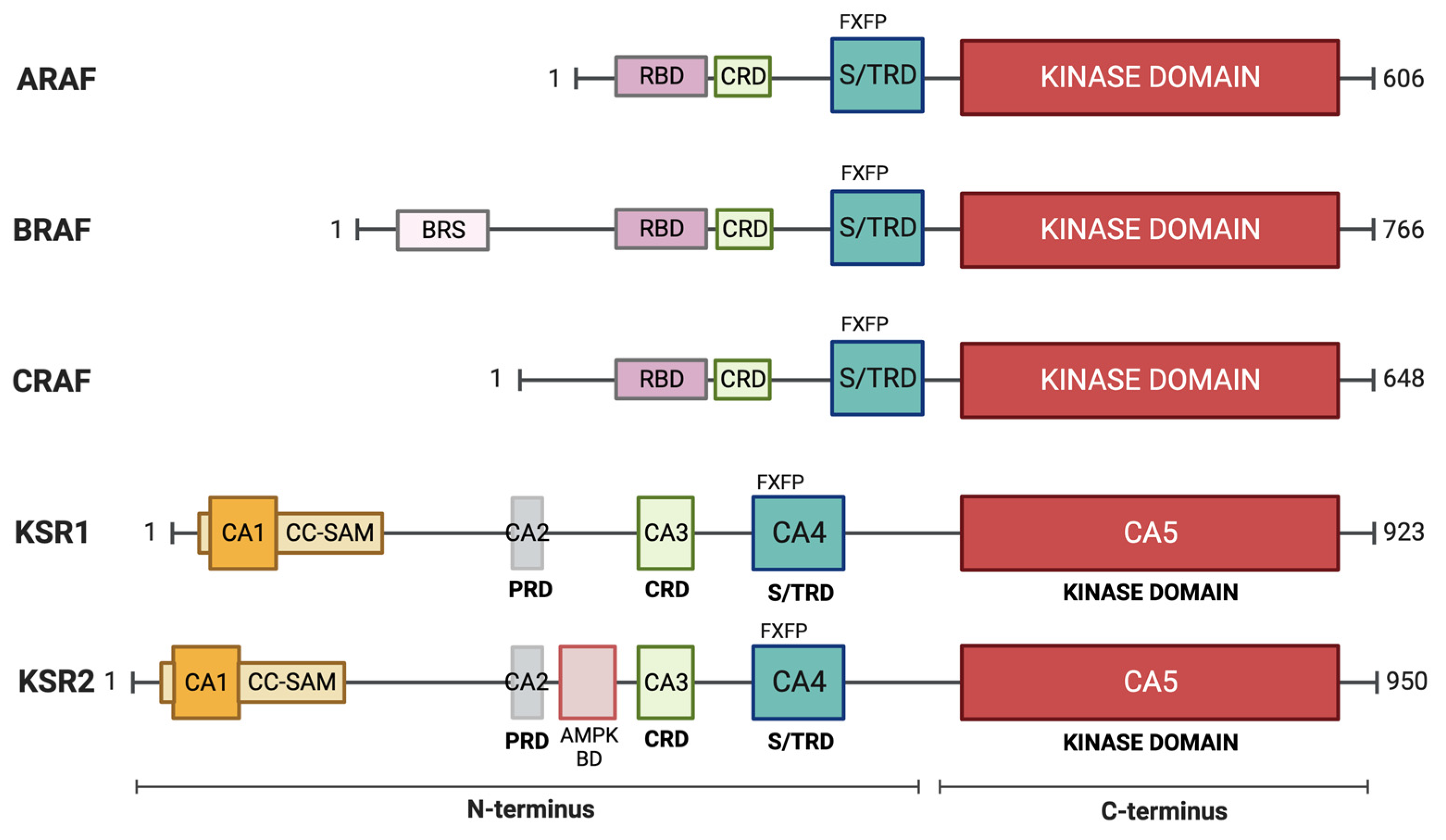
RAF consists of a RAS binding domain (RBD) and a cysteine-rich domain (CRD), both in the N-terminal regulatory region of the protein and the kinase domain in the C-terminus. RAF was formerly divided into three conserved regions named CR1, CR2, and CR3. CR1 contains the RBD and the CRD, CR2 is a serine/threonine-rich domain, and CR3 refers to the kinase domain (reviewed in [46]). CRD facilitates RAF localization at the plasma membrane [47,48] and together with RBD both interact with RAS [49,50]. Furthermore, the kinase domain is predicted to directly interact with the plasma membrane [51]. In quiescent cells, the N-terminal regulatory region interacts with the kinase domain as an autoinhibitory mechanism, preventing RAF activation. Besides the inhibitory intramolecular interactions, a 14-3-3 dimer binds to two phospho-sites, S365 and S729 in BRAF, stabilizing its inactive monomeric conformation. Active RAS recruits RAF to the plasma membrane opening the monomeric conformation [52]. The S365 (in BRAF; S259 in CRAF) inhibitory site is exposed and the ternary complex SHOC2-MRAS-PP1 dephosphorylates it, releasing the autoinhibitory conformation [53,54,55]. In the meantime, one of the 14-3-3 protomers remains bound to the pS729, while the other one is available to bind another RAF molecule stabilizing an active RAF dimer [42,56]. Other molecules such as KSR and MEK participate in RAF activation, adding to its complex nature [57,58]. The primary substrates of RAF are the MAP2Ks MEK1 and MEK2 [59].
Of the three isoforms, BRAF is the most frequently mutated in cancer. BRAF mutations are present in 7.7% across all cancer types [60]. Overall, BRAF mutation prevalence in human cancers is almost 100% for hairy cell leukemia, 50% for melanoma, 45% for papillary thyroid cancer, 10% for colon cancer, and 10% for non-small cell lung cancer cases [61]. The most frequent mutation in BRAF is V to E substitution at residue 600 (V600E), accounting for 90% of BRAF mutations [62]. Around 200 low-frequency cancer-associated mutations have been described in BRAF [63]. Most of them are believed to reverse the autoinhibitory conformation in the absence of upstream signaling [64]. BRAF mutations have been classified into three groups as follows: class I mutations are those that affect BRAF V600 position, most commonly V600E but also V600K/D/R/M which are able to transduce the signal downstream as monomers; class II mutations function as RAS-independent dimers with reported increased kinase activity but still weaker than BRAF V600 mutants; and class III mutants show impaired kinase activity and do not directly phosphorylate MEK but retain the ability to bind RAS and heterodimerize with CRAF [65,66]. BRAF fusions have also been found which lack the inhibitory N-terminal domain leading to constitutive BRAF dimerization and are considered in class II [67].
2.3. MEK
Downstream of RAF are the two ERK-specific MAP2Ks [68,69], MEKs 1 and 2 of 44 and 45 kDa, respectively [70]. The MEK kinase domain contains an ERK-selective binding site near the N-terminus and a nuclear export sequence (NES) that favors its cytosolic localization [71,72]. MEK1 is activated by phosphorylation of two activation loop serine residues (S218 and S222, S222 and S226 in MEK2) [73]. Apart from canonical activation by RAF, MEK can also be activated by other MAP3Ks, including MOS [74], MAP3K1/MEKK1 [75], MAP3K8 (aka Tpl2/Cot) [76], and MAST1 [77], linking activation to distinct upstream stimuli (Figure 1). In addition, PAK1 phosphorylates MEK1 on S298 to increase the interaction with ERK [78,79,80], and it was found later that it was also able to phosphorylate MEK1/2 in the two canonical serines of the activation loop by the canonical activation of serine [81].
MEK1 is retro-phosphorylated by ERK at T292 [82]. This phosphorylation event is specific to MEK1 and enhances the accessibility of phosphatases facilitating the dephosphorylation of S218 and S222 in the activation loop [80]. As MEK1 and MEK2 form heterodimers and this residue is exclusive to MEK1, in the absence of MEK1, MEK2 is not subjected to negative regulation, thus leading to ERK-sustained activation via MEK2 [83]. As in the case of ERK1/2, MEK1 and MEK2 seem to play non-redundant roles in development [84,85]; however, this may be due to a dosage effect [86]. Nevertheless, some studies emphasize specific functions at least in some settings [87,88,89]. MEK acts as a cytoplasmic anchor for ERK, binding to it in the cytoplasm and mediating its export from the nucleus via its XPO1/CRM1, initially demonstrated by its sensitivity to leptomycin B [90,91].
MEK mutations are rare, found in less than 1% of all human cancers, and not concentrated in a specific region or codon [63], in contrast to the upstream activators RAF and RAS. Mutations in MEK have been functionally classified into three groups. (1) RAF-independent mutations consist of deletions that result in autophosphorylation in the activation loop, resulting in a potent downstream activation. (2) RAF-regulated alterations lead to some basal activation which is potentiated in the presence of RAF. Some of these mutations have been found in resistant tumors. (3) RAF-dependent mutations display the lowest activity of all the mutants and depend on RAF to be active acting as signal amplifiers [92].
2.4. ERK
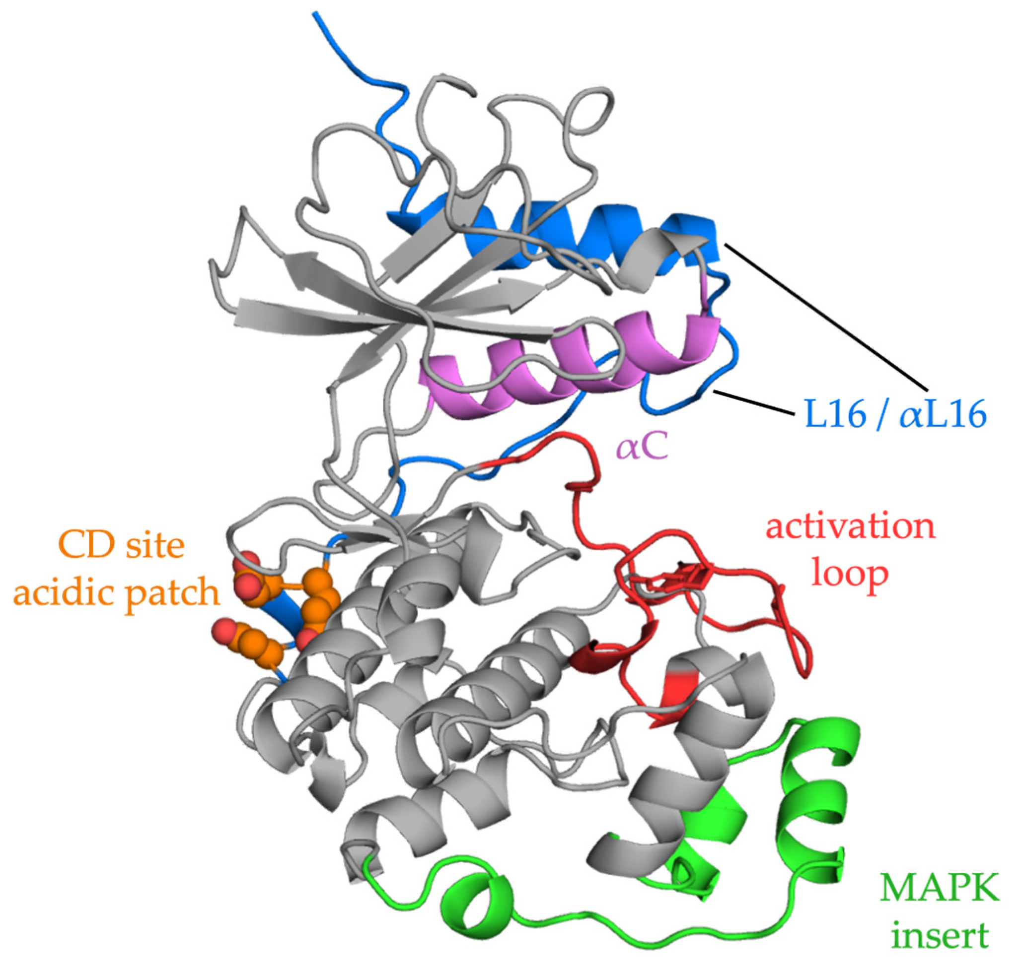
ERKs contain the usual two-domain protein kinase structure (reviewed in [93]). The N-terminal domain binds ATP and contains an essential ATP-binding lysine, often referred to as the catalytic lysine. Protein substrates and regulators bind across the C-terminal domain as discussed more fully below. ERKs also include a MAPK insert in the C-terminal domain and other regions that are important for MEK recognition [94,95] and an essential C-terminal segment that extends across both domains (Figure 2).
Activation of ERK by MEK results from the phosphorylation of tyrosine followed by threonine in the TEY sequence within the activation loop [11,96,97,98]. Following phosphorylation by MEK, this C-terminal segment aids in repositioning alpha helix C to achieve an activated ERK conformation [98]. The combined phosphorylation of both residues increases ERK activity 50,000-fold [99].
The similarities and overlapping substrate specificities of ERK1 and ERK2 have fueled the long-running debate over their functional specificity [100,101,102,103]. Genetic studies using knockout mouse models showed that the deletion of either ERK1 or ERK2 led to different phenotypic outcomes. Although loss of either ERK gene impairs neuronal development [104], ERK1−/− mice are viable and fertile [105], while whole body ERK2 knockout results in embryonic lethality regardless of ERK1 expression [106]. Other work has reported divergent roles in specific contexts [107,108,109,110,111,112]. Phenotypic differences have been attributed to the allele number and differential expression of each isoform. Ectopic overexpression of one isoform compensated for the lack of the other, supporting the conclusion of redundancy [100]. It has been suggested that variations in the expression of either isoform will be compensated for to maintain a similar amount of total ERK [103]. A detailed analysis of ERKs across many species demonstrated that two ERK genes first appeared in bony vertebrates by a gene duplication, although both genes are not always expressed in all tetrapod species [102]. Phylogenetic analysis indicated that ERK1 is evolving faster than ERK2, probably related to the much smaller size of the ERK1 gene and consistent with the complex regulation of ERK2 expression suggested by work in Drosophila [113].
N-terminal alanine-rich (NTAR) sequences in ERK1/2 are now known to improve their translation. NTAR sequences ensure precise translation and prevent frameshifts by reducing the speed of elongating ribosomes. Human ERK1/2 possess multiple Ala codons immediately downstream of the AUG codon with an additional in-frame start codon also present. Notably, most of these Ala codons are rare codons that are repeated in tandem. Collectively, these features optimize accurate translation minimizing errors, which reflects the need for proper ERK protein synthesis within cells [114].
2.4.1. ERK Substrate Recognition
ERKs can interact with hundreds of substrates in any location in the cell [119,120]. In addition to the many phosphorylation sites that have been identified by examining defined targets, many more sites in putative effectors have been predicted by in silico approaches with some validated in vivo [121,122]. ERK1/2 show a preference for sites in the consensus phosphorylation sequence, PX(S/T)P, with a proline in the P+1 residue, following the phosphorylation site, and are thus considered proline-directed kinases. Many substrates contain only a minimal (S/T)P sequence. Unlike many other kinases, in MAPKs the pocket for the P+1 residue in the substrate is shallow creating a preference for proline at the position following the serine/threonine phosphorylation site [98,123]. Glycine and rarely alanine can also be accommodated at the P+1 site.
The huge number of phospho-sites found in proteins [121] raises the question of how specificity is achieved. While scaffolds (see Section 4.2) can form three-way complexes with enzymes and substrates, docking sites are important MAPK recognition mechanisms [124,125]. The best characterized docking sites identified on ERK are the common docking (CD) or D-recruitment site (DRS) and the F-recruitment site (FRS) or docking site for ERK FXF (DEF) (see Figure 2) [126]. The CD site, accessible in both inactive and active ERK configurations, binds a basic/hydrophobic motif, typically K/RX2-4LXL, referred to as a D motif, kinase interaction motif (KIM), or more generally as a short linear motif (SLiM). In addition to many substrates, MEK1/2 also bind ERK1/2 through a CD–D motif interaction, as does the MAP kinase phosphatase DUSP6. The FRS site binds proteins containing FXF motifs, including nuclear pore proteins [127], and is more accessible after ERK activation [128,129]. Some ERK substrates, such as Elk1, contain both interacting domains and some appear to lack either motif [130,131,132]. When a protein possesses multiple (S/T)P residues, docking motifs may play a crucial role in guiding the phosphorylation towards a particular residue [133]. Additional strategies are being developed to identify interaction mechanisms, including a recent study expanding our understanding of CD–D motif interactions [134]. Moreover, targeting the DRS to disrupt protein–protein interaction with ERK interactors has already been assessed [135,136].
2.4.2. ERK Localization
ERK1/2 are found in cytoplasmic-, nuclear-, and membrane-associated compartments, and their subcellular distribution defines access to substrates. Localization is dynamic and is influenced by the duration and nature of activating stimulation. Two ERK monomers can associate to form a free dimer in the cytoplasm to activate cytoplasmic substrates. ERK dimers can be assembled on scaffold proteins in specific subcellular localizations where the ERK monomer not associated with the scaffold will phosphorylate specific substrates [137]. Binding the FG repeat regions of nuclear pore proteins, nucleoporins (Nup) Nup214, Nup153, TPR, and others, allows passive entry of ERK into the nucleus [138,139,140]. A study by the Nishida lab showed numerous interactions of ERK2 with nuclear pore proteins with regulatory impact [141]. Nup-mediated facilitated diffusion allows rapid exchange of free ERK2 between the cytoplasmic and nuclear pools and reveals the great regulatory importance of binding partners with restricted localizations that can influence where ERK2 acts.
While active nuclear import mechanisms have been identified, ERK scaffolding, to retain the kinases in the relevant compartment, and ERK nuclear export through binding to exported partners, appear to dominate processes determining ERK location. As noted earlier, binding to MEK promotes ERK nuclear export and favors cytoplasmic retention. Dual specificity phosphatases also influence ERK location by related mechanisms. The phosphatase DUSP6 is primarily cytoplasmic and binds ERK through a D motif–CD domain interaction; DUSP6 also possesses an NES, promoting ERK nuclear export. In the nucleus, ERK associates with many transcription factors and is also retained by de novo-synthesized nuclear anchors, such as another phosphatase, DUSP5 [142] and Vanishin [143]. Chromatin association of ERK1/2 is widespread as deduced from chromatin immunoprecipitation [144,145] (reviewed in [146]). Cytoplasmic anchors include microtubules which may bind as much as one-third to half of ERK2 [147]. PEA-15 can also anchor ERK in the cytoplasm [148] and prevents ERK2 phosphorylation by MEK1. PEA-15 directly binds ERK2 through the MAPK insert, which is also required for a productive interaction with MEK [149]. Another cytoplasmic anchor is Sef1, which interferes with ERK nuclear translocation without affecting its activation state or cytoplasmic functions. Sef1 binds to MEK1 and prevents MEK–ERK complex dissociation [150].
2.4.3. ERK in Cancer
In contrast to upstream cascade components, ERK1/2 are rarely mutated and oncogenic activity has not been unequivocally demonstrated. ERK mutations recognized as of 2020 have been thoroughly reviewed by Smorodinsky-Atias (2020) [151]. Most of these mutations have been generated in the laboratory based on ERK orthologs, protein structure and two ancestors as follows: one common to ERK1/2/5 with autophosphorylation capabilities and another common to ERK1 and ERK2 that lacks the autophosphorylation ability. A few of them rendered some activation but usually minimal compared to that achieved by MEK [152]. Yet, many mutations have been found in cancer patients. The Johanessen group created a library with almost 7000 missense ERK2 mutations and identified some of them as gain- or loss-of-function in tumor samples. The ERK2 missense mutants were also assessed for resistance to different pathway therapeutics [153].
An ERK mutation with potential oncogenic activities is an intrinsically active mutant ERK1 with the mutation R84S, initially found in a screen for ERK MEK-independent mutants in an ERK yeast ortholog [154]. This mutant was able to transform NIH3T3 cells [155] and was activated in developmental assays in a Drosophila cancer model, especially combined with the sevenmaker mutation [156]. However, the equivalent mutation in ERK2 did not replicate the transformation outcomes. As of September 2023, it has not been reported in human cancer samples (COSMIC database [157]). The only mutation found in that residue in patients is R84H, which showed resistance to ERK inhibitors [158].
According to the COSMIC database, as of September 2023, ERK1 accounts for 0.5% of all the cancer samples analyzed. These mutations can be found throughout the protein with no hotspots identified, most of them were found only in one single sample with a maximum of four cases [157]. Mutations in ERK2 are slightly more frequent, representing 1.2% of the tested samples. Alterations also spread across the whole protein; however, there is a hotspot in residues 321 and 322 within the common docking domain. In fact, the most common mutation is E322K, which occurs in only 0.1% of all the assessed samples but accounts for 7% of all ERK2 mutations found in cancer samples. Although the majority of the mutations in ERK1 and ERK2 do not follow a tissue distribution pattern, mutations in the CD site of ERK2 are found mainly in squamous cell carcinoma, most frequently in cervix, oral-esophagus, head and neck, lung and oral tissues [159]. E322K was originally identified in oral squamous cell carcinoma [160]. Interestingly, the other most frequent mutation in this hotspot, D321N [161], and E322K are both considered gain-of-function due to decreased interaction with phosphatases [162,163] and are MEK activation-dependent [164]; however, they display functional differences [163,164]. The equivalent mutations in ERK1 do not produce gain-of-function mutants [158].
3. ERK Functions and Context Specificity
ERK1/2 are critical for integrating extracellular ligand binding cues with intracellular conditions to elicit coordinated cellular responses such as exit from a stem cell state, proliferation, terminal differentiation, and tissue-specific differentiated functions [165,166]. As noted above, ERK2 is essential early in embryonic development, and variation in a single allele is associated with developmental and cognitive impairment [104,167,168,169]. ERK2 can phosphorylate hundreds of downstream targets, including more than half a dozen protein kinase substrates which expand its regulatory reach in many subcellular locations [122,141,170,171,172,173]. The ability to communicate directly and indirectly with such a breadth and distribution of downstream proteins results in both the intrinsic potential of ERK2 to carry out ligand-specific directives and also the malleability of ERK2-elicited responses to context, tightly linked to the cellular proteome expressed at any specific time [174,175,176,177,178]. This plasticity also makes cells vulnerable to diseases arising from disturbances in ERK2 regulation. The activities of ERK1/2 must be adjusted up and down during the cell cycle, as development progresses, and in cells of the adult animal. This fluctuation in ERK1/2 activity is required to maintain or exit from a stem cell state, to facilitate terminal differentiation, and to maintain cell identity. ERK1/2 activities are necessary participants that mediate acute endocrine, paracrine, and autocrine stimulation, and adjust outputs due to changes in nutrient availability and other cellular stresses to support the context-specified cellular responses. Given the broad variety of ERK substrates and their intricate interactions, ERK orchestrates many cellular functions in physiological conditions and diseases. While ERK functions have been reviewed extensively elsewhere [165], a concise overview of some aspects is provided here.
3.1. Cell Cycle, Growth, Migration, and Survival
ERK regulates G1 to S phase transition, and its depletion results in cell cycle arrest [179] (reviewed in [180]). Cell growth is essentially biomass accumulation due to anabolic growth pathways [181]. ERK participates in the regulation of several processes implicated in growth for the preparation of cell division (reviewed in [182]). For instance, ERK stimulates the de novo synthesis of pyrimidine nucleotides to generate DNA and RNA. ERK phosphorylates the CAD protein (carbamoyl-phosphate synthetase 2, aspartate transcarbamylase, and dihydroorotase), increasing pyrimidine biosynthesis and antagonizing the negative effect of PKA CAD phosphorylation [183]. Ribosomes are necessary to handle new protein synthesis. ERK stimulates ribosome biogenesis by phosphorylation of RNA polymerase I [184] and III [185]. Finally, ERK is involved in protein translation by regulation of mammalian target of rapamycin complex 1 (mTORC1), eukaryotic translation initiation factor 4E (eIF4E) through MNK1/2, and cytoplasmic polyadenylation element binding protein 1 (CPEB1) [186] (reviewed in [187]). ERK increases mRNA polyadenylation and translation [188].
ERK regulates many steps of cell migration including membrane protrusion, cell matrix adhesion and cell contraction. ERK signaling promotes epithelial to mesenchymal transition (EMT) [189] by regulating pro-motile and pro-invasion gene expression, such as TWIST1 [190], SNAI2/Slug, and ZEB1/2 [191], and through DOCK10-dependent Rac1/FoxO1 activation [192].
ERK has both pro-apoptotic and anti-apoptotic functions. In response to specific stimuli such as stress or signaling from the death receptors, tumor necrosis factor (TNF), Fas, and TNF-related apoptosis-inducing ligand (TRAIL) receptors, ERK protects against apoptosis [193] by phosphorylating BCL-2 (B cell lymphoma 2) family pro-survival proteins and promoting degradation of pro-apoptotic proteins such as BAD [194], BIM [195,196] and caspase 9 [197]. ERK-mediated phosphorylation of cAMP response element-binding protein (CREB) leads to the transcription of pro-survival genes, even though sustained ERK activation negatively regulates CREB [198]. ERK may have dual pro- and anti-apoptotic functions, as in the case of thymocytes, depending on the activating stimulus [199] (reviewed in [200,201,202]).
3.2. Context Dependence of ERK Functions in Pancreatic Beta Cells
In pancreatic beta cells, circulating glucose regulates insulin secretion and production. ERK1/2 are essential in nutrient sensing, and their activities rise and fall as a function of glucose concentration over the physiologic range, in parallel with insulin secretion [203]. Glucose metabolism triggers calcium influx and release from intracellular stores to activate ERK1/2. Calcium influx also activates the calcium-dependent phosphatase calcineurin, which is required for maximal ERK1/2 activation by glucose [204]. Calcineurin is required for activation of ERK1/2 by other stimuli that induce insulin secretion as well, including depolarization and glucagon-like peptide 1, but not by phorbol ester or insulin. BRAF is also a calcineurin substrate; calcineurin dephosphorylates threonine 401 on BRAF, which is a site of negative feedback phosphorylation by ERK1/2 [205]. The major calcineurin-dependent event in glucose sensing by ERK1/2 is the activation of BRAF in beta cells [206].
The composition and localization of substrates molds cell characteristics and defines the potential regulatory consequences of ERK activation [207,208,209]. Changing the concentration of ERK substrates in pancreatic beta cells clearly demonstrates how ERK substrate composition influences differentiated properties of cells. In a matter of hours, ERK activity switches from inducing insulin gene transcription, by phosphorylating the glucose-sensitive transcription factors NEUROD1, PDX1, and MAFA, to inhibiting insulin gene transcription through the hyperglycemic induction of the substrate C/EBP beta, a transcription factor that inhibits insulin gene transcription [174,203,210,211]. Changes in substrate concentrations are induced by metabolic signals, not by ERKs themselves. Nevertheless, ERK actions on substrates induced by hyperglycemia strongly promote beta cell dysfunction, demonstrating how ERK may enhance or interfere with normal physiological functions of differentiated cells. Similar cell context-specific properties allow ERK to interconvert from its common role as a cancer-promoting pathway to a tumor suppressor in small cell lung cancer (SCLC) [212,213,214,215].
3.3. ERK in Stemness
The requirement for ERK in stem cells depends on the pluripotency state of the cells. There are two states of pluripotency as follows: naïve, that resembles an early-stage embryo (pre-implantation) and primed stem cells, that evoke a later stage (post-implantation) [216]. Naïve pluripotent cells require LIF/STAT3 (leukemia inhibitory factor/signal transducer and activator of transcription 3) signaling, whereas primed pluripotency depends on FGF (fibroblast growth factor)/ERK and activin signaling [217]. ERK activation is dispensable for naïve but essential for primed pluripotency. In the case of mouse embryonic stem cells, the activation of the LIF/STAT3 pathway [218,219] and BMP (bone morphogenetic protein) triggering Id (inhibition of differentiation) genes are enough to maintain pluripotency. To avoid differentiation, they are cultured in the presence of LIF [218] and BMP4 [220]. In this setting, ERK antagonizes STAT3 signaling promoting differentiation [221], but BMP4 suppresses ERK signaling to sustain self-renewal through Smad activation and DUSP9 upregulation [222,223].
Meanwhile, GSK3 (glycogen synthase kinase-3) inhibits Wnt/β-catenin signaling, which is essential for maintaining self-renewal and inhibiting differentiation in mESCs [224,225]. mESCs require MEK and GSK3 inhibitors in the culture medium for stability; this combination is known as 2i-culture [226,227]. ERK activation is crucial in mESCs to end self-renewal and initiate differentiation [228,229]. Nevertheless, attempts to completely abolish ERK expression by genetic means in mESCs have resulted in cell death, which indicates that ERK activity is still required for survival in these cells although at a low level [230].
Stem cells from mice (mESCs) and human (hESCs) do not behave identically. mESCs are derived from pre-implantation embryos [231,232], while hESCs are acquired from post-implantation embryos. Thus, mESCs display naïve pluripotency [233,234,235], whereas hESCs belong to a primed pluripotent state [236]. Accordingly, they display striking differences regarding culture conditions, activated signaling pathways and gene expression profiles.
While ERK signaling destabilizes pluripotency in mESCs, it has the opposite effect in the human counterparts that require the activation of the FGF/ERK pathway as well as the presence of activin/transforming growth factor β-1 (TGFβ1) to preserve self-renewal and pluripotency [236,237,238]. Indeed, mESCs and hESCs have an inverse response to FGF/ERK signaling. In human cells, high levels of exogenous FGF-2 are enough to maintain pluripotency [239,240,241], and ERK2 and ELK1 cooperate to regulate stemness repressing differentiation genes by binding their promoters [242]. In contrast to mice, FGF inhibition leads to differentiation [240,243]. In summary, ERK has context-specific actions in stem cells at different states of pluripotency and from different species as a powerful example of the complexity of its actions.
3.4. ERK as an Allosteric Regulator
ERK possesses multiple functions independent of its catalytic activity. In addition to the many activity-dependent actions on transcription factors (e.g., Elk1, NeuroD1) noted above, ERK also influences gene expression transcription factors, co-activators, and co-repressors through direct interactions. For instance, ERK acts as a transcriptional repressor for interferon gamma (IFNγ)-induced genes as both ERK2 and C/EBP-β compete to bind GATE element, and only when C/EBP-β is bound are the downstream genes expressed [244]. ERK directly assembles on a promoter element of the IL4 gene, aiding in recruitment of transcription factors that initiate IL4 gene transcription. Association with the promoter and IL4 transcription induction is necessary for human Th2-cell differentiation [245]. ERK2 directly binds the MYC promoter and recruits CDK9 which binds and activates RNA polymerase II, resulting in ERK-induced MYC expression independently of ERK activity [246]. Another non-catalytic ERK function as a transcriptional activator occurs by disrupting the interaction of Rb with Lamin A. Activated ERK translocates into the nucleus and displaces Rb from Lamin A. Thus, Rb is phosphorylated by other kinases in the nucleoplasm which disturbs Rb inhibitory binding to EF2 in the DNA, inducing transcription and cell cycle progression [247].
ERK allosterically regulates certain other proteins, notably DUSP6, independent of its kinase activity. A D motif in the non-catalytic amino-terminal domain of DUSP6 binds the ERK CD site. This interaction stabilizes the active conformation of DUSP6 by inducing the closure of the general acid loop in the phosphatase [248]. Thus, ERK provokes a conformational change in the DUSP6 catalytic domain increasing its phosphatase activity to negatively regulate ERK pathway activation [249]. In a similar manner, ERK is believed to activate Topoisomerase IIα independently of its kinase activity, yet in this case it needs to be phosphorylated [250]. Recent studies indicate that ERK phosphorylates Topoisomerase IIα, and both inhibition of ERK2 kinase activity and knock down alters Topoisomerase IIα functions [251]. When phosphorylated, ERK2 allosterically activates poly (ADP-ribose) polymerase 1 (PARP1) through mechanisms that are independent of its catalytic activity. PARP1 activation is crucial in the cellular response to DNA damage and DNA repair mechanisms [252]. In these cases, the active conformation of ERK may be required even though activity is apparently not [253]. Non-catalytic regions of ERK are also being exploited as therapeutic strategies (see inhibitor section below).
4. ERK Regulation
4.1. ERK Negative Feedback Loops
Among its extensive substrates, ERK can bind and phosphorylate upstream members of the cascade to exert a positive or negative impact on the downstream signal. Negative feedback loops downregulate the signaling maintaining pathway balance. In addition to upstream components, ERK can phosphorylate scaffolds and adaptor proteins to rapidly suppress pathway activity, referred to as direct regulation. ERK can also phosphorylate transcription factors responsible for the expression of negative regulators, giving rise to a delayed but long-term response known as indirect regulation (reviewed in [254]).
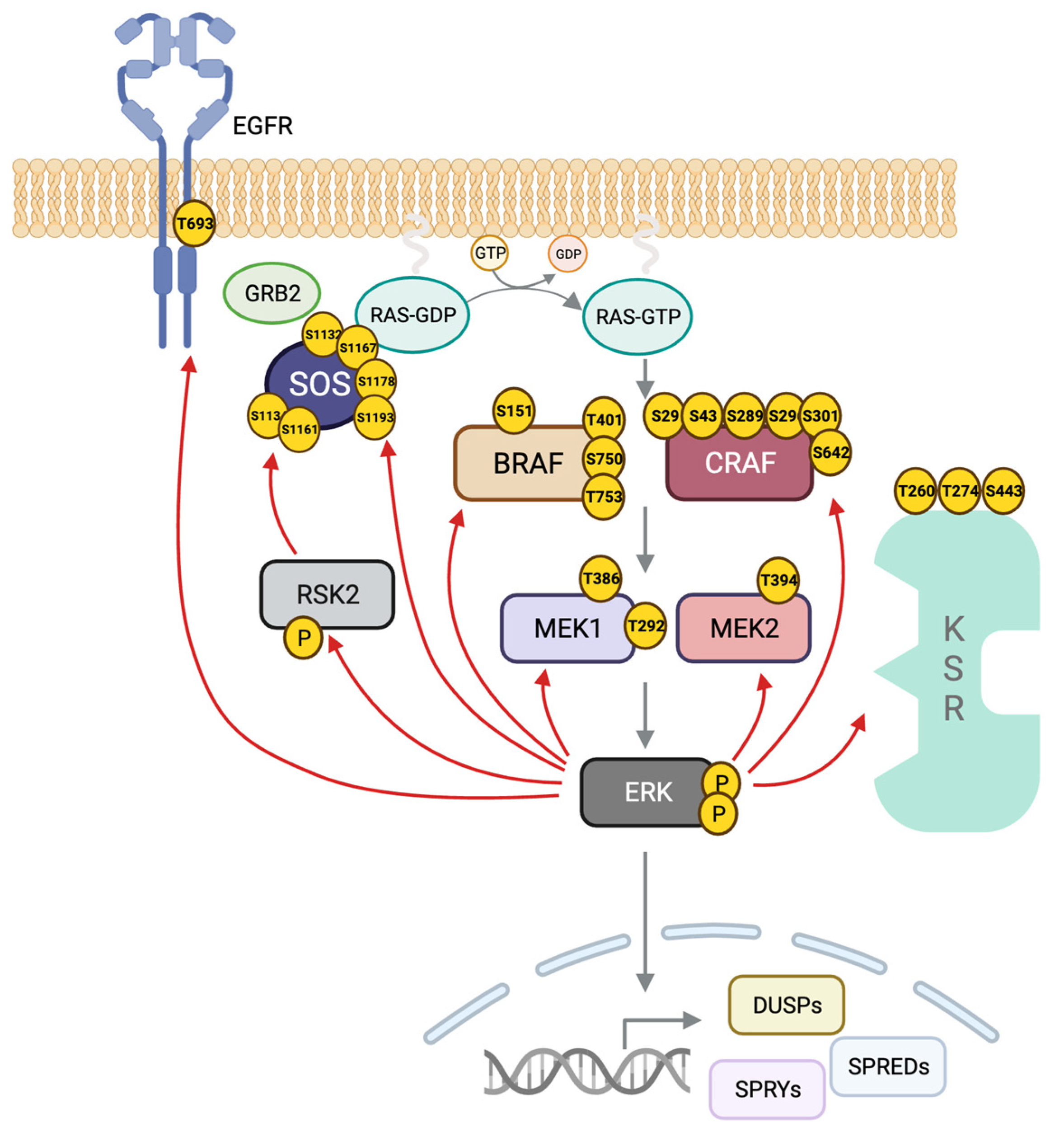
ERK can phosphorylate multiple sites on BRAF and CRAF, some preventing their heterodimerization and others the interaction with RAS and localization at the plasma membrane [255,256] (Figure 3). In the case of BRAF, ERK also phosphorylates several residues with a negative outcome [205]. The conserved motif SPKTP, not present in the other RAF isoforms, is found in the C-terminus of BRAF, and it is also phosphorylated by ERK and implicated in the negative regulation of the pathway [257]. Three other residues in BRAF, T401, S750 and T753, are phosphorylated by ERK and prevent heterodimerization with CRAF [205,258].
ERK also can negatively regulate the scaffold protein KSR1, while ERK phosphorylates T260, T274, S320 and S443 in response to growth factor stimulation or RAS activation [259,260,261]. These events are proposed to contribute to BRAF–KSR1 dissociation and relocation of KSR1 from the plasma membrane to the cytoplasm [262].
Another ERK-regulated negative feedback loop targets the guanine nucleotide exchange factor SOS. SOS is phosphorylated by ERK at residues S1132, S1167, S1178 and S1193 to decrease binding affinity with Grb2 and, consequently, block SOS translocation to the plasma membrane [263,264]. RSK2 activated by ERK is responsible for SOS phosphorylation at S1134 and S1161 [265,266,267]. These phosphorylations generate a binding site for 14-3-3 proteins sequestering SOS and prevent its participation in RAS activation [268,269].
Indirect regulation by ERK leads to the de novo synthesis of negative regulators by transcriptional upregulation of dual-specificity phosphatases (DUSPs) DUSP1, DUSP2, DUSP5, DUSP6 and DUSP9 [270]. In addition, ERK phosphorylates DUSP1 to stabilize the protein and increase its half-life [271,272]. ERK increases the expression of sprouty (SPRY) genes [273]. Sprouty1 and sprouty2 proteins act upstream of RAS by binding the adaptor protein Grb2 and impeding its interaction with SOS [274]. SPRY4 inhibits the pathway by binding CRAF [275].
4.2. ERK Scaffolds
Many actions of the MAPK cascade are targeted to subsets of substrates through association with scaffolding proteins. Nevertheless, the concentrations and affinities of pathway components, which vary widely among cell types [276], are also important for pathway outputs. Early efforts to quantify relative amounts of these signaling molecules in HeLa and Cos-7 cells suggested that MEK is the most abundant protein of the pathway, followed by ERK, then RAS, and RAF is the least abundant in these cell lines [277]. More recent studies have suggested increases in the molecules moving down the cascade with ERK2 as the enzyme of highest abundance [278]. Numerous studies have arrived at affinities for pairs of components (Table 1). Of course, Kd values determined from in vitro experiments may not reflect the cellular value as a result of differences in concentrations of scaffolds and interacting proteins in cells. Affinity predictions under physiological conditions are approximations at best [279]. Table 1 summarizes data from multiple in vitro studies.
Dozens of protein scaffolds have been linked to a function in the RAS–ERK pathway. These include adaptor or accessory proteins which are sometimes classified as scaffolds. Sticking to the benchmark that a scaffold binds simultaneously to at least two members of the same signaling cascade forming a functional, stable complex [292,293], in this review the discussion is limited to three types of the nearly 20 proteins that physically associate with at least two members of the cascade. Table 2 contains the current list.
The first scaffold characterized in a MAPK cascade was Ste5, identified in the budding yeast Saccharomyces cerevisiae. Ste5 binds Fus3 (a MAPK), Ste7 (a MAP2K) and Ste11 (a MAP3K) forming a multiprotein complex at the tips of mating projections following stimulation with mating pheromones [294,295]. Before the discovery of Ste5, it was generally assumed that kinases did not form stable complexes with substrates. Interestingly, no mammalian Ste5 homologue has been identified suggesting context specificity [296,297]. In contrast, scaffold proteins such as IQGAP [298] are evolutionary conserved from yeast to humans which suggests an essential cellular function.
Why are scaffolds important? Scaffolds are the least understood components of MAPK pathways, but they connect more directly to functional specificity than the individual cascade kinases themselves. For example, IQGAP1 [299], paxillin [300] and GIT1 [301] ERK scaffolds are responsible for localization of active ERK in focal adhesions and actin filaments. Here, ERK phosphorylates several substrates involved in actin polymerization, myosin activation and Rac/Cdc42 inactivation, among others (reviewed in [302]).
They incorporate diverse regulatory properties, as detailed below, in addition to serving as cascade assembly sites [303]. Scaffolds share a number of essential features ensuring enzymes like ERK1/2, which can phosphorylate hundreds of proteins, do so at the right place and time. Scaffold proteins optimize signaling by clustering enzymes and substrates together, increasing their effective concentrations. Scaffolds also position proteins in advantageous orientations, often to facilitate phospho-transfer reactions [304,305]. Other proteins or small molecules may allosterically regulate the pathway through a specific scaffold to increase or inhibit the cascade output [57,306]. Scaffolds can also sequester and protect the components of the cascade not only from dephosphorylation from phosphatases [293,305,307,308] but also from unwanted crosstalk, for example, by segregating a kinase involved in two cascades to the proximity of the components of one of them [309,310]. Obviously, this is not the case when two pathways, that cross-activate one another on purpose, share the same scaffold. In addition, scaffolds control the ERK signal in space; by localizing ERK activity in a specific subcellular localization [311,312], scaffolds dictate ERK substrate specificity [313]. Scaffolds can also work as dimerization platforms to promote the formation of ERK dimers. This function may also participate in substrate selectivity, as one ERK monomer binds to the scaffold and the other to a specific cytoplasmic substrate [314].
Also central to their function, scaffolds must be at an optimal concentration to produce maximum signal output. With too few scaffold proteins, cascade assembly may fall below the optimum, while too many scaffold proteins will separate the kinase cascade components, yielding unproductive complexes; in the latter case, more is less. This phenomenon has been named “combinatorial inhibition” [315] and the “prozone effect” [305,307,316,317]. Ideally, each scaffold should maintain the aforementioned optimum level that results in maximum signal efficiency. Nevertheless, to balance the necessary pathway outputs in a given cell context, these optimum levels may vary based on other scaffold concentrations that are competing for the same pool of kinases, as well as the upstream activating signal. For example, overexpression of a cytoplasmic scaffold enhances ERK activity in the cytoplasm and, as a consequence, downregulates nuclear ERK signaling, consistent with competition for the same pool of kinases [150,318,319,320]. Because of the dependence on scaffolds, regulating scaffold concentrations might be an effective method to control MAPK output, titrating activity down without fully suppressing it, perhaps with output selectivity. With a more precise understanding of scaffold function, such a strategy might offer an alternative to bypass resistance mechanisms. Modeling approaches have sought to fill this gap in our knowledge of scaffold behavior but have not yet developed models that have been validated experimentally [321].
Variations in the expression of a single scaffold have altered total ERK activity to a greater extent than just interfering with a single scaffold [322,323,324,325,326,327]. Furthermore, scaffolds can associate with each other to form macro-complexes. Interactions have been described between MP1 and MORG1 [328]; IQGAP1 and MP1 [329]; paxillin and GAB1 [330]; IQGAP1 and β-arrestin-2 [331]; and IQGAP1 and KSR1 [332]. It has been proposed that scaffolds coordinate and cross-talk [137]. This premise was substantiated a few years later by the functional interaction between KSR1 and IQGAP1. ERK bound to a KSR1 mutant unable to bind MEK can be transactivated from MEK bound to IQGAP1, and this cross-activation mechanism has been named trans-phosphorylation. This mechanism takes place in specific cellular settings, and it depends on the concentration of the scaffolds and proper stoichiometry among them and the kinases. Additionally, the downregulation of one scaffold can lead to the upregulation of another scaffold with compensatory abilities [332].
4.2.1. KSR1/2
In 1995, KSR1 was simultaneously discovered in Drosophila virilis/Drosophila melanogaster [333] and Caenorhabditis elegans [334,335] by a genetic screen for RAS downstream effectors. The phenotypes of the KSR1 loss-of-function mutants revealed inhibition of RAS signals, and for this reason the protein was named Kinase Suppressor of RAS (KSR) [333,334]. KSR was recognized as a mammalian functional equivalent of the yeast MAPK scaffold protein Ste5, even though they do not share sequence homology.
The KSR family is composed of five members, the pseudokinases KSR1 [333] and KSR2 [336] and their relatives, the three RAF protein kinases (Figure 4). Despite close sequence similarity between KSR1 and KSR2, these two proteins have distinct functions which are due in part to differences in expression. While KSR1 is widely expressed, KSR2 has a restricted tissue expression pattern, generally with neuroendocrine enrichment. Regarding function, both are scaffolds of the RAS–ERK pathway, but the knockout phenotypes are markedly different. Mice lacking KSR1 show a mild phenotype with disorganized hair follicles but no developmental issues, while RAS-directed oncogenesis is reduced [323]. On the other hand, KSR2-deficient mice are less fertile and develop metabolic disorders such as obesity, insulin resistance and type 2 diabetes [337,338]. Structurally, KSR2 is larger than KSR1 [336], incorporating a distinct motif that binds the AMP-activated protein kinase, AMPK. Binding AMPK likely confers, at least in part, the unique association with metabolic disorders [337].
Why are KSR1/2 considered pseudokinases? Although mammalian KSR1/2 were initially classified as pseudokinases because they contain arginine in place of the lysine essential for catalytic activity, this has been a point of contention [348,349,350,351,352,353]. Some studies have reported residual kinase activity [306,351,354,355] not detected by others [342,356,357]. Indirect evidence suggesting catalytic function comes from the findings that ectopic expression of KSR1 or KSR2 is sufficient to induce proliferation in a RAS-independent manner, attributed to heterodimerization with RAF. Still, the exact mechanism by which KSR overexpression leads to ERK activation has not been defined [358]. Some work has reported that ATP binding is essential for KSR1 activation, although how KSR1 binds ATP was not addressed [306,358]. Adding to the puzzle, unlike mammalian KSRs, Drosophila and C. elegans KSRs possesses the same catalytic sequence as conventional protein kinases (VAVK) [344], raising the question of why arginine replaced lysine in the mammalian proteins. On the other hand, mutation of the ATP-binding lysine in Drosophila and C. elegans KSR does not compromise the function of either; in these organisms, mutant KSR still supports RAF activation and increases ERK signaling [356,357], consistent with a non-catalytic function.
KSR1 is usually cytoplasmic and thought to be constitutively bound to MEK [345,346,347,359]. Upon stimulation and RAS activation, KSR1 rapidly translocates to the plasma membrane where it interacts with a RAF isoform, especially with BRAF [58]. At the plasma membrane, KSR exhibits selectivity towards defined microdomains, responding preferentially to RAS signals from lipid rafts [339]. Interaction with the E3 ligase IMP (Impedes Mitogenic signal Propagation) promotes the recruitment of KSR to Triton-resistant structures that sequester KSR1 and block ERK activation [360,361]. RAS activation leads to IMP proteasomal degradation, facilitating KSR-mediated ERK activation [362]. In this respect, it has been shown that KSR selectively couples RAS signals from lipid rafts to the activation of cPLA2 by ERK [16]. The ERK-mediated KSR1 phosphorylation sites (Section 4.1) are proposed to contribute to BRAF–KSR1 dissociation and relocation of KSR1 from the plasma membrane to the cytoplasm [363]. Phosphorylation of KSR1 on S297 and S392 by the kinase C-TAK1 creates 14-3-3 binding sites; these sites must be dephosphorylated, most likely by PP2A, to localize KSR1 to lipid rafts [259,364,365]. KSR1 phosphorylation may be associated with its nucleo-cytoplasmic shuttling [366]. Fewer sites have been reported in KSR2 and none are well characterized, but some may be controlled by calcineurin dephosphorylation [255].
Homo- and heterodimerization of KSR and RAF proteins underscores the remarkable complexity of ERK regulation at the MAP3K level, some of which was originally uncovered through studies of RAF inhibitors. A depth of structural data has revealed multiple dimeric structures of these molecules [57,306]. KSR and RAF can form side-to-side heterodimers believed to trigger RAF activation through a nearly identical dimer interface that is conserved across all family members. In the same way, KSR1 can also homodimerize through its C-terminus, forming side-to-side dimers as well [57,367]. Apart from KSR–RAF interaction through the kinase domains, selective heterodimerization of RAF with KSR1 occurs through direct contacts between the N-terminal regulatory regions of each protein, including the KSR coiled-coil–sterile alpha motif (CCSAM) [340]. Interestingly, MEK binding to the kinase domain of KSR1 promotes KSR heterodimerization with RAF. As a result of KSR–RAF dimerization, KSR acts as an allosteric activator of RAF to enhance its catalytic activity in addition to its function as a scaffold connecting RAF with its substrate MEK [57]. On the other hand, according to the model proposed by Brennan and collaborators in 2011 [306], once KSR and BRAF dimerize, a conformational change in KSR exposes the MEK activation loop phosphorylation sites, S218 and S222. Once these sites in the MEK activation loop are exposed, an active RAF molecule from a second complex will phosphorylate MEK.
4.2.2. IQGAP1/2 and 3
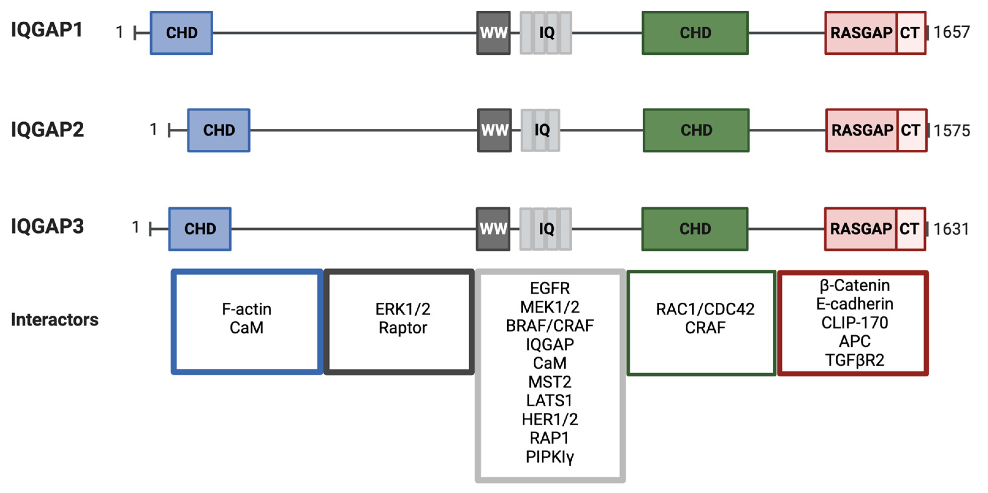
IQ motif-containing GTPase Activating Protein (IQGAP) 1 was first identified in 1994 by Weissbach and collaborators in a screen to discover novel matrix metalloproteases [368]. Nevertheless, it was not until 2004 that IQGAP was classified as a Ras/ERK pathway scaffold [324]. Mammalian IQGAP1, 2, and 3 are multidomain proteins of ~190 kDa [369,370,371]. IQGAPs exhibit different tissue expression patterns; while IQGAP1 is ubiquitous [368], IQGAP2 is mainly expressed in the liver and the gastro-intestinal and urogenital track [372,373], and IQGAP3 is found mainly in the brain, lung and testes [374]. IQGAP1 is cytoplasmic and mainly associated with the cytoskeleton, with particular enrichment in cell–cell contacts [375]. Distributions of IQGAP2 and IQGAP3 are not well described, but they have been noted throughout the cytoplasm and in cell–cell junctions, respectively [376,377]. A recent study reported IQGAP3 primarily in the nucleus [378].
IQGAP isoforms share a similar domain composition (Figure 5). Abundant protein-interacting motifs make them key players in numerous cellular processes. For example, more than 100 binding partners have been described for IQGAP1 [379]. At the N-termini of IQGAPs is a calponin homology domain (CHD) that binds cytoskeletal proteins, in particular F-actin [380]. This domain can also bind calmodulin/Ca2+ but at a lower affinity than to the IQ domain and in competition with F-actin [381]. The CHD is followed by a WW domain with two conserved Trp residues (W). WW domains specify proline-rich regions in binding partners. Interestingly, a major IQGAP WW domain-binding partner, ERK, apparently binds through a different mechanism [324]. Four tandem isoleucine/glutamine-containing (IQ) motifs are multipurpose domains that bind partners including calmodulin [381,382], the EGF receptor [383], MEK [384], BRAF [385] and the small GTPase RAP1 [386]. IQ domains are also important as they mediate the formation of IQGAP dimers and oligomers [387]. IQ domains participate in the interaction with phosphoinositide signaling elements such as phosphatidylinositol phosphate kinase type Iγ (PIPKIγ) at the leading edge. PIPKIγ binding favors the IQGAP open conformation to recruit the actin polymerization machinery and participate in cell motility [388]. At the C- terminus, there is the GAP-related domain (GRD), which defines IQGAP as a GTPase-activating protein (GAP). In spite of the family name and structural similarity to GAPs, IQGAPs do not promote GTP hydrolysis. A conserved arginine in GAPs is replaced by a threonine, which allows IQGAP1 to stabilize small GTPases in their GTP-bound states [369,389], similar to the Rho family GTPases RAC1 and CDC42 [389,390]. At its distal C-terminus, there is an RGCT (Ras GAP C-terminus) domain unique to IQGAP. This region binds diverse targets, such as phosphoinositides [388], E-cadherin [391] and β-catenin [392] among others. Overall, IQGAPs are unique complex proteins with exceptional binding capabilities, involving them in a broad variety of signaling pathways and cellular processes.
Despite the long list of interactors, a major role of IQGAP1 is as a scaffold for the RAS/ERK pathway [324]. IQGAP1 binds both MEK and ERK [384] and can also interact with BRAF and modulate its functions. Generally, IQGAP1–BRAF association is decreased by Ca2+/calmodulin binding to IQGAP1, as this interaction provokes a conformational change [393]. IQGAP1-bound BRAF has higher kinase activity than free BRAF; it is unclear whether binding to IQGAP1 promotes high BRAF activity or whether highly activated BRAF preferentially binds IQGAP [393]. EGF stimulates IQGAP1 binding to MEK1 and decreases binding to MEK2. It has been suggested that MEK1 binding to IQGAP1 promotes proliferation, whereas binding to MEK2 induces differentiation [394]. In contrast to MEK and BRAF, ERK1/2 constitutively bind IQGAP [324]. Overexpressed IQGAP3 also interacts with H-RAS [374], whereas IQGAP1 interacts with K-RAS [395,396]. IQGAP1 binding to EGFR modulates its activation [383], perhaps because the IQGAP1 scaffolded complex promotes EGFR phosphorylation by ERK when RAS is signaling from lipid raft domains [313]. IQGAP1 can also form complexes with other ERK pathway scaffolds such as β-arrestin-2 [397], MP1 [329], and KSR1 [332].
Under the control of RAC1/Cdc42 and antagonizing Ca2+/calmodulin signals, IQGAP1 participates in cell migration through focal adhesions and cytoskeleton dynamics by mediating co-localization of N-WASP with the Arp2/3 complex in lamellipodia which assemble to catalyze actin polymerization [398,399,400]. Stimuli that promote the formation of focal adhesions, such as PDGF, stimulate the assembly of IQGAP1 complexes with the focal adhesion proteins vinculin and paxillin [401]. During migration, a coordinated assembly and disassembly of focal adhesions is necessary. MP1 (MEK partner 1 aka LAMTOR3) plays a role in this process by anchoring MEK to late endosomes via p14 (LAMTOR2) [327]. Knock down of MP1 impairs migration due to the formation of abnormal focal adhesions that accumulate IQGAP1, suggesting that the interaction between IQGAP1, MP1 and the ERK cascade is crucial in this process [329]. IQGAP1 at the leading edge in migrating cells promotes migration in a RAC1- and Cdc42-dependent fashion. In response to integrin engagement, IQGAP1 forms a complex with filamin A to recruit RAC1GAP, which inactivates RAC1. Decreased expression of any of these three proteins results in uncontrolled formation of membrane protrusions and impairs directional cell migration [402]. F-actin crosslinking is key in the regulation of cytoskeletal processes such as the formation of focal adhesions and filopodia, essential for cellular migration [403]. IQGAP1 also aids in stabilizing F-actin crosslinking stimulated by high Cdc42 [404,405]. Interestingly, only IQGAP1 dimers, formed following Cdc42 stimulation, are competent for F-actin crosslinking and the generation of strong adherent junctions [406]. Recently, IQGAP1 has been found to participate as a scaffold in the Hippo pathway. IQGAP1 interacts with kinases MST2 and LATS1 to suppress their kinase activity. IQGAP1 is also a negative regulator of the non-canonical pro-apoptotic pathway [407].
IQGAP1, like KSR1, is not required for embryonic development. Mice lacking IQGAP1 are viable and fertile with gastric hyperplasia as a mild phenotype [408]. However, these mice are resistant to developing tumors induced by oncogenic H-RAS [325]. In the case of IQGAP2 knockout mice, they develop age-dependent hepatocellular carcinoma (HCC) with increased IQGAP1 expression [409]. These two phenotypes align with IQGAP1 pro-tumorigenic and IQGAP2 anti-tumorigenic roles. Indeed, in HCC, IQGAP1 protein and mRNA are increased, while IQGAP2 mRNA is downregulated [410,411] and IQGAP3 is high in gastric cancer [412]. Furthermore, the IQGAP1–MST2-LATS1 (Hippo pathway) complex is regulated by bile acid in HCC cells, likely to inhibit MST2-dependent apoptosis and cell transformation [407].
No definitive oncogenic mutations have been identified in IQGAPs in tumors; however, missense mutations have been identified that correlate with specific tumor types (Table 2), further suggesting that IQGAP1 participates in oncogenic processes [413]. Also, IQGAP gene amplifications have been found in tumors [414]. IQGAP1 was found overexpressed in lung, endometrial, ovarian, gastric, colon, and breast cancer, as well as hepatocellular carcinoma (HCC) and melanoma [415]. In fact, IQGAP1 has been proposed as an oncogene because its overexpression can stimulate cell proliferation and favor transformation of human epithelial cells [415]. Similarly, downregulating IQGAP1 in MCF7 cells using siRNAs diminishes cell migration, proliferation and prevents tumor formation in xenograft mice [299,416].
4.2.3. HPIP
HPIP (hematopoietic PBX (pre-B cell leukemia homeobox)-interacting protein or pre-B cell leukemia transcription factor-interacting protein (PBXIP1)) is a 731-amino acid protein that was discovered in 2000 in a yeast two-hybrid screen using PBX1a as bait. HPIP interacts with PBX transcription factors, negatively regulating PBX1-mediated hematopoiesis [417]. Although HPIP is predominantly cytoplasmic, specifically localizing on microtubules, this protein contains both a nuclear localization signal (NLS) and a nuclear export signal (NES) [418].
It was not until ten years after its discovery that HPIP was recognized as an ERK scaffold. HPIP associates with the estrogen receptor (ERα), Src and PI3K, which, in turn, activate ERK1/2 and AKT signaling pathways [419]. Thus, HPIP’s function as a scaffold protein is not exclusive to the RAS–ERK pathway. HPIP participates in signal transduction via transcription factors, C/EBPα and GATA1, ERα and ERβ, LEF1/ β-catenin and p53 as well as through tyrosine and serine/threonine protein kinases, FAK/Src, and TBK1 and CK1α, and it has also been implicated in the regulation by TGF-β. This great variety of interactors connect HPIP with processes including hematopoiesis, cell proliferation and tumorigenesis, germ cell proliferation, EMT, renal fibrosis, cell migration and invasion, and osteoarthritis. HPIP is widely expressed in different tissues, and it is considered a proto-oncogene since it is overexpressed in more than 15 types of cancer [420]. The only HPIP knockout mouse study revealed a defect in articular cartilage and bone development, but the lack of this protein protects against osteoarthritis [421].
In a recent study, HPIP was shown to facilitate the interaction of the canonical components of the RAS–ERK pathway in a phosphorylation-dependent manner. HPIP interacts with all the components of the pathway, from the EGFR to ERK, including all RAS isoforms (K-, N-, H-RAS), giving rise to two different functional complexes. First, HPIP associates with EGFR, SHC, Grb2, SOS1, RAS, RAF, and MEK to form complex 1. Subsequently, the scaffold leaves behind a few upstream components to bind ERK and make complex 2 with SHC, RAS, RAF, MEK, and ERK [422]. Further research is needed to fully understand the importance of this little-studied scaffold in health, disease, and potential therapeutic implications.

Table 2.
Interactors, subcellular localization, tissue expression and mutations in RAS–ERK pathway scaffolds found in cancer. The information presented in this table has been retrieved from multiples sources, including string-db.org (accessed on 3 July 2023) [423], genecards.org (accessed on 3 July 2023) [424] and phosphosite.org (accessed on 3 July 2023) [425], as of July 2023. Data for subcellular localization obtained from GeneCards was collected from UniProtKB/Swiss-Prot, the Human Protein Atlas (HPA) and Gene Ontology (GO) (Confidence > 3). Tissue expression information is based on mRNA expression in normal human tissues from GTEx, Illumina, BioGPS, and SAGE (expression was considered ubiquitous when detected in over seven different tissues). The mutation frequency was obtained from TCGA (The Cancer Genome Atlas) in 15 types of cancer. The table lists up to the five most common cancer types with recurrent potential oncogenic missense mutations in each scaffold. The cancer types are listed in descending order of prevalence. Protein name is given in alphabetical order, and the gene name is in italics under the protein name.
| Scaffold Name | RAS-ERK Pathway Interactors (Source: STRING and References) | Subcellular Localization (Source: GeneCards) | Tissue Expression (Source: GeneCards) | Mutations in Cancer (Source: Phospho-Site) | References |
|---|---|---|---|---|---|
| Archvillin/Supervillain Spliced isoform of SVIL | B-RAF, MEK, ERK | Cytosol, Cytoskeleton, PM, Nucleus | Ubiquitous | Stomach, bladder, endometrial, lung squamous, LUAD… | [426] |
| β-Arrestin-1 ARRB1 | C-RAF, MEK, ERK | Nucleus, GA, Lysosome, Cytosol, PM | Ubiquitous | Lung squamous, Stomach, Endometrial, Bladder, CRC… | [427] |
| β-Arrestin-2 ARRB2 | C-RAF, MEK, ERK | Cytosol, Nucleus, PM, Endosomes | Ubiquitous | Endometrial, LUAD, HNSCC, Stomach… | [427] |
| CaM CALM1 Calmodulin | EGFR, RAS, RAF | Cytoskeleton, PM, Cytosol, Nucleus | Ubiquitous | LUAD, Endometrial, Bladder, Stomach, HNSCC. | [393] |
| CNK1 CNKSR1 Connector Enhancer Of Kinase Suppressor Of Ras 1 | RAS, RAF | Cytosol, PM, Nucleus | Highest in Muscle | Stomach, Endometrial, Bladder, CRC, LUAD… | [428] |
| CNK2/MAGUIN-1 CNKSR2 Connector Enhancer Of Kinase Suppressor Of Ras 1 | RAS, RAF | Cytosol, PM, Nucleus | Nervous system | Lung squamous, Endometrial, Stomach, CRC, LUAD… | [429] |
| DYRK1A DYRK1A Dual Specificity Tyrosine Phosphorylation Regulated Kinase 1A | RAS, B-RAF, MEK1 | Nucleus, Cytosol, Cytoskeleton | Ubiquitous | Endometrial, Lung squamous, Stomach, CRC, HNSCC… | [430] |
| β-Dystroglycan-1 DAG1 Dystrophin-Associated Glycoprotein 1 | MEK, ERK | PM, Nucleus, Extracellular, Cytosol, GA, ER, Cytoskeleton | Ubiquitous | Stomach, CRC, Endometrial, LUAD, Lung squamous… | [431] |
| FHL1 FHL1 Four And A Half LIM Domains 1 | CRAF, MEK2, ERK2 | Cytosol, Nucleus, PM | Ubiquitous | Endometrial, Stomach, LUAD, Lung squamous, Breast… | [432] |
| FLOT1 FLOT1 Flotillin 1 | RAF, MEK, ERK | Endosomes, PM, Lysosome, Extracellular, Cytosol, Cytoskeleton | Ubiquitous | Stomach, CRC, Lung squamous, Endometrial, HNSCC… | [433] |
| GIT1 GIT1 G Protein-Coupled Receptor Kinase Interactor 1 | MEK1, ERK1/2 | Cytosol, Cytoskeleton, Mitochondria | Ubiquitous | Endometrial, bladder, Stomach, Lung squamous, kidney… | [434] |
| GAB1 GAB1 Grb2 Associated Binding Protein 1 | Grb2, SOS1 | Cytosol | Ubiquitous | CRC, Endometrial, bladder, Lung squamous, Stomach… | [435] |
| Galectin1 LGALS1 Putative MAPK-Activating Protein PM12 | HRAS, CRAF | Cytosol, ER, Nucleus | Ubiquitous | HNSCC, Breast Lung squamous, CRC, LUAD… | [436] |
| Grb10 GRB10 Growth Factor Receptor-Bound Protein 10 | C-RAF, MEK | Cytosol, PM, Nucleus | Ubiquitous | Endometrial, LUAD, Stomach, Lung squamous, bladder… | [437] |
| HPIP/PBXIP1 PBXIP1 Hematopoietic PBX (Pre-B cell leukemia homeobox)-interacting protein | EGFR, SHC, Grb2, SOS1, RAS, RAF, MEK, ERK | Cytosol, nucleus, Cytoskeleton | Ubiquitous | Stomach, Bladder, CRC, LUAD, Endometrial… | [419] |
| IQGAP1 IQGAP1 IQ Motif Containing GTPase Activating Protein 1 | EGFR, K-RAS, B-RAF, MEK, ERK | Cytosol, Nucleus, Cytoskeleton, PM, Focal Adhesions | Ubiquitous | Stomach, CRC, Endometrial, Lung squamous, HNSCC… | [324] |
| IQGAP2 IQGAP2 IQ Motif Containing GTPase Activating Protein 2 | RAS, B-RAF, MEK, ERK | Cytosol, Cytoskeleton, PM, Nucleus | Ubiquitous | Endometrial, CRC, LUAD, Lung squamous, Stomach | [413] |
| IQGAP3 IQGAP3 IQ Motif Containing GTPase Activating Protein 3 | H-RAS, B-RAF, MEK, ERK | Cytosol, Nucleus, PM, cell-cell junctions | Liver, lung, pancreas | Lung squamous, CRC, Endometrial, Stomach, Bladder… | [413] |
| KSR1 KSR1 Kinase Suppressor of Ras 1 | RAF, MEK, ERK | Cytosol, ER, PM | Ubiquitous | Endometrial, ovarian. | [333,334,335] |
| KSR2 KSR2 Kinase Suppressor of Ras 2 | RAF, MEK, ERK | Cytosol, PM | Nervous system | Stomach, Endometrial, LUAD, HNSCC, Lung squamous… | [336] |
| MERLIN NF2 Moesin-Ezrin-Radixin Like (MERLIN) Tumor Suppressor | RAS, RAF | Cytosol, Cytoskeleton, PM, Nucleus, Mitochondria | Ubiquitous | Endometrial, Bladder, HNSCC, CRC, Lung squamous… | [438] |
| MORG1 WDR83 Mitogen-Activated Protein Kinase Organizer 1 | C-RAF, MEK, ERK | Endosomes, Nucleus | Kidney, Muscle, Nervous system | Endometrial, Bladder, Prostate, Lung squamous, CRC… | [328] |
| MP1 LAMTOR3 MEK Partner 1 | MEK1, ERK1, ERK2 | Endosomes, Lysosome, PM, Extracellular | Ubiquitous | HNSCC, Glioblastoma. | [439,440] |
| Nucleophosmin NPM1 | KRAS, RAF | Cytosol, Cytoskeleton, Nucleus, | Ubiquitous | Leukemia, LUAD, Endometrial, CRC, HNSCC… | [441] |
| Paxillin PXN | MEK, ERK | Cytosol, Focal Adhesions, Cytoskeleton, PM, Nucleus | Ubiquitous | Endometrial, Stomach, Lung squamous, HNSCC, Glioblastoma… | [300] |
| PEA15 PEA15 Phosphoprotein Enriched in Astrocytes, 15 KDa Proliferation And Apoptosis Adaptor Protein 15 | ERK, RSK | Cytosol, Cytoskeleton, Nucleus | Ubiquitous | Lung squamous, Endometrial, CRC, LUAD, HNSCC. | [148] |
| RGS12 RGS12 Regulator Of G Protein Signaling 12 | RAS, RAF, MEK2 | Cytosol, Nucleus, PM | Nervous system, Lung, Kidney | Stomach, Lung squamous, bladder, Endometrial, CRC… | [442] |
| RGS14 RGS14 Regulator Of G Protein Signaling 14 | RAS, RAF | Nucleus, Cytoskeleton, Cytosol, PM | Ubiquitous | Endometrial, bladder, Stomach, CRC, LUAD… | [443] |
| SEF1 IL17RD Interleukin-17 Receptor D | MEK, ERK | GA, Nucleus, PM | Nervous system, heart, Kidney | Endometrial, CRC, Stomach, Leukemia, Breast… | [150] |
| SHOC2/SUR8 SHOC2 Leucine Rich Repeat Scaffold Protein | RAS, RAF | Cytosol, Nucleus | Ubiquitous | Endometrial, Stomach, CRC, LUAD, HNSCC… | [444] |
Abbreviations: LUAD = lung adenocarcinoma; HNSCC = head and neck squamous cell carcinoma; CRC = colorectal carcinoma; ER = endoplasmic reticulum; PM = plasma membrane; GA = Golgi apparatus. All data refers to Homo sapiens.
5. Pathway Inhibitors
Pharmacological small molecule inhibitors of great scientific utility have been developed for the enzymes in the cascade, while those targeting RAF and MEK dominate clinical strategies to block the pathway. Table 3 lists some of these compounds. Poor pharmacokinetics and other issues with small molecule kinase inhibitors have provoked the development of other strategies to inhibit high-impact targets. In this section we focus on several alternative strategies to manipulate the ERK pathway via docking site inhibitors, protein degraders for multiple pathway components, molecular glues, and optically activated MEK inhibitors. Some of these may also yield access to proteins considered undruggable such as transcription factors and scaffolds.
5.1. Classical Inhibitors
Many classical inhibitors for the different members of the cascade have been developed. Many of them are in clinical trials but only a few have received FDA approval, mostly those targeting RAF and MEK. Remarkably, two covalent inhibitors, sotorasib and adragasib, have been FDA-approved in the last couple of years to treat KRASG12C lung cancers (reviewed in [487]), and several promising clinical trials are currently in progress [30]. As of today, no ERK inhibitors are in clinical use, although several are in clinical trials [488]. Unfortunately, single-therapy approaches have led to the development of mechanisms of resistance. To overcome this issue, many combination strategies are being assessed and some have been already approved [489].
In contrast to many other kinases, ERK2 undergoes relatively small conformational changes in the active site upon activation. Solution measurements have found evidence for phosphorylated ERK2 (2P-ERK2) in two conformational states, R and L, which interconvert on a millisecond time scale [490]. Surprisingly, the L state resembles the active site of unphosphorylated ERK (0P-ERK2). A recent study found ERK inhibitors that interact with both states and ones that selectively interact with the R or L states. Inhibitors were noted that could shift the equilibrium towards the R or L states. As some ERK inhibitors have been shown to affect specific protein–protein interactions, this study provides new insights for inhibitor design [491].
Apart from the large number of classical small molecule inhibitors directed at the ATP site [492] (representative list in Table 4), some alternative inhibitors target protein–protein interactions. An example is the ERK dimerization inhibitor DEL-22379. This small molecule disrupts ERK dimerization by binding within the dimerization interface without affecting the ERK phosphorylation state [493,494]. Another strategy is to target ERK1/2 substrate docking sites [135,136], e.g., SF-3-030, which selectively interferes with a subset of ERK functions dependent on the ERK FXF docking site [495].
5.2. PROTAC Technology Applied to the ERK Pathway
As its name implies, Targeted Protein Degradation (TPD) covers several methods to induce the selective degradation of a specific molecule either via the proteasome, lysosome or autophagy system. One of these methods using the proteasome degradation system is PROTAC (Proteolysis-Targeting Chimera) [514], initially developed in 2001 [515,516,517,518]. The strategy involves recruitment of an E3 ligase, usually VHL (von Hippel-Lindau VHL) or CRBN (Cereblon), in current versions of PROTACs, to the target protein to tag it for degradation. Two ligands, one that binds the target protein and a second that recruits an E3 ligase, are connected through a chemical linker. The target protein will be ubiquitinylated by the E3 and sent to the proteasome resulting in its near elimination [519]. Advantages of protein degradation versus classical small molecule inhibitors are many. PROTACs can reach the undruggable proteome as long as a suitable target ligand is available. Degradation prevents target protein accumulation, thus avoiding compensatory upregulation and drug resistance. PROTACs overcome the generation of resistant mutations under selective pressures or even the emergence of non-enzymatic functions of the targeted proteins, which might provoke a signaling rebound. With an appropriate high-specificity ligand, PROTAC may only target the pathogenic, mutated version of the protein, thus minimizing side effects. The catalytic nature allows them to be used at sub-stoichiometric concentrations to reduce toxicities, as the same PROTAC transiently binds the target protein, and after tagging it for degradation, it releases and binds another target protein. Protein degradation achieves an extended pharmacodynamic effect similar to that of covalent inhibitors, depending on the turnover rate of the target. Unlike small molecule inhibitors that function by an occupancy-driven mechanism, PROTACs possess an event-driven mode of action, which means that a brief interaction with the protein of interest is enough to trigger its degradation [519,520,521,522].
The major limitation of PROTACs continues to be poor oral bioavailability and low cell permeability due to their large sizes [523]. For this reason, it was not until 2019 that the first PROTAC progressed to clinical trials [524]. Although none are employed in the RAS–ERK pathway, a few FDA-approved therapies use degraders [525]. Nevertheless, due to their promising advantages, PROTACs are being developed to inhibit components within the RAS/ERK cascade, summarized here and in Table 5.
The first attempt to generate a PROTAC against KRASG12C was based on the inhibitor ARS1620, and a thalidomide analog, pomalidomide, an FDA-approved drug with high affinity for the ubiquitin ligase CRBN. The compound was named XY-4-88, but failed to induce endogenous KRASG12C degradation in cancer cells [526]. A parallel study generated LC-2, the first successful PROTAC against KRASG12C, which used MRTX849 as the parental inhibitor [527]. A new collection of KRASG12C-selective PROTACs were synthesized based on the AMG-510 covalent inhibitor and were effective, but the IC50s were above 10 µM [528].
The first series of compounds acting as PROTACs for BRAF were created based on the inhibitor rigosertib, which binds to RAF in the RAS binding domain (RBD) and should, therefore, bind RAF mutants as activating mutations are in the kinase domain [529], although this was not tested. P4B was the first effective PROTAC with selectivity for the BRAF mutant V600E. The ligand was based on the inhibitor BI882370 and provides degradative efficacy in the nanomolar range [530]. A year later a mutant-selective PROTAC based on vemurafenib, SJF-0628, was shown to work at nanomolar concentrations on all BRAF mutants tested, including those that showed resistance to vemurafenib, while it had no effect on the WT form [531]. Another BRAFV600E-selective PROTAC, CRBN(BRAF)-24, was based on PLX8394 [532]. In parallel, encorafenib-based PROTACs were developed but none of them resulted in degradation of the mutant BRAF, probably due to failure to recruit the ubiquitin ligase [533].
The first-in-class highly selective degrader of MEK1 and MEK2 was the PD0325901-based compound MS432. It was effective in colorectal cancer and melanoma cell proliferation, with good bioavailability in mice [534]. A few PROTAC compounds against MEK1 and MEK2 were developed based on allosteric MEK inhibitors, such as PD0325901 and refametinib, using VHL as the E3 ligase. They were assessed in cell proliferation and pERK levels to compare the compound with the parental inhibitor. Although these compounds had modest degradation efficiencies, two of them were able to completely suppress proliferation in melanoma cells [535]. Following the development of the first-in-class MEK degrader, the Lin laboratory also characterized three more, MS928, MS934 and MS910. Compared with previously reported VHL-recruiting degraders, these new compounds were more potent in preserving the high selectivity. Of these three degraders, MS934 appeared as the best MEK1/2 degrader for in vivo studies to date. In addition, combinations of MS934 with either BRAF or PI3K inhibitors provided a potent antiproliferative response in CRC and melanoma cells [536]. A new study has reported that MS934 collaterally degrades CRAF in KRAS mutant cells, which makes this compound a first-in-class dual PROTAC [537]. Remarkably, other PD0325901-based degraders did not cause CRAF collateral degradation. Therefore, MS934 inhibiting both CRAF and MEK would not be expected to induce resistance mechanisms via non-catalytic CRAF functions [537]. The aforementioned PROTACs were designed based on a diarylamine scaffold common to most of the inhibitors. Recently, MEK1/2 degraders were synthesized based on a non-diarylamine allosteric MEK1/2 inhibitor, which was a coumarin derivative [538]. From all of them, compound P6b is the first MEK PROTAC with a non-diarylamine scaffold showing a potent degradation effect in human cancer cells [539]. At this time, MEK appears to be the most promising target for PROTACs due to demonstrated functional efficacy in the nanomolar range, and the fact that the only known substrate of MEK is ERK minimizes toxicities by avoiding interference with other pathways.
Table 5.
PROTAC-targeted protein degraders.
| Name | Target (Protein of Interest) | Leading Compound (“Warhead”) | Linker | E3 Ubiquitin Ligase Target | E3 Recruiter (“Anchor”) | Reference |
|---|---|---|---|---|---|---|
| XY-4-88 | KRASG12C | ARS-1620 | Cyclic tertiary amine-containing linker | CRBN | pomalidomide (thalidomide analog) | [526] |
| LC-2 | KRASG12C | MRTX849 | Alkyl | VHL | VH032 | [527] |
| III-2 | KRASG12C | AMG-510 | polyethylene glycol (PEG) | VHL | (S,R,S)-AHPC (VH032-NH2) hydrochloride | [539] |
| P4B | BRAFV600E BRAFG466V | BI882370 | polyethylene glycol (PEG) | CRBN | pomalidomide (thalidomide analog) | [530] |
| SJF-0628 | All mutant BRAF | vemurafenib | Rigid piperzine-based linker | VHL | VHL ligand (not specified) | [531] |
| CRBN(BRAF)-24 | BRAFV600E | PLX8394 | Cyclic tertiary amine-containing linker | CRBN | pomalidomide (thalidomide analog) | [532] |
| MS432 | MEK1/2 | PD0325901 | alkyl | VHL | (S,R,S)-AHPC-Me | [534] |
| Several compounds | MEK1/2 | Allosteric MEKi | Alkyl | VHL | VHL ligand (not specified) | [535] |
| MS928 | MEK1/2 | PD0325901 | Alkyl | VHL | VH032 | [536] |
| MS910 | MEK1/2 | PD0325901 | polyethylene glycol (PEG) | CRBN | pomalidomide (thalidomide analog) | [536] |
| MS934 | MEK1/2 CRAF | PD0325901 | Alkyl | VHL | VH032 | [536,537] |
| P6b | MEK 1/2 | Substituted 3-benzylcoumarins allosteric MEKi | Alkyl | VHL | VHL ligand 22 | [539] |
CRBN, Cereblon. VHL, von Hippel–Lindau.
An enhanced PROTAC technology, known as CLIPTAC (In-Cell Click-Formed Proteolysis-Targeting Chimeras), involves the independent entry of the protein-binding ligand and the ubiquitin ligase anchor into the cell, where these precursors assemble and function as a regular PROTAC. This technique requires sequential treatment with each precursor because their assembly outside the cell will not result in protein degradation. Based on an ERK covalent inhibitor, this tool became the first degrader against ERK1/2 to show efficacy in cells [540]. On the other hand, ubiquibodies (uAbs) are newly engineered molecules able to mark proteins for degradation inside the cell. The uAbs consist of a universal E3 ligase fused to a synthetic binding protein such as a single-chain antibody fragment (scFv), a designed ankyrin repeat protein (DARPin), or a fibronectin type III (FN3) monobody [541]. Their main advantage is easy customizability by simply exchanging synthetic binding proteins with specificity for the target. The difference from regular PROTACs is that uAbs do not need to recruit ubiquitin ligase but they are the ligase themselves. This approach has been successfully applied for ERK degradation. In this case, the uAbs contain DARPins that bind to both phosphorylated and non-phosphorylated ERK1/2 [542].
5.3. Molecular Glues Targeting MEK
The natural products cyclosporin A and FK506 were among the earliest described as molecular glues by Schreiber in 1992 for their ability to promote interactions between two molecules that do not otherwise bind. Following this molecular glue concept, Simonetta (2019) [543] set out to develop a compound that would enhance a protein–protein interaction between a transcription factor and the E3 that normally degrades it. Subsequently, more molecular glues have been identified by high-throughput chemical screens and target validation [544,545]. This strategy involves a monovalent molecule able to stabilize a protein–protein interaction to modify the interactor’s surface to facilitate its binding with a new partner, even if they do not interact under normal conditions. Molecular glues can act as degraders when one of the binding proteins is an E3 ligase [546].
Because molecular glues are small molecules, unlike PROTACs, they should have superior membrane penetrability and oral bioavailability. On the other hand, PROTACs display a higher adaptability to target a protein of interest, while the original molecular glues had unknown ligands [523,546]. Recent studies are focusing on molecular glue optimization, as is the case for the novel MEKi trametiglue. Although approved for clinical use for a decade, trametinib was identified through phenotypic screens, and for this reason, the mechanism of action of trametinib remains incompletely understood. The Dar laboratory elucidated the binding mechanism of this small molecule inhibitor to its target by determining the crystal structure of trametinib-MEK-KSR. Trametinib simultaneously binds to MEK and its scaffold protein KSR, while it destabilizes the interaction of MEK with RAF. Trametiglue was created based on these notions, exhibiting an enhanced affinity for the MEK–KSR complex and demonstrating a better growth inhibition of KRAS mutant cells and lower signaling rebound than the original compound [547,548].
5.4. Optically Activated MEK Inhibitors
Coupling light with therapeutics offers spatial selectivity. The fundamental principle of this strategy is to administer a photoactive drug with low intrinsic (dark) toxicity to a patient followed by irradiation with light to activate it only in a defined region, thus minimizing on-target side effects and enhancing treatment precision. Three light-dependent pharmacological approaches have been developed as follows: photodynamic reactive therapy, irreversible photopharmacology (photodecaging), and reversible photopharmacology with a ligand that is active only in one structural state [549].
Light-dependent pharmacological approaches, specifically photo-decaging, have been proposed to prevent MEKi toxicities. The strategy consists of generating an inactive locked MEK inhibitor precursor, by linking a photo-cleavable protecting group (the ‘cage’) to the small molecule inhibitor. This drug protector is irreversibly cleaved from the MEK inhibitor by exposure to light, and then the inhibitor will bind MEK where it has been uncaged. This cutting-edge strategy has been carried out with the potent allosteric inhibitor PD0325901, which was previously discontinued in clinical trials due to its harmful effects which are presumably on-target. The optically activatable MEK1/2 inhibitors, named opti-MEKi, have shown promising efficacy in vivo in a melanoma xenograft zebrafish model [550]. Combinations of these approaches, e.g., photo-switchable linkers or light-activated degraders, are also in development [551].
6. Concluding Remarks
In conclusion, this review has offered an overview of pathway components and their functions, shedding light on the diverse range of scaffolds involved in pathway regulation. We have also emphasized emerging interventional strategies that hold promise for future advancements in this field. The intricate network and potential interventions discussed here underscore the exciting prospects for further research and therapeutic development within this pathway.
Author Contributions
Writing—original draft preparation, A.M.-V.; Writing—review and editing, M.H.C.; funding acquisition, M.H.C. All authors have read and agreed to the published version of the manuscript.
Funding
This research was funded by the Welch Foundation grant number I1243 and the National Institutes of Health grant R01 GM141699 to M.H.C. and the Cancer Prevention and Research Institute of Texas training grant RP210041 to A.M.-V.
Acknowledgments
Special thanks go to Clinton Taylor for the figure showing the ERK2 structure. We acknowledge the help and support of all members of the Cobb lab for their thoughts on the ERK1/2 pathway. We also thank Dionne Ware for administrative assistance.
Conflicts of Interest
The authors declare no conflict of interest.
References
- Cooper, J.A.; Bowen-Pope, D.F.; Raines, E.; Ross, R.; Hunter, T. Similar Effects of Platelet-Derived Growth Factor and Epidermal Growth Factor on the Phosphorylation of Tyrosine in Cellular Proteins. Cell 1982, 31, 263–273. [Google Scholar] [CrossRef]
- Ray, L.B.; Sturgill, T.W. Characterization of Insulin-Stimulated Microtubule-Associated Protein Kinase. Rapid Isolation and Stabilization of a Novel Serine/Threonine Kinase from 3T3-L1 Cells. J. Biol. Chem. 1988, 263, 12721–12727. [Google Scholar] [CrossRef]
- Sturgill, T.W.; Ray, L.B.; Erikson, E.; Maller, J.L. Insulin-Stimulated MAP-2 Kinase Phosphorylates and Activates Ribosomal Protein S6 Kinase II. Nature 1988, 334, 715–718. [Google Scholar] [CrossRef] [PubMed]
- Boulton, T.G.; Yancopoulos, G.D.; Gregory, J.S.; Slaughter, C.; Moomaw, C.; Hsu, J.; Cobb, M.H. An Insulin-Stimulated Protein Kinase Similar to Yeast Kinases Involved in Cell Cycle Control. Science 1990, 249, 64–67. [Google Scholar] [CrossRef] [PubMed]
- Boulton, T.G.; Cobb, M.H. Identification of Multiple Extracellular Signal-Regulated Kinases (ERKs) with Antipeptide Antibodies. Mol. Biol. Cell 1991, 2, 357–371. [Google Scholar] [CrossRef]
- Boulton, T.G.; Gregory, J.S.; Cobb, M.H. Purification and Properties of Extracellular Signal-Regulated Kinase 1, an Insulin-Stimulated Microtubule-Associated Protein 2 Kinase. Biochemistry 1991, 30, 278–286. [Google Scholar] [CrossRef] [PubMed]
- English, J.; Pearson, G.; Wilsbacher, J.; Swantek, J.; Karandikar, M.; Xu, S.; Cobb, M.H. New Insights into the Control of MAP Kinase Pathways. Exp. Cell Res. 1999, 253, 255–270. [Google Scholar] [CrossRef]
- Coulombe, P.; Meloche, S. Atypical Mitogen-Activated Protein Kinases: Structure, Regulation and Functions. Biochim. Biophys. Acta-Mol. Cell Res. 2007, 1773, 1376–1387. [Google Scholar] [CrossRef]
- Cargnello, M.; Roux, P.P. Activation and Function of the MAPKs and Their Substrates, the MAPK-Activated Protein Kinases. Microbiol. Mol. Biol. Rev. 2011, 75, 50–83. [Google Scholar] [CrossRef]
- Chartier, M.; Chénard, T.; Barker, J.; Najmanovich, R. Kinome Render: A Stand-Alone and Web-Accessible Tool to Annotate the Human Protein Kinome Tree. PeerJ 2013, 1, e126. [Google Scholar] [CrossRef] [PubMed]
- Robbins, D.J.; Zhen, E.; Cheng, M.; Xu, S.; Vanderbilt, C.A.; Ebert, D.; Garcia, C.; Dang, A.; Cobb, M.H. Regulation and Properties of Extracellular Signal-Regulated Protein Kinases 1, 2, and 3. J. Am. Soc. Nephrol. 1993, 4, 1104–1110. [Google Scholar] [CrossRef]
- Pearson, G.; Robinson, F.; Gibson, T.B.; Xu, B.E.; Karandikar, M.; Berman, K.; Cobb, M.H. Mitogen-Activated Protein (MAP) Kinase Pathways: Regulation and Physiological Functions. Endocr. Rev. 2001, 22, 153–183. [Google Scholar] [CrossRef]
- Cox, A.D.; Fesik, S.W.; Kimmelman, A.C.; Luo, J.; Der, C.J. Drugging the Undruggable RAS: Mission Possible? Nat. Rev. Drug Discov. 2014, 13, 828–851. [Google Scholar] [CrossRef]
- Arozarena, I.; Calvo, F.; Crespo, P. Ras, an Actor on Many Stages: Posttranslational Modifications, Localization, and Site-Specified Events. Genes Cancer 2011, 2, 182–194. [Google Scholar] [CrossRef]
- Prior, A.I.; Hancock, J.F. Ras Trafficking, Localization and Compartmentalized Signalling. Semin. Cell Dev. Biol. 2012, 32, 145–153. [Google Scholar] [CrossRef]
- Casar, B.; Pinto, A.; Crespo, P. ERK Dimers and Scaffold Proteins: Unexpected Partners for a Forgotten (Cytoplasmic) Task. Cell Cycle 2009, 8, 1007–1013. [Google Scholar] [CrossRef]
- Hall, B.E.; Bar-Sagi, D.; Nassar, N. The Structural Basis for the Transition from Ras-GTP to Ras-GDP. Proc. Natl. Acad. Sci. USA 2002, 99, 12138–12142. [Google Scholar] [CrossRef] [PubMed]
- Egan, S.E.; Giddings, B.W.; Brooks, M.W.; Buday, L.; Sizeland, A.M.; Weinberg, R.A. Association of Sos Ras Exchange Protein with Grb2 Is Implicated in Tyrosine Kinase Signal Transduction and Transformation. Nature 1993, 363, 45–51. [Google Scholar] [CrossRef]
- Kiel, C.; Matallanas, D.; Kolch, W. The Ins and Outs of Ras Effector Complexes. Biomolecules 2021, 11, 236. [Google Scholar] [CrossRef] [PubMed]
- Kolch, W.; Berta, D.; Rosta, E. Dynamic Regulation of RAS and RAS Signaling. Biochem. J. 2023, 480, 1–23. [Google Scholar] [CrossRef] [PubMed]
- Barbacid, M. ras Genes. Annu. Rev. Biochem. 1987, 56, 779–827. [Google Scholar] [CrossRef]
- Ninomiya, Y.; Kato, K.; Takahashi, A.; Ueoka, Y.; Kamikihara, T.; Arima, T.; Matsuda, T.; Kato, H.; Nishida, J.I.; Wake, N. K-Ras and H-Ras Activation Promote Distinct Consequences on Endometrial Cell Survival. Cancer Res. 2004, 64, 2759–2765. [Google Scholar] [CrossRef] [PubMed]
- Voice, J.K.; Klemke, R.L.; Le, A.; Jackson, J.H. Four Human Ras Homologs Differ in Their Abilities to Activate Raf-1, Induce Transformation, and Stimulate Cell Motility. J. Biol. Chem. 1999, 274, 17164–17170. [Google Scholar] [CrossRef] [PubMed]
- Johnson, L.; Greenbaum, D.; Cichowski, K.; Mercer, K.; Murphy, E.; Schmitt, E.; Bronson, R.T.; Umanoff, H.; Edelmann, W.; Kucherlapati, R.; et al. K-Ras Is an Essential Gene in the Mouse with Partial Functional Overlap with N-Ras. Genes Dev. 1997, 11, 2468–2481. [Google Scholar] [CrossRef]
- Koera, K.; Nakamura, K.; Nakao, K.; Miyoshi, J.; Toyoshima, K.; Hatta, T.; Otani, H.; Aiba, A.; Katsuki, M. K-Ras Is Essential for the Development of the Mouse Embryo. Oncogene 1997, 15, 1151–1159. [Google Scholar] [CrossRef] [PubMed]
- Esteban, L.M.; Vicario-Abejón, C.; Fernández-Salguero, P.; Fernández-Medarde, A.; Swaminathan, N.; Yienger, K.; Lopez, E.; Malumbres, M.; McKay, R.; Ward, J.M.; et al. Targeted Genomic Disruption of H-Ras and N-Ras, Individually or in Combination, Reveals the Dispensability of Both Loci for Mouse Growth and Development. Mol. Cell. Biol. 2001, 21, 1444–1452. [Google Scholar] [CrossRef]
- Potenza, N.; Vecchione, C.; Notte, A.; De Rienzo, A.; Rosica, A.; Bauer, L.; Affuso, A.; De Felice, M.; Russo, T.; Poulet, R.; et al. Replacement of K-Ras with H-Ras Supports Normal Embryonic Development despite Inducing Cardiovascular Pathology in Adult Mice. EMBO Rep. 2005, 6, 432–437. [Google Scholar] [CrossRef]
- Drosten, M.; Simon-Carrasco, L.; Hernandez-Porras, I.; Lechuga, C.G.; Blasco, M.T.; Jacob, H.K.C.; Fabbiano, S.; Potenza, N.; Bustelo, X.R.; Guerra, C.; et al. H-Ras and K-Ras Oncoproteins Induce Different Tumor Spectra When Driven by the Same Regulatory Sequences. Cancer Res. 2017, 77, 707–718. [Google Scholar] [CrossRef]
- Prior, I.A.; Lewis, P.D.; Mattos, C. A Comprehensive Survey of Ras Mutations in Cancer. Cancer Res. 2012, 72, 2457–2467. [Google Scholar] [CrossRef]
- Liu, C.; Ye, D.; Yang, H.; Chen, X.; Su, Z.; Li, X.; Ding, M.; Liu, Y. RAS-targeted Cancer Therapy: Advances in Drugging Specific Mutations. MedComm 2023, 4, e285. [Google Scholar] [CrossRef]
- Lampson, B.L.; Pershing, N.L.K.; Prinz, J.A.; Lacsina, J.R.; Marzluff, W.F.; Nicchitta, C.V.; MacAlpine, D.M.; Counter, C.M. Rare Codons Regulate KRas Oncogenesis. Curr. Biol. 2013, 23, 70–75. [Google Scholar] [CrossRef]
- Pershing, N.L.K.; Lampson, B.L.; Belsky, J.A.; Kaltenbrun, E.; MacAlpine, D.M.; Counter, C.M. Rare Codons Capacitate Kras-Driven de Novo Tumorigenesis. J. Clin. Investig. 2015, 125, 222–233. [Google Scholar] [CrossRef] [PubMed]
- Fu, J.; Dang, Y.; Counter, C.; Liu, Y. Codon Usage Regulates Human KRAS Expression at Both Transcriptional and Translational Levels. J. Biol. Chem. 2018, 293, 17929–17940. [Google Scholar] [CrossRef]
- Benisty, H.; Weber, M.; Hernandez-Alias, X.; Schaefer, M.H.; Serrano, L. Mutation Bias within Oncogene Families Is Related to Proliferation-Specific Codon Usage. Proc. Natl. Acad. Sci. USA 2020, 117, 30848–30856. [Google Scholar] [CrossRef]
- Moore, A.R.; Rosenberg, S.C.; McCormick, F.; Malek, S. RAS-Targeted Therapies: Is the Undruggable Drugged? Nat. Rev. Drug Discov. 2020, 19, 533–552. [Google Scholar] [CrossRef]
- Li, S.; Balmain, A.; Counter, C.M. A Model for RAS Mutation Patterns in Cancers: Finding the Sweet Spot. Nat. Rev. Cancer 2018, 18, 767–777. [Google Scholar] [CrossRef] [PubMed]
- Li, S.; MacAlpine, D.M.; Counter, C.M. Capturing the Primordial Kras Mutation Initiating Urethane Carcinogenesis. Nat. Commun. 2020, 11, 1800. [Google Scholar] [CrossRef] [PubMed]
- Li, S.; Counter, C.M. Signaling Levels Mold the Ras Mutation Tropism of Urethane. eLife 2021, 10, e67172. [Google Scholar] [CrossRef]
- Rapp, U.R.; Goldsborough, M.D.; Mark, G.E.; Bonner, T.I.; Groffen, J.; Reynolds, F.H.; Stephenson, J.R. Structure and Biological Activity of V-Raf, a Unique Oncogene Transduced by a Retrovirus. Proc. Natl. Acad. Sci. USA 1983, 80, 4218–4222. [Google Scholar] [CrossRef]
- Surve, S.V.; Myers, P.J.; Clayton, S.A.; Watkins, S.C.; Lazzara, M.J.; Sorkina, A. Localization Dynamics of Endogenous Fluorescently Labeled RAF1 in EGF-Stimulated Cells. Mol. Biol. Cell 2019, 30, 506–523. [Google Scholar] [CrossRef]
- Hekman, M.; Hamm, H.; Villar, A.V.; Bader, B.; Kuhlmann, J.; Nickel, J.; Rapp, U.R. Associations of B- and C-Raf with Cholesterol, Phosphatidylserine, and Lipid Second Messengers: Preferential Binding of Raf to Artificial Lipid Rafts. J. Biol. Chem. 2002, 277, 24090–24102. [Google Scholar] [CrossRef] [PubMed]
- Park, E.; Rawson, S.; Schmoker, A.; Kim, B.-W.; Oh, S.; Song, K.; Jeon, H.; Eck, M.J. Cryo-EM Structure of a RAS/RAF Recruitment Complex. Nat. Commun. 2023, 14, 4580. [Google Scholar] [CrossRef]
- Terrell, E.M.; Morrison, D.K. Ras-Mediated Activation of the Raf Family Kinases. Cold Spring Harb. Perspect. Med. 2019, 9, a033746. [Google Scholar] [CrossRef]
- Lavoie, H.; Therrien, M. Regulation of RAF Protein Kinases in ERK Signalling. Nat. Rev. Mol. Cell Biol. 2015, 16, 281–298. [Google Scholar] [CrossRef]
- Matallanas, D.; Birtwistle, M.; Romano, D.; Zebisch, A.; Rauch, J.; von Kriegsheim, A.; Kolch, W. Raf Family Kinases: Old Dogs Have Learned New Tricks. Genes Cancer 2011, 2, 232. [Google Scholar] [CrossRef] [PubMed]
- Wellbrock, C.; Karasarides, M.; Marais, R. The RAF Proteins Take Centre Stage. Nat. Rev. Mol. Cell Biol. 2004, 5, 875–885. [Google Scholar] [CrossRef] [PubMed]
- Li, S.; Jang, H.; Zhang, J.; Nussinov, R. Raf-1 Cysteine-Rich Domain Increases the Affinity of K-Ras/Raf at the Membrane, Promoting MAPK Signaling. Structure 2018, 26, 513–525.e2. [Google Scholar] [CrossRef]
- Nguyen, K.; López, C.A.; Neale, C.; Van, Q.N.; Carpenter, T.S.; Di Natale, F.; Travers, T.; Tran, T.H.; Chan, A.H.; Bhatia, H.; et al. Exploring CRD Mobility during RAS/RAF Engagement at the Membrane. Biophys. J. 2022, 121, 3630–3650. [Google Scholar] [CrossRef] [PubMed]
- Fang, Z.; Lee, K.Y.; Huo, K.G.; Gasmi-Seabrook, G.; Zheng, L.; Moghal, N.; Tsao, M.S.; Ikura, M.; Marshall, C.B. Multivalent Assembly of KRAS with the RAS-Binding and Cysteine-Rich Domains of CRAF on the Membrane. Proc. Natl. Acad. Sci. USA 2020, 117, 12101–12108. [Google Scholar] [CrossRef]
- Tran, T.H.; Chan, A.H.; Young, L.C.; Bindu, L.; Neale, C.; Messing, S.; Dharmaiah, S.; Taylor, T.; Denson, J.P.; Esposito, D.; et al. KRAS Interaction with RAF1 RAS-Binding Domain and Cysteine-Rich Domain Provides Insights into RAS-Mediated RAF Activation. Nat. Commun. 2021, 12, 1176. [Google Scholar] [CrossRef]
- Prakash, P.; Hancock, J.F.; Gorfe, A.A. Three Distinct Regions of CRaf Kinase Domain Interact with Membrane. Sci. Rep. 2019, 9, 2057. [Google Scholar] [CrossRef]
- Martinez Fiesco, J.A.; Durrant, D.E.; Morrison, D.K.; Zhang, P. Structural Insights into the BRAF Monomer-to-Dimer Transition Mediated by RAS Binding. Nat. Commun. 2022, 13, 486. [Google Scholar] [CrossRef]
- Young, L.C.; Hartig, N.; Del Río, I.B.; Sari, S.; Ringham-Terry, B.; Wainwright, J.R.; Jones, G.G.; McCormick, F.; Rodriguez-Viciana, P. SHOC2-MRAS-PP1 Complex Positively Regulates RAF Activity and Contributes to Noonan Syndrome Pathogenesis. Proc. Natl. Acad. Sci. USA 2018, 115, E10576–E10585. [Google Scholar] [CrossRef] [PubMed]
- Kwon, J.J.; Hajian, B.; Bian, Y.; Young, L.C.; Amor, A.J.; Fuller, J.R.; Fraley, C.V.; Sykes, A.M.; So, J.; Pan, J.; et al. Structure-Function Analysis of the SHOC2-MRAS-PP1C Holophosphatase Complex. Nature 2022, 609, 408–415. [Google Scholar] [CrossRef] [PubMed]
- Hauseman, Z.J.; Fodor, M.; Dhembi, A.; Viscomi, J.; Egli, D.; Bleu, M.; Katz, S.; Park, E.; Jang, D.M.; Porter, K.A.; et al. Structure of the MRAS–SHOC2–PP1C Phosphatase Complex. Nature 2022, 609, 416–423. [Google Scholar] [CrossRef] [PubMed]
- Zhang, M.; Maloney, R.; Jang, H.; Nussinov, R. The Mechanism of Raf Activation through Dimerization. Chem. Sci. 2021, 12, 15609–15619. [Google Scholar] [CrossRef] [PubMed]
- Rajakulendran, T.; Sahmi, M.; Lefrançois, M.; Sicheri, F.; Therrien, M. A Dimerization-Dependent Mechanism Drives RAF Catalytic Activation. Nature 2009, 461, 542–545. [Google Scholar] [CrossRef]
- Lavoie, H.; Sahmi, M.; Maisonneuve, P.; Marullo, S.A.; Thevakumaran, N.; Jin, T.; Kurinov, I.; Sicheri, F.; Therrien, M. MEK Drives BRAF Activation through Allosteric Control of KSR Proteins. Nature 2018, 554, 549–553. [Google Scholar] [CrossRef]
- Holderfield, M.; Deuker, M.M.; McCormick, F.; McMahon, M. Targeting RAF Kinases for Cancer Therapy: BRAF-Mutated Melanoma and Beyond. Nat. Rev. Cancer 2014, 14, 455–467. [Google Scholar] [CrossRef] [PubMed]
- Yi, Q.; Peng, J.; Xu, Z.; Liang, Q.; Cai, Y.; Peng, B.; He, Q.; Yan, Y. Spectrum of BRAF Aberrations and Its Potential Clinical Implications: Insights From Integrative Pan-Cancer Analysis. Front. Bioeng. Biotechnol. 2022, 10, 806851. [Google Scholar] [CrossRef]
- Zhao, J.; Luo, Z. Discovery of Raf Family Is a Milestone in Deciphering the Ras-Mediated Intracellular Signaling Pathway. Int. J. Mol. Sci. 2022, 23, 5158. [Google Scholar] [CrossRef] [PubMed]
- Davies, H.; Bignell, G.R.; Cox, C.; Stephens, P.; Edkins, S.; Clegg, S.; Teague, J.; Woffendin, H.; Garnett, M.J.; Bottomley, W.; et al. Mutations of the BRAF Gene in Human Cancer. Nature 2002, 417, 949–954. [Google Scholar] [CrossRef] [PubMed]
- Ullah, R.; Yin, Q.; Snell, A.H.; Wan, L. RAF-MEK-ERK Pathway in Cancer Evolution and Treatment. Semin. Cancer Biol. 2022, 85, 123–154. [Google Scholar] [CrossRef]
- Wan, P.T.C.; Garnett, M.J.; Roe, S.M.; Lee, S.; Niculescu-Duvaz, D.; Good, V.M.; Project, C.G.; Jones, C.M.; Marshall, C.J.; Springer, C.J.; et al. Mechanism of Activation of the RAF-ERK Signaling Pathway by Oncogenic Mutations of B-RAF. Cell 2004, 116, 855–867. [Google Scholar] [CrossRef]
- Garnett, M.J.; Rana, S.; Paterson, H.; Barford, D.; Marais, R. Wild-Type and Mutant B-RAF Activate C-RAF through Distinct Mechanisms Involving Heterodimerization. Mol. Cell 2005, 20, 963–969. [Google Scholar] [CrossRef] [PubMed]
- Owsley, J.; Stein, M.K.; Porter, J.; In, G.K.; Salem, M.; O’Day, S.; Elliott, A.; Poorman, K.; Gibney, G.; VanderWalde, A. Prevalence of Class I–III BRAF Mutations among 114,662 Cancer Patients in a Large Genomic Database. Exp. Biol. Med. 2021, 246, 31–39. [Google Scholar] [CrossRef] [PubMed]
- Ross, J.S.; Wang, K.; Chmielecki, J.; Gay, L.; Johnson, A.; Chudnovsky, J.; Yelensky, R.; Lipson, D.; Ali, S.M.; Elvin, J.A.; et al. The Distribution of BRAF Gene Fusions in Solid Tumors and Response to Targeted Therapy. Int. J. Cancer 2016, 138, 881–890. [Google Scholar] [CrossRef] [PubMed]
- Seger, R.; Ahn, N.G.; Posada, J.; Munar, E.S.; Jensen, A.M.; Cooper, J.A.; Cobb, M.H.; Krebs, E.G. Purification and Characterization of Mitogen-Activated Protein Kinase Activator(s) from Epidermal Growth Factor-Stimulated A431 Cells. J. Biol. Chem. 1992, 267, 14373–14381. [Google Scholar] [CrossRef]
- Crews, C.M.; Alessandrini, A.; Erikson, R.L. The Primary Structure of MEK, a Protein Kinase That Phosphorylates the ERK Gene Product. Science 1992, 258, 478–480. [Google Scholar] [CrossRef]
- Kocieniewski, P.; Lipniacki, T. MEK1 and MEK2 Differentially Control the Duration and Amplitude of the ERK Cascade Response. Phys. Biol. 2013, 10, 035006. [Google Scholar] [CrossRef]
- Fukuda, M.; Gotoh, Y.; Nishida, E. Interaction of MAP Kinase with MAP Kinase Kinase: Its Possible Role in the Control of Nucleocytoplasmic Transport of MAP Kinase. EMBO J. 1997, 16, 1901–1908. [Google Scholar] [CrossRef] [PubMed]
- Adachi, M.; Fukuda, M.; Nishida, E. Nuclear Export of MAP Kinase (ERK) Involves a MAP Kinase Kinase (MEK)-Dependent Active Transport Mechanism. J. Cell Biol. 2000, 148, 849–856. [Google Scholar] [CrossRef] [PubMed]
- Zheng, C.F.; Guan, K.L. Activation of MEK Family Kinases Requires Phosphorylation of Two Conserved Ser/Thr Residues. EMBO J. 1994, 13, 1123–1131. [Google Scholar] [CrossRef] [PubMed]
- Nebreda, A.R.; Hill, C.; Gomez, N.; Cohen, P.; Hunt, T. The Protein Kinase Mos Activates MAP Kinase Kinase in Vitro and Stimulates the MAP Kinase Pathway in Mammalian Somatic Cells in Vivo. FEBS Lett. 1993, 333, 183–187. [Google Scholar] [CrossRef]
- Xu, S.; Robbins, D.; Frost, J.; Dang, A.; Lange-Carter, C.; Cobb, M.H. MEKK1 Phosphorylates MEK1 and MEK2 but Does Not Cause Activation of Mitogen-Activated Protein Kinase. Proc. Natl. Acad. Sci. USA 1995, 92, 6808–6812. [Google Scholar] [CrossRef]
- Waterfield, M.R.; Zhang, M.; Norman, L.P.; Sun, S.C. NF-ΚB1/P105 Regulates Lipopolysaccharide-Stimulated MAP Kinase Signaling by Governing the Stability and Function of the Tpl2 Kinase. Mol. Cell 2003, 11, 685–694. [Google Scholar] [CrossRef] [PubMed]
- Jin, L.; Chun, J.; Pan, C.; Li, D.; Lin, R.; Alesi, G.N.; Wang, X.; Kang, H.B.; Song, L.; Wang, D.; et al. MAST1 Drives Cisplatin Resistance in Human Cancers by Rewiring CRaf-Independent MEK Activation. Cancer Cell 2018, 34, 315–330. [Google Scholar] [CrossRef] [PubMed]
- Frost, J.A.; Steen, H.; Shapiro, P.; Lewis, T.; Ahn, N.; Shaw, P.E.; Cobb, M.H. Cross-Cascade Activation of ERKs and Ternary Complex Factors by Rho Family Proteins. EMBO J. 1997, 16, 6426–6438. [Google Scholar] [CrossRef] [PubMed]
- Slack-Davis, J.K.; Eblen, S.T.; Zecevic, M.; Boerner, S.A.; Tarcsafalvi, A.; Diaz, H.B.; Marshall, M.S.; Weber, M.J.; Parsons, J.T.; Catling, A.D. PAK1 Phosphorylation of MEK1 Regulates Fibronectin-Stimulated MAPK Activation. J. Cell Biol. 2003, 162, 281–291. [Google Scholar] [CrossRef]
- Eblen, S.T.; Slack-Davis, J.K.; Tarcsafalvi, A.; Parsons, J.T.; Weber, M.J.; Catling, A.D. Mitogen-Activated Protein Kinase Feedback Phosphorylation Regulates MEK1 Complex Formation and Activation during Cellular Adhesion. Mol. Cell. Biol. 2004, 24, 2308–2317. [Google Scholar] [CrossRef]
- Wang, Z.; Pedersen, E.; Basse, A.; Lefever, T.; Peyrollier, K.; Kapoor, S.; Mei, Q.; Karlsson, R.; Chrostek-Grashoff, A.; Brakebusch, C. Rac1 Is Crucial for Ras-Dependent Skin Tumor Formation by Controlling Pak1-Mek-Erk Hyperactivation and Hyperproliferation in Vivo. Oncogene 2010, 29, 3362–3373. [Google Scholar] [CrossRef] [PubMed]
- Brunet, A.; Pagès, G.; Pouysségur, J. Growth Factor-Stimulated MAP Kinase Induces Rapid Retrophosphorylation and Inhibition of MAP Kinase Kinase (MEK1). FEBS Lett. 1994, 346, 299–303. [Google Scholar] [CrossRef] [PubMed]
- Catalanotti, F.; Reyes, G.; Jesenberger, V.; Galabova-Kovacs, G.; De Matos Simoes, R.; Carugo, O.; Baccarini, M. A Mek1-Mek2 Heterodimer Determines the Strength and Duration of the Erk Signal. Nat. Struct. Mol. Biol. 2009, 16, 294–303. [Google Scholar] [CrossRef]
- Bélanger, L.-F.; Roy, S.; Tremblay, M.; Brott, B.; Steff, A.-M.; Mourad, W.; Hugo, P.; Erikson, R.; Charron, J. Mek2 Is Dispensable for Mouse Growth and Development. Mol. Cell. Biol. 2003, 23, 4778–4787. [Google Scholar] [CrossRef] [PubMed]
- Bissonauth, V.; Roy, S.; Gravel, M.; Guillemette, S.; Charron, J. Requirement for Map2k1 (Mek1) in Extra-Embryonic Ectoderm during Placentogenesis. Development 2006, 133, 3429–3440. [Google Scholar] [CrossRef]
- Aoidi, R.; Maltais, A.; Charron, J. Functional Redundancy of the Kinases MEK1 and MEK2: Rescue of the Mek1 Mutant Phenotype by Mek2 Knock-in Reveals a Protein Threshold Effect. Sci. Signal. 2016, 9, ra9. [Google Scholar] [CrossRef]
- Skarpen, E.; Flinder, L.I.; Rosseland, C.M.; Ørstavik, S.; Wierød, L.; Oksvold, M.P.; Skålhegg, B.S.; Huitfeldt, H.S. MEK1 and MEK2 Regulate Distinct Functions by Sorting ERK2 to Different Intracellular Compartments. FASEB J. 2008, 22, 466–476. [Google Scholar] [CrossRef] [PubMed]
- Houde, N.; Beuret, L.; Bonaud, A.; Fortier-Beaulieu, S.P.; Truchon-Landry, K.; Aoidi, R.; Pic, É.; Alouche, N.; Rondeau, V.; Schlecht-Louf, G.; et al. Fine-Tuning of MEK Signaling Is Pivotal for Limiting B and T Cell Activation. Cell Rep. 2022, 38, 110223. [Google Scholar] [CrossRef] [PubMed]
- Zhou, L.; Tan, X.; Kamohara, H.; Wang, W.; Wang, B.; Liu, J.; Egami, H.; Baba, H.; Dai, X. MEK1 and MEK2 Isoforms Regulate Distinct Functions in Pancreatic Cancer Cells. Oncol. Rep. 2010, 24, 251–255. [Google Scholar] [CrossRef]
- Jaaro, H.; Rubinfeld, H.; Hanoch, T.; Seger, R. Nuclear Translocation of Mitogen-Activated Protein Kinase Kinase (MEK1) in Response to Mitogenic Stimulation. Proc. Natl. Acad. Sci. USA 1997, 94, 3742–3747. [Google Scholar] [CrossRef]
- Kudo, N.; Wolff, B.; Sekimoto, T.; Schreiner, E.P.; Yoneda, Y.; Yanagida, M.; Horinouchi, S.; Yoshida, M. Leptomycin B Inhibition of Signal-Mediated Nuclear Export by Direct Binding to CRM1. Exp. Cell Res. 1998, 242, 540–547. [Google Scholar] [CrossRef]
- Gao, Y.; Chang, M.T.; McKay, D.; Na, N.; Zhou, B.; Yaeger, R.; Torres, N.M.; Muniz, K.; Drosten, M.; Barbacid, M.; et al. Allele-Specific Mechanisms of Activation of Mek1 Mutants Determine Their Properties. Cancer Discov. 2018, 8, 648–661. [Google Scholar] [CrossRef] [PubMed]
- Roskoski, R. Invited Review ERK1/2 MAP Kinases: Structure, Function, and Regulation. Pharmacol. Res. 2012, 66, 105–143. [Google Scholar] [CrossRef]
- Eblen, S.T.; Catling, A.D.; Assanah, M.C.; Weber, M.J. Biochemical and Biological Functions of the N-Terminal, Noncatalytic Domain of Extracellular Signal-Regulated Kinase 2. Mol. Cell. Biol. 2001, 21, 249–259. [Google Scholar] [CrossRef]
- Robinson, F.L.; Whitehurst, A.W.; Raman, M.; Cobb, M.H. Identification of Novel Point Mutations in ERK2 That Selectively Disrupt Binding to MEK1. J. Biol. Chem. 2002, 277, 14844–14852. [Google Scholar] [CrossRef] [PubMed]
- Payne, D.M.; Rossomando, A.J.; Martino, P.; Erickson, A.K.; Her, J.H.; Shabanowitz, J.; Hunt, D.F.; Weber, M.J.; Sturgill, T.W. Identification of the Regulatory Phosphorylation Sites in Pp42/Mitogen-Activated Protein Kinase (MAP Kinase). EMBO J. 1991, 10, 885. [Google Scholar] [CrossRef]
- Robbins, D.J.; Cobb, M.H. Extracellular Signal-Regulated Kinases 2 Autophosphorylates on a Subset of Peptides Phosphorylated in Intact Cells in Response to Insulin and Nerve Growth Factor: Analysis by Peptide Mapping. Mol. Biol. Cell 1992, 3, 299–308. [Google Scholar] [CrossRef] [PubMed]
- Canagarajah, B.J.; Khokhlatchev, A.; Cobb, M.H.; Goldsmith, E.J. Activation Mechanism of the MAP Kinase ERK2 by Dual Phosphorylation. Cell 1997, 90, 859–869. [Google Scholar] [CrossRef]
- Prowse, C.N.; Lew, J. Mechanism of Activation of ERK2 by Dual Phosphorylation. J. Biol. Chem. 2001, 276, 99–103. [Google Scholar] [CrossRef]
- Frémin, C.; Saba-El-Leil, M.K.; Lévesque, K.; Ang, S.L.; Meloche, S. Functional Redundancy of ERK1 and ERK2 MAP Kinases during Development. Cell Rep. 2015, 12, 913–921. [Google Scholar] [CrossRef]
- Saba-El-Leil, M.K.; Frémin, C.; Meloche, S. Redundancy in the World of MAP Kinases: All for One. Front. Cell Dev. Biol. 2016, 4, 67. [Google Scholar] [CrossRef] [PubMed]
- Buscà, R.; Christen, R.; Lovern, M.; Clifford, A.M.; Yue, J.X.; Goss, G.G.; Pouysségur, J.; Lenormand, P. ERK1 and ERK2 Present Functional Redundancy in Tetrapods despite Higher Evolution Rate of ERK1. BMC Evol. Biol. 2015, 15, 179. [Google Scholar] [CrossRef]
- Buscà, R.; Pouysségur, J.; Lenormand, P. ERK1 and ERK2 Map Kinases: Specific Roles or Functional Redundancy? Front. Cell Dev. Biol. 2016, 4, 53. [Google Scholar] [CrossRef]
- Vithayathil, J.; Pucilowska, J.; Goodnough, L.H.; Atit, R.P.; Landreth, G.E. Dentate Gyrus Development Requires ERK Activity to Maintain Progenitor Population and MAPK Pathway Feedback Regulation. J. Neurosci. 2015, 35, 6836–6848. [Google Scholar] [CrossRef] [PubMed]
- Pagès, G.; Guérin, S.; Grall, D.; Bonino, F.; Smith, A.; Anjuere, F.; Auberger, P.; Pouysségur, J. Defective Thymocyte Maturation in P44 MAP Kinase (Erk 1) Knockout Mice. Science 1999, 286, 1374–1377. [Google Scholar] [CrossRef]
- Hatano, N.; Mori, Y.; Oh-hora, M.; Kosugi, A.; Fujikawa, T.; Nakai, N.; Niwa, H.; Miyazaki, J.I.; Hamaoka, T.; Ogata, M. Essential Role for ERK2 Mitogen-Activated Protein Kinase in Placental Development. Genes Cells 2003, 8, 847–856. [Google Scholar] [CrossRef] [PubMed]
- Krens, S.F.G.; Corredor-Adámez, M.; He, S.; Snaar-Jagalska, B.E.; Spaink, H.P. ERK1 and ERK2 MAPK Are Key Regulators of Distinct Gene Sets in Zebrafish Embryogenesis. BMC Genom. 2008, 9, 196. [Google Scholar] [CrossRef] [PubMed]
- Nguyen, H.N.; Raisley, B.; Hadwiger, J.A. MAP Kinases Have Different Functions in Dictyostelium G Protein-Mediated Signaling. Cell. Signal. 2010, 22, 836–847. [Google Scholar] [CrossRef]
- von Thun, A.; Birtwistle, M.; Kalna, G.; Grindlay, J.; Strachan, D.; Kolch, W.; von Kriegsheim, A.; Norman, J.C. ERK2 Drives Tumour Cell Migration in Threedimensional Microenvironments by Suppressing Expression of Rab17 and Liprin-Β2. J. Cell Sci. 2012, 125, 1465–1477. [Google Scholar] [CrossRef]
- Guégan, J.P.; Ezan, F.; Théret, N.; Langoüt, S.; Baffet, G. MAPK Signaling in Cisplatin-Induced Death: Predominant Role of ERK1 over ERK2 in Human Hepatocellular Carcinoma Cells. Carcinogenesis 2013, 34, 38–47. [Google Scholar] [CrossRef] [PubMed]
- Bar-Gill, A.B.; Efergan, A.; Seger, R.; Fukuda, M.; Sagi-Eisenberg, R. The Extra-Cellular Signal Regulated Kinases ERK1 and ERK2 Segregate Displaying Distinct Spatiotemporal Characteristics in Activated Mast Cells. Biochim. Biophys. Acta-Mol. Cell Res. 2013, 1833, 2070–2082. [Google Scholar] [CrossRef] [PubMed]
- Yu, S.; Zhang, M.; Huang, L.; Ma, Z.; Gong, X.; Liu, W.; Zhang, J.; Chen, L.; Yu, Z.; Zhao, W.; et al. ERK1 Indicates Good Prognosis and Inhibits Breast Cancer Progression by Suppressing YAP1 Signaling. Aging 2019, 11, 12295–12314. [Google Scholar] [CrossRef] [PubMed]
- Ashton-Beaucage, D.; Udell, C.M.; Gendron, P.; Sahmi, M.; Lefrançois, M. A Functional Screen Reveals an Extensive Layer of Transcriptional and Splicing Control Underlying RAS/MAPK Signaling in Drosophila. PLoS Biol. 2014, 12, 1001809. [Google Scholar] [CrossRef] [PubMed]
- Buscà, R.; Onesto, C.; Egensperger, M.; Pouysségur, J.; Pagès, G.; Lenormand, P. N-Terminal Alanine-Rich (NTAR) Sequences Drive Precise Start Codon Selection Resulting in Elevated Translation of Multiple Proteins Including ERK1/2. Nucleic Acids Res. 2023, 51, 7714–7735. [Google Scholar] [CrossRef]
- Zhou, T.; Sun, L.; Humphreys, J.; Goldsmith, E.J. Docking Interactions Induce Exposure of Activation Loop in the MAP Kinase ERK2. Structure 2006, 14, 1011–1019. [Google Scholar] [CrossRef]
- Sours, K.M.; Xiao, Y.; Ahn, N.G. Extracellular-Regulated Kinase 2 Is Activated by the Enhancement of Hinge Flexibility. J. Mol. Biol. 2014, 426, 1925–1935. [Google Scholar] [CrossRef]
- Lopez, E.D.; Burastero, O.; Arcon, J.P.; Defelipe, L.A.; Ahn, N.G.; Marti, M.A.; Turjanski, A.G. Kinase Activation by Small Conformational Changes. J. Chem. Inf. Model. 2020, 60, 821–832. [Google Scholar] [CrossRef]
- Iverson, D.B.; Xiao, Y.; Jones, D.N.; Eisenmesser, E.Z.; Ahn, N.G. Activation Loop Dynamics Are Coupled to Core Motions in Extracellular Signal-Regulated Kinase-2. Biochemistry 2020, 59, 2698–2706. [Google Scholar] [CrossRef]
- Ünal, E.B.; Uhlitz, F.; Blüthgen, N. A Compendium of ERK Targets. FEBS Lett. 2017, 591, 2607–2615. [Google Scholar] [CrossRef]
- Yang, L.; Zheng, L.; Chng, W.J.; Ding, J.L. Comprehensive Analysis of ERK1/2 Substrates for Potential Combination Immunotherapies. Trends Pharmacol. Sci. 2019, 40, 897–910. [Google Scholar] [CrossRef]
- Johnson, J.L.; Yaron, T.M.; Huntsman, E.M.; Kerelsky, A.; Song, J.; Regev, A.; Lin, T.Y.; Liberatore, K.; Cizin, D.M.; Cohen, B.M.; et al. An Atlas of Substrate Specificities for the Human Serine/Threonine Kinome. Nature 2023, 613, 759–766. [Google Scholar] [CrossRef]
- Santini, C.C.; Longden, J.; Schoof, E.M.; Simpson, C.D.; Jeschke, G.R.; Creixell, P.; Kim, J.; Wu, X.; Turk, B.E.; Rosen, N.; et al. Global View of the RAF-MEK-ERK Module and Its Immediate Downstream Effectors. Sci. Rep. 2019, 9, 10865. [Google Scholar] [CrossRef] [PubMed]
- Zhang, F.; Strand, A.; Robbins, D.; Cobb, M.H.; Goldsmith, E.J. Atomic Structure of the MAP Kinase ERK2 at 2.3 Å Resolution. Nature 1994, 367, 704–711. [Google Scholar] [CrossRef]
- Tanoue, T.; Maeda, R.; Adachi, M.; Nishida, E. Identification of a Docking Groove on ERK and P38 MAP Kinases That Regulates the Specificity of Docking Interactions. EMBO J. 2001, 20, 466–479. [Google Scholar] [CrossRef] [PubMed]
- Tanoue, T.; Adachi, M.; Moriguchi, T.; Nishida, E. A Conserved Docking Motif in MAP Kinases Common to Substrates, Activators and Regulators. Nat. Cell Biol. 2000, 2, 110–116. [Google Scholar] [CrossRef] [PubMed]
- Jacobs, D.; Glossip, D.; Xing, H.; Muslin, A.J.; Kornfeld, K. Multiple Docking Sites on Substrate Proteins Form a Modular System That Mediates Recognition by ERK MAP Kinase. Genes Dev. 1999, 13, 163–175. [Google Scholar] [CrossRef]
- Ahn, N.G. PORE-Ing over ERK Substrates. Nat. Struct. Mol. Biol. 2009, 16, 1004–1005. [Google Scholar] [CrossRef]
- Lee, T.; Hoofnagle, A.N.; Kabuyama, Y.; Stroud, J.; Min, X.; Goldsmith, E.J.; Chen, L.; Resing, K.A.; Ahn, N.G. Docking Motif Interactions in Map Kinases Revealed by Hydrogen Exchange Mass Spectrometry. Mol. Cell 2004, 14, 43–55. [Google Scholar] [CrossRef]
- Piserchio, A.; Ramakrishan, V.; Wang, H.; Kaoud, T.S.; Arshava, B.; Dutta, K.; Dalby, K.N.; Ghose, R. Structural and Dynamic Features of F-Recruitment Site Driven Substrate Phosphorylation by ERK2. Sci. Rep. 2015, 5, 11127. [Google Scholar] [CrossRef]
- Burkhard, K.A.; Chen, F.; Shapiro, P. Quantitative Analysis of ERK2 Interactions with Substrate Proteins: Roles for Kinase Docking Domains and Activity in Determining Binding Affinity. J. Biol. Chem. 2011, 286, 2477–2485. [Google Scholar] [CrossRef]
- Piserchio, A.; Warthaka, M.; Kaoud, T.S.; Callaway, K.; Dalby, K.N.; Ghose, R. Local Destabilization, Rigid Body, and Fuzzy Docking Facilitate the Phosphorylation of the Transcription Factor Ets-1 by the Mitogen-Activated Protein Kinase ERK2. Proc. Natl. Acad. Sci. USA 2017, 114, E6287–E6296. [Google Scholar] [CrossRef] [PubMed]
- MacE, P.D.; Wallez, Y.; Egger, M.F.; Dobaczewska, M.K.; Robinson, H.; Pasquale, E.B.; Riedl, S.J. Structure of ERK2 Bound to PEA-15 Reveals a Mechanism for Rapid Release of Activated MAPK. Nat. Commun. 2013, 4, 1681. [Google Scholar] [CrossRef]
- Fantz, D.A.; Jacobs, D.; Glossip, D.; Kornfeld, K. Docking Sites on Substrate Proteins Direct Extracellular Signal-Regulated Kinase to Phosphorylate Specific Residues. J. Biol. Chem. 2001, 276, 27256–27265. [Google Scholar] [CrossRef]
- Shi, G.; Song, C.; Robles, J.T.; Salichos, L.; Lou, H.J.; Lam, T.K.T.; Gerstein, M.; Turk, B.E. Proteome-Wide Screening for Mitogen-Activated Protein Kinase Docking Motifs and Interactors. Sci. Signal. 2023, 16, eabm5518. [Google Scholar] [CrossRef]
- Hancock, C.N.; Macias, A.; Lee, E.K.; Yu, S.Y.; MacKerell, A.D.; Shapiro, P. Identification of Novel Extracellular Signal-Regulated Kinase Docking Domain Inhibitors. J. Med. Chem. 2005, 48, 4586–4595. [Google Scholar] [CrossRef] [PubMed]
- Sammons, R.M.; Perry, N.A.; Li, Y.; Cho, E.J.; Piserchio, A.; Zamora-Olivares, D.P.; Ghose, R.; Kaoud, T.S.; Debevec, G.; Bartholomeusz, C.; et al. A Novel Class of Common Docking Domain Inhibitors That Prevent ERK2 Activation and Substrate Phosphorylation. ACS Chem. Biol. 2019, 14, 1183–1194. [Google Scholar] [CrossRef] [PubMed]
- Casar, B.; Crespo, P. ERK Signals: Scaffolding Scaffolds? Front. Cell Dev. Biol. 2016, 4, 49. [Google Scholar] [CrossRef]
- Matsubayashi, Y.; Fukuda, M.; Nishida, E. Evidence for Existence of a Nuclear Pore Complex-Mediated, Cytosol-Independent Pathway of Nuclear Translocation of ERK MAP Kinase in Permeabilized Cells. J. Biol. Chem. 2001, 276, 41755–41760. [Google Scholar] [CrossRef]
- Whitehurst, A.W.; Wilsbacher, J.L.; You, Y.; Luby-Phelps, K.; Moore, M.S.; Cobb, M.H. ERK2 Enters the Nucleus by a Carrier-Independent Mechanism. Proc. Natl. Acad. Sci. USA 2002, 99, 7496–7501. [Google Scholar] [CrossRef]
- Vomastek, T.; Iwanicki, M.P.; Burack, W.R.; Tiwari, D.; Kumar, D.; Parsons, J.T.; Weber, M.J.; Nandicoori, V.K. Extracellular Signal-Regulated Kinase 2 (ERK2) Phosphorylation Sites and Docking Domain on the Nuclear Pore Complex Protein Tpr Cooperatively Regulate ERK2-Tpr Interaction. Mol. Cell. Biol. 2008, 28, 6954–6966. [Google Scholar] [CrossRef]
- Kosako, H.; Yamaguchi, N.; Aranami, C.; Ushiyama, M.; Kose, S.; Imamoto, N.; Taniguchi, H.; Nishida, E.; Hattori, S. Phosphoproteomics Reveals New ERK MAP Kinase Targets and Links ERK to Nucleoporin-Mediated Nuclear Transport. Nat. Struct. Mol. Biol. 2009, 16, 1026–1035. [Google Scholar] [CrossRef] [PubMed]
- Mandl, M.; Slack, D.N.; Keyse, S.M. Specific Inactivation and Nuclear Anchoring of Extracellular Signal-Regulated Kinase 2 by the Inducible Dual-Specificity Protein Phosphatase DUSP5. Mol. Cell. Biol. 2005, 25, 1830–1845. [Google Scholar] [CrossRef] [PubMed]
- Sur, R.; Ramos, J.W. Vanishin Is a Novel Ubiquitinylated Death-Effector Domain Protein That Blocks ERK Activation. Biochem. J. 2005, 387, 315–324. [Google Scholar] [CrossRef]
- Edmunds, J.W.; Mahadevan, L.C. MAP Kinases as Structural Adaptors and Enzymatic Activators in Transcription Complexes. J. Cell Sci. 2004, 117, 3715–3723. [Google Scholar] [CrossRef]
- Lawrence, M.C.; Shao, C.; McGlynn, K.; Naziruddin, B.; Levy, M.F.; Cobb, M.H. Multiple Chromatin-Bound Protein Kinases Assemble Factors That Regulate Insulin Gene Transcription. Proc. Natl. Acad. Sci. USA 2009, 106, 22181. [Google Scholar] [CrossRef] [PubMed]
- Klein, A.M.; Zaganjor, E.; Cobb, M.H. Chromatin-Tethered MAPKs. Curr. Opin. Cell Biol. 2013, 25, 272–277. [Google Scholar] [CrossRef]
- Reszka, A.A.; Seger, R.; Diltz, C.D.; Krebs, E.G.; Fischer, E.H. Association of Mitogen-Activated Protein Kinase with the Microtubule Cytoskeleton. Proc. Natl. Acad. Sci. USA 1995, 92, 8881–8885. [Google Scholar] [CrossRef] [PubMed]
- Formstecher, E.; Ramos, J.W.; Fauquet, M.; Calderwood, D.A.; Hsieh, J.C.; Canton, B.; Nguyen, X.T.; Barnier, J.V.; Camonis, J.; Ginsberg, M.H.; et al. PEA-15 Mediates Cytoplasmic Sequestration of ERK MAP Kinase. Dev. Cell 2001, 1, 239–250. [Google Scholar] [CrossRef]
- Whitehurst, A.W.; Robinson, F.L.; Moore, M.S.; Cobb, M.H. The Death Effector Domain Protein PEA-15 Prevents Nuclear Entry of ERK2 by Inhibiting Required Interactions. J. Biol. Chem. 2004, 279, 12840–12847. [Google Scholar] [CrossRef]
- Torii, S.; Kusakabe, M.; Yamamoto, T.; Maekawa, M.; Nishida, E. Sef Is a Spatial Regulator for Ras/MAP Kinase Signaling. Dev. Cell 2004, 7, 33–44. [Google Scholar] [CrossRef]
- Atias, K.S.; Soudah, N.; Engelberg, D. Mutations That Confer Drug-Resistance, Oncogenicity and Intrinsic Activity on the ERK MAP Kinases—Current State of the Art. Cells 2020, 9, 129. [Google Scholar] [CrossRef] [PubMed]
- Emrick, M.A.; Hoofnagle, A.N.; Miller, A.S.; Ten Eyck, L.F.; Ahn, N.G. Constitutive Activation of Extracellular Signal-Regulated Kinase 2 by Synergistic Point Mutations. J. Biol. Chem. 2001, 276, 46469–46479. [Google Scholar] [CrossRef] [PubMed]
- Brenan, L.; Andreev, A.; Cohen, O.; Pantel, S.; Kamburov, A.; Cacchiarelli, D.; Persky, N.S.; Zhu, C.; Bagul, M.; Goetz, E.M.; et al. Phenotypic Characterization of a Comprehensive Set of MAPK1/ERK2 Missense Mutants. Cell Rep. 2016, 17, 1171–1183. [Google Scholar] [CrossRef]
- Levin-Salomon, V.; Kogan, K.; Ahn, N.G.; Livnah, O.; Engelberg, D. Isolation of Intrinsically Active (MEK-Independent) Variants of the ERK Family of Mitogen-Activated Protein (MAP) Kinases. J. Biol. Chem. 2008, 283, 34500–34510. [Google Scholar] [CrossRef] [PubMed]
- Smorodinsky-Atias, K.; Goshen-Lago, T.; Goldberg-Carp, A.; Melamed, D.; Shir, A.; Mooshayef, N.; Beenstock, J.; Karamansha, Y.; Darlyuk-Saadon, I.; Livnah, O.; et al. Intrinsically Active Variants of Erk Oncogenically Transform Cells and Disclose Unexpected Autophosphorylation Capability That Is Independent of TEY Phosphorylation. Mol. Biol. Cell 2015, 27, 1026–1039. [Google Scholar] [CrossRef]
- Kushnir, T.; Bar-Cohen, S.; Mooshayef, N.; Lange, R.; Bar-Sinai, A.; Rozen, H.; Salzberg, A.; Engelberg, D.; Paroush, Z. An Activating Mutation in ERK Causes Hyperplastic Tumors in a Scribble Mutant Tissue in Drosophila. Genetics 2020, 214, 109–120. [Google Scholar] [CrossRef]
- COSMIC: Catalog of Somatic Mutations in Cancer. Available online: https://cancer.sanger.ac.uk/cosmic/gene/analysis?ln=MAPK3 (accessed on 29 September 2023).
- Goetz, E.M.; Ghandi, M.; Treacy, D.J.; Wagle, N.; Garraway, L.A. ERK Mutations Confer Resistance to Mitogen-Activated Protein Kinase Pathway Inhibitors. Cancer Res. 2014, 74, 7079. [Google Scholar] [CrossRef]
- COSMIC: Catalog of Somatic Mutations in Cancer. Available online: https://cancer.sanger.ac.uk/cosmic/gene/analysis?ln=MAPK1_ENST00000398822 (accessed on 29 September 2023).
- Arvind, R.; Shimamoto, H.; Momose, F.; Amagasa, T.; Omura, K.; Tsuchida, N. A Mutation in the Common Docking Domain of ERK2 in a Human Cancer Cell Line, Which Was Associated with Its Constitutive Phosphorylation. Int. J. Oncol. 2005, 27, 1499–1504. [Google Scholar] [CrossRef] [PubMed]
- Brunner, D.; Oellers, N.; Szabad, J.; Biggs, W.H.; Zipursky, S.L.; Hafen, E. A Gain-of-Function Mutation in Drosophila MAP Kinase Activates Multiple Receptor Tyrosine Kinase Signaling Pathways. Cell 1994, 76, 875–888. [Google Scholar] [CrossRef] [PubMed]
- Bott, C.M.; Thorneycroft, S.G.; Marshall, C.J. The Sevenmaker Gain-of-Function Mutation in P42 MAP Kinase Leads to Enhanced Signalling and Reduced Sensitivity to Dual Specificity Phosphatase Action. FEBS Lett. 1994, 352, 201–205. [Google Scholar] [CrossRef]
- Taylor, C.A.; Cormier, K.W.; Keenan, S.E.; Earnest, S.; Stippec, S.; Wichaidit, C.; Juang, Y.C.; Wang, J.; Shvartsman, S.Y.; Goldsmith, E.J.; et al. Functional Divergence Caused by Mutations in an Energetic Hotspot in ERK2. Proc. Natl. Acad. Sci. USA 2019, 116, 15514–15523. [Google Scholar] [CrossRef] [PubMed]
- Taylor, C.A.; Cormier, K.W.; Martin-Vega, A.; Earnest, S.; Stippec, S.; Wichaidit, C.; Cobb, M.H. ERK2 Mutations Affect Interactions, Localization, and Dimerization. Biochemistry 2023, 62, 1433–1442. [Google Scholar] [CrossRef] [PubMed]
- Lavoie, H.; Gagnon, J.; Therrien, M. ERK Signalling: A Master Regulator of Cell Behaviour, Life and Fate. Nat. Rev. Mol. Cell Biol. 2020, 21, 607–632. [Google Scholar] [CrossRef]
- Fey, D.; Croucher, D.R.; Kolch, W.; Kholodenko, B.N. Crosstalk and Signaling Switches in Mitogen-Activated Protein Kinase Cascades. Front. Physiol. 2012, 3, 355. [Google Scholar] [CrossRef]
- Saba-El-Leil, M.K.; Vella, F.D.J.; Vernay, B.; Voisin, L.; Chen, L.; Labrecque, N.; Ang, S.L.; Meloche, S. An Essential Function of the Mitogen-Activated Protein Kinase Erk2 in Mouse Trophoblast Development. EMBO Rep. 2003, 4, 964–968. [Google Scholar] [CrossRef]
- Pucilowska, J.; Puzerey, P.A.; Karlo, J.C.; Galán, R.F.; Landreth, G.E. Development/Plasticity/Repair Disrupted ERK Signaling during Cortical Development Leads to Abnormal Progenitor Proliferation, Neuronal and Network Excitability and Behavior, Modeling Human Neuro-Cardio-Facial-Cutaneous and Related Syndromes. J. Neurosci. 2012, 32, 8663–8677. [Google Scholar] [CrossRef]
- Seaberg, B.; Henslee, G.; Wang, S.; Paez-Colasante, X.; Landreth, G.E.; Rimer, M. Muscle-Derived Extracellular Signal-Regulated Kinases 1 and 2 Are Required for the Maintenance of Adult Myofibers and Their Neuromuscular Junctions. Mol. Cell. Biol. 2015, 35, 1238–1253. [Google Scholar] [CrossRef]
- Courcelles, M.; Frémin, C.; Voisin, L.; Lemieux, S.; Meloche, S.; Thibault, P. Phosphoproteome Dynamics Reveal Novel ERK1/2 MAP Kinase Substrates with Broad Spectrum of Functions. Mol. Syst. Biol. 2013, 9, 669. [Google Scholar] [CrossRef]
- Carlson, S.M.; Chouinard, C.R.; Labadorf, A.; Lam, C.J.; Schmelzle, K.; Fraenkel, E.; White, F.M. Large-Scale Discovery of ERK2 Substrates Identifies ERK-Mediated Transcriptional Regulation by ETV3. Sci. Signal. 2011, 4, rs11. [Google Scholar] [CrossRef]
- Zeke, A.; Bastys, T.; Alexa, A.; Garai, Á.; Mészáros, B.; Kirsch, K.; Dosztányi, Z.; Kalinina, O.V.; Reményi, A. Systematic Discovery of Linear Binding Motifs Targeting an Ancient Protein Interaction Surface on MAP Kinases. Mol. Syst. Biol. 2015, 11, 837. [Google Scholar] [CrossRef]
- Gógl, G.; Biri-Kovács, B.; Póti, Á.L.; Vadászi, H.; Szeder, B.; Bodor, A.; Schlosser, G.; Ács, A.; Turiák, L.; Buday, L.; et al. Dynamic Control of RSK Complexes by Phosphoswitch-Based Regulation. FEBS J. 2018, 285, 46–71. [Google Scholar] [CrossRef] [PubMed]
- Lawrence, M.C.; McGlynn, K.; Park, B.H.; Cobb, M.H. ERK1/2-Dependent Activation of Transcription Factors Required for Acute and Chronic Effects of Glucose on the Insulin Gene Promoter. J. Biol. Chem. 2005, 280, 26751–26759. [Google Scholar] [CrossRef]
- Lawrence, M.; Shao, C.; Duan, L.; McGlynn, K.; Cobb, M.H. The Protein Kinases ERK1/2 and Their Roles in Pancreatic Beta Cells. Acta Physiol. 2008, 192, 11–17. [Google Scholar] [CrossRef]
- Chamberlain, C.E.; Scheel, D.W.; McGlynn, K.; Kim, H.; Miyatsuka, T.; Wang, J.; Nguyen, V.; Zhao, S.; Mavropoulos, A.; Abraham, A.G.; et al. Menin Determines K-RAS Proliferative Outputs in Endocrine Cells. J. Clin. Investig. 2014, 124, 4093–4101. [Google Scholar] [CrossRef] [PubMed]
- Konieczkowski, D.J.; Johannessen, C.M.; Abudayyeh, O.; Kim, J.W.; Cooper, Z.A.; Piris, A.; Frederick, D.T.; Barzily-Rokni, M.; Straussman, R.; Haq, R.; et al. A Melanoma Cell State Distinction Influences Sensitivity to MAPK Pathway Inhibitors. Cancer Discov. 2014, 4, 816–827. [Google Scholar] [CrossRef]
- Wang, Y.; Yang, F.; Fu, Y.; Huang, X.; Wang, W.; Jiang, X.; Gritsenko, M.A.; Zhao, R.; Monore, M.E.; Pertz, O.C.; et al. Spatial Phosphoprotein Profiling Reveals a Compartmentalized Extracellular Signal-Regulated Kinase Switch Governing Neurite Growth and Retraction. J. Biol. Chem. 2011, 286, 18190–18201. [Google Scholar] [CrossRef] [PubMed]
- Drosten, M.; Dhawahir, A.; Sum, E.Y.M.; Urosevic, J.; Lechuga, C.G.; Esteban, L.M.; Castellano, E.; Guerra, C.; Santos, E.; Barbacid, M. Genetic Analysis of Ras Signalling Pathways in Cell Proliferation, Migration and Survival. EMBO J. 2010, 29, 1091–1104. [Google Scholar] [CrossRef] [PubMed]
- Wang, Z. Regulation of Cell Cycle Progression by Growth Factor-Induced Cell Signaling. Cells 2021, 10, 3327. [Google Scholar] [CrossRef] [PubMed]
- Ginzberg, M.B.; Kafri, R.; Kirschner, M. On Being the Right (Cell) Size. Science 2015, 348, 1245075. [Google Scholar] [CrossRef]
- Chambard, J.C.; Lefloch, R.; Pouysségur, J.; Lenormand, P. ERK Implication in Cell Cycle Regulation. Biochim. Biophys. Acta-Mol. Cell Res. 2007, 1773, 1299–1310. [Google Scholar] [CrossRef]
- Kotsis, D.H.; Masko, E.M.; Sigoillot, F.D.; Gregorio, R.D.; Guy-Evans, H.I.; Evans, D.R. Protein Kinase A Phosphorylation of the Multifunctional Protein CAD Antagonizes Activation by the MAP Kinase Cascade. Mol. Cell. Biochem. 2007, 301, 69–81. [Google Scholar] [CrossRef]
- Stefanovsky, V.; Langlois, F.; Gagnon-Kugler, T.; Rothblum, L.I.; Moss, T. Growth Factor Signaling Regulates Elongation of RNA Polymerase I Transcription in Mammals via UBF Phosphorylation and R-Chromatin Remodeling. Mol. Cell 2006, 21, 629–639. [Google Scholar] [CrossRef] [PubMed]
- Felton-Edkins, Z.A.; Fairley, J.A.; Graham, E.L.; Johnston, I.M.; White, R.J.; Scott, P.H. The Mitogen-Activated Protein (MAP) Kinase ERK Induces TRNA Synthesis by Phosphorylating TFIIIB. EMBO J. 2003, 22, 2422–2432. [Google Scholar] [CrossRef] [PubMed]
- Scheper, G.C.; Morrice, N.A.; Kleijn, M.; Proud, C.G. The Mitogen-Activated Protein Kinase Signal-Integrating Kinase Mnk2 Is a Eukaryotic Initiation Factor 4E Kinase with High Levels of Basal Activity in Mammalian Cells. Mol. Cell. Biol. 2001, 21, 743–754. [Google Scholar] [CrossRef]
- Kalous, J.; Tetkova, A.; Kubelka, M.; Susor, A. Importance of ERK1/2 in Regulation of Protein Translation during Oocyte Meiosis. Int. J. Mol. Sci. 2018, 19, 698. [Google Scholar] [CrossRef] [PubMed]
- Jiang, J.C.; Zhang, H.; Cao, L.R.; Dai, X.X.; Zhao, L.W.; Liu, H.B.; Fan, H.Y. Oocyte Meiosis-Coupled Poly(A) Polymerase α Phosphorylation and Activation Trigger Maternal MRNA Translation in Mice. Nucleic Acids Res. 2021, 49, 5867. [Google Scholar] [CrossRef]
- Xie, L.; Law, B.K.; Chytil, A.M.; Brown, K.A.; Aakre, M.E.; Moses, H.L. Activation of the Erk Pathway Is Required for TGF-Β1-Induced EMT in Vitro. Neoplasia 2004, 6, 603–610. [Google Scholar] [CrossRef]
- Weiss, M.B.; Abel, E.V.; Mayberry, M.M.; Basile, K.J.; Berger, A.C.; Aplin, A.E. TWIST1 Is an ERK1/2 Effector That Promotes Invasion and Regulates MMP-1 Expression in Human Melanoma Cells. Cancer Res. 2012, 72, 6382–6392. [Google Scholar] [CrossRef]
- Shin, S.; Dimitri, C.A.; Yoon, S.O.; Dowdle, W.; Blenis, J. ERK2 but Not ERK1 Induces Epithelial-to-Mesenchymal Transformation via DEF Motif-Dependent Signaling Events. Mol. Cell 2010, 38, 114–127. [Google Scholar] [CrossRef]
- Shin, S.; Buel, G.R.; Nagiec, M.J.; Han, M.J.; Roux, P.P.; Blenis, J.; Yoon, S.O. ERK2 Regulates Epithelial-to-Mesenchymal Plasticity through DOCK10-Dependent Rac1/FoxO1 Activation. Proc. Natl. Acad. Sci. USA 2019, 116, 2967–2976. [Google Scholar] [CrossRef]
- Tran, S.E.F.; Holmström, T.H.; Ahonen, M.; Kähäri, V.M.; Eriksson, J.E. MAPK/ERK Overrides the Apoptotic Signaling from Fas, TNF, and TRAIL Receptors. J. Biol. Chem. 2001, 276, 16484–16490. [Google Scholar] [CrossRef] [PubMed]
- Harada, H.; Andersen, J.S.; Mann, M.; Terada, N.; Korsmeyer, S.J. P70S6 Kinase Signals Cell Survival as Well as Growth, Inactivating the pro-Apoptotic Molecule BAD. Proc. Natl. Acad. Sci. USA 2001, 98, 9666–9670. [Google Scholar] [CrossRef]
- Biswas, S.C.; Greene, L.A. Nerve Growth Factor (NGF) down-Regulates the Bcl-2 Homology 3 (BH3) Domain-Only Protein Bim and Suppresses Its Proapoptotic Activity by Phosphorylation. J. Biol. Chem. 2002, 277, 49511–49516. [Google Scholar] [CrossRef] [PubMed]
- Hübner, A.; Barrett, T.; Flavell, R.A.; Davis, R.J. Multisite Phosphorylation Regulates Bim Stability and Apoptotic Activity. Mol. Cell 2008, 30, 415–425. [Google Scholar] [CrossRef]
- Allan, L.A.; Morrice, N.; Brady, S.; Magee, G.; Pathak, S.; Clarke, P.R. Inhibition of Caspase-9 through Phosphorylation at Thr 125 by ERK MAPK. Nat. Cell Biol. 2003, 5, 647–654. [Google Scholar] [CrossRef]
- Wang, Z.; Zhang, B.; Wang, M.; Carr, B.I. Persistent ERK Phosphorylation Negatively Regulates CAMP Response Element-Binding Protein (CREB) Activity via Recruitment of CREB-Binding Protein to Pp90RSK. J. Biol. Chem. 2003, 278, 11138–11144. [Google Scholar] [CrossRef]
- Smirnova, I.S.; Chang, S.; Forsthuber, T.G. Prosurvival and Proapoptotic Functions of ERK1/2 Activation in Murine Thymocytes in Vitro. Cell. Immunol. 2010, 261, 29–36. [Google Scholar] [CrossRef]
- Zhuang, S.; Schnellmann, R.G. A Death-Promoting Role for Extracellular Signal-Regulated Kinase. J. Pharmacol. Exp. Ther. 2006, 319, 991–997. [Google Scholar] [CrossRef]
- Lu, Z.; Xu, S. ERK1/2 MAP Kinases in Cell Survival and Apoptosis. IUBMB Life 2006, 58, 621–631. [Google Scholar] [CrossRef]
- Yue, J.; López, J.M. Understanding MAPK Signaling Pathways in Apoptosis. Int. J. Mol. Sci. 2020, 21, 2346. [Google Scholar] [CrossRef] [PubMed]
- Khoo, S.; Griffen, S.C.; Xia, Y.; Baer, R.J.; German, M.S.; Cobb, M.H. Regulation of Insulin Gene Transcription by ERK1 and ERK2 in Pancreatic β Cells. J. Biol. Chem. 2003, 278, 32969–32977. [Google Scholar] [CrossRef] [PubMed]
- Arnette, D.; Gibson, T.B.; Lawrence, M.C.; January, B.; Khoo, S.; McGlynn, K.; Vanderbilt, C.A.; Cobb, M.H. Regulation of ERK1 and ERK2 by Glucose and Peptide Hormones in Pancreatic β Cells. J. Biol. Chem. 2003, 278, 32517–32525. [Google Scholar] [CrossRef]
- Ritt, D.A.; Monson, D.M.; Specht, S.I.; Morrison, D.K. Impact of Feedback Phosphorylation and Raf Heterodimerization on Normal and Mutant B-Raf Signaling. Mol. Cell. Biol. 2010, 30, 806–819. [Google Scholar] [CrossRef]
- Duan, L.; Cobb, M.H. Calcineurin Increases Glucose Activation of ERK1/2 by Reversing Negative Feedback. Proc. Natl. Acad. Sci. USA 2010, 107, 22314–22319. [Google Scholar] [CrossRef]
- Whitehurst, A.; Cobb, M.H.; White, M.A. Stimulus-Coupled Spatial Restriction of Extracellular Signal-Regulated Kinase 1/2 Activity Contributes to the Specificity of Signal-Response Pathways. Mol. Cell. Biol. 2004, 24, 10145–10150. [Google Scholar] [CrossRef] [PubMed]
- Lawrence, M.C.; McGlynn, K.; Shao, C.; Duan, L.; Naziruddin, B.; Levy, M.F.; Cobb, M.H. Chromatin-Bound Mitogen-Activated Protein Kinases Transmit Dynamic Signals in Transcription Complexes in β-Cells. Proc. Natl. Acad. Sci. USA 2008, 105, 13315–13320. [Google Scholar] [CrossRef]
- Kidger, A.M.; Saville, M.K.; Rushworth, L.K.; Davidson, J.; Stellzig, J.; Ono, M.; Kuebelsbeck, L.A.; Janssen, K.P.; Holzmann, B.; Morton, J.P.; et al. Suppression of Mutant Kirsten-RAS (KRASG12D)-Driven Pancreatic Carcinogenesis by Dual-Specificity MAP Kinase Phosphatases 5 and 6. Oncogene 2022, 41, 2811–2823. [Google Scholar] [CrossRef]
- Guo, S.; Burnette, R.; Zhao, L.; Vanderford, N.L.; Poitout, V.; Hagman, D.K.; Henderson, E.; Özcan, S.; Wadzinski, B.E.; Stein, R. The Stability and Transactivation Potential of the Mammalian MafA Transcription Factor Are Regulated by Serine 65 Phosphorylation. J. Biol. Chem. 2009, 284, 759–765. [Google Scholar] [CrossRef]
- Seufert, J.; Weir, G.C.; Habener, J.F. Differential Expression of the Insulin Gene Transcriptional Repressor CCAAT/Enhancer-Binding Protein Beta and Transactivator Islet Duodenum Homeobox-1 in Rat Pancreatic Beta Cells during the Development of Diabetes Mellitus. J. Clin. Investig. 1998, 101, 2528–2539. [Google Scholar] [CrossRef]
- Caeser, R.; Hulton, C.; Costa, E.; Durani, V.; Little, M.; Chen, X.; Tischfield, S.E.; Asher, M.; Kombak, F.E.; Chavan, S.S.; et al. MAPK Pathway Activation Selectively Inhibits ASCL1-Driven Small Cell Lung Cancer. iScience 2021, 24, 103224. [Google Scholar] [CrossRef]
- Inoue, Y.; Nikolic, A.; Farnsworth, D.; Shi, R.; Johnson, F.D.; Liu, A.; Ladanyi, M.; Somwar, R.; Gallo, M.; Lockwood, W.W. Extracellular Signal-Regulated Kinase Mediates Chromatin Rewiring and Lineage Transformation in Lung Cancer. eLife 2021, 10, e66524. [Google Scholar] [CrossRef] [PubMed]
- Unni, A.M.; Harbourne, B.; Oh, M.H.; Wild, S.; Ferrarone, J.R.; Lockwood, W.W.; Varmus, H. Hyperactivation of ERK by Multiple Mechanisms Is Toxic to RTK-RAS Mutation-Driven Lung Adenocarcinoma Cells. eLife 2018, 7, e33718. [Google Scholar] [CrossRef]
- Martin-Vega, A.; Earnest, S.; Augustyn, A.; Wichaidit, C.; Gazdar, A.; Girard, L.; Peyton, M.; Kollipara, R.K.; Minna, J.D.; Johnson, J.E.; et al. ASCL1-ERK1/2 Axis: ASCL1 Restrains ERK1/2 via the Dual Specificity Phosphatase DUSP6 to Promote Survival of a Subset of Neuroendocrine Lung Cancers. bioRxiv 2023. [Google Scholar] [CrossRef]
- Nichols, J.; Smith, A. Naive and Primed Pluripotent States. Cell Stem Cell 2009, 4, 487–492. [Google Scholar] [CrossRef] [PubMed]
- Huang, Y.; Osorno, R.; Tsakiridis, A.; Wilson, V. In Vivo Differentiation Potential of Epiblast Stem Cells Revealed by Chimeric Embryo Formation. Cell Rep. 2012, 2, 1571–1578. [Google Scholar] [CrossRef] [PubMed]
- Williams, R.L.; Hilton, D.J.; Pease, S.; Willson, T.A.; Stewart, C.L.; Gearing, D.P.; Wagner, E.F.; Metcalf, D.; Nicola, N.A.; Gough, N.M. Myeloid Leukaemia Inhibitory Factor Maintains the Developmental Potential of Embryonic Stem Cells. Nature 1988, 336, 684–687. [Google Scholar] [CrossRef] [PubMed]
- Niwa, H.; Ogawa, K.; Shimosato, D.; Adachi, K. A Parallel Circuit of LIF Signalling Pathways Maintains Pluripotency of Mouse ES Cells. Nature 2009, 460, 118–122. [Google Scholar] [CrossRef]
- Ying, Q.L.; Nichols, J.; Chambers, I.; Smith, A. BMP Induction of Id Proteins Suppresses Differentiation and Sustains Embryonic Stem Cell Self-Renewal in Collaboration with STAT3. Cell 2003, 115, 281–292. [Google Scholar] [CrossRef]
- Chazaud, C.; Yamanaka, Y.; Pawson, T.; Rossant, J. Early Lineage Segregation between Epiblast and Primitive Endoderm in Mouse Blastocysts through the Grb2-MAPK Pathway. Dev. Cell 2006, 10, 615–624. [Google Scholar] [CrossRef]
- Qi, X.; Li, T.G.; Hao, J.; Hu, J.; Wang, J.; Simmons, H.; Miura, S.; Mishina, Y.; Zhao, G.Q. BMP4 Supports Self-Renewal of Embryonic Stem Cells by Inhibiting Mitogen-Activated Protein Kinase Pathways. Proc. Natl. Acad. Sci. USA 2004, 101, 6027–6032. [Google Scholar] [CrossRef]
- Li, Z.; Fei, T.; Zhang, J.; Zhu, G.; Wang, L.; Lu, D.; Chi, X.; Teng, Y.; Hou, N.; Yang, X.; et al. BMP4 Signaling Acts via Dual-Specificity Phosphatase 9 to Control ERK Activity in Mouse Embryonic Stem Cells. Cell Stem Cell 2012, 10, 171–182. [Google Scholar] [CrossRef] [PubMed]
- Sato, N.; Meijer, L.; Skaltsounis, L.; Greengard, P.; Brivanlou, A.H. Maintenance of Pluripotency in Human and Mouse Embryonic Stem Cells through Activation of Wnt Signaling by a Pharmacological GSK-3-Specific Inhibitor. Nat. Med. 2004, 10, 55–63. [Google Scholar] [CrossRef] [PubMed]
- Bone, H.K.; Damiano, T.; Bartlett, S.; Perry, A.; Letchford, J.; Ripoll, Y.S.; Nelson, A.S.; Welham, M.J. Involvement of GSK-3 in Regulation of Murine Embryonic Stem Cell Self-Renewal Revealed by a Series of Bisindolylmaleimides. Chem. Biol. 2009, 16, 15–27. [Google Scholar] [CrossRef]
- Ying, Q.L.; Wray, J.; Nichols, J.; Batlle-Morera, L.; Doble, B.; Woodgett, J.; Cohen, P.; Smith, A. The Ground State of Embryonic Stem Cell Self-Renewal. Nature 2008, 453, 519–523. [Google Scholar] [CrossRef]
- Silva, J.; Barrandon, O.; Nichols, J.; Kawaguchi, J.; Theunissen, T.W.; Smith, A. Promotion of Reprogramming to Ground State Pluripotency by Signal Inhibition. PLoS Biol. 2008, 6, 2237–2247. [Google Scholar] [CrossRef] [PubMed]
- Stavridis, M.P.; Simon Lunn, J.; Collins, B.J.; Storey, K.G. A Discrete Period of FGF-Induced Erk1/2 Signalling Is Required for Vertebrate Neural Specification. Development 2007, 134, 2889–2894. [Google Scholar] [CrossRef] [PubMed]
- Hamilton, W.B.; Kaji, K.; Kunath, T. ERK2 Suppresses Self-Renewal Capacity of Embryonic Stem Cells, but Is Not Required for Multi-Lineage Commitment. PLoS ONE 2013, 8, e60907. [Google Scholar] [CrossRef]
- Chen, H.; Guo, R.; Zhang, Q.; Guo, H.; Yang, M.; Wu, Z.; Gao, S.; Liu, L.; Chen, L. Erk Signaling Is Indispensable for Genomic Stability and Self-Renewal of Mouse Embryonic Stem Cells. Proc. Natl. Acad. Sci. USA 2015, 112, E5936–E5943. [Google Scholar] [CrossRef]
- Martin, G.R. Isolation of a Pluripotent Cell Line from Early Mouse Embryos Cultured in Medium Conditioned by Teratocarcinoma Stem Cells. Proc. Natl. Acad. Sci. USA 1981, 78, 7634–7638. [Google Scholar] [CrossRef]
- Evans, M.J.; Kaufman, M.H. Establishment in Culture of Pluripotential Cells from Mouse Embryos. Nature 1981, 292, 154–156. [Google Scholar] [CrossRef]
- Brons, I.G.M.; Smithers, L.E.; Trotter, M.W.B.; Rugg-Gunn, P.; Sun, B.; Chuva De Sousa Lopes, S.M.; Howlett, S.K.; Clarkson, A.; Ahrlund-Richter, L.; Pedersen, R.A.; et al. Derivation of Pluripotent Epiblast Stem Cells from Mammalian Embryos. Nature 2007, 448, 191–195. [Google Scholar] [CrossRef]
- Vallier, L.; Touboul, T.; Chng, Z.; Brimpari, M.; Hannan, N.; Millan, E.; Smithers, L.E.; Trotter, M.; Rugg-Gunn, P.; Weber, A.; et al. Early Cell Fate Decisions of Human Embryonic Stem Cells and Mouse Epiblast Stem Cells Are Controlled by the Same Signalling Pathways. PLoS ONE 2009, 4, e6082. [Google Scholar] [CrossRef]
- Weinberger, L.; Ayyash, M.; Novershtern, N.; Hanna, J.H. Dynamic Stem Cell States: Naive to Primed Pluripotency in Rodents and Humans. Nat. Rev. Mol. Cell Biol. 2016, 17, 155–169. [Google Scholar] [CrossRef] [PubMed]
- Tesar, P.J.; Chenoweth, J.G.; Brook, F.A.; Davies, T.J.; Evans, E.P.; Mack, D.L.; Gardner, R.L.; McKay, R.D.G. New Cell Lines from Mouse Epiblast Share Defining Features with Human Embryonic Stem Cells. Nature 2007, 448, 196–199. [Google Scholar] [CrossRef] [PubMed]
- Vallier, L.; Alexander, M.; Pedersen, R.A. Activin/Nodal and FGF Pathways Cooperate to Maintain Pluripotency of Human Embryonic Stem Cells. J. Cell Sci. 2005, 118, 4495–4509. [Google Scholar] [CrossRef] [PubMed]
- James, D.; Levine, A.J.; Besser, D.; Hemmati-Brivanlou, A. TGFβ/Activin/Nodal Signaling Is Necessary for the Maintenance of Pluripotency in Human Embryonic Stem Cells. Development 2005, 132, 1273–1282. [Google Scholar] [CrossRef]
- Ding, V.M.Y.; Ling, L.; Natarajan, S.; Yap, M.G.S.; Cool, S.M.; Choo, A.B.H. FGF-2 Modulates Wnt Signaling in Undifferentiated HESC and IPS Cells through Activated PI3-K/GSK3β Signaling. J. Cell. Physiol. 2010, 225, 417–428. [Google Scholar] [CrossRef] [PubMed]
- Dvorak, P.; Dvorakova, D.; Koskova, S.; Vodinska, M.; Najvirtova, M.; Krekac, D.; Hampl, A. Expression and Potential Role of Fibroblast Growth Factor 2 and Its Receptors in Human Embryonic Stem Cells. Stem Cells 2005, 23, 1200–1211. [Google Scholar] [CrossRef]
- Levenstein, M.E.; Ludwig, T.E.; Xu, R.-H.; Llanas, R.A.; VanDenHeuvel-Kramer, K.; Manning, D.; Thomson, J.A. Basic Fibroblast Growth Factor Support of Human Embryonic Stem Cell Self-Renewal. Stem Cells 2006, 24, 568–574. [Google Scholar] [CrossRef] [PubMed]
- Göke, J.; Chan, Y.S.; Yan, J.; Vingron, M.; Ng, H.H. Genome-Wide Kinase-Chromatin Interactions Reveal the Regulatory Network of ERK Signaling in Human Embryonic Stem Cells. Mol. Cell 2013, 50, 844–855. [Google Scholar] [CrossRef]
- Li, Z.; Theus, M.H.; Wei, L. Role of ERK 1/2 Signaling in Neuronal Differentiation of Cultured Embryonic Stem Cells. Dev. Growth Differ. 2006, 48, 513–523. [Google Scholar] [CrossRef]
- Hu, S.; Xie, Z.; Onishi, A.; Yu, X.; Jiang, L.; Lin, J.; Rho, H.S.; Woodard, C.; Wang, H.; Jeong, J.S.; et al. Profiling the Human Protein-DNA Interactome Reveals ERK2 as a Transcriptional Repressor of Interferon Signaling. Cell 2009, 139, 610–622. [Google Scholar] [CrossRef]
- Tripathi, P.; Sahoo, N.; Ullah, U.; Kallionpää, H.; Suneja, A.; Lahesmaa, R.; Rao, K.V.S. A Novel Mechanism for ERK-Dependent Regulation of IL4 Transcription during Human Th2-Cell Differentiation. Immunol. Cell Biol. 2012, 90, 676–687. [Google Scholar] [CrossRef] [PubMed]
- Agudo-Ibáñez, L.; Morante, M.; García-Gutiérrez, L.; Quintanilla, A.; Rodríguez, J.; Muñoz, A.; León, J.; Crespo, P. ERK2 Stimulates MYC Transcription by Anchoring CDK9 to the MYC Promoter in a Kinase Activity-Independent Manner. Sci. Signal. 2023, 16, eadg4193. [Google Scholar] [CrossRef] [PubMed]
- Rodríguez, J.; Calvo, F.; González, J.M.; Casar, B.; Andrés, V.; Crespo, P. ERK1/2 MAP Kinases Promote Cell Cycle Entry by Rapid, Kinase-Independent Disruption of Retinoblastoma–Lamin A Complexes. J. Cell Biol. 2010, 191, 967. [Google Scholar] [CrossRef] [PubMed]
- Fjeld, C.C.; Rice, A.E.; Kim, Y.; Gee, K.R.; Denu, J.M. Mechanistic Basis for Catalytic Activation of Mitogen-Activated Protein Kinase Phosphatase 3 by Extracellular Signal-Regulated Kinase. J. Biol. Chem. 2000, 275, 6749–6757. [Google Scholar] [CrossRef]
- Camps, M.; Nichols, A.; Gillieron, C.; Antonsson, B.; Muda, M.; Chabert, C.; Boschert, U.; Arkinstall, S. Catalytic Activation of the Phosphatase MKP-3 by ERK2 Mitogen-Activated Protein Kinase. Science 1998, 280, 1262–1265. [Google Scholar] [CrossRef]
- Shapiro, P.S.; Whalen, A.M.; Tolwinski, N.S.; Wilsbacher, J.; Froelich-Ammon, S.J.; Garcia, M.; Osheroff, N.; Ahn, N.G. Extracellular Signal-Regulated Kinase Activates Topoisomerase IIα through a Mechanism Independent of Phosphorylation. Mol. Cell. Biol. 1999, 19, 3551–3560. [Google Scholar] [CrossRef]
- Kim, D.; Naganuma, M.; Nakagawa, R.; Cong, A.; Ehara, H.; Vu, H.; Jeong, J.; Chang, J.H.; Schellenberg, M.J.; Sekine, S.-I.; et al. ERK2-Topoisomerase II Regulatory Axis Is Important for Gene Activation in Immediate Early Genes. bioRxiv 2022. [CrossRef]
- Cohen-Armon, M.; Visochek, L.; Rozensal, D.; Kalal, A.; Geistrikh, I.; Klein, R.; Bendetz-Nezer, S.; Yao, Z.; Seger, R. DNA-Independent PARP-1 Activation by Phosphorylated ERK2 Increases Elk1 Activity: A Link to Histone Acetylation. Mol. Cell 2007, 25, 297–308. [Google Scholar] [CrossRef]
- Kung, J.E.; Jura, N. Structural Basis for the Non-Catalytic Functions of Protein Kinases. Structure 2016, 24, 7–24. [Google Scholar] [CrossRef]
- Lake, D.; Corrêa, S.A.L.; Müller, J. Negative Feedback Regulation of the ERK1/2 MAPK Pathway. Cell. Mol. Life Sci. 2016, 73, 4397–4413. [Google Scholar] [CrossRef]
- Dougherty, M.K.; Müller, J.; Ritt, D.A.; Zhou, M.; Zhou, X.Z.; Copeland, T.D.; Conrads, T.P.; Veenstra, T.D.; Lu, K.P.; Morrison, D.K. Regulation of Raf-1 by Direct Feedback Phosphorylation. Mol. Cell 2005, 17, 215–224. [Google Scholar] [CrossRef]
- Hekman, M.; Fischer, A.; Wennogle, L.P.; Wang, Y.K.; Campbell, S.L.; Rapp, U.R. Novel C-Raf Phosphorylation Sites: Serine 296 and 301 Participate in Raf Regulation. FEBS Lett. 2005, 579, 464–468. [Google Scholar] [CrossRef] [PubMed]
- Brummer, T.; Naegele, H.; Reth, M.; Misawa, Y. Identification of Novel ERK-Mediated Feedback Phosphorylation Sites at the C-Terminus of B-Raf. Oncogene 2003, 22, 8823–8834. [Google Scholar] [CrossRef] [PubMed]
- Rushworth, L.K.; Hindley, A.D.; O’Neill, E.; Kolch, W. Regulation and Role of Raf-1/B-Raf Heterodimerization. Mol. Cell. Biol. 2006, 26, 2262–2272. [Google Scholar] [CrossRef]
- Cacace, A.M.; Michaud, N.R.; Therrien, M.; Mathes, K.; Copeland, T.; Rubin, G.M.; Morrison, D.K. Identification of Constitutive and Ras-Inducible Phosphorylation Sites of KSR: Implications for 14-3-3 Binding, Mitogen-Activated Protein Kinase Binding, and KSR Overexpression. Mol. Cell. Biol. 1999, 19, 229–240. [Google Scholar] [CrossRef]
- Volle, D.J.; Fulton, J.A.; Chaika, O.V.; McDermott, K.; Huang, H.; Steinke, L.A.; Lewis, R.E. Phosphorylation of the Kinase Suppressor of Ras by Associated Kinases. Biochemistry 1999, 38, 5130–5137. [Google Scholar] [CrossRef] [PubMed]
- McKay, M.M.; Ritt, D.A.; Morrison, D.K. Signaling Dynamics of the KSR1 Scaffold Complex. Proc. Natl. Acad. Sci. USA 2009, 106, 11022–11027. [Google Scholar] [CrossRef]
- McKay, M.M.; Morrison, D.K. Caspase-Dependent Cleavage Disrupts the ERK Cascade Scaffolding Function of KSR1. J. Biol. Chem. 2007, 282, 26225–26234. [Google Scholar] [CrossRef]
- Chen, D.; Waters, S.B.; Holt, K.H.; Pessin, J.E. SOS Phosphorylation and Disassociation of the Grb2-SOS Complex by the ERK and JNK Signaling Pathways. J. Biol. Chem. 1996, 271, 6328–6332. [Google Scholar] [CrossRef]
- Corbalan-Garcia, S.; Yang, S.S.; Degenhardt, K.R.; Bar-Sagi, D. Identification of the Mitogen-Activated Protein Kinase Phosphorylation Sites on Human Sos1 That Regulate Interaction with Grb2. Mol. Cell. Biol. 1996, 16, 5674–5682. [Google Scholar] [CrossRef]
- Porfiri, E.; McCormick, F. Regulation of Epidermal Growth Factor Receptor Signaling by Phosphorylation of the Ras Exchange Factor HSOS1. J. Biol. Chem. 1996, 271, 5871–5877. [Google Scholar] [CrossRef]
- Shin, S.Y.; Rath, O.; Choo, S.M.; Fee, F.; McFerran, B.; Kolch, W.; Cho, K.H. Positive- and Negative-Feedback Regulations Coordinate the Dynamic Behavior of the Ras-Raf-MEK-ERK Signal Transduction Pathway. J. Cell Sci. 2009, 122, 425–435. [Google Scholar] [CrossRef] [PubMed]
- Kamioka, Y.; Yasuda, S.; Fujita, Y.; Aoki, K.; Matsuda, M. Multiple Decisive Phosphorylation Sites for the Negative Feedback Regulation of SOS1 via ERK. J. Biol. Chem. 2010, 285, 33540–33548. [Google Scholar] [CrossRef] [PubMed]
- Douville, E.; Downward, J. EGF Induced SOS Phosphorylation in PC12 Cells Involves P90 RSK-2. Oncogene 1997, 15, 373–383. [Google Scholar] [CrossRef] [PubMed]
- Saha, M.; Carriere, A.; Cheerathodi, M.; Zhang, X.; Lavoie, G.; Rush, J.; Roux, P.P.; Ballif, B.A. RSK Phosphorylates SOS1 Creating 14-3-3-Docking Sites and Negatively Regulating MAPK Activation. Biochem. J. 2012, 447, 159–166. [Google Scholar] [CrossRef]
- Owens, D.M.; Keyse, S.M. Differential Regulation of MAP Kinase Signalling by Dual-Specificity Protein Phosphatases. Oncogene 2007, 26, 3203–3213. [Google Scholar] [CrossRef]
- Brondello, J.M.; Brunet, A.; Pouysségur, J.; McKenzie, F.R. The Dual Specificity Mitogen-Activated Protein Kinase Phosphatase-1 and -2 Are Induced by the P42/P44(MAPK) Cascade. J. Biol. Chem. 1997, 272, 1368–1376. [Google Scholar] [CrossRef]
- Brondello, J.M.; Pouysségur, J.; McKenzie, F.R. Reduced MAP Kinase Phosphatase-1 Degradation after P42/P44(MAPK)- Dependent Phosphorylation. Science 1999, 286, 2514–2517. [Google Scholar] [CrossRef]
- Ozaki, K.; Kadomoto, R.; Asato, K.; Tanimura, S.; Itoh, N.; Kohno, M. Erk Pathway Positively Regulates the Expression of Sprouty Genes. Biochem. Biophys. Res. Commun. 2001, 285, 1084–1088. [Google Scholar] [CrossRef]
- Hanafusa, H.; Torii, S.; Yasunaga, T.; Nishida, E. Sprouty1 and Sprouty2 Provide a Control Mechanism for the Ras/MAPK Signalling Pathway. Nat. Cell Biol. 2002, 4, 850–858. [Google Scholar] [CrossRef]
- Sasaki, A.; Taketomi, T.; Kato, R.; Saeki, K.; Nonami, A.; Sasaki, M.; Kuriyama, M.; Saito, N.; Shibuya, M.; Yoshimura, A. Mammalian Sprouty4 Suppresses Ras-Independent ERK Activation by Binding to Raf1. Nat. Cell Biol. 2003, 5, 427–432. [Google Scholar] [CrossRef] [PubMed]
- Wilhelm, M.; Schlegl, J.; Hahne, H.; Gholami, A.M.; Lieberenz, M.; Savitski, M.M.; Ziegler, E.; Butzmann, L.; Gessulat, S.; Marx, H.; et al. Mass-Spectrometry-Based Draft of the Human Proteome. Nature 2014, 509, 582–587. [Google Scholar] [CrossRef] [PubMed]
- Fujioka, A.; Terai, K.; Itoh, R.E.; Aoki, K.; Nakamura, T.; Kuroda, S.; Nishida, E.; Matsuda, M. Dynamics of the Ras/ERK MAPK Cascade as Monitored by Fluorescent Probes. J. Biol. Chem. 2006, 281, 8917–8926. [Google Scholar] [CrossRef]
- Itzhak, D.N.; Tyanova, S.; Cox, J.; Borner, G.H.H. Global, Quantitative and Dynamic Mapping of Protein Subcellular Localization. eLife 2016, 5, e16950. [Google Scholar] [CrossRef]
- Rukhlenko, O.; Kholodenko, B.N.; Kolch, W. Systems Biology Approaches to Macromolecules: The Role of Dynamic Protein Assemblies in Information Processing. Curr. Opin. Struct. Biol. 2021, 67, 61–68. [Google Scholar] [CrossRef] [PubMed]
- Lin, W.C.; Iversen, L.; Tu, H.L.; Rhodes, C.; Christensen, S.M.; Iwig, J.S.; Hansen, S.D.; Huang, W.Y.C.; Groves, J.T. H-Ras Forms Dimers on Membrane Surfaces via a Protein—Protein Interface. Proc. Natl. Acad. Sci. USA 2014, 111, 2996–3001. [Google Scholar] [CrossRef]
- Muratcioglu, S.; Chavan, T.S.; Freed, B.C.; Jang, H.; Khavrutskii, L.; Natasha Freed, R.; Dyba, M.A.; Stefanisko, K.; Tarasov, S.G.; Gursoy, A.; et al. GTP-Dependent K-Ras Dimerization. Structure 2015, 23, 1325–1335. [Google Scholar] [CrossRef]
- Lee, K.Y.; Fang, Z.; Enomoto, M.; Gasmi-Seabrook, G.; Zheng, L.; Koide, S.; Ikura, M.; Marshall, C.B. Two Distinct Structures of Membrane-Associated Homodimers of GTP- and GDP-Bound KRAS4B Revealed by Paramagnetic Relaxation Enhancement. Angew. Chem. Int. Ed. 2020, 59, 11037–11045. [Google Scholar] [CrossRef]
- Yuan, J.; Ng, W.H.; Tian, Z.; Yap, J.; Baccarini, M.; Chen, Z.; Hu, J. Activating Mutations in MEK1 Enhance Homodimerization and Promote Tumorigenesis. Sci. Signal. 2018, 11, eaar6795. [Google Scholar] [CrossRef]
- Fischer, A.; Hekman, M.; Kuhlmann, J.; Rubio, I.; Wiese, S.; Rapp, U.R. B- and C-RAF Display Essential Differences in Their Binding to Ras: The Isotype-Specific N Terminus of B-RAF Facilitates Ras Binding. J. Biol. Chem. 2007, 282, 26503–26516. [Google Scholar] [CrossRef]
- Block, C.; Janknecht, R.; Herrmann, C.; Nassar, N.; Wittinghofer, A. Quantitative Structure-Activity Analysis Correlating Ras/Raf Interaction in Vitro to Raf Activation in Vivo. Nat. Struct. Biol. 1996, 3, 244–251. [Google Scholar] [CrossRef] [PubMed]
- Haling, J.R.; Sudhamsu, J.; Yen, I.; Sideris, S.; Sandoval, W.; Phung, W.; Bravo, B.J.; Giannetti, A.M.; Peck, A.; Masselot, A.; et al. Structure of the BRAF-MEK Complex Reveals a Kinase Activity Independent Role for BRAF in MAPK Signaling. Cancer Cell 2014, 26, 402–413. [Google Scholar] [CrossRef]
- Lito, P.; Saborowski, A.; Yue, J.; Solomon, M.; Joseph, E.; Gadal, S.; Saborowski, M.; Kastenhuber, E.; Fellmann, C.; Ohara, K.; et al. Disruption of CRAF-Mediated MEK Activation Is Required for Effective Mek Inhibition in KRAS Mutant Tumors. Cancer Cell 2014, 25, 697–710. [Google Scholar] [CrossRef]
- Dhawan, N.S.; Scopton, A.P.; Dar, A.C. Small Molecule Stabilization of the KSR Inactive State Antagonizes Oncogenic Ras Signalling. Nature 2016, 537, 112–116. [Google Scholar] [CrossRef] [PubMed]
- Barbero, N.; Napione, L.; Visentin, S.; Alvaro, M.; Veglio, A.; Bussolino, F.; Viscardi, G. A Transient Kinetic Study between Signaling Proteins: The Case of the MEK-ERK Interaction. Chem. Sci. 2011, 2, 1804–1809. [Google Scholar] [CrossRef]
- Mansour, S.J.; Candia, J.M.; Matsuura, J.E.; Manning, M.C.; Ahn, N.G. Interdependent Domains Controlling the Enzymatic Activity of Mitogen Activated Protein Kinase Kinase 1. Biochemistry 1996, 35, 15529–15536. [Google Scholar] [CrossRef] [PubMed]
- Khokhlatchev, A.V.; Canagarajah, B.; Wilsbacher, J.; Robinson, M.; Atkinson, M.; Goldsmith, E.; Cobb, M.H. Phosphorylation of the MAP Kinase ERK2 Promotes Its Homodimerization and Nuclear Translocation. Cell 1998, 93, 605–615. [Google Scholar] [CrossRef]
- Burack, W.R.; Shaw, A.S. Signal Transduction: Hanging on a Scaffold. Curr. Opin. Cell Biol. 2000, 12, 211–216. [Google Scholar] [CrossRef]
- Good, M.C.; Zalatan, J.G.; Lim, W.A. Scaffold Proteins: Hubs for Controlling the Flow of Cellular Information. Science 2011, 332, 680–686. [Google Scholar] [CrossRef] [PubMed]
- Choi, K.Y.; Satterberg, B.; Lyons, D.M.; Elion, E.A. Ste5 Tethers Multiple Protein Kinases in the MAP Kinase Cascade Required for Mating in S. Cerevisiae. Cell 1994, 78, 499–512. [Google Scholar] [CrossRef]
- Yablonski, D.; Marbach, I.; Levitzki, A. Dimerization of Ste5, a Mitogen-Activated Protein Kinase Cascade Scaffold Protein, Is Required for Signal Transduction. Proc. Natl. Acad. Sci. USA 1996, 93, 13864–13869. [Google Scholar] [CrossRef]
- Kolch, W. Coordinating ERK/MAPK Signalling through Scaffolds and Inhibitors. Nat. Rev. Mol. Cell Biol. 2005, 6, 827–837. [Google Scholar] [CrossRef] [PubMed]
- Dhanasekaran, D.N.; Kashef, K.; Lee, C.M.; Xu, H.; Reddy, E.P. Scaffold Proteins of MAP-Kinase Modules. Oncogene 2007, 26, 3185–3202. [Google Scholar] [CrossRef] [PubMed]
- Briggs, M.W.; Sacks, D.B. IQGAP Proteins Are Integral Components of Cytoskeletal Regulation. EMBO Rep. 2003, 4, 571–574. [Google Scholar] [CrossRef]
- Mataraza, J.M.; Briggs, M.W.; Li, Z.; Entwistle, A.; Ridley, A.J.; Sacks, D.B. IQGAP1 Promotes Cell Motility and Invasion. J. Biol. Chem. 2003, 278, 41237–41245. [Google Scholar] [CrossRef]
- Ishibe, S.; Joly, D.; Liu, Z.X.; Cantley, L.G. Paxillin Serves as an ERK-Regulated Scaffold for Coordinating FAK and Rac Activation in Epithelial Morphogenesis. Mol. Cell 2004, 16, 257–267. [Google Scholar] [CrossRef]
- Yin, G.; Zheng, Q.; Yan, C.; Berk, B.C. GIT1 Is a Scaffold for ERK1/2 Activation in Focal Adhesions. J. Biol. Chem. 2005, 280, 27705–27712. [Google Scholar] [CrossRef]
- Tanimura, S.; Takeda, K. ERK Signalling as a Regulator of Cell Motility. J. Biochem. 2017, 162, 145–154. [Google Scholar] [CrossRef]
- Witzel, F.; Maddison, L.; Blüthgen, N. How Scaffolds Shape MAPK Signaling: What We Know and Opportunities for Systems Approaches. Front. Physiol. 2012, 3, 475. [Google Scholar] [CrossRef] [PubMed]
- Scott, A.; Haystead, C.M.; Haystead, T.A. Purification of a 12,020-Dalton Protein That Enhances the Activation of Mitogen-Activated Protein (MAP) Kinase by MAP Kinase Kinase. J. Biol. Chem. 1995, 270, 24540–24547. [Google Scholar] [CrossRef]
- Levchenko, A.; Bruck, J.; Sternberg, P.W. Scaffold Proteins May Biphasically Affect the Levels of Mitogen-Activated Protein Kinase Signaling and Reduce Its Threshold Properties. Proc. Natl. Acad. Sci. USA 2000, 97, 5818–5823. [Google Scholar] [CrossRef] [PubMed]
- Brennan, D.F.; Dar, A.C.; Hertz, N.T.; Chao, W.C.H.; Burlingame, A.L.; Shokat, K.M.; Barford, D. A Raf-Induced Allosteric Transition of KSR Stimulates Phosphorylation of MEK. Nature 2011, 472, 366–369. [Google Scholar] [CrossRef] [PubMed]
- Locasale, J.W.; Shaw, A.S.; Chakraborty, A.K. Scaffold Proteins Confer Diverse Regulatory Properties to Protein Kinase Cascades. Proc. Natl. Acad. Sci. USA 2007, 104, 13307–13312. [Google Scholar] [CrossRef]
- Shaw, A.S.; Filbert, E.L. Scaffold Proteins and Immune-Cell Signalling. Nat. Rev. Immunol. 2009, 9, 47–56. [Google Scholar] [CrossRef]
- McClean, M.N.; Mody, A.; Broach, J.R.; Ramanathan, S. Cross-Talk and Decision Making in MAP Kinase Pathways. Nat. Genet. 2007, 39, 409–414. [Google Scholar] [CrossRef]
- Patterson, J.C.; Klimenko, E.S.; Thorner, J. Single-Cell Analysis Reveals That Insulation Maintains Signaling Specificity between Two Yeast MAPK Pathways with Common Components. Sci. Signal. 2010, 3, ra75. [Google Scholar] [CrossRef]
- Calvo, F.; Agudo-Ibáñez, L.; Crespo, P. The Ras-ERK Pathway: Understanding Site-Specific Signaling Provides Hope of New Anti-Tumor Therapies. BioEssays 2010, 32, 412–421. [Google Scholar] [CrossRef]
- Abel, A.M.; Schuldt, K.M.; Rajasekaran, K.; Hwang, D.; Riese, M.J.; Rao, S.; Thakar, M.S.; Malarkannan, S. IQGAP1: Insights into the Function of a Molecular Puppeteer. Mol. Immunol. 2015, 65, 336–349. [Google Scholar] [CrossRef]
- Casar, B.; Arozarena, I.; Sanz-Moreno, V.; Pinto, A.; Agudo-Ibáñez, L.; Marais, R.; Lewis, R.E.; Berciano, M.T.; Crespo, P. Ras Subcellular Localization Defines Extracellular Signal-Regulated Kinase 1 and 2 Substrate Specificity through Distinct Utilization of Scaffold Proteins. Mol. Cell. Biol. 2009, 29, 1338–1353. [Google Scholar] [CrossRef] [PubMed]
- Casar, B.; Pinto, A.; Crespo, P. Essential Role of ERK Dimers in the Activation of Cytoplasmic but Not Nuclear Substrates by ERK-Scaffold Complexes. Mol. Cell 2008, 31, 708–721. [Google Scholar] [CrossRef] [PubMed]
- Ha, S.H.; Kim, S.Y.; Ferrell, J.E. The Prozone Effect Accounts for the Paradoxical Function of the Cdk-Binding Protein Suc1/Cks. Cell Rep. 2016, 14, 1408–1421. [Google Scholar] [CrossRef]
- Heinrich, R.; Neel, B.G.; Rapoport, T.A. Mathematical Models of Protein Kinase Signal Transduction. Mol. Cell 2002, 9, 957–970. [Google Scholar] [CrossRef]
- Chapman, S.A.; Asthagiri, A.R. Quantitative Effect of Scaffold Abundance on Signal Propagation. Mol. Syst. Biol. 2009, 5, 313. [Google Scholar] [CrossRef]
- Sugimoto, T.; Stewart, S.; Han, M.; Guan, K.L. The Kinase Suppressor of Ras (KSR) Modulates Growth Factor and Ras Signaling by Uncoupling Elk-1 Phosphorylation from MAP Kinase Activation. EMBO J. 1998, 17, 1717–1727. [Google Scholar] [CrossRef]
- DeFea, K.A.; Zalevsky, J.; Thoma, M.S.; Dery, O.; Mullins, R.D.; Bunnett, N.W. β-Arrestin-Dependent Endocytosis of Proteinase-Activated Receptor 2 Is Required for Intracellular Targeting of Activated ERK1/2. J. Cell Biol. 2000, 148, 1267–1281. [Google Scholar] [CrossRef] [PubMed]
- Tohgo, A.; Pierce, K.L.; Choy, E.W.; Lefkowitz, R.J.; Luttrell, L.M. β-Arrestin Scaffolding of the ERK Cascade Enhances Cytosolic ERK Activity but Inhibits ERK-Mediated Transcription Following Angiotensin AT1a Receptor Stimulation. J. Biol. Chem. 2002, 277, 9429–9436. [Google Scholar] [CrossRef] [PubMed]
- Suderman, R.; Schauer, A.; Deeds, E.J. Understanding the Dynamics of Scaffold-Mediated Signaling. bioRxiv 2017, 1–36. [Google Scholar] [CrossRef]
- Nguyen, A.; Burack, W.R.; Stock, J.L.; Kortum, R.; Chaika, O.V.; Afkarian, M.; Muller, W.J.; Murphy, K.M.; Morrison, D.K.; Lewis, R.E.; et al. Kinase Suppressor of Ras (KSR) Is a Scaffold Which Facilitates Mitogen-Activated Protein Kinase Activation in Vivo. Mol. Cell. Biol. 2002, 22, 3035–3045. [Google Scholar] [CrossRef] [PubMed]
- Lozano, J.; Xing, R.; Cai, Z.; Jensen, H.L.; Trempus, C.; Mark, W.; Cannon, R.; Kolesnick, R. Deficiency of Kinase Suppressor of Ras1 Prevents Oncogenic Ras Signaling in Mice. Cancer Res. 2003, 63, 4232–4238. [Google Scholar]
- Roy, M.; Li, Z.; Sacks, D.B. IQGAP1 Binds ERK2 and Modulates Its Activity. J. Biol. Chem. 2004, 279, 17329–17337. [Google Scholar] [CrossRef] [PubMed]
- Jameson, K.L.; Mazur, P.K.; Zehnder, A.M.; Zhang, J.; Zarnegar, B.; Sage, J.; Khavari, P.A. IQGAP1 Scaffold-Kinase Interaction Blockade Selectively Targets RAS-MAP Kinase-Driven Tumors. Nat. Med. 2013, 19, 626–630. [Google Scholar] [CrossRef] [PubMed]
- Sharma, C.; Vomastek, T.; Tarcsafalvi, A.; Catling, A.D.; Schaeffer, H.J.; Eblen, S.T.; Weber, M.J. MEK Partner 1 (MP1): Regulation of Oligomerization in MAP Kinase Signaling. J. Cell. Biochem. 2005, 94, 708–719. [Google Scholar] [CrossRef] [PubMed]
- Teis, D.; Taub, N.; Kurzbauer, R.; Hilber, D.; De Araujo, M.E.; Erlacher, M.; Offterdinger, M.; Villunger, A.; Geley, S.; Bohn, G.; et al. P14-MP1-MEK1 Signaling Regulates Endosomal Traffic and Cellular Proliferation during Tissue Homeostasis. J. Cell Biol. 2006, 175, 861–868. [Google Scholar] [CrossRef]
- Vomastek, T.; Schaeffer, H.J.; Tarcsafalvi, A.; Smolkin, M.E.; Bissonette, E.A.; Weber, M.J. Modular Construction of a Signaling Scaffold: MORG1 Interacts with Components of the ERK Cascade and Links ERK Signaling to Specific Agonists. Proc. Natl. Acad. Sci. USA 2004, 101, 6981–6986. [Google Scholar] [CrossRef] [PubMed]
- Schiefermeier, N.; Scheffler, J.M.; de Araujo, M.E.G.; Stasyk, T.; Yordanov, T.; Ebner, H.L.; Offterdinger, M.; Munck, S.; Hess, M.W.; Wickström, S.A.; et al. The Late Endosomal P14-MP1 (LAMTOR2/3) Complex Regulates Focal Adhesion Dynamics during Cell Migration. J. Cell Biol. 2014, 205, 525–540. [Google Scholar] [CrossRef]
- Ren, Y.; Meng, S.; Mei, L.; Zhao, Z.J.; Jove, R.; Wu, J. Roles of Gab1 and SHP2 in Paxillin Tyrosine Dephosphorylation and Src Activation in Response to Epidermal Growth Factor. J. Biol. Chem. 2004, 279, 8497–8505. [Google Scholar] [CrossRef]
- Feigin, M.E.; Xue, B.; Hammell, M.C.; Muthuswamy, S.K. G-Protein-Coupled Receptor GPR161 Is Overexpressed in Breast Cancer and Is a Promoter of Cell Proliferation and Invasion. Proc. Natl. Acad. Sci. USA 2014, 111, 4191–4196. [Google Scholar] [CrossRef]
- Martín-Vega, A.; Ruiz-Peinado, L.; García-Gómez, R.; Herrero, A.; de la Fuente-Vivas, D.; Parvathaneni, S.; Caloto, R.; Morante, M.; von Kriegsheim, A.; Bustelo, X.R.; et al. Scaffold Coupling: ERK Activation by Trans-Phosphorylation across Different Scaffold Protein Species. Sci. Adv. 2023, 9, eadd7969. [Google Scholar] [CrossRef]
- Therrien, M.; Chang, H.C.; Solomon, N.M.; Karim, F.D.; Wassarman, D.A.; Rubin, G.M. KSR, a Novel Protein Kinase Required for RAS Signal Transduction. Cell 1995, 83, 879–888. [Google Scholar] [CrossRef] [PubMed]
- Kornfeld, K.; Hom, D.B.; Horvitz, H.R. The Ksr-1 Gene Encodes a Novel Protein Kinase Involved in Ras-Mediated Signaling in C. Elegans. Cell 1995, 83, 903–913. [Google Scholar] [CrossRef]
- Sundaram, M.; Han, M. The C. Elegans Ksr-1 Gene Encodes a Novel Raf-Related Kinase Involved in Ras-Mediated Signal Transduction. Cell 1995, 83, 889–901. [Google Scholar] [CrossRef] [PubMed]
- Channavajhala, P.L.; Wu, L.; Cuozzo, J.W.; Hall, J.P.; Liu, W.; Lin, L.L.; Zhang, Y. Identification of a Novel Human Kinase Supporter of Ras (HKSR-2) That Functions as a Negative Regulator of Cot (Tp12) Signaling. J. Biol. Chem. 2003, 278, 47089–47097. [Google Scholar] [CrossRef]
- Costanzo-Garvey, D.L.; Pfluger, P.T.; Dougherty, M.K.; Stock, J.L.; Boehm, M.; Chaika, O.; Fernandez, M.R.; Fisher, K.; Kortum, R.L.; Hong, E.G.; et al. KSR2 Is an Essential Regulator of AMP Kinase, Energy Expenditure, and Insulin Sensitivity. Cell Metab. 2009, 10, 366–378. [Google Scholar] [CrossRef]
- Pilbrow, A.P. Discovery of an Obesity Susceptibility Gene, KSR2, Provides New Insight into Energy Homeostasis Pathways. Circ. Cardiovasc. Genet. 2014, 7, 218–219. [Google Scholar] [CrossRef]
- Yin, X.; Zafrullah, M.; Lee, H.; Haimovitz-Friedman, A.; Fuks, Z.; Kolesnick, R. A Ceramide-Binding C1 Domain Mediates Kinase Suppressor of Ras Membrane Translocation. Cell. Physiol. Biochem. 2009, 24, 219–230. [Google Scholar] [CrossRef]
- Koveal, D.; Schuh-Nuhfer, N.; Ritt, D.; Page, R.; Morrison, D.K.; Peti, W. A CC-SAM, for Coiled Coil-Sterile α Motif, Domain Targets the Scaffold KSR-1 to Specific Sites in the Plasma Membrane. Sci. Signal. 2012, 5, ra94. [Google Scholar] [CrossRef] [PubMed]
- Driedger, P.E.; Blumberg, P.M. Specific Binding of Phorbol Ester Tumor Promoters. Proc. Natl. Acad. Sci. USA 1980, 77, 567–571. [Google Scholar] [CrossRef]
- Michaud, N.R.; Therrien, M.; Cacace, A.; Edsall, L.C.; Spiegel, S.; Rubin, G.M.; Morrison, D.K. KSR Stimulates Raf-1 Activity in a Kinase-Independent Manner. Proc. Natl. Acad. Sci. USA 1997, 94, 12792–12796. [Google Scholar] [CrossRef]
- Zhou, M.; Horita, D.A.; Waugh, D.S.; Byrd, R.A.; Morrison, D.K. Solution Structure and Functional Analysis of the Cysteine-Rich C1 Domain of Kinase Suppressor of Ras (KSR). J. Mol. Biol. 2002, 315, 435–446. [Google Scholar] [CrossRef]
- Clapéron, A.; Therrien, M. KSR and CNK: Two Scaffolds Regulating RAS-Mediated RAF Activation. Oncogene 2007, 26, 3143–3158. [Google Scholar] [CrossRef] [PubMed]
- Therrien, M.; Michaud, N.R.; Rubin, G.M.; Morrison, D.K. KSR Modulates Signal Propagation within the MAPK Cascade. Genes Dev. 1996, 10, 2684–2695. [Google Scholar] [CrossRef] [PubMed]
- Denouel-Galy, A.; Douville, E.M.; Warne, P.H.; Papin, C.; Laugier, D.; Calothy, G.; Downward, J.; Eychène, A. Murine Ksr Interacts with MEK and Inhibits Ras-Induced Transformation. Curr. Biol. 1998, 8, 46–55. [Google Scholar] [CrossRef] [PubMed]
- Xing, H.; Kornfeld, K.; Muslin, A.J. The Protein Kinase KSR Interacts with 14-3-3 Protein and Raf. Curr. Biol. 1997, 7, 294–300. [Google Scholar] [CrossRef]
- Weinmaster, G.; Zoller, M.J.; Pawson, T. A Lysine in the ATP-Binding Site of P130gag-Fps Is Essential for Protein-Tyrosine Kinase Activity. EMBO J. 1986, 5, 69–76. [Google Scholar] [CrossRef]
- Hanks, S.K.; Quinn, A.M.; Hunter, T. The Protein Kinase Family: Conserved Features and Deduced Phylogeny of the Catalytic Domains. Science 1988, 241, 42–52. [Google Scholar] [CrossRef]
- Boudeau, J.; Miranda-Saavedra, D.; Barton, G.J.; Alessi, D.R. Emerging Roles of Pseudokinases. Trends Cell Biol. 2006, 16, 443–452. [Google Scholar] [CrossRef]
- Hu, J.; Yu, H.; Kornev, A.P.; Zhao, J.; Filbert, E.L.; Taylor, S.S.; Shaw, A.S. Mutation That Blocks ATP Binding Creates a Pseudokinase Stabilizing the Scaffolding Function of Kinase Suppressor of Ras, CRAF and BRAF. Proc. Natl. Acad. Sci. USA 2011, 108, 6067–6072. [Google Scholar] [CrossRef]
- Eyers, P.A.; Murphy, J.M. Exploring Kinomes: Pseudokinases and beyond: Dawn of the Dead: Protein Pseudokinases Signal New Adventures in Cell Biology. Biochem. Soc. Trans. 2013, 41, 969–974. [Google Scholar] [CrossRef]
- Zhang, H.; Koo, C.Y.; Stebbing, J.; Giamas, G. The Dual Function of KSR1: A Pseudokinase and Beyond. Biochem. Soc. Trans. 2013, 41, 1078–1082. [Google Scholar] [CrossRef] [PubMed]
- Zhang, Y.; Bei, Y.; Delikat, S.; Bayoumy, S.; Lin, X.H.; Basu, S.; McGinley, M.; Chan-Hui, P.-Y.; Lichenstein, H.; Kolesnick, R. Kinase Suppressor of Ras Is Ceramide-Activated Protein Kinase. Cell 1997, 89, 63–72. [Google Scholar] [CrossRef]
- Goettel, J.A.; Liang, D.; Hilliard, V.C.; Edelblum, K.L.; Broadus, M.R.; Gould, K.L.; Hanks, S.K.; Polk, D.B. KSR1 Is a Functional Protein Kinase Capable of Serine Autophosphorylation and Direct Phosphorylation of MEK1. Exp. Cell Res. 2011, 317, 452–463. [Google Scholar] [CrossRef] [PubMed]
- Stewart, S.; Sundaram, M.; Zhang, Y.; Lee, J.; Han, M.; Guan, K.-L. Kinase Suppressor of Ras Forms a Multiprotein Signaling Complex and Modulates MEK Localization. Mol. Cell. Biol. 1999, 19, 5523–5534. [Google Scholar] [CrossRef] [PubMed]
- Roy, F.; Laberge, G.; Douziech, M.; Ferland-McCollough, D.; Therrien, M. KSR Is a Scaffold Required for Activation of the ERK/MAPK Module. Genes Dev. 2002, 16, 427–438. [Google Scholar] [CrossRef]
- Paniagua, G.; Jacob, H.K.C.; Brehey, O.; García-Alonso, S.; Lechuga, C.G.; Pons, T.; Musteanu, M.; Guerra, C.; Drosten, M.; Barbacid, M. KSR Induces RAS-Independent MAPK Pathway Activation and Modulates the Efficacy of KRAS Inhibitors. Mol. Oncol. 2022, 16, 3066–3081. [Google Scholar] [CrossRef]
- Yu, W.; Fantl, W.J.; Harrowe, G.; Williams, L.T. Regulation of the MAP Kinase Pathway by Mammalian Ksr through Direct Interaction with MEK and ERK. Curr. Biol. 1998, 8, 56–64. [Google Scholar] [CrossRef]
- Matheny, S.A.; Chen, C.; Kortum, R.L.; Razidlo, G.L.; Lewis, R.E.; White, M.A. Ras Regulates Assembly of Mitogenic Signalling Complexes through the Effector Protein IMP. Nature 2004, 427, 256–260. [Google Scholar] [CrossRef]
- Chen, C.; Lewis, R.E.; White, M.A. IMP Modulates KSR1-Dependent Multivalent Complex Formation to Specify ERK1/2 Pathway Activation and Response Thresholds. J. Biol. Chem. 2008, 283, 12789–12796. [Google Scholar] [CrossRef]
- Matheny, S.A.; White, M.A. Ras-Sensitive IMP Modulation of the Raf/MEK/ERK Cascade through KSR1. In Methods in Enzymology; Academic Press: Cambridge, MA, USA, 2006; Volume 407, pp. 237–247. ISBN 9780121828127. [Google Scholar]
- Mckay, M.M.; Morrison, D.K. Integrating Signals from RTKs to ERK/MAPK. Oncogene 2007, 26, 3113–3121. [Google Scholar] [CrossRef] [PubMed]
- Müller, J.; Ory, S.; Copeland, T.; Piwnica-Worms, H.; Morrison, D.K. C-TAK1 Regulates Ras Signaling by Phosphorylating the MAPK Scaffold, KSR1. Mol. Cell 2001, 8, 983–993. [Google Scholar] [CrossRef]
- Ory, S.; Zhou, M.; Conrads, T.P.; Veenstra, T.D.; Morrison, D.K. Protein Phosphatase 2A Positively Regulates Ras Signaling by Dephosphorylating KSR1 and Raf-1 on Critical 14-3-3 Binding Sites. Curr. Biol. 2003, 13, 1356–1364. [Google Scholar] [CrossRef] [PubMed]
- Brennan, J.A.; Volle, D.J.; Chaika, O.V.; Lewis, R.E. Phosphorylation Regulates the Nucleocytoplasmic Distribution of Kinase Suppressor of Ras. J. Biol. Chem. 2002, 277, 5369–5377. [Google Scholar] [CrossRef] [PubMed]
- Verlande, A.; Krafčíková, M.; Potěšil, D.; Trantírek, L.; Zdráhal, Z.; Elkalaf, M.; Trnka, J.; Souček, K.; Rauch, N.; Rauch, J.; et al. Metabolic Stress Regulates ERK Activity by Controlling KSR-RAF Heterodimerization. EMBO Rep. 2018, 19, 320–336. [Google Scholar] [CrossRef] [PubMed]
- Weissbach, L.; Settleman, J.; Kalady, M.F.; Snijders, A.J.; Murthy, A.E.; Yan, Y.X.; Bernards, A. Identification of a Human RasGAP-Related Protein Containing Calmodulin-Binding Motifs. J. Biol. Chem. 1994, 269, 20517–20521. [Google Scholar] [CrossRef]
- Brill, S.; Li, S.; Lyman, C.W.; Church, D.M.; Wasmuth, J.J.; Weissbach, L.; Bernards, A.; Snijders, A.J. The Ras GTPase-Activating-Protein-Related Human Protein IQGAP2 Harbors a Potential Actin Binding Domain and Interacts with Calmodulin and Rho Family GTPases. Mol. Cell. Biol. 1996, 16, 4869–4878. [Google Scholar] [CrossRef] [PubMed]
- McCallum, S.J.; Wu, W.J.; Cerione, R.A. Identification of a Putative Effector for Cdc42Hs with High Sequence Similarity to the RasGAP-Related Protein IQGAP1 and a Cdc42Hs Binding Partner with Similarity to IQGAP2. J. Biol. Chem. 1996, 271, 21732–21737. [Google Scholar] [CrossRef]
- Wang, S.; Watanabe, T.; Noritake, J.; Fukuta, M.; Yoshimura, T.; Itoh, N.; Harada, T.; Nakagawa, M.; Matsuura, Y.; Arimura, N.; et al. IQGAP3, a Novel Effector of Rac1 and Cdc42, Regulates Neurite Outgrowth. J. Cell Sci. 2007, 120, 567–577. [Google Scholar] [CrossRef]
- Schmidt, V.A.; Scudder, L.; Devoe, C.E.; Bernards, A.; Cupit, L.D.; Bahou, W.F. IQGAP2 Functions as a GTP-Dependent Effector Protein in Thrombin-Induced Platelet Cytoskeletal Reorganization. Blood 2003, 101, 3021–3028. [Google Scholar] [CrossRef]
- Cupit, L.D.; Schmidt, V.A.; Miller, F.; Bahou, W.F. Distinct PAR/IQGAP Expression Patterns Durinq Murine Development: Implications for Thrombin-Associated Cytoskeletal Reorganization. Mamm. Genome 2004, 15, 618–629. [Google Scholar] [CrossRef]
- Nojima, H.; Adachi, M.; Matsui, T.; Okawa, K.; Tsukita, S.; Tsukita, S. IQGAP3 Regulates Cell Proliferation through the Ras/ERK Signalling Cascade. Nat. Cell Biol. 2008, 10, 971–978. [Google Scholar] [CrossRef]
- Li, Z.; Kim, S.H.; Higgins, J.M.G.; Brenner, M.B.; Sacks, D.B. IQGAP1 and Calmodulin Modulate E-Cadherin Function. J. Biol. Chem. 1999, 274, 37885–37892. [Google Scholar] [CrossRef]
- Zhou, R.; Guo, Z.; Watson, C.; Chen, E.; Kong, R.; Wang, W.; Yao, X. Polarized Distribution of IQGAP Proteins in Gastric Parietal Cells and Their Roles in Regulated Epithelial Cell Secretion. Mol. Biol. Cell 2003, 14, 1097–1108. [Google Scholar] [CrossRef]
- Chew, C.S.; Okamoto, C.T.; Chen, X.; Qin, H.Y. IQGAPs Are Differentially Expressed and Regulated in Polarized Gastric Epithelial Cells. Am. J. Physiol.-Gastrointest. Liver Physiol. 2005, 288, G376–G387. [Google Scholar] [CrossRef]
- Kumar, D.; Hassan, M.K.; Pattnaik, N.; Mohapatra, N.; Dixit, M. Reduced Expression of IQGAP2 and Higher Expression of IQGAP3 Correlates with Poor Prognosis in Cancers. PLoS ONE 2017, 12, e0186977. [Google Scholar] [CrossRef]
- Hedman, A.C.; Smith, J.M.; Sacks, D.B. The Biology of IQGAP Proteins: Beyond the Cytoskeleton. EMBO Rep. 2015, 16, 427–446. [Google Scholar] [CrossRef]
- Mateer, S.C.; Morris, L.E.; Cromer, D.A.; Benseñor, L.B.; Bloom, G.S. Actin Filament Binding by a Monomeric IQGAP1 Fragment with a Single Calponin Homology Domain. Cell Motil. Cytoskelet. 2004, 58, 231–241. [Google Scholar] [CrossRef] [PubMed]
- Ho, Y.D.; Joyal, J.L.; Li, Z.; Sacks, D.B. IQGAP1 Integrates Ca2+/Calmodulin and Cdc42 Signaling. J. Biol. Chem. 1999, 274, 464–470. [Google Scholar] [CrossRef] [PubMed]
- Li, Z.; Sacks, D.B. Elucidation of the Interaction of Calmodulin with the IQ Motifs of IQGAP1. J. Biol. Chem. 2003, 278, 4347–4352. [Google Scholar] [CrossRef] [PubMed]
- McNulty, D.E.; Li, Z.; White, C.D.; Sacks, D.B.; Annan, R.S. MAPK Scaffold IQGAP1 Binds the EGF Receptor and Modulates Its Activation. J. Biol. Chem. 2011, 286, 15010–15021. [Google Scholar] [CrossRef]
- Roy, M.; Li, Z.; Sacks, D.B. IQGAP1 Is a Scaffold for Mitogen-Activated Protein Kinase Signaling. Mol. Cell. Biol. 2005, 25, 7940–7952. [Google Scholar] [CrossRef]
- Ren, J.G.; Li, Z.; Sacks, D.B. IQGAP1 Modulates Activation of B-Raf. Proc. Natl. Acad. Sci. USA 2007, 104, 10465–10469. [Google Scholar] [CrossRef] [PubMed]
- Jeong, H.W.; Li, Z.; Brown, M.D.; Sacks, D.B. IQGAP1 Binds Rap1 and Modulates Its Activity. J. Biol. Chem. 2007, 282, 20752–20762. [Google Scholar] [CrossRef]
- Ren, J.G.; Li, Z.; Crimmins, D.L.; Sacks, D.B. Self-Association of IQGAP1: Characterization and Functional Sequelae. J. Biol. Chem. 2005, 280, 34548–34557. [Google Scholar] [CrossRef] [PubMed]
- Choi, S.; Thapa, N.; Hedman, A.C.; Li, Z.; Sacks, D.B.; Anderson, R.A. IQGAP1 Is a Novel Phosphatidylinositol 4,5 Bisphosphate Effector in Regulation of Directional Cell Migration. EMBO J. 2013, 32, 2617–2630. [Google Scholar] [CrossRef] [PubMed]
- Hart, M.J.; Callow, M.G.; Souza, B.; Polakis, P. IQGAP1, a Calmodulin-Binding Protein with a RasGAP-Related Domain, Is a Potential Effector for Cdc42Hs. EMBO J. 1996, 15, 2997–3005. [Google Scholar] [CrossRef] [PubMed]
- Swart-Mataraza, J.M.; Li, Z.; Sacks, D.B. IQGAP1 Is a Component of Cdc42 Signaling to the Cytoskeleton. J. Biol. Chem. 2002, 277, 24753–24763. [Google Scholar] [CrossRef] [PubMed]
- Kuroda, S.; Fukata, M.; Nakagawa, M.; Fujii, K.; Nakamura, T.; Ookubo, T.; Izawa, I.; Nagase, T.; Nomura, N.; Tani, H.; et al. Role of IQGAP1, a Target of the Small GTPases Cdc42 and Rac1, in Regulation of E-Cadherin- Mediated Cell-Cell Adhesion. Science 1998, 281, 832–835. [Google Scholar] [CrossRef]
- Fukata, M.; Kuroda, S.; Nakagawa, M.; Kawajiri, A.; Itoh, N.; Shoji, I.; Matsuura, Y.; Yonehara, S.; Fujisawa, H.; Kikuchi, A.; et al. Cdc42 and Rac1 Regulate the Interaction of IQGAP1 with β-Catenin. J. Biol. Chem. 1999, 274, 26044–26050. [Google Scholar] [CrossRef]
- Ren, J.G.; Li, Z.; Sacks, D.B. IQGAP1 Integrates Ca2+/Calmodulin and B-Raf Signaling. J. Biol. Chem. 2008, 283, 22972–22982. [Google Scholar] [CrossRef]
- Ussar, S.; Voss, T. MEK1 and MEK2, Different Regulators of the G1/S Transition. J. Biol. Chem. 2004, 279, 43861–43869. [Google Scholar] [CrossRef]
- Matsunaga, H.; Kubota, K.; Inoue, T.; Isono, F.; Ando, O. IQGAP1 Selectively Interacts with K-Ras but Not with H-Ras and Modulates K-Ras Function. Biochem. Biophys. Res. Commun. 2014, 444, 360–364. [Google Scholar] [CrossRef]
- Morgan, C.J.; Hedman, A.C.; Li, Z.; Sacks, D.B. Endogenous IQGAP1 and IQGAP3 Do Not Functionally Interact with Ras. Sci. Rep. 2019, 9, 11057. [Google Scholar] [CrossRef] [PubMed]
- Alemayehu, M.; Dragan, M.; Pape, C.; Siddiqui, I.; Sacks, D.B.; Di Guglielmo, G.M.; Babwah, A.V.; Bhattacharya, M. β-Arrestin2 Regulates Lysophosphatidic Acid-Induced Human Breast Tumor Cell Migration and Invasion via Rap1 and IQGAP1. PLoS ONE 2013, 8, e56174. [Google Scholar] [CrossRef]
- Noritake, J.; Watanabe, T.; Sato, K.; Wang, S.; Kaibuchi, K. IQGAP1: A Key Regulator of Adhesion and Migration. J. Cell Sci. 2005, 118, 2085–2092. [Google Scholar] [CrossRef]
- Le Clainche, C.; Schlaepfer, D.; Ferrari, A.; Klingauf, M.; Grohmanova, K.; Veligodskiy, A.; Didry, D.; Le, D.; Egile, C.; Carlier, M.F.; et al. IQGAP1 Stimulates Actin Assembly through the N-Wasp-Arp2/3 Pathway. J. Biol. Chem. 2007, 282, 426–435. [Google Scholar] [CrossRef] [PubMed]
- Benseñor, L.B.; Kan, H.M.; Wang, N.; Wallrabe, H.; Davidson, L.A.; Cai, Y.; Schafer, D.A.; Bloom, G.S. IQGAP1 Regulates Cell Motility by Linking Growth Factor Signaling to Actin Assembly. J. Cell Sci. 2007, 120, 658–669. [Google Scholar] [CrossRef] [PubMed]
- Kohno, T.; Urao, N.; Ashino, T.; Sudhahar, V.; Inomata, H.; Yamaoka-Tojo, M.; McKinney, R.D.; Fukai, T.; Ushio-Fukai, M. IQGAP1 Links PDGF Receptor-β Signal to Focal Adhesions Involved in Vascular Smooth Muscle Cell Migration: Role in Neointimal Formation after Vascular Injury. Am. J. Physiol.-Cell Physiol. 2013, 305, C591–C600. [Google Scholar] [CrossRef]
- Jacquemet, G.; Morgan, M.R.; Byron, A.; Humphries, J.D.; Choi, C.K.; Chen, C.S.; Caswell, P.T.; Humphries, M.J. Rac1 Is Deactivated at Integrin Activation Sites through an IQGAP1-Filamin-A-RacGAP1 Pathway. J. Cell Sci. 2013, 126, 4121–4135. [Google Scholar] [CrossRef]
- Watanabe, T.; Wang, S.; Noritake, J.; Sato, K.; Fukata, M.; Takefuji, M.; Nakagawa, M.; Izumi, N.; Akiyama, T.; Kaibuchi, K. Interaction with IQGAP1 Links APC to Rac1, Cdc42, and Actin Filaments during Cell Polarization and Migration. Dev. Cell 2004, 7, 871–883. [Google Scholar] [CrossRef]
- Fukata, M.; Kuroda, S.; Fujii, K.; Nakamura, T.; Shoji, I.; Matsuura, Y.; Okawa, K.; Iwamatsu, A.; Kikuchi, A.; Kaibuchi, K. Regulation of Cross-Linking of Actin Filament by IQGAP1, a Target for Cdc42. J. Biol. Chem. 1997, 272, 29579–29583. [Google Scholar] [CrossRef]
- Fukata, M.; Watanabe, T.; Noritake, J.; Nakagawa, M.; Yamaga, M.; Kuroda, S.; Matsuura, Y.; Iwamatsu, A.; Perez, F.; Kaibuchi, K. Rac1 and Cdc42 Capture Microtubules through IQGAP1 and CLIP-170. Cell 2002, 109, 873–885. [Google Scholar] [CrossRef] [PubMed]
- LeCour, L.; Boyapati, V.K.; Liu, J.; Li, Z.; Sacks, D.B.; Worthylake, D.K. The Structural Basis for Cdc42-Induced Dimerization of IQGAPs. Structure 2016, 24, 1499–1508. [Google Scholar] [CrossRef] [PubMed]
- Quinn, N.P.; García-gutiérrez, L.; Doherty, C.; von Kriegsheim, A.; Fallahi, E.; Sacks, D.B.; Matallanas, D. Iqgap1 Is a Scaffold of the Core Proteins of the Hippo Pathway and Negatively Regulates the Pro-apoptotic Signal Mediated by This Pathway. Cells 2021, 10, 478. [Google Scholar] [CrossRef] [PubMed]
- Li, S.; Wang, Q.; Chakladar, A.; Bronson, R.T.; Bernards, A. Gastric Hyperplasia in Mice Lacking the Putative Cdc42 Effector IQGAP1. Mol. Cell. Biol. 2000, 20, 697–701. [Google Scholar] [CrossRef]
- Schmidt, V.A.; Chiariello, C.S.; Capilla, E.; Miller, F.; Bahou, W.F. Development of Hepatocellular Carcinoma in Iqgap2-Deficient Mice Is IQGAP1 Dependent. Mol. Cell. Biol. 2008, 28, 1489–1502. [Google Scholar] [CrossRef]
- Gnatenko, D.V.; Xu, X.; Zhu, W.; Schmidt, V.A. Transcript Profiling Identifies Iqgap2 2/2 Mouse as a Model for Advanced Human Hepatocellular Carcinoma. PLoS ONE 2013, 8, 71826. [Google Scholar] [CrossRef]
- White, C.D.; Khurana, H.; Gnatenko, D.V.; Li, Z.; Odze, R.D.; Sacks, D.B.; Schmidt, V.A. IQGAP1 and IQGAP2 Are Reciprocally Altered in Hepatocellular Carcinoma. BMC Gastroenterol. 2010, 10, 125. [Google Scholar] [CrossRef]
- Jinawath, N.; Shiao, M.S.; Chanpanitkitchote, P.; Svasti, J.; Furukawa, Y.; Nakamura, Y. Enhancement of Migration and Invasion of Gastric Cancer Cells by IQGAP3. Biomolecules 2020, 10, 1194. [Google Scholar] [CrossRef]
- White, C.D.; Brown, M.D.; Sacks, D.B. IQGAPs in Cancer: A Family of Scaffold Proteins Underlying Tumorigenesis. FEBS Lett. 2009, 583, 1817–1824. [Google Scholar] [CrossRef]
- Sugimoto, N.; Imoto, I.; Fukuda, Y.; Kurihara, N.; Kuroda, S.; Tanigami, A.; Kaibuchi, K.; Kamiyama, R.; Inazawa, J. IQGAP1, a Negative Regulator of Cell-Cell Adhesion, Is Upregulated by Gene Amplification at 15q26 in Gastric Cancer Cell Lines HSC39 and 40A. J. Hum. Genet. 2001, 46, 21–25. [Google Scholar] [CrossRef]
- Johnson, M.; Sharma, M.; Henderson, B.R. IQGAP1 Regulation and Roles in Cancer. Cell. Signal. 2009, 21, 1471–1478. [Google Scholar] [CrossRef] [PubMed]
- Jadeski, L.; Mataraza, J.M.; Jeong, H.W.; Li, Z.; Sacks, D.B. IQGAP1 Stimulates Proliferation and Enhances Tumorigenesis of Human Breast Epithelial Cells. J. Biol. Chem. 2008, 283, 1008–1017. [Google Scholar] [CrossRef] [PubMed]
- Abramovich, C.; Shen, W.F.; Pineault, N.; Imren, S.; Montpetit, B.; Largman, C.; Keith Humphries, R. Functional Cloning and Characterization of a Novel Nonhomeodomain Protein That Inhibits the Binding of PBX1-HOX Complexes to DNA. J. Biol. Chem. 2000, 275, 26172–26177. [Google Scholar] [CrossRef]
- Abramovich, C.; Chavez, E.A.; Lansdorp, P.M.; Humphries, R.K. Functional Characterization of Multiple Domains Involved in the Subcellular Localization of the Hematopoietic Pbx Interacting Protein (Hpip). Oncogene 2002, 21, 6766–6771. [Google Scholar] [CrossRef]
- Manavathi, B.; Acconcia, F.; Rayala, S.K.; Kumar, R. An Inherent Role of Microtubule Network in the Action of Nuclear Receptor. Proc. Natl. Acad. Sci. USA 2006, 103, 15981–15986. [Google Scholar] [CrossRef]
- Khumukcham, S.S.; Manavathi, B. Two Decades of a Protooncogene HPIP/PBXIP1: Uncovering the Tale from Germ Cell to Cancer. Biochim. Biophys. Acta-Rev. Cancer 2021, 1876, 188576. [Google Scholar] [CrossRef]
- Ji, Q.; Xu, X.; Kang, L.; Xu, Y.; Xiao, J.; Goodman, S.B.; Zhu, X.; Li, W.; Liu, J.; Gao, X.; et al. Hematopoietic PBX-Interacting Protein Mediates Cartilage Degeneration during the Pathogenesis of Osteoarthritis. Nat. Commun. 2019, 10, 313. [Google Scholar] [CrossRef]
- Jiang, Q.; Zhang, D.; Liu, J.; Liang, C.; Yang, R.; Zhang, C.; Wu, J.; Lin, J.; Ye, T.; Ding, L.; et al. HPIP Is an Essential Scaffolding Protein Running through the EGFR-RAS-ERK Pathway and Drives Tumorigenesis. Sci. Adv. 2023, 9, eade1155. [Google Scholar] [CrossRef]
- STRING. Available online: https://string-db.org/ (accessed on 3 July 2023).
- GeneCards: The Human Gene Database. Available online: https://www.genecards.org/ (accessed on 3 July 2023).
- PhosphoSitePlus®. Available online: https://www.phosphosite.org/homeAction.action (accessed on 3 July 2023).
- Gangopadhyay, S.S.; Kengni, E.; Appel, S.; Gallant, C.; Kim, H.R.; Leavis, P.; DeGnore, J.; Morgan, K.G. Smooth Muscle Archvillin Is an ERK Scaffolding Protein. J. Biol. Chem. 2009, 284, 17607–17615. [Google Scholar] [CrossRef] [PubMed]
- Lefkowitz, R.J.; Shenoy, S.K. Transduction of Receptor Signals by SS-Arrestins. Science 2005, 308, 512–517. [Google Scholar] [CrossRef]
- Therrien, M.; Wong, A.M.; Rubin, G.M. CNK, a RAF-Binding Multidomain Protein Required for RAS Signaling. Cell 1998, 95, 343–353. [Google Scholar] [CrossRef] [PubMed]
- Lanigan, T.M.; Liu, A.; Huang, Y.Z.; Mei, L.; Margolis, B.; Guan, K.-L. Human Homologue of Drosophila CNK Interacts with Ras Effector Proteins Raf and Rlf1. FASEB J. 2003, 17, 2048–2060. [Google Scholar] [CrossRef]
- Kelly, P.A.; Rahmani, Z. DYRK1A Enhances the Mitogen-Activated Protein Kinase Cascade in PC12 Cells by Forming a Complex with Ras, B-Raf, and MEK1. Mol. Biol. Cell 2005, 16, 3562–3573. [Google Scholar] [CrossRef]
- Spence, H.J.; Dhillon, A.S.; James, M.; Winder, S.J. Dystroglycan, a Scaffold for the ERK-MAP Kinase Cascade. EMBO Rep. 2004, 5, 484–489. [Google Scholar] [CrossRef]
- Sheikh, F.; Raskin, A.; Chu, P.H.; Lange, S.; Domenighetti, A.A.; Zheng, M.; Liang, X.; Zhang, T.; Yajima, T.; Gu, Y.; et al. An FHL1-Containing Complex within the Cardiomyocyte Sarcomere Mediates Hypertrophic Biomechanical Stress Responses in Mice. J. Clin. Investig. 2008, 118, 3870–3880. [Google Scholar] [CrossRef] [PubMed]
- Amaddii, M.; Meister, M.; Banning, A.; Tomasovic, A.; Mooz, J.; Rajalingam, K.; Tikkanen, R. Flotillin-1/Reggie-2 Protein Plays Dual Role in Activation of Receptor-Tyrosine Kinase/Mitogen-Activated Protein Kinase Signaling. J. Biol. Chem. 2012, 287, 7265–7278. [Google Scholar] [CrossRef]
- Yin, G.; Haendeler, J.; Yan, C.; Berk, B.C. GIT1 Functions as a Scaffold for MEK1–Extracellular Signal-Regulated Kinase 1 and 2 Activation by Angiotensin II and Epidermal Growth Factor. Mol. Cell. Biol. 2004, 24, 875–885. [Google Scholar] [CrossRef]
- Kiyatkin, A.; Aksamitiene, E.; Markevich, N.I.; Borisov, N.M.; Hoek, J.B.; Kholodenko, B.N. Scaffolding Protein Grb2-Associated Binder 1 Sustains Epidermal Growth Factor-Induced Mitogenic and Survival Signaling by Multiple Positive Feedback Loops. J. Biol. Chem. 2006, 281, 19925–19938. [Google Scholar] [CrossRef] [PubMed]
- Belanis, L.; Plowman, S.J.; Rotblat, B.; Hancock, J.F.; Kloog, Y. Galectin-1 Is a Novel Structural Component and a Major Regulator of H-Ras Nanoclusters. Mol. Biol. Cell 2008, 19, 1404–1414. [Google Scholar] [CrossRef]
- Nantel, A.; Mohammad-Ali, K.; Sherk, J.; Posner, B.I.; Thomas, D.Y. Interaction of the Grb10 Adapter Protein with the Raf1 and MEK1 Kinases. J. Biol. Chem. 1998, 273, 10475–10484. [Google Scholar] [CrossRef]
- Morrison, H.; Sperka, T.; Manent, J.; Giovannini, M.; Ponta, H.; Herrlich, P. Merlin/Neurofibromatosis Type 2 Suppresses Growth by Inhibiting the Activation of Ras and Rac. Cancer Res. 2007, 67, 520–527. [Google Scholar] [CrossRef]
- Schaeffer, H.J.; Catling, A.D.; Eblen, S.T.; Collier, L.S.; Krauss, A.; Weber, M.J. MP1: A MEK Binding Partner That Enhances Enzymatic Activation of the MAP Kinase Cascade. Science 1998, 281, 1668–1671. [Google Scholar] [CrossRef]
- Wunderlich, W.; Fialka, I.; Teis, D.; Alpi, A.; Pfeifer, A.; Parton, R.G.; Lottspeich, F.; Huber, L.A. A Novel 14-Kilodalton Protein Interacts with the Mitogen-Activated Protein Kinase Scaffold MP1 on a Late Endosomal/Lysosomal Compartment. J. Cell Biol. 2001, 152, 765–776. [Google Scholar] [CrossRef]
- Inder, K.L.; Lau, C.; Loo, D.; Chaudhary, N.; Goodall, A.; Martin, S.; Jones, A.; Van der Hoeven, D.; Parton, R.G.; Hill, M.M.; et al. Nucleophosmin and Nucleolin Regulate K-Ras Plasma Membrane Interactions and MAPK Signal Transduction. J. Biol. Chem. 2009, 284, 28410–28419. [Google Scholar] [CrossRef]
- Willard, M.D.; Willard, F.S.; Li, X.; Cappell, S.D.; Snider, W.D.; Siderovski, D.P. Selective Role for RGS12 as a Ras/Raf/MEK Scaffold in Nerve Growth Factor-Mediated Differentiation. EMBO J. 2007, 26, 2029–2040. [Google Scholar] [CrossRef]
- Shu, F.J.; Ramineni, S.; Hepler, J.R. RGS14 Is a Multifunctional Scaffold That Integrates G Protein and Ras/Raf MAPkinase Signalling Pathways. Cell. Signal. 2010, 22, 366–376. [Google Scholar] [CrossRef] [PubMed]
- Li, W.; Han, M.; Guan, K.L. The Leucine-Rich Repeat Protein SUR-8 Enhances MAP Kinase Activation and Forms a Complex with Ras and Raf. Genes Dev. 2000, 14, 895–900. [Google Scholar] [CrossRef] [PubMed]
- Wauson, E.M.; Guerra, M.L.; Barylko, B.; Albanesi, J.P.; Cobb, M.H. Off-Target Effects of MEK Inhibitors. Biochemistry 2013, 52, 5164–5166. [Google Scholar] [CrossRef] [PubMed]
- Pang, L.; Sawada, T.; Decker, S.J.; Saltiel, A.R. Inhibition of MAP Kinase Kinase Blocks the Differentiation of PC-12 Cells Induced by Nerve Growth Factor. J. Biol. Chem. 1995, 270, 13585–13588. [Google Scholar] [CrossRef]
- Dudley, D.T.; Pang, L.; Decker, S.J.; Bridges, A.J.; Saltiel, A.R. A Synthetic Inhibitor of the Mitogen-Activated Protein Kinase Cascade. Proc. Natl. Acad. Sci. USA 1995, 92, 7686–7689. [Google Scholar] [CrossRef]
- DeSilva, D.R.; Jones, E.A.; Favata, M.F.; Jaffee, B.D.; Magolda, R.L.; Trzaskos, J.M.; Scherle, P.A. Inhibition of Mitogen-Activated Protein Kinase Kinase Blocks T Cell Proliferation But Does Not Induce or Prevent Anergy. J. Immunol. 1998, 160, 4175–4181. [Google Scholar] [CrossRef] [PubMed]
- Favata, M.F.; Horiuchi, K.Y.; Manos, E.J.; Daulerio, A.J.; Stradley, D.A.; Feeser, W.S.; Van Dyk, D.E.; Pitts, W.J.; Earl, R.A.; Hobbs, F.; et al. Identification of a Novel Inhibitor of Mitogen-Activated Protein Kinase Kinase. J. Biol. Chem. 1998, 273, 18623–18632. [Google Scholar] [CrossRef] [PubMed]
- Sebolt-Leopold, J.S.; Dudley, D.T.; Herrera, R.; Van Becelaere, K.; Wiland, A.; Gowan, R.C.; Tecle, H.; Barret, S.D.; Bridges, A.; Przybranowski, S.; et al. Blockade of the MAP Kinase Pathway Suppresses Growth of Colon Tumors in Vivo. Nat. Med. 1999, 5, 810–816. [Google Scholar] [CrossRef]
- Barrett, S.D.; Bridges, A.J.; Dudley, D.T.; Saltiel, A.R.; Fergus, J.H.; Flamme, C.M.; Delaney, A.M.; Kaufman, M.; LePage, S.; Leopold, W.R.; et al. The Discovery of the Benzhydroxamate MEK Inhibitors CI-1040 and PD 0325901. Bioorg. Med. Chem. Lett. 2008, 18, 6501–6504. [Google Scholar] [CrossRef] [PubMed]
- Sebolt-Leopold, J.S.; Merriman, R.; Omer, C.; Tecle, H.; Bridges, A.; Klohs, W.; Loi, C.M.; Valik, H.; Przybranowski, S.; Meyer, M.; et al. The Biological Profile of PD 0325901: A Second Generation Analog of CI-1040 with Improved Pharmaceutical Potential. Cancer Res. 2004, 64, 925. [Google Scholar]
- Klein, P.J.; Schmidt, C.M.; Wiesenauer, C.A.; Choi, J.N.; Gage, E.A.; Yip-Schneider, M.T.; Wiebke, E.A.; Wang, Y.; Omer, C.; Sebolt-Leopold, J.S. The Effects of a Novel MEK Inhibitor PD184161 on MEK-ERK Signaling and Growth in Human Liver Cancer. Neoplasia 2006, 8, 1–8. [Google Scholar] [CrossRef]
- Pheneger, J.; Wallace, E.; Marlow, A.; Hurley, B.; Lyssikatos, J.; Bendele, A.M.; Lee, P.A. Characterization of ARRY438162, a Potent MEK Inhibitor in Combination with Methotrexate or Ibuprofen in in Vivo Models of Arthritis. In Arthritis and Rheumatism; John Wiley & Sons, Inc.: Hoboken, NJ, USA, 2006; Volume 54, p. S354. [Google Scholar]
- Yeh, T.C.; Marsh, V.; Bernat, B.A.; Ballard, J.; Colwell, H.; Evans, R.J.; Parry, J.; Smith, D.; Brandhuber, B.J.; Gross, S.; et al. Biological Characterization of ARRY-142886 (AZD6244), a Potent, Highly Selective Mitogen-Activated Protein Kinase Kinase 1/2 Inhibitor. Clin. Cancer Res. 2007, 13, 1576–1583. [Google Scholar] [CrossRef]
- Iverson, C.; Larson, G.; Lai, C.; Yeh, L.-T.; Dadson, C.; Weingarten, P.; Appleby, T.; Vo, T.; Maderna, A.; Vernier, J.-M.; et al. RDEA119/BAY 869766: A Potent, Selective, Allosteric Inhibitor of MEK1/2 for the Treatment of Cancer. Cancer Res. 2009, 69, 6839–6847. [Google Scholar] [CrossRef]
- Lee, L.; Niu, H.; Rueger, R.; Igawa, Y.; Deutsch, J.; Ishii, N.; Mu, S.; Sakamoto, Y.; Busse-Reid, R.; Gimmi, C.; et al. The Safety, Tolerability, Pharmacokinetics, and Pharmacodynamics of Single Oral Doses of CH4987655 in Healthy Volunteers: Target Suppression Using a Biomarker. Clin. Cancer Res. 2009, 15, 7368–7374. [Google Scholar] [CrossRef]
- Isshiki, Y.; Kohchi, Y.; Iikura, H.; Matsubara, Y.; Asoh, K.; Murata, T.; Kohchi, M.; Mizuguchi, E.; Tsujii, S.; Hattori, K.; et al. Design and Synthesis of Novel Allosteric MEK Inhibitor CH4987655 as an Orally Available Anticancer Agent. Bioorg. Med. Chem. Lett. 2011, 21, 1795–1801. [Google Scholar] [CrossRef]
- Wallace, E.M.; Lyssikatos, J.; Blake, J.F.; Marlow, A.; Greschuk, J.; Yeh, T.C.; Callejo, M.; Marsh, V.; Poch, G.; Otten, J.; et al. AZD8330 (ARRY-424704): Preclinical Evaluation of a Potent, Selective MEK 1/2 Inhibitor Currently in Phase I Trials. In Proceedings of the 100th AACR Annual Meeting, Denver, CO, USA, 18–22 April 2009. [Google Scholar]
- Goto, M.; Chow, J.; Muramoto, K.; Chiba, K.-I.; Yamamoto, S.; Fujita, M.; Obaishi, H.; Tai, K.; Mizui, Y.; Tanaka, I.; et al. E6201 [(3S,4R,5Z,8S,9S,11E)-14-(Ethylamino)-8, 9,16-trihydroxy-3,4-dimethyl-3,4,9,19-tetrahydro-1H-2-benzoxacyclotetradecine-1,7(8H)-dione], a Novel Kinase Inhibitor of Mitogen-Activated Protein Kinase/Extracellular Signal-Regulated Kinase Kinase (MEK)-1 and MEK Kinase-1: In Vitro Characterization of Its Anti-Inflammatory and Antihyperproliferative Activities. J. Pharmacol. Exp. Ther. 2009, 331, 485–495. [Google Scholar] [CrossRef]
- Kim, K.; Kong, S.-Y.; Fulciniti, M.; Li, X.; Song, W.; Nahar, S.; Burger, P.; Rumizen, M.J.; Podar, K.; Chauhan, D.; et al. Blockade of the MEK/ERK Signaling Cascade by AS703026, a Novel Selective MEK1/2 Inhibitor, Induces Pleiotropic Anti-Myeloma Activity in Vitro and in Vivo. Br. J. Haematol. 2010, 149, 537–549. [Google Scholar] [CrossRef] [PubMed]
- Mala, C.; Neville, N.G.; Haindl, E.; Buergle, M.; Schmalix, W.; Bevan, P. A Phase I, First-in-Human Single Ascending Dose Study of the MEK Inhibitor WX-554 given to Healthy Male Subjects. J. Clin. Oncol. 2010, 28, e13666. [Google Scholar] [CrossRef]
- Daouti, S.; Higgins, B.; Kolinsky, K.; Packman, K.; Wang, H.; Rizzo, C.; Moliterni, J.; Huby, N.; Fotouhi, N.; Liu, M.; et al. Preclinical in Vivo Evaluation of Efficacy, Pharmacokinetics, and Pharmacodynamics of a Novel MEK1/2 Kinase Inhibitor RO5068760 in Multiple Tumor Models. Mol. Cancer Ther. 2010, 9, 134–144. [Google Scholar] [CrossRef]
- Choo, E.F.; Belvin, M.; Chan, J.; Hoeflich, K.; Orr, C.; Robarge, K.; Yang, X.; Zak, M.; Boggs, J. Preclinical Disposition and Pharmacokinetics-Pharmacodynamic Modeling of Biomarker Response and Tumour Growth Inhibition in Xenograft Mouse Models of G-573, a MEK Inhibitor. Xenobiotica 2010, 40, 751–762. [Google Scholar] [CrossRef] [PubMed]
- Dong, Q.; Dougan, D.R.; Gong, X.; Halkowycz, P.; Jin, B.; Kanouni, T.; O’Connell, S.M.; Scorah, N.; Shi, L.; Wallace, M.B.; et al. Discovery of TAK-733, a Potent and Selective MEK Allosteric Site Inhibitor for the Treatment of Cancer. Bioorg. Med. Chem. Lett. 2011, 21, 1315–1319. [Google Scholar] [CrossRef]
- Khire, U.R.; Bhargava, A.; Hart, B.; Chordia, M.D. Abstract 5470: Potent MEK Inhibitor CIP-137401: Preclinical Studies. Cancer Res. 2011, 71, 5470. [Google Scholar] [CrossRef]
- Yamaguchi, T.; Kakefuda, R.; Tajima, N.; Sowa, Y.; Sakai, T. Antitumor Activities of JTP-74057 (GSK1120212), a Novel MEK1/2 Inhibitor, on Colorectal Cancer Cell Lines in Vitro and in Vivo. Int. J. Oncol. 2011, 39, 23–31. [Google Scholar] [CrossRef]
- Abe, H.; Kikuchi, S.; Hayakawa, K.; Iida, T.; Nagahashi, N.; Maeda, K.; Sakamoto, J.; Matsumoto, N.; Miura, T.; Matsumura, K.; et al. Discovery of a Highly Potent and Selective MEK Inhibitor: GSK1120212 (JTP-74057 DMSO Solvate). ACS Med. Chem. Lett. 2011, 2, 320–324. [Google Scholar] [CrossRef]
- Martinez-Garcia, M.; Banerji, U.; Albanell, J.; Bahleda, R.; Dolly, S.; Kraeber-Bodéré, F.; Rojo, F.; Routier, E.; Guarin, E.; Xu, Z.X.; et al. First-in-Human, Phase I Dose-Escalation Study of the Safety, Pharmacokinetics, and Pharmacodynamics of RO5126766, a First-in-Class Dual MEK/RAF Inhibitor in Patients with Solid Tumors. Clin. Cancer Res. 2012, 18, 4806–4819. [Google Scholar] [CrossRef] [PubMed]
- Wong, H.; Vernillet, L.; Peterson, A.; Ware, J.A.; Lee, L.; Martini, J.-F.; Yu, P.; Li, C.; Del Rosario, G.; Choo, E.F.; et al. Bridging the Gap between Preclinical and Clinical Studies Using Pharmacokinetic–Pharmacodynamic Modeling: An Analysis of GDC-0973, a MEK Inhibitor. Clin. Cancer Res. 2012, 18, 3090–3099. [Google Scholar] [CrossRef] [PubMed]
- Heald, R.A.; Jackson, P.; Savy, P.; Jones, M.; Gancia, E.; Burton, B.; Newman, R.; Boggs, J.; Chan, E.; Chan, J.; et al. Discovery of Novel Allosteric Mitogen-Activated Protein Kinase Kinase (MEK) 1,2 Inhibitors Possessing Bidentate Ser212 Interactions. J. Med. Chem. 2012, 55, 4594–4604. [Google Scholar] [CrossRef] [PubMed]
- Hatzivassiliou, G.; Haling, J.R.; Chen, H.; Song, K.; Price, S.; Heald, R.; Hewitt, J.F.M.; Zak, M.; Peck, A.; Orr, C.; et al. Mechanism of MEK Inhibition Determines Efficacy in Mutant KRAS- versus BRAF-Driven Cancers. Nature 2013, 501, 232–236. [Google Scholar] [CrossRef]
- Cheng, Y.; Tian, H. Current Development Status of MEK Inhibitors. Molecules 2017, 22, 1551. [Google Scholar] [CrossRef]
- Zhao, Q.; Wang, T.; Wang, H.; Cui, C.; Zhong, W.; Fu, D.; Xi, W.; Si, L.; Guo, J.; Cheng, Y.; et al. Phase I Pharmacokinetic Study of an Oral, Small-Molecule MEK Inhibitor Tunlametinib in Patients with Advanced NRAS Mutant Melanoma. Front. Pharmacol. 2022, 13, 1039416. [Google Scholar] [CrossRef]
- Song, J.Y.; Kim, C.S.; Lee, J.H.; Jang, S.J.; Lee, S.W.; Hwang, J.J.; Lim, C.; Lee, G.; Seo, J.; Cho, S.Y.; et al. Dual Inhibition of MEK1/2 and EGFR Synergistically Induces Caspase-3-Dependent Apoptosis in EGFR Inhibitor-Resistant Lung Cancer Cells via BIM Upregulation. Investig. New Drugs 2013, 31, 1458–1465. [Google Scholar] [CrossRef]
- Phadke, M.S.; Sini, P.; Smalley, K.S.M. The Novel ATP-Competitive MEK/Aurora Kinase Inhibitor BI-847325 Overcomes Acquired BRAF Inhibitor Resistance through Suppression of Mcl-1 and MEK Expression. Mol. Cancer Ther. 2015, 14, 1354–1364. [Google Scholar] [CrossRef]
- Lu, B.; Huang, S.; Cao, J.; Hu, Q.; Shen, R.; Wan, H.; Wang, D.; Yuan, J.; Zhang, L.; Zhang, J.; et al. Discovery of EBI-1051: A Novel and Orally Efficacious MEK Inhibitor with Benzofuran Scaffold. Bioorg. Med. Chem. 2018, 26, 581–589. [Google Scholar] [CrossRef]
- Chakrabarty, S.; Monlish, D.A.; Gupta, M.; Wright, T.D.; Hoang, V.T.; Fedak, M.; Chopra, I.; Flaherty, P.T.; Madura, J.; Mannepelli, S.; et al. Structure Activity Relationships of Anthranilic Acid-Based Compounds on Cellular and in Vivo Mitogen Activated Protein Kinase-5 Signaling Pathways. Bioorg. Med. Chem. Lett. 2018, 28, 2294–2301. [Google Scholar] [CrossRef]
- Goulielmaki, M.; Assimomytis, N.; Rozanc, J.; Taki, E.; Christodoulou, I.; Alexopoulos, L.G.; Zoumpourlis, V.; Pintzas, A.; Papahatjis, D. DPS-2: A Novel Dual MEK/ERK and PI3K/AKT Pathway Inhibitor with Powerful Ex Vivo and In Vivo Anticancer Properties. Transl. Oncol. 2019, 12, 932–950. [Google Scholar] [CrossRef]
- Cheng, Y.; Wang, X.; Xia, X.; Zhang, W.; Tian, H. A Benzoxazole Compound as a Novel MEK Inhibitor for the Treatment of RAS/RAF Mutant Cancer. Int. J. Cancer 2019, 145, 586–596. [Google Scholar] [CrossRef]
- Rowswell-Turner, R.B.; Rutishauser, J.A.; Kim, K.K.; Khazan, N.; Sivagnanalingam, U.; Jones, A.M.; Singh, R.K.; Moore, R.G. Novel Small Molecule MEK Inhibitor URML-3881 Enhances Cisplatin Sensitivity in Clear Cell Ovarian Cancer. Transl. Oncol. 2019, 12, 917–924. [Google Scholar] [CrossRef] [PubMed]
- Mao, L.; Guo, J.; Zhu, L.; Jiang, Y.; Yan, W.; Zhang, J.; Hui, A.M.; Yang, Y.; Diao, L.; Tan, Y.; et al. A First-in-Human, Phase 1a Dose-Escalation Study of the Selective MEK1/2 Inhibitor FCN-159 in Patients with Advanced NRAS-Mutant Melanoma. Eur. J. Cancer 2022, 175, 125–135. [Google Scholar] [CrossRef]
- Lin, S.; Zhao, X.; Zhou, Z.; Tan, H.; Chen, L.; Tan, R.; Zhang, W.; Jiang, L.; Linghu, L.; Sun, J.; et al. Abstract 1951: FCN-159: A Novel, Potent and Selective Oral Inhibitor of MEK1/2 for the Treatment of Solid Tumors. Cancer Res. 2020, 80, 1951. [Google Scholar] [CrossRef]
- Poddutoori, R.; Aardalen, K.; Aithal, K.; Barahagar, S.S.; Belliappa, C.; Bock, M.; Chelur, S.; Gerken, A.; Gopinath, S.; Gruenenfelder, B.; et al. Discovery of MAP855, an Efficacious and Selective MEK1/2 Inhibitor with an ATP-Competitive Mode of Action. J. Med. Chem. 2022, 65, 4350–4366. [Google Scholar] [CrossRef]
- Chen, C.; Huang, C.; Xu, M.; Chen, Y.; Zhao, L.; Chen, Y.; Hu, Y. Abstract 475: Preclinical Development of ABM-168, a Novel MEK Inhibitor to Treat Cancer with Brain Tumors. Cancer Res. 2023, 83, 475. [Google Scholar] [CrossRef]
- Takano, K.; Munehira, Y.; Hatanaka, M.; Murakami, R.; Shibata, Y.; Shida, T.; Takeuchi, K.; Takechi, S.; Tabata, T.; Shimada, T.; et al. Discovery of a Novel ATP-Competitive MEK Inhibitor DS03090629 That Overcomes Resistance Conferred by BRAF Overexpression in BRAF-Mutated Melanoma. Mol. Cancer Ther. 2023, 22, 317–332. [Google Scholar] [CrossRef]
- Punekar, S.R.; Velcheti, V.; Neel, B.G.; Wong, K.K. The Current State of the Art and Future Trends in RAS-Targeted Cancer Therapies. Nat. Rev. Clin. Oncol. 2022, 19, 637–655. [Google Scholar] [CrossRef] [PubMed]
- Song, Y.; Bi, Z.; Liu, Y.; Qin, F.; Wei, Y.; Wei, X. Targeting RAS–RAF–MEK–ERK Signaling Pathway in Human Cancer: Current Status in Clinical Trials. Genes Dis. 2023, 10, 76–88. [Google Scholar] [CrossRef] [PubMed]
- Morante, M.; Pandiella, A.; Crespo, P.; Herrero, A. Immune Checkpoint Inhibitors and RAS–ERK Pathway-Targeted Drugs as Combined Therapy for the Treatment of Melanoma. Biomolecules 2022, 12, 1562. [Google Scholar] [CrossRef]
- Xiao, Y.; Lee, T.; Latham, M.P.; Warner, L.R.; Tanimoto, A.; Pardi, A.; Ahn, N.G. Phosphorylation Releases Constraints to Domain Motion in ERK2. Proc. Natl. Acad. Sci. USA 2014, 111, 2506–2511. [Google Scholar] [CrossRef]
- Anderson, J.W.; Vaisar, D.; Jones, D.N.; Pegram, L.M.; Chen, H.; Moffat, J.G.; Ahn, N.G. Conformation Selection by ATP-Competitive Inhibitors and Allosteric Communication in ERK2. bioRxiv 2023. [Google Scholar] [CrossRef]
- Pan, X.; Pei, J.; Wang, A.; Shuai, W.; Feng, L.; Bu, F.; Zhu, Y.; Zhang, L.; Wang, G.; Ouyang, L. Development of Small Molecule Extracellular Signal-Regulated Kinases (ERKs) Inhibitors for Cancer Therapy. Acta Pharm. Sin. B 2022, 12, 2171–2192. [Google Scholar] [CrossRef]
- Herrero, A.; Pinto, A.; Colón-Bolea, P.; Casar, B.; Jones, M.; Agudo-Ibáñez, L.; Vidal, R.; Tenbaum, S.P.; Nuciforo, P.; Valdizán, E.M.; et al. Small Molecule Inhibition of ERK Dimerization Prevents Tumorigenesis by RAS-ERK Pathway Oncogenes. Cancer Cell 2015, 28, 170–182. [Google Scholar] [CrossRef] [PubMed]
- Zaballos, M.A.; Acuña-Ruiz, A.; Morante, M.; Riesco-Eizaguirre, G.; Crespo, P.; Santisteban, P. Inhibiting ERK Dimerization Ameliorates BRAF-Driven Anaplastic Thyroid Cancer. Cell. Mol. Life Sci. 2022, 79, 504. [Google Scholar] [CrossRef] [PubMed]
- Martinez, R.; Huang, W.; Samadani, R.; Mackowiak, B.; Centola, G.; Chen, L.; Conlon, I.L.; Hom, K.; Kane, M.A.; Fletcher, S.; et al. Mechanistic Analysis of an Extracellular Signal–Regulated Kinase 2–Interacting Compound That Inhibits Mutant BRAF-Expressing Melanoma Cells by Inducing Oxidative Stress. J. Pharmacol. Exp. Ther. 2021, 376, 84–97. [Google Scholar] [CrossRef] [PubMed]
- Ohori, M.; Kinoshita, T.; Okubo, M.; Sato, K.; Yamazaki, A.; Arakawa, H.; Nishimura, S.; Inamura, N.; Nakajima, H.; Neya, M.; et al. Identification of a Selective ERK Inhibitor and Structural Determination of the Inhibitor-ERK2 Complex. Biochem. Biophys. Res. Commun. 2005, 336, 357–363. [Google Scholar] [CrossRef]
- Aronov, A.M.; Qing, T.; Martinez-Botella, G.; Bemis, G.W.; Cao, J.; Chen, G.; Ewing, N.P.; Ford, P.J.; Germann, U.A.; Green, J.; et al. Structure-Guided Design of Potent and Selective Pyrimidylpyrrole Inhibitors of Extracellular Signal-Regulated Kinase (ERK) Using Conformational Control. J. Med. Chem. 2009, 52, 6362–6368. [Google Scholar] [CrossRef] [PubMed]
- Gerlach, M.; Seipelt, I.; Blumenstein, L.; Jung, K.; Schuster, T.; Mueller, G.; Guenther, E.; Engel, J.; Teifel, M. Dual PI3K/Erk Inhibitor AEZS-136, A Potent Anti-Tumor Compound Under Preclinical Development. In Proceedings of the 244th National Meeting of the American Chemistry Society, Philadelphia, PA, USA, 19–23 August 2012. [Google Scholar]
- Morris, E.J.; Jha, S.; Restaino, C.R.; Dayananth, P.; Zhu, H.; Cooper, A.; Carr, D.; Deng, Y.; Jin, W.; Black, S.; et al. Discovery of a Novel ERK Inhibitor with Activity in Models of Acquired Resistance to BRAF and MEK Inhibitors. Cancer Discov. 2013, 3, 742–750. [Google Scholar] [CrossRef]
- Robarge, K.; Schwarz, J.; Blake, J.; Burkard, M.; Chan, J.; Chen, H.; Chou, K.-J.; Diaz, D.; Gaudino, J.; Gould, S.; et al. Discovery of GDC-0994, a Potent and Selective ERK1/2 Inhibitor in Early Clinical Development. In Proceedings of the AACR Annual Meeting, San Diego, CA, USA, 5–9 April 2014. [Google Scholar]
- Germann, U.; Furey, B.; Roix, J.; Markland, W.; Hoover, R.; Aronov, A.; Hale, M.; Chen, G.; Martinez-Botella, G.; Alargova, R.; et al. The Selective ERK Inhibitor BVD-523 Is Active in Models of MAPK Pathway-Dependent Cancers, Including Those with Intrinsic and Acquired Drug Resistance. In Proceedings of the AACR Annual Meeting, Philadelphia, PA, USA, 18–22 April 2015. [Google Scholar]
- Ward, R.A.; Colclough, N.; Challinor, M.; Debreczeni, J.E.; Eckersley, K.; Fairley, G.; Feron, L.; Flemington, V.; Graham, M.A.; Greenwood, R.; et al. Structure-Guided Design of Highly Selective and Potent Covalent Inhibitors of ERK1/2. J. Med. Chem. 2015, 58, 4790–4801. [Google Scholar] [CrossRef]
- Bhagwat, S.V.; McMillen, W.T.; Cai, S.; Zhao, B.; Whitesell, M.; Kindler, L.; Flack, R.S.; Wu, W.; Huss, K.; Anderson, B.; et al. Discovery of LY3214996, a Selective and Novel ERK1/2 Inhibitor with Potent Antitumor Activities in Cancer Models with MAPK Pathway Alterations. In Proceedings of the AACR Annual Meeting, Washington, DC, USA, 1–5 April 2017. [Google Scholar]
- Bhagwat, S.V.; McMillen, W.T.; Cai, S.; Zhao, B.; Whitesell, M.; Shen, W.; Kindler, L.; Flack, R.S.; Wu, W.; Anderson, B.; et al. ERK Inhibitor LY3214996 Targets ERK Pathway–Driven Cancers: A Therapeutic Approach toward Precision Medicine. Mol. Cancer Ther. 2020, 19, 325–336. [Google Scholar] [CrossRef] [PubMed]
- Burrows, F.; Kessler, L.; Chen, J.; Gao, X.; Hansen, R.; Li, S.; Thach, C.; Darjania, L.; Yao, Y.; Wang, Y.; et al. KO-947, a potent E. inhibitor with robust preclinical single agent activity in M. pathway dysregulated tumors KO-947, a Potent ERK Inhibitor with Robust Preclinical Single Agent Activity in MAPK Pathway Dysregulated Tumors. In Proceedings of the AACR Annual Meeting, Washington, DC, USA, 1–5 April 2017. [Google Scholar]
- Reddy, S.P.; Sivanandhan, D.; Dewang, P.; Rao, N.; Smith, R.A.; Thompson, S.K. ASN007, a Novel Oral ERK Inhibitor, Shows Robust Antitumor Activity in RAS Mutant Cancer Models. In Proceedings of the AACR Annual Meeting, Chicago, IL, USA, 14–18 April 2018. [Google Scholar]
- Boga, S.B.; Deng, Y.; Zhu, L.; Nan, Y.; Cooper, A.B.; Shipps, G.W.; Doll, R.; Shih, N.Y.; Zhu, H.; Sun, R.; et al. MK-8353: Discovery of an Orally Bioavailable Dual Mechanism ERK Inhibitor for Oncology. ACS Med. Chem. Lett. 2018, 9, 761–767. [Google Scholar] [CrossRef]
- Moschos, S.J.; Sullivan, R.J.; Hwu, W.J.; Ramanathan, R.K.; Adjei, A.A.; Fong, P.C.; Shapira-Frommer, R.; Tawbi, H.A.; Rubino, J.; Rush, T.S.; et al. Development of MK-8353, an Orally Administered ERK1/2 Inhibitor, in Patients with Advanced Solid Tumors. JCI Insight 2018, 3, e92352. [Google Scholar] [CrossRef]
- Aronchik, I.; Dai, Y.; Labenski, M.; Barnes, C.; Jones, T.; Qiao, L.; Beebe, L.; Malek, M.; Elis, W.; Shi, T.; et al. Efficacy of a Covalent ERK1/2 Inhibitor, CC-90003, in KRAS-Mutant Cancer Models Reveals Novel Mechanisms of Response and Resistance. Mol. Cancer Res. 2019, 17, 642–654. [Google Scholar] [CrossRef] [PubMed]
- Ward, R.A.; Anderton, M.J.; Bethel, P.; Breed, J.; Cook, C.; Davies, E.J.; Dobson, A.; Dong, Z.; Fairley, G.; Farrington, P.; et al. Discovery of a Potent and Selective Oral Inhibitor of ERK1/2 (AZD0364) That Is Efficacious in Both Monotherapy and Combination Therapy in Models of Nonsmall Cell Lung Cancer (NSCLC). J. Med. Chem. 2019, 62, 11004–11018. [Google Scholar] [CrossRef] [PubMed]
- Li, L.; Huang, M.; Li, L.; Chen, Y.; Tang, S.; Su, Y.; Dong, R.; Ding, J.; Geng, M. HH2710, a Highly Potent and Selective Erk1/2 Inhibitor for the Treatment of Mapk Mutant Tumors. In Proceedings of the AACR Annual Meeting, Virtual meeting, 24–29 April 2020. [Google Scholar]
- Li, A.; Jian, S.; Yuan, X.; Song, F.; Yang, S.; Du, C.; Tao, Y.; Wang, L.; Pan, M.; Dong, P.; et al. The ERK1/2 Inhibitor, JSI-1187, Demonstrates Preclinical Efficacy in Tumor Models with MAPK Pathway Mutations. In Proceedings of the AACR Annual Meeting, Virtual meeting, 24–29 April 2020. [Google Scholar]
- Munck, J.M.; Berdini, V.; Bevan, L.; Brothwood, J.L.; Castro, J.; Courtin, A.; East, C.; Ferraldeschi, R.; Heightman, T.D.; Hindley, C.J.; et al. ASTX029, a Novel Dual-Mechanism ERK Inhibitor, Modulates Both the Phosphorylation and Catalytic Activity of ERK. Mol. Cancer Ther. 2021, 20, 1757–1768. [Google Scholar] [CrossRef]
- Zhao, L.; Zhao, J.; Zhong, K.; Tong, A.; Jia, D. Targeted Protein Degradation: Mechanisms, Strategies and Application. Signal Transduct. Target. Ther. 2022, 7, 113. [Google Scholar] [CrossRef]
- Sakamoto, K.M.; Kim, K.B.; Kumagai, A.; Mercurio, F.; Crews, C.M.; Deshaies, R.J. Protacs: Chimeric Molecules That Target Proteins to the Skp1-Cullin-F Box Complex for Ubiquitination and Degradation. Proc. Natl. Acad. Sci. USA 2001, 98, 8554–8559. [Google Scholar] [CrossRef]
- Schneekloth, A.R.; Pucheault, M.; Tae, H.S.; Crews, C.M. Targeted Intracellular Protein Degradation Induced by a Small Molecule: En Route to Chemical Proteomics. Bioorg. Med. Chem. Lett. 2008, 18, 5904–5908. [Google Scholar] [CrossRef]
- Lai, A.C.; Toure, M.; Hellerschmied, D.; Salami, J.; Jaime-Figueroa, S.; Ko, E.; Hines, J.; Crews, C.M. Modular PROTAC Design for the Degradation of Oncogenic BCR-ABL. Angew. Chem. Int. Ed. 2016, 55, 807–810. [Google Scholar] [CrossRef] [PubMed]
- Gadd, M.S.; Testa, A.; Lucas, X.; Chan, K.H.; Chen, W.; Lamont, D.J.; Zengerle, M.; Ciulli, A. Structural Basis of PROTAC Cooperative Recognition for Selective Protein Degradation. Nat. Chem. Biol. 2017, 13, 514–521. [Google Scholar] [CrossRef]
- Nalawansha, D.A.; Crews, C.M. PROTACs: An Emerging Therapeutic Modality in Precision Medicine. Cell Chem. Biol. 2020, 27, 998–1014. [Google Scholar] [CrossRef] [PubMed]
- Neklesa, T.K.; Winkler, J.D.; Crews, C.M. Targeted Protein Degradation by PROTACs. Pharmacol. Ther. 2017, 174, 138–144. [Google Scholar] [CrossRef]
- Gao, H.; Sun, X.; Rao, Y. PROTAC Technology: Opportunities and Challenges. ACS Med. Chem. Lett. 2020, 11, 237–240. [Google Scholar] [CrossRef]
- Martín-Acosta, P.; Xiao, X. PROTACs to Address the Challenges Facing Small Molecule Inhibitors Graphical Abstract HHS Public Access. Eur. J. Med. Chem. 2021, 210, 112993. [Google Scholar] [CrossRef] [PubMed]
- Fang, Y.; Wang, S.; Han, S.; Zhao, Y.; Yu, C.; Liu, H.; Li, N. Targeted Protein Degrader Development for Cancer: Advances, Challenges, and Opportunities. Trends Pharmacol. Sci. 2023, 44, 303–317. [Google Scholar] [CrossRef] [PubMed]
- Mullard, A. Arvinas’s PROTACs Pass First Safety and PK Analysis. Nat. Rev. Drug Discov. 2019, 18, 895. [Google Scholar] [CrossRef] [PubMed]
- Kelm, J.M.; Pandey, D.S.; Malin, E.; Kansou, H.; Arora, S.; Kumar, R.; Gavande, N.S. PROTAC’ing Oncoproteins: Targeted Protein Degradation for Cancer Therapy. Mol. Cancer 2023, 22, 62. [Google Scholar] [CrossRef]
- Zeng, M.; Xiong, Y.; Safaee, N.; Nowak, R.P.; Donovan, K.A.; Yuan, C.J.; Nabet, B.; Gero, T.W.; Feru, F.; Li, L.; et al. Exploring Targeted Degradation Strategy for Oncogenic KRASG12C. Cell Chem. Biol. 2020, 27, 19–31.e6. [Google Scholar] [CrossRef] [PubMed]
- Bond, M.J.; Chu, L.; Nalawansha, D.A.; Li, K.; Crews, C.M. Targeted Degradation of Oncogenic KRASG12Cby VHL-Recruiting PROTACs. ACS Cent. Sci. 2020, 6, 1367–1375. [Google Scholar] [CrossRef]
- Zhang, X.; Zhao, T.; Sun, M.; Li, P.; Lai, M.; Xie, L.; Chen, J.; Ding, J.; Xie, H.; Zhou, J.; et al. Design, Synthesis and Biological Evaluation of KRASG12C-PROTACs. Bioorg. Med. Chem. 2023, 78, 117153. [Google Scholar] [CrossRef] [PubMed]
- Chen, H.; Chen, F.; Pei, S.; Gou, S. Pomalidomide Hybrids Act as Proteolysis Targeting Chimeras: Synthesis, Anticancer Activity and B-Raf Degradation. Bioorg. Chem. 2019, 87, 191–199. [Google Scholar] [CrossRef]
- Posternak, G.; Tang, X.; Maisonneuve, P.; Jin, T.; Lavoie, H.; Daou, S.; Orlicky, S.; Goullet de Rugy, T.; Caldwell, L.; Chan, K.; et al. Functional Characterization of a PROTAC Directed against BRAF Mutant V600E. Nat. Chem. Biol. 2020, 16, 1170–1178. [Google Scholar] [CrossRef]
- Alabi, S.; Jaime-Figueroa, S.; Yao, Z.; Gao, Y.; Hines, J.; Samarasinghe, K.T.G.; Vogt, L.; Rosen, N.; Crews, C.M. Mutant-Selective Degradation by BRAF-Targeting PROTACs. Nat. Commun. 2021, 12, 920. [Google Scholar] [CrossRef]
- Ohoka, N.; Suzuki, M.; Uchida, T.; Tsukumo, Y.; Yoshida, M.; Inoue, T.; Ohki, H.; Naito, M. Development of a Potent Small-Molecule Degrader against Oncogenic BRAFV600E Protein That Evades Paradoxical MAPK Activation. Cancer Sci. 2022, 113, 2828–2838. [Google Scholar] [CrossRef] [PubMed]
- Marini, E.; Marino, M.; Gionfriddo, G.; Maione, F.; Pandini, M.; Oddo, D.; Giorgis, M.; Rolando, B.; Blua, F.; Gastaldi, S.; et al. Investigation into the Use of Encorafenib to Develop Potential PROTACs Directed against BRAFV600E Protein. Molecules 2022, 27, 8513. [Google Scholar] [CrossRef]
- Wei, J.; Hu, J.; Wang, L.; Xie, L.; Jin, M.S.; Chen, X.; Liu, J.; Jin, J. Discovery of a First-in-Class Mitogen-Activated Protein Kinase Kinase 1/2 Degrader. J. Med. Chem. 2019, 62, 10897–10911. [Google Scholar] [CrossRef]
- Vollmer, S.; Cunoosamy, D.; Lv, H.; Feng, H.; Li, X.; Nan, Z.; Yang, W.; Perry, M.W.D. Design, Synthesis, and Biological Evaluation of MEK PROTACs. J. Med. Chem. 2020, 63, 157–162. [Google Scholar] [CrossRef] [PubMed]
- Hu, J.; Wei, J.; Yim, H.; Wang, L.; Xie, L.; Jin, M.S.; Kabir, M.; Qin, L.; Chen, X.; Liu, J.; et al. Potent and Selective Mitogen-Activated Protein Kinase Kinase 1/2 (MEK1/2) Heterobifunctional Small-Molecule Degraders. J. Med. Chem. 2020, 63, 15883–15905. [Google Scholar] [CrossRef]
- Herrera-montávez, C.; Kurimchak, A.M.; Hu, X.; Hu, J.; Jin, J. MEK1/2-Targeting PROTACs Promote the Collateral Degradation of CRAF in KRAS Mutant Cells. bioRxiv 2023. [Google Scholar] [CrossRef]
- Wang, C.; Zhang, H.; Xu, F.; Niu, Y.; Wu, Y.; Wang, X.; Peng, Y.; Sun, J.; Liang, L.; Xu, P. Substituted 3-Benzylcoumarins as Allosteric MEK1 Inhibitors: Design, Synthesis and Biological Evaluation as Antiviral Agents. Molecules 2013, 18, 6057–6091. [Google Scholar] [CrossRef]
- Wang, C.; Wang, H.; Zheng, C.; Li, B.; Liu, Z.; Zhang, L.; Yuan, L.; Xu, P. Discovery of Coumarin-Based MEK1/2 PROTAC Effective in Human Cancer Cells. ACS Med. Chem. Lett. 2023, 14, 92–102. [Google Scholar] [CrossRef]
- Lebraud, H.; Wright, D.J.; Johnson, C.N.; Heightman, T.D. Protein Degradation by In-Cell Self-Assembly of Proteolysis Targeting Chimeras. ACS Cent. Sci. 2016, 2, 927–934. [Google Scholar] [CrossRef] [PubMed]
- Portnoff, A.D.; Stephens, E.A.; Varner, J.D.; DeLisa, M.P. Ubiquibodies, Synthetic E3 Ubiquitin Ligases Endowed with Unnatural Substrate Specificity for Targeted Protein Silencing. J. Biol. Chem. 2014, 289, 7844–7855. [Google Scholar] [CrossRef]
- Stephens, E.A.; Ludwicki, M.B.; Meksiriporn, B.; Li, M.; Ye, T.; Monticello, C.; Forsythe, K.J.; Kummer, L.; Zhou, P.; Plückthun, A.; et al. Engineering Single Pan-Specific Ubiquibodies for Targeted Degradation of All Forms of Endogenous ERK Protein Kinase. ACS Synth. Biol. 2021, 10, 2396–2408. [Google Scholar] [CrossRef] [PubMed]
- Simonetta, K.R.; Taygerly, J.; Boyle, K.; Basham, S.E.; Padovani, C.; Lou, Y.; Cummins, T.J.; Yung, S.L.; von Soly, S.K.; Kayser, F.; et al. Prospective Discovery of Small Molecule Enhancers of an E3 Ligase-Substrate Interaction. Nat. Commun. 2019, 10, 1402. [Google Scholar] [CrossRef]
- Mayor-Ruiz, C.; Bauer, S.; Brand, M.; Kozicka, Z.; Siklos, M.; Imrichova, H.; Kaltheuner, I.H.; Hahn, E.; Seiler, K.; Koren, A.; et al. Rational Discovery of Molecular Glue Degraders via Scalable Chemical Profiling. Nat. Chem. Biol. 2020, 16, 1199–1207. [Google Scholar] [CrossRef]
- Słabicki, M.; Kozicka, Z.; Petzold, G.; Li, Y.D.; Manojkumar, M.; Bunker, R.D.; Donovan, K.A.; Sievers, Q.L.; Koeppel, J.; Suchyta, D.; et al. The CDK Inhibitor CR8 Acts as a Molecular Glue Degrader That Depletes Cyclin K. Nature 2020, 585, 293–297. [Google Scholar] [CrossRef]
- Sasso, J.M.; Tenchov, R.; Wang, D.S.; Johnson, L.S.; Wang, X.; Zhou, Q.A. Molecular Glues: The Adhesive Connecting Targeted Protein Degradation to the Clinic. Biochemistry 2023, 62, 601–623. [Google Scholar] [CrossRef]
- Khan, Z.M.; Real, A.M.; Marsiglia, W.M.; Chow, A.; Duffy, M.E.; Yerabolu, J.R.; Scopton, A.P.; Dar, A.C. Structural Basis for the Action of the Drug Trametinib at KSR-Bound MEK. Nature 2020, 588, 509–514. [Google Scholar] [CrossRef]
- Chow, A.; Khan, Z.M.; Marsiglia, W.M.; Dar, A.C. Conformational Control and Regulation of the Pseudokinase KSR via Small Molecule Binding Interactions. Methods Enzymol. 2022, 667, 365–402. [Google Scholar] [CrossRef] [PubMed]
- Fuchter, M.J. On the Promise of Photopharmacology Using Photoswitches: A Medicinal Chemist’s Perspective. J. Med. Chem. 2020, 63, 11436–11447. [Google Scholar] [CrossRef] [PubMed]
- Hao, C.; Li, X.; Wang, Z.; Liu, L.; He, F.; Pan, Z. Optically Activated MEK1/2 Inhibitors (Opti-MEKi) as Potential Antimelanoma Agents. Eur. J. Med. Chem. 2023, 251, 115236. [Google Scholar] [CrossRef] [PubMed]
- Pfaff, P.; Samarasinghe, K.T.G.; Crews, C.M.; Carreira, E.M. Reversible Spatiotemporal Control of Induced Protein Degradation by Bistable PhotoPROTACs. ACS Cent. Sci. 2019, 5, 1682–1690. [Google Scholar] [CrossRef]
Figure 1.
Activation of MEK by different upstream activators causes diverse outcomes. MEK can be activated by different MAP3Ks in a stimulus- and cell type-specific manner. The canonical pathway involves signal transduction through RAF. The synthesis of MOS induces oocyte maturation by direct phosphorylation of MEK and consequent activation of ERK. Upon stimulation, the kinase Tpl2/Cot is released from a ternary complex and is stabilized by phosphorylation of specific residues. The signaling module Tpl2/Cot-MEK-ERK is important in inflammation and immune response. MEKK1 is an upstream regulator of stress-responsive kinases, and it is also able to phosphorylate MEK1/2 to induce ERK activation in inflammatory settings. MAST1 initial or induced expression leads to cisplatin resistance.

Figure 2.
ERK2 structure. The ribbon diagram shows the two-domain structure of ERK2. Activation of the kinase occurs upon phosphorylation of the TEY motif in the activation loop (red), which lies in front of the active site. Phosphorylation causes a small domain closure, relative to other protein kinases. Reorganization of loop L16 (blue), part of a C-terminal extension that wraps up the back of the kinase, causes repositioning of alpha helix C (αC, magenta), allowing formation of a salt bridge between the glutamate in the helix to the essential lysine in beta strand 3 in the interior of the active site. The acidic residues that make up the CD domain are shown in orange. The FXF binding site lies between the MAPK insert (green) and the kinase core. Interactions through these two docking motifs may provoke conformational changes in ERK2 [115,116,117,118].

Figure 3.
ERK negative feedback loops. ERK phosphorylates MEK1 at T292, thus impeding dimerization with MEK2 and PAK1-mediated phosphorylation of MEK1 in S298. ERK also phosphorylates T386 in MEK1 and the homolog residue in MEK2, T394. In CRAF, there are five ERK phospho-sites throughout the protein as follows: S29 and S43 in the N-terminus, S642 at the C-terminus, and S289, S296 and S301 at the flexible hinge between the regulatory and catalytic domains. The result is impaired localization at the plasma membrane, loss of CRAF–RAS interaction, and enhanced access for PP2A facilitating CRAF dephosphorylation. In BRAF, ERK phosphorylates S151 next to the RAS binding domain (RBD) of RAF, impairing RAS–BRAF interaction. ERK phosphorylates T401, S750 and T753 in BRAF, interfering with BRAF–CRAF dimerization. Pathway activation induces phosphorylation of KSR1 on T260, T274, S320 and S443 by ERK impairing dimerization with RAF and localization in the plasma membrane. ERK directly phosphorylates SOS on S1132, S1167, S1178 and S1193 and indirectly in S1134 and S1161 through RSK2. ERK phosphorylates the EGFR on T693 in the mature form of the receptor.

Figure 4.
Schematic of the RAF and KSR protein structures. Domain organization of RAF consists of a RAS binding domain (RBD) followed by a cysteine-rich domain (CRD), both in the N-terminal regulatory region of the protein, and the C-terminal part is a kinase domain. BRAF presents an additional sequence at its N-terminal region, the BRAF specific sequence (BRS). KSR1 and KSR2 are composed of the same five conserved areas, CA1-CA5 [333]. CA1 to CA4 comprise the N-terminal region and constitute the regulatory domain, while CA5 is the pseudokinase region at the C-terminus. CA1 has a coiled-coil fused to a sterile α-motif (CC-SAM, not present in RAF) implicated in KSR1 localization at membrane ruffles [339]; this is not as clear for KSR2 since it has been considerably less studied. CA1, together with CA5, participates in B–RAF binding in a CC-SAM-independent manner [261,340]. CA2 is a proline-rich region, and it is believed to function as an SH3 binding domain. CA3, a cysteine-rich domain (CRD), is a lipid binding domain also involved in KSR subcellular localization, and it is essential to target KSR to the plasma membrane upon RAS activation [341,342,343,344]. CA4 is a serine/threonine-rich region which includes the FXFP docking motif for ERK binding [126,133,345]. Lastly, the CA5 kinase domain contains the MEK [346] and RAF [347] binding domains. Colors indicate structural similarities between family members including the cysteine-rich domain (CRD), serine/threonine-rich domain (S/TRD) and kinase domain.

Figure 5.
Schematic of IQGAP1/2 and 3 domain organization and interactors. IQGAPs are divided into five conserved domains, from N- to C-terminus. CHD, calponin homology domain; WW domain, a poly-proline protein–protein domain with two conserved Trp (W) residues; IQ domain, with four repeats (three for IQGAP2) of isoleucine- and glutamine-containing motifs; GRD, GAP-related domain; and RGCT, RAS GAP C-terminus domain. Some representative binding partners for each domain are organized and displayed in the color-coded boxes.

Table 1.
Dissociation constant between different pairs of pathways components. Kd values have been calculated with data obtained from different in vitro methods as indicated in the corresponding reference.
Table 1.
Dissociation constant between different pairs of pathways components. Kd values have been calculated with data obtained from different in vitro methods as indicated in the corresponding reference.
| Interaction | Kd | Reference |
|---|---|---|
| HRAS (GDP and GTP-bound) dimer | [280] | |
| in vitro (in solution) | NB | |
| in vitro (membrane-associated) | 500 µM | |
| KRAS (GTP-bound) dimer | ||
| in vitro (in solution) | 0.77 µM | [281] |
| in vitro (membrane-associated) | 530 µM | [282] |
| BRAF L487G dimer in vitro (in solution) | 2 µM | [57] |
| MEK1 homodimers | ||
| in vitro (in solution) | 1.5 µM | [283] |
| Computational prediction | 1.2 × 10⁻¹¹ M−1 | [83] |
| MEK2 homodimers Computational prediction | 3.7 × 10⁻⁹ M−1 | [83] |
| MEK1-MEK2 dimers Computational prediction | 1.1 × 10⁻¹¹ M−1 | [83] |
| HRAS-GTP and ARAF-RBD | 76.6 nM | [284] |
| HRAS-GTP and BRAF-RBD | 11.2 nM | |
| HRAS-GTP and CRAF-RBD in vitro (in solution) | 21.2 nM | |
| RAS-GTP and CRAF-RBD in vitro (in solution) (physiological ionic strength) | 130 nM | [285] |
| KRAS-GTP and CRAF-RBDGRD in vitro (in solution) | 152 nM | [50] |
| MEK1 and BRAF in vitro (in solution) | 43 nM | [286] |
| 55 nM | [287] | |
| 51 nM | [288] | |
| MEK1 and CRAF in vitro (in solution) | 38 nM | [287] |
| ppMEK1-ERK1 | 58 nM | [289] |
| ppMEK1-ERK2 | 340 nM (Km) | [290] |
| ERK2 and DUSP6 in vitro (in solution) | 1.1 µM | [163] |
| ERK2 and RSK1 | 150 nM | [130] |
| ppERK2 and RSK1 in vitro (in solution) | 120 nM | |
| ERK2 and ELK1 | 250 nM | [130] |
| ppERK2 and ELK1 in vitro (in solution) | >10 µM | |
| ERK2 and cFos | 970 nM | [130] |
| ppERK2 and cFos in vitro (in solution) | 1.3 µM | |
| ERK dimer in vitro (in solution) | [291] | |
| Phosphorylated | 7.5 nM | |
| Unphosphorylated | 20 µM |
Table 3.
List of representative allosteric or ATP-competitive MEK inhibitors. Some of the earliest inhibitors present major off-target effects, such as PD98059 and U0126 [445]. Among the most selective is PD0325901. The two values MEK1 and MEK2, respectively. (ND = not determined).
Table 3.
List of representative allosteric or ATP-competitive MEK inhibitors. Some of the earliest inhibitors present major off-target effects, such as PD98059 and U0126 [445]. Among the most selective is PD0325901. The two values MEK1 and MEK2, respectively. (ND = not determined).
| Name | IC50 | Type | Target | Reported | Reference |
|---|---|---|---|---|---|
| PD98059 | 2–7 µM 50 µM | non-ATP-competitive | MEK1/2 | 1995 | [446,447] |
| U0126 | 72 nM 58 nM | non-ATP-competitive | MEK1/2 | 1998 | [448,449] |
| CI-1040/ PD184352 | 17 nM | non-ATP-competitive | MEK1 | 1999 | [450,451] |
| Mirdametinib/PD0325901 | 0.33 nM | non-ATP-competitive | MEK1/2 | 2004 | [451,452] |
| PD184161 | 10–100 nM | non-ATP-competitive | MEK1/2 | 2006 | [453] |
| Binimetinib/ MEK162 (MEKTOVI) | 12 nM | non-ATP-competitive | MEK1/2 | 2006 (FDA-approved: 2018) | [454] |
| Selumetinib/AZD6244/ARRY-142886 (KOSELUGO) | 14 nM | non-ATP-competitive | MEK1 | 2007 (FDA-approved: 2020) | [455] |
| Refametinib/RDEA-119/BAY 869766 | 19 nM 47 nM | non-ATP-competitive | MEK1/2 | 2009 | [456] |
| RO4987655/CH4987655/ RG7167 | 5.2 nM | non-ATP-competitive | MEK1 | 2009 | [457,458] |
| AZD8330/ARRY-704 | 7 nM | non-ATP-competitive | MEK1/2 | 2009 | [459] |
| E6201 | 50 nM | ATP-competitive | MEK1 | 2009 | [460] |
| Pimasertib/ AS703026/MSC1936369B | ND | non-ATP-competitive | MEK1/2 | 2010 | [461] |
| WX-554/UCB1366554 | 4.7 nM 10.7 nM | non-ATP-competitive | MEK1/2 | 2010 | [462] |
| RO5068760 | 25 nM | non-ATP-competitive | MEK1 | 2010 | [463] |
| G-573 | 16 nM | non-ATP-competitive | MEK | 2010 | [464] |
| TAK-733 | 3.2 nM | non-ATP-competitive | MEK1/2 | 2011 | [465] |
| CIP-137401 | 21 nM | non-ATP-competitive | MEK1 | 2011 | [466] |
| Trametinib/GSK1120212/JTP-74057 (MEKINIST) | 2 nM | non-ATP-competitive | MEK1/2 | 2011 (FDA-approved: 2013) | [467,468] |
| Avutometinib/RO5126766/ RG-7304/ CH-5126766, CKI-27, R-7304/VS-6766 | 8.2 nM 56 nM 160 nM 190 nM | non-ATP-competitive | BRAFV600E CRAF MEK BRAF | 2012 | [469] |
| Cobimetinib/ GDC-0973 (COTELLIC) | 4.2 nM | non-ATP-competitive | MEK1 | 2012 (FDA-approved: 2015) | [470] |
| G-894 | 13 nM | non-ATP-competitive | MEK1/2 | 2012 | [471] |
| GDC-0623/RG 7421 | 0.13 nM | non-ATP-competitive | MEK1 | 2013 | [472] |
| Tunlametinib/HL-085 | 1.9–10 nM | non-ATP-competitive | MEK1 | 2013 | [473,474] |
| CZ0775 | >10 μM | non-ATP-competitive | MEK1/2 | 2013 | [475] |
| BI-847325 | 25/4 nM 25/15 nM | ATP-competitive | MEK1/2 Aurora kinase A/C | 2015 | [476] |
| EBI-1051 | 3.9 nM | non-ATP-competitive | MEK | 2018 | [477] |
| SC-1-151 | 26 nM | non-ATP-competitive | MEK1/2/5 | 2018 | [478] |
| DPS-2 | <5 µM | non-ATP-competitive | MEK/ERK PI3K/AKT | 2019 | [479] |
| KZ-001 | 7.4 nM | non-ATP-competitive | MEK1 | 2019 | [480] |
| URML-3881 | 30 nM | non-ATP-competitive | MEK1/2 | 2019 | [481] |
| FCN-159 | 8.8 nM | non-ATP-competitive | MEK1/2 | 2020 | [482,483] |
| MAP855 | 3nM EC50 5 nM (pERK) | ATP-competitive | MEK1/2 | 2022 | [484] |
| ABM-168 | <30 nM | non-ATP-competitive | MEK1/2 | 2023 | [485] |
| DS03090629 | 74 nM | ATP-competitive | MEK1/2 | 2023 | [486] |
Table 4.
List of representative ATP-competitive ERK inhibitors. ND = not determined; NA = not applicable.
Table 4.
List of representative ATP-competitive ERK inhibitors. ND = not determined; NA = not applicable.
| Name | IC50 | Type | Target | Reported | Reference |
|---|---|---|---|---|---|
| FR 180204 | 0.31 µM 0.14 µM | ATP-competitive | ERK1/2 | 2005 | [496] |
| VTX-11e (Vertex-11e) | KI < 2 nM | ATP-competitive | ERK1/2 | 2009 | [497] |
| AEZS-136 | 50 nM | ATP-competitive | ERK1/2 PI3K | 2012 | [498] |
| SCH772984 | 4 nM 1 nM | ATP-competitive | ERK1/2 | 2013 | [499] |
| Ravoxertinib (GDC-0994) | 1.1 nM 0.3 nM | ATP-competitive | ERK1/2 | 2014 | [500] |
| Ulixertinib (BVD-523, VRT752271) | <0.3 nM (ERK2) | ATP-competitive | ERK1/2 | 2015 | [501,502] |
| LTT462 | ND | ATP-competitive | ERK1/2 | 2016 | NA (ClinicalTrials.gov Identifier: NCT02711345) |
| Temuterkib (LY3214996) | 5 nM | ATP-competitive | ERK1/2 | 2017 | [503,504] |
| KO-947 | 10 nM | ATP-competitive | ERK1/2 | 2017 | [505] |
| ASN007 (ERK-IN-3) | Low nM | ATP-competitive | ERK1/2 | 2018 | [506] |
| MK-8353 (SCH900353) | 20 nM 7 nM | ATP-competitive | ERK1/2 | 2018 | [507,508] |
| CC-90003 | 10–20 nM | ATP-competitive | ERK1/2 | 2019 | [509] |
| AZD0364 | 6 nM | ATP-competitive | ERK1/2 | 2019 | [510] |
| HH2710 | 4.5 nM 4.9 nM | ATP-competitive | ERK1/2 | 2020 | [511] |
| JSI-1187 | KI < 1 nM | ATP-competitive | ERK1/2 | 2020 | [512] |
| ASTX029 | 3 nM 2.7 nM | ATP-competitive | ERK1/2 | 2021 | [513] |
| HMPL-295 | ND | ND | ERK1/2 | 2021 | NA (ClinicalTrials.gov Identifier: NCT04908046) |
Disclaimer/Publisher’s Note: The statements, opinions and data contained in all publications are solely those of the individual author(s) and contributor(s) and not of MDPI and/or the editor(s). MDPI and/or the editor(s) disclaim responsibility for any injury to people or property resulting from any ideas, methods, instructions or products referred to in the content. |
© 2023 by the authors. Licensee MDPI, Basel, Switzerland. This article is an open access article distributed under the terms and conditions of the Creative Commons Attribution (CC BY) license (https://creativecommons.org/licenses/by/4.0/).
Share and Cite
MDPI and ACS Style
Martin-Vega, A.; Cobb, M.H. Navigating the ERK1/2 MAPK Cascade. Biomolecules 2023, 13, 1555. https://doi.org/10.3390/biom13101555
AMA Style
Martin-Vega A, Cobb MH. Navigating the ERK1/2 MAPK Cascade. Biomolecules. 2023; 13(10):1555. https://doi.org/10.3390/biom13101555
Chicago/Turabian StyleMartin-Vega, Ana, and Melanie H. Cobb. 2023. "Navigating the ERK1/2 MAPK Cascade" Biomolecules 13, no. 10: 1555. https://doi.org/10.3390/biom13101555
Note that from the first issue of 2016, this journal uses article numbers instead of page numbers. See further details here.