Article

Notch Missense Mutations in Drosophila Reveal Functions of Specific EGF-like Repeats in Notch Folding, Trafficking, and Signaling

1
Department of Biological Science, Graduate School of Science, Osaka University, Toyonaka 560-0043, Japan
2
Department of Molecular and Human Genetics, Baylor College of Medicine, Houston, TX 77030, USA
3
Jan and Dan Duncan Neurological Research Institute, Texas Children’s Hospital, Houston, TX 77030, USA
*
Authors to whom correspondence should be addressed.
Biomolecules 2022, 12(12), 1752; https://doi.org/10.3390/biom12121752
Submission received: 17 July 2022 / Revised: 10 October 2022 / Accepted: 14 October 2022 / Published: 25 November 2022
(This article belongs to the Special Issue Notch Signalling and Cell Fate)

Abstract

:
Notch signaling plays various roles in cell-fate specification through direct cell–cell interactions. Notch receptors are evolutionarily conserved transmembrane proteins with multiple epidermal growth factor (EGF)-like repeats. Drosophila Notch has 36 EGF-like repeats, and while some play a role in Notch signaling, the specific functions of most remain unclear. To investigate the role of each EGF-like repeat, we used 19 previously identified missense mutations of Notch with unique amino acid substitutions in various EGF-like repeats and a transmembrane domain; 17 of these were identified through a single genetic screen. We assessed these mutants’ phenotypes in the nervous system and hindgut during embryogenesis, and found that 10 of the 19 Notch mutants had defects in both lateral inhibition and inductive Notch signaling, showing context dependency. Of these 10 mutants, six accumulated Notch in the endoplasmic reticulum (ER), and these six were located in EGF-like repeats 8–10 or 25. Mutations with cysteine substitutions were not always coupled with ER accumulation. This suggests that certain EGF-like repeats may be particularly susceptible to structural perturbation, resulting in a misfolded and inactive Notch product that accumulates in the ER. Thus, we propose that these EGF-like repeats may be integral to Notch folding.

1. Introduction

Cell signaling is essential in the regulation of various biological processes. Notch signaling plays crucial roles in development and homeostasis across phyla [1,2], and regulates cell-fate specification, cell physiology, apoptosis, and pattern formation through direct cell–cell interactions [3]. Components of the Notch signaling pathway are evolutionarily conserved, and studies of Notch signaling in invertebrate model organisms such as Drosophila melanogaster have contributed significantly to a mechanistic understanding of how the pathway works in vertebrates [1,2]. In humans, Notch signaling plays vital roles in development and homeostasis, and aberrant Notch signaling causes various diseases [4]. The major steps of the Notch signaling cascade have been revealed through genetic, biochemical, and cell biology studies [3]. Notch receptor and its ligands, designated as DSL (Delta/Serrate/Lag-2) ligands, are single-pass transmembrane proteins that are synthesized in the endoplasmic reticulum (ER) and trafficked to the cell surface through the Golgi complex [5]. When DSL ligands presented by a signaling cell bind the extracellular domain of Notch on the signal-receiving cell, Notch undergoes two successive proteolytic cleavages by ADAM-family metalloproteases and the γ-secretase complex [6]. Consequently, the Notch intracellular domain (NICD) is freed from the plasma membrane and translocated into the nucleus, where NICD forms a complex with CSL (CBF1/Suppressor of Hairless/Lag-1) transcription factor and promotes the transcription of target genes [6,7,8].
The extracellular domains of Drosophila Notch and mammalian Notch-1 and Notch-2 receptors have 36 EGF-like repeats that serve as sites for cis- and trans-interactions with ligands [9]. An EGF-like repeat generally consists of ~40 amino acid residues that mostly form two β-sheet structures and contain six conserved cysteines (C1-C6) forming three disulfide bonds in the following interactions: C1-C3, C2-C4, and C5-C6 [10,11,12,13,14]. Regardless of similarities in sequence and structure in the EGF-like repeats, each repeat likely plays its own distinct roles in ligand–receptor interactions and signaling. For example, among the 36 EGF-like repeats of Drosophila Notch and mammalian Notch-1, an EGF-like repeat of any number (assigned by its position in the sequence of EGF-like repeats, counted out from the N-terminal) is most similar to the corresponding EGF-like repeats of the other Notch paralogs, compared with any other EGF-like repeat located in a different region of the same protein [2]. This observation suggests that the alignment sequence of the EGF-like repeats is important and that each EGF-like repeat has position-specific roles. In fact, EGF-like repeats 11–12 were identified as the core ligand-binding site (Figure 1) [15]. More recently, studies involving structural biology revealed that EGF-like repeats 11–12 and 8–12 interface with the ligands Delta-like 4 and Jagged1, respectively [15,16].
Protein glycosylation adds another layer of specificity to EGF-like repeats in Notch signaling because of specific glycan modifications present in various EGF-like repeats, including O-fucosylation, O-glucosylation, and N-glycosylation [17]. These glycan modifications have unique and redundant roles in Notch signaling. For example, O-fucose glycan added to EGF-like repeats in the Notch ligand-binding site (EGF-like repeats 11–12) directly contributes to ligand–receptor interactions, as it lies within the binding pocket and modulates the specificity of the interaction between Notch and the two ligand types, Delta and Serrate/Jagged [15,16]. Although about two thirds of EGF-like repeats have some of these O-fucose glycan modifications, the modifications are important only for specific EGF-like repeats, such as EGF-like 6, 8, 9, 12, and 36, in regulating Notch–ligand interactions [18,19]. Thus, individual EGF-like repeats with O-fucose glycan modifications play specific roles. In addition, we previously reported that O-fucose and O-glucose glycans have redundant functions in folding Notch in vivo [20]. In our previous study of Drosophila missense mutations in Notch EGF-like repeats, we revealed that Notch accumulates in the ER when O-fucose and O-glucose glycans are simultaneously removed, but not when either glycan alone is depleted [20]. Since some of the EGF-like repeats lack the modification sites for these O-glycans, each EGF-like repeat likely differs in its response to the structural perturbation induced by depleting these glycans. The hypothesis that different EGF-like repeats on Notch receptors have specific functions is also supported by genetic evidence [21,22,23]. For example, a class of Notch gain-of-function alleles, called Abruptex mutants, were found to carry missense mutations introducing amino acid substitutions in EGF-like repeats 24–29, designated as the Abruptex domain [21]. Therefore, it has been suggested that EGF-like repeats 24–29 negatively regulate the Notch receptor [21,22,23]. However, the specific roles of most of the EGF-like repeats or clusters of repeats in Notch signaling are not clear.
A previous study obtained a collection of new Notch mutants isolated on an isogenized chromosome through a forward genetic screen based on lethality and morphological phenotypes in the wing and mechanosensory bristles (Figure 1) [24]. Sixteen Notch mutant alleles isolated from this single genetic screen carry unique single amino acid substitutions in the different EGF-like repeats, which gave us an opportunity to investigate specific functions of the individual EGF-like repeats. Notch signaling is known to contribute to three conceptually and molecularly distinct classes of signaling events: lateral inhibition, inductive signaling, and asymmetric cell division [3]. Phenotypes associated with the wing margin and wing veins were screened to isolate these mutants through clonal analysis; these phenotypes are associated with the disruption of Notch activity in inductive signaling and lateral inhibition during larval development [25,26,27,28]. Morphological defects in mechanosensory bristle, screened in the dorsal thorax, often reflect defects in lateral inhibition and asymmetric cell division of the peripheral nervous system during pupal development [24,27,28].
In the present study, we also considered phenotypes associated with defects in Notch signaling in neurogenesis and hindgut development. Neural hyperplasia of the embryonic central nervous system, a neurogenic phenotype, occurs when Notch signaling activity is depleted in lateral inhibition during early embryogenesis, since wild-type Notch signaling restricts the number of neuroblasts through lateral inhibition in the developing neuroectoderm [29]. In later embryogenesis, Notch is important for patterning the digestive tract through inductive signaling, which can be analyzed by observing the formation of boundary cells between the dorsal and ventral compartments of the hindgut epithelium in embryos [30]. The hindgut epithelium is also useful for analyzing intracellular Notch localization [31], since wild-type Notch mostly localizes to adherens junctions (AJs) in the epithelium of the embryonic hindgut and other epithelia [31,32], but defective Notch folding causes Notch to accumulate in the ER instead of the AJs [33]. Aberrant vesicular Notch trafficking and endocytosis can be assessed by Notch accumulation in endocytic compartments of various epithelial tissues [20,33,34,35]. Therefore, the epithelium of the embryonic hindgut was useful for studying defective Notch folding and trafficking in the Notch mutants.
Here, we analyzed Notch mutant alleles isolated through the genetic screening noted earlier, along with two classic Notch mutants that affect EGF-like repeats, and systematically investigated their effects on Notch signaling activity during lateral inhibition in the developing embryonic central nervous system and inductive signaling in the embryonic hindgut. We found that EGF-like repeats with different missense mutations produce specific defects in the activity, folding, and trafficking of Notch, and some mutations form a discrete cluster. These results suggest that each EGF-like repeat or cluster of repeats is specific in its contributions to Notch structure and function.

2. Materials and Methods

2.1. Drosophila Stocks and Crosses

All experiments were performed at 25 °C using standard Drosophila culture media. Canton-S was used as a wild-type control line. Our collection of Notch mutants with missense mutations that introduce an amino acid substitution to an EGF-like repeat has been described [24,27,28]. These Notch mutants, which are recessive lethal, were maintained on an FM7c Kr > GFP balancer chromosome [24,27,28].
We used the following Notch mutants from our collection: NX (DGRC 116669), NOmicron (DGRC 116715), Njigsaw (DGRC 116622), NGamma (DGRC 116750), NS (DGRC 116605), NIota (DGRC 116608), NG (DGRC 116671), NI (DGRC 116689), NZeta (DGRC 116597), NH (DGRC 116684), NJ (DGRC 116700), NB (DGRC 116625), NQ (DGRC 116732), NPi (DGRC 116764), NDelta (DGRC 116573), NAlpha [24] and NLambda [24]. We also used the classic Notch alleles Nspl−1 (BDSC 182), NAx−16 (BDSC 52014), and N55e11 (BDSC 28813) in some experiments. We used a Pdi-GFP protein trap line (DGRC 110624) to detect protein disulfide isomerase (Pdi), a typical marker of the ER [36]. To observe Pdi-GFP in the epithelium of the embryonic hindgut, females heterozygous for each Notch mutant (Nmutant/FM7c Kr > GFP) were crossed with males of +/Y; Pdi-GFP/Pdi-GFP. Male embryos hemizygous for each Notch mutant (Nmutant/Y) were selected based on their neurogenic phenotype and the absence of FM7c Kr > GFP.

2.2. Immunostaining

Embryos were stained with antibodies as previously described [37] and observed with a Zeiss LSM 700 or LSM 810 confocal laser microscope, and the results were analyzed using the LSM image browser (Zeiss, Jena, Germany) and ImageJ software (Version 13.0.6, NIH, Bethesda, MD, USA) [38]. We used the following primary antibodies: rat anti-Elav (7E8A10, 1:500) [39], mouse anti-NICD (C17.9C6, 1:250) [40], mouse anti-Crumbs (Cq4, 1:250) [41], rat anti-E-Cadherin (DCAD2 1:500) [42], guinea pig anti-FL-Hrs (GP30, 1:1000) [43], rabbit anti-Rab7 (1:5000) [44], rabbit anti-Rab11 (1:4000) [44], rabbit anti-GM130 (1:50, Abcam, Cambridge, MA, USA) [45], rabbit anti-GFP (1:250, 598 MBL) [46], and rat anti-GFP (1:250, Nacalai Tesque, Kyoto, Japan).
We used the following secondary antibodies: Cy3-conjugated donkey anti-mouse, Cy5-conjugated anti-rabbit, Cy-5 conjugated anti-rat, Cy5-conjugated anti-guinea pig, Alexa488-conjugated donkey anti-rat, and Alexa488-conjugated donkey anti-rabbit (all from Jackson Immunoresearch, West Grove, PA, USA).

3. Results

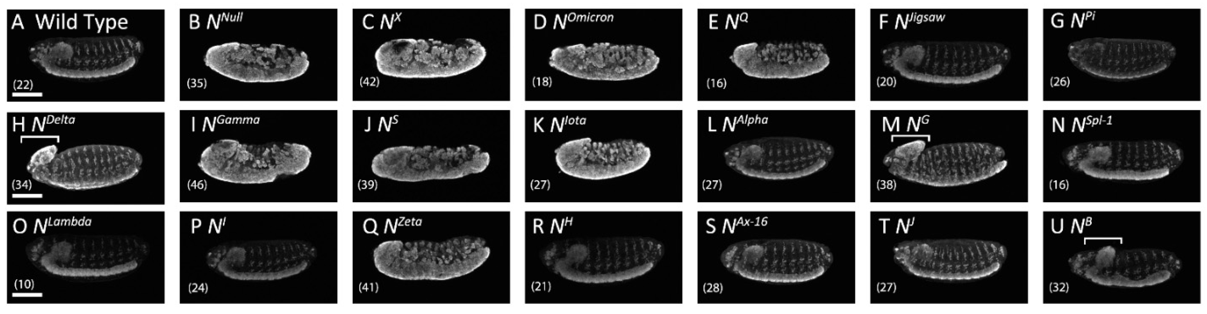
3.1. Notch Missense Mutations That Affected the Development of the Embryonic Nervous System

We investigated the roles of individual EGF-like repeats in Notch signaling using a previously established collection of 17 missense mutations of Notch [24,27,28]. These mutants carry a single missense mutation, identified by DNA sequencing, in the Notch locus corresponding to the EGF-like repeats (n = 16) or in the transmembrane domain (n = 1) [24,27,28]; Figure 1 and Table 1 show the amino acid substitution, position, and EGF-like repeat affected for each mutant. We also included two classic missense alleles of Notch, NSpl−1 and NAx−16, bringing the total number of Notch missense mutants examined in this study to 19.
In the embryonic central nervous system, depleted Notch signaling causes neural hyperplasia, designated as a neurogenic phenotype [47]. In this study, we observed neuronal cells by immunostaining with an antibody against the neuron-specific nuclear protein Elav (embryonic lethal abnormal vision) [48]. In wild-type embryos, anti-Elav stained the neuronal nuclei of the ladder-like nervous system (Figure 2A); in contrast, the classic Notch amorphic (null) allele Notch55e11 produced a severe neurogenic phenotype, with nearly the entire embryo stained by anti-Elav (Figure 2B) [47]. Of the 19 Notch alleles tested, each carrying a different missense mutation (Figure 2), 10 had a neurogenic or brain deformation phenotype (Figure 2C–E,H–K,M,Q,U; Table 1). Although the nature of brain deformation phenotype remained unclear, intensity of anti-Elav staining increased in these deformed brains, suggesting their neural hyperplasia that implies region-specific reduction of Notch signaling (Figure 2H,M,U; Table 1). The remaining nine mutants exhibited a wild-type nervous system, even though the same alleles produced a Notch signaling-related phenotype in other contexts (Figure 2F,G,L,N–T; Table 1) [24]. Considering that the role of Notch signaling is context-dependent in various tissues and organs [24,49], these missense mutations likely disrupt Notch signaling in a context-dependent manner.

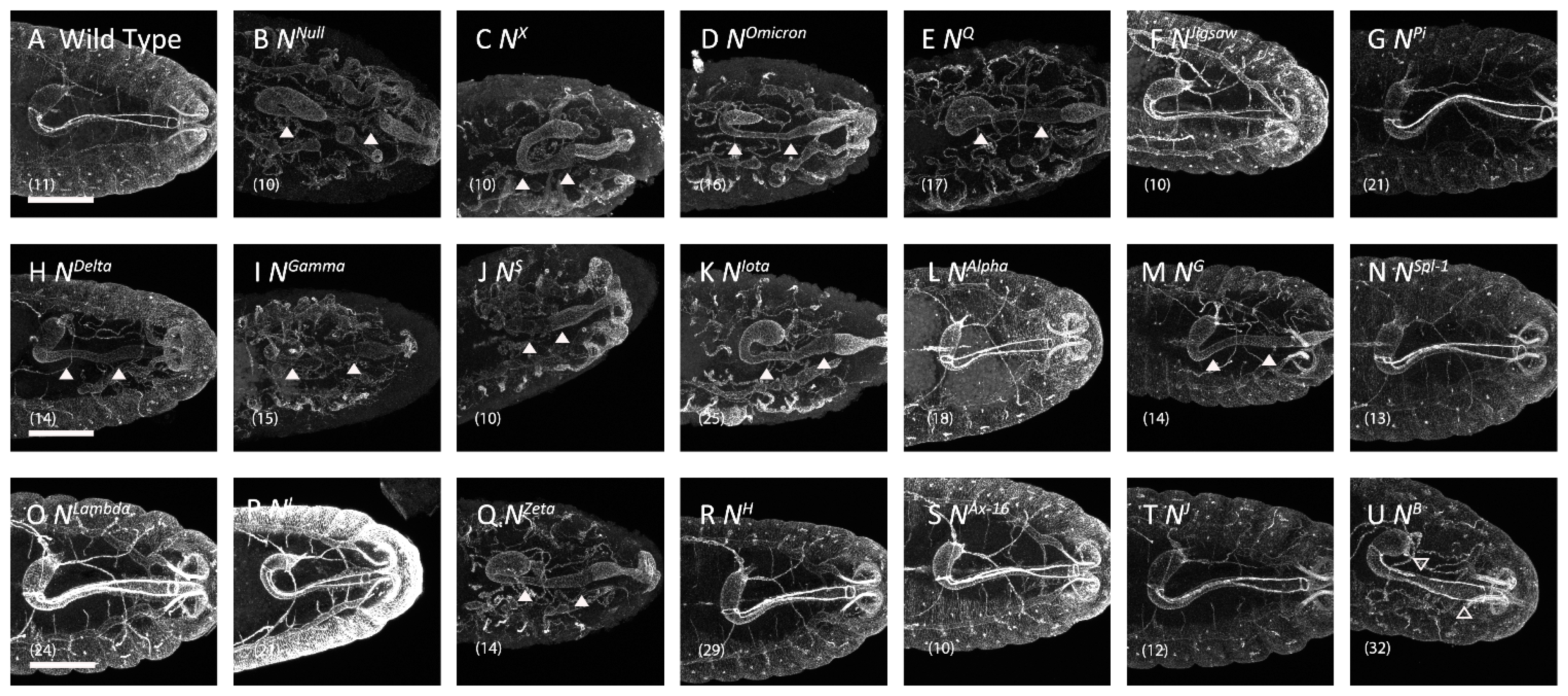
3.2. Missense Notch Mutations That Affected Boundary Cell Formation in the Hindgut

We next examined these Notch mutants for defects in boundary cells in the embryonic digestive system, since boundary cell formation is a typical example of inductive Notch signaling [30,50]. The expression of the ligand Delta is limited to the ventral compartment of the hindgut because engrailed, which suppresses Delta expression, is expressed in the dorsal compartment [30]. In the ventral cells where Delta is expressed, Notch signaling is suppressed in most cells by cis-inhibition of Notch via Delta [30]. However, since Delta presented from the ventral cells can signal Notch receptors expressed in the dorsal cells, where cis-inhibition does not take place, Notch signaling is activated in the single row of dorsal cells that subsequently differentiates into boundary cells [30].
Thus, we analyzed boundary cell formation to determine whether Notch signaling was disrupted in the Notch missense mutants during the development of the digestive system. The boundary cells highly express crumbs, which is required to establish apical-basal cell polarity and contributes to the organization of zonula adherens [51]. When stained with an anti-Crumbs antibody, boundary cells were observed as two narrow bands, each composed of a single row of boundary cells (Figure 3A) [51]. We confirmed that crumbs expression is lost in embryos hemizygous for Notch55e11 as previously described (Figure 3B), demonstrating that our assay has sufficient sensitivity for our purposes [30]. We assessed the presence or absence of boundary cells in embryos hemizygous for each Notch missense mutation and found that crumbs expression was depleted or showed abnormal gaps in 10 of the 19 Notch missense mutants (Figure 3C–E,H–K,M,Q,U; Table 1). However, the remaining nine missense mutations did not affect crumbs expression, indicating that inductive signaling was normal in this context (Figure 3F,G,L,N–P,R–T; Table 1) [24]. The 10 mutants with defective inductive signaling were the same 10 mutants with neurogenic or brain deformation phenotype (Table 1). Therefore, we speculate that these 10 missense mutations are relatively severe loss-of-function alleles of Notch, whereas the other alleles are hypomorphic or context-dependent. We noticed that seven of these 10 mutations affect cysteine residues that form disulfide bonds and most of them are clustered in EGF-like repeats 8–10 (Table 1). Thus, Notch may be particularly sensitive to disruption of the basic structure of EGF-like repeats 8–10.

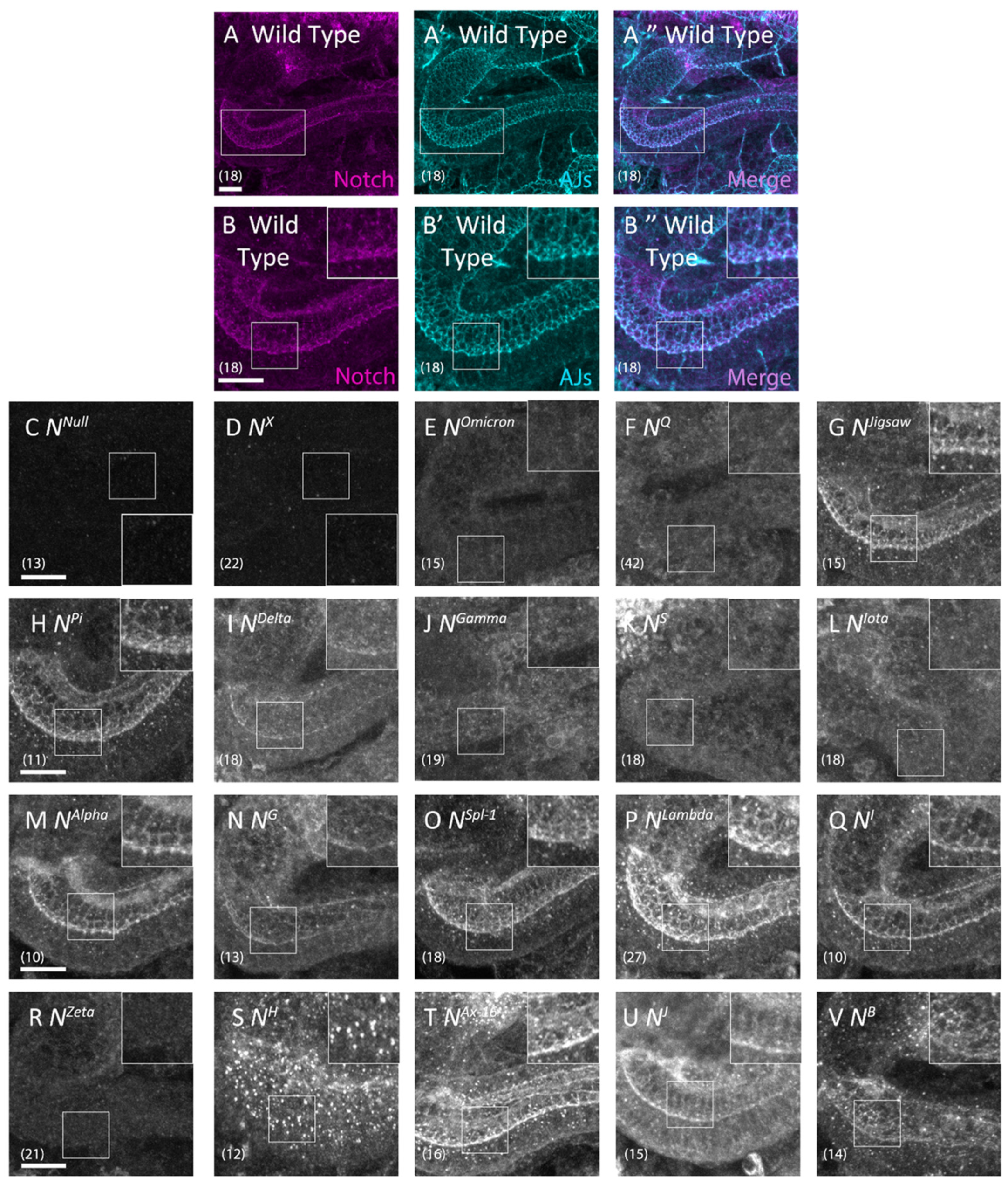
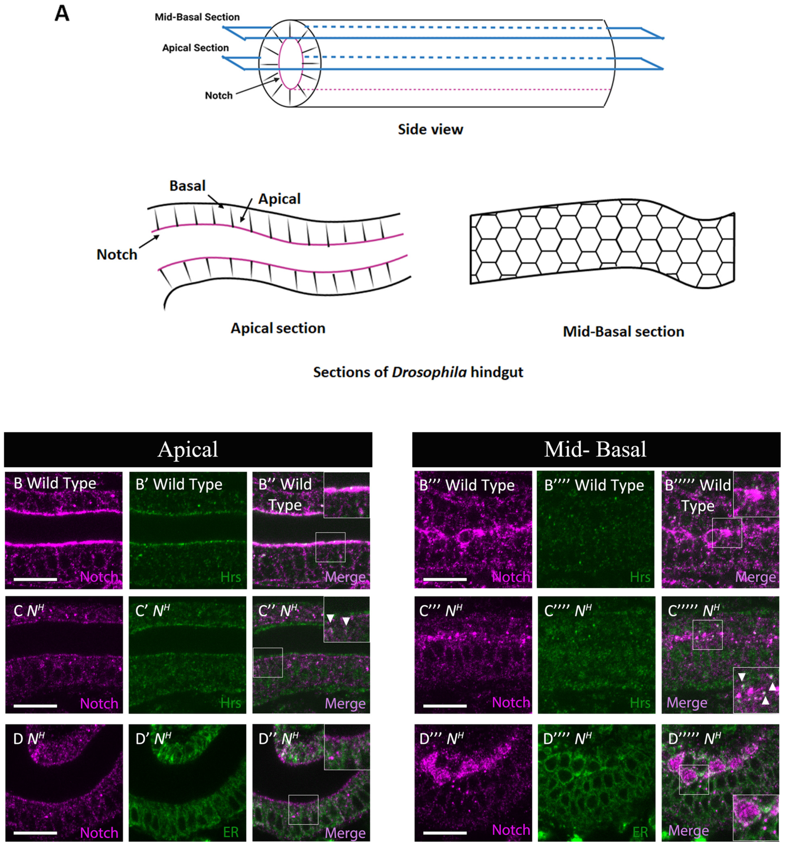
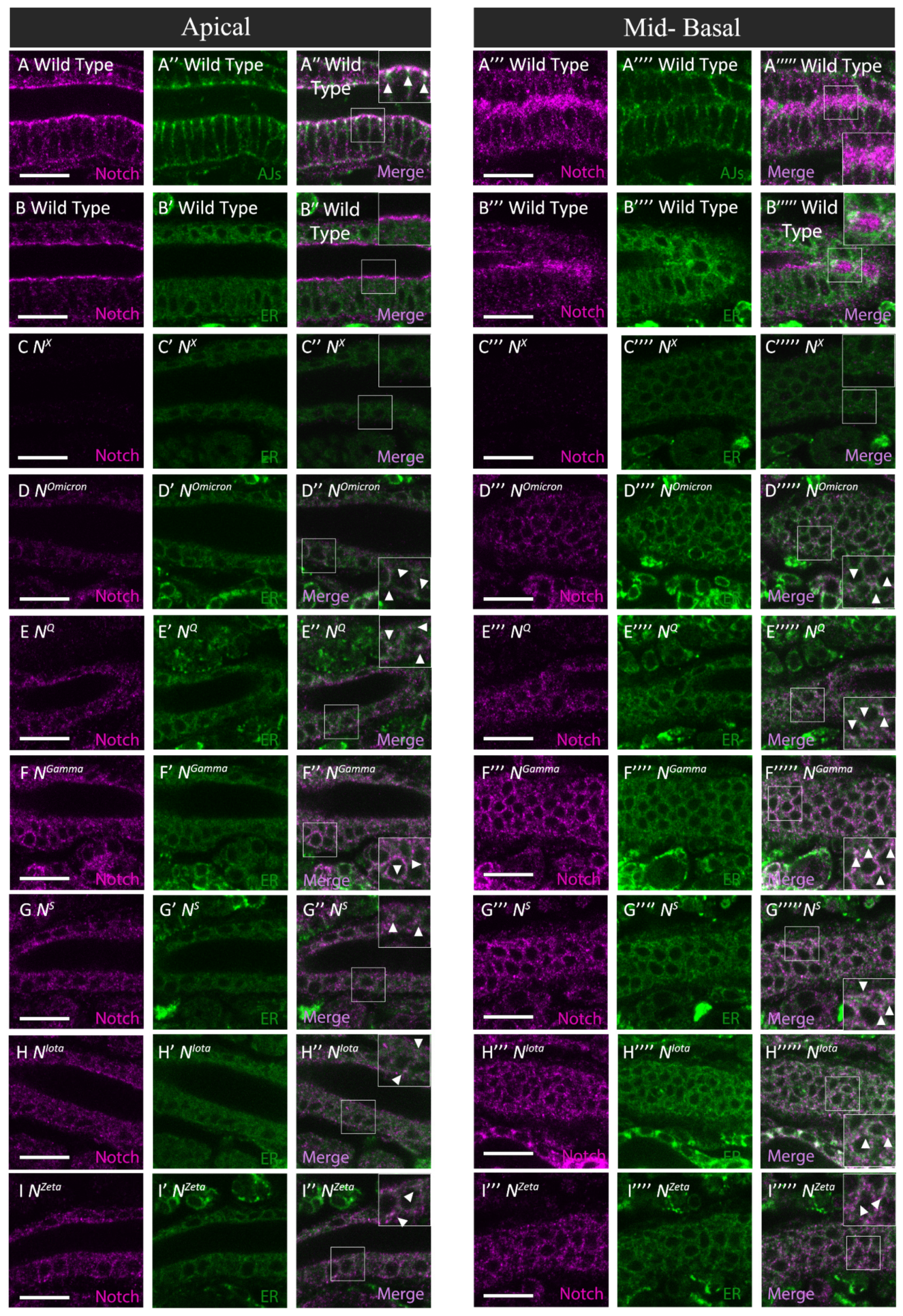
3.3. Notch Missense Mutations That Disrupted Intracellular Notch Trafficking

Notch mutations may disrupt Notch signaling by introducing defects in its folding or trafficking, with Notch consequently accumulating in the ER and/or endosomes [33]. An accumulation of Notch in the ER can, in turn, lead to the loss of Notch from AJs in epithelial cells. This loss is easily detected in the hindgut epithelium [31,33]. Thus, we analyzed the subcellular localization of Notch in the hindgut epithelium of embryo hemizygous for each of the 19 Notch missense mutations, and found abnormal intracellular distribution of Notch in 8 of the 19 mutants (Figure 4D–F,J–L,R,S; Table 1).
We compared signaling defects found in the central nervous system and boundary cells, assessed through Notch mutant phenotypes, with cellular defects related to Notch trafficking. We divided the Notch mutants accordingly into four classes based on the types of defects observed (Table 2), as follows: Class I comprised eight Notch mutants with normal Notch trafficking and normal Notch activity in the boundary cells and central nervous system. Class II comprised one Notch mutant that disrupted Notch trafficking but did not affect Notch activity in the boundary cells or nervous system. Class III comprised three Notch mutants that disrupted Notch activity in both the boundary cells and nervous system, but did not affect Notch trafficking. Class IV comprised seven Notch mutants that disrupted Notch trafficking and Notch activity in the boundary cells and nervous system. Based on these results, we conclude that a change in the amino acids in an EGF-like repeat can differ in its effect on Notch trafficking and activity, and that signaling defects and trafficking defects are not necessarily linked. Considering that amino acid substitutions in EGF-like repeats induced a range of defects in Notch trafficking and activities, the specific amino acid sequences within certain EGF-like repeats are likely crucial for normal Notch activity or trafficking [52].

3.4. Defects in Notch Trafficking and Loss of Notch Activity Were Not Always Coupled

The only Class II mutant in this study, NH, carries an amino acid substitution in the 29th EGF-like repeat with a cysteine (C) to serine (S) amino acid substitution at the 1155th amino acid residue (EGF-29, C1155S); this mutation affected the intracellular trafficking of Notch but not Notch function in lateral inhibition or inductive signaling in embryogenesis (Figure 2R, Figure 3R and Figure 4S). Notch was not detected in AJs in the hindgut of NH hemizygote embryos, where Notch is highly enriched in wild-type flies, but was instead found in punctate structures in the cytoplasm. To reveal the nature of such punctae, we analyzed the potential colocalization of Notch with markers of various intracellular compartments. We found that Notch colocalized with the early endosome marker Hrs (Hepatocyte growth factor regulated tyrosine kinase substrate) in the hindgut epithelium of NH hemizygote embryos (Figure 5C’’,C’’’’’) but not wild-type embryos (Figure 5B’’,B’’’’’; Table 1). On the other hand, Notch did not colocalize with markers for the ER (PDI-GFP) (Figure 5D’’,D’’’’’), cis-Golgi, recycling endosomes, or late endosomes under the same conditions (Figures S1–S5). Therefore, in NH hemizygotes, Notch is absent from AJs and accumulates in early endosomes in the hindgut epithelium, although such mislocalization of Notch does not appear to affect Notch signaling activity in this context. Under this condition, Notch presented at the plasma membrane appeared to be severely reduced, whereas the activity of Notch signaling was maintained normally. We speculated that this phenomenon can be explained by the nature of the NH mutation, which introduces an amino acid substitution in EGF-like repeat 29, included in the Abruptex domain [23]. Since mutations in the Abruptex domain often result in gain-of-function Notch alleles [23], it is possible that NH encodes a gain-of-function Notch while simultaneously reducing Notch presentation at the plasma membrane, which should reduce Notch signaling activity. Therefore, we speculate that a balance of these opposing effects on Notch activity belonging to the NH mutation may account for our observation that Notch signaling activity was normal in this mutant.
Conversely, our analyses revealed that the Class III alleles NDelta (EGF-9, D389N), NG (EGF-13, C535S), and NB (TMD, I1751K) showed attenuation in Notch activity in lateral inhibition, as predicted from brain deformation phenotype (Figure 2H,M,U) and in inductive signaling (Figure 3H,M,U) during embryogenesis, whereas Notch trafficking was normal in the hindgut epithelium (Figure 4I,N,V). These results suggest that the disruption of Notch activity is not always coupled with Notch trafficking defects. Considering the many factors that regulate Notch signaling at various layers within a cell, we speculate that these Notch missense mutations might disrupt some processes other than normal Notch trafficking. For example, NDelta and NG might disrupt ligand–receptor binding, since these mutations introduce amino acid substitutions into EGF-like repeats 9 and 13, respectively.

3.5. Notch Missense Mutations That Coupled Trafficking Defects with Loss of Notch Activity

In total, 7 of the 19 Notch mutant alleles tested were Class IV, which exhibit trafficking defects and loss of Notch activity in both neural development and the formation of hindgut border cells (Figure 2C–E,I–K,Q; Figure 3C–E,I–K,Q; Table 1). The Class IV mutants include NX (EGF-8, C343S), NOmicron (EGF-8, C343Y), NQ (EGF-8, D331N), NGamma (EGF-9, C398Y), NS (EGF-9, C407S), NIota (EGF-10, C413S), and NZeta (EGF-25, C993S). Notch was absent from AJs in all Class IV alleles (Figure 4D–F,J–L,R). Six of the seven Class IV mutants produced Notch proteins that accumulated in the ER, as shown by colocalization studies with Pdi-GFP (Figure 6D–I’’’’’), whereas hardly any Notch was detected in this organelle in wild-type embryos (Figure 6B’’,B’’’’’). On the other hand, Notch proteins derived from these six mutants did not colocalize with markers of other intracellular compartments, such as cis-Golgi, early endosomes, recycling endosomes, late endosomes, or of AJs (Figures S1–S5). Previous studies show that misfolded Notch protein was not transported to AJs because it was trapped in the ER [20,33]. These six mutants may also produce misfolded Notch that is not exported from the ER.
Importantly, we found that six of the seven Class IV mutants have amino acid substitutions in EGF-like repeats 8–10. Thus, the EGF-like repeats in this region may be especially sensitive to structural perturbations (Figure 7). We speculate that these three EGF-like repeats may be particularly important in folding the whole extracellular domain of Notch.
The Class IV mutant NZeta, which has an amino acid substitution in EGF-like repeat 25, accumulates Notch in the ER, suggesting that the mutation induces a severely misfolded product. EGF-like repeat 25 is a part of the Abruptex domain (EGF-like repeats 24–29) [21]. Amino acid substitutions within the Abruptex domain are known to induce gain-of-function mutations of Notch, suggesting that the Abruptex domain is involved in suppressing Notch activation [21]. It has also been suggested that the Abruptex domain contributes to forming Notch dimer proteins [53]. Given the apparent sensitivity of EGF-like repeat 25 to structural perturbation (Figure 7), the Abruptex domain may also be involved in the high-order organization of EGF-like repeats.

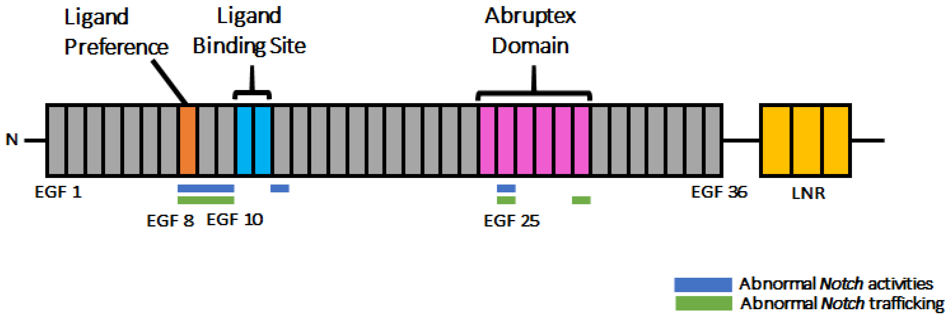
3.6. Disrupting Conserved Disulfide Bonds in Different EGF-like Repeats Induced Distinct Defects in Notch Activity and Trafficking

Although we observed different phenotypes associated with amino acid substitutions in individual EGF-like repeats, some differences may depend on the specific amino acids that replace the original residue rather than the position of the repeat. Four of the Notch missense mutants tested here—NX (EGF-8, C343S), NG (EGF-13, C535S), NZeta (EGF-25, C993S), and NH (EGF-29, C1155S)—have the same amino acid substitution at the conserved second cysteine though occurring in different EGF-like repeats, and these cysteines were replaced with serine residues. Considering the differences in the behavior of these variants in our assay system, our data argue that, at least among the mutants we tested, the matter of which EGF-like repeat contains the mutation has important biological consequences (Figure 7). These results also suggest that our analysis of the defects induced in the various mutants also indicate, at least to some degree, a specific function of the EGF-like repeats containing the amino acid substitutions. On the other hand, our analyses also revealed that the NG (EGF-13, C535S) and NJ (EGF-34, C1341Y) mutants, which have amino acid substitutions at conserved cysteines, did not accumulate Notch in the ER (Figure 4). This observation suggests that these EGF-like repeats are tolerant to structural perturbation with consequent misfolding. This also supports our idea that each EGF-like repeat plays specific roles in Notch folding.

4. Discussion

Notch has 36 EGF-like repeats in its extracellular domain [1]. Although these EGF-like repeats share a conserved structure, they play diverse roles as individual repeats and as clusters [3]. For example, EGF-like repeats 11–12 form the core ligand-binding site [54]. EGF-like repeats 10–12, 11–12, and 8 specifically contribute to cis-inhibition [55], trans-activation [9], and ligand selection [24], respectively. Genetic evidence suggests that EGF-like repeats 24–29, designated as the Abruptex domain, negatively regulate the Notch receptor [21,22,23]. However, relatively little is known about the specific roles of individual EGF-like repeats, and a complete high-order structure of Notch and its 36 EGF-like repeats in action has not been solved through structural analysis. In this study, we attempted to reveal the specific contributions of each EGF-like repeat to the activity, folding, and intracellular trafficking of Notch by studying the effect of missense mutations.
We analyzed 19 Notch mutants carrying missense mutations that were identified through a recent forward genetic screen [24] or as classic alleles. These mutations introduce unique amino acid substitutions into EGF-like repeats in 18 cases, and into the transmembrane domain in one case [24]. The mutants collected through genetic screening were isolated by clinical observation of Notch-related phenotypes in the wing or mechanosensory bristles [24]. To further characterize these mutants, we examined two other well-studied Notch-related phenotypes in embryonic tissues: lateral inhibition during central nervous system development and inductive signaling during boundary cell formation in the hindgut (Table 1). Our comparative analyses revealed that 10 out of 19 alleles exhibited either a neurogenic or brain deformation phenotype and boundary cells abnormalities (Table 1). In all cases, these two defects were observed coincidently. Therefore, the behavior of each of these 10 missense mutations was the same for lateral inhibition and for inductive signaling during embryogenesis. Although context dependency in Notch signaling has been studied extensively, it is still difficult to explain how it operates differently in various tissues [56]. Clear differences and similarities in the behaviors of the Notch missense mutants observed in this study provide an excellent opportunity to understand the molecular mechanisms of context-dependent Notch signaling.
As summarized in Figure 7, our analysis revealed that the EGF-like repeats sensitive to the amino acid substitutions with respect to the depletion of Notch activity are found in two regions within the 36 EGF-like repeats. One of these regions is EGF-like repeats 8–10, as revealed in the Notch missense mutants NX, NOmicron, NQ, NGamma, NS, and NIota. Intriguingly, the importance of EGF-like repeats 8–10 agrees with previous findings. For example, O-fucose modifications on EGF-like repeats 8 and 12 in Notch1 engage the EGF-like repeat 3 and the C2 domain, respectively, of the Jagged1 ligand [16]. Moreover, EGF-like repeat 8 modulates ligand binding selectivity in Drosophila [24]. EGF-like repeats 8–10 of Notch1 are required for DLL1- and DLL4-induced Notch signaling [57]. The importance of EGF-like repeats 8–10 has also been shown by analyzing O-fucose glycan modifications. O-fucose glycan modifications in EGF-like repeats 8, 9, and 12 of Drosophila Notch and in EGF-like repeats 8 and 12 of Notch1 specifically play important roles in modulating Notch–ligand binding [18,19]. Collectively, these results highlight the importance of EGF-like repeats 8–10 in Notch functions.
Another sensitive region was found in the EGF-like repeat 25, although this region was identified based on only one Notch mutant, Nzeta. This region overlaps with the Abruptex domain (EGF-like repeats 24–29), which is known to negatively regulate Notch activity [21]. Genetic interaction analysis suggests that the Abruptex domain can be divided into two different clusters—EGF-like repeats 24–25, known as “suppressor of Notch”, and EGF-like repeats 27–29, known as “enhancer of Notch” [23]. The precise molecular function of Abruptex domain is unknown, and it is not clear why the Nzeta mutation found in this region leads to a loss-of-function rather than a gain-of-function Notch phenotype. A more detailed study of this mutation along with other Abruptex alleles of Notch will likely provide insights into this mysterious domain. In summary, these two missense-sensitive clusters of EGF-like repeats correspond well to the EGF-like repeats that have been shown to play specific roles in Notch functions.
Our results also revealed that of seven Notch mutants with an amino acid substitution in one of the sensitive clusters, six accumulated Notch abnormally in the ER of the hindgut epithelium. We found seven Class IV Notch mutations in this study—NX, NOmicron, NQ, NGamma, NS, NIota, and NZeta, which disrupted Notch trafficking and Notch activity (Table 2). Notch misfolding is known to cause Notch to accumulate in the ER [33]. Therefore, we speculated that amino acid substitutions in the EGF-like repeats of the sensitive clusters may induce global misfolding of Notch, which prevents the export of Notch from the ER by quality control mechanisms [20,33]. On the other hand, in Class III mutants, including NDelta, NG, and NB, Notch trafficking was normal, although Notch activity was reduced. However, in Class III mutants, defects in neural development were observed only in the brain, but not in the other part of the central nervous system. Considering that all Class IV mutants showed neurogenic phenotype in the entire central nervous system, underlying defects in Notch signaling may be different between Class III and Class IV, although all of them showed defects in inductive Notch signaling, as judged by the disruption of boundary cell formation. It is known that the activation of Notch signaling requires several steps in addition to proper Notch folding, such as ligand binding and Notch processing. Therefore, we speculate that some of these other steps might be disrupted in the Class III mutants, which may also explain the difference of neuronal phenotypes between Class III and Class IV.
As a potential limitation of this study, one could argue that the type of amino acid substitution found in the Notch mutants might be more important than which EGF-like repeat is affected. However, our analysis of the missense mutations in the Notch mutants NX, NG, NZeta, and NH, which introduce the same amino acid substitution in the conserved second cysteine to serine, but in different EGF-like repeats, argues that identical amino acid changes introduced into different EGF-like repeats can differ in effect. Therefore, despite the limitation in the number of Notch alleles used here, our analyses successfully demonstrate, at least to some extent, the specificity of individual EGF-like repeats in Notch folding and activity.
Based on the results of our study, we propose that the EGF-like repeats 8–10 and 25 are particularly susceptible to structural perturbation with consequent misfolding and inactivation of Notch. We speculate that the ER may monitor the folding of these particular EGF-like repeats more strictly than other repeats because of their critical roles in Notch receptor functions. This idea should provide insights for further studies of correlations between Notch structure and function, and may provide molecular handles to assist in the functional interpretation of the missense variants that are found in human Notch receptors and are linked to diverse genetic disorders or cancers.

Supplementary Materials

The following supporting information can be downloaded at: https://www.mdpi.com/article/10.3390/biom12121752/s1, Figure S1. Notch localized to AJs in Class II and IV Notch mutants. Figure S2. Class II and IV Notch mutants did not accumulate Notch in cis-Golgi. Figure S3. Class IV Notch mutants did not accumulate Notch in early endosomes. Figure S4. Class II and IV Notch mutants did not accumulate Notch in late endosomes. Figure S5. Class II and Class IV Notch mutants did not accumulate Notch in recycling endosomes.

Author Contributions

H.N. and K.M. designed the experiments. H.N., E.Y.M., M.H. and S.Y. conducted the experiments. T.S., M.I., S.Y., T.Y. and K.M., provided instructions. H.N. and K.M., analyzed the data. H.N, S.Y. and K.M., wrote the paper. All authors have read and agreed to the published version of the manuscript.

Funding

This research was supported by JSPS KAKENHI Grant Number JP18K14697 and 2020 Toyota Riken Scholar Program.

Institutional Review Board Statement

Not applicable.

Informed Consent Statement

Not applicable.

Data Availability Statement

All data reported are included and represented in the manuscript.

Acknowledgments

We thank Martin Baron for fruitful discussions. We are grateful to Hugo Bellen for his generous gift of anti-HRS antibody. We thank Kyoto Stock Center (DGRC) and Bloomington Drosophila Stock Center (BDSC) for Drosophila stock, and the Developmental Studies of Hybridoma Bank (DSHB) for antibodies.

Conflicts of Interest

The authors declare no conflict of interest.

References

  1. Artavanis-Tsakonas, S.; Rand, M.D.; Lake, R.J. Notch signaling: Cell fate control and signal integration in development. Science 1999, 284, 770–776. [Google Scholar] [CrossRef] [PubMed] [Green Version]
  2. Gazave, E.; Lapébie, P.; Richards, G.S.; Brunet, F.; Ereskovsky, A.; Degnan, B.M.; Borchiellini, C.; Vervoort, M.; Renard, E. Origin and evolution of the Notch signalling pathway: An overview from eukaryotic genomes. BMC Evol. Biol. 2009, 9, 249. [Google Scholar] [CrossRef] [Green Version]
  3. Bray, S.J. Notch signalling in context. Nat. Rev. Mol. Cell Biol. 2016, 17, 722–735. [Google Scholar] [CrossRef]
  4. Guruharsha, K.G.; Kankel, M.W.; Artavanis-Tsakonas, S. The Notch signalling system: Recent insights into the complexity of a conserved pathway. Nat. Rev. Genet. 2012, 13, 654–666. [Google Scholar] [CrossRef] [PubMed] [Green Version]
  5. Baron, M. An overview of the Notch signalling pathway. Semin. Cell Dev. Biol. 2003, 14, 113–119. [Google Scholar] [CrossRef] [PubMed]
  6. Bray, S.J. Notch signalling: A simple pathway becomes complex. Nat. Rev. Mol. Cell Biol. 2006, 7, 678–689. [Google Scholar] [CrossRef] [PubMed]
  7. Schweisguth, F. Regulation of Notch signaling activity. Curr. Biol. 2004, 14, R129–R138. [Google Scholar] [CrossRef]
  8. Hori, K.; Sen, A.; Artavanis-Tsakonas, S. Notch signaling at a glance. J. Cell Sci. 2015, 52, 797–803. [Google Scholar] [CrossRef] [PubMed] [Green Version]
  9. Chillakuri, C.R.; Sheppard, D.; Lea, S.M.; Handford, P.A. Notch receptor–ligand binding and activation: Insights from molecular studies. Semin. Cell Dev. Biol. 2012, 23, 421–428. [Google Scholar] [CrossRef] [Green Version]
  10. Kelley, M.R.; Kidd, S.; Deutsch, W.A.; Young, M.W. Mutations altering the structure of epidermal growth factor-like coding sequences at the Drosophila Notch locus. Cell 1987, 51, 539–548. [Google Scholar] [CrossRef] [PubMed]
  11. Haltom, A.R.; Jafar-Nejad, H. The multiple roles of epidermal growth factor repeat O-glycans in animal development. Glycobiology 2015, 25, 1027–1042. [Google Scholar] [CrossRef] [PubMed] [Green Version]
  12. Wouters, M.A.; Rigoutsos, I.; Chu, C.K.; Feng, L.L.; Sparrow, D.B.; Dunwoodie, S.L. Evolution of distinct EGF domains with specific functions. Protein Sci. 2005, 14, 1091–1103. [Google Scholar] [CrossRef] [Green Version]
  13. Rand, M.D.; Lindblom, A.; Carlson, J.; Villoutreix, B.O.; Stenflo, J. Calcium binding to tandem repeats of EGF-like modules. Expression and characterization of the EGF-like modules of human Notch-1 implicated in receptor-ligand interactions. Protein Sci. 2008, 6, 2059–2071. [Google Scholar] [CrossRef] [PubMed] [Green Version]
  14. Tombling, B.J.; Wang, C.K.; Craik, D.J. EGF-like and other disulfide-rich microdomains as therapeutic scaffolds. Angew. Chem. Int. Ed. 2020, 59, 11218–11232. [Google Scholar] [CrossRef]
  15. Luca, V.C.; Jude, K.M.; Pierce, N.W.; Nachury M v Fischer, S.; Garcia, K.C. Structural basis for Notch1 engagement of Delta-like 4. Science 2015, 347, 847–853. [Google Scholar] [CrossRef] [PubMed] [Green Version]
  16. Luca, V.C.; Kim, B.C.; Ge, C.; Kakuda, S.; Wu, D.; Roein-Peikar, M.; Haltiwanger, R.S.; Zhu, C.; Ha, T.; Garcia, K.C. Notch-Jagged complex structure implicates a catch bond in tuning ligand sensitivity. Science 2017, 355, 1320–1324. [Google Scholar] [CrossRef] [Green Version]
  17. Stanley, P.; Okajima, T. Roles of Glycosylation in Notch Signaling. In Current Topics in Developmental Biology: Notch Signaling; Kopan, R., Ed.; Elsevier: Berkeley, CA, USA, 2010; pp. 131–164. [Google Scholar] [CrossRef]
  18. Pandey, A.; Harvey, B.M.; Lopez, M.F.; Ito, A.; Haltiwanger, R.S.; Jafar-Nejad, H. Glycosylation of specific Notch EGF repeats by O-Fut1 and fringe regulates notch signaling in drosophila. Cell Rep. 2019, 29, 2054–2066.e6. [Google Scholar] [CrossRef] [Green Version]
  19. Kakuda, S.; Haltiwanger, R.S. Deciphering the fringe-mediated Notch code: Identification of activating and inhibiting sites allowing discrimination between ligands. Dev. Cell 2017, 40, 193–201. [Google Scholar] [CrossRef] [PubMed] [Green Version]
  20. Matsumoto, K.; Ayukawa, T.; Ishio, A.; Sasamura, T.; Yamakawa, T.; Matsuno, K. Dual roles of O-Glucose glycans redundant with monosaccharide O-Fucose on Notch in Notch trafficking. J. Biol. Chem. 2016, 291, 13743–13752. [Google Scholar] [CrossRef] [PubMed]
  21. De Celis, J.F.; Bray, S.J. The Abruptex domain of Notch regulates negative interactions between Notch, its ligands and Fringe. Development 2000, 127, 1291–1302. [Google Scholar] [CrossRef] [PubMed]
  22. Baron, M. Combining genetic and biophysical approaches to probe the structure and function relationships of the Notch receptor. Mol. Membr. Biol. 2017, 34, 33–49. [Google Scholar] [CrossRef] [PubMed] [Green Version]
  23. Yamamoto, S. Making sense out of missense mutations: Mechanistic dissection of Notch receptors through structure-function studies in Drosophila. Dev. Growth Differ. 2000, 62, 15–34. [Google Scholar] [CrossRef] [PubMed] [Green Version]
  24. Yamamoto, S.; Charng, W.-L.; Rana, N.A.; Kakuda, S.; Jaiswal, M.; Bayat, V.; Xiong, B.; Zhang, K.; Sandoval, H.; David, G.; et al. A mutation in EGF Repeat-8 of Notch discriminates between serrate/jagged and delta family ligands. Science 2012, 338, 1229–1232. [Google Scholar] [CrossRef] [Green Version]
  25. Johannes, B.; Preiss, A. Wing vein formation in Drosophila melanogaster: Hairless is involved in the cross-talk between Notch and EGF signaling pathways. Mech. Dev. 2002, 115, 3–14. [Google Scholar] [CrossRef] [PubMed]
  26. De Celis, J.F.; Garcia-Bellido, A.; Bray, S.J. Activation and function of Notch at the dorsal-ventral boundary of the wing imaginal disc. Development 1996, 122, 359–369. [Google Scholar] [CrossRef]
  27. Yamamoto, S.; Jaiswal, M.; Charng, W.-L.; Gambin, T.; Karaca, E.; Mirzaa, G.; Wiszniewski, W.; Sandoval, H.; Haelterman, N.A.; Xiong, B.; et al. A drosophila genetic resource of mutants to study mechanisms underlying human genetic diseases. Cell 2014, 159, 200–214. [Google Scholar] [CrossRef] [Green Version]
  28. Haelterman, N.A.; Jiang, L.; Li, Y.; Bayat, V.; Sandoval, H.; Ugur, B.; Tan, K.L.; Zhang, K.; Bei, D.; Xiong, B.; et al. Large-scale identification of chemically induced mutations in Drosophila melanogaster. Genome Res. 2014, 24, 1707–1718. [Google Scholar] [CrossRef] [Green Version]
  29. Sjöqvist, M.; Andersson, E.R. Do as I say, Not(ch) as I do: Lateral control of cell fate. Dev. Biol. 2019, 447, 58–70. [Google Scholar] [CrossRef]
  30. Takashima, S.; Yoshimori, H.; Yamasaki, N.; Matsuno, K.; Murakami, R. Cell-fate choice and boundary formation by combined action of Notch and engrailed in the Drosophila hindgut. Dev. Genes Evol. 2002, 212, 534–541. [Google Scholar] [CrossRef]
  31. Fuß, B.; Hoch, M. Notch signaling controls cell fate specification along the dorsoventral axis of the drosophila gut. Curr. Biol. 2002, 12, 171–179. [Google Scholar] [CrossRef] [Green Version]
  32. Sasaki, N.; Sasamura, T.; Ishikawa, H.O.; Kanai, M.; Ueda, R.; Saigo, K.; Matsuno, K. Polarized exocytosis and transcytosis of Notch during its apical localization in Drosophila epithelial cells. Genes Cells 2007, 12, 89–103. [Google Scholar] [CrossRef] [PubMed]
  33. Okajima, T.; Xu, A.; Lei, L.; Irvine, K.D. Chaperone activity of protein O-Fucosyltransferase 1 promotes Notch receptor folding. Science 2005, 307, 1599–1603. [Google Scholar] [CrossRef] [PubMed] [Green Version]
  34. Yamamoto, S.; Charng, W.-L.; Bellen, H.J. Endocytosis and Intracellular Trafficking of Notch and Its Ligands. In Current Topics in Developmental Biology: Notch Signaling; Kopan, R., Ed.; Elsevier: Berkeley, CA, USA, 2010; pp. 165–200. [Google Scholar] [CrossRef]
  35. Hounjet, J.; Vooijs, M. The role of intracellular trafficking of notch receptors in ligand-independent notch activation. Biomolecules 2021, 11, 1369. [Google Scholar] [CrossRef] [PubMed]
  36. Roth, R.A.; Pierce, S.B. In vivo cross-linking of protein disulfide isomerase to immunoglobulins. Biochemistry 1987, 26, 4179–4182. [Google Scholar] [CrossRef] [PubMed]
  37. Rhyu, M.S.; Jan, L.Y.; Jan, Y.N. Asymmetric distribution of numb protein during division of the sensory organ precursor cell confers distinct fates to daughter cells. Cell 1994, 76, 477–491. [Google Scholar] [CrossRef]
  38. Schindelin, J.; Arganda-Carreras, I.; Frise, E.; Kaynig, V.; Longair, M.; Pietzsch, T.; Preibisch, S.; Rueden, C.; Saalfeld, S.; Schmid, B.; et al. Fiji: An open-source platform for biological-image analysis. Nat. Methods 2012, 9, 676–682. [Google Scholar] [CrossRef] [Green Version]
  39. O’Neill, E.M.; Rebay, I.; Tjian, R.; Rubin, G.M. The activities of two Ets-related transcription factors required for drosophila eye development are modulated by the Ras/MAPK pathway. Cell 1994, 78, 137–147. [Google Scholar] [CrossRef]
  40. Fehon, R.G.; Kooh, P.J.; Rebay, I.; Regan, C.L.; Xu, T.; Muskavitch MA, T.; Artavanis-Tsakonas, S. Molecular interactions between the protein products of the neurogenic loci Notch and Delta, two EGF-homologous genes in Drosophila. Cell 1990, 61, 523–534. [Google Scholar] [CrossRef]
  41. Tepass, U.; Knust, E. crumbs and stardust act in a genetic pathway that controls the organization of epithelia in drosophila melanogaster. Dev. Biol. 1993, 159, 311–326. [Google Scholar] [CrossRef]
  42. Oda, H.; Uemura, T.; Harada, Y.; Iwai, Y.; Takeichi, M. A drosophila homolog of cadherin associated with armadillo and essential for embryonic cell-cell adhesion. Dev. Biol. 1994, 165, 716–726. [Google Scholar] [CrossRef]
  43. Lloyd, T.E.; Atkinson, R.; Wu, M.N.; Zhou, Y.; Pennetta, G.; Bellen, H.J. Hrs regulates endosome membrane invagination and tyrosine kinase receptor signaling in drosophila. Cell 2002, 108, 261–269. [Google Scholar] [CrossRef] [PubMed] [Green Version]
  44. Tanaka, T.; Nakamura, A. The endocytic pathway acts downstream of Oskar in Drosophila germ plasm assembly. Development 2008, 135, 1107–1117. [Google Scholar] [CrossRef] [PubMed] [Green Version]
  45. O’Sullivan, N.C.; Jahn, T.R.; Reid, E.; O’Kane, C.J. Reticulon-like-1, the Drosophila orthologue of the Hereditary Spastic Paraplegia gene reticulon 2, is required for organization of endoplasmic reticulum and of distal motor axons. Hum. Mol. Genet. 2012, 21, 3356–3365. [Google Scholar] [CrossRef] [PubMed] [Green Version]
  46. Suzuki, M.; Hara, Y.; Takagi, C.; Yamamoto, T.S.; Ueno, N. MID1 and MID2 are required for Xenopus neural tube closure through the regulation of microtubule organization. Development 2010, 137, 2329–2339. [Google Scholar] [CrossRef] [Green Version]
  47. Lehmann, R.; Jimenez, F.; Dietrich, U.; Campos-Ortega, J.A. On the phenotype and development of mutants of early neurogenesis in Drosophila melanogaster. Wilhelm Roux’s Arch. Dev. Biol. 1983, 192, 62–74. [Google Scholar] [CrossRef]
  48. Berger, C.; Renner, S.; Lüer, K.; Technau, G.M. The commonly used marker ELAV is transiently expressed in neuroblasts and glial cells in the Drosophila embryonic CNS. Dev. Dyn. 2007, 236, 3562–3568. [Google Scholar] [CrossRef]
  49. Schweisguth, F. Asymmetric cell division in the Drosophila bristle lineage: From the polarization of sensory organ precursor cells to Notch-mediated binary fate decision. Wiley Interdiscip. Rev. Dev. Biol. 2015, 4, 299–309. [Google Scholar] [CrossRef]
  50. Iwaki, D.D.; Lengyel, J.A. A DeltaNotch signaling border regulated by Engrailed/Invected repression specifies boundary cells in the Drosophila hindgut. Mech. Dev. 2002, 114, 71–84. [Google Scholar] [CrossRef]
  51. Kumichel, A.; Knust, E. Apical localisation of crumbs in the boundary cells of the drosophila hindgut is independent of its canonical interaction partner stardust. PLoS ONE 2014, 9, e94038. [Google Scholar] [CrossRef] [Green Version]
  52. Brennan, K.; Tateson, R.; Lewis, K.; Arias, A.M. A functional analysis of Notch mutations in Drosophila. Genetics 1997, 147, 177–188. [Google Scholar] [CrossRef]
  53. Pei, Z.; Baker, N.E. Competition between Delta and the Abruptex domain of Notch. BMC Dev. Biol. 2008, 8, 4. [Google Scholar] [CrossRef] [Green Version]
  54. Rebay, I.; Fleming, R.J.; Fehon, R.G.; Cherbas, L.; Cherbas, P.; Artavanis-Tsakonas, S. Specific EGF repeats of Notch mediate interactions with Delta and serrate: Implications for Notch as a multifunctional receptor. Cell 1991, 67, 687–699. [Google Scholar] [CrossRef] [PubMed]
  55. Becam, I.; Fiuza, U.-M.; Arias, A.M.; Milán, M. A role of receptor Notch in ligand cis-inhibition in drosophila. Curr. Biol. 2010, 20, 554–560. [Google Scholar] [CrossRef] [PubMed] [Green Version]
  56. Siebel, C.; Lendahl, U. Notch signaling in development, tissue homeostasis, and disease. Physiol. Rev. 2017, 97, 1235–1294. [Google Scholar] [CrossRef] [PubMed] [Green Version]
  57. Andrawes, M.B.; Xu, X.; Liu, H.; Ficarro, S.B.; Marto, J.A.; Aster, J.C.; Blacklow, S.C. Intrinsic selectivity of Notch 1 for delta-like 4 over delta-like 1. J. Biol. Chem. 2013, 288, 25477–25489. [Google Scholar] [CrossRef] [PubMed]
Figure 1. A schematic diagram of the Notch extracellular domain and the positions of EGF-like repeats with amino acid substitutions in Notch mutations analyzed in this study. The extracellular domain of Drosophila Notch has 36 epidermal growth factor (EGF)-like repeats (gray), three lin12/Notch repeats (LNR) (orange), and a transmembrane domain (blue lines). We investigated 19 missense mutations of Notch with amino acid substitutions in EGF-like repeats and the transmembrane domain. The mutant alleles are listed across the top, with arrows pointing to the position of the EGF-like repeat and the transmembrane domain with the substitution. EGF-like repeats are numbered in order starting with the repeat closest to the N-terminal (EGF-1).
Biomolecules 12 01752 g001
Figure 2. Notch alleles that disrupted lateral inhibition in the embryonic central nervous system. Lateral views show the embryonic nervous system, stained with an anti-Elav antibody (white), in (A) wild-type Drosophila and hemizygotes for (B) N55e11, an amorphic allele of Notch; (C) NX, (D) NOmicron, (E) NQ, (F) NJigsaw, (G) Npi, (H) NDelta, (I) NGamma, (J) NS, (K) NIota, (L) NAlpha, (M) NG, (N) NSpl−1, (O) NLambda, (P) NI, (Q) NZeta, (R) NH, (S) NAx−16, (T) NJ, and (U) NB. White brackets show the regions with a brain deformation phenotype. The number of embryos analyzed is shown in parentheses. Scale bars: 100 μm.
Biomolecules 12 01752 g002
Figure 3. Notch alleles that induced defects in boundary cell formation. Dorsal views of the embryonic hindgut epithelium, stained with an anti-Crumbs antibody to detect boundary cells (white), in (A) wild-type embryos and the following hemizygotes: (B) N55e11, an amorphic allele of Notch; (C) NX, (D) NOmicron, (E) NQ, (F) NJigsaw, (G) Npi, (H) NDelta, (I) NGamma, (J) NS, (K) NIota, (L) NAlpha, (M) NG, (N) NSpl−1, (O) NLambda, (P) NI, (Q) NZeta, (R) NH, (S) NAx−16, (T) NJ, and (U) NB. Filled white arrows indicate regions where anti-Crumbs antibody staining is depleted. Arrows outlined in white indicate regions where anti-Crumbs antibody staining showed abnormal gaps. The number of embryos analyzed is shown in parentheses. Scale bars: 50 μm.
Biomolecules 12 01752 g003
Figure 4. Notch alleles that disrupted intracellular Notch trafficking. (A,A’’,B,B’’) Notch and E-cadherin, a marker of adherens junctions (AJs), were detected in wild-type hindgut epithelium by anti-Notch (magenta in (A,B)) and anti-E-cadherin (turquoise in (A’,B’)) antibody staining. (B,B’,B’’) show high-magnification views of the regions outlined in (A,A’,A’’), respectively. Panels (A’’,B’’) are merged images of panels (A,A’,B,B’), respectively. (CV) Notch was detected by anti-Notch antibody staining (white) in the hindgut epithelium of (C) N55e11, an amorphic allele of Notch; (D) NX, (E) NOmicron, (F) NQ, (G) NJigsaw, (H) NPi, (I) NDelta, (J) NGamma, (K) NS, (L) NIota, (M) NAlpha, (N) NG, (O) NSpl−1, (P) NLambda, (Q) NI, (R) NZeta, (S) NH, (T) NAx−16, (U) NJ, and (V) NB hemizygotes. Insets are highly magnified images of regions outlined by white rectangles. The number of hindgut samples analyzed is shown in parentheses. Scale bars: 10 μm.
Biomolecules 12 01752 g004
Figure 5. Notch accumulated abnormally in early endosomes of the hindgut epithelium in a Class II Notch mutant. (A) A diagram showing optical sections of the apical and mid-basal regions of the hindgut epithelium (Figures were made with the application from Biorender.com). The upper diagram shows the side view of the hindgut tube with apical and mid-basal sections, and the lower diagrams show the dorsal views of apical (left) and mid-basal (right) sections of the hindgut epithelium. The apical and mid-basal sections correspond to the microscopic images in (BD’’) and (B’’’D’’’’’), respectively, as indicated in the top of (BD’’’’’). (BB’’’’’) In wild-type hindgut epithelium, Notch (magenta) and Hrs (green), a marker of early endosomes, were stained with an anti-Notch (B,B’’,B’’’,B’’’’’) and anti-Hrs antibodies (B’,B’’,B’’’’,B’’’’’), respectively. (CD’’’’’) Hindgut epithelium in the NH hemizygote, a Class II Notch mutant, stained for Notch (magenta in C,C’’,C’’’,C’’’’’,D,D’’,D’’’,D’’’’’), Hrs (green in C’,C’’,C’’’’,C’’’’’), and Pdi-GFP, an ER marker (green in D’,D’’,D’’’’,D’’’’’) were observed by anti-Notch, anti-Hrs, and anti-GFP antibody staining, respectively. Insets in (B’’,B’’’’’,C’’,C’’’’’,D’’,D’’’’’) are highly magnified images of regions outlined by white rectangles. White arrowheads point colocalized expression. All results were confirmed by staining biological triplicates. Scale bars: 10 μm.
Biomolecules 12 01752 g005
Figure 6. Notch accumulated abnormally in the ER of the hindgut epithelium in Class IV Notch mutants. (AI’’’’’) Apical and mid-basal images corresponding to the diagrams of apical and mid-basal planes in Figure 5. (AB’’’’’) Wild-type hindgut epithelium stained for Notch (magenta in A,A’’,A’’’,A’’’’’,B,B’’,B’’’,B’’’’’), E-Cadherin (green in A’,A’’,A’’’’,A’’’’’), and the ER marker Pdi-GFP (green in B’,B’’,B’’’’,B’’’’’) using anti-Notch, anti-E-Cadherin, and anti-GFP antibodies. (CI’’’’’) Notch (magenta, left panels) and Pdi-GFP (green, middle panels) were observed by anti-Notch and anti-GFP antibody staining, respectively, in the hindgut epithelium of (CC’’’’’) NX, (DD’’’’’) NOmicron, (EE’’’’’) NQ, (FF’’’’’) NGamma, (GG’’’’’) NS, (HH’’’’’) Niota, and (II’’’’’) NZeta hemizygotes. Right-side panels in apical and mid-basal images, indicated by ’’ and ’’’’’, respectively, are merged from the left and middle images. Insets in the right panels indicated by ’’ and ’’’’’ are highly magnified views of regions in white rectangles. Intracellular punctae where Notch and Pdi-GFP colocalized are shown by white arrowheads. All results were confirmed by staining biological triplicates. Scale bars: 10 μm.
Biomolecules 12 01752 g006
Figure 7. EGF-like repeats 8–10 and 25 are particularly sensitive to amino acid substitutions. A diagram showing EGF-like repeats with amino acid substitutions that disrupted Notch activity (blue bars) and Notch trafficking (green bars). The Abruptex domain (magenta), ligand-preference site (brown), and ligand-binding site (light blue) are also indicated. LNR repeats are shown in yellow.
Biomolecules 12 01752 g007
Table 1. Notch missense mutations and their defects.
NoNameEGF-like Repeat AMutation PositionNotch ActivityNotch TraffickingNotch Localization B
Bristle FormationLateral InhibitionInductive Signaling
1NXEGF 8C343S (C2S)AbsentNeurogenicDepletionAbnormalLoss
2NOmicronEGF 8C343Y (C2Y)AbsentNeurogenicDepletionAbnormalER
3NQEGF 8D331NAbsentNeurogenicDepletionAbnormalER
4NJigsawEGF 8V361MNormalNormalNormalNormalAJs
5NPiEGF 9D374GAbsentNormalNormalNormalAJs
6NDeltaEGF 9D389NAbsentBrain deformationDepletionNormalAJs
7NGammaEGF 9C398Y (C5Y)AbsentNeurogenicDepletionAbnormalER
8NSEGF 9C407S (C6S)AbsentNeurogenicDepletionAbnormalER
9NIotaEGF 10C413S (C1S)AbsentNeurogenicDepletionAbnormalER
10NAlphaEGF 11E452KAbsentNormalNormalNormalAJs
11NGEGF 13C535S (C2S)AbsentBrain deformationDepletionNormalAJs
12NSpl1EGF 14I578TReducedNormalNormalNormalAJs
13NLambdaEGF 16G668RAbsentNormalNormalNormalAJs
14NIEGF 16G671DAbsentNormalNormalNormalAJs
15NZetaEGF 25C993S (C2S)AbsentNeurogenicDepletionAbnormalER
16NHEGF 29C1155S (C2S)AbsentNormalNormalAbnormalEarly endosomes
17NAx−16EGF 29G1174AReducedNormalNormalNormalAJs
18NJEGF 34C1341Y(C1Y)AbsentNormalNormalNormalAJs
19NBTMDI1751KNormalBrain deformationAbnormal GapsNormalAJs
A EGF-like repeats are numbered in order starting with the repeat closest to the N terminal (EGF 1). B AJs: adherens junction. ER: endoplasmic reticulum.
Table 2. Notch missense mutations classified by types of defects in Notch activity and trafficking.
ClassesNotch Activity in Neuron & Boundary CellNotch TraffickingNotch Alleles
INormalNormalNJigsaw, NPi, NAlpha, NSpl−1, NLambda, NI, NAx−16, NJ
IINormalAbnormalNH
IIIAbnormalNormalNDelta, NG, NB
IVAbnormalAbnormalNX, NOmicron, NQ, NS, NGamma, NIota, NZeta
Publisher’s Note: MDPI stays neutral with regard to jurisdictional claims in published maps and institutional affiliations.

