Article

Gene Editing of the Follicle-Stimulating Hormone Gene to Sterilize Channel Catfish, Ictalurus punctatus, Using a Modified Transcription Activator-like Effector Nuclease Technology with Electroporation

School of Fisheries, Aquaculture and Aquatic Sciences, Auburn University, Auburn, AL 36849, USA
*
Author to whom correspondence should be addressed.
Current address: Ministry of Education Key Laboratory of Marine Genetics and Breeding, College of Marine Life Sciences, Ocean University of China, Qingdao 266003, China.
Current address: Department of Animal Wealth Development, Faculty of Veterinary Medicine, Suez Canal University, Ismailia 41522, Egypt.
§
Current address: Department of Fisheries Biology and Genetics, Bangladesh Agriculture University, Mymensingh 2202, Bangladesh.
Biology 2023, 12(3), 392; https://doi.org/10.3390/biology12030392
Submission received: 11 February 2023 / Accepted: 27 February 2023 / Published: 1 March 2023
(This article belongs to the Section Marine Biology)

Abstract

:

Simple Summary

Follicle-stimulating hormone (fsh) is crucial to the reproduction of fish. In the current investigation, transcription activator-like effector nuclease (TALEN) plasmids targeting the channel catfish fsh gene were electroporated into fertilized eggs to generate infertile channel catfish (Ictalurus punctatus). Human chorionic gonadotropin (HCG) hormone treatment enhanced the spawning and hatching rates of catfish fsh-knockout mutants when lowered infertility was detected. Gene editing of channel catfish offers promise for reproductive confinement of genetically modified, native, and invasive fish to eliminate genetic drift into external ecosystems.

Abstract

Follicle-stimulating hormone (fsh) plays an important role in sexual maturation in catfish. Knocking out the fsh gene in the fish zygote should suppress the reproduction of channel catfish (Ictalurus punctatus). In this study, transcription activator-like effector nuclease (TALEN) plasmids targeting the fsh gene were electroporated into fertilized eggs with the standard double electroporation technique. Targeted fsh cleavage efficiency was 63.2% in P1 fsh-knockout catfish. Ten of fifteen (66.7%) control pairs spawned, and their eggs had 32.3–74.3% average hatch rates in 2016 and 2017. Without hormone therapy, the spawning rates of P1 mutants ranged from 33.3 to 40.0%, with an average egg hatching rate of 0.75%. After confirmation of the low fertility of P1 mutants in 2016, human chorionic gonadotropin (HCG) hormone therapy improved the spawning rates by 80% for female mutants and 88.9% for male mutants, and the mean hatch rate was 35.0% for F1 embryos, similar to that of the controls (p > 0.05). Polymerase chain reaction (PCR) identification showed no potential TALEN plasmid integration into the P1 channel catfish genome. Neither the P1 nor the F1 mutant fish showed any noticeable changes in in body weight, survival rate, and hatching rate when the reproductive gene was knocked out. F1 families had a mean inheritance rate of 50.3%. The results brought us one step closer to allowing implementation of certain genetic techniques to aquaculture and fisheries management, while essentially eliminating the potential environment risk posed by transgenic, hybrid, and exotic fish as well as domestic fish.

1. Introduction

The genetic modification of catfish has the great potential to increase yield, disease resistance, environmental adaptation, and the release of particular proteins [1,2,3]. Nevertheless, the possible genetic and ecological implications of these fish have raised some concerns [4]. Effective fish sterilization technology could eliminate or significantly lower the environmental threats posed by transgenic fish.
Gonadotropins (GTHs), such as follicle-stimulating hormone (fsh) and luteinizing hormone (lh), have been discovered to regulate the in vitro production and discharge of steroid hormones to drive ovarian maturity in female fish and spermatogeny in male fish, respectively [5,6,7,8,9]. The gonadotropin fsh is a member of the glycoprotein hormone family [10]. Functional glycoproteins consist of a complex of α and β subunits. All α subunits are identical within a single species of fish; however, the different β subunits communicate the hormone’s physiological specialness [11]. In salmon, the maximal levels of fsh formation and release correspond to the yolk formation stage (vitellogenesis) in the annual breeding cycle, whereas lh generation peaks amid egg maturity and spawning [12]. Salmonid fish have high fsh levels during spermatogenesis and optimum lh levels in the spawning season according to [13]. During the breeding cycle, the occurrence of oocytes at different stages of maturation necessitates the simultaneous presence of ovarian lh and fsh levels [14]. The periodicity of hypothalamic activity level and the characteristics of gonadotropin receptor activation in the gonads provide evidence of a fish’s proper reproductive system [9]. Insulin-like growth factor 1 (Igf1) and growth hormone, in addition to gonadotropins, influence the ovaries’ maturation process. Igf1 promotes the rate of oocyte maturation and increases the production of essential sex steroids for maturation [15]. Our hypothesis is that fsh gene mutations cause abnormal gene functions and finally sterilize the fish.
This study contains the goals as follows, (1) utilize transcription activator-like effector nucleases (TALENs) and double electroporation technologies to knock out the fsh gene, (2) produce genetically engineered sterile channel catfish (Ictalurus punctatus) via TALEN-mediated mutagenesis at the DNA level, (3) evaluate the reproductive ability of mutant catfish via fish mating experiments, (4) examine F1 fish for mutations to confirm genetic inheritance, (5) bring back the fertility of fsh-mutant fish through injecting hormones, and (6) assess the pleiotropic effects of gene knockout in the F1 offspring. The effective sterilization technology could be used to ensure biosafety and prevent catfish and other gene-edited fish from spreading out in the environment. This technology could also be applied to domestic and invasive fishes.

2. Materials and Methods

2.1. TALEN Plasmid Construction and Preparation

TALEN plasmids were constructed by Transposagen Company (Lexington, KY, USA). The schematic representation of TALEN plasmids is showed in Supplementary Figure S1. The β subunit of the channel catfish fsh gene was chosen for the TALENs’ target sites. The fsh gene (GenBank Accession number is NM_001200079.1) confers the physiological specificity of the hormone. The left DNA binding site is 5′-TACCAACATCTCCATCAC, and the right DNA binding site is 5′-TGTGGCAGCTGCATCA-3′. TALEN plasmids were replicated, extracted, and diluted followed the protocol described in Qin et al. 2022 [16].

2.2. Broodstock and Gamete Preparation

The Kansas Random strain of channel catfish were chosen as the experimental brood stock. The luteinizing hormone releasing hormone analog (LHRHa) (Reproboost® Implants, Center of Marine Biotechnology, Columbus Center, Baltimore, MD, USA) with a dose of 90 μg/kg was implanted into the fish for spawning. Fish eggs were hand stripped and 200 eggs were collected for each group. Double electroporation was performed to transfer the TALEN plasmids into the eggs [17]. Our lab has applied double electroporation for plasmid delivery to make transgenics for a long while and has achieved great successes [16,18,19], although single electroporation such as that after fertilization eggs/embryos were electroporated only once with the plasmid was also feasible. In the absence of TALEN plasmids, the same procedures were utilized to generate the control group. After electroporation and fertilization, the embryos were transferred into 10 L tubs with 7.0 L Holtfreter’s solution containing 10 ppm doxycycline for hatching.

2.3. Mutant Detection amd Plasmid Integration Inspection for P1 Fish

The pelvic fin and barbel samples were collected for DNA extraction when the fish reached 6 months old [20]. Firstly, the Roche Expand High FidelityPlus PCR System (Roche, Indianapolis, IN, USA) was used to amplify the fsh gene utilizing the following primers, reverse primer 5′-CAGAATTCCGTGGCCATTTA-3′; forward primer 5′-CACAACTCCAGCTGTGACAA-3′ (Supplementary Table S1). The PCR program was set as follows: 94 °C 2 min; 94 °C 30 s; 60 °C 30 s; 72 °C 40 s; go to step 2 for 29 cycles; and 72 °C 10 min.
The Surveyor® Mutation Detection Kit (Integrated DNA Technologies, Coralville, IA, USA) was utilized to test gene mutation [21,22]. Surveyor Nuclease has proven to be an effective and repeatable tool for mutation testing. It has been used to detect mutations and polymorphisms in the genomes of humans, mammals, bacteria, and plants [23,24]. TA cloning and Sanger sequencing were conducted to confirm the presence of the mutations. The details were described in Qin et al. 2022 [16]. Ten single colonies were randomly selected and cultured in LB broth. After overnight shaking, samples were sent to Eurofins Genomics Company for sequencing. The T-coffee online tool was used to align the DNA sequences.
Integration of plasmids into the fish genome or retention of the plasmids in the cytoplasm was further investigated by PCR; two sets of primers directed to the CMV promoter and TAL repeats region of the plasmids were utilized for amplification (Supplementary Table S1). The PCR conditions are as follows: 94 °C 2 min; 94 °C 30 s; 60 °C 30 s; 72 °C 40 s; go to step 2 for 29 cycles; and 72 °C 10 min for final extension.

2.4. Fish Culture and Mutant Detection for F1 Fish

F1 egg masses were put in hatching troughs that had aeration and constant water flow. To achieve a 40–50 ppm hardness, calcium chloride solution was continuously added into the hatching trough using home-made dripping equipment [25]. Two hours after spawning collection, a paddlewheel was used to gently stir the eggs. The egg masses were given a prophylactic treatment every 8 h with 100 ppm formalin or 32 ppm copper sulfate to inhibit the growth of fungus [26]. Twelve hours before the hatch, the treatments were stopped.
With water temperatures ranging from 26 to 28 °C, fish embryos started hatching after one week. They consumed their yolk sac three days after hatching and started the swim-up stage. Before being stocked into a recirculating system, they were given artemia (Brine Shrimp Eggs, Carolina Biological) three times daily.
Pelvic fins were sampled from 30 fish in each family when the 2016 spawned F1 fish were 1.5 years old. When 2017 spawned F1 fish were 1 year old, ventral fins were sampled from 20–30 fish in each family. Surveyor mutation analysis was conducted following the procedures described in 2.3.

2.5. Reproductive Evaluation for P1 fish

When the fingerlings were 15–20 cm long, they were moved to a recirculating system or a clay pond to continue growing and maturing. Mutant fish were selected if they had detected mutations and polymorphisms in amplified DNA. On 20 June 2016, mating tests were performed. Five mutant (M) males and three mutant females were selected to mate with wild-type (Wt) females and males (Wt♀ × M♂, M♀ × Wt♂). Five pairs of the Kansas Random strain control channel catfish were chosen for natural spawning without hormone injection. Each pair of fish were kept in a 48 cm × 36 cm × 21 cm overhead aquarium with compressed air and had a steady water flow. Excellent secondary sexual traits were present in all fish. Males that were sexually mature possessed large and reddish genital papillae, well-developed head muscles, and black mottling on their lower jaw and abdomen. Compared with immature females, females at full maturity displayed a broader, rounder belly and a reddening genital region.
In order to test the fertility of mutant males, only the wild-type females were mated with the mutant males in aquariums and implanted with 75 μg/kg LHRHa to induce spawning. Then, 90 μg/kg LHRHa was re-implanted in the wild-type females that did not spawn within 8 days. If the females did not spawn after the first two implants, they were replaced with other wild-type females twice, and the replaced females were implanted for ovulation. To determine the fertility of the mutant females, they were allowed 17 days to mate with males of the wild-type and spawn naturally, without hormone injection.

2.6. Hormone Therapy for P1 fish

Two fsh mutant males and two fsh mutant females mated with each other and were implanted with 50 μg/kg LHRHa and 300 IU/kg pregnant mare gonadotropin (M♀ × M♂) in 2016. The additional mating design between mutant female with wild-type male and wild-type female with mutant male was implemented for fertility evaluation. Hormone therapy was carried out in 2017 after it was determined that infertility existed in 2016. Nine mutant males and ten mutant females with exceptional secondary sexual traits were chosen to mate together (M♀ × M♂). Human chorionic gonadotropin (HCG) (1200 IU) was injected into both males and females. Males and females were injected with 400 IU HCG once again if they did not spawn within 5 days. Five pairs of wild-type channel catfish as controls were selected to perform natural spawning without injecting the hormone.

2.7. Pleiotropic Effects Evaluation

The survival rates of P1 embryos, P1 fingerlings, and P1 adults were calculated. F1 fish that were 1 and 1.5 years old had their survival rate and body weight measured. A low dissolved oxygen incident in one tank resulted in dead fish and 30 of them were sampled. The reproductive behavior of P1 fish was observed regularly through the transparent bottom of overhead fish aquariums. Normally, courtship behavior includes the male catfish using their tails to cover the female’s eyes and body quivering.

2.8. Statistical Analysis

To calculate and compare body weight, hatch rate, and fish survival rate, the R studio program was used. To assess mutation, survival, and hatch rates when the sample size was small, the Fisher’s exact test was used. The hatch and spawning rates of P1 fish were compared between treatments and controls using Student’s t-test. Student’s t-test was also used to compare body weight in each F1 family and survival and mutation rates among F1 families before and after hormone therapy. The normality of the data was examined using the Shapiro–Wilk test. A p value of 0.05 was the threshold for statistically significant comparisons, and all data were given as the mean ± standard error (SEM).

3. Results

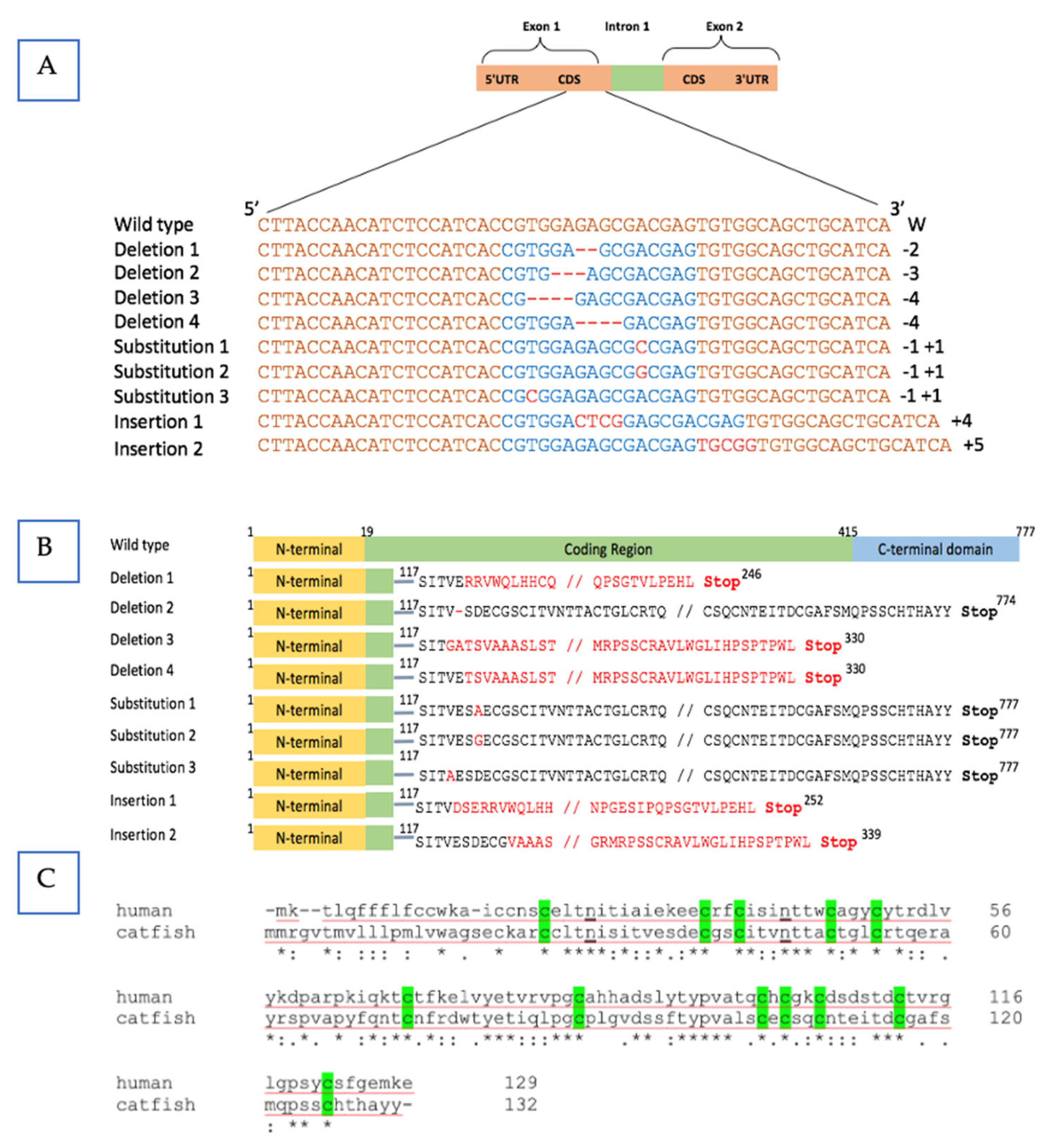
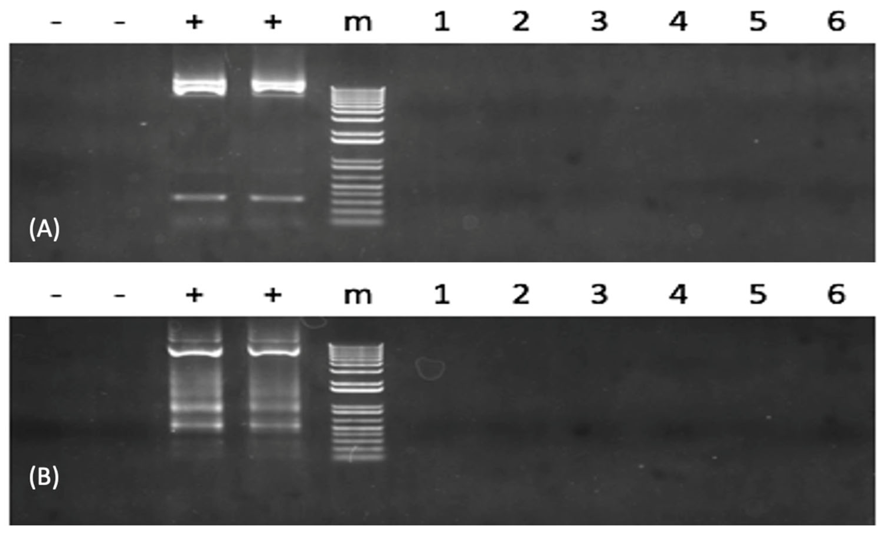
3.1. Mutation Evaluation and Plasmid Integration in the P1 Fish

In 31 of 57 P1 fingerlings that were 6 months old, the mutation rate was 54.4%. Five bands were visible on the gel based on the surveyor mutation assay finds. The fsh gene was edited at the right position because a band of 350 bp was resolved on the gel (Figure 1 and Figure S2).
The mutation of the fsh gene in channel catfish was validated by sequencing and sequence comparison. All the modifications happened within the TALEN cutting region. In the coding area of the fsh gene, we found distinct mutation types, including substitutions, base deletions, and insertions, which determines the biological responsiveness of the hormone (Figure 2A). The fsh gene’s product is expected to be affected by these mutations, which are also expected to prevent the gene from functioning properly (Figure 2B). We compared these five frame shift mutations with that of the known normal human fsh gene structure; four of five resulted in losing of the N-glycosylation sites, which act as the attaching sites for oligosaccharides to bind and then assembly subunit and dimerization afterwards. Therefore, in silico analysis indicates the protein structures might be destroyed and the biological function of fsh may be reduced or eliminated (Figure 2C). The correct assembly of the subunit determined the metabolic clearance of the gonadotrophin.
Because neither sequence from the TAL repeats region nor CMV promoter region could be PCR amplified in the plasmid integration assessment, it suggests that no plasmid DNA was present using PCR for all TALEN-fsh-defective fish. The results concluded that none of the tested samples harbored the foreign DNA (Figure 3 and Figure S3).

3.2. Fertility Evaluation and Hormone Treatment and Intervention

Without hormone intervention, three of five (60.0%) in the control groups reproduced with a mean hatch rate of 74.3% in 2016. Fish receiving LHRHa and pregnant mare gonadotropin did not spawn. In 2017, without LHRHa hormone therapy, five of seven (71.4%) pairs of controls spawned and an average of 32.3% eggs were hatched, whereas using LHRHa hormone treatment, two of three (66.7%) pairs of fish spawned and an average of 56.5% eggs were hatched (Table 1).
Spawnability of P1 mutants was evaluated without hormone treatment in 2016. There are 33.3% P1 female mutants and 40% P1 male mutants spawned, with egg hatch rates ranging from 0.45 to 1.0%. The hatch rate differed considerably between the mutated group and the control group (p = 0.001). In 2017, a 1200 IU HCG hormone treatment was implemented. Hormone therapy appeared to improve the spawning rate, 80.0% for female mutants and 88.9% for male mutants, which was similar to that of controls, which were 71.4% without LHRHa injection (p = 1.000) and 66.7% with LHRHa injection (p = 0.737). A second 400 IU of HCG was injected into the female and male mutants that did not spawn after 5 days; however, the females still did not lay any eggs. F1 embryo mean hatch rates for both mutated females and males that underwent hormone treatment were improved to 35.0%, which did not differ from controls, which were 32.3% without LHRHa treatment (p = 0.259) and 56.5% with LHRHa treatment (p = 0.351) (Table 1).

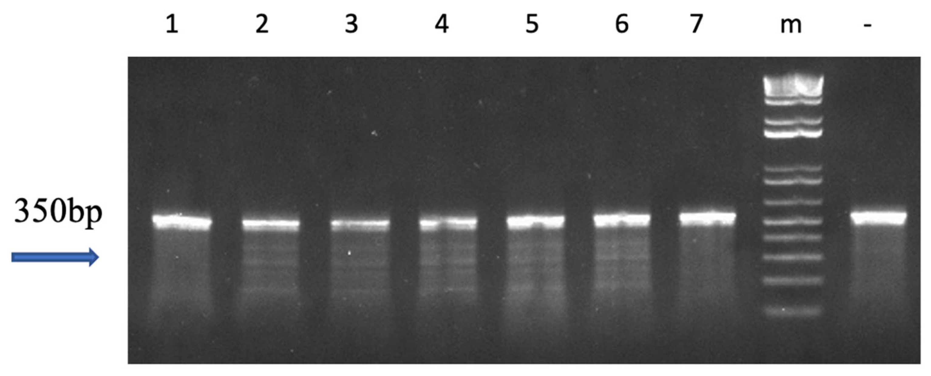
3.3. Mutation Evaluation for the F1 Fish

There was one F1 family (M♀ × Wt♂) obtained in 2016 and seven F1 families (M♀ × M♂) obtained in 2017. The mutations were successfully inherited by the offspring, with inheritance rates ranging from 45.5% to 64% (Table 2). Surveyor assay results showed multiple bands for mutants (Figure 4 and Figure S4).

3.4. Pleiotropic Effects

Pleiotropic effects were evaluated based on behavior, hatch rate, earlier fingerling survival rate, later adult survival rate in two environments, and bodyweight. For P1 embryos and 6-month-old fingerlings, no substantial differences were found for hatch rate (p = 0.760) and survival rate (p = 0.602) between the plasmid electroporated group and the control group. For 4-year-old P1 fish, mutants did not differ from non-mutants in survival rate cultured in a recirculating system (p = 1.000), whereas mutants had higher survival rates than non-mutants when cultured in the pond (p = 0.023) (Table 3).
The adult fish with outstanding secondary sexual characteristics were selected for the spawning trial. Abnormal reproductive behaviors were observed in three of the five male mutants and two of the three female mutants. The mutant males and wild-type females were seen engaging in an anomalous courtship between the wild-type females and the mutant males that was visible through the transparent bottoms of aquariums. Females of the wild-type had been observed laying immature eggs without forming an egg mass. In 2017, after receiving 1200 IU of HCG hormone, normal reproductive behavior was seen in two of five (40.0%) mutant males and one of three (33.3%) mutant females.
For the offspring, six families showed no difference regarding body weight between mutant fish and non-mutant fish (Table 2). However, the family 2017 FSH-3 showed a lower body weight for non-mutants (p = 0.009). The survival rate was not different among F1 families except one family of 2017 fsh-5 (p = 0.110). A low dissolved oxygen incident happened in one family of 2017 FSH-5 that killed thirty fish in one tank, in which there was detected a 56.7% mutation rate. This mutation rate for the dead progeny did not differ from their siblings (p = 0.475) in the family of 2017 FSH-5 in another tank.

4. Discussion

At the targeted region of the fsh gene, various mutations were identified, such as base deletions, substitutions, and insertions, all of which were efficiently transmitted to F1 offspring. Indels have the potential to cause a nonsense codon to inhibit the function of fsh. In a scenario, a single nucleotide in fsh was altered, which led to a significant impact on the function and expression of the gene because some nucleotide alterations either caused the coding sequence to terminate or mutate into a new codon corresponding a specific amino acid. The spawning rate of P1 mutants and their egg hatch rates were suppressed. The P1 brood stocks were mostly mosaic. This is also the explanation about how reproduction capacity was not completely eliminated. For the F1 generation, genome editing should happen in all the tissues. Hormone therapy with HCG reversed the sterility of P1 mutants. Seven families of F1 offspring exhibited no evident pleiotropic effects, but only one family of F1 offspring showed a decreased body weight for mutants, which might be attributed to the small sample size. Four-year-old P1 mutants had a greater survival rate.
TALENs are typically developed to target genes with great precision and optimized to pose a minimal amount of cytotoxicity [27,28,29,30]. In the P1 generation, the TALEN-treated group had a similar embryo hatch rate to that of the control group; a comparable fry survival rate was also observed between treated and control groups, indicating that the highly precise TALEN plasmids targeting catfish fsh were exceedingly specific and had minimal unintended consequences. The observation that the mortality percentage of mutated P1 fish and their full-sibling controls maintained in recirculating systems was the same even after four years provides more support for this conclusion. P1 mutants exhibited a higher survival rate in the pond than their full-sibling controls.
The TALENs’ target sites were designed and intended to disable the translation of the β subunit of the fsh gene, which provides the biological uniqueness of the hormone. Another possibility is that an aberrant variation in this multi-subunit structure is incapable of working in concert with the remaining subunits, rendering the whole molecule inactive. A mutant of this kind would react biologically as an antimorph, causing disruption to proteolytic activity in a dose-response manner [31,32]. The greatest amounts of fsh production and release correlate to the vitellogenesis stage of the salmon’s annual breeding process [12]. The membrane receptors for fsh are located on the granulosa and theca cells of the follicle. When fsh binds to its receptors on these cells, it regulates the production of hormones (estrogens and androgens), the growth and maturity of follicles, and ultimately the overall functioning of the ovaries. Amago salmon (Oncorhynchus rhodurus) [33] and murrel (Channa punctatus) [34] are two fish species that have proven establishment of their particular binding sites in the ovary. The ablation of the fsh gene should lead to a suppression of the production and release of pituitary gonadotropin, which in turn would cause a reduction in the production of steroid hormones, notably estradiol and testosterone.
Notwithstanding these findings on fsh in teleosts over the past 20 years, our understanding of the functions of fsh in directing fish reproduction remains limited. In salmonids, it has been hypothesized that fsh is primarily accountable for ovarian development, whereas lh is essential for triggering final oocyte maturation and ovulation [6,35,36]. Catfishes have a cyclical breeding system with various stages governed by a hormone regulatory signaling pathway, comprising predominantly gonadotropin-releasing hormone (gnrh), lh, growth hormone, melatonin, and sex steroid hormones. In nature, a gonadotropin burst typically promotes natural oocyte maturity, ovulation, or spermiation [37]. The effects of sex steroids on the teleost pituitary are complex and vary with the species studied, the animal’s physiological status or sex, and dose or mode of delivery [38]. The G protein-coupled receptor that responds to follicle-stimulating hormone is termed the FSH receptor (GPCR). When FSH hormone binds to the receptor on the cell surface, it initiates a chain of events within the cell by activating one or more G proteins, which are signal-transducing proteins that bind to guanine nucleotides. This activation controls the expression of genes, the release of hormones, and the access of ion channels, among other functions [39].
Using TALENs in zebrafish (Danio rerio), the hormone-specific β-subunits of both fsh and lh were individually or collectively knocked out. For fsh-deficient zebrafish, the establishment of the ovary, testis, and initiation of puberty was postponed. However, both sexes are still fertile. Also, fsh appeared to be involved in retaining the female condition, as sexual reversal was detected in the fsh-ablation zebrafish. The dual disruption of the fsh and lh genes resulted in all male offspring, albeit with severely delayed testicular development [40]. Our results with channel catfish vary from those with zebrafish. This is not entirely unexpected given the diversity of fish reproductive strategies [41]. Some fsh-deficient channel catfish with exceptional secondary sexual traits are unable to reproduce due to aberrant reproductive behaviors. The F1 embryo from fsh-deficient P1 females or males had a low hatch rate without HCG hormone therapy, whereas both spawning and hatch rates increased after hormone therapy.
However, whether this is also holds true for other groups of teleosts, such as cyprinids, has yet to be determined, particularly considering that the species-specificity of Fsh receptors varies considerably. In coho salmon (Oncorhynchus kisutch), the fsh interacted less with the type II receptor (Lh receptor) and associated exclusively with the type I receptor (now known as Fsh receptor) [42]. Purified and recombinant Lh and Fsh were able to increase intracellular cAMP levels in the African catfish (Clarias gariepinus) [43]. The scenario is substantially more convoluted in the Indian carp (Labeo rohita) because the ovarian follicles’ enriched gonadotropin receptors reacted to both salmon Fsh and Lh but preferred their respective ligands [44].
Glycoprotein hormone receptors (GPHRs) contain three subfamilies, including the luteinizing hormone/choriogonadotrophin receptor (LHCGR), follicle-stimulating hormone receptor (FSHR), and the thyroid-stimulating hormone receptor (TSHR) [45,46]. Equine chorionic gonadotropin, like LH, FSH, and thyroid-stimulating hormone, is a member of the glycoprotein family and its hormone has been employed to promote reproduction in horses and cows [47]. Chorionic gonadotropins are only found in primates and Equidae have both LH-like and FSH-like activity [48,49]. Various fish species are reproductively diverse, and HCG has been used to spawn many fish species, including channel catfish, but with variable success depending upon the species [49,50,51,52]. HCG and a cocktail of Fsh/Lh were demonstrated to be equivalently effective in increasing oocyte maturation and inhibiting the beginning of degeneration in grey mullet (Mugil cephalus L.) [53]. In perch (Perca fluviatilis), similar situations were observed after hormonal treatment of the common carp, Cyprinus carpio, with pituitary extract and HCG [54]. For therapeutic purposes, both Lh and HCG have been employed because of the activities they trigger while binding to the Lh/HCG receptor. This interaction is triggered by the fact that the two Lh/HCG hormones share the same Lh/HCG receptor. Because of its unique structure, the Lh/HCG receptor is known to be a member of the superfamily of G protein-coupled receptors [55]. Interacting with specific surface receptors, Fsh stimulates the development and proliferation of both theca cells and granulosa cells [56].
Although GTHs determine the steroidogenic potency, which in turn regulates FSH production and secretion, sex steroids do not alter the transcription of the Fshβ gene in rats in in vitro conditions [57]. In ovariectomized sheep, estradiol therapy represses the expression of Fshβ mRNA [58]. In the current research, the bottom line is sterility, which is the ultimate goal. Therefore, the direct effect of an FSH knockout mutation on the HPG axis, the GTH axis, and steroidogenesis may be very species-specific and complicated. Future research will need to examine whether the fsh gene knockout has any pleiotropic effects on the levels of Fsh mRNA, protein, or hormone in both control and mutant fishes before and after hormone therapy.

5. Conclusions

In the current investigation, TALEN plasmids targeting the channel catfish fsh gene were introduced into the sperm and early embryos through double electroporation. The mutagenesis of fsh was detected at the genomic level and resulted in sterile channel catfish via a TALEN-mediated effect. Using fish mating trials, the reproductive capacity of fsh-mutant channel catfish were evaluated and found to be negligible. F1 generation fish were subjected to mutation assays to confirm the inheritance of the mutations. No substantial pleiotropic effects were discovered in the F1 generation. Correction of sterility caused by the mutated fsh-gene in channel catfish is feasible with hormone treatment. The successful development and demonstration of this sterilization technology could be used for the bioconfinement of catfish and other genetically engineered fish, as well as domestic and invasive fish.

Supplementary Materials

The following supporting information can be downloaded at: https://www.mdpi.com/article/10.3390/biology12030392/s1, Figure S1: Schematic representation of transcription activator-like effector nucleases (TALENs) plasmid structure targeting follicle-stimulating hormone (fsh) gene of channel catfish (Ictalurus punctatus). Figure S2: Original images of gels. The result of Surveyor mutation detection in Figure 1 was shown. Red box represents the cropped area. Figure S3: Original images of gels. The result of PCR inspection in Figure 3 was shown. Red box represents the cropped area. Figure S4: Original images of gels. The result of Surveyor mutation detection in Figure 4 was shown. Table S1: Primer sequences used for the amplification of follicle-stimulating hormone (fsh), cytomegalovirus (CMV) promoter, and transcription activator-like (TAL) repeats regions in channel catfish (Ictalurus punctatus).

Author Contributions

G.Q., Z.Q., B.S. and R.A.D. conceived and designed the experiment and performed the statistical analyses; G.Q., Z.Q., A.E. and B.S. analyzed the results; G.Q., Z.Q., Z.Y., A.E., C.L., M.G.Q.K. and Y.J. conducted the experiment; G.Q., Z.Q., C.L., A.E. and M.G.Q.K. fed the fish and conducted the field work. G.Q., B.S. and R.A.D. wrote the manuscript. All authors have read and agreed to the published version of the manuscript.

Funding

This project is supported by Biotechnology Risk Assessment Grant Program competitive grant no. 2014-33522-22263 from the USDA National Institute of Food.

Institutional Review Board Statement

All experimental protocols used in this experiment were approved by the Auburn University Institutional Animal Care and Use Committee (AU-IACUC) before the experiment was initiated and followed Association for Assessment and Accreditation of Laboratory Animal Care (AAALAC) protocols and guidelines.

Informed Consent Statement

Not applicable.

Data Availability Statement

Not applicable.

Conflicts of Interest

The authors declare no conflict of interest.

References

  1. Dunham, R.A.; Ramboux, A.C.; Duncan, P.L.; Hayat, M.; Chen, T.T.; Lin, C.M.; Kight, K.; Gonzalez-Villasenor, I.; Powers, D.A. Transfer, Expression, and Inheritance of Salmonid Growth Hormone Genes in Channel Catfish, Ictalurus punctatus, and Effects on Performance Traits. Mol. Mar. Biol. Biotechnol. 1992, 1, 380–389. [Google Scholar]
  2. Dunham, R.A.; Chitmanat, C.; Nichols, A.; Argue, B.; Powers, D.A.; Chen, T.T. Predator Avoidance of Transgenic Channel Catfish Containing Salmonid Growth Hormone Genes. Mar. Biotechnol. 1999, 1, 545–551. [Google Scholar] [CrossRef] [PubMed]
  3. Dunham, R.A.; Warr, G.W.; Nichols, A.; Duncan, P.L.; Argue, B.; Middleton, D.; Kucuktas, H. Enhanced Bacterial Disease Resistance of Transgenic Channel Catfish Ictalurus punctatus Possessing Cecropin Genes. Mar. Biotechnol. 2002, 4, 338–344. [Google Scholar] [CrossRef] [PubMed]
  4. Hedrick, P.W. Invasion of Transgenes from Salmon or Other Genetically Modified Organisms into Natural Populations. Can. J. Fish. Aquat. Sci. 2001, 58, 2317. [Google Scholar] [CrossRef]
  5. Patiño, R.; Thomas, P. Effects of Gonadotropin on Ovarian Intrafollicular Processes during the Development of Oocyte Maturational Competence in a Teleost, the Atlantic Croaker: Evidence for Two Distinct Stages of Gonadotropic Control of Final Oocyte Maturation. Biol. Reprod. 1990, 43, 818–827. [Google Scholar] [CrossRef]
  6. Prat, F.; Sumpter, J.P.; Tyler, C.R. Validation of Radioimmunoassays for Two Salmon Gonadotropins (GTH I and GTH II) and Their Plasma Concentrations throughout the Reproductive Cycle in Male and Female Rainbow Trout (Oncorhynchus mykiss). Biol. Reprod. 1996, 54, 1375–1382. [Google Scholar] [CrossRef] [Green Version]
  7. Swanson, P. Salmon Gonadotropins: Reconciling Old and New Ideas. Reprod. Physiol. Fish 1991, 2–7. [Google Scholar]
  8. Swanson, P.; Dickey, J.T.; Campbell, B. Biochemistry and Physiology of Fish Gonadotropins. Fish Physiol. Biochem. 2003, 28, 53–59. [Google Scholar] [CrossRef]
  9. Kwok, H.F.; So, W.K.; Wang, Y.; Ge, W. Zebrafish Gonadotropins and Their Receptors: I. Cloning and Characterization of Zebrafish Follicle Stimulating Hormone and Luteinizing Hormone Receptors-Evidence for Their Distinct Functions in Follicle Development. Biol. Reprod. 2005, 72, 1370–1381. [Google Scholar] [CrossRef] [PubMed]
  10. Hsu, S.Y.; Nakabayashi, K.; Bhalla, A. Evolution of Glycoprotein Hormone Subunit Genes in Bilateral Metazoa: Identification of Two Novel Human Glycoprotein Hormone Subunit Family Genes, GPA2 and GPB5. Mol. Endocrinol. 2002, 16, 1538–1551. [Google Scholar] [CrossRef]
  11. Liu, Z.; Kim, S.; Karsi, A. Channel Catfish Follicle-Stimulating Hormone and Luteinizing Hormone: Complementary DNA Cloning and Expression during Ovulation. Mar. Biotechnol. 2001, 3, 590–599. [Google Scholar] [CrossRef]
  12. Dolomatov, S.I.; Zukow, W.; Novikov, N.Y.; Muszkieta, R.; Bulatowicz, I.; Dzierzanowski, M.; Kazmierczak, U.; Strojek, K. The Regulation of Osmotic and Ionic Balance in Fish Reproduction and in the Early Stages of Ontogeny. Russ. J. Mar. Biol. 2012, 38, 365–374. [Google Scholar] [CrossRef]
  13. Schulz, R.W.; Goos, H.J.T. Puberty in Male Fish: Concepts and Recent Developments with Special Reference to the African Catfish (Clarias gariepinus). Aquaculture 1999, 177, 5–12. [Google Scholar] [CrossRef]
  14. Ko, H.; Park, W.; Kim, D.J.; Kobayashi, M.; Sohn, Y.C. Biological Activities of Recombinant Manchurian Trout FSH and LH: Their Receptor Specificity, Steroidogenic and Vitellogenic Potencies. J. Mol. Endocrinol. 2007, 38, 99–111. [Google Scholar] [CrossRef] [PubMed] [Green Version]
  15. Campbell, B.; Dickey, J.; Beckman, B.; Young, G.; Pierce, A.; Fukada, H.; Swanson, P. Previtellogenic Oocyte Growth in Salmon: Relationships among Body Growth, Plasma Insulin-like Growth Factor-1, Estradiol-17beta, Follicle-Stimulating Hormone and Expression of Ovarian Genes for Insulin-like Growth Factors, Steroidogenic-Acute Regulatory Protein and Receptors for Gonadotropins, Growth Hormone, and Somatolactin. Biol. Reprod. 2006, 75, 34–44. [Google Scholar] [CrossRef]
  16. Qin, G.; Qin, Z.; Lu, C.; Ye, Z.; Elaswad, A.; Bangs, M.; Li, H.; Zhang, Y.; Huang, Y.; Shi, H.; et al. Gene Editing of the Catfish Gonadotropin-Releasing Hormone Gene and Hormone Therapy to Control the Reproduction in Channel Catfish, Ictalurus punctatus. Biology 2022, 11, 649. [Google Scholar] [CrossRef]
  17. Su, B.; Peatman, E.; Shang, M.; Thresher, R.; Grewe, P.; Patil, J.; Pinkert, C.A.; Irwin, M.H.; Li, C.; Perera, D.A.; et al. Expression and Knockdown of Primordial Germ Cell Genes, Vasa, Nanos and Dead End in Common Carp (Cyprinus carpio) Embryos for Transgenic Sterilization and Reduced Sexual Maturity. Aquaculture 2014, 420, S72–S84. [Google Scholar] [CrossRef]
  18. Su, B.; Shang, M.; Li, C.; Perera, D.A.; Pinkert, C.A.; Irwin, M.H.; Peatman, E.; Grewe, P.; Patil, J.G.; Dunham, R.A. Effects of Transgenic Sterilization Constructs and Their Repressor Compounds on Hatch, Developmental Rate and Early Survival of Electroporated Channel Catfish Embryos and Fry. Transgenic Res. 2015, 24, 333–352. [Google Scholar] [CrossRef]
  19. Su, B.; Shang, M.; Grewe, P.M.; Patil, J.G.; Peatman, E.; Perera, D.A.; Cheng, Q.; Li, C.; Weng, C.-C.; Li, P.; et al. Suppression and Restoration of Primordial Germ Cell Marker Gene Expression in Channel Catfish, Ictalurus punctatus, Using Knockdown Constructs Regulated by Copper Transport Protein Gene Promoters: Potential for Reversible Transgenic Sterilization. Theriogenology 2015, 84, 1499–1512. [Google Scholar] [CrossRef]
  20. Cheng, Q.; Su, B.; Qin, Z.; Weng, C.-C.; Yin, F.; Zhou, Y.; Fobes, M.; Perera, D.A.; Shang, M.; Soller, F.; et al. Interaction of Diet and the Masou Salmon Δ5-Desaturase Transgene on Δ6-Desaturase and Stearoyl-CoA Desaturase Gene Expression and N-3 Fatty Acid Level in Common Carp (Cyprinus carpio). Transgenic Res. 2014, 23, 729–742. [Google Scholar] [CrossRef]
  21. Qin, Z. Gene Editing of Luteinizing Hormone, Follicle-Stimulating Hormone and Gonadotropin-Releasing Hormone Genes to Sterilize Channel Catfish, Ictalurus Punctatus, Using Zinc Finger Nuclease, Transcription Activator-Like Effector Nuclease and Clustered Regularly Interspaced Short Palindromic Repeats/Cas9 Technologies. Ph.D. Thesis, Auburn University, Auburn, AL, USA, 2015. [Google Scholar]
  22. Qiu, P.; Shandilya, H.; D’Alessio, J.M.; O’Connor, K.; Durocher, J.; Gerard, G.F. Mutation Detection Using Surveyor Nuclease. Biotechniques 2004, 36, 702–707. [Google Scholar] [CrossRef]
  23. Pimkin, M.; Caretti, E.; Canutescu, A.; Yeung, J.B.; Cohn, H.; Chen, Y.; Oleykowski, C.; Bellacosa, A.; Yeung, A.T. Recombinant Nucleases CEL I from Celery and SP I from Spinach for Mutation Detection. BMC Biotechnol. 2007, 7, 29. [Google Scholar] [CrossRef] [PubMed] [Green Version]
  24. Tsuji, T.; Niida, Y. Development of a Simple and Highly Sensitive Mutation Screening System by Enzyme Mismatch Cleavage with Optimized Conditions for Standard Laboratories. Electrophoresis 2008, 29, 1473–1483. [Google Scholar] [CrossRef] [Green Version]
  25. Steeby, J.; Avery, J. Channel Catfish Broodfish and Hatchery Management; Southern Regional Aquaculture Center: Stoneville, MS, USA, 2005. [Google Scholar]
  26. Small, B.C.; Chatakondi, N. Efficacy of Formalin as an Egg Disinfectant for Improving Hybrid Catfish (Channel Catfish × Blue Catfish) Hatching Success. N. Am. J. Aquac. 2006, 68, 9–13. [Google Scholar] [CrossRef]
  27. Liu, J.; Gaj, T.; Patterson, J.T.; Sirk, S.J.; Barbas, C.F., 3rd. Cell-Penetrating Peptide-Mediated Delivery of TALEN Proteins via Bioconjugation for Genome Engineering. PLoS ONE 2014, 9, e85755. [Google Scholar] [CrossRef] [Green Version]
  28. Mussolino, C.; Alzubi, J.; Fine, E.J.; Morbitzer, R.; Cradick, T.J.; Lahaye, T.; Bao, G.; Cathomen, T. TALENs Facilitate Targeted Genome Editing in Human Cells with High Specificity and Low Cytotoxicity. Nucleic Acids Res. 2014, 42, 6762–6773. [Google Scholar] [CrossRef] [PubMed] [Green Version]
  29. Dreyer, A.K.; Hoffmann, D.; Lachmann, N.; Ackermann, M.; Steinemann, D.; Timm, B.; Siler, U.; Reichenbach, J.; Grez, M.; Moritz, T.; et al. TALEN-Mediated Functional Correction of X-Linked Chronic Granulomatous Disease in Patient-Derived Induced Pluripotent Stem Cells. Biomaterials 2015, 69, 191–200. [Google Scholar] [CrossRef]
  30. Veres, A.; Gosis, B.S.; Ding, Q.; Collins, R.; Ragavendran, A.; Brand, H.; Erdin, S.; Cowan, C.A.; Talkowski, M.E.; Musunuru, K. Low Incidence of Off-Target Mutations in Individual CRISPR-Cas9 and TALEN Targeted Human Stem Cell Clones Detected by Whole-Genome Sequencing. Cell Stem Cell 2014, 15, 254. [Google Scholar] [CrossRef] [Green Version]
  31. Saville, K.J.; Belote, J.M. Identification of an Essential Gene, l(3)73Ai, with a Dominant Temperature-Sensitive Lethal Allele, Encoding a Drosophila Proteasome Subunit. Proc. Natl. Acad. Sci. USA 1993, 90, 8842–8846. [Google Scholar] [CrossRef] [PubMed] [Green Version]
  32. Schweisguth, F. Dominant-Negative Mutation in the β2 and β6 Proteasome Subunit Genes Affect Alternative Cell Fate Decisions in the Drosophila Sense Organ Lineage. Proc. Natl. Acad. Sci. USA 1999, 96, 11382–11386. [Google Scholar] [CrossRef] [Green Version]
  33. Kanamori, A.; Kagawa, H.; Nagahama, Y. Gonadotropin Receptors in the Postovulatory Ovary of Amago Salmon (Oncorhynchus rhodurus). Gen. Comp. Endocrinol. 1987, 66, 210–217. [Google Scholar] [CrossRef] [PubMed]
  34. Jamal Uddin, M.; Bhattacharya, S. In-Vitro Binding of Gonadotrophin to Fish Ovary. J. Endocrinol. 1986, 111, 407–413. [Google Scholar] [CrossRef] [PubMed]
  35. Breton, B.; Govoroun, M.; Mikolajczyk, T. GTH I and GTH II Secretion Profiles during the Reproductive Cycle in Female Rainbow Trout: Relationship with Pituitary Responsiveness to GnRH-A Stimulation. Gen. Comp. Endocrinol. 1998, 111, 38–50. [Google Scholar] [CrossRef] [PubMed]
  36. Weil, C.; Bougoussa-Houadec, M.; Gallais, C.; Itoh, S.; Sekine, S.; Valotaire, Y. Preliminary Evidence Suggesting Variations of GtH1 and GtH2 MRNA Levels at Different Stages of Gonadal Development in Rainbow Trout, Oncorhynchus mykiss. Gen. Comp. Endocrinol. 1995, 100, 327–333. [Google Scholar] [CrossRef] [PubMed]
  37. Senthilkumaran, B.; Kar, S. Advances in Reproductive Endocrinology and Neuroendocrine Research Using Catfish Models. Cells 2021, 10, 2807. [Google Scholar] [CrossRef]
  38. Fontaine, R.; Royan, M.R.; von Krogh, K.; Weltzien, F.-A.; Baker, D.M. Direct and Indirect Effects of Sex Steroids on Gonadotrope Cell Plasticity in the Teleost Fish Pituitary. Front. Endocrinol. 2020, 11, 605068. [Google Scholar] [CrossRef]
  39. Ulloa-Aguirre, A.; Timossi, C. Structure-Function Relationship of Follicle-Stimulating Hormone and Its Receptor. Hum. Reprod. Update 1998, 4, 260–283. [Google Scholar] [CrossRef] [Green Version]
  40. Zhang, Z.; Zhu, B.; Ge, W. Genetic Analysis of Zebrafish Gonadotropin (FSH and LH) Functions by TALEN-Mediated Gene Disruption. Mol. Endocrinol. 2015, 29, 76–98. [Google Scholar] [CrossRef] [Green Version]
  41. Dunham, R.A. Aquaculture and Fisheries Biotechnology: Genetic Approaches; CABI Publishing: Wallingford, UK, 2023. [Google Scholar]
  42. Yan, L.; Swanson, P.; Dickhoff, W.W. A Two-Receptor Model for Salmon Gonadotropins (GTH I and GTH II). Biol. Reprod. 1992, 47, 418–427. [Google Scholar] [CrossRef]
  43. Vischer, H.F.; Granneman, J.C.M.; Linskens, M.H.K.; Schulz, R.W.; Bogerd, J. Both Recombinant African Catfish LH and FSH Are Able to Activate the African Catfish FSH Receptor. J. Mol. Endocrinol. 2003, 31, 133–140. [Google Scholar] [CrossRef] [PubMed] [Green Version]
  44. Basu, D.; Bhattacharya, S. Purification of Two Types of Gonadotropin Receptors from Carp Ovarian Follicles: Over-Lapping Recognition by Two Different Ligands. Gen. Comp. Endocrinol. 2002, 129, 152–162. [Google Scholar] [CrossRef] [PubMed]
  45. Ascoli, M.; Fanelli, F.; Segaloff, D.L. The Lutropin/Choriogonadotropin Receptor, a 2002 Perspective. Endocr. Rev. 2002, 23, 141–174. [Google Scholar] [CrossRef]
  46. Troppmann, B.; Kleinau, G.; Krause, G.; Gromoll, J. Structural and Functional Plasticity of the Luteinizing Hormone/Choriogonadotrophin Receptor. Hum. Reprod. Update 2013, 19, 583–602. [Google Scholar] [CrossRef] [Green Version]
  47. De Rensis, F.; López-Gatius, F. Use of Equine Chorionic Gonadotropin to Control Reproduction of the Dairy Cow: A Review. Reprod. Domest. Anim. 2014, 49, 177–182. [Google Scholar] [CrossRef]
  48. Min, K.-S.; Park, J.-J.; Byambaragchaa, M.; Kang, M.-H. Characterization of Tethered Equine Chorionic Gonadotropin and Its Deglycosylated Mutants by Ovulation Stimulation in Mice. BMC Biotechnol. 2019, 19, 60. [Google Scholar] [CrossRef] [Green Version]
  49. Zohar, Y.; Mylonas, C.C. Endocrine Manipulations of Spawning in Cultured Fish: From Hormones to Genes. Aquaculture 2001, 197, 99–136. [Google Scholar] [CrossRef]
  50. Prentice, J.A.; Thomas, P. Successful Spawning of Orangemouth Corvina Following Injection with Des-Gly10,[D-Ala6]-Luteinizing Hormone-Releasing Hormone (1-9) Ethylamide and Pimozide. Prog. Fish-Cult. 1987, 49, 66–69. [Google Scholar] [CrossRef]
  51. Rottmann, R.W.; Shireman, J.V.; Chapman, F.A. Hormonal Control of Reproduction in Fish for Induced Spawning; Southern Regional Aquaculture Center: Stoneville, MS, USA, 1991. [Google Scholar]
  52. Goudie, C.A.; Simco, B.A.; Davis, K.B.; Parker, N.C. Reproductive Performance of Pigmented and Albino Female Channel Catfish Induced to Spawn with HCG or Ovaprim. J. World Aquac. Soc. 1992, 23, 138–145. [Google Scholar] [CrossRef]
  53. Shehadeh, Z.H.; Madden, W.D.; Dohl, T.P. The Effect of Exogenous Hormone Treatment on Spermiation and Vitellogenesis in the Grey Mullet, Mugil cephalus L. J. Fish Biol. 1973, 5, 479–487. [Google Scholar] [CrossRef]
  54. Kucharczyk, D.; Kujawa, R.; Murmurz, A.; Skrzypczak, A.; Wyszomirska, E. Induced Spawning in Perch, Perca fluviatilis L. Using Carp Pituitary Extract and HCG. Aquac. Res. 1996, 27, 847–852. [Google Scholar] [CrossRef]
  55. Kido, A.; Mori, M.; Adachi, Y.; Yukaya, H.; Ishida, T.; Sugimachi, K. Immunohistochemical Ex-Pression of β-Human Chorionic Gonadotropin in Colorectal Carcinoma. Surg. Today 1996, 26, 966–970. [Google Scholar] [CrossRef] [PubMed]
  56. Filicori, M.; Fazleabas, A.T.; Huhtaniemi, I.; Licht, P.; Rao, C.V.; Tesarik, J.; Zygmunt, M. Novel Concepts of Human Chorionic Gonadotropin: Reproductive System Interactions and Potential in the Management of Infertility. Fertil. Steril. 2005, 84, 275–284. [Google Scholar] [CrossRef] [PubMed]
  57. Shupnik, M.A.; Gharib, S.D.; Chin, W.W. Divergent Effects of Estradiol on Gonadotropin Gene Transcription in Pituitary Fragments. Mol. Endocrinol. 1989, 3, 474–480. [Google Scholar] [CrossRef] [PubMed] [Green Version]
  58. Mercer, J.E.; Clements, J.A.; Funder, J.W.; Clarke, I.J. Regulation of Follicle-Stimulating Hormone β and Common α-Subunit Messenger Ribonucleic Acid by Gonadotropin-Releasing Hormone and Estrogen in the Sheep Pituitary. Neuroendocrinology 1989, 50, 321–326. [Google Scholar] [CrossRef] [PubMed]
Figure 1. Surveyor mutation detection test results of P1 channel catfish (Ictalurus punctatus). The mutation was targeted at the follicle-stimulating hormone (fsh) gene. The “-” on the left of the molecular marker indicates the control, which is wild-type fish DNA. The right “-” denotes the negative control using water as the DNA template; “m” indicates 1 kb molecular marker. Mutated channel catfish are shown in lanes 1, 3, 4, and 5, and wild-type channel catfish are displayed in lanes 2 and 6. Figure 1 was derived from the full-length gel shown in Supplementary Figure S2.
Biology 12 00392 g001
Figure 2. Sequence analysis of the mutated follicle-stimulating hormone (fsh) gene in channel catfish (Ictalurus punctatus). (A) Sequence analysis of the nucleic acid in the mutated fsh gene. The channel catfish fsh nucleotide sequence of the wild-type is displayed at the top. Orange highlighted sequences represent the target binding sites of the transcription activator-like effector nucleases (TALENs). Blue colored sequences represent the predicted cleavage sites of the nucleases. The mutation types (deletion/insertion/substitution) of nucleotides are shown by red dashes and letters. Numbers of nucleotides deleted (-) or inserted (+) in the edited fsh gene are denoted on the right side of the sequences. (B) Sequence analysis of corresponding predicted amino acid in the fsh mutants. Frameshift reading resulted in a premature stop codon (red colored). Single amino acid deletions or substitutions were caused by changes in one nucleotide mutations or three nucleotide deletions that are highlighted in red. (C) Bright-green shaded areas are the Cys residues, and N-linked glycosylation sites are underlined. The asterisk “*” indicated the same amino acid between normal human and wild-type catfish.
Biology 12 00392 g002
Figure 3. Analysis of transcription activator-like effector nuclease (TALEN) plasmid integration into the genome of channel catfish (Ictalurus punctatus) using polymerase chain reaction (PCR) genome. In (A,B), from the left to the right lanes of “-” denote the negative controls. The left one indicates using water as a template, and the right one indicates the wild-type channel fish sample; the two “+” denote the positives. The left one indicates the TALEN plasmids containing the left DNA binding site, and the right one indicates TALEN plasmids containing the right DNA binding site; “m” indicates 1 kb molecular weight standards. The same six fsh-mutated channel catfish individuals (numbered 1–6) were checked for plasmid DNA element integration. The cytomegalovirus (CMV) promoter region was resolved in (A). The transcription activator-like (TAL) repeats region was resolved in (B). The original full gel image of Figure 3 can be found in Supplementary Figure S3.
Biology 12 00392 g003
Figure 4. Mutation analysis of follicle-stimulating hormone (fsh) gene edited F1 channel catfish (Ictalurus punctatus) using the Surveyor mutation detection test. “-” represents the wild-type DNA as negative control; “m” represents 1 kb DNA molecular weight standards; numbers (2, 3, 4, 5 and 6) show the mutated channel catfish individuals; lanes numbered 1 and 7 show progeny without anticipated mutation. The full gel picture of Figure 4 can be found in Supplementary Figure S4.
Biology 12 00392 g004
Table 1. Spawnability evaluation in terms of spawning rate and embryo hatch rate in two spawning seasons, 2016 and 2017. For the purpose of evaluating spawning, two different types of controls were used: the non-hormone-injected control (nCTRL) and the injected-hormone control (iCTRL) using a 90 μg/kg luteinizing hormone releasing hormone analog (LHRHa) implant. In 2016, wild-type fish were coupled with 3 mutant females and 5 mutant males. The 90 μg/kg LHRHa was only given to the wild-type female fish. In 2017, 10 female mutants were paired with 9 mutant males. A total of 1200 IU human chorionic gonadotropin (HCG) was given to both females and males. To calculate the hatch rate, divide the number of eggs hatched by the number of eggs in total, then multiply by 100. Mean hatch rate data were displayed as the mean of all families ± standard error (SEM).
Spawning in 2016Spawning in 2017
Fish NSpawned Fish NSpawning Rate (%) aMean Hatch Rate (%)Fish NSpawned Fish NSpawning Rate b (%)Mean Hatch Rate (%) c
fshF3133.31.0 *10880.035.0 ± 0.14
M5240.00.5 ± 0.05 *9888.935.0 ± 0.14
iCTRLM and F ////3266.756.5 ± 0.34
nCTRLM and F5360.074.3 ± 0.02 *7571.432.3 ± 0.20
* In 2016, there were significant changes in the hatch rate between 2 groups, including the fsh gene edited group and the control group (Student’s t test, p = 0.001). a In 2016, there were no significant differences regarding the spawning rates among mutant males, mutant females, and wild-type fish (Fisher’s exact test, p = 1.000). b In 2017, there were no significant differences in the spawning rates among mutant fish, iCTRL, and nCTRL after hormone therapy (Fisher’s exact test, p = 0.737). c In 2017, there were no significant differences between the fsh mutants and the nCTRL group. There were no significant differences between fsh mutants and the iCTRL group after hormone therapy (Student’s t test, p = 0.259, p = 0.351).
Table 2. Pleiotropic effects evaluation, including survival rate, mutation rate, and mean body weight of channel catfish (Ictalurus punctatus). There are a total of 8 families of F1 progeny, including one 1.5-year-old 2016 FSH family without performing hormone therapy and seven 1-year-old 2017 FSH families with hormone therapy (2017 FSH-1, FSH-2, FSH-3, FSH-4, FSH-5, FSH-6, and FSH-7). The 2016 FSH family was generated from one fsh-mutated female fish paired with wild-type channel catfish without hormone treatment. However, 2017 FSH families were generated from mutant females mated with mutant males with hormone therapy. The 2017 FSH-5 family of fish perished as a result of low dissolved oxygen. Data on body weight are displayed as the mean of the body weight ± standard error (SEM).
Family Names of F1 OffspringSurvival EvaluationMutation EvaluationMean Body Weight (g)
Mutant Fish a
Body Weight (g) of
Non-Mutant Fish a
N FishN Fish SurvivedSurvival Rate (%)N Fish SampledN Mutant FishMutation Rate (%)
2016 FSH25022288.9301963.336.4 ± 2.1335.6 ± 2.21
2017 FSH-1222091.0201155.030.2 ± 4.7628.6 ± 5.26
2017 FSH-230027993.0301550.09.5 ± 0.5510.9 ± 0.81
2017 FSH-3242083.3251664.015.2 ± 1.09 *22.8 ± 2.19 *
2017 FSH-430026688.7211047.624.6 ± 3.2226.6 ± 1.89
2017 FSH-530016856.0301860.0 b12.8 ± 0.8211.9 ± 0.62
2017 FSH-5 dead fish///301860.0 b16.67 ± 0.81117.67 ± 1.509
2017 FSH-630025986.3281346.418.3 ± 1.4816.2 ± 0.78
2017 FSH-730026990.0221045.58.4 ± 0.709.3 ± 1.41
* Mutant fish in the family 2017 FSH-3 had a considerably lower body weight (Student’s t test, p = 0.009) than non-mutant fish in the same family. a The mutant fish and non-mutant fish had no significant differences in body weight in seven F1 families, including 2016 FSH (p = 0.807), 2017 FSH-1 (p = 0.827), 2017 FSH-2 (p = 0.156), 2017 FSH-4 (p = 0.585), 2017 FSH-5 (p = 0.373), 2017 FSH-6 (p = 0.223), and 2017 FSH-7 (p = 0.578) (Student’s t test). b Mutation rate was not significantly different (p = 0.475) between the 2017 FSH-5 family of fish and the sibling fish that perished as a result of oxygen depletion in this family (Fisher’s exact test).
Table 3. Pleiotropic effects of the P1 embryo, 6-month-old fingerlings and 4-year-old P1 fish. Survival rate and hatch rate were calculated. Transcription activator-like effector nuclease (TALEN)-plasmid electroporated and no-plasmid electroporated embryos and fingerlings were compared. Mutant and non-mutant channel catfish (Ictalurus punctatus) cultured in different environment were compared.
TreatmentP1 Embryos and 6-Month-Old FingerlingsGenotype4-Year-Old P1 Fish in 2017
Recirculating SystemPond
N EggsN HatchedHatch Rate (%) aSurvival Rate (%) bN FishSurvival Rate (%) cN FishSurvival Rate (%) *
Electroporated with plasmids2008044.071.3fsh Mutants4254.83754.1
Electroporated without plasmids2008444.075.0fsh Non-mutants250.060
ab No significant differences in hatch rate (p = 0.760) and survival rate (p = 0.602) were detected between the electroporated with TALEN plasmids group and the electroporated without plasmids group for P1 embryos and 6-month-old fingerlings (Fisher’s exact test). c P1 mutants and non-mutants at 4-years of age had similar survival rates (p = 1.000) in a circulating system (Fisher’s exact test). However, the sample size is very small for non-mutants. * P1 mutants had a higher (p = 0.023) survival rate than non-mutants at 4-years of age in the pond (Fisher’s exact test).
Disclaimer/Publisher’s Note: The statements, opinions and data contained in all publications are solely those of the individual author(s) and contributor(s) and not of MDPI and/or the editor(s). MDPI and/or the editor(s) disclaim responsibility for any injury to people or property resulting from any ideas, methods, instructions or products referred to in the content.

