Article

DNA Methylation Levels of the TBX5 Gene Promoter Are Associated with Congenital Septal Defects in Mexican Paediatric Patients

by
Esbeidy García-Flores
1,2,†,
José Manuel Rodríguez-Pérez
1,†,
Verónica Marusa Borgonio-Cuadra
3,
Gilberto Vargas-Alarcón
1,
Juan Calderón-Colmenero
4,
Juan Pablo Sandoval
5,
José Antonio García-Montes
5,
Víctor Manuel Espinoza-Gutiérrez
6,
Juan Gerardo Reyes-García
2,
Benny Giovanni Cazarín-Santos
1,2,
Antonio Miranda-Duarte
3,
Armando Gamboa-Domínguez
7 and
Nonanzit Pérez-Hernández
1,*
1
Departamento de Biología Molecular, Instituto Nacional de Cardiología Ignacio Chávez, Ciudad de Mexico 14080, Mexico
2
Sección de Estudios de Posgrado e Investigación, Escuela Superior de Medicina, Instituto Politécnico Nacional, Ciudad de Mexico 11340, Mexico
3
Departamento de Medicina Genómica, Instituto Nacional de Rehabilitación Luis Guillermo Ibarra Ibarra, Ciudad de Mexico 14389, Mexico
4
Departamento de Cardiología Pediátrica, Instituto Nacional de Cardiología Ignacio Chávez, Ciudad de Mexico 14080, Mexico
5
Laboratorio de Hemodinámica e Intervención en Cardiopatías Congénitas, Instituto Nacional de Cardiología Ignacio Chávez, Ciudad de Mexico 14080, Mexico
6
Hospital General de Zona No. 18, Playa del Carmen 77730, Mexico
7
Departamento de Patología, Instituto Nacional de Ciencias Médicas y de la Nutrición Salvador Zubirán, Ciudad de Mexico 14000, Mexico
*
Author to whom correspondence should be addressed.
These authors contributed equally to this work.
Biology 2022, 11(1), 96; https://doi.org/10.3390/biology11010096
Submission received: 30 November 2021 / Revised: 31 December 2021 / Accepted: 7 January 2022 / Published: 8 January 2022
(This article belongs to the Special Issue DNA Methylation and Epigenetics)

Abstract

:

Simple Summary

One of the most important health problems in the paediatric population, due to its prevalence worldwide, is the occurrence of congenital heart defects. The integration of multidisciplinary approaches to clinical diagnosis will allow us to detect this type of problem in patients in a timely way. The TBX5 gene has an important participation in cardiogenesis. Therefore, we performed a case-control study, that involved the DNA methylation assessment of the TBX5 gene promoter region in patients that were non-syndromic with congenital septal defects, to identify an epigenetic marker. Moreover, we evaluated the exposure to environmental factors during pregnancy in mothers of these patients. Additionally, we used bioinformatic tools to identify transcription factors binding to the TBX5 gene region of interest and to learn their possible functional effect. These results could help to clarify the mechanisms that regulate these pathologies and establish risk markers, which can be used in future in clinical practice.

Abstract

The TBX5 gene regulates morphological changes during heart development, and it has been associated with epigenetic abnormalities observed in congenital heart defects (CHD). The aim of this research was to evaluate the association between DNA methylation levels of the TBX5 gene promoter and congenital septal defects. DNA methylation levels of six CpG sites in the TBX5 gene promoter were evaluated using pyrosequencing analysis in 35 patients with congenital septal defects and 48 controls. Average methylation levels were higher in individuals with congenital septal defects than in the controls (p < 0.004). In five CpG sites, we also found higher methylation levels in patients than in the controls (p < 0.05). High methylation levels were associated with congenital septal defects (OR = 3.91; 95% CI = 1.02–14.8; p = 0.045). The analysis of Receiver Operating Characteristic (ROC) showed that the methylation levels of the TBX5 gene could be used as a risk marker for congenital septal defects (AUC = 0.68, 95% CI = 0.56–0.80; p = 0.004). Finally, an analysis of environmental factors indicated that maternal infections increased the risk (OR = 2.90; 95% CI = 1.01–8.33; p = 0.048) of congenital septal defects. Our data suggest that a high DNA methylation of the TBX5 gene could be associated with congenital septal defects.

1. Introduction

Congenital heart defects (CHDs) are a group of complex diseases defined as developmental defects in the human heart; they represent an important problem of public health worldwide, with a prevalence of 4–50 per 1000 live births [1]. The development of CHDs are the result of an interaction between genetic and environmental factors. In the last decade, several studies have focused on genes involved in the development of heart defects, such as the T-box gene family and have found clinical evidence that this gene family contributes to the development of CHDs [2,3,4,5].
The T-box genes encode a family of transcription factors that share similar sequences within the DNA-binding domain (T-domain). In vertebrates, five subfamilies of genes involved in cardiogenesis have been identified: T family, Tbx1, Tbx2, Tbx6, and Tbr1 [6,7]. Recently, genes that belong to the Tbx1 (TBX1, TBX18, and TBX20) and Tbx2 (TBX2, TBX5) subfamilies have been reported as transcriptional activators and gene repressors involved in the formation of chamber myocardium. These genes have also been associated with the development of CHDs, including atrial septal defect (ASD), ventricular septal defect (VSD), mitral valve disease (MVD) and tetralogy of Fallot (TOF), among others [5,8,9,10,11].
In particular, the TBX5 gene is a preponderant transcriptional activator of genes related and required for morphological changes in the septation and in cardiac conduction system [11]. The TBX5 gene interacts with other genes in signalling pathways that modulate atrial septation; this finding is supported with functional evidence that shows its participation in the ontogeny of CHDs [12]. In addition, it has been reported that the TBX5 gene interacts with the nucleosome remodelling and deacetylase (NuRD) repressor complex. This TBX5-NuRD complex is necessary for the normal cardiac development, therefore, any alteration could lead to CHDs [13].
On the other hand, it has been demonstrated that the TBX5 gene is modified by different epigenetic mechanisms, including long non-coding RNAs (lncRNAs), histone modification and polycomb group proteins. These epigenetic modifications play a key role in the pathophysiology of CHDs [14,15,16,17]. Furthermore, some studies have shown that an imbalance in the status of DNA methylation could change the expression of genes that participate in cardiogenesis [8,10,18,19,20].
Nevertheless, the epigenetic mechanisms of the TBX5 gene associated with congenital septal defects have been scarcely studied. Thus, we investigated the association of DNA methylation levels of the TBX5 gene promoter with the risk of developing congenital septal defects. We also, analysed the association of methylation levels of TBX5 gene with the exposure to environmental factors in mothers of patients with septal defects. Finally, we performed an in silico analysis to predict the possible functional role of the CpG sites studied by pyrosequencing.

2. Materials and Methods

2.1. Individuals Studied

This study included a total of 83 participants (35 patients with congenital septal defects and 48 controls). The selection criteria for this study were individuals of both sexes, including newborn to eighteen years, with a single diagnosis non-syndromic of ASD, VSD and patent ductus arteriosus (PDA); the exclusion criteria were patients with another type of CHD, other types of heart malformations and other types of infectious heart diseases, additionally, all participants were assessed by expert clinical geneticists with the aim to exclude syndromic forms of CHD.
For this study, we integrated these two types of CHDs (ASD and VSD) as a group of patients, even though both congenital septal defects seem to be different diseases, both conditions occur in related territories, they share the same characteristic of an incomplete septal closure and the same potential aetiology. Therefore, both malformations lead to similar clinical complications, for example, pulmonary hypertension, cerebral infarction, and arrhythmias [21,22]. These individuals came from different regions of Mexico and were attended in the Paediatric Cardiology Department at the “Instituto Nacional de Cardiología Ignacio Chávez” in Mexico City. The study complied with the Declaration of Helsinki and was approved by institutional ethical and research committees (Register No. 19-1138).
ASD was defined as a failure to close the communication between the right and left atria. VSD was defined as a failure in hemodynamic mechanisms and disturbance in the communication between left and right ventricles. In both cases, the congenital defects can lead to pulmonary hypertension, stroke and dysrhythmias [21,22]. The control group included individuals with a diagnosis of PDA, which was defined as a non-septal structure formed during foetal development, located between the pulmonary artery and the descending aorta, and usually undergoes spontaneous closure after birth [23].
The diagnosis of septal defects in all patients was made using echocardiography characteristics; additionally, clinical and physical examinations, as well as chest X-rays were taken into consideration. Information regarding environmental risk factors that could predispose to the development of septal defects (drug addiction, diseases during pregnancy, exposure to pollutants, infections during pregnancy, medications and vitamins intake) were obtained from the mothers of these patients during gestation. These data are presented in the Supplementary Table S1.

2.2. DNA Extraction and Bisulfite Treatment

DNA was isolated from 3–5 mL of peripheral blood samples using the Gentra Pure DNA isolation kit (QIAGEN, Hilden, Germany); the concentration and purity of the DNA extracted were calculated using a nanodrop 2000 spectrophotometer (Thermo Fhiser Scientific, Waltham, MA, USA). Genomic DNA was stored at −20 °C prior to bisulfite modification. A total of 500 ng DNA went into bisulfite modification using the Epitect Bisulfite Conversion Kit (QIAGEN, Hilden, Germany) following the manufacturer’s instructions, each sample of bisulfite-modified DNA was suspended in 20 μL of TE buffer for the following pyrosequencing assay.

2.3. DNA Methylation

The CpG island region (chr12: 114409768-114409805) in the genomic sequence GRGh38.p10 of the TBX5 promoter was selected for PyroMark CpG assays to quantify site-specific methylation levels. This region included 6 CpG sites, represented in bold in the following sequence (CGCGCTGTAGCCGGCTCCGGAGTTTACTGCCCGAACGA) from base −3623 to base −3661 relative to the transcription start site (TSS) (Figure 1).
PCR was performed using PyroMark PCR Kits (QIAGEN, Hilden, Germany) following the standard manufacturer’s protocol. A total volume of 25 μL was amplified with 25 ng bisulfite-converted DNA, 0.2 μM forward and reverse primers were incorporated, 12.5 μL of PyroMark PCR master mix 2X and 2.5 μL of Coral load concentrate 10X.
Thermal cycling conditions included an initial denaturation at 95 °C for 15 min, followed by 45 cycles of 94 °C for 30 s, 56 °C for 30 s and 72 °C for 30 s, followed by a final extension of 72 °C for 10 min to obtain an amplicon length of 227 bp. The product quality of PCR was confirmed using 1.5% agarose gels with ethidium bromide staining.
Then, by the processes of PCR and pyrosequencing, the sequence, after bisulfite treatment (YGYGTTGTAGTYGGTTTYGGAGTTTATTGTTYGAAYGA), was analysed with the Hs_TBX5_04_PM 148 PyroMark CpG assay (GeneGlobe ID: PM000509609) commercially available from the supplier (QIAGEN, Hilden, Germany). Pyrosequencing was performed to detect the methylation levels of CpG sites through PyroMark Q24 (QIAGEN, Hilden, Germany), wherein PCR products were used to prepare single-stranded DNA templates combined with the pyrosequencing primer following the manufacturer’s protocol. Each pyrosequencing experiment included an EpiTect DNA methylated and unmethylated control (QIAGEN Cat. Num. 59695), as well as a negative control. The pyrosequencing quality control was assessed for each sample using PyroMark Q24 Advanced Software V.3.0.0, which allowed us to analyse and visualize the methylation levels expressed as a percentage.

2.4. In Silico Analysis

The prediction of possible functional effects for the TBX5 promoter region, was analysed with PROMO, TFBIND and AliBaba 2.1 software [24,25,26] which identify and predict transcription factors binding affinities to DNA region evaluated by pyrosequencing.

2.5. Statistical Analysis

For quantitative variables, data were expressed as medians and interquartile ranges, whereas qualitative variables were expressed as frequencies and percentages. The comparison between categorical variables was obtained by a χ2 test. To evaluate the methylation levels of each CpG site between the study groups, the Mann–Whitney U test was applied. To determine the correlations between methylation levels of the TBX5 gene and age, the Spearman test was performed. The association of environmental factors with methylation levels was calculated by univariate logistic regression. Methylation levels of the CpG sites were classified as quartiles; the lowest quartile was used as a reference group with the aim to calculate the risk of septal defects according to the distribution of methylation levels by a model of logistic regression. A Receiver Operating Characteristic (ROC) curve was plotted, the area under the curve (AUC) and their confidence intervals were estimated for the prediction capacity. The statistical analysis was two-tailed, and the alpha level of significance was <0.05. The analysis was performed using the SPSS 22.0 Software (SPSS Inc., Chicago, IL, USA) and GraphPad Prism Software 6.01 (GraphPad Software, La Jolla, CA, USA).

3. Results

We included 35 non-syndromic paediatric patients with septal congenital defects, 48.6% were females and 51.4% were males with a median age of 8 years [IQR 4–13]. For the control group, we included 48 individuals of whom 70.8% were females and 29.2% were males, with a median age of 3 years [IQR 2–6]. The population study characteristics are described in Table 1.

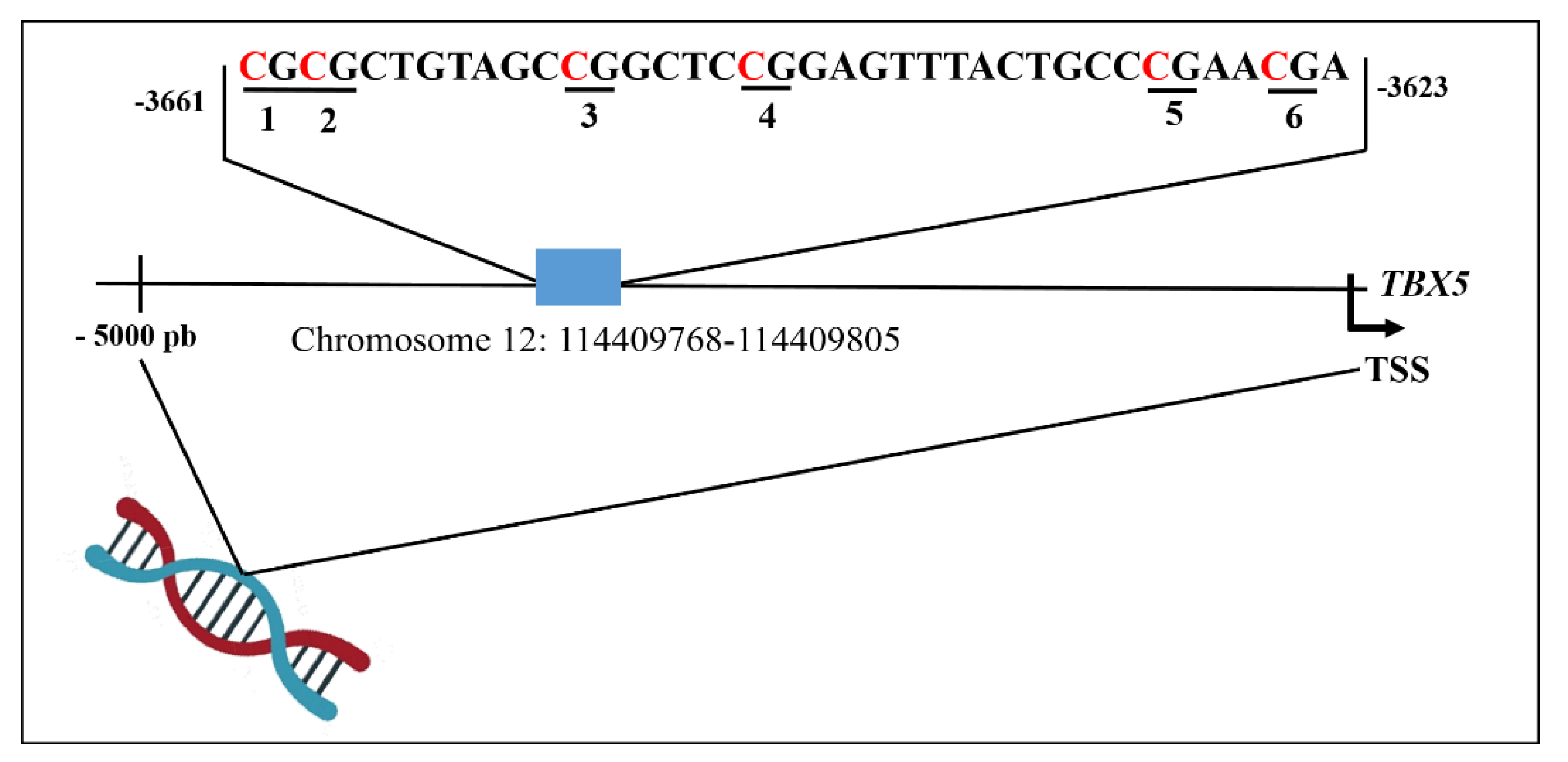
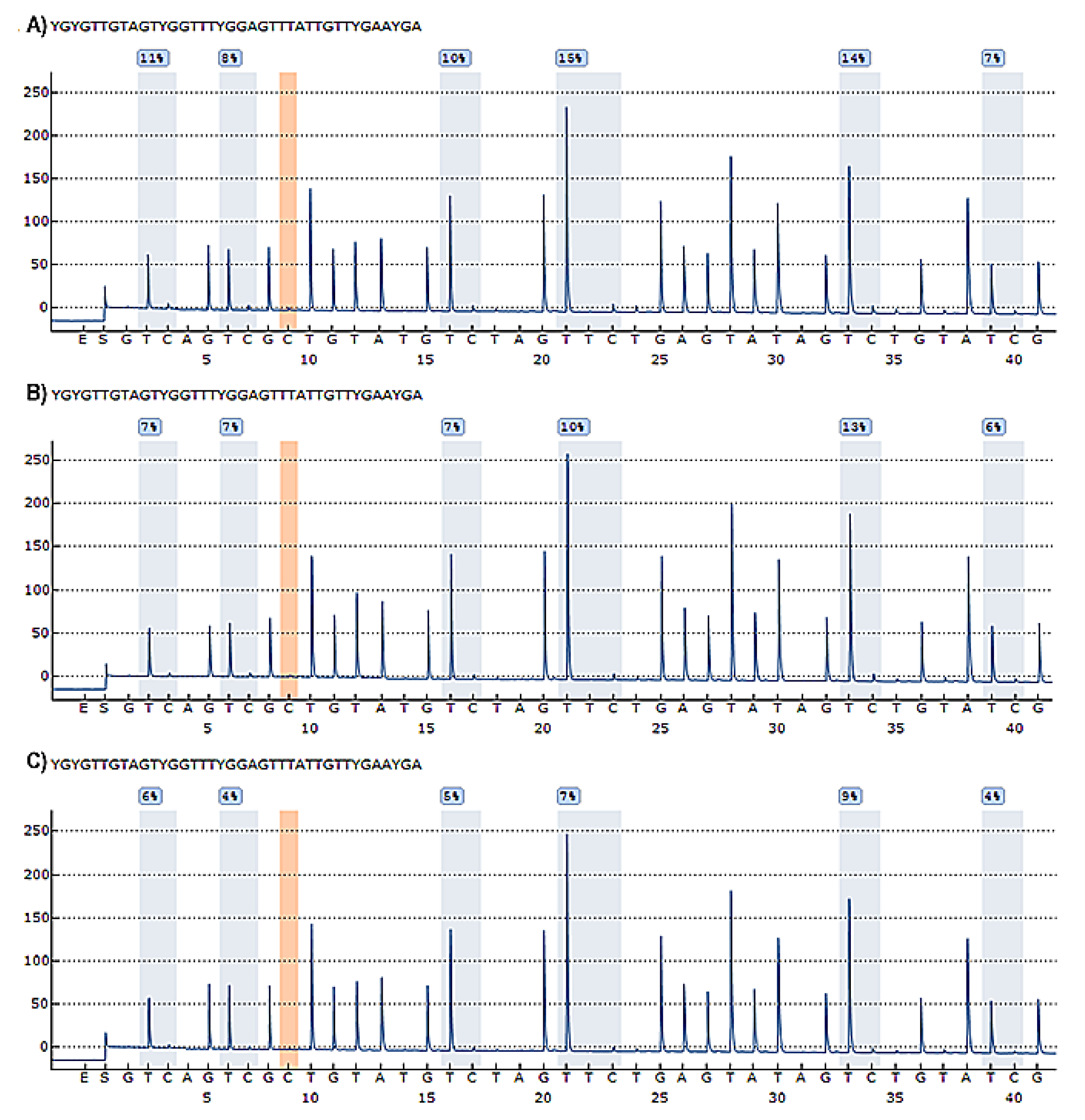
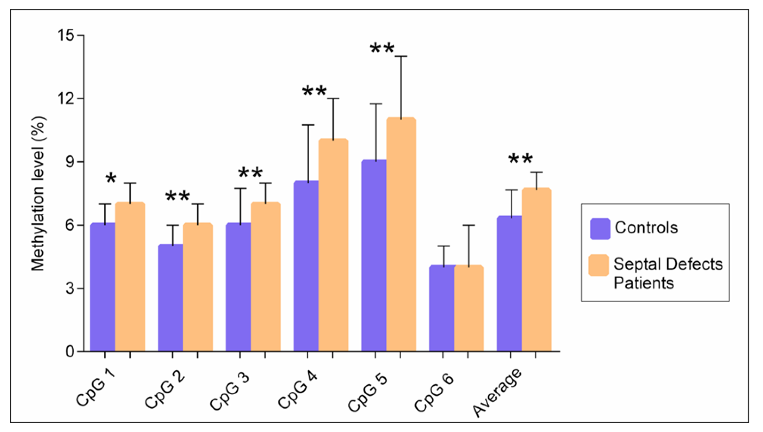
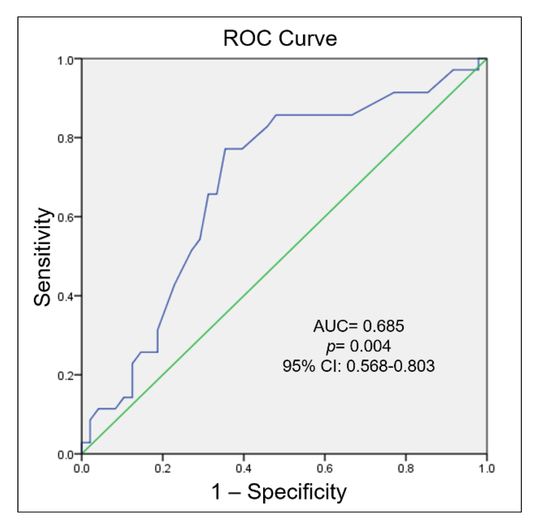
3.1. DNA Methylation of the TBX5 Gene Promoter and Its Association with Septal Defects

Six CpG sites in the promoter region of the TBX5 gene were analysed. We included an example of a pyrogram result, which contains the methylation quantification of each CpG site expressed as a percentage, in Figure 2.
We found significant differences in the levels of methylation represented as median [interquartile range] between the patient and control groups. The average methylation level of all CpG sites was significantly higher in patients with congenital septal defects 7.6% [IQR 7–8.5%] than in the controls 6.3% [IQR 6–7.6%], p < 0.004. The methylation level of five CpG sites was significantly higher in patients with congenital septal defects than in the controls, with a median value at site 1: 7% [IQR 6–8%] vs. 6% [IQR 6–7%] p = 0.032; site 2: 6% [IQR 5–7%] vs. 5% [4–6%] p = 0.001; site 3: 7% [IQR 7–8%] vs. 6% [IQR 5–7.7%] p = 0.004; site 4: 10% [IQR 9–12%] vs. 8% [IQR 7–10.7%] p = 0.004; site 5: 11% [IQR 10–14%] p = 0.001. In site 6, no differences were observed (Figure 3).
Additionally, we analysed whether the methylation levels of all CpG sites confers a risk in both study groups, by stratifying the methylation levels into quartiles. We observed a significant increased risk of developing septal defects in the highest quartile [OR (95% CI) = 3.91 (1.02–14.8); p = 0.045) (Table 2).
Then, we performed a ROC curve analysis to the discriminatory value of the methylation levels of the TBX5 gene in patients with septal defects when comparing to the control group; the result showed an AUC of 0.685 (95% CI = 0.568–0.803; p = 0.004) (Figure 4).
We also evaluated the association between environmental factors of mothers of patients with CHD during pregnancy and methylation levels of the TBX5 gene. We observed that maternal infections during pregnancy were associated with an increased risk of development septal defects [OR (95% CI) = 2.90 (1.01–8.33); p = 0.048] (Table 3).
Correlations between methylation levels and age or sex were then analysed. No correlation with age was found, either in controls (r = 0.14, p = 0.33), or in patients with congenital septal defect (r = 0.16, p = 0.35). Likewise, no correlation between DNA methylation levels and sex were observed: control group females (r = 0.28, p = 0.10), males (r = 0.28, p = 0.39); patient group females (r = 0.17, p = 0.45), males (r = 0.38, p = 0.11). (Supplementary Figure S1).

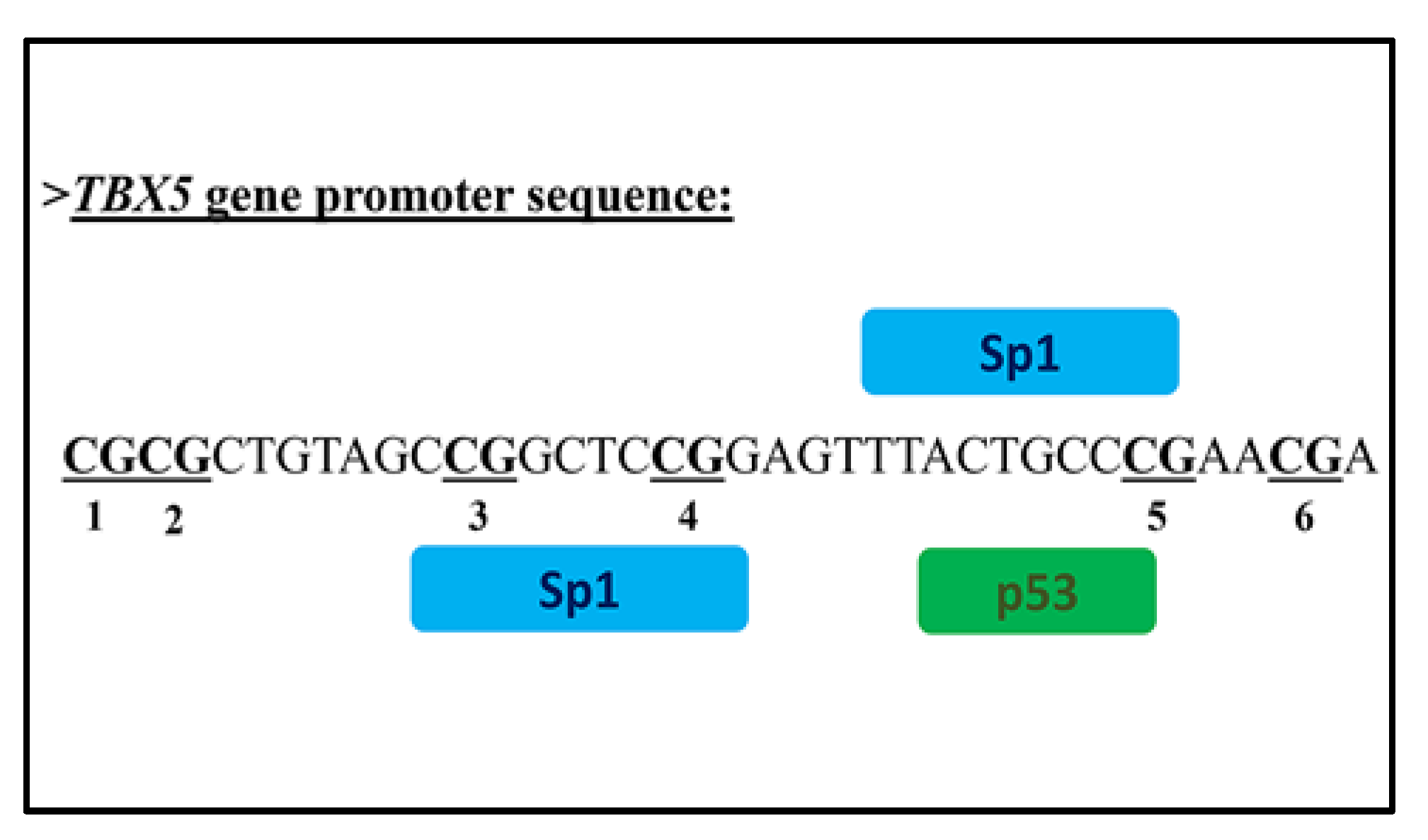
3.2. In Silico Analysis of Transcription Factor Binding Sites to TBX5 Promoter Region

The in silico analysis with PROMO, TFBIND and AliBaba 2.1 (http://gene-regulation.com/pub/programs/alibaba2/index.html accessed on 26 September 2021) software for the possible prediction of potential transcription factors binding sites to the TBX5 promoter region indicated the binding of Sp1 (Specificity protein 1) and p53 (Tumor suppressor Trp53) transcription factors. See Figure 5.

4. Discussion

To date, the epigenetic involvement of the TBX5 gene in CHDs is poorly understood. Thus, in this research, a quantitative DNA methylation analysis of the TBX5 gene promoter in non-syndromic patients with congenital septal defects was performed. Additionally, the mothers of individuals with septal defects were analysed with regard to their exposure to environmental risk factors during pregnancy; likewise, we made a bioinformatics analysis in the promoter region of the TBX5 gene.
Our results showed significant differences in methylation levels when comparing patients with congenital septal defects to the controls, which suggests that the TBX5 gene could be a marker for risk association with septal defects. This assertion is based on the results of the ROC analysis, suggesting that methylation levels may be an epigenetic marker to detect individuals with congenital septal defects.
As far as we know, this the first study with an approach that included congenital septal defect patients and the TBX5 gene; however, various investigations have been carried out in patients with TOF, with different genes implicated in cardiogenesis. TOF is a frequent type of CHD with implications in four different malformations such as overriding aorta, pulmonic stenosis, ventricular septal defect (VSD) and right ventricular hypertrophy. Therefore, we consider it pertinent that TOF and the pathology included in our study share relationships such as septal defects, although some authors include other relevant genes during the cardiogenesis [20,27,28,29,30].
For instance, Sheng et al. analysed the methylation levels of 71 candidate genes for CHDs in 41 patients with TOF and six controls. The authors found significant differences in 26 genes and within these, the TBX5 gene showed increased methylation levels in patients with TOF when compared to the controls [27]. Additionally, Zhang et al. reported methylation levels of the RXRA gene promoter in 26 patients with TOF and in six controls by means of quantitative methods. The authors found that the overall methylation levels in the promoter region, was significantly higher in patients with TOF than in the controls [28]. In another research, Yuan et al. studied the methylation status of the VANGL2 gene in 15 patients with TOF and in 5 controls. Their results showed that overall methylation levels of the VANGL2 gene were significantly higher in patients with TOF than in the controls [20]. These results are in agreement with our findings, as we found that patients also presented higher methylation levels than the controls.
Evidence has shown that the foetus development could be influenced alterations in maternal DNA methylation; in this sense, the uterine microenvironment has the capacity to induce important changes in the global DNA methylation and can result in direct changes in gene expression in the developing foetus [31,32,33,34,35]. Therefore, we assessed environmental and lifestyle exposures factors during pregnancy in mothers of individuals with CHDs to understand this relationship. We observed differences in DNA methylation levels in individuals with CHDs when the mothers were exposed to harmful environmental factors during pregnancy. This evidence suggests at least in part, the participation of these epigenetic events in CHDs; nevertheless, the molecular mechanisms of these phenomena are still not fully understood.
The results of in silico analysis showed that modifications in the promoter region of the TBX5 gene produced binding sites for the transcription factors Sp1 (Specificity protein 1) and p53 (Tumor suppressor Trp53). Factor p53 is implicated in the transrepression and transactivation of multiple genes. The increased level of p53 expression contributes mainly to cell cycle arrest, apoptosis, metabolism disorder and autophagy; it also plays a role in some cardiovascular diseases and CHDs, as it is required for embryonic heart development. During the embryonic and post-natal development, an inappropriate p53 activation can disrupt the development of the heart by initiating apoptosis and limiting cell proliferation [36,37]. Moreover, under physiological conditions, p53 participates in providing support to the cardiac structure, as well as in functions related to mechanisms of DNA repair, cell division, and cellular senescence [38,39].
Sp1 is a transcription factor implicated in several biological processes including cell growth, cell differentiation and chromatin remodelling [40]. This factor can increase or decrease the transcriptional activity, because of its affinity to the promoter region of target genes [41]. Factor Sp1 has also been studied in congenital heart defects; Li et al. for instance, studied the NKX2.5 gene in murine models and, using a transcriptional regulatory networks analysis, found a dysregulation of Sp1 and other transcriptional factors, providing evidence of the Sp1 participation in the pathogenesis of CHDs [42]. Moreover, a relationship has been reported between Sp1 and other genes of the T-box family including the TBX20 gene. Gong et al. for instance, evaluated DNA methylation levels in the promoter region of this gene in patients with TOF. The study provided evidence of the bond with Sp1 at the sequence TBX20_M1, which describes the influence of Sp1 in the expression of TBX20. This evidence suggests the participation of Sp1 in congenital septal defects [40].
It is important to highlight the main strengths of our study: (a) we included non-syndromic individuals, (b) the experimental strategy used was pyrosequencing, which is a quantitative method, (c) we analysed the exposure of environmental factors in mothers of patients with CHDs in order to determinate the possible association of the environment with the methylation levels of TBX5 gene (this aspect has not been evaluated in previous studies of patients with congenital septal defects), and (d) we performed an in silico analysis that suggests binding of transcriptional factors to the CpG sequence studied. Interestingly, these transcriptional factors are related to cardiac pathologies. Nevertheless, limitations of present study should also be recognized: (a) we were not able to assess the functional effect of promoter methylation of the TBX5 gene due to the impossibility of obtaining heart biopsies without clinical compromise, (b) we analysed six CpG sites in the promoter region, additional CpG sites are needed to establish the true role of methylation of the TBX5 gene, (c) it would be interesting to address other regulatory regions and other epigenetic mechanisms of the TBX5 gene, (d) it is advisable to expand the sample size in order to validate the present findings.

5. Conclusions

In conclusion, our data suggest that a high DNA methylation of the TBX5 gene could be associated with congenital septal defects; additionally, environmental factors during pregnancy also confer a risk of developing congenital septal defects. These findings represent a first step to better understanding the epigenetic factors implicated in CHDs. Nevertheless, the exact molecular and epigenetic mechanisms remain unknown. Additional research in this field is necessary to provide new markers for clinical diagnosis in early onset of these pathologies.

Supplementary Materials

The following supporting information can be downloaded at: https://www.mdpi.com/article/10.3390/biology11010096/s1, Figure S1: Correlation of methylation levels (%) in the TBX5 gene promoter and age expressed in years in the population study; Table S1: Exposure to environmental factors during pregnancy in mothers of patients with congenital septal defects.

Author Contributions

Conceptualization, N.P.-H., E.G.-F., J.M.R.-P. and V.M.B.-C.; methodology, E.G.-F., B.G.C.-S. and A.G.-D.; formal analysis, E.G.-F., B.G.C.-S. and A.M.-D.; resources, N.P.-H.; visualization and supervision, N.P.-H., J.C.-C., J.P.S., J.A.G.-M., V.M.E.-G. and J.G.R.-G.; project administration, N.P.-H.; funding acquisition, N.P.-H.; data curation, E.G.-F., B.G.C.-S., A.M.-D. and G.V.-A.; writing—original draft preparation, N.P.-H., J.M.R.-P., V.M.B.-C., G.V.-A. and A.M.-D.; writing—review and editing, N.P.-H., J.M.R.-P., V.M.B.-C., A.M.-D. and G.V.-A. All authors have read and agreed to the published version of the manuscript.

Funding

This research was funded by a CONACyT grant [Project number: 181931]. In addition, the “Instituto Nacional de Cardiogía Ignacio Chávez” covered the costs of publication in open access.

Institutional Review Board Statement

The study was conducted according to the guidelines of the Declaration of Helsinki, and approved by institutional ethical and research committees of the “Instituto Nacional de Cardiología Ignacio Chávez” (Register No. 19-1138).

Informed Consent Statement

A written informed consent and an informed assent were obtained from each participant and their parents or relatives after receiving a complete information concerning to the study.

Data Availability Statement

All data are given in the main manuscript, or in the Supplementary Material.

Acknowledgments

The researchers are grateful to every participant, as well as to the staff of the Paediatric Cardiology, Hemodynamic Laboratory and Intervention in Congenital Heart Defects and Molecular Biology Departments, for the support received in the present research. The authors are grateful to Marva Arellano-González and Silvestre Ramírez-Fuentes for their technical assistance. Also, the researchers are grateful to Beatriz Eugenia Sánchez-Hernández, for the support provided in the validation of the pyrograms. Part of the data was obtained by Esbeidy García-Flores in order to obtain a PhD in Research in Medicine by the Superior Medicine School of the “Instituto Politécnico Nacional” (IPN) and CONACyT that provided a scholarship (CVU 756752).

Conflicts of Interest

The authors declare no conflict of interest.

References

  1. Yoshida, A.; Morisaki, H.; Nakaji, M.; Kitano, M.; Kim, K.S.; Sagawa, K.; Ishikawa, S.; Satokata, I.; Mitani, Y.; Kato, H.; et al. Genetic mutation analysis in Japanese patients with non-syndromic congenital heart disease. J. Hum. Genet. 2016, 61, 157–162. [Google Scholar] [CrossRef] [PubMed]
  2. Zhang, H.; Zhao, X.; Wang, C.; Du, R.; Wang, X.; Fu, J.; Sun, Q. A Preliminary Study of the Association between Apolipoprotein E Promoter Methylation and Atherosclerotic Cerebral Infarction. J. Stroke Cerebrovasc. Dis. Off. J. Natl. Stroke Assoc. 2019, 28, 1056–1061. [Google Scholar] [CrossRef]
  3. Thomford, N.E.; Dzobo, K.; Yao, N.A.; Chimusa, E.; Evans, J.; Okai, E.; Kruszka, P.; Muenke, M.; Awandare, G.; Wonkam, A.; et al. Genomics and Epigenomics of Congenital Heart Defects: Expert Review and Lessons Learned in Africa. Omics A J. Integr. Biol. 2018, 22, 301–321. [Google Scholar] [CrossRef]
  4. Xie, H.; Zhang, E.; Hong, N.; Fu, Q.; Li, F.; Chen, S.; Yu, Y.; Sun, K. Identification of TBX2 and TBX3 variants in patients with conotruncal heart defects by target sequencing. Hum. Genom. 2018, 12, 44. [Google Scholar] [CrossRef]
  5. Greulich, F.; Rudat, C.; Kispert, A. Mechanisms of T-box gene function in the developing heart. Cardiovasc. Res. 2011, 91, 212–222. [Google Scholar] [CrossRef] [Green Version]
  6. Papaioannou, V.E. The T-box gene family: Emerging roles in development, stem cells and cancer. Development 2014, 141, 3819–3833. [Google Scholar] [CrossRef] [PubMed] [Green Version]
  7. Showell, C.; Binder, O.; Conlon, F.L. T-box genes in early embryogenesis. Dev. Dyn. Off. Publ. Am. Assoc. Anat. 2004, 229, 201–218. [Google Scholar] [CrossRef] [PubMed] [Green Version]
  8. Yang, X.; Kong, Q.; Li, Z.; Xu, M.; Cai, Z.; Zhao, C. Association between the promoter methylation of the TBX20 gene and tetralogy of fallot. Scand. Cardiovasc. J. SCJ 2018, 52, 287–291. [Google Scholar] [CrossRef]
  9. Yang, X.F.; Zhang, Y.F.; Zhao, C.F.; Liu, M.M.; Si, J.P.; Fang, Y.F.; Xing, W.W.; Wang, F.L. Relationship between TBX20 gene polymorphism and congenital heart disease. Genet. Mol. Res. GMR 2016, 1–9. [Google Scholar] [CrossRef]
  10. Boogerd, C.J.; Zhu, X.; Aneas, I.; Sakabe, N.; Zhang, L.; Sobreira, D.R.; Montefiori, L.; Bogomolovas, J.; Joslin, A.C.; Zhou, B.; et al. Tbx20 Is Required in Mid-Gestation Cardiomyocytes and Plays a Central Role in Atrial Development. Circ. Res. 2018, 123, 428–442. [Google Scholar] [CrossRef]
  11. Steimle, J.D.; Moskowitz, I.P. TBX5: A Key Regulator of Heart Development. Curr. Top. Dev. Biol. 2017, 122, 195–221. [Google Scholar]
  12. Zhang, K.K.; Xiang, M.; Zhou, L.; Liu, J.; Curry, N.; Heine Suñer, D.; Garcia-Pavia, P.; Zhang, X.; Wang, Q.; Xie, L. Gene network and familial analyses uncover a gene network involving Tbx5/Osr1/Pcsk6 interaction in the second heart field for atrial septation. Hum. Mol. Genet. 2016, 25, 1140–1151. [Google Scholar] [CrossRef] [PubMed] [Green Version]
  13. Waldron, L.; Steimle, J.D.; Greco, T.M.; Gomez, N.C.; Dorr, K.M.; Kweon, J.; Temple, B.; Yang, X.H.; Wilczewski, C.M.; Davis, I.J.; et al. The Cardiac TBX5 Interactome Reveals a Chromatin Remodeling Network Essential for Cardiac Septation. Dev. Cell 2016, 36, 262–275. [Google Scholar] [CrossRef] [Green Version]
  14. Ma, J.; Chen, S.; Hao, L.; Sheng, W.; Chen, W.; Ma, X.; Zhang, B.; Ma, D.; Huang, G. Hypermethylation-mediated down-regulation of lncRNA TBX5-AS1:2 in Tetralogy of Fallot inhibits cell proliferation by reducing TBX5 expression. J. Cell. Mol. Med. 2020, 24, 6472–6484. [Google Scholar] [CrossRef]
  15. Moore-Morris, T.; van Vliet, P.P.; Andelfinger, G.; Puceat, M. Role of Epigenetics in Cardiac Development and Congenital Diseases. Physiol. Rev. 2018, 98, 2453–2475. [Google Scholar] [CrossRef] [Green Version]
  16. Jarrell, D.K.; Lennon, M.L.; Jacot, J.G. Epigenetics and Mechanobiology in Heart Development and Congenital Heart Disease. Diseases 2019, 7, 52. [Google Scholar] [CrossRef] [PubMed] [Green Version]
  17. Lewandowski, S.L.; Janardhan, H.P.; Smee, K.M.; Bachman, M.; Sun, Z.; Lazar, M.A.; Trivedi, C.M. Histone deacetylase 3 modulates Tbx5 activity to regulate early cardiogenesis. Hum. Mol. Genet. 2014, 23, 3801–3809. [Google Scholar] [CrossRef] [Green Version]
  18. Grunert, M.; Dorn, C.; Cui, H.; Dunkel, I.; Schulz, K.; Schoenhals, S.; Sun, W.; Berger, F.; Chen, W.; Sperling, S.R. Comparative DNA methylation and gene expression analysis identifies novel genes for structural congenital heart diseases. Cardiovasc. Res. 2016, 112, 464–477. [Google Scholar] [CrossRef] [PubMed]
  19. Mittal, A.; Sharma, R.; Prasad, R.; Bahl, A.; Khullar, M. Role of cardiac TBX20 in dilated cardiomyopathy. Mol. Cell. Biochem. 2016, 414, 129–136. [Google Scholar] [CrossRef] [PubMed]
  20. Yuan, Y.; Gao, Y.; Wang, H.; Ma, X.; Ma, D.; Huang, G. Promoter methylation and expression of the VANGL2 gene in the myocardium of pediatric patients with tetralogy of fallot. Birth Defects Res. Part A Clin. Mol. Teratol. 2014, 100, 973–984. [Google Scholar] [CrossRef]
  21. Dakkak, W.; Oliver, T.I. Ventricular Septal Defect. In StatPearls; Copyright © 2022; StatPearls Publishing LLC: Treasure Island, FL, USA, 2021. [Google Scholar]
  22. Menillo, A.M.; Lee, L.S.; Pearson-Shaver, A.L. Atrial Septal Defect. In StatPearls; Copyright © 2022; StatPearls Publishing LLC: Treasure Island, FL, USA, 2021. [Google Scholar]
  23. Backer, C.L.; Eltayeb, O.; Mongé, M.C.; Mazwi, M.L.; Costello, J.M. Shunt Lesions Part I: Patent Ductus Arteriosus, Atrial Septal Defect, Ventricular Septal Defect, and Atrioventricular Septal Defect. Pediatric Crit. Care Med. A J. Soc. Crit. Care Med. World Fed. Pediatric Intensive Crit. Care Soc. 2016, 17, S302–S309. [Google Scholar] [CrossRef]
  24. Farré, D.; Roset, R.; Huerta, M.; Adsuara, J.E.; Roselló, L.; Albà, M.M.; Messeguer, X. Identification of patterns in biological sequences at the ALGGEN server: PROMO and MALGEN. Nucleic Acids Res. 2003, 31, 3651–3653. [Google Scholar] [CrossRef] [Green Version]
  25. Messeguer, X.; Escudero, R.; Farré, D.; Núñez, O.; Martínez, J.; Albà, M.M. PROMO: Detection of known transcription regulatory elements using species-tailored searches. Bioinformatics 2002, 18, 333–334. [Google Scholar] [CrossRef]
  26. Tsunoda, T.; Takagi, T. Estimating transcription factor bindability on DNA. Bioinformatics 1999, 15, 622–630. [Google Scholar] [CrossRef] [PubMed]
  27. Sheng, W.; Qian, Y.; Zhang, P.; Wu, Y.; Wang, H.; Ma, X.; Chen, L.; Ma, D.; Huang, G. Association of promoter methylation statuses of congenital heart defect candidate genes with Tetralogy of Fallot. J. Transl. Med. 2014, 12, 31. [Google Scholar] [CrossRef] [PubMed] [Green Version]
  28. Zhang, J.; Ma, X.; Wang, H.; Ma, D.; Huang, G. Elevated methylation of the RXRA promoter region may be responsible for its downregulated expression in the myocardium of patients with TOF. Pediatric Res. 2014, 75, 588–594. [Google Scholar] [CrossRef] [Green Version]
  29. Song, X.; Cao, H.; Hong, L.; Zhang, L.; Li, M.; Shi, J.; Liu, J.; Ma, J.; Cui, L.; Zhang, Y.; et al. Ventricular Myocardial Deformation in Fetuses With Tetralogy of Fallot: A Necessary Field of Investigation. Front. Cardiovasc. Med. 2021, 8, 764676. [Google Scholar] [CrossRef] [PubMed]
  30. Diaz-Frias, J.; Guillaume, M. Tetralogy of Fallot. In StatPearls; Copyright © 2022; StatPearls Publishing LLC: Treasure Island, FL, USA, 2021. [Google Scholar]
  31. Green, B.B.; Marsit, C.J. Select Prenatal Environmental Exposures and Subsequent Alterations of Gene-Specific and Repetitive Element DNA Methylation in Fetal Tissues. Curr. Environ. Health Rep. 2015, 2, 126–136. [Google Scholar] [CrossRef] [PubMed] [Green Version]
  32. Nielsen, C.H.; Larsen, A.; Nielsen, A.L. DNA methylation alterations in response to prenatal exposure of maternal cigarette smoking: A persistent epigenetic impact on health from maternal lifestyle? Arch. Toxicol. 2016, 90, 231–245. [Google Scholar] [CrossRef] [PubMed]
  33. Alvarado-Cruz, I.; Alegría-Torres, J.A.; Montes-Castro, N.; Jiménez-Garza, O.; Quintanilla-Vega, B. Environmental Epigenetic Changes, as Risk Factors for the Development of Diseases in Children: A Systematic Review. Ann. Glob. Health 2018, 84, 212–224. [Google Scholar] [CrossRef] [Green Version]
  34. Rauschert, S.; Melton, P.E.; Burdge, G.; Craig, J.M.; Godfrey, K.M.; Holbrook, J.D.; Lillycrop, K.; Mori, T.A.; Beilin, L.J.; Oddy, W.H.; et al. Maternal Smoking During Pregnancy Induces Persistent Epigenetic Changes Into Adolescence, Independent of Postnatal Smoke Exposure and Is Associated With Cardiometabolic Risk. Front. Genet. 2019, 10, 770. [Google Scholar] [CrossRef] [PubMed]
  35. Cao, J.; Wu, Q.; Huang, Y.; Wang, L.; Su, Z.; Ye, H. The role of DNA methylation in syndromic and non-syndromic congenital heart disease. Clin. Epigenetics 2021, 13, 93. [Google Scholar] [CrossRef] [PubMed]
  36. Men, H.; Cai, H.; Cheng, Q.; Zhou, W.; Wang, X.; Huang, S.; Zheng, Y.; Cai, L. The regulatory roles of p53 in cardiovascular health and disease. Cell. Mol. Life Sci. CMLS 2021, 78, 2001–2018. [Google Scholar] [CrossRef]
  37. Zhang, M.; Zhang, Y.; Xu, E.; Mohibi, S.; de Anda, D.M.; Jiang, Y.; Zhang, J.; Chen, X. Rbm24, a target of p53, is necessary for proper expression of p53 and heart development. Cell Death Differ. 2018, 25, 1118–1130. [Google Scholar] [CrossRef] [Green Version]
  38. Lahalle, A.; Lacroix, M.; De Blasio, C.; Cissé, M.Y.; Linares, L.K.; Le Cam, L. The p53 Pathway and Metabolism: The Tree That Hides the Forest. Cancers 2021, 13, 133. [Google Scholar] [CrossRef]
  39. Van Nostrand, J.L.; Brady, C.A.; Jung, H.; Fuentes, D.R.; Kozak, M.M.; Johnson, T.M.; Lin, C.Y.; Lin, C.J.; Swiderski, D.L.; Vogel, H.; et al. Inappropriate p53 activation during development induces features of CHARGE syndrome. Nature 2014, 514, 228–232. [Google Scholar] [CrossRef] [PubMed] [Green Version]
  40. Gong, J.; Sheng, W.; Ma, D.; Huang, G.; Liu, F. DNA methylation status of TBX20 in patients with tetralogy of Fallot. BMC Med. Genom. 2019, 12, 75. [Google Scholar] [CrossRef] [Green Version]
  41. Beishline, K.; Azizkhan-Clifford, J. Sp1 and the ‘hallmarks of cancer’. FEBS J. 2015, 282, 224–258. [Google Scholar] [CrossRef]
  42. Li, J.; Cao, Y.; Wu, Y.; Chen, W.; Yuan, Y.; Ma, X.; Huang, G. The expression profile analysis of NKX2-5 knock-out embryonic mice to explore the pathogenesis of congenital heart disease. J. Cardiol. 2015, 66, 527–531. [Google Scholar] [CrossRef] [Green Version]
Figure 1. Schematic representation of distribution the CpG sites analysed in the TBX5 gene promoter. Numbers 1-6 refer to CpG site within the sequence of TBX5. TSS, transcription start site.
Biology 11 00096 g001
Figure 2. Pyrograms resulting from pyrosequencing process. (A) Pyrogram of patient with ASD; (B) Pyrogram of a patient with VSD; (C) Pyrogram of a control individual. In each pyrogram, the evaluated CpG sites are identified with the letter “Y”. The X-axis describes the order of dispensing during the pyrosequencing process, starting with the enzyme and the substrate, followed by the corresponding nucleotides; the Y-axis represents the relative light intensity corresponding to bioluminescence. The height of each peak in the CpG sites is proportional to the number of nucleotides incorporated in the analysed sequence. The blue shaded areas represent the CpG sites with their corresponding percentages of methylation; the presence of an orange shaded area indicates a control bisulfite dispensing, representing a suitable bisulfite conversion process.
Biology 11 00096 g002
Figure 3. Median methylation levels for 6 CpG sites in the TBX5 gene between patients with septal defects and the controls. * p < 0.05, ** p < 0.001; Mann–Whitney U test.
Biology 11 00096 g003
Figure 4. Receptor-operator characteristic (ROC) curve analysis associated to the presence of septal defects by TBX5 gene methylation level.
Biology 11 00096 g004
Figure 5. The potential binding sites of transcription factors identified with PROMO, TFBIND and AliBaba 2.1 software.
Biology 11 00096 g005
Table 1. Demographic characteristics of the study population.
Congenital Septal Defects Patients (n = 35)Controls
(n = 48)
p *
Age (in years) a8 (4–13)3 (2–6)0.005
Sex (%) b 0.040
Females 17 (48.6)34 (70.8)
Males 18 (51.4)14 (29.2)
a median [IQR], b n (%) for categorical variables. * Mann–Whitney U test for continues variables and χ2 test for categorical variables.
Table 2. Association between TBX5 methylation levels and risk of congenital heart defects.
TBX5 GeneCongenital Septal Defect Patients
(n = 35)
Controls
(n = 48)
OR (95% CI)p *
Methylation Levelsn (%)n (%)
Highest quartile
(>75%)
11 (31.4)9 (18.8)3.91 (1.02–14.8)0.045
Medium quartile
(25–75%)
19 (54.3)23 (47.9)2.64 (0.8–8.5)0.105
Lowest quartile
(<25%)
5 (14.3)16 (33.3)1.0 (Reference)
OR, odds ratio, CI, confidence intervals. * Logistic regression.
Table 3. Environmental factors analysed to determine the risk in the study population.
Environmental Risk FactorsOR (95% CI)p *
Drug addiction0.75 (0.26–2.15)0.593
Diseases during pregnancy1.77 (0.68–4.57)0.236
Exposure to pollutants1.14 (0.47–2.77)0.765
Maternal infections2.90 (1.01–8.33)0.048
Medication consumption2.03 (0.84–4.93)0.115
Consumption of vitamins0.22 (0.02–2.28)0.208
OR, odds ratio, CI, confidence intervals. * Univariate logistic regression.
Publisher’s Note: MDPI stays neutral with regard to jurisdictional claims in published maps and institutional affiliations.

