

Correction of Arterial-Phase Motion Artifacts in Gadoxetic Acid-Enhanced Liver MRI Using an Innovative Unsupervised Network
 , , ,
, , ,
Abstract
:
1. Introduction
2. Materials and Methods
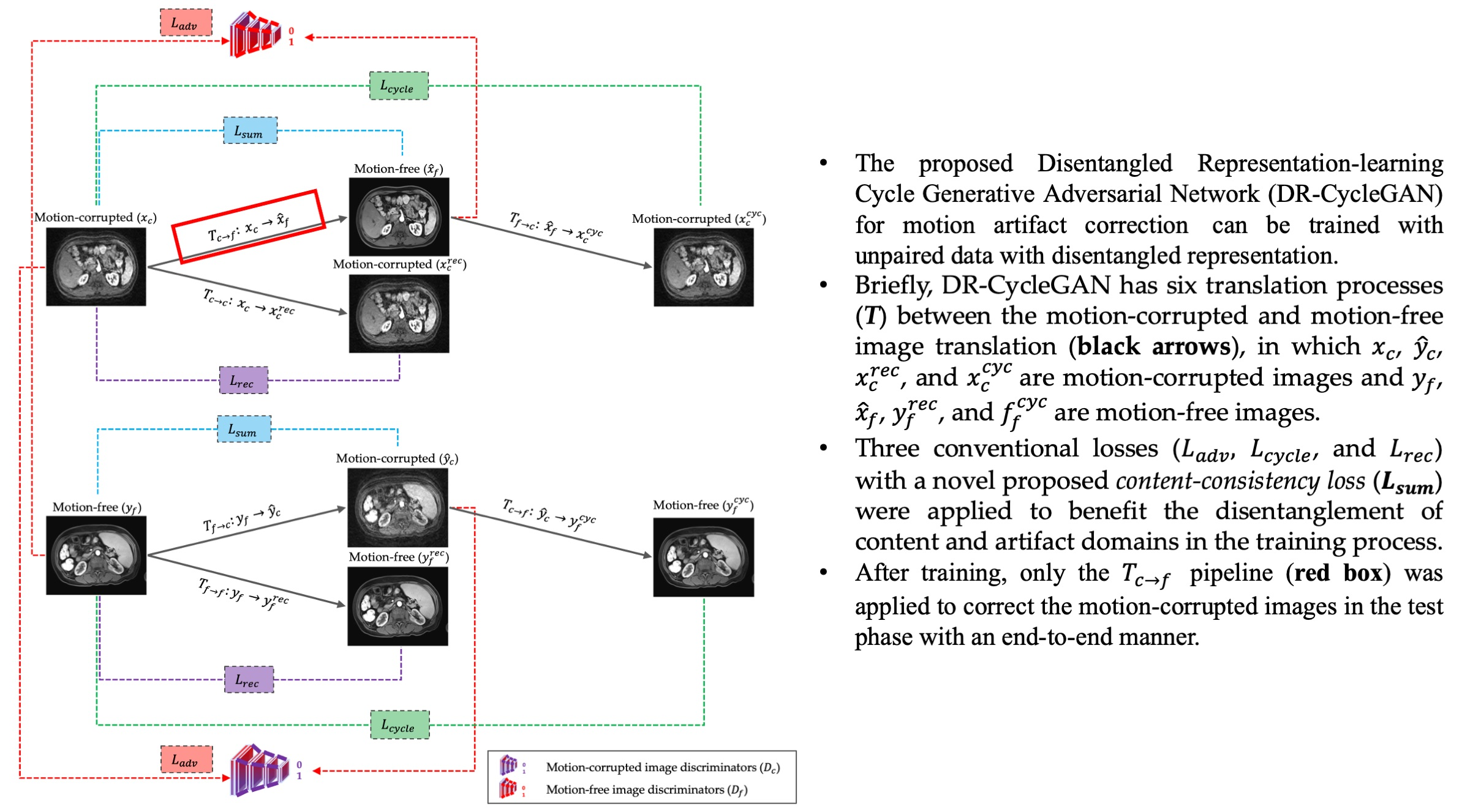
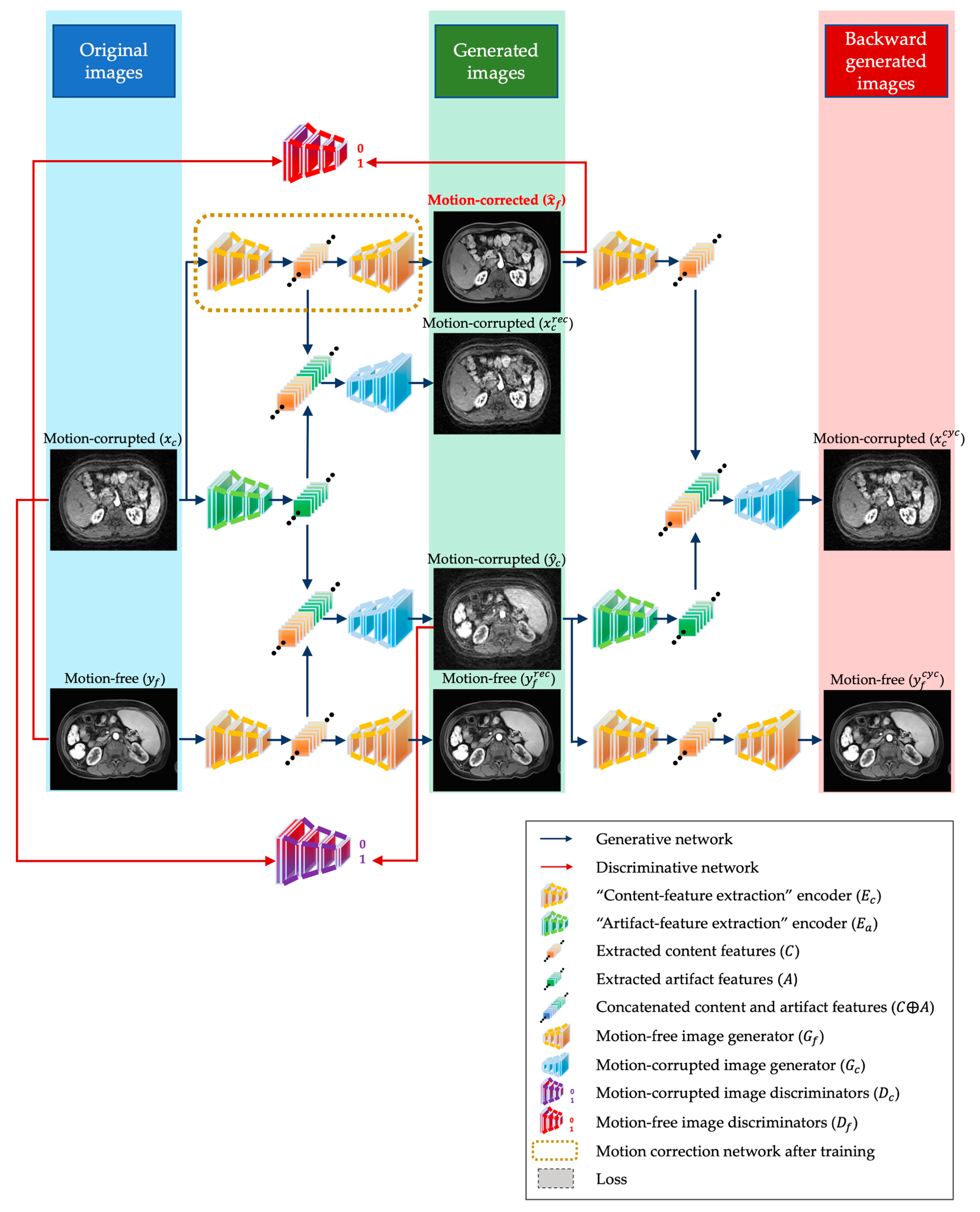
2.1. The Proposed DR-CycleGAN Structure
- Two Encoders: A conventional “content-feature extraction” encoder () and an extra “artifact-feature extraction” encoder (). They can enhance the disentanglement of content features () and artifact features () in motion-corrupted images (). By employing both encoders ( and ), the content and artifact features ( and ) in motion-corrupted images () are separated.
- Two Generators: and , which were introduced to specifically generate motion-free and motion-corrupted images, respectively. can generate “motion-free” images (, , and ) reconstructed from content features (), while can generate different “motion-corrupted” images (, , and ) reconstructed from concatenated content and artifact features ().
- Two discriminators: and , which are employed to distinguish between reconstructed motion-free images () and real motion-free images (), as well as between fake motion-corrupted images () and real motion-corrupted images ().

2.2. Loss in the Training
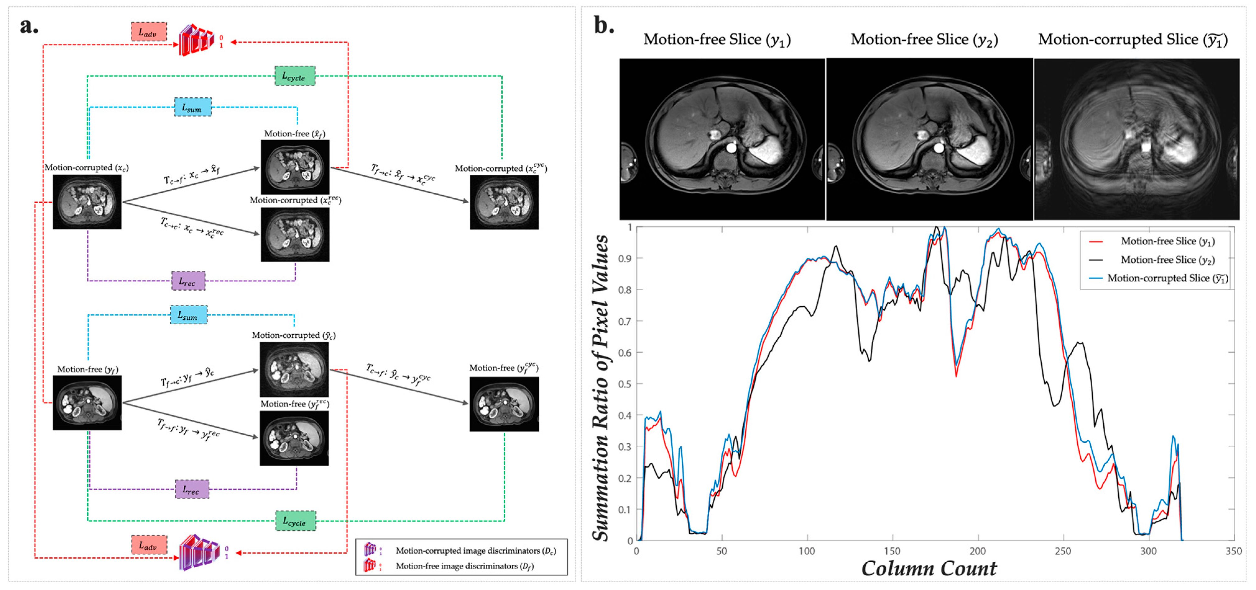
- Adversarial domain loss (): it supervises the resemblance between motion-corrected images and the original motion-free images ( vs. ), as well as the similarity between generated motion-corrupted images and the original motion-corrupted images ( vs. ) (Figure 2a and Supplementary S2).
- Reconstruction loss (): It is designed to minimize the pixel-wise difference between the input image and its reconstructed counterpart in the same domain translation ( vs. and vs. ). Its primary objective is to ensure that eligible encoders and generators do not introduce any significant discrepancies during the reconstruction process (Figure 2a and Supplementary S2).
- Cycle-consistency loss (): it is another classic loss in CycleGAN and guarantees that the images generated backward closely resemble the originals ( vs. and vs. ) (Figure 2a and Supplementary S2).

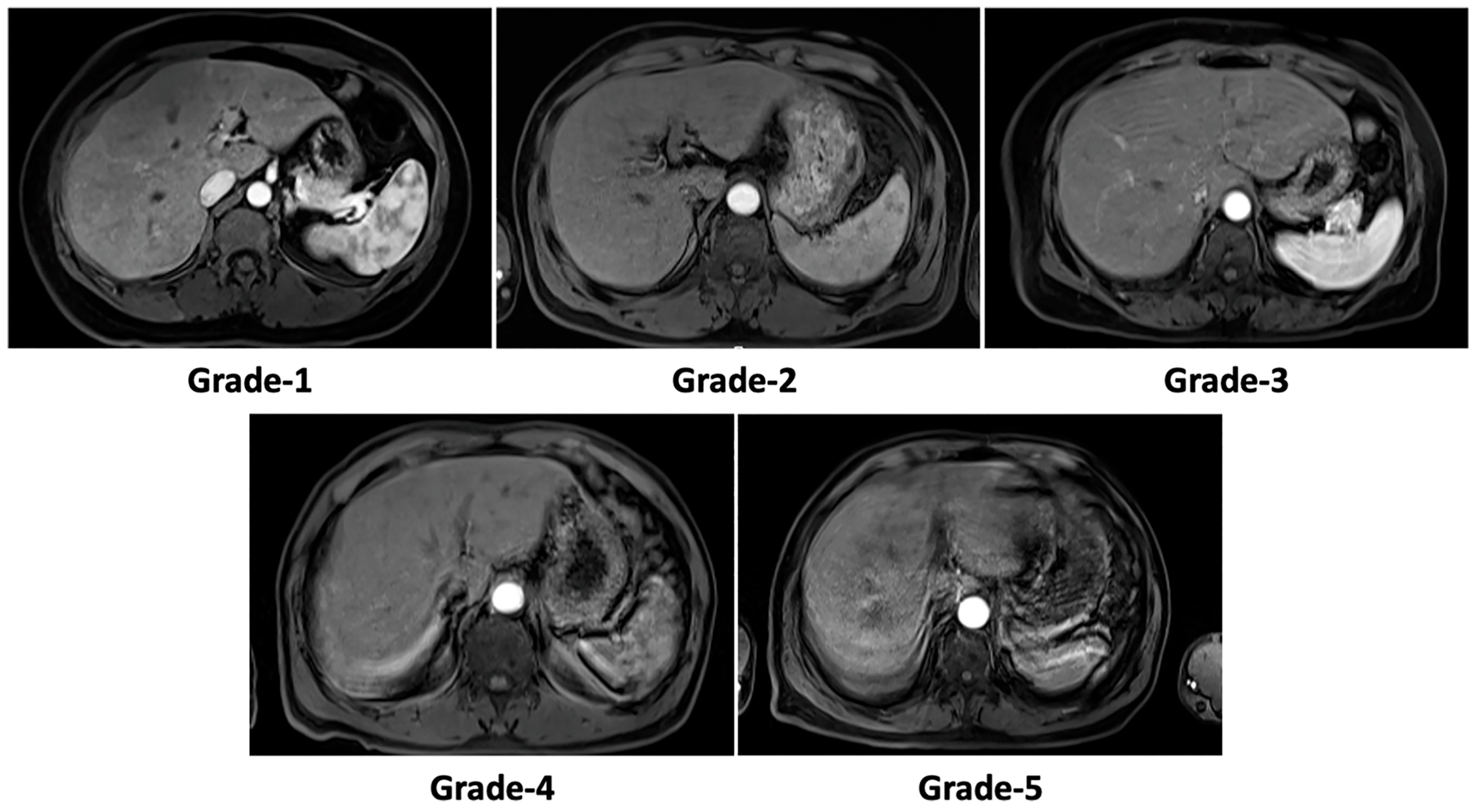
2.3. Motion Artifact Grading
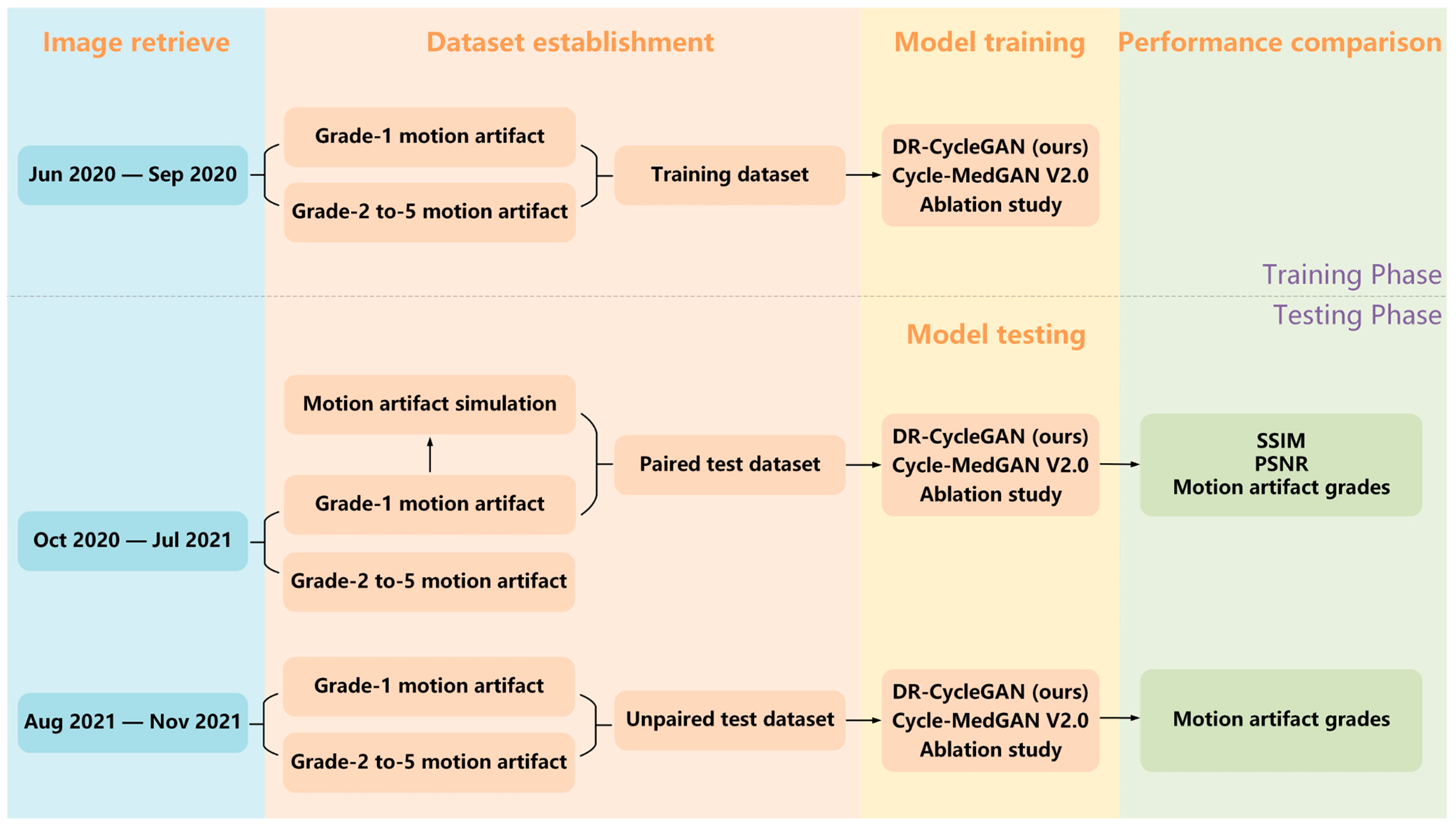
2.4. Training and Test Datasets
2.5. Training, Performance Comparisons, and Ablation Study
2.6. Statistical Analysis
3. Results
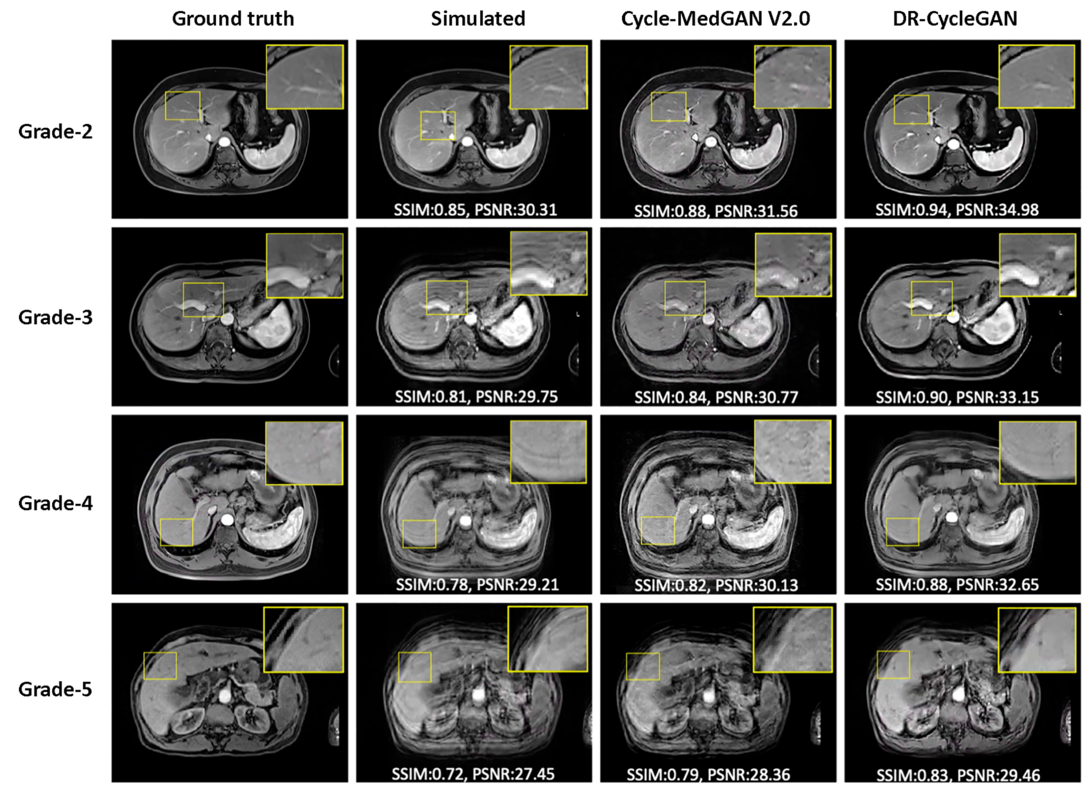
3.1. Evaluation of Paired Simulated Test Dataset
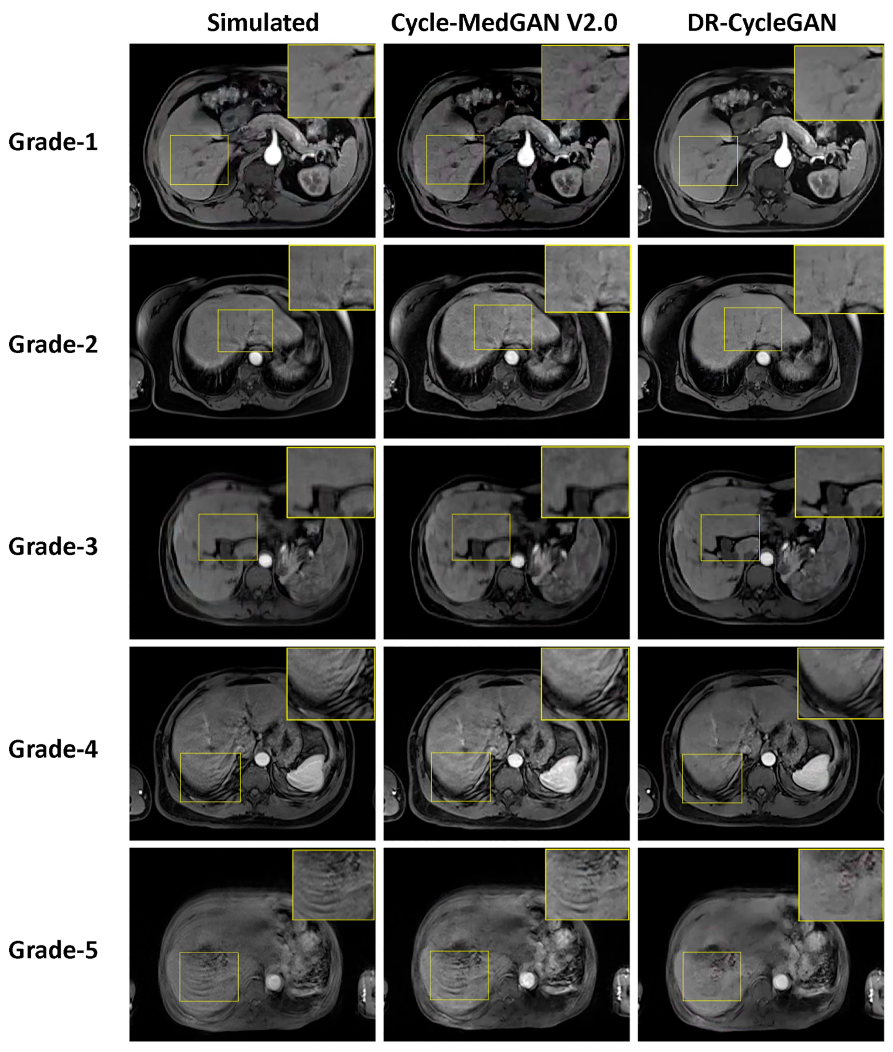
3.2. Evaluation of Unpaired Test Dataset
3.3. Ablation Study
3.4. Inter-Observer Agreement in Semi-Quantitative Motion Artifact Grading
4. Discussion
5. Conclusions
Supplementary Materials
Author Contributions
Funding
Institutional Review Board Statement
Informed Consent Statement
Data Availability Statement
Acknowledgments
Conflicts of Interest
References
- Park, Y.S.; Lee, C.H.; Yoo, J.L.; Kim, I.S.; Kiefer, B.; Woo, S.T.; Kim, K.A.; Park, C.M. Hepatic Arterial Phase in Gadoxetic Acid-Enhanced Liver Magnetic Resonance Imaging: Analysis of Respiratory Patterns and Their Effect on Image Quality. Investig. Radiol. 2016, 51, 127–133. [Google Scholar] [CrossRef]
- Ichikawa, T.; Saito, K.; Yoshioka, N.; Tanimoto, A.; Gokan, T.; Takehara, Y.; Kamura, T.; Gabata, T.; Murakami, T.; Ito, K.; et al. Detection and characterization of focal liver lesions: A Japanese phase III, multicenter comparison between gadoxetic acid disodium-enhanced magnetic resonance imaging and contrast-enhanced computed tomography predominantly in patients with hepatocellular carcinoma and chronic liver disease. Investig. Radiol. 2010, 45, 133–141. [Google Scholar] [CrossRef]
- Seo, H.J.; Kim, M.J.; Lee, J.D.; Chung, W.S.; Kim, Y.E. Gadoxetate disodium-enhanced magnetic resonance imaging versus contrast-enhanced 18F-fluorodeoxyglucose positron emission tomography/computed tomography for the detection of colorectal liver metastases. Investig. Radiol. 2011, 46, 548–555. [Google Scholar] [CrossRef]
- Zhuo, L.Y.; Xing, L.H.; Ma, X.; Zhang, Y.; Ma, Z.P.; Yin, X.P.; Wang, J.N. “Nondefect” of arterial enhancing rim on hepatobiliary phase in 3.0-T gadolinium-ethoxybenzyl-diethylenetriamine pentaacetic acid-enhanced liver magnetic resonance imaging: Distinguishing hepatic abscess from metastasis. J. Comput. Assist. Tomogr. 2013, 37, 849–855. [Google Scholar] [CrossRef]
- Kim, A.; Lee, C.H.; Kim, B.H.; Lee, J.; Choi, J.W.; Park, Y.S.; Kim, K.A.; Park, C.M. Gadoxetic acid-enhanced 3.0T MRI for the evaluation of hepatic metastasis from colorectal cancer: Metastasis is not always seen as a “defect” on the hepatobiliary phase. Eur. J. Radiol. 2012, 81, 3998–4004. [Google Scholar] [CrossRef]
- Park, Y.S.; Lee, C.H.; Kim, B.H.; Lee, J.; Choi, J.W.; Kim, K.A.; Ahn, J.H.; Park, C.M. Using Gd-EOB-DTPA-enhanced 3-T MRI for the differentiation of infiltrative hepatocellular carcinoma and focal confluent fibrosis in liver cirrhosis. Magn. Reason. Imaging 2013, 31, 1137–1142. [Google Scholar] [CrossRef]
- Zhuo, J.; Gullapalli, R.P. AAPM/RSNA physics tutorial for residents: MR artifacts, safety, and quality control. Radiographics 2006, 26, 275–297. [Google Scholar] [CrossRef]
- Ikram, N.S.; Yee, J.; Weinstein, S.; Yeh, B.M.; Corvera, C.U.; Monto, A.; Hope, T.A. Multiple arterial phase MRI of arterial hypervascular hepatic lesions: Improved arterial phase capture and lesion enhancement. Abdom. Radiol. 2017, 42, 870–876. [Google Scholar] [CrossRef]
- Rimola, J.; Sapena, V.; Brancatelli, G.; Darnell, A.; Forzenigo, L.; Mähringer-Kunz, A.; Paisant, A.; Renzulli, M.; Schima, W.; Terraz, S.; et al. Reliability of extracellular contrast versus gadoxetic acid in assessing small liver lesions using liver imaging reporting and data system v.2018 and European association for the study of the liver criteria. Hepatology 2022, 76, 1318–1328. [Google Scholar] [CrossRef]
- Davenport, M.S.; Viglianti, B.L.; Al-Hawary, M.M.; Caoili, E.M.; Kaza, R.K.; Liu, P.S.; Maturen, K.E.; Chenevert, T.L.; Hussain, H.K. Comparison of acute transient dyspnea after intravenous administration of gadoxetate disodium and gadobenate dimeglumine: Effect on arterial phase image quality. Radiology 2013, 266, 452–461. [Google Scholar] [CrossRef]
- Davenport, M.S.; Caoili, E.M.; Kaza, R.K.; Hussain, H.K. Matched within-patient cohort study of transient arterial phase respiratory motion-related artifact in MR imaging of the liver: Gadoxetate disodium versus gadobenate dimeglumine. Radiology 2014, 272, 123–131. [Google Scholar] [CrossRef] [PubMed]
- Well, L.; Rausch, V.H.; Adam, G.; Henes, F.O.; Bannas, P. Transient Severe Motion Artifact Related to Gadoxetate Disodium-Enhanced Liver MRI: Frequency and Risk Evaluation at a German Institution. Rofo 2017, 189, 651–660. [Google Scholar] [CrossRef] [PubMed]
- Motosugi, U.; Bannas, P.; Bookwalter, C.A.; Sano, K.; Reeder, S.B. An Investigation of Transient Severe Motion Related to Gadoxetic Acid-enhanced MR Imaging. Radiology 2016, 279, 93–102. [Google Scholar] [CrossRef] [PubMed]
- Inoue, Y.; Hata, H.; Nakajima, A.; Iwadate, Y.; Ogasawara, G.; Matsunaga, K. Optimal techniques for magnetic resonance imaging of the liver using a respiratory navigator-gated three-dimensional spoiled gradient-recalled echo sequence. Magn. Reason. Imaging 2014, 32, 975–980. [Google Scholar] [CrossRef]
- Zaitsev, M.; Maclaren, J.; Herbst, M. Motion artifacts in MRI: A complex problem with many partial solutions. J. Magn. Reason. Imaging 2015, 42, 887–901. [Google Scholar] [CrossRef]
- Oksuz, I.; Clough, J.; Bustin, A.; Cruz, G.; Prieto, C.; Botnar, R.; Rueckert, D.; Schnabel, J.A.; King, A.P. Cardiac MR motion artefact correction from k-space using deep learning-based reconstruction. In Proceedings of the 1st Workshop on Machine Learning for Medical Image Reconstruction (MLMIR) held as part of the 21st Conference on Medical Image Computing and Computer Assisted Intervention (MICCAI), Granda, Spain, 12 September 2018; Springer International Publishing: Granada, Spain, 2018; pp. 21–29. [Google Scholar]
- Armanious, K.; Jiang, C.; Fischer, M.; Küstner, T.; Hepp, T.; Nikolaou, K.; Gatidis, S.; Yang, B. MedGAN: Medical image translation using GANs. Comput. Med. Imaging Graph. 2020, 79, 101684. [Google Scholar] [CrossRef]
- Tamada, D.; Kromrey, M.L.; Ichikawa, S.; Onishi, H.; Motosugi, U. Motion Artifact Reduction Using a Convolutional Neural Network for Dynamic Contrast Enhanced MR Imaging of the Liver. Magn. Reason. Med. Sci. 2020, 19, 64–76. [Google Scholar] [CrossRef]
- Armanious, K.; Jiang, C.; Abdulatif, S.; Küstner, T.; Gatidis, S.; Yang, B. Unsupervised medical image translation using Cycle-MedGAN. In Proceedings of the 27th European Signal Processing Conference (EUSIPCO), Coruna, Spain, 2–6 September 2019. [Google Scholar]
- Zhu, J.Y.; Park, T.; Isola, P.; Efros, A.A. Unpaired image-to-image translation using cycle-consistent adversarial networks. In Proceedings of the 16th IEEE International Conference on Computer Vision (ICCV), Venice, Italy, 22–29 October 2017; pp. 2242–2251. [Google Scholar]
- Ghodrati, V.; Bydder, M.; Ali, F.; Gao, C.; Prosper, A.; Nguyen, K.L.; Hu, P. Retrospective respiratory motion correction in cardiac cine MRI reconstruction using adversarial autoencoder and unsupervised learning. NMR Biomed. 2021, 34, e4433. [Google Scholar] [CrossRef]
- Makhzani, A.; Shlens, J.; Jaitly, N.; Goodfellow, I.; Frey, B. Adversarial autoencoders. arXiv 2015, arXiv:1511.05644. [Google Scholar]
- Oh, G.; Lee, J.E.; Ye, J.C. Unsupervised MR motion artifact deep learning using outlier-rejecting bootstrap aggregation. arXiv 2020, arXiv:2011.06337. [Google Scholar]
- Breiman, L. Bagging predictors. Mach. Learn 1996, 24, 123–140. [Google Scholar] [CrossRef]
- Chung, H.; Kim, J.; Yoon, J.H.; Lee, J.M.; Ye, J.C. Simultaneous super-resolution and motion artifact removal in diffusion-weighted MRI using unsupervised deep learning. arXiv 2021, arXiv:2105.00240. [Google Scholar]
- Liu, S.; Thung, K.-H.; Qu, L.; Lin, W.; Shen, D.; Yap, P.-T. Learning MRI artefact removal with unpaired data. Nat. Mach. Intell. 2021, 3, 60–67. [Google Scholar] [CrossRef]
- Goodfellow, I.; Pouget-Abadie, J.; Mirza, M.; Xu, B.; Warde-Farley, D.; Ozair, S.; Courville, A.; Bengio, Y. Generative adversarial networks. Commun. ACM 2020, 63, 139–144. [Google Scholar] [CrossRef]
- Armanious, K.; Tanwar, A.; Abdulatif, S.; Küstner, T.; Gatidis, S.; Yang, B. Unsupervised adversarial correction of rigid MR motion artifacts. In Proceedings IEEE 17th International Symposium on Biomedical Imaging (ISBI). Iowa, IA, USA, 3–7 April 2020; pp. 1494–1498. [Google Scholar]
- Bao, Q.; Chen, Y.; Bai, C.; Li, P.; Liu, K.; Li, Z.; Zhang, Z.; Wang, J.; Liu, C. Retrospective motion correction for preclinical/clinical magnetic resonance imaging based on a conditional generative adversarial network with entropy loss. NMR Biomed. 2022, 35, e4809. [Google Scholar] [CrossRef]
- Bai, C.; Liu, K.; Chen, S.; Li, Z.; Xie, W.; Bao, Q.; Liu, C. Dual-domain unsupervised network for removing motion artifact related to Gadoxetic acid-enhanced MRI. J. Phys. Conf. Ser. IOP Publ. 2022, 2258, 012037. [Google Scholar] [CrossRef]
- Du, W.; Chen, H.; Yang, H. Learning invariant representation for unsupervised image restoration. In Proceedings of the 2020 IEEE/CVF Conference on Computer Vision and Pattern Recognition (CVPR), Seattle, WA, USA, 13–19 June 2020; pp. 14471–14480. [Google Scholar]
- Pietryga, J.A.; Burke, L.M.; Marin, D.; Jaffe, T.A.; Bashir, M.R. Respiratory motion artifact affecting hepatic arterial phase imaging with gadoxetate disodium: Examination recovery with a multiple arterial phase acquisition. Radiology 2014, 271, 426–434. [Google Scholar] [CrossRef] [PubMed]
- ACR ACOR. Liver Imaging Reporting and Data System Version 2018. ACR Web Site. Available online: https://www.acr.org/-/media/ACR/Files/RADS/LI-RADS/LI-RADS-2018-Core.pdf (accessed on 29 September 2023).
- Yang, H.; Han, P.; Huang, M.; Yue, X.; Wu, L.; Li, X.; Fan, W.; Li, Q.; Ma, G.; Lei, P. The role of gadoxetic acid-enhanced MRI features for predicting microvascular invasion in patients with hepatocellular carcinoma. Abdom. Radiol. 2022, 47, 948–956. [Google Scholar] [CrossRef] [PubMed]
- Takahashi, R.; Matsubara, T.; Uehara, K. Data augmentation using random image cropping and patching for deep CNNs. IEEE Trans. Circuits Syst. Video Technol. 2019, 30, 2917–2931. [Google Scholar] [CrossRef]
- Wang, Z.; Bovik, A.C.; Sheikh, H.R.; Simoncelli, E.P. Image quality assessment: From error visibility to structural similarity. IEEE Trans. Image Process 2004, 13, 600–612. [Google Scholar] [CrossRef]
- Poobathy, D.; Chezian, R.M. Edge Detection Operators: Peak Signal to Noise Ratio Based Comparison. Int. J. Image Graph. Signal Process. 2014, 6, 55–61. [Google Scholar] [CrossRef]


| Mean ± Standard Deviation | |
|---|---|
| SSIM # | |
| DR-CycleGAN | 0.89 ± 0.07 * |
| Cycle-MedGAN V2.0 | 0.84 ± 0.09 * |
| Simulated corrupted image data | 0.81 ± 0.11 * |
| PSNR # | |
| DR-CycleGAN | 32.88 ± 2.11 * |
| Cycle-MedGAN V2.0 | 30.81 ± 2.64 * |
| Simulated corrupted image data | 30.13 ± 3.81 * |
| Motion artifact grades | |
| DR-CycleGAN | 2.7 ± 0.7 * |
| Cycle-MedGAN V2.0 | 3.0 ± 0.9 * |
| Simulated corrupted image data | 4.0 ± 0.8 * |
| Mean ± Standard Deviation | |
|---|---|
| Total (n = 474 examinations) | |
| Before correction | 2.9 ± 1.3 * |
| DR-CycleGAN | 2.0 ± 0.6 * |
| Cycle-MedGAN V2.0 | 2.4 ± 0.9 * |
| Motion artifact grade-1 (n = 60 examinations) | |
| DR-CycleGAN | 1.0 ± 0.0 |
| Cycle-MedGAN V2.0 | 1.0 ± 0.0 |
| Motion artifact grade-2 (n = 157 examinations) | |
| DR-CycleGAN | 1.9 ± 0.3 |
| Cycle-MedGAN V2.0 | 2.0 ± 0.4 |
| Motion artifact grade-3 (n = 110 examinations) | |
| DR-CycleGAN | 2.1 ± 0.5 # |
| Cycle-MedGAN V2.0 | 2.4 ± 0.6 |
| Motion artifact grade-4 (n = 78 examinations) | |
| DR-CycleGAN | 2.4 ± 0.5 # |
| Cycle-MedGAN V2.0 | 3.0 ± 0.7 |
| Motion artifact grade-5 (n = 69 examinations) | |
| DR-CycleGAN | 2.7 ± 0.6 # |
| Cycle-MedGAN V2.0 | 3.8 ± 0.7 |
| Mean ± Standard Deviation | |
| Paired test dataset (n = 160 examinations) | |
| SSIM # | |
| Before correction | 0.81 ± 0.11 * |
| DR-CycleGAN | 0.89 ± 0.07 |
| Without | 0.86 ± 0.12 |
| With artifact encoder for motion-free images | 0.85 ± 0.09 |
| PSNR # | |
| Before correction | 30.13 ± 3.81 * |
| DR-CycleGAN | 32.88 ± 2.11 |
| Without | 32.10 ± 3.04 |
| With artifact encoder for motion-free images | 32.71 ± 2.47 |
| Motion artifact grades | |
| Before correction | 4.0 ± 0.8 * |
| DR-CycleGAN | 2.6 ± 0.7 |
| Without | 2.8 ± 0.8 |
| With artifact encoder for motion-free images | 2.7 ± 0.7 |
| Unpaired test dataset (n = 474 examinations) | |
| Motion artifact grades | |
| Before correction | 2.9 ± 1.3 * |
| DR-CycleGAN | 2.0 ± 0.6 |
| Without | 2.2 ± 0.7 |
| With artifact encoder for motion-free images | 2.2 ± 0.6 |
Disclaimer/Publisher’s Note: The statements, opinions and data contained in all publications are solely those of the individual author(s) and contributor(s) and not of MDPI and/or the editor(s). MDPI and/or the editor(s) disclaim responsibility for any injury to people or property resulting from any ideas, methods, instructions or products referred to in the content. |
© 2023 by the authors. Licensee MDPI, Basel, Switzerland. This article is an open access article distributed under the terms and conditions of the Creative Commons Attribution (CC BY) license (https://creativecommons.org/licenses/by/4.0/).
Share and Cite
Pan, F.; Fan, Q.; Xie, H.; Bai, C.; Zhang, Z.; Chen, H.; Yang, L.; Zhou, X.; Bao, Q.; Liu, C. Correction of Arterial-Phase Motion Artifacts in Gadoxetic Acid-Enhanced Liver MRI Using an Innovative Unsupervised Network. Bioengineering 2023, 10, 1192. https://doi.org/10.3390/bioengineering10101192
Pan F, Fan Q, Xie H, Bai C, Zhang Z, Chen H, Yang L, Zhou X, Bao Q, Liu C. Correction of Arterial-Phase Motion Artifacts in Gadoxetic Acid-Enhanced Liver MRI Using an Innovative Unsupervised Network. Bioengineering. 2023; 10(10):1192. https://doi.org/10.3390/bioengineering10101192
Chicago/Turabian StylePan, Feng, Qianqian Fan, Han Xie, Chongxin Bai, Zhi Zhang, Hebing Chen, Lian Yang, Xin Zhou, Qingjia Bao, and Chaoyang Liu. 2023. "Correction of Arterial-Phase Motion Artifacts in Gadoxetic Acid-Enhanced Liver MRI Using an Innovative Unsupervised Network" Bioengineering 10, no. 10: 1192. https://doi.org/10.3390/bioengineering10101192