Case Report

Does Apical Papilla Survive and Develop in Apical Periodontitis Presence after Regenerative Endodontic Procedures?

1
Institute of Endodontics, Faculty of Medicine, University of Coimbra, 3000-075 Coimbra, Portugal
2
CNC-Center for Neuroscience and Cell Biology, University of Coimbra, 3004-504 Coimbra, Portugal
3
Institute for Interdisciplinary Research (IIIUC), University of Coimbra, 3030-789 Coimbra, Portugal
4
PhD Program in Experimental Biology and Biomedicine (PDBEB), University of Coimbra, 3004-504 Coimbra, Portugal
5
Institute of Operative Dentistry, Faculty of Medicine, University of Coimbra, 3000-075 Coimbra, Portugal
6
Department of Endodontics, University of Texas Health Science Center at San Antonio, San Antonio, TX 78229, USA
*
Author to whom correspondence should be addressed.
Appl. Sci. 2019, 9(19), 3942; https://doi.org/10.3390/app9193942
Submission received: 30 July 2019 / Revised: 16 September 2019 / Accepted: 16 September 2019 / Published: 20 September 2019
(This article belongs to the Special Issue Applied Sciences in Dentistry)

Abstract

:

Featured Application

The apical papilla can survive and further develop after an endodontic infection.

Abstract

Regenerative endodontic procedures (REPs) have emerged as a treatment option for immature necrotic teeth to allow the reestablishment of a newly formed vital tissue and enable continued root development. The apical papilla stem cells (SCAPs) play an important role in physiologic root development and may also contribute to further root development during REPs. The goal of these case reports is to show evidence of the apical papilla survival and development, in human teeth with apical periodontitis, after REPs, with 5-year clinical and radiographic follow-up. In the first case, an 11-year-old girl with acute apical abscess of tooth 15 was referred for a REP. Treatment was performed with an intracanal medication followed by induction of a blood clot and a Mineral Trioxide Aggregate (MTA) cervical barrier. The 5-year follow-up showed an appreciable increase in root length as well as root canal thickness. In case 2, a 16-year-old girl was referred for endodontic treatment of tooth 21. The parents of the patient recalled a previous dental trauma (no specified on the patient records) on tooth 21 at age 7. The dental history reports a previous endodontic treatment failure and presence of a long-standing sinus tract. A mineralized tissue beyond the root apical portion could be seen at the preoperative X-ray. Nonsurgical root canal retreatment with an apical barrier was suggested as the treatment plan and accepted by the patient. After 2 weeks, the patient was recalled for a follow-up appointment presenting spontaneous pain, swelling, and sinus tract. Apical surgery was performed. Histopathological assessment of the apical root fragment collected showed the presence of dentin, cementum and pulp tissue, including odontoblasts. The 5-year follow-up depicted complete apical healing. The present case reports support survival and continued potential differentiation of the apical papilla after endodontic infection.

1. Introduction

Regenerative endodontic procedures (REPs) have emerged as a treatment option for immature necrotic teeth with apical periodontitis to allow the reestablishment of a newly formed vital tissue and enable continued root development. There is no standard protocol of REPs; hence, the American Association of Endodontists (AAE) as well as the European Society of Endodontology (ESE) suggest Quality Clinical Guidelines for a regenerative procedure which was adapted in both of the case reports hereby described [1,2]. The clinical protocol of REPs differs mostly in terms of concentrations of sodium hypochlorite irrigant, intracanal medication, the method to induce the bleeding, and the use of biological matrices (Table 1).
One of the goals of REP is root thickness and length augmentation to reinforce root canal walls [3]. The appropriate management of infected canals can create a favorable environment for tissue regeneration since open-apex immature teeth favor the reestablishment of a rich blood supply and stem cells [4]. Considerable advances in disinfection techniques that combine the balance between antimicrobial effect and biocompatibility with mesenchymal stem cells (MSCs) have been developed. Similarly, it has been demonstrated that intracanal bleeding evoked from the apical tissues brings substantial numbers of MSCs into the canal system during REPs in immature teeth [5]. However, the characteristics and phenotype of this tissue in the presence of inflammation are still largely unknown [6].
The apical papilla is the source of a particular type of MSC, namely, stem cells of the apical papilla (SCAPs), which plays an important role in physiologic root development and may also contribute to further root development during REPs [4,7,8].
The development of a normal dentin–pulp complex implies not only the survival of SCAPs but also the presence of epithelial cells, derived from Hertwig’s epithelial root sheath (HERS) or its defragmentation as epithelial rests of Malassez (ERM), that allows their interaction with each other and recapitulates the early events of tooth development [9].Moreover, HERS acts as a barrier between the dental follicle and dental papilla cells as well as a regulator for shape, size, and number of roots and dentin and cementum development [10].
HERS and the apical papilla are two embryologic structures which coordinate all the radicular development through epithelial–mesenchymal interactions. During endodontic infection in immature teeth with an open apex, diagnosed with pulp necrosis and apical periodontitis, the microenvironment created in the apical area as well as the presence of a microbial biofilm may provoke the death of the undifferentiated cells from the apical papilla and HERS and consequently root development will be impaired [11].
In the literature concerning this theme, there have been some clinical cases in which radiology confirmed remarkable progress in root development after apexogenesis/maturogenesis or REPs [12,13,14,15], or the formation of a root-tip further away from the corresponding root wall [16]. Moreover, recent histologic findings from animal studies [17,18] and human clinical cases [19] give information about the nature of the newly formed tissues after REPs.
The main goal of those two case reports is to show evidence of the apical papilla survival and development, in human teeth with apical periodontitis, after regenerative endodontic procedures, and with 5-years clinical and radiographic follow-up.

2. Case Report

2.1. Case 1

An 11-year-old girl was referred for REP of tooth 15. Her medical history was noncontributory. Sensibility tests (heat, cold, and electrical pulp testing) of the tooth 15 were negative, with evidence of coronal restoration leakage. Clinical examination showed that tooth 15 had edema and suppuration with sinus tract (chronic apical abscess), pain with vertical and lateral percussion, no mobility and the probing depth was within normal limits. Radiographically, the root canal was wide with a divergent open apical foramen (Figure 1A). After assessing the possibility of restoring the tooth without intracanal post and explaining all therapeutic alternatives to the patient and guardians, informed consent was obtained, and REP was performed. Local anesthesia was achieved with 3% mepivacaine (Mepivastesin®, 3M ESPE, Neuss, Germany). After isolation with a rubber dam, restorative material and carious lesions were removed and the access cavity was performed. After access, the wide-open canal was observed by dental microscope (Leica M320 F12, Leica Microsystems, Heerbrugg Switzerland) with 10x magnification depicting that some vital pulp tissue remained inside the root canal. The working length to this vital tissue was confirmed with paper points (size #80, Dentsply-Maillefer, Balaigues, Switzerland) by the presence of bleeding at 15 millimeters (mm). The canal was irrigated with 20 mL of 3% sodium hypochlorite (NaOCl; CanalProTM 3%, Coltène/Whaledent, Langenau, Germany) using a NavitipTM needle (Ultradent Products Inc, South Jordan, UT) at 13 mm, followed by rinsing with 20 mL of a saline solution for 5 minutes (min). The canal was dried with paper points, and intracanal medication of 45% calcium hydroxide (Calcicur Voco, Cuxhaven, Germany) (CH) paste was placed into the canal. The access cavity was sealed with a sterilized cotton pellet and CavitTM (3M, Neuss, Germany) and intermediate restorative material (IRM®, DentsplyDetrey GmgH, Konstanz, Germany). The patient was scheduled for a second visit after 4 weeks.
The tooth (15) was asymptomatic during the entire postoperative period, with no sinus tract or edema, and the temporary filling was intact. In the second session, local anesthesia was accomplished with 3% mepivacaine (Mepivastesin®, 3M ESPE, Neuss, Germany). Under the rubber dam, temporary material and cotton pellet were removed from the access cavity and the canal was rinsed with 20 mL of EDTA 17% (Coltène/Whaledent, Langenau, Germany) for 5 min, followed by irrigation with 5 mL of a sterile saline solution. The root canal space was dried with paper points and a microsurgical suction tip (Roeko Surgitip-endo, Coltène/Whaledent, Langenau, Germany). Subsequently, a manual sterilized k-file (size #25, Dentsply-Maillefer, Balaigues, Switzerland) was introduced until reaching the 15 mm working length and then deliberately pushed 2 mm past the working length to induce bleeding into the canal. After blood filled the canal up to 2 mm bellow cementoenamel junction (CEJ), an absorbable hemostatic gelatin matrix (Spongostan DentalTM, MS0005, Ethicon Inc, New Jersey, EUA) was placed over the blood clot. Finally, a 3 mm mineral trioxide aggregate barrier (ProRoot® MTA, Dentsply Tulsa Dental, Johnson City, TN, USA) was placed in the cervical third of the root canal with a coronal barrier (Buchanan Hand Barrierger #2; SybronEndo, Orange, CA, USA) over the blood clot. After placing a moist cotton pellet over the MTA, the access cavity was sealed with a temporary restoration material (CavitTM) and two weeks later replaced with a resin-based composite (SDRTM flow and Spectrum®, DentsplyDetrey GmgH, Konstanz, Germany).
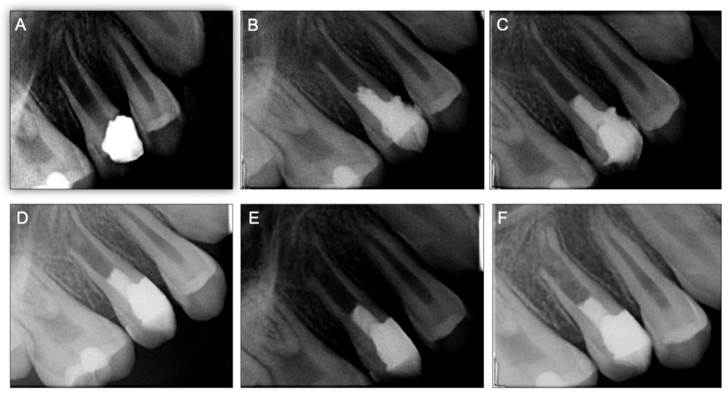
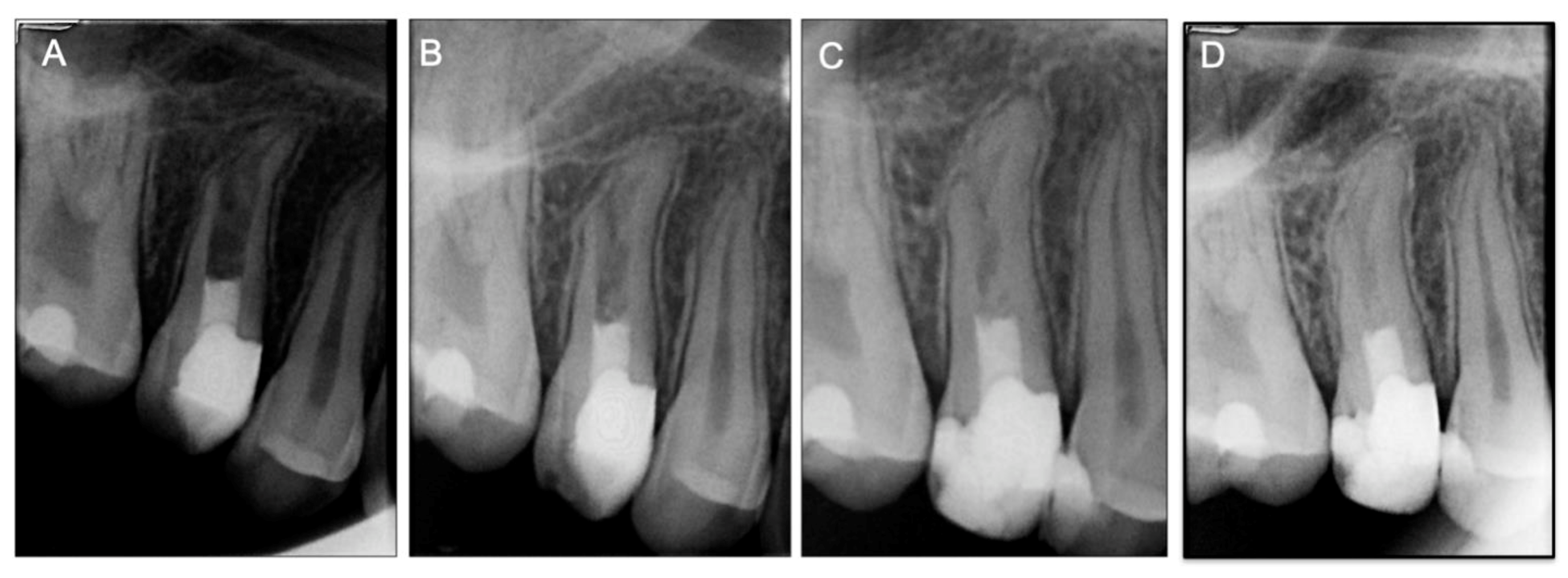
After REPs, the tooth (15) was asymptomatic without signs of coronal discoloration. Follow-up was successively performed at 3, 6, 12 months, 3 years, and 5 years (Figure 1E,F and Figure 2). The radiographic evaluation depicted a continuity of the periodontal ligament space (confirming the absence of an apical radiolucency) as well as an appreciable increase in the root length and thickness. With the progression in time of follow-up, the partial separation of the apical root portion observed at 3, 6, and 12 months decreased significantly compared to the 2- and 5-year follow-up X-rays. After 5 years, a fully developed root structure was observed at the periapical radiography, and on the cone-beam computed tomographic (CBCT) scan (Supplementary materials: Video S1A, S1B), with an increase in length and thickness of the root walls as well as a decrease of the root canal lumen width.

2.2. Case 2

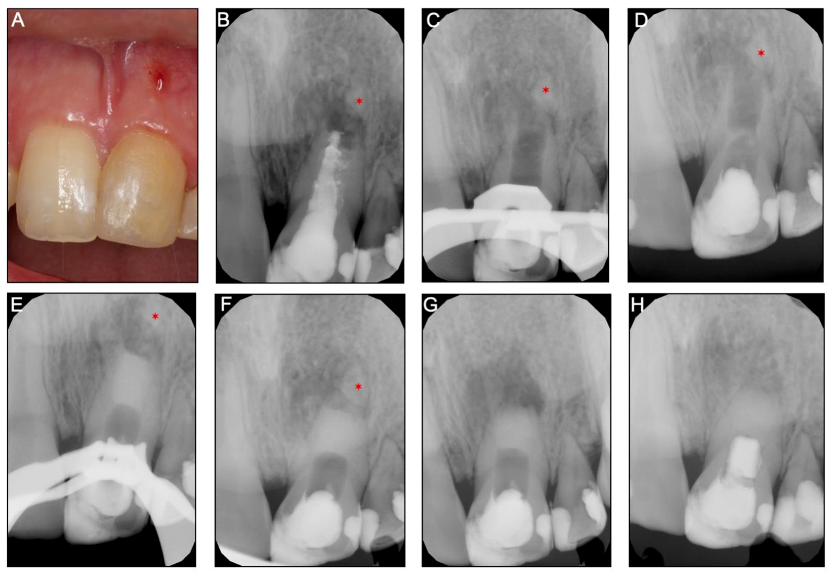
A 16-year-old girl was referred for the nonsurgical endodontic retreatment of the maxillary left central incisor (21) with pulp necrosis and chronic apical periodontitis with the presence of a long-standing sinus tract and a previous endodontic treatment failure. The periapical radiograph and CBTC revealed the maxillary left central incisor with pulp chamber and the canal underfilled with core material and a mineralized tissue beyond the tooth apical portion, separated from the root (Figure 3A,B and Supplementary Materials: Video S2). The agenesis of several teeth was confirmed (12, 22, 32, and 42) with the patient records. Previous dental trauma (non-specified on the parent’s records) on tooth 21 at age 7, followed by an attempt of REP by a generalist dentist, two years before the visit was referred by the child parents. However, the patient remained with edema, swelling, and a sinus tract associated with tooth 21. At the day of evaluation, tooth 21 had normal periodontal probing, and response to sensibility tests (thermal and electrical pulp testing) was negative. After an extended discussion with the children parents about the several treatment options, a nonsurgical root canal retreatment with an apical barrier with a biomaterial (BiodentineTM, Septodont, Saint-Maur-des-Fossés Cedex, France) was elected.
At the first appointment, anesthesia was performed with 3% mepivacaine (Mepivastesin®, 3M ESPE, Neuss, Germany) and under the rubber dam, an access cavity was made with a diamond bur (size #12, Dentsply Sirona, Ballaigues, Switzerland) and refined with an Endo-Z drill (Dentsply Maillefer, Ballaigues, Switzerland). After accessing the pulp chamber, a previous root filling was removed under magnification. Then, a long-neck (LN) bur (size #10, Dentsply Sirona, Ballaigues Switzerland) and an ultrasonic tip (size #3, Start-XTM, Dentsply Sirona, Ballaigues Switzerland) were used to remove the hard material from the canal walls, which was similar to a hydraulic silicate cement (Figure 3C). The same tip was used for ultrasonic agitation of the irrigant. After minimal manual instrumentation with a K-file (size #50, Dentsply-Maillefer, Balaigues, Switzerland) of the canal walls, the disinfection procedure was achieved with copious irrigation of 3% sodium hypochlorite (NaOCl; CanalProTM, Coltène/Whaledent, Langenau, Germany). Intracanal medication (45% CH, Calcicur) was placed in the coronal portion of the canal (Figure 3C) and the access cavity closed with a temporary restorative material (CavitTM).
Three weeks later, the tooth (21) was asymptomatic, and no edema or sinus tract were observed. In the second session, local anesthesia was performed with 2% lidocaine with 1:100,000 epinephrine (XylesteinTM 2%, 3M ESPE, Neuss, Germany), a rubber dam placed, and the provisional material was removed.
The intracanal medication was removed with continuous irrigation of 3% sodium hypochlorite (NaOCl; CanalProTM, Coltène/Whaledent, Langenau, Germany) and saline solution. The canal was dried with paper points and a bioceramic material (BiodentineTM) was prepared in accordance with the manufacturer’s instructions. It was smoothly carried into the canal and compacted apically with a hand barrierger (size #2, Buchanan Hand Plugger) to form an apical barrier of 5 mm in thickness. The excess of material from the walls was removed with paper points, a moist cotton pellet was added, and the access cavity was temporarily closed (CavitTM). Intraoperative and postoperative radiographs were taken to confirm the apical barrier position.
After 2 weeks, the patient developed spontaneous pain, swelling, and a sinus tract. At this time, the surgical endodontic treatment was proposed and explained to the patient and parents. After informed consent was obtained, the surgical procedure was performed by the same Endodontist who performed earlier the apical barrier. Anesthesia was achieved with 2% lidocaine with 1:80,000 epinephrine (XylestesinTM, 3M ESPE, Neuss, Germany), and an Ochsenbein Luebke flap was raised. Following flap retraction, the lesion was accessible and the granulation tissue was carefully removed by curettage. Approximately 2 mm of the root-end was resected with a surgical bur in an air impact high-speed handpiece. After root-end resection (~2 mm), the previous Biodentine barrier was inspected under magnification—nor voids nor chromogenic pigmentation or discoloration detected—as well as mechanically tested with a probe to evaluate its resistance and the lack of voids showing a well condensed barrier and for these reasons no retrograde root-end filling was done. The flap was repositioned and sutured (Figure 3G). One week later, the patient returned with no signs or symptoms, and a resin-based composite (SDRTM flow and Spectrum
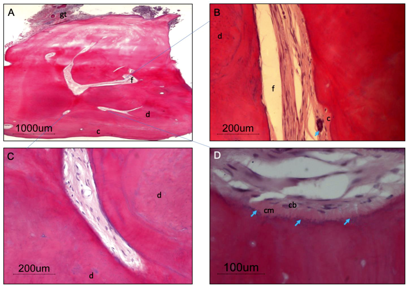
®) was placed into the root canal structure above the apical barrier all the way to the incisal edge. The resected apical root portion was processed for histologic evaluation. Histopathological assessment of the apical root fragment removed during the surgical root canal treatment was performed (Figure 4). Staining with hematoxylin and eosin (H&E) depicts the presence of a root tip, with pulp tissue at the main foramen as well as small foramina, surrounded by dentin and cement. The presence of a giant multinucleated cell in the pulp tissue near the major foramen shall be emphasized. Granulation tissue was observed in and around the excised root tip.
A 5-year follow-up after endodontic surgery as well as clinical and radiographic (Figure 3H) examination revealed almost complete apical healing and healthy soft tissues.

3. Discussion

The literature documenting follow-up of REPs teeth beyond 18 months is limited, and its long-term effects and full impact in a young patient remain unknown [20]. In this case report, we show the follow up of two cases at 5 years and their respective radiographic and clinical success. Despite the loss of pulp vitality with the development of a chronic abscess in both presented clinical cases, a successful outcome with the resolution of the symptoms and tooth survival was achieved. As reported on a meta-analysis by Kahler et al. [20], corroborated by Tong et al. [21] and He et al. [22], the periapical healing and tooth survival after REPs are excellent (above 90%), showing also that periapical pathology resolution after REPs was comparable with treatment with an apical barrier or apexification.
In case 1, the progressive and continuous apical root formation can be seen through the follow-up X-rays. This phenomenon might be explained by Hertwig’s sheath survival, retaining its ability to perform its role on root development and organization (when the inflammatory process was eliminated), and associated to SCAP survival (source of odontoblasts and thereafter can develop root dentin).
Trauma, infection, or iatrogenic factors can be responsible for apical papilla and HERS dislodgement from the main root walls [18]. However, if these two structures stay largely uncompromised, they might continue their development independently of periapical pathology resolution [15]. Case report 2 shows that after the first endodontic treatment (by the previous dentist), the apical fragment of the root was dislodged but continued its development separated from the main root even without the resolution of the apical periodontitis.
To the best of our knowledge, Sonoyama et al. [7] were the first to demonstrate that the apical papilla was a reservoir for mesenchymal stem cells (SCAPs) fundamental for root development of immature teeth. It appears that SCAP has the inherent aptitude to survive and thrive in the hypoxic environment due to its niche normally being poorly vascularized, its nutrition relying on passive diffusion from the surrounding tissues [17,19,23]. Indeed, periradicular granuloma is a highly vascularized granulomatous tissue and is likely to provide even a greater vascular bed than in normal and non-pathological states. From the same research, investigators affirmed that HERS cells are essential for controlling stem cell differentiation in a complement to cementum formation through a process of epithelial–mesenchymal transition in which cells undergo a developmental switch from a polarized epithelial phenotype to a greatly motile mesenchymal phenotype [4]. In addition, it was described that in vitro HERS can promote differentiation of periodontal ligaments stem cells into cementum forming cells [24].
It has been described that the physiologic continuous root development of revascularized immature permanent necrotic teeth depends on whether the HERS survives or not in apical periodontitis as well as in abscess cases [25]. Subsequently, unaffected HERS regulate the continuous root development due to apical cementum deposition after revascularization procedures. However, a normal dentin–pulp complex development requires not only the survival of HERS and ERM but also SCAP.
Previously, our research group [18] showed that 79% of necrotic immature teeth with periapical lesions had histological resolution with an increase in root thickness and length (74%). Complete apical closure was observed in 42% and partial closure in 32%. These findings suggest that SCAPS and HERS cells remain vital in continuity or close to the main root after infection. The present work supports these data with the clinical observation that even in larger periapical lesions, SCAP, HERS, and ERM are more resilient than expected, surviving to clinical adverse conditions and continue their development, as shown by the root tip growth in the clinical case 1. In the follow-up plain radiographs as well as the CBCT scan, a substantial increase in root thickness and length can be appreciated with a root tip development in which, in the beginning, one side is partially detached from the main root. These radiographic findings are supported by our results that note the almost constant presence of an associated, sometimes totally or partially detached, periapical radiopacity, surrounded by a well-defined periodontal ligament space, suggestive of a newly developed root tip. To our knowledge, only a few reports [26,27] about the histology of the newly formed root have been conducted in detail. Gibson et al. [26] reported the presence of disturbed dentinogenesis and the formation beginning stages of pulp stone formation after a traumatic avulsion of an immature permanent incisor, while studies conducted by Yang et al. [27] revealed that the detached root fragments had a normal histological root structure, although no previous studies showed the survival of the apical papilla and HERS cells (or ERM). In spite of the absence of signs and symptoms, the provided clinical evidence of pulp regeneration and the radiographic and CBCT findings were consistent with continued root formation, and the tooth remained unresponsive to pulp sensitivity tests. This was probably because of the distance between the crown and the pulp tissue, which was most likely confined to the apical portion of the tooth.
Concerning the late-stage drawbacks from the REPs, such as undesirable discoloration, pulp canal obliteration, atypical root morphology development, and loss of vitality after apical closure, the available literature is sparse. From the Tong et al. meta-analysis [21], tooth discoloration after REPs treatment was reported in 50% of studies, when minocycline as intracanal medication (known to cause tooth discoloration) as well as grey MTA (bismuth oxide content in MTA has also been associated with coronal discoloration) were used [28]. In both clinical cases, the clinicians decided to use calcium hydroxide as intracanal medication, since calcium hydroxide has a lower risk of later discoloration in comparation to the antibiotic paste [29]. Several studies showed that MTA presents even greater color change after contact with blood [28,30,31]. However, it should be kept in mind that during REPs, MTA is placed over the blood clot on the cervical third of the root, just below the CEJ, increasing the potential for discoloration. In the first case report, where REP was performed, even though there is no discoloration at the 5-year follow-up, a higher risk of discoloration is still possible to occur in a longer-term follow-up.
The literature also points out that the risk of intracanal calcification after REPs was higher in cases medicated with calcium hydroxide (76.9%) compared with teeth medicated with antibiotic pastes (46.2%) [32] Altai et al. [33] in a comprehensive review highlighted that all the studies that used MTA directly on blood clot or on other scaffolds showed intracanal calcification. Correspondingly, other case reports have shown the formation of hard tissue bridges under MTA, and scattered calcification inside the canal, when some portion of the MTA was pushed into the canal. However, our data from case report 1, after five years of follow-up, where calcium hydroxide was used as intracanal medication, show that the root canal is still discernible at the X-ray, which allows being confident that revascularization-associated intracanal calcification did not occur.
The ESE position statement about revitalization procedures advises the use of 1.5–3% sodium hypochlorite [1]. The choice of 3% sodium hypochlorite concentration reflects the need for adequate disinfection and at the same time the preservation of the apical tissues, allowing the further development of the root depicting the success of the clinical procedure in case report 1.
In clinical case 2, an apexification procedure with an apical barrier of Biodentine was the treatment of choice due to the presence of wide root canal and open apex (blunderbuss) [34]. It should be emphasized that in the most recent ESE guidelines, irrigation with EDTA is advised when an apical barrier is placed. Despite this, EDTA irrigation was not performed when performing case report 2 as the irrigation of EDTA did not seem like it would impact the outcome of treatment. However, it is known that Biodentine has significant potential to stimulate TGF-b1 release from the root dentin with similar effects to the irrigation with EDTA [35]. These high levels of TGF-b1 were explained by alkaline property (high pH levels of Biodentine) on solubilize of dentin matrix. We should stress that even after the apical barrier placement, there were no signs of periapical healing—pain, swelling, as well as a fistula were present after 2 weeks of the apical barrier. The persistence of apical infection might be explained by the presence of an extraradicular biofilm—as shown by the histologic slides after the apical surgery—which was not removed by the intrarradicular disinfection procedures [19]. As reported by Siqueira et al. [36], biofilms that adhered to the apical root surface (extraradicular biofilms) have been a possible cause of post-treatment apical periodontitis.
Furthermore, our findings provide evidence that in clinical case 2, the mineralized structures observed radiographically had histologic features of a root tip—composed by cellular cementum, periodontal ligament, dentin, predentin with an intact odontoblastic layer, and, in some cases, the presence of apical foramen were also seen. Thus, according to our findings, it is reasonable to suggest that even when partially or completely detached from the main root, the apical papilla associated with HERS preserves their potential of continuing root development [18]. These findings confirm the results observed in a study by Yang et al. [27] that the separation of the soft tissues from mineralized tissues and their development as an independent root tip was associated with HERS injure and its following detachment caused by the apexification procedure.

4. Conclusions

The present report demonstrates the survival and continued potential differentiation of the apical papilla and HERS/ERM after endodontic infection, with clinical radiographic and histological evidence that a root tip can form separately from the main root and can continue its development and eventually reconnect with the coronal portion.
Thus, a conservative approach should be considered as the first choice of treatment for immature teeth with necrotic pulp. Endodontic therapies applied to immature teeth that do not affect the apical papilla viability are a key determining factor, guiding successful root development.

Supplementary Materials

The following are available online at https://www.mdpi.com/2076-3417/9/19/3942/s1, Video S1. Follow-up after 5-year of REPs (Case 1—tooth 15), the decrease of the root canal lumen width was observed on cone-beam computed tomographic (CBCT) scan: (S1A) sagittal view; (S1B) axial view. Video S2. Preoperative Case 2 (tooth 21) were the cone-beam computed tomographic (CBCT) scan showed a persistent periapical lesion, and the root tip (mineralized tissue) beyond the tooth apical portion, separated from the root.

Funding

This research did not receive any specific grant from funding agencies in the public, commercial, or not-for-profit sectors.

Conflicts of Interest

The authors declare no conflict of interest.

References

  1. Galler, K.M.; Krastl, G.; Simon, S.; Van Gorp, G.; Meschi, N.; Vahedi, B.; Lambrechts, P. European Society of Endodontology position statement: Revitalization procedures. Int. Endod. J. 2016, 49, 717–723. [Google Scholar] [CrossRef] [PubMed]
  2. American Association of Endodontists. Considerations for Regenerative Procedures (Internet). Chicago: American Association of Endodontists. 2018 (Revised 4/1/2018). Available online: https://f3f142zs0k2w1kg84k5p9i1o-wpengine.netdna-ssl.com/specialty/wp-content/uploads/sites/2/2018/06/ConsiderationsForRegEndo_AsOfApril2018.pdf (accessed on 12 May 2019).
  3. Hargreaves, K.M.; Diogenes, A.; Teixeira, F.B. Treatment options: Biological basis of regenerative endodontic procedures. Pediatr. Dent. 2013, 35, 129–140. [Google Scholar] [CrossRef] [PubMed]
  4. Huang, G.T.; Sonoyama, W.; Liu, Y.; Liu, H.; Wang, S.; Shi, S. The hidden treasure in apical papilla: The potential role in pulp/dentin regeneration and bioroot engineering. J. Endod. 2008, 34, 645–651. [Google Scholar] [CrossRef] [PubMed]
  5. Lovelace, T.W.; Henry, M.A.; Hargreaves, K.M.; Diogenes, A. Evaluation of the delivery of mesenchymal stem cells into the root canal space of necrotic immature teeth after clinical regenerative endodontic procedure. J. Endod. 2011, 37, 133–138. [Google Scholar] [CrossRef] [PubMed]
  6. Chrepa, V.; Henry, M.A.; Daniel, B.J.; Diogenes, A. Delivery of apical mesenchymal stem cells into root canals of mature teeth. J. Dent. Res. 2015, 94, 1653–1659. [Google Scholar] [CrossRef] [PubMed]
  7. Sonoyama, W.; Liu, Y.; Fang, D.; Yamaza, T.; Seo, B.M.; Zhang, C.; Liu, H.; Gronthos, S.; Wang, C.-Y.; Shi, S.; et al. Mesenchymal stem cell-mediated functional tooth regeneration in swine. PLoS ONE 2006, 1, e79. [Google Scholar] [CrossRef] [PubMed]
  8. Sequeira, D.B.; Seabra, C.M.; Palma, P.J.; Cardoso, A.L.; Peca, J.; Santos, J.M. Effects of a new bioceramic material on human apical papilla cells. J. Funct. Biomater. 2018, 9, 74. [Google Scholar] [CrossRef] [PubMed]
  9. Nanci, A.; Ten Cate, A.R. Ten Cate´s Oral Histology: Development, Structure, and Function, 8th ed.; Mosby: St. Louis, MO, USA, 2012. [Google Scholar]
  10. Xiong, J.; Mrozik, K.; Gronthos, S.; Bartold, P.M. Epithelial cell rests of Malassez contain unique stem cell populations capable of undergoing epithelial-mesenchymal transition. Stem Cells Dev. 2012, 21, 2012–2025. [Google Scholar] [CrossRef]
  11. Diogenes, A.; Hargreaves, K.M. Microbial modulation of stem cells and future directions in regenerative endodontics. J. Endod. 2017, 43, S95–S101. [Google Scholar] [CrossRef]
  12. Jung, I.Y.; Lee, S.J.; Hargreaves, K.M. Biologically based treatment of immature permanent teeth with pulpal necrosis: A case series. J. Endod. 2008, 34, 876–887. [Google Scholar] [CrossRef]
  13. Bose, R.; Nummikoski, P.; Hargreaves, K. A retrospective evaluation of radiographic outcomes in immature teeth with necrotic root canal systems treated with regenerative endodontic procedures. J. Endod. 2009, 35, 1343–1349. [Google Scholar] [CrossRef] [PubMed]
  14. Ding, R.Y.; Cheung, G.S.; Chen, J.; Yin, X.Z.; Wang, Q.Q.; Zhang, C.F. Pulp revascularization of immature teeth with apical periodontitis: a clinical study. J. Endod. 2009, 35, 745–749. [Google Scholar] [CrossRef] [PubMed]
  15. Chen, M.Y.; Chen, K.L.; Chen, C.A.; Tayebaty, F.; Rosenberg, P.A.; Lin, L.M. Responses of immature permanent teeth with infected necrotic pulp tissue and apical periodontitis/abscess to revascularization procedures. Int. Endod. J. 2012, 45, 294–305. [Google Scholar] [CrossRef] [PubMed]
  16. Jung, I.Y.; Kim, E.S.; Lee, C.Y.; Lee, S.J. Continued development of the root separated from the main root. J. Endod. 2011, 37, 711–714. [Google Scholar] [CrossRef] [PubMed]
  17. Tobias Duarte, P.C.; Gomes-Filho, J.E.; Ervolino, E.; Marcal Mazza Sundefeld, M.L.; Tadahirowayama, M.; Lodi, C.S.; Dezan-Júnior, E.; Cintra, L.T.A. Histopathological condition of the remaining tissues after endodontic infection of rat immature teeth. J. Endod. 2014, 40, 538–542. [Google Scholar] [CrossRef] [PubMed]
  18. Palma, P.J.; Ramos, J.C.; Martins, J.B.; Diogenes, A.; Figueiredo, M.H.; Ferreira, P.; Viegas, C.; Santos, J.M. Histologic evaluation of regenerative endodontic procedures with the use of chitosan scaffolds in immature dog teeth with apical periodontitis. J. Endod. 2017, 43, 1279–1287. [Google Scholar] [CrossRef] [PubMed]
  19. Meschi, N.; Hilkens, P.; Van Gorp, G.; Strijbos, O.; Mavridou, A.; Cadenas de Llano Perula, M.; Lambrichts, I.; Verbeken, E.; Lambrechets, P. Regenerative endodontic procedures posttrauma: Immunohistologic analysis of a retrospective series of failed cases. J. Endod. 2019, 45, 427–434. [Google Scholar] [CrossRef] [PubMed]
  20. Kahler, B.; Rossi-Fedele, G.; Chugal, N.; Lin, L.M. An evidence-based review of the efficacy of treatment approaches for immature permanent teeth with pulp necrosis. J. Endod. 2017, 43, 1052–1057. [Google Scholar] [CrossRef] [PubMed]
  21. Tong, H.J.; Rajan, S.; Bhujel, N.; Kang, J.; Duggal, M.; Nazzal, H. Regenerative endodontic therapy in the management of nonvital immature permanent teeth: A systematic review-outcome evaluation and meta-analysis. J. Endod. 2017, 43, 1453–1464. [Google Scholar] [CrossRef] [PubMed]
  22. He, L.; Zhong, J.; Gong, Q.; Kim, S.G.; Zeichner, S.J.; Xiang, L.; Ye, L.; Zhou, X.; Zheng, J.; Liu, Y.; et al. Treatment of necrotic teeth by apical revascularization: meta-analysis. Sci. Rep. 2017, 7, 13941. [Google Scholar] [CrossRef]
  23. Vanacker, J.; Viswanath, A.; De Berdt, P.; Everard, A.; Cani, P.D.; Bouzin, C.; Feron, O.; Diogenes, A.; Leprince, J.G.; Des Rieux, A. Hypoxia modulates the differentiation potential of stem cells of the apical papilla. J. Endod. 2014, 40, 1410–1418. [Google Scholar] [CrossRef] [PubMed]
  24. Sonoyama, W.; Seo, B.M.; Yamaza, T.; Shi, S. Human Hertwig’s epithelial root sheath cells play crucial roles in cementum formation. J. Dent. Res. 2007, 86, 594–599. [Google Scholar] [CrossRef] [PubMed]
  25. Lin, L.M.; Rosenberg, P.A. Repair and regeneration in endodontics. Int. Endod. J. 2011, 44, 889–906. [Google Scholar] [CrossRef] [PubMed]
  26. Gibson, A.C. Continued root development after traumatic avulsion of partly-formed permanent incisor. Br. Dent. J. 1969, 126, 356–357. [Google Scholar] [PubMed]
  27. Yang, S.F.; Yang, Z.P.; Chang, K.W. Continuing root formation following apexification treatment. Endod. Dent. Traumatol. 1990, 6, 232–235. [Google Scholar] [CrossRef]
  28. Lenherr, P.; Allgayer, N.; Weiger, R.; Filippi, A.; Attin, T.; Krastl, G. Tooth discoloration induced by endodontic materials: A laboratory study. Int. Endod. J. 2012, 45, 942–949. [Google Scholar] [CrossRef]
  29. Nagata, J.Y.; Gomes, B.P.; Rocha Lima, T.F.; Murakami, L.S.; De Faria, D.E.; Campos, G.R.; Souza-Filho, F.J.; De Jesus Soares, A. Traumatized immature teeth treated with 2 protocols of pulp revascularization. J. Endod. 2014, 40, 606–612. [Google Scholar] [CrossRef]
  30. Palma, P.J.; Marques, J.A.; Falacho, R.I.; Correia, E.; Vinagre, A.; Santos, J.M.; Ramos, J.C. Six-month color stability assessment of two calcium silicate-based cements used in regenerative endodontic procedures. J. Funct. Biomater. 2019, 10, 14. [Google Scholar] [CrossRef]
  31. Ramos, J.C.; Palma, P.J.; Nascimento, R.; Caramelo, F.; Messias, A.; Vinagre, A.; Santos, J.M. 1-year in vitro evaluation of tooth discoloration induced by 2 calcium silicate-based cements. J. Endod. 2016, 42, 1403–1407. [Google Scholar] [CrossRef]
  32. Song, M.; Cao, Y.; Shin, S.J.; Shon, W.J.; Chugal, N.; Kim, R.H.; Kim, E.; Kang, M.K. Revascularization-associated intracanal calcification: assessment of prevalence and contributing factors. J. Endod. 2017, 43, 2025–2033. [Google Scholar] [CrossRef]
  33. Altaii, M.; Richards, L.; Rossi-Fedele, G. Histological assessment of regenerative endodontic treatment in animal studies with different scaffolds: A systematic review. Dent. Traumatol. 2017, 33, 235–244. [Google Scholar] [CrossRef] [Green Version]
  34. Palma, P.J.; Marques, J.A.; Falacho, R.I.; Vinagre, A.; Santos, J.M.; Ramos, J.C. Does delayed restoration improve shear bond strength of different restorative protocols to calcium silicate-based cements? Materials 2018, 11, 2216. [Google Scholar] [CrossRef] [PubMed]
  35. Wattanapakkavong, K.; Srisuwan, T. Release of transforming growth factor beta 1 from human tooth dentin after application of either ProRoot MTA or Biodentine as a coronal barrier. J. Endod. 2019, 45, 701–705. [Google Scholar] [CrossRef] [PubMed]
  36. Siqueira, J.F.; Jr Rocas, I.N.; Ricucci, D. Biofilms in endodontic infection. Endod. Topics 2012, 22, 33–49. [Google Scholar] [CrossRef]
Figure 1. Case report 1 with radiographic images of teeth 15: (A) preoperative periapical radiograph of tooth indicating the root canal was wide with a divergent open apical foramen, the existence of periapical lesion and thin root dentinal walls; (B) radiograph of the tooth after chemomechanical debridement with 3% of NaOCl irrigation and calcium hydroxide application; (C) after 4 weeks, the cervical barrier with 4 mm of MTA; (D) final restoration with direct composite resin restoration; (E) 3-month follow-up periapical radiograph; (F) after six months of following, the radiograph shows periapical healing as well as mesial and apical root formation with larger dentinal walls.
Applsci 09 03942 g001
Figure 2. Case report 1 with controls radiographic images: (A) 12-month postoperative periapical radiograph control of tooth the continuity of the periodontal ligament space with absence of apical radiolucency was observed at radiographic evaluation showed appreciable root length increase; (B) after 2 years; (C) a total root formation is observed in radiographs after 3-years; (D) at the 5-year follow-up, an increase was observed in the root length and thickness and a decrease of the root canal lumen width.
Applsci 09 03942 g002
Figure 3. Clinical and radiographic images of Case 2 of teeth 21 with endodontic treatment failure. (A) Initial photograph showing the presence of a sinus tract. (B) Initial radiographic depicting a periapical radiolucency. The periapical radiograph revealed the maxillary left central incisor with pulp chamber and the canal underfilled with core material and a mineralized tissue beyond the tooth apical portion, separated from the root (*). (C) Nonsurgical root canal retreatment was used to remove the hydraulic silicate cement from the canal walls. (D) Periapical radiograph after the access cavity was closed with temporary material, and the intracanal medication (CH, Calcicur) was placed in the coronal portion of the canal. (E) An operative and (F) and postoperative to confirm the apical barrier position. (G) Postoperative radiograph after the surgical procedure. (H) Five-year follow-up after endodontic surgery.
Applsci 09 03942 g003
Figure 4. Histologic microphotographs of the apical fragment surgical removed during the apical surgery of the Case 2. Histologic sections depict the presence of mineralized tissue with the ordinary structure of a root apex (due to mineralization of the apical papilla): (A) small magnification of apical root tip with pulp tissue at the main foramen as well as small foramina (f), surrounded by dentin (d) and cement (c). Fragments of granulation tissue (gt) can be observed on the outside root wall. (B) High magnification of the cementum–dentin junction (CDJ) of the main canal showing the connective pulp tissue (p), cementum (c) on the inner portion and dentin (d). (C) High magnification of foramina depicting in the central portion the pulp tissue surrounded by dentin (d) on the right upper side and by cement (c) on the lower left side. The dentinal tubules are show in a transversal section. (D) High magnification of the apposition/formative phase during cementum formation. Hematoxylin mineralization granules (blue arrow) can be observed on the non-mineralized cementum matrix (cm) and cementoblasts (cb). (Hematoxylin–eosin, H&E).
Applsci 09 03942 g004
Table 1. Comparison between intraoperative clinical aspects of European Society of Endodontology (ESE) Position Statement: Revitalization procedures adapted from ESE [1] and Clinical Considerations for a Regenerative Procedure adapted from American Association of Endodontists (AAE) [2].
European Society of Endodontology Position Statement: Revitalization ProceduresAmerican Association of Endodontists Clinical Considerations for a Regenerative Procedure
 First Appointment
  • Perform clinical diagnostics according to a clinical and radiographical assessment checklist.
  • Tooth cleaning, local anesthesia (optional), field isolation and disinfection (with, e.g., Iso-Betadine).
  • Prepare access cavity.
  • Remove loose or necrotic pulp tissue using suitable endodontic instruments.
  • Avoid mechanical instrumentation of the root canal walls.
  • Irrigate with 1.5–3% sodium hypochlorite (20 mL, 5 min), use of side-vented needle, place 2 mm above vital tissue (which can be controlled using an operating microscope or when the patient reports pain).
  • Bleeding or exudate (control with paper points) may require extended irrigation; irrigate with sterile physiological saline (5 mL) to minimize the cytotoxic effects of sodium hypochlorite on vital tissues.
  • Dry with paper points.
  • Irrigate with 20 mL of 17% EDTA.
  • Insert a non discolouring calcium hydroxide product homogenously into the root canal. The antibiotics, mainly triple antibiotic paste (TAP) consisting of ciprofloxacin, metronidazole and minocycline were used with good results. Drawbacks such as discoloration, cytotoxicity, sensitization, development of resistance and difficulty of removal from the root canal need to be considered.
  • Place coronal seal directly onto intracanal dressing with a minimum thickness according to the material selected.
Second Appointment (2–4 weeks later)
  • Clinical diagnostics according to the checklist.
  • If signs of inflammation have not subsided, refresh calcium hydroxide. Administration of systemic antibiotics may be considered if the patient reports general health alteration such as fever or dysphagia (for further information see recommendations of the European Academy of Pediatric Dentistry/EAPD).
  • Cleaning, anesthesia, field isolation and disinfection of operating field. The chosen anesthesia should feature optimal bone penetration. Current recommendations specify the use of anesthetics without vasoconstrictor; however, the creation of a blood clot is mostly hampered by the patients’ sensation of pain, and evidence describing improved bleeding without vasoconstrictor is sparse. Experiences from the first visit regarding patient compliance, anxiety, and pain control should be considered.
  • Remove temporary seal; irrigate with 17% EDTA (20 mL, 5 minutes (min)), use of side-vented needle and place 2 millimeters (mm) above vital tissue; irrigate with sterile physiological saline (5 mL) to reduce adverse effects of irrigants on target cells.
  • Remove excess liquid with paper points.
  • Induce bleeding by mechanical irritation of periapical tissue and rotational movement of an apically pre-bent hand-file (e.g., size 40 Hedström).
  • Allow the canal to fill with blood until 2 mm below the gingival margin to wait for blood clot formation for 15 min.
  • Cut a collagen matrix (e.g., Parasorb Cone (Resorba, Medical GmbH, Germany), Collaplug (Integra LifeSciences Corp., Plainsboro, NJ, USA) or Hemocollagene (Septodont, Saint Maur des Fossés, France) to a diameter larger than the coronal part of the root canal and a height of 2–3 mm, place on top of the blood clot, allow the matrix to soak with liquid to avoid formation of a hollow space.
  • Place a hydraulic silicate cement (e.g., Mineral Trioxide Aggregate (MTA) or tricalcium silicate cement) on top of the collagen matrix in a thin homogeneous layer of about 2 mm underneath the cement–enamel junction and beware of potential discoloration after contact of the material with blood.
  • Apply a flowable, light-curable glass-ionomer or calcium hydroxide cement; refresh the cavity walls with a diamond bur or grit blast with aluminum oxide; seal with adhesive restoration.
 First Appointment
  • Local anesthesia, dental dam isolation and access.
  • Copious, gentle irrigation with 20 mL NaOCl using an irrigation system that minimizes the possibility of extrusion of irrigants into the periapical space (e.g., needle with closed end and side-vents, or EndoVac™). Lower concentrations of NaOCl are advised (1.5% NaOCl (20 mL/canal, 5 min)) and then irrigated with saline or EDTA (20 mL/canal, 5 min), with irrigating needle positioned about 1 mm from root-end, to minimize cytotoxicity to stem cells in the apical tissues.
  • Dry canals with paper points.
  • Place calcium hydroxide or low concentration of triple antibiotic paste. If the triple antibiotic paste (TAP) is used: 1) consider sealing pulp chamber with a dentin bonding agent (to minimize risk of staining) and 2) mix 1:1:1 ciprofloxacin: Metronidazole: Minocycline to a final concentration of 1–5 mg/mL. TAP has been associated with tooth discoloration. Double antibiotic paste (DAP) without minocycline paste or substitution of minocycline for other antibiotic (e.g., clindamycin; amoxicillin; cefaclor) is another possible alternative as root canal disinfectant. Clinicians should be aware that studies have been done using higher concentrations of TAP/DAP, but a recommendation to a higher concentration cannot be made at this time due to limited studies.
  • Deliver into canal system via syringe, if triple antibiotic is used, ensure that it remains below cement-enamel junction (CEJ) (minimize crown staining).
  • Seal with 3–4 mm of a temporary restorative material such as Cavit™, IRM™, glass-ionomer or another temporary material. Dismiss patient for 1–4 week
Second Appointment (1–4 weeks after first visit)
  • Assess response to initial treatment. If there are signs/symptoms of persistent infection, consider additional treatment time with antimicrobial, or alternative antimicrobial.
  • Anesthesia with 3% mepivacaine without vasoconstrictor, dental dam isolation.
  • Copious, gentle irrigation with 20 ml of 17% EDTA.
  • Dry with paper points.
  • Create bleeding into canal system by over instrumenting (endo file, endo explorer) (induce by rotating a pre-curved K-file at 2 mm past the apical foramen with the goal of having the entire canal filled with blood to the level of the cementoenamel junction). An alternative to creating of a blood clot is the use of platelet-rich plasma (PRP), platelet rich fibrin (PRF) or autologous fibrin matrix (AFM).
  • Stop bleeding at a level that allows for 3–4 mm of restorative material.
  • Place a resorbable matrix such as CollaPlug™, Collacote™, CollaTape™ over the blood clot if necessary and white MTA as capping material.
  • A 3–4 mm layer of glass-ionomer (e.g., Fuji IX™, GC America, Alsip, IL) is flowed gently over the capping material and light-cured for 40 s. As MTA has been associated with discoloration, possible alternatives (such as bioceramics or tricalcium silicate cements (e.g., BiodentineTM, Septodont, Lancasted, PA, USA, EndoSequence® BC RRM-Fast Set Putty, Brasseler, USA)) should be considered in teeth where there is an esthetic concern.
  • Anterior and Premolar teeth—Consider use of Collatape/Collaplug and restoring with 3 mm of a non-staining restorative material followed by bonding a filled composite to the beveled enamel margin.
  • Molar teeth or teeth with porcelain fused metal (PFM) crown—Consider use of Collatape/Collaplug and restoring with 3 mm of MTA, followed by RMGI, composite or alloy.

