Core@shell Nanoparticles: Greener Synthesis Using Natural Plant Products


1
NanoBioelectrochemistry Research Center, Bam University of Medical Sciences, Bam, Iran
2
Nanomedicine and Nanobiology Research Center, Shiraz University of Medical Sciences, Shiraz, Iran
3
Leishmaniasis Research Center, Kerman University of Medical Sciences, Kerman, Iran
4
Research Center for Tropical and Infectious Diseases, Kerman University of Medical Sciences, Kerman, Iran
5
Regional Centre of Advanced Technologies and Materials, Department of Physical Chemistry, Faculty of Science, Palacky University, Šlechtitelů 27, 783 71 Olomouc, Czech Republic
*
Authors to whom correspondence should be addressed.
Appl. Sci. 2018, 8(3), 411; https://doi.org/10.3390/app8030411
Submission received: 17 February 2018
/
Revised: 5 March 2018
/
Accepted: 7 March 2018
/
Published: 10 March 2018
(This article belongs to the Section Nanotechnology and Applied Nanosciences)
Abstract
:Featured Application
Greener synthesis and applications of Core-shell nanoparticles.
Abstract
Among an array of hybrid nanoparticles, core-shell nanoparticles comprise of two or more materials, such as metals and biomolecules, wherein one of them forms the core at the center, while the other material/materials that were located around the central core develops a shell. Core-shell nanostructures are useful entities with high thermal and chemical stability, lower toxicity, greater solubility, and higher permeability to specific target cells. Plant or natural products-mediated synthesis of nanostructures refers to the use of plants or its extracts for the synthesis of nanostructures, an emerging field of sustainable nanotechnology. Various physiochemical and greener methods have been advanced for the synthesis of nanostructures, in contrast to conventional approaches that require the use of synthetic compounds for the assembly of nanostructures. Although several biological resources have been exploited for the synthesis of core-shell nanoparticles, but plant-based materials appear to be the ideal candidates for large-scale green synthesis of core-shell nanoparticles. This review summarizes the known strategies for the greener production of core-shell nanoparticles using plants extract or their derivatives and highlights their salient attributes, such as low costs, the lack of dependence on the use of any toxic materials, and the environmental friendliness for the sustainable assembly of stabile nanostructures.
1. Introduction
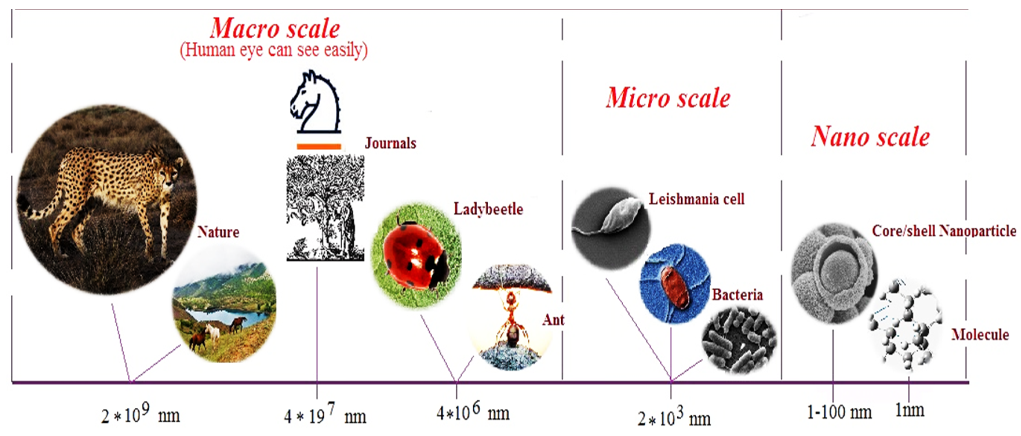
In global standard norms, materials or particles that in at least one dimension have a length of 1 to 100 nanometers (nm) are termed nanoparticles (nanomaterials) [1,2]. Nanoparticles (NPs) are aggregates of between a few and many millions of atoms. Materials at the nano-scale (Figure 1) display novel biological and physicochemical properties that are never observed for their bulk counterparts [3]. The material in this size have high surface to volume ratio and they can pass across cell membranes by passive diffusion [4].
Nanoparticles (NPs) are sort of bridge between ions (atomic) and bulk phases of materials [5]. Nanomaterials based on structure, appearance, and their usage are defined by different names, but the common feature for all of them is to have at least one dimension in the nano-scale (1 to 100 nm) [6]; unique nano-specific phenomena of particulates are most likely to occur in the size range of 1 and 100 nm [7].
In view of the increased knowledge and understanding about the synthesis and the characterization of NPs, scientists have now succeeded in designing a useful newer hybrid class of NPs termed core shell (core-shell, core@shell or core/shell) NPs that can be defined as well-organized nanomaterials consisting of two, three, or more types of individual nanocomponents [8].
The surface of nanoparticles can be modified (functionalized) with a wide range of organic/inorganic materials such as polymers, biomolecules, silica, metals, and non-metal components. Stability, biocompatibility, newer specific performance, targeted delivery, treatment, or diagnosis of biological entities, such as cells, proteins, nucleic acids, enzymes, bacteria, etc., are some of the reasons for the surface modification of nanoparticles via functionalization. The coating of organic/inorganic materials that surrounds the NPs can actually be the shell or second shell, which are called core-shell nanoparticles. The value of the coating materials could be enormous when their presence not only protects the core, but also may play a significant functional role as exemplified in the protection afforded by the ZnO shell for the oxidation of the Fe core in the Fe@ZnO NPs.
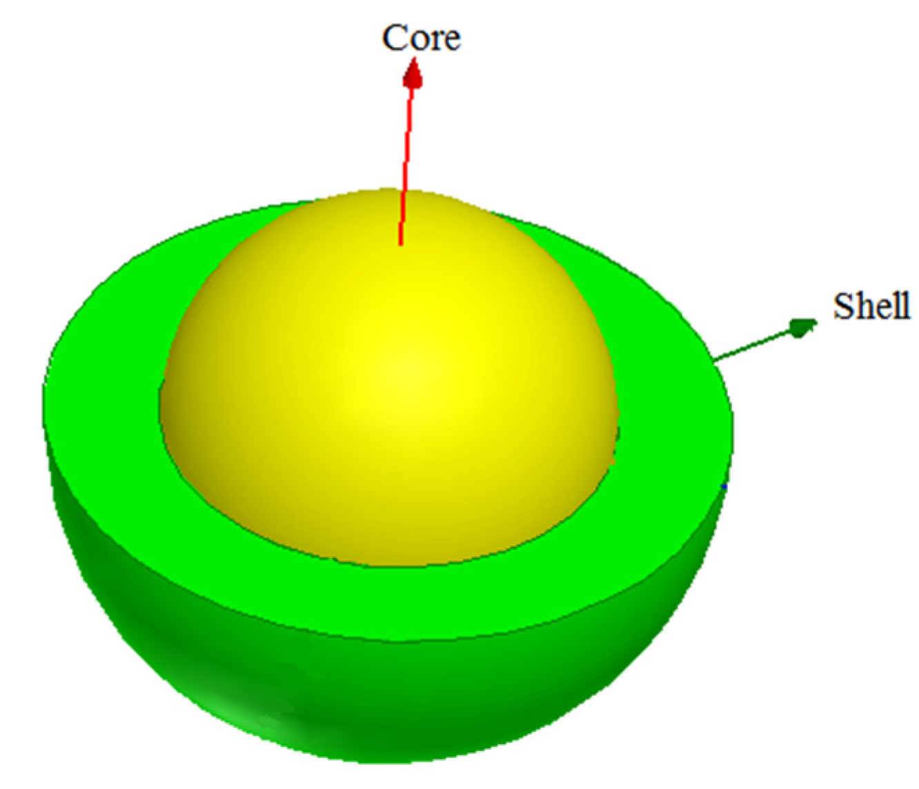
Core-shell nanoparticles are heterogeneous NPs composed of two or more materials (metal, element, or biomolecules); one nanomaterial acts as a core in the center while the other material/materials located around the central core (shell) (Figure 2). Essentially, core -shell NPs are a type of biphasic nanomaterial, which have an inner core and an outer shell made up of different components [9].
A wide range of organic and inorganic nanomaterial can be used for forming both, the core and the shell comprising core-shell NP. A combination of several diverse materials, where the building components are easily distinguishable from each other, is referred to as composite or nanocomposite, when the dimensions are less than 100 nm [10]. The properties of nanocomposites need not necessarily originate from each individual part. Briefly, a component of nanocomposite may serve the role of an adhesive without which they may not stick together and the ensuing blend of components will not possess any new properties of the nanocomposite.
The core-shell NPs can be labeled as nanocomposites but they may not necessarily be nanocomposites which tend to be uniform in microscopic view (mixed up components). In contrast, core-shell NPs usually have a separation that can be seen between the shell and the core. The nanocomposite properties are not expected from core-shell NPs which can be bimetallic or trimetallic [11,12].
The synthesis of core-shell NPs has the ability to use a wide range of materials as core or shell that can impart desired and unique features and functions. The properties of core-shell NPs, such as physicochemical, biological, optical, etc. can be altered by changing the ingredients constituting the core or shell layer; this ability to manipulate can produce an array of core-shell NPs, which finds numerous applications in diverse fields, such as medicine, pharmacy, engineering, and material science.
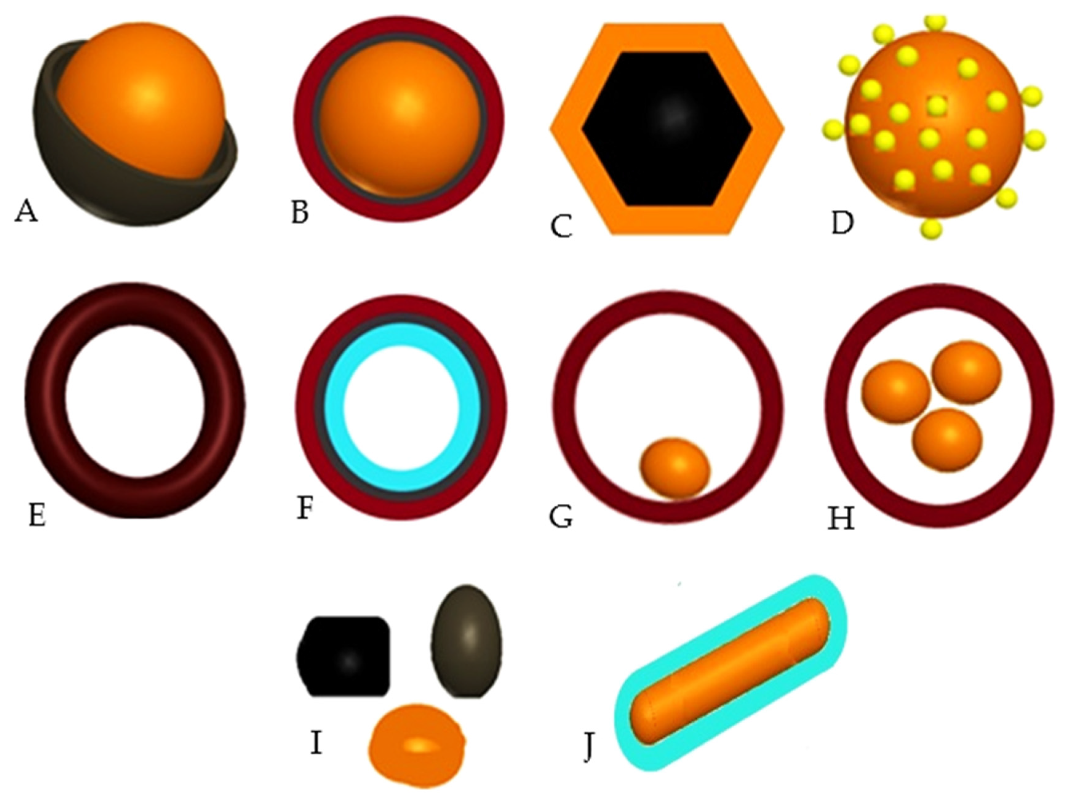
Based on the structural configuration, core-shell NPs can be classified in to following categories (Figure 3):
Each of these classes of core shell NPs have their unique and exclusive properties. For example, after synthesis of core-shell NPs with organic core and its subsequent removal (by dissolution in organic solvents or simply heating at elevated temperatures) can generate hollow-core shell NPs. Additionally, the core multi-shell are yet another variety of core-shell NPs that have two or more shells. The protection of the deeper layers and use the interface between the core and shell layers that are not compatible with each other are some of the advantages of this type of core-shell NPs.
2. Synthesis Processes for Assembly of Core-Shell Nanoparticles
A variety of methods with bottom-up and top-down approach have been introduced, optimized, and are essentially accomplished via physical, chemical [13,14,15,16], or biosynthetic means or combination thereof (Table 1). In bottom-up approach, which is often preferred one, self-assembly of atoms lead to formation of nanosize particles, while in the top-down approach, bulk material is broken down to nano size particle (not explored for core shell nanoparticles).
3. Biosynthesis of Core Shell Nanoparticles
In today’s world, the synthetic interest for engineered nanomaterials should ideally be inspired by nature instead of using complicated procedures; biosynthesized nanoparticles have beneficial use in clinical trials and in the design of novel therapeutic strategies. Chemically synthesized NPs may be coated with compounds that are used in the synthesis process that limits their application in clinical studies because even the associated impurities in trace amounts can emanate from these experiments. In contrast, the biosynthesis nanoparticles may not have these restrictions [21,22,23,24].
Biosynthetic methods are cheaper and eco-friendlier than the physicochemical synthesis methods [25,26]. The synthesis process time, depending on the type of bioresource that is used, varies from a few minutes to a few days. In brief, expeditious, inexpensive, and endowed with greater ease of manipulation are some of the salient features of biosynthesis processes.
Each method has their advantages and disadvantage, but in generalm the use of biological resources for nanoparticle synthesis is easier, cost-effective, and generates less pollution and circumvent environmental damage than the physicochemical methods in terms of human health and the environment. Nature has provided countless biological resources [27] with millions of bacteria, fungi, and plants bearing numerous structural types that suitable for specific applications [28,29,30,31,32]. The synthesized NPs decompose after a certain period of time. However, the stability of NPs generated via the biosynthesis method can be enhanced by changing the physicochemical parameters, such as varying the extract concentration or metal ion source, etc.
Studies have shown that the hydrophobic surface of NPs, if not covered or capped, will bind to each other due to hydrophobic interactions, and particle size will increase, and eventually the formation of interconnected agglomerated nanoparticles will ensue. Aggregation phenomena, a challenge for chemically synthesized NPs, can be addressed by the use of natural protecting biomolecules that are present in bioresource extracts. The advantage of green synthesis of NPs using natural resources such as plants, fungal or bacterial strains is that these extracts contain a wide variety of biomolecules which gets immobilized on surface of nanoparticles during the synthesis process. Therefore, biosynthesized nanoparticles using natural resources would lead to coating of hydrophobic surfaces of nanoparticles that results in increased stability and stability of biosynthesized nanoparticles.
3.1. Biosynthesis of Core-Shell Nanoparticles Using Bacterial and Fungal Strains
Reports on the synthesis of hybrid NPs using fungi are rather limited, especially on fungal-mediated synthesis or the biocompatibility studies of ensuing hybrid/core shell NPs, presumably due to the dependence on costly cell cultures, and longer synthesis time often required as compared to plant-mediated synthesis [33,34]. Exposure of the bacteria live cells or their lysis cell extracts to aqueous metal ions have resulted in the formation of NPs [35]. Only a handful of investigations have been conducted on prokaryotes synthesis of core-shell NPs and limited interested in bacterial-mediated synthesis of hybrid/core shell NPs is reminiscent of fungal-mediated approach. The typical disadvantages of bacterial- and fungal-mediated synthesis of core-shell NPs are that they often excessively time-consuming and are culture-dependence, which entails the use of expensive culture media, a trait that is not shared by the plant-mediated methods.
3.2. Greener Synthesis of Core-Shell Nanoparticles Using Plants
Plants-mediated synthesis of core-shell nanoparticles is advantageous when compared to processes that are assisted by bacteria and fungi [36]. Prasad et al. (2017) [37] reported one-step biosynthesized magnetic nickel/iron-oxide core shell NPs using 20 grams of Moringa oleifera extract. Gosh et al. [38] reported one-step biosynthesized gold/silver core shell NPs using Dioscorea bulbifera. Venkateswarlu et al. [39] reported two-step biosynthesized Fe3O4-Ag core/shell NPs (50 nm) using Vitis vinifera stem extract. Au-Ag core shell NPs (10 nm) were synthesized via one-pot method using grape water [40], a herbal medicinal drug. Plants seem to be the ideal candidates as they are suitable for expeditious large-scale green synthesis of core-shell NPs; the rate of synthesis being faster than those observed for processes aided by microorganisms [41]. Various core shell NPs with diverse structures, shapes and sizes have been synthesized using plants and some are documented in Table 2.
The plant extracts contain chemical entities with numerous functional groups that are involved in the reduction of metal salts and capping of ensuing nanoparticles and therefore, are ideally suited for biological evaluation [50]. Additionally, some effective factors are widely distributed in biological systems and their combined influence have been reported in the biosynthesis of nanoparticles using plants. The individual effect of enzymes, such as nitrate reductase enzyme [51,52], amino acid (Tyrosine) [53], carbohydrate, such as starch and glucose (with 17 months, stability) [54], terpenoids, polyphenols, alkaloids, phenolic acids, flower pollen, and proteins have been implicated in the synthesis of nanoparticles [55].
In plant mediated-synthesis of nanoparticles, the nature of extract has a significant effect on the availability of functional groups such as aldehyde, hydroxyl, carboxyl, amino, etc. on the surface of biosynthesized nanoparticles. Accordingly, the coated functional groups on surface of these nanoparticles can bind to a variety of biological structures, including antibodies, proteins, DNAs, enzymes, polymers, etc. Thus, one of the added advantages of the greener synthesis using the plant extract, in contrast to other synthesis methods, is that the choice of extract not only reduce the metal salts, but also provides certain functional groups on their surface. Importantly, the selection of plant extract is critical for achieving the suitable functionalization of nanoparticles as the avoidance of aggregation of biosynthesized nanoparticles and attaining a suspension of stable nanoparticles is a prerequisite for advancement of research. The functional groups on biomolecules can be divided into three types of hydrophilic (such as ammonium salt, polyol, lysine, an amino acid, citric acid, vitamin), hydrophobic (fatty acids, alkyl phenol), and amphiphilic functional groups (soluble in water and acids, such as lysine).
Natural plant-derived polymers are an interesting group of compounds used for coating of NPs (Table 3). These are high molecular weight polymeric compounds that are built-up of small repeated units (monomers) and can be sub-divided into natural and synthetic classes. Among often used natural polymers deployed for coating of NPs are: chitosan (hydrophilic, non-toxic, alkaline, and biocompatible), dextran (stable and biocompatible), and starch (biocompatible and ideal for the design of drug delivery systems). The friendlier members of synthetic polymers are polyethylene glycol (PEG, increased solubility in water), polyvinyl alcohol (PVA, preventing the accumulation of particles), poly lactic acid (PLA, low cytotoxicity for humans) and alginate (stability improvement).
For instance, the use of Stevia (sweetener) leaf extract, due to its high Steviol glycosidic content, culminates in glycoside surface coating of the synthesized surface of the nanoparticles. Consequently, for greener synthesis of nanoparticles, one can select an extract that maintains the desired functional groups on their surface, which in addition to reducing the metal precursors, can provide the avenue to attach the selected desired material on to the surface of ensuing biosynthesized NPs.
The careful selection of the reaction conditions, bioresource and its inherent coating material can lead to desired custom applications of engineered NPs. Biomolecules, such as collagen protein [56], amino acids, antibodies, polypeptides, nucleic acids, avidin, and biotin, etc., can also cover the surfaces of NPs, thus making them targeted NPs.
Nanoparticles coated with biomolecules invariably lead to their enhanced biocompatibility property. The strong conviction for the deployment of biological resources is due to their better biocompatibility, chemical stability, and solubility of synthesized NPs in water in contrast to conventional chemical methods. Greener synthetic methods can also reduce the burden of environmental pollution to some extent, thus enhancing the economic growth story via green chemistry [57].
4. Effect of Environmental and Physiochemical Parameters in the Synthesis of Nanoparticles
It is imperative to learn the critical impact of environmental and physicochemical parameters on the synthesis processes for NPs [66]. Studies have shown that even the smallest changes in the value of these parameters can have major impact on the overall synthetic outcome for NPs, which is variable. Sometimes a change in any of the parameter may prevent the synthesis of NPs or change in the parameter lead to an alteration in the stability, size or shape of the synthesized NPs.
Furthermore, it need to be emphasized here that scientists carefully conduct their studies and record the affecting parameters involved in the synthesis process to help improve the reproducibility of their work; optimizing separately the effect of each parameter on the synthesis process of NPs is thus crucial. Such thorough and meticulously conducted studies can help us to synthesize NPs with desired structure, stability, size, or shape. Sometimes, the synthesis of NPs using specific biological source may not be successful initially. In such cases, it is possible to explore synthesis of NPs by changing the environment and physico-chemical parameters. Raju et al. [67] studied the effect of temperature on the synthesis of gold NPs using extract from Semecarpus anacardium L. They reported that higher temperatures lead to synthesis of spherical NPs, while lower temperatures lead to the synthesis of mainly triangular NPs. Sheny et al. [48] reported that the synthesis at elevated temperatures lead to large-sized and more stable NPs than at lower temperatures.
The effect of pH is not limited only on the synthesis process for NPs, but is equally important in the performance of the ensuing NPs. Ren et al. (in 2017) showed that an increase in the pH values ranging from 4 to 7 leads to 30 times enhancement in the removal efficiency of heavy metals ions by magnetic CoFe2O4@SiO2 NPs. Joshi et al. [68] showed that increase in shell thickness of Fe3O4-SiOx core shell NPs led to decreases on r2 relaxivity in magnetic resonance imaging (MRI). Yang et al. studied the influence of shell thickness of Au@Ag core shell NPs on the antimicrobial activity against Escherichia coli and Staphylococcus aureus; Au@Ag NPs with average shell thickness of 8.8 nm showed the higher antibacterial activity when compared with Au@Ag NPs with the shell thickness of 1.5 nm at the same concentration [69].
Khanchandani et al. [70] reported citric acid mediated synthesis ZnO@CdS core shell nanorods with variable shell thickness (10–30 nm) and showed that the core shell nanorods with shell thickness of 30 have higher photocatalytic efficiency for the degradation of rhodamine B under simulated solar compared to ZnO@CdS core shell nanorods with shell thickness of 10 nm.
5. Applications of Core Shell Nanoparticles
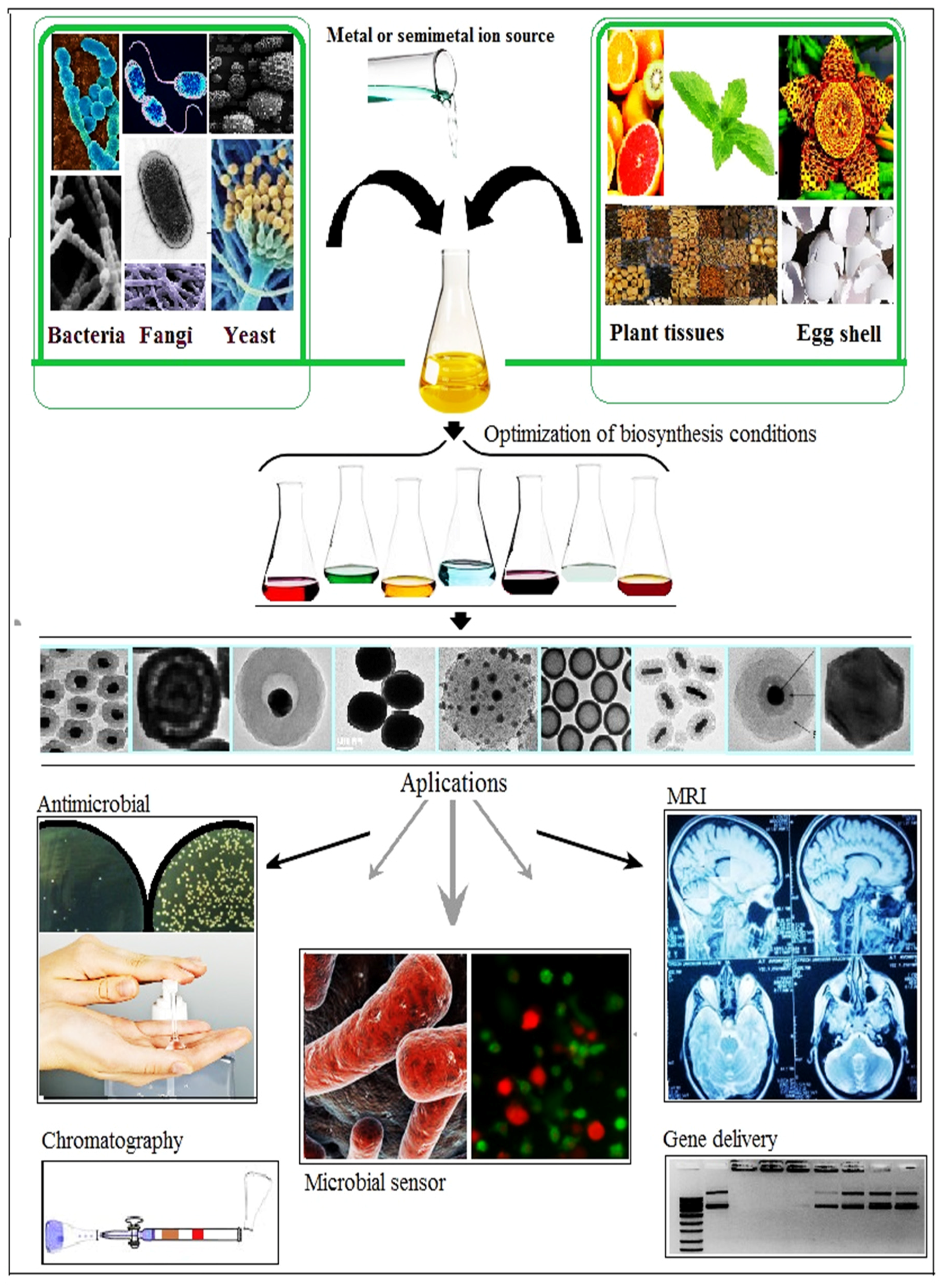
Nanoscience is an interdisciplinary field of science that has countless applications (Figure 4). Core-shell NPs display improved properties when compared to single NPs due to their increased performance, durability and the breadth of their applications with special economic value [31,71,72,73,74,75]. Core-shell NPs have garnered special scientific interest as they exhibit some unique properties arising from their design, geometry core, shell or combination of core and shell materials. Not surprisingly, therefore, they have been used in assorted fields, such as medicine, engineering, industry, and material science, etc. [76,77,78,79,80,81,82,83,84,85,86,87]. These structures have several optimized features, namely their ability to function over a wide range of temperatures and pH, antimicrobial properties, and magnetic conductivity [88].
With the gradual decline of fossil-derived natural resources (oil, gas, and gasoline) and environmental degradation, there has been an expanded effort to develop green energy using abundant wind and sun light. The use of metallic oxide semiconductor NPs with strong absorption capability of sun light is one of the important application in green energy development. Core-shell nanostructures are especially attractive due to their optimal morphology and adjustable pore size (NPs with porous shell), and free space between core and shell (which imparts more stability under harsh environments). Finally, the enhanced biocompatibility and economic validation for large-scale production of powerful absorbents to eliminate contaminants from polluted waters are some of the other valued attributes.
Core-shell nanostructures, although small but are dominant entities with high thermal and chemical stability, lower toxicity, greater solubility, and higher permeability to specific target cells. Some of the core-shell nanostructures are multifunctional and thus can be used for several applications (Table 4) with magnetic and luminescent parts. Their magnetic component can help improve the penetration of NPs into cells and also enhance the resolution of traditional magnetic resonance images (MRI), while luminescent part can assist in luminescent-based detection [89]. Further, such multifunctional NPs can be used for delivery of biomolecules, such as antibodies, proteins, and genes for treatment [90].
Core-shell nanogels, with intelligent and bio-degradable properties, have made significant inroads in the biomedical applications as drug carrier; remarkable feature being the dual loading drug capacity and controlled release at the target site [97].
Simultaneous transmission of DNA and drug to achieve therapeutic effects of combining drugs and gene therapy has been suggested through the use of biodegradable cationic amphiphilic core shell polymeric NPs that have the ability to load hydrophobic drugs via their core and the ability to bind to DNA through their cationic shells.
The architecture and synthesis of core-shell NPs with desired structure and their usage for a wide range of applications (Table 4) will attract the attention of scientists towards this class of biosynthesized core-shell NPs. The design of custom core-shell NPs structures and their applications appear truly endless. Commercial products will be offered in this area, which has very strong economic implications in the future.
6. Conclusions
The ability to manipulate particle structures at nanoscale has led to the emergence of a new class of engineered hybrid NPs, core-shell NPs. The developments in the field of greener synthesis of NPs in general and for core-shell NPs in particular has prompted us to summarize the current status in this field. The focus has been on introducing core-shell NPs, their synthesis with emphasis on greener methods, and their applications in a variety of scientific disciplines. Successful further scientific research and the industrial implementation of applications in this area may help to improve the quality of life for the world’s population in the future. Greener and sustainable synthetic methods can reduce environmental pollution to some extent, while attaining economic growth via green chemistry.
The importance and necessity of future progress in core-shell NPs encourages us to put forth the greener synthesis possibilities for core-shell NPs. We hope that some active researchers may feel stimulated to explore the use of this class of hybrid core-shell NPs instead of single NPs.
Acknowledgments
The authors acknowledged from Bam University of Medical Sciences for the financial support from Grant “mubam 96”.
Author Contributions
All author contributed different sections to this review article; M.K. and H.Q.A. did the literature survey, M.S.N. did the drawings and R.S.V. wrote, corrected and edited for English the entire manuscript.
Conflicts of Interest
The author declares no competing financial interests.
References
- Sharifi, F.; Sharififar, F.; Sharifi, I.; Alijani, H.; Khatami, M. Cytotoxicity, Leishmanicidal and Antioxidant Activity of Biosynthesized Zinc Sulfide Nanoparticles Using Phoenix dactylifera. IET Nanobiotechnol. 2018, 12. [Google Scholar] [CrossRef]
- Khatami, M.; Alijani, H.; Sharifi, I.; Sharifi, F.; Pourseyedi, S.; Kharazi, S.; Lima Nobre, M.A.; Khatami, M. Leishmanicidal Activity of Biogenic Fe3O4 Nanoparticles. Sci. Pharm. 2017, 85, 36. [Google Scholar] [CrossRef] [PubMed]
- Nadagouda, M.N.; Varma, R.S. Green Synthesis of Silver and Palladium Nanoparticles at Room Temperature Using Coffee and Tea Extract. Green Chem. 2008, 10, 859–862. [Google Scholar] [CrossRef]
- Mohammadinejad, R.; Karimi, S.; Iravani, S.; Varma, R.S. Plant-Derived Nanostructures: Types and Applications. Green Chem. 2016, 18, 20–52. [Google Scholar] [CrossRef]
- Mélinon, P.; Begin-Colin, S.; Duvail, J.L.; Gauffre, F.; Boime, N.H.; Ledoux, G.; Plain, J.; Reiss, P.; Silly, F.; Warot-Fonrose, B. Engineered Inorganic Core/Shell Nanoparticles. Phys. Rep. 2014, 543, 163–197. [Google Scholar] [CrossRef] [Green Version]
- Khatami, M.; Mehnipor, R.; Poor, M.H.S.; Jouzani, G.S. Facile Biosynthesis of Silver Nanoparticles Using Descurainia sophia and Evaluation of Their Antibacterial and Antifungal Properties. J. Clust. Sci. 2016, 27, 1601–1612. [Google Scholar] [CrossRef]
- Singh, P.; Kim, Y.-J.; Zhang, D.; Yang, D.-C. Biological Synthesis of Nanoparticles from Plants and Microorganisms. Trends Biotechnol. 2016, 34, 588–599. [Google Scholar] [CrossRef] [PubMed]
- Gawande, M.B.; Goswami, A.; Asefa, T.; Guo, H.; Biradar, A.V.; Peng, D.-L.; Zboril, R.; Varma, R.S. Core-Shell Nanoparticles: Synthesis and Applications in Catalysis and Electrocatalysis. Chem. Soc. Rev. 2015, 44, 7540–7590. [Google Scholar] [CrossRef] [PubMed]
- López-Lorente, A.I.; Simonet, B.M.; Valcárcel, M. Analytical Potential of Hybrid Nanoparticles. Anal. Bioanal. Chem. 2011, 399, 43–54. [Google Scholar] [CrossRef] [PubMed]
- Nomoev, A.V.; Bardakhanov, S.P.; Schreiber, M.; Bazarova, D.G.; Romanov, N.A.; Baldanov, B.B.; Radnaev, B.R.; Syzrantsev, V.V. Structure and Mechanism of the Formation of Core–Shell Nanoparticles Obtained through a One-Step Gas-Phase Synthesis by Electron Beam Evaporation. Beilstein J. Nanotechnol. 2015, 6, 874–880. [Google Scholar] [CrossRef] [PubMed]
- Allafchian, A.; Jalali, S.A.H.; Bahramian, H.; Ahmadvand, H. Preparation, Characterization, and Antibacterial Activity of NiFe2O4/Pama/Ag–TiO2 Nanocomposite. J. Mag. Mag. Mater. 2016, 404, 14–20. [Google Scholar] [CrossRef]
- Nadagouda, M.N.; Castle, A.; Murdock, R.C.; Hussain, S.M.; Varma, R.S. In Vitro Biocompatibility of Nanoscale Zerovalent Iron Particles (NZVI) Synthesized Using Tea Polyphenols. Green Chem. 2010, 12, 114–122. [Google Scholar] [CrossRef]
- Karthik, K.; Dhanuskodi, S.; Gobinath, C.; Prabukumar, S.; Sivaramakrishnan, S. Nanostructured CdO-NiO Composite for Multifunctional Applications. J. Phys. Chem. Sol. 2018, 112, 106–118. [Google Scholar] [CrossRef]
- Karthik, K.; Dhanuskodi, S.; Prabu Kumar, S.; Gobinath, C.; Sivaramakrishnan, S. Microwave Assisted Green Synthesis of Mgo Nanorods and Their Antibacterial and Anti-Breast Cancer Activities. Mater. Lett. 2017, 206, 217–220. [Google Scholar] [CrossRef]
- Hameed, A.S.H.; Karthikeyan, C.; Ahamed, A.P.; Thajuddin, N.; Alharbi, N.S.; Alharbi, S.A.; Ravi, G. In Vitro Antibacterial Activity of ZnO and Nd Doped ZnO Nanoparticles against Esbl Producing Escherichia coli and Klebsiella pneumoniae. Sci. Rep. 2016, 6, 24312. [Google Scholar] [CrossRef] [PubMed]
- Karthik, K.; Dhanuskodi, S.; Gobinath, C.; Prabukumar, S.; Sivaramakrishnan, S. Dielectric and Antibacterial Studies of Microwave Assisted Calcium Hydroxide Nanoparticles. J. Mater. Sci. Mater. Electron. 2017, 28, 16509–16518. [Google Scholar] [CrossRef]
- Karthik, K.; Dhanuskodi, S.; Gobinath, C.; Prabukumar, S.; Sivaramakrishnan, S. Andrographis Paniculata Extract Mediated Green Synthesis of CdO Nanoparticles and Its Electrochemical and Antibacterial Studies. J. Mater. Sci. Mater. Electron. 2017, 28, 7991–8001. [Google Scholar] [CrossRef]
- Rafiee, E.; Khodayari, M. Starch as a Green Source for Fe3O4@Carbon Core–Shell Nanoparticles Synthesis: A Support for 12-Tungstophosphoric Acid, Synthesis, Characterization, and Application as an Efficient Catalyst. Res. Chem. Intermed. 2016, 42, 3523–3536. [Google Scholar] [CrossRef]
- Miyakawa, M.; Hiyoshi, N.; Nishioka, M.; Koda, H.; Sato, K.; Miyazawa, A.; Suzuki, T.M. Continuous Syntheses of Pd@Pt and Cu@Ag Core-Shell Nanoparticles Using Microwave-Assisted Core Particle Formation Coupled with Galvanic Metal Displacement. Nanoscale 2014, 6, 8720–8725. [Google Scholar] [CrossRef] [PubMed]
- Devi, P.; Patil, S.D.; Jeevanandam, P.; Navani, N.K.; Singla, M.L. Synthesis, Characterization and Bactericidal Activity of Silica/Silver Core–Shell Nanoparticles. J. Mater. Sci. Mater. Med. 2014, 25, 1267–1273. [Google Scholar] [CrossRef] [PubMed]
- Iyanna, N.; Lalley, J.; Han, C.; Dionysiou, D.D.; Varma, R.S.; Nadagouda, M.N. Synthesis of Silver and Gold Nanoparticles Using Antioxidants from Blackberry, Blueberry, Pomegranate, and Turmeric Extracts. ACS Sustain. Chem. Eng. 2014, 2, 1717–1723. [Google Scholar]
- Allafchian, A.R.; Farhang, H.R.; Jalali, S.A.H.; Vahabi, M.R. Gundelia tournefortii L.: A Natural Source for the Green Synthesis of Silver Nanoparticles. IET Nanobiotechnol. 2017, 11, 815–820. [Google Scholar] [CrossRef]
- Zandpour, F.; Allafchian, A.; vahabi, M.R.; Jalali, S.A.H. The Green Synthesize of Silver Nanoparticles by Arial Part of Dorema ammoniacum D. Extract with Antimicrobial Analysis. IET Nanobiotechnol. 2018, 12. [Google Scholar] [CrossRef]
- Allafchian, A.; Jalali, S.A.H.; Aghaei, F.; farhang, H.R. Green Synthesis of Silver Nanoparticles Using Glaucium corniculatum (L.) Curtis Extract and Evaluation of Its Antibacterial Activity. IET Nanobiotechnol. 2018, 12. [Google Scholar] [CrossRef]
- Markova, Z.; Novak, P.; Kaslik, J.; Plachtova, P.; Brazdova, M.; Jancula, D.; Siskova, K.M.; Machala, L.; Marsalek, B.; Zboril, R.; et al. Iron(II,III)–Polyphenol Complex Nanoparticles Derived from Green Tea with Remarkable Ecotoxicological Impact. ACS Sustain. Chem. Eng. 2014, 2, 1674–1680. [Google Scholar] [CrossRef]
- Charbgoo, F.; Ramezani, M.; Darroudi, M. Bio-Sensing Applications of Cerium Oxide Nanoparticles: Advantages and Disadvantages. Biosen. Bioelectron. 2017, 96, 33–43. [Google Scholar] [CrossRef] [PubMed]
- Soltanzadeh, M.; Soltani Nejad, M.; Shahidi Bonjar, G.H. Application of Soil-Borne Actinomycetes for Biological Control against Fusarium Wilt of Chickpea (Cicer arietinum) Caused by Fusarium solani Fsp Pisi. J. Phytopathol. 2016, 164, 967–978. [Google Scholar] [CrossRef]
- Taghavizadeh Yazdi, M.E.; Khara, J.; Sadeghnia, H.R.; Esmaeilzadeh Bahabadi, S.; Darroudi, M. Biosynthesis, Characterization, and Antibacterial Activity of Silver Nanoparticles Using Rheum Turkestanicum Shoots Extract. Res. Chem. Intermed. 2018, 44, 1325–1334. [Google Scholar] [CrossRef]
- Miri, A.; Darroudi, M.; Entezari, R.; Sarani, M. Biosynthesis of Gold Nanoparticles Using Prosopis farcta Extract and Its in Vitro Toxicity on Colon Cancer Cells. Res. Chem. Intermed. 2018, 1–9. [Google Scholar] [CrossRef]
- Darroudi, M.; Sarani, M.; Kazemi Oskuee, R.; Khorsand Zak, A.; Amiri, M.S. Nanoceria: Gum Mediated Synthesis and in Vitro Viability Assay. Ceram. Int. 2014, 40, 2863–2868. [Google Scholar] [CrossRef]
- Pishahang, J.; Amiri, H.B.; Heli, H. Synthesis of Carbon Nanoparticles-Poly(Ortho-Aminophenol) Nanocomposite and Its Application for Electroanalysis of Iodate. Sens. Actuators B Chem. 2018, 256, 878–887. [Google Scholar] [CrossRef]
- Mirzaei, H.; Darroudi, M. Zinc Oxide Nanoparticles: Biological Synthesis and Biomedical Applications. Ceram. Int. 2017, 43, 907–914. [Google Scholar] [CrossRef]
- Jamdagni, P.; Khatri, P.; Rana, J.S. Nanoparticles Based DNA Conjugates for Detection of Pathogenic Microorganisms. Int. Nano Lett. 2016, 6, 139–146. [Google Scholar] [CrossRef]
- Jamdagni, P.; Khatri, P.; Rana, J.S. Green Synthesis of Zinc Oxide Nanoparticles Using Flower Extract of Nyctanthes Arbor-Tristis and Their Antifungal Activity. J. King Saud Univ. Sci. 2016. [Google Scholar] [CrossRef]
- Hebbalalu, D.; Lalley, J.; Nadagouda, M.N.; Varma, R.S. Greener Techniques for the Synthesis of Silver Nanoparticles Using Plant Extracts, Enzymes, Bacteria, Biodegradable Polymers, and Microwaves. ACS Sustain. Chem. Eng. 2013, 1, 703–712. [Google Scholar] [CrossRef]
- Ahn, S.; Singh, P.; Jang, M.; Kim, Y.J.; Castro-Aceituno, V.; Simu, S.Y.; Kim, Y.J.; Yang, D.C. Gold Nanoflowers Synthesized Using Acanthopanacis Cortex Extract Inhibit Inflammatory Mediators in Lps-Induced Raw264.7 Macrophages via Nf-Kappab and Ap-1 Pathways. Colloids Surf. B Biointerfaces 2017, 160, 423–428. [Google Scholar] [PubMed]
- Prasad, C.; Sreenivasulu, K.; Gangadhara, S.; Venkateswarlu, P. Bio Inspired Green Synthesis of Ni/Fe3O4 Magnetic Nanoparticles Using Moringa Oleifera Leaves Extract: A Magnetically Recoverable Catalyst for Organic Dye Degradation in Aqueous Solution. J. Alloy Comp. 2017, 700, 252–258. [Google Scholar] [CrossRef]
- Ghosh, S.; Jagtap, S.; More, P.; Shete, U.J.; Maheshwari, N.O.; Rao, S.J.; Kitture, R.; Kale, S.; Bellare, J.; Patil, S.; et al. Dioscorea Bulbifera Mediated Synthesis of Novel Aucoreagshell Nanoparticles with Potent Antibiofilm and Antileishmanial Activity. J. Nanomater. 2015, 16, 161. [Google Scholar]
- Venkateswarlu, S.; Natesh Kumar, B.; Prathima, B.; Anitha, K.; Jyothi, N.V.V. A Novel Green Synthesis of Fe3o4-Ag Core Shell Recyclable Nanoparticles Using Vitis vinifera Stem Extract and Its Enhanced Antibacterial Performance. Phys. B Condens. Matter 2015, 457, 30–35. [Google Scholar] [CrossRef]
- Kirubha, E.; Palanisamy, P.K. Green Synthesis, Characterization of Au–Ag Core–Shell Nanoparticles Using Gripe Water and Their Applications in Nonlinear Optics and Surface Enhanced Raman Studies. Adv. Nat. Sci. Nanosci. Nanotechnol. 2014, 5, 045006. [Google Scholar] [CrossRef]
- Poor, M.H.S.; Khatami, M.; Azizi, H.; Abazari, Y. Cytotoxic Activity of Biosynthesized Ag Nanoparticles by Plantago Major Towards a Human Breast Cancer Cell Line. Rendioconti Lincei 2017, 28, 693–699. [Google Scholar] [CrossRef]
- Ganaie, S.U.; Abbasi, T.; Abbasi, S.A. Rapid and Green Synthesis of Bimetallic Au–Ag Nanoparticles Using an Otherwise Worthless Weed Antigonon Leptopus. J. Exp. Nanosci. 2016, 11, 395–417. [Google Scholar] [CrossRef]
- Song, J.Y.; Kim, B.S. Biological Synthesis of Bimetallic Au/Ag Nanoparticles Using Persimmon (Diopyros kaki) Leaf Extract. Korean J. Chem. Eng. 2008, 25, 808–811. [Google Scholar] [CrossRef]
- Shankar, S.S.; Rai, A.; Ahmad, A.; Sastry, M. Rapid Synthesis of Au, Ag, and Bimetallic Au Core–Ag Shell Nanoparticles Using Neem (Azadirachta Indica) Leaf Broth. J. Colloid Interface Sci. 2004, 275, 496–502. [Google Scholar] [CrossRef] [PubMed]
- Zhan, G.; Huang, J.; Du, M.; Abdul-Rauf, I.; Ma, Y.; Li, Q. Green Synthesis of Au–Pd Bimetallic Nanoparticles: Single-Step Bioreduction Method with Plant Extract. Mater. Lett. 2011, 65, 2989–2991. [Google Scholar] [CrossRef]
- AbdelHamid, A.A.; Al-Ghobashy, M.A.; Fawzy, M.; Mohamed, M.B.; Abdel-Mottaleb, M.M.S.A. Phytosynthesis of Au, Ag, and Au–Ag Bimetallic Nanoparticles Using Aqueous Extract of Sago Pondweed (Potamogeton pectinatus L.). ACS Sustain. Chem. Eng. 2013, 1, 1520–1529. [Google Scholar] [CrossRef]
- Usha Rani, P.; Rajasekharreddy, P. Green Synthesis of Silver-Protein (Core–Shell) Nanoparticles Using Piper Betle, L. Leaf Extract and Its Ecotoxicological Studies on Daphnia Magna. Colloids Surf. A Physicochem. Eng. Asp. 2011, 389, 188–194. [Google Scholar] [CrossRef]
- Sheny, D.S.; Mathew, J.; Philip, D. Phytosynthesis of Au, Ag and Au–Ag Bimetallic Nanoparticles Using Aqueous Extract and Dried Leaf of Anacardium Occidentale. Spectrochim. Acta Part A Mol. Biol. Spectrosc. 2011, 79, 254–262. [Google Scholar] [CrossRef] [PubMed]
- Sharma, A.; Tapadia, K. Greener Production of Magnetic Nanoparticles and Their Fabrication. Curr. Sci. 2016, 111, 2018. [Google Scholar] [CrossRef]
- Baruwati, B.; Simmons, S.O.; Varma, R.S.; Veronesi, B. “Green” Synthesized and Coated Nanosilver Alters the Membrane Permeability of Barrier (Intestinal, Brain Endothelial) Cells and Stimulates Oxidative Stress Pathways in Neurons. ACS Sustain. Chem. Eng. 2013, 1, 753–759. [Google Scholar] [CrossRef]
- Luo, C.-H.; Shanmugam, V.; Yeh, C.-S. Nanoparticle Biosynthesis Using Unicellular and Subcellular Supports. NPG Asia Mater. 2015, 7, e209. [Google Scholar] [CrossRef]
- Khodashenas, B.; Ghorbani, H.R. Optimisation of Nitrate Reductase Enzyme Activity to Synthesise Silver Nanoparticles. IET Nanobiotechnol. 2016, 10, 158–161. [Google Scholar] [CrossRef] [PubMed]
- Selvakannan, P.; Swami, A.; Srisathiyanarayanan, D.; Shirude, P.S.; Pasricha, R.; Mandale, A.B.; Sastry, M. Synthesis of Aqueous Au Core-Ag Shell Nanoparticles Using Tyrosine as a Ph-Dependent Reducing Agent and Assembling Phase-Transferred Silver Nanoparticles at the Air-Water Interface. Langmuir 2004, 20, 7825–7836. [Google Scholar] [CrossRef] [PubMed]
- Engelbrekt, C.; Sorensen, K.H.; Zhang, J.; Welinder, A.C.; Jensen, P.S.; Ulstrup, J. Green Synthesis of Gold Nanoparticles with Starch-Glucose and Application in Bioelectrochemistry. J. Mater. Chem. 2009, 19, 7839–7847. [Google Scholar] [CrossRef]
- Virkutyte, J.; Varma, R.S. Green Synthesis of Metal Nanoparticles: Biodegradable Polymers and Enzymes in Stabilization and Surface Functionalization. Chem. Sci. 2011, 2, 837–846. [Google Scholar] [CrossRef]
- Xing, R.; Jiao, T.; Yan, L.; Ma, G.; Liu, L.; Dai, L.; Li, J.; Möhwald, H.; Yan, X. Colloidal Gold–Collagen Protein Core–Shell Nanoconjugate: One-Step Biomimetic Synthesis, Layer-by-Layer Assembled Film, and Controlled Cell Growth. ACS Appl. Mater. Interfaces 2015, 7, 24733–24740. [Google Scholar] [CrossRef] [PubMed]
- Wu, W.; He, Q.; Jiang, C. Magnetic Iron Oxide Nanoparticles: Synthesis and Surface Functionalization Strategies. Nanoscale Res. Lett. 2008, 3, 397–415. [Google Scholar] [CrossRef] [PubMed]
- Valodkar, M.; Modi, S.; Pal, A.; Thakore, S. Synthesis and Anti-Bacterial Activity of Cu, Ag and Cu–Ag Alloy Nanoparticles: A Green Approach. Mater. Res. Bull. 2011, 46, 384–389. [Google Scholar] [CrossRef]
- Srivastava, S.K.; Hasegawa, T.; Yamada, R.; Ogino, C.; Mizuhata, M.; Kondo, A. Green Synthesis of Au, Pd and Au@Pd Core-Shell Nanoparticles Via a Tryptophan Induced Supramolecular Interface. RSC Adv. 2013, 3, 18367–18372. [Google Scholar] [CrossRef]
- Alarfaj, N.A.; El-Tohamy, M.F. Eco-Friendly Synthesis of Gelatin-Capped Bimetallic Au–Ag Nanoparticles for Chemiluminescence Detection of Anticancer Raloxifene Hydrochloride. Luminescence 2016, 31, 1194–1200. [Google Scholar] [CrossRef] [PubMed]
- Huang, X.; Wu, H.; Pu, S.; Zhang, W.; Liao, X.; Shi, B. One-Step Room-Temperature Synthesis of Au@Pd Core-Shell Nanoparticles with Tunable Structure Using Plant Tannin as Reductant and Stabilizer. Green Chem. 2011, 13, 950–957. [Google Scholar] [CrossRef]
- Zhang, G.; Zheng, H.; Shen, M.; Wang, L.; Wang, X. Green Synthesis and Characterization of Au@Pt Core–Shell Bimetallic Nanoparticles Using Gallic Acid. J. Phys. Chem. Solids 2015, 81, 79–87. [Google Scholar] [CrossRef]
- Nadagouda, M.N.; Varma, R.S. A Greener Synthesis of Core (Fe, Cu)-Shell (Au, Pt, Pd, and Ag) Nanocrystals Using Aqueous Vitamin C. Cryst. Growth Des. 2007, 7, 2582–2587. [Google Scholar] [CrossRef]
- Makarov, V.V.; Love, A.J.; Sinitsyna, O.V.; Makarova, S.S.; Yaminsky, I.V.; Taliansky, M.E.; Kalinina, N.O. “Green” Nanotechnologies: Synthesis of Metal Nanoparticles Using Plants. Acta Nat. 2014, 6, 35–44. [Google Scholar]
- Raju, D.; Mehta, U.J.; Hazra, S. Synthesis of Gold Nanoparticles by Various Leaf Fractions of Semecarpus anacardium L. tree. Trees 2011, 25, 145–151. [Google Scholar] [CrossRef]
- Joshi, H.M.; De, M.; Richter, F.; He, J.; Prasad, P.V.; Dravid, V.P. Effect of Silica Shell Thickness of FeO–Sio(X) Core–Shell Nanostructures on MRI Contrast. Contrast Med. Mol. Imaging 2012, 7, 460–468. [Google Scholar]
- Yang, L.; Yan, W.; Wang, H.; Zhuang, H.; Zhang, J. Shell Thickness-Dependent Antibacterial Activity and Biocompatibility of Gold@Silver Core-Shell Nanoparticles. RSC Adv. 2017, 7, 11355–11361. [Google Scholar] [CrossRef]
- Khanchandani, S.; Kundu, S.; Patra, A.; Ganguli, A.K. Shell Thickness Dependent Photocatalytic Properties of Zno/Cds Core–Shell Nanorods. J. Phys. Chem. C 2012, 116, 23653–23662. [Google Scholar] [CrossRef]
- Sattarahmady, N.; Rahi, A.; Heli, H. A Signal-on Built in-Marker Electrochemical Aptasensor for Human Prostate-Specific Antigen Based on a Hairbrush-Like Gold Nanostructure. Sci. Rep. 2017, 7, 11238. [Google Scholar] [CrossRef] [PubMed]
- Sattarahmady, N.; Tondro, G.H.; Gholchin, M.; Heli, H. Gold Nanoparticles Biosensor of Brucella S Genomic DNA: Visual and Spectrophotometric Detections. Biochem. Eng. J. 2015, 97, 1–7. [Google Scholar] [CrossRef]
- Hossein, H.; Masoud, N. Applications of Nanoflowers in Biomedicine. Recent Pat. Nanotechnol. 2017, 11, 1–12. [Google Scholar]
- Mortazavi, S.M.; Khatami, M.; Sharifi, I.; Heli, H.; Kaykavousi, K.; Sobhani Poor, M.H.; Kharazi, S.; Nobre, M.A.L. Bacterial Biosynthesis of Gold Nanoparticles Using Salmonella enterica Subsp. Enterica Serovar Typhi Isolated from Blood and Stool Specimens of Patients. J. Clust. Sci. 2017, 28, 2997–3007. [Google Scholar] [CrossRef]
- Khorrami, M.B.; Sadeghnia, H.R.; Pasdar, A.; Ghayour-Mobarhan, M.; Riahi-Zanjani, B.; Darroudi, M. Role of Pullulan in Preparation of Ceria Nanoparticles and Investigation of Their Biological Activities. J. Mol. Struct. 2018, 1157, 127–131. [Google Scholar] [CrossRef]
- Varma, R.S. Greener Approach to Nanomaterials and Their Sustainable Applications. Curr. Opin. Chem. Eng. 2012, 1, 123–128. [Google Scholar] [CrossRef]
- Moghaddam, H.M.; Beitollahi, H.; Tajik, S.; Jahani, S.; Khabazzadeh, H.; Alizadeh, R. Voltammetric Determination of Droxidopa in the Presence of Carbidopa Using a Nanostructured Base Electrochemical Sensor. Russ. J. Electrochem. 2017, 53, 452–460. [Google Scholar] [CrossRef]
- Beitollai, H.; Garkani Nejad, F.; Tajik, S.; Jahani, S.; Biparva, P. Voltammetric Determination of Amitriptyline Based on Graphite Screen Printed Electrode Modified with a Copper Oxide Nanoparticles. Int. J. Nano Dimens. 2017, 8, 197–205. [Google Scholar]
- Jahani, S.; Beitollahi, H. Selective Detection of Dopamine in the Presence of Uric Acid Using Nio Nanoparticles Decorated on Graphene Nanosheets Modified Screen-Printed Electrodes. Electroanalysis 2016, 28, 2022–2028. [Google Scholar] [CrossRef]
- Khorasani-Motlagh, M.; Noroozifar, M.; Jahani, S. Preparation and Characterization of Nano-Sized Magnetic Particles Lacoo3 by Ultrasonic-Assisted Co-precipitation Method. Synth. React. Inorg. Metal-Org. Nano-Metal Chem. 2015, 45, 1591–1595. [Google Scholar] [CrossRef]
- Niroomand, S.; Khorasani-Motlagh, M.; Noroozifar, M.; Jahani, S.; Moodi, A. Photochemical and Dft Studies on DNA-Binding Ability and Antibacterial Activity of Lanthanum(Iii)-Phenanthroline Complex. J. Mol. Struct. 2017, 1130, 940–950. [Google Scholar] [CrossRef]
- Beitollahi, H.; Tajik, S.; Jahani, S. Electrocatalytic Determination of Hydrazine and Phenol Using a Carbon Paste Electrode Modified with Ionic Liquids and Magnetic Core-Shell Fe3O4@SiO2/MWSNT Nanocomposite. Electroanalysis 2016, 28, 1093–1099. [Google Scholar] [CrossRef]
- Jahani, S.; Khorasani-Motlagh, M.; Noroozifar, M. DNA Interaction of Europium(Iii) Complex Containing 2,2′-Bipyridine and Its Antimicrobial Activity. J. Biomol. Struct. Dyn. 2016, 34, 612–624. [Google Scholar] [CrossRef] [PubMed]
- Jahani, S.; Beitollahi, H. Carbon Paste Electrode Modified with TiO2/Fe3O4/MWCNT Nanocomposite and Ionic Liquids as a Voltammetric Sensor for Sensitive Ascorbic Acid and Tryptophan Detection. Anal. Bioanal. Electrochem. 2016, 8, 158–168. [Google Scholar]
- Singh, P.; Singh, H.; Ahn, S.; Castro-Aceituno, V.; Jimenez, Z.; Simu, S.Y.; Kim, Y.J.; Yang, D.C. Pharmacological Importance, Characterization and Applications of Gold and Silver Nanoparticles Synthesized by Panax Ginseng Fresh Leaves. Artif. Cells Nanomed. Biotechnol. 2017, 45, 1415–1424. [Google Scholar] [CrossRef] [PubMed]
- Singh, A.K.; Srivastava, O.N. One-Step Green Synthesis of Gold Nanoparticles Using Black Cardamom and Effect of Ph on Its Synthesis. Nano Res. Lett. 2015, 10, 353. [Google Scholar] [CrossRef] [PubMed]
- Singh, P.; Singh, H.; Castro-Aceituno, V.; Ahn, S.; Kim, Y.J.; Farh, M.E.-A.; Yang, D.C. Engineering of Mesoporous Silica Nanoparticles for Release of Ginsenoside Ck and Rh2 to Enhance Their Anticancer and Anti-Inflammatory Efficacy: In Vitro Studies. J. Nanopart. Res. 2017, 19, 257. [Google Scholar] [CrossRef]
- Singh, P.; Ahn, S.; Kang, J.P.; Veronika, S.; Huo, Y.; Singh, H.; Chokkaligam, M.; El-Agamy Farh, M.; Aceituno, V.C.; Kim, Y.J.; et al. In Vitro Anti-Inflammatory Activity of Spherical Silver Nanoparticles and Monodisperse Hexagonal Gold Nanoparticles by Fruit Extract of Prunus serrulata: A Green Synthetic Approach. Artif. Cells Nanomed. Biotechnol. 2017, 30, 1–11. [Google Scholar] [CrossRef] [PubMed]
- Seddighi, N.S.; Salari, S.; Izadi, A.R. Evaluation of Antifungal Effect of Iron-Oxide Nanoparticles against Different Candida Species. IET Nanobiotechnol. 2017, 11. [Google Scholar] [CrossRef]
- Zaimy, M.A.; Saffarzadeh, N.; Mohammadi, A.; Pourghadamyari, H.; Izadi, P.; Sarli, A.; Moghaddam, L.K.; Paschepari, S.R.; Azizi, H.; Torkamandi, S.; et al. New Methods in the Diagnosis of Cancer and Gene Therapy of Cancer Based on Nanoparticles. Cancer Gene Ther. 2017, 24, 233–243. [Google Scholar] [CrossRef] [PubMed]
- Zaimy, M.A.; Jebali, A.; Bazrafshan, B.; Mehrtashfar, S.; Shabani, S.; Tavakoli, A.; Hekmatimoghaddam, S.H.; Sarli, A.; Azizi, H.; Izadi, P.; et al. Coinhibition of Overexpressed Genes in Acute Myeloid Leukemia Subtype M2 by Gold Nanoparticles Functionalized with Five Antisense Oligonucleotides and One Anti-Cd33(+)/Cd34(+) Aptamer. Cancer Gene Ther. 2016, 23, 315–320. [Google Scholar] [CrossRef] [PubMed]
- Yang, Y.; Shi, J.; Kawamura, G.; Nogami, M. Preparation of Au–Ag, Ag–Au Core–Shell Bimetallic Nanoparticles for Surface-Enhanced Raman Scattering. Scripta Mater. 2008, 58, 862–865. [Google Scholar] [CrossRef]
- Ren, C.; Ding, X.; Fu, H.; Li, W.; Wu, H.; Yang, H. Core-Shell Superparamagnetic Monodisperse Nanospheres Based on Amino-Functionalized CoFe2O4@SiO2 for Removal of Heavy Metals from Aqueous Solutions. RSC Adv. 2017, 7, 6911–6921. [Google Scholar] [CrossRef]
- Byers, C.P.; Zhang, H.; Swearer, D.F.; Yorulmaz, M.; Hoener, B.S.; Huang, D.; Hoggard, A.; Chang, W.-S.; Mulvaney, P.; Ringe, E.; et al. From Tunable Core-Shell Nanoparticles to Plasmonic Drawbridges: Active Control of Nanoparticle Optical Properties. Sci. Adv. 2015, 1, e1500988. [Google Scholar] [CrossRef] [PubMed]
- Howes, P.D.; Chandrawati, R.; Stevens, M.M. Colloidal Nanoparticles as Advanced Biological Sensors. Science 2014, 346, 1247390. [Google Scholar] [CrossRef] [PubMed]
- Lee, H.; Yoon, T.-J.; Weissleder, R. Ultrasensitive Detection of Bacteria Using Core-Shell Nanoparticles and a Nmr-Filter System. Angew. Chem. Int. Ed. 2009, 48, 5657–5660. [Google Scholar] [CrossRef] [PubMed]
- Park, J.C.; Lee, G.T.; Seo, J.H. Mannose-Functionalized Core@Shell Nanoparticles and Their Interactions with Bacteria. J. Mater. Sci. 2017, 52, 1534–1545. [Google Scholar] [CrossRef]
- Gontero, D.; Veglia, A.V.; Bracamonte, A.G.; Boudreau, D. Synthesis of Ultraluminescent Gold Core-Shell Nanoparticles as Nanoimaging Platforms for Biosensing Applications Based on Metal-Enhanced Fluorescence. RSC Adv. 2017, 7, 10252–10258. [Google Scholar] [CrossRef]
- Kemin, W.; Weihong, T.; Xiaoxiao, H. Biomedical Applications Based on Core-Shell Nanoparticles. In Proceedings of the 27th Annual International Conference of the Engineering in Medicine and Biology Society, Shanghai, China, 17–18 January 2006. [Google Scholar]
- Yang, G.; Chen, D.; Lv, P.; Kong, X.; Sun, Y.; Wang, Z.; Yuan, Z.; Liu, H.; Yang, J. Core-Shell Au-Pd Nanoparticles as Cathode Catalysts for Microbial Fuel Cell Applications. Sci. Rep. 2016, 6, 35252. [Google Scholar] [CrossRef] [PubMed]
- Behrendt, J.M.; Nagel, D.; Chundoo, E.; Alexander, L.M.; Dupin, D.; Hine, A.V.; Bradley, M.; Sutherland, A.J. Synthesis and Characterization of Dual-Functionalized Core-Shell Fluorescent Microspheres for Bioconjugation and Cellular Delivery. PLoS ONE 2013, 8, e50713. [Google Scholar] [CrossRef] [PubMed] [Green Version]
- Mitsudome, T.; Matoba, M.; Mizugaki, T.; Jitsukawa, K.; Kaneda, K. Core–Shell AgNP@CeO2 Nanocomposite Catalyst for Highly Chemoselective Reductions of Unsaturated Aldehydes. Chem. Eur. J. 2013, 19, 5255–5258. [Google Scholar] [CrossRef] [PubMed]
Figure 1.
Comparison of macro, micro, and nano scales.

Figure 2.
Schematic of core-shell nanoparticles: core in the center (yellow part) and other material located around the central core (green part).
Figure 2.
Schematic of core-shell nanoparticles: core in the center (yellow part) and other material located around the central core (green part).

Figure 3.
Schematic (A–J) pictures of different structure of core shell nanoparticles: (A) Core shell nanoparticles; (B) Core double-shell particles or core multi-shell nanoparticle; (C) Polyhedral core/shell nanoparticles; (D) Core porous-shell nanoparticles; (E) Hollow-core shell nanoparticles or single-shell nanoparticles; (F) Hollow-core double-shell nanoparticles; (G) Moveable-core shell nanoparticles; (H) Multi-core shell nanoparticles; (I) Irregular shape core shell nanoparticles; and, (J) Rod core shell nanoparticles.

Figure 4.
Green Synthesis and applications of core-shell nanoparticles in some biomedical, engineering and environmental Fields.
Figure 4.
Green Synthesis and applications of core-shell nanoparticles in some biomedical, engineering and environmental Fields.


Table 1.
Some examples of chemical and physical synthesis for core shell nanoparticles.
| No. | Core Shell NPs | Synthesis Method | Synthesis Details (Brief) | Ref. |
|---|---|---|---|---|
| 1 | Fe3O4@carbon | Combination of hydrothermal and chemical co precipitation | Ferric chloride hexahydrate and ferrous chloride tetrahydrate upon addition of NH4OH. Then a carbon layer was coated on Fe3O4 NPs by using starch as the carbon precursor to protect the Fe3O4 magnetic core from oxidation. | [17] |
| 2 | Cu@silica and Ag@Si | Through evaporation via a high powered electron beam | One-step: Silica and Cu were placed in a graphite crucible with a weight ratio of 20:1 with Cu on the bottom. The Ag@Si particles were synthesized with a Si:Ag weight ratio of 10:1 with Ag at the bottom. | [9] |
| 3 | Gold@Silver | Sodium citrate | Two-step: 4-mercaptobenzoic acid ligand (8 μL, 0.57 mM in 0.25 M NaOH) was added under vortex to the as synthesized citrate stabilized AuNPs solution (1 mL). The contents were incubated for 2 h at room temperature, then hydroquinone (30 μL, 10 mM in H2O) and AgNO3 (30 μL, 10 mM in H2O) were added under vigorous stirring for 12 h. | [18] |
| 4 | Pd@Pt and Cu@Ag | Microwave-assisted | An EG solution containing (PdCl4, 100 mM) and PVP (15 wt %) was introduced continuously into the quartz tube reactor placed in the microwave cavity. EG (bp 195 °C) was heated to 200 °C using MW irradiation under pressure. | [19] |
| 5 | Silica@silver | Electroless reduction | Silica core synthesized by hydrolysis and condensation of TEOS in an alkaline medium. TEOS (3.6 mL) was added to ammonium hydroxide (11.9 mL) in ethanol (88 mL) solution and stirred for 15 h. | [20] |
Table 2.
Biosynthesis of core shell nanoparticles using plants.
| No. | Plant | Nanoparticles Type | Method for Extract Preparation | Size (nm) | Ref. |
|---|---|---|---|---|---|
| 1 | Moringa Oleifera | Ni/Fe3O4 | 20 g leaves extract at 80 °C with 100 mL of de-ionized water for 2 h. | 16–20 | [37] |
| 2 | Dioscorea bulbifera | Au/Ag | 5 g of the dried tubers powder boiled in 100 mL of distilled water for 5 min. | 9 nm core total diameter 15 | [38] |
| 3 | Vitis vinifera | Fe3O4-Ag | 10 g stem powder mixed with 100 mL distilled water. | Below 50 | [39] |
| 4 | Antigonon leptopus | Au/Ag | 2 g of plant parts boiled in 100 mL of water for 5 min. | 10–60 | [42] |
| 5 | Diopyros kaki (Persimmon) | Au/Ag | 5 g of dried cut leaves in 100 mL of distilled water and boiling the mixture for 5 min. | 50–500 | [43] |
| 6 | Azadirachta indica (Neem) | Au/Ag | 20 g powder leaf boiled (2 min) in 100 mL of distilled water and finally decantation. | 50–100 | [44] |
| 7 | Cacumen platycladi | Au-Pd | 1 g leaf was dispersed in 100 mL deionized water on a shaker for 4 h. and filtrated. | ~7 nm | [45] |
| 8 | Potamogeton pectinatus | Au-Ag | 1.5 g in 30 mL distilled water, and then heated at 80 °C while stirring for 10 min. | 5 ± 10.6 nm | [46] |
| 9 | Piper betle L. | Silver-protein | 10 g of finely cut leaves in 200 mL of double-distilled deionized water and then boiling the mixture for 5 min. | 17–28 | [47] |
| 10 | Anacadium occidentale | Au-Ag | Conditions not described. | Below 25 | [48] |
| 11 | Green tea extract | Fe3O4@SiO2 | 50 g of green tea leaves boiled in 500 mL double-distilled-water, until reached boiling temperature. The extract was then vacuum filtered after 1 h. | - | [49] |
Table 3.
Greener synthesis of hybrid nanoparticles using natural products.
| No. | Bioresource Derivatives | Type | Ions Source | Size (nm) | Biosynthesis Detail | Ref. |
|---|---|---|---|---|---|---|
| 1 | Starch | Cu-Ag | Cu(NO3)2·3H2O and (AgNO3) and ascorbic acid and starch | 20 ± 5 | To the starch solution, equal volume of metal salt solution and ascorbic acid (10%, v/v, of starch) were added and reaction was carried out under microwave at full power for 90 s. | [58] |
| 2 | Tryptophan | Au@Pd | HAuCl4 and PdCl2 | ~60 | Palladium precursor solution (5 mL) was introduced to the gold NP seed solution (5 mL) along with 1 mL of tryptophan solution. The resulting mixture was stirred for 2 h followed by intermittent sonication for 10 s at every 30 min. | [59] |
| 3 | Gelatin | Au-Ag | HAuCl4 and AgNO3 | 30 | 5.0 mL of 1.0 × 10⁻² mol/L HAuCl4 solution to 50 mL of (1.0 wt %) gelatin solution, followed by 5.0 mL of 1.0 × 10⁻² mol/L AgNO3 with vigorous stirring at 50 °C for 6 h | [60] |
| 4 | Plant tannin (bay berry) | Au@Pd | HAuCl4 and PdCl2 | 12 ± 6 | 10.0 mL of HAuCl4/PdCl2 mixture with different molar ratios was mixed with 40.0 mL of BT solution (0.02 g). The resultant mixture was shaken at 30 °C | [61] |
| 5 | Gallic acid | Au@Pt | H2PtCl6 and HauCl4 | 40–60 | HAuCl4 and H2PtCl6 in a total volume of 0.5 mL) were mixed in 17.5 mL of doubly distilled water in a round-bottom flask of 50 mL, and heated to boiling under magnetic stirring, when 2.0 mL of gallic acid solution was rapidly added. | [62] |
| 6 | Ascorbic acid (vitamin C) | Core (Fe, Cu)-Shell (Au, Pt, Pd, and Ag) | CuCl2 and HAuCl4·3H2O | _ | 10 mL of 0.1 N ascorbic acid (vitamin C) was reacted with core CuCl2 (2 mL, 0.1 N) at room temperature, and then shell HAuCl4·3H2O (2 mL, 0.01 N) was added and allowed to react at room temperature for 1 h. | [63] |
| Fe3O4 /Au | Fe3O4 and HAuCl4 | 16 | 70 mg citrate-coated Fe3O4 nanoparticles were dispersed in 20 mL double distilled water and were sonicated for 20 min. To this, 700 mL of freshly prepared gold chloride solution (0.1 g/mL) was then added maintaining a Fe3O4 nanoparticle to gold chloride solution is in the ratio of 1:1 (w/w, 60–70 °C). The solution was kept under constant stirring condition. After 5 min, 560 mL of freshly prepared ascorbic acid solution (0.5 g/mL) was added maintaining gold chloride to ascorbic acid ratio of 1:4 (w/w, constant stirring). | [64] | ||
| 7 | Paraffin oleic acid | CdSe/CdS/ZnS | Se and CdO and Na2S·9H2O and zinc acetate | 0.2 mmol of Se was dissolved in 18 mL of paraffin in a three-necked flask at 220 °C under vigorous stirring. In another flask, the cadmium precursor was prepared by dissolving CdO (2 mmol) in a mixture of 0.6 mmol oleic acid (2 mL) and paraffin liquid (8 mL) at 160 °C. 2 mL of the cadmium precursor solution was quickly injected into the Se solution, and the temperature was kept at 220 °C. After 20 mins, the solution was cooled to 100 °C, followed by the addition of Na2S·9H2O (0.5 mmol) into the solution. The solution was heated to 160 °C under vigorous stirring followed by dropwise addition of the Zn precursor (3 mL) prepared by dissolving zinc acetate in liquid paraffin. The mixture was cooled to 100 °C and the reaction was allowed to continue for 90 mins to monitor the growth of the ZnS shell. | [65] |
Table 4.
Some applications of core shell nanoparticle in various scientific disciplines.
| No. | Nanoparticles Type | Application | Details | Ref. |
|---|---|---|---|---|
| 1 | CoFe2O4@SiO2-NH2 | Remove heavy metals | The regenerable magnetic nanospheres could be potential adsorbents for the effective and removal of heavy metal ions (Cd(II), Cu(II) and Pb(II) from aqueous solutions. | [91] |
| 2 | Au-Ag, Ag-Au | Chemical and biomolecular Sensor | Surface-enhanced Raman scattering | [90] |
| 3 | Au-Ag | Detection drugs | Chemiluminescence detection of anticancer drug, Raloxifene hydrochloride | [60] |
| 4 | Au/Ag core-shell nanoparticles | Sensor | Demonstrated fully reversible active control of plasmon resonances in both isolated and interacting plasmonic nanoparticles | [92,93,94] |
| 5 | Fe core and a thin ferrite shell | Biosensor development of new diagnostic platforms | Multiplexed intracellular sensing of forum RNAs in MCF-7 human breast cancer cells using AuNPs with a dense shell of multicolored molecular beacons. These show high nuclease resistance and biocompatibility. | [95,96,97] |
| 6 | Uniform-sized amino-modified silica NH2SiNPs | Sensor | Detection of breast cancer cells at early apoptosis | [88] |
| 7 | Au-Pd | Cathode catalysts | The core-shell Au-Pd nanoparticles could be used in MFCs as a good alternative to the Pt-based cathode catalysts for the production of electricity without losing power efficiency and stability. | [98] |
| 8 | Dual-Functionalized Core-Shell Fluorescent Microspheres | Vector | Far-red-labeled microspheres are ideal candidates for in vitro, cellular delivery of proteins. | [99] |
| 9 | AgNPs@CeO2 | Nanocatalysts | Catalyze the chemoselective reduction of unsaturated aldehydes to the corresponding unsaturated alcohols with H2 | [100] |
| 10 | Au-Ag | Parasitology | Antileishmanial Activity | [38] |
© 2018 by the authors. Licensee MDPI, Basel, Switzerland. This article is an open access article distributed under the terms and conditions of the Creative Commons Attribution (CC BY) license (http://creativecommons.org/licenses/by/4.0/).
Share and Cite
MDPI and ACS Style
Khatami, M.; Alijani, H.Q.; Nejad, M.S.; Varma, R.S. Core@shell Nanoparticles: Greener Synthesis Using Natural Plant Products. Appl. Sci. 2018, 8, 411. https://doi.org/10.3390/app8030411
AMA Style
Khatami M, Alijani HQ, Nejad MS, Varma RS. Core@shell Nanoparticles: Greener Synthesis Using Natural Plant Products. Applied Sciences. 2018; 8(3):411. https://doi.org/10.3390/app8030411
Chicago/Turabian StyleKhatami, Mehrdad, Hajar Q. Alijani, Meysam S. Nejad, and Rajender S. Varma. 2018. "Core@shell Nanoparticles: Greener Synthesis Using Natural Plant Products" Applied Sciences 8, no. 3: 411. https://doi.org/10.3390/app8030411
Note that from the first issue of 2016, this journal uses article numbers instead of page numbers. See further details here.