
Nanomaterials in Bone Regeneration
by
 , ,
, ,
Vaclav Babuska
* ,
,
Phanindra Babu Kasi
,
Petra Chocholata
,
Lucie Wiesnerova
,
Jana Dvorakova
,
Radana Vrzakova
,
Anna Nekleionova
,
Lukas Landsmann
and
Vlastimil Kulda

Department of Medical Chemistry and Biochemistry, Faculty of Medicine in Pilsen, Charles University, Karlovarska 48, 301 66 Plzen, Czech Republic
*
Author to whom correspondence should be addressed.
Appl. Sci. 2022, 12(13), 6793; https://doi.org/10.3390/app12136793
Submission received: 6 June 2022
/
Revised: 29 June 2022
/
Accepted: 3 July 2022
/
Published: 5 July 2022
(This article belongs to the Special Issue Biocompatible Materials for Bone Regeneration)
Abstract
:Nanomaterials are promising in the development of innovative therapeutic options that include tissue and organ replacement, as well as bone repair and regeneration. The expansion of new nanoscaled biomaterials is based on progress in the field of nanotechnologies, material sciences, and biomedicine. In recent decades, nanomaterial systems have bridged the line between the synthetic and natural worlds, leading to the emergence of a new science called nanomaterial design for biological applications. Nanomaterials replicating bone properties and providing unique functions help in bone tissue engineering. This review article is focused on nanomaterials utilized in or being explored for the purpose of bone repair and regeneration. After a brief overview of bone biology, including a description of bone cells, matrix, and development, nanostructured materials and different types of nanoparticles are discussed in detail.
1. Introduction
Nanomaterials are currently in the spotlight of bone regeneration and tissue engineering in general. Due to their size on a nanoscale, the nanoparticles express higher availability in biological systems. Rapid progress in the field of biomaterials nanotechnologies contributes to the solution to growing demands on the number of functional bone grafts and implants. To date, the biomedical uses of nanomaterials have been extensively reviewed [1,2,3]. Tissue engineering is a multidisciplinary field that applies the principles of mechanical engineering, material science, and life sciences with the aim of developing biological substitutes that restore, maintain, and improve tissue function by preparing porous three-dimensional scaffolds [4].
With the ever-increasing age of the population, the need for various biomedical implants and scaffolds is becoming a significant economic burden to the health care system [5,6]. Conventional tissue replacements (such as autografts and allografts), although there are revolutionary methods, encounter many problems that must be addressed for individual patients. Considering that cells interact with tissues that are nanometer in size and form a nanostructured matrix, nanomaterials play a key role in stimulating cell migration and proliferation, which is very important for the regeneration of bone tissue in particular.
Bones can be thought of as the scaffold of the body supporting organ systems and are accountable for protection, load-bearing, and locomotion. The bone tissue is a dynamic, macro-, micro-, and nanostructured system that consists of both inorganic and organic material [7,8]. A bone has the ability to be constantly remodeled and repaired during its lifetime [5]. Unfortunately, natural repair and reconstruction of hard bone tissue lead to defects, and bone damage as a result of resection of tumors, trauma, infection, or congenital diseases still needs external conventions [9,10]. Of course, the healing of the bone itself is also influenced by many other factors related to the patients, such as their age, gender, and other illnesses [11]. Therefore, bone regeneration requires a material that is biocompatible, osteoconductive, osteoinductive, and tolerated by the patient’s immune system. Cells mediating the process of osteogenesis, osteoblasts, are derived from mesenchymal stem cells (MSCs) that differentiate through pre-osteoblasts and finally mature into secretive osteoblasts producing bone matrix. The bone matrix becomes hard due to mineralization by insoluble inorganic compounds, mostly calcium phosphate hydroxide (hydroxyapatite) [12].
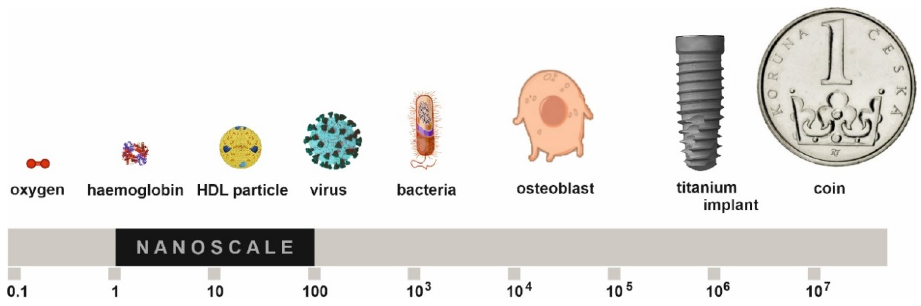
Nanotechnologies have received a large amount of attention in bone tissue engineering and are connected to many disciplines such as physics, chemistry, engineering, life sciences, and medicine, representing a real combination of different fields. Nanomaterials are currently defined as materials with basic structural units smaller than 100 nm in at least one dimension and exhibit unique properties different from their bulkier counterparts that have made them highly effective in numerous biomedical applications (Figure 1) [13,14,15]. There are included not only nanoparticles but also nanoclusters, nanocrystals, nanotubes, nanofibers, nanowires, nanorods, nanofilms, etc. The decrease in material size into the nanoscale radically increases surface area, roughness, and the ratio of surface area to volume leading to superior physico-chemical properties. These nanoparticles affect cell signaling, proliferation, cell viability, and integration [8]. Nanoparticles do not only support the cells; they also regulate the osteoblast function, proliferation, differentiation, and migration [16].
A variety of materials can be used to prepare nanoparticles, such as metals, ceramics, polymers, and organic materials [17,18,19], but usually, the nanoparticles are part of a different matrix, so nanocomposite scaffolds are created [20,21]. Currently, numerous top-down and bottom-up nanofabrication technologies (such as electrospinning, phase separation, self-assembly processes, thin film deposition, physical and chemical vapor deposition, chemical etching, nanoimprinting, photolithography, and electron beam or nanosphere lithographies) [22,23,24,25,26,27,28,29] are available to synthesize nanomaterials with ordered or random nanotopographies. Scaffolds with nanoparticles can be self-assembled into nanotubes/nanofibers, which can even more accurately simulate the dimensions of natural entities, such as collagen fibers [30,31].
In this review, we will focus on the recent progress in the use of nanomaterials for bone tissue engineering. After a brief introduction of bone biology, development, and an overview of the application of various nanomaterials, nanoparticles and nanocomposites are addressed.
2. Bone Biology
Bone is a hard connective tissue that offers, besides the classical functions of locomotion, protection, and support for soft organs, storage of calcium and phosphate ions, and a harbor for bone marrow. Additionally, the bone has endocrine functions capable of affecting other organs [32].
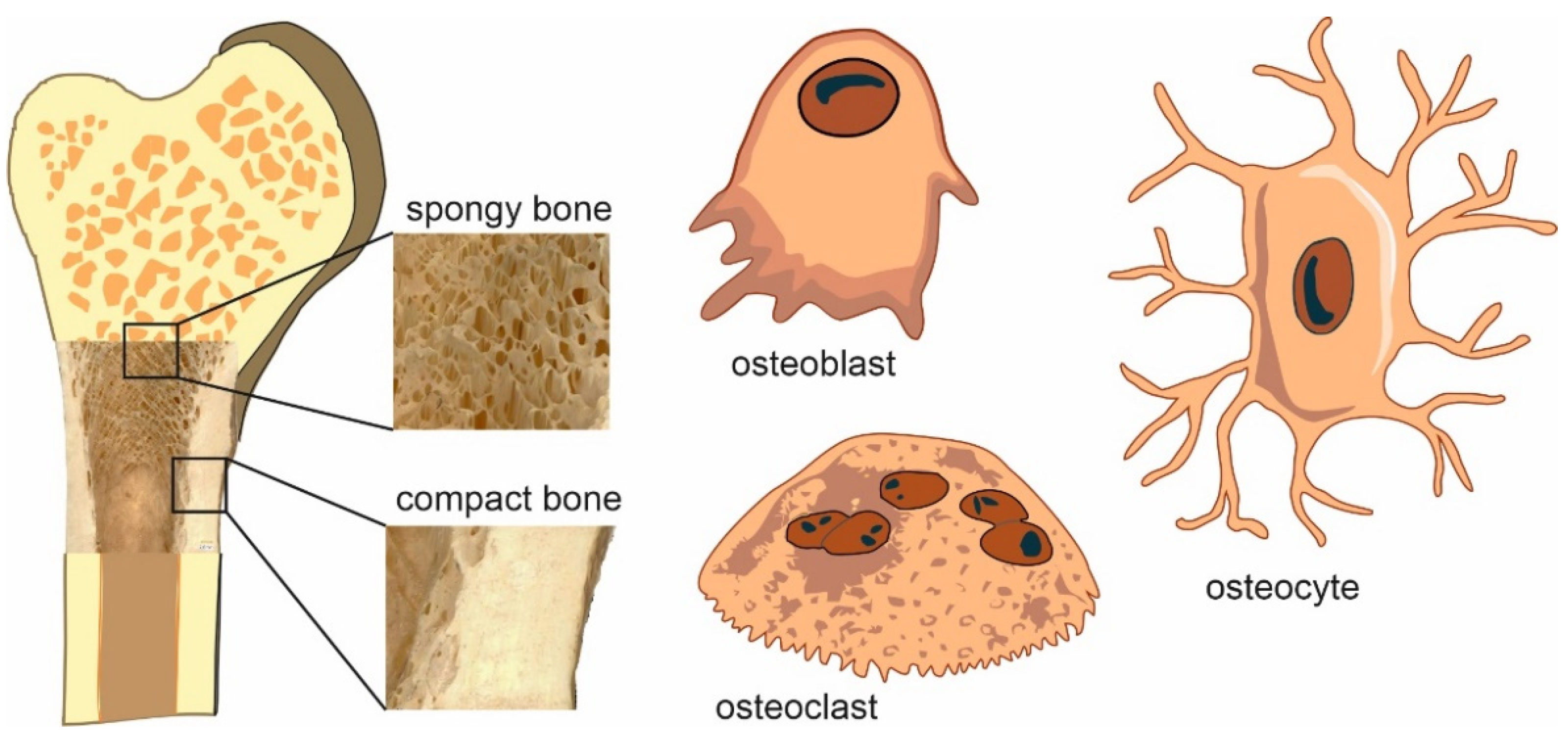
Two histological types of bone tissue can be distinguished, fibrous (primary, immature) and lamellar (secondary, mature). Mature lamellar bone is further divided into two structural subtypes according to its location and load, compact (cortical) and cancellous (trabecular, spongy) bone (Figure 2) [33].
Lamellar bone appears in the body much more often than fibrous bone. The lamellae are collagen fibers embedded in parallel in a mineralized amorphous matrix. They cluster concentrically around the central canal or form a system of parallel mantle lamellae on the bone surface. Compact bone is a system of parallel arranged osteons; the surface consists of parallel bone lamellae. Cancellous bone forms a fine network of thin bone beams (trabeculae) with a large internal surface. The orientation of trabecular networks reflects the optimal directional transmission of force. It plays a significant role in the remodeling and healing process of bone tissue [34]. Flat bones are formed by two outer layers of compact bone and one cancellous layer in the middle. The space in the cancellous bone among trabeculae is occupied by bone marrow containing pluripotent MSCs. These MSCs have the potential to differentiate into bone, cartilage, muscle, and adipose tissue.
Bone tissue is composed of four different cell types, osteoblasts, osteocytes, osteoclasts, and bone lining cells variously stored in the extracellular bone matrix [35,36].
2.1. Bone Cells
The major types of bone cells are osteoblasts, osteoclasts, osteocytes, and bone lining cells. All four types of bone cells are involved in growth, repair, regulation, and all other functions of skeletal tissues (Figure 2).
The term “osteoblast”, as a combination of the Greek ὀστέo- (bone) and βλαστάνω (germinate), was first used in the early 20th century as a cell from which literally “germinate bone” [37]. Morphologically, it is a cubic to cylindrical cell with a strongly basic cytoplasm located at the interface of newly synthesized bone. Osteoblasts are present in bone throughout life, but their activity is highest during embryonic skeletal development and growth. In the adult organism, osteoblasts are activated when there is a need to regenerate damaged bones or to form bone matrix [38]. Osteoblasts produce bone matrix proteins such as type I collagen (Col1), osteocalcin (OC), or alkaline phosphatase (ALP). Osteoblasts are post-mitotic cells but are not finally differentiated. Osteoblasts gradually surround the bone matrix and can differentiate into osteocytes, which are interconnected stellate cells. Osteoblasts that remain on the outer layer toward the periosteum can either turn into bone lining cells or undergo apoptosis. If the amount of osteoblasts decreases, either as a result of natural turnover or during an excessive regenerative process, new cells are differentiated from mesenchymal progenitor cells [39]. Osteoblasts are able to form osteoids (collagen I rich matrix), which are subsequently calcified. The corresponding impulse directs the active osteoblast characterized by a large number of endoplasmic reticulum as well as a large number of Golgi complexes from its mesenchymal progenitor cell located in the bone marrow.
Osteoclasts are multinucleated cells responsible for bone matrix resorption. They are derived from hematopoietic stem cells, specifically from the macrophage lineage. Through their activity, they dissolve bone minerals and create an optimal environment for enzymes involved in the degradation of the demineralized extracellular bone matrix. As osteoblast antagonists, they maintain homeostasis between bone formation and resorption. This complex activity is primarily controlled by the nuclear factor κβ receptor activator (RANK) and its ligand (RANKL). Under the anabolic processes or under the influence of osteoresorption factors such as glucocorticoids, calcitriol, or parathyroid hormone, osteoblasts release RANKL, which binds to the RANK receptor on proosteoclast cells and turns them into fully functional osteoclasts [40]. The mature osteoclast is a functionally polarized cell adjacent to the mineralized bone matrix by its apical pole. This connection is formed by a specific interaction between an adhesion molecule integrated into the cell membrane (integrin) and specific bone matrix proteins. The apical part of the osteoclast is invaginated, creating a free space between its roughly wrinkled membrane and the adjacent bone, into which it actively draws the substances needed for resorption [41]. One of the main tasks of the osteoclast is to acidify the resulting resorption space. The cell is richly equipped with the enzyme carbonic anhydrase, which catalyzes the reaction of water and carbon dioxide to form a proton (H+) and bicarbonate (HCO3−) anion. The protons are then actively pumped by a proton pump into the resorption space [42]. The regulation of this transport, which is closely linked to the regulation of intracellular pH and membrane potential, is maintained by ion pumps and channels on the basolateral side of the cell. The activity of the mature osteoclast is negatively regulated by calcitonin (CT), for which a large number of receptors are expressed [43].
Bone-lining cells are post-proliferative quiescent flat-shaped osteoblasts that cover the inactive surface of the bone, maintain the gap junction with osteocytes, control mineral homeostasis, and keep bone vitality. They produce osteoprotegerin (OPG), a factor involved in osteoclast differentiation. The secretory activity depends on the bone’s physiological status, whereas the cells can regain their secretory activity, increase their size and adopt a cuboidal form.
Osteocytes are terminally differentiated cells of the osteoprogenitor line anchored in the mineralized bone matrix. The single osteocytes communicate with each other and with cells on the bone surface through long intercellular protrusions. Their location and morphology make them particularly suitable for the transfer of information among cells in bone. In the case of mechanical pressure, osteocytes are able to register changes in bone, which elicit specific chemical signals leading to bone surface cells that ensure subsequent bone formation or resorption [44,45]. Their special location inside the bone makes them inaccessible for possible isolation and cultivation in in vitro studies.
All processes of osteogenesis are controlled by osteoblasts in close cooperation with bone-resorbing osteoclasts, together forming a multicellular unit. While osteoblasts synthesize the bone extracellular matrix, osteoclasts resorb this mass and contribute to physiological bone remodeling. Very fine and perfect control of this system is absolutely essential for bone development, fracture healing, and the proper development of the skeleton during life. This fragile balance can be shifted in favor of resorption, e.g., in osteoporosis, which affects postmenopausal women and older men and leads to more frequent fractures [46].
2.2. Bone Extracellular Matrix
The mechanical properties of bone tissue strongly depend on the specific structure and organization of the bony extracellular matrix (ECM). The bone tissue consists of 20% organic and 65% inorganic substances; the remaining 15% is water and fat. The bone ECM is a dynamic network composed of collagenous and non-collagenous proteins, which are the main components of the trabeculae and osteon units. The most abundant constituent of the organic matrix is collagen, a triple-helical molecule formed by three amino acid chains (two identical and one different), which form the collagen fibrils. A small amount of collagen III and V regulate the fiber diameter and fibrillogenesis of type I collagen. The crosslinking of collagen is key to its mechanical properties. The main function of collagens is to act as a scaffold for bone cells. The remaining organic part of ECM is other different non-collagenous proteins, such as OC, osteonectin (ON), osteopontin (OPN), bone sialoprotein, fibronectin, bone morphogenetic protein (BMP), and other types of proteoglycans involved in the regulation of bone matrix mineralization, organization, and other bone cell activity [47].
Osteocalcin (OC, γ-carboxyglutamic acid protein) is a 46–50 amino acid residues long protein secreted primarily by osteoblasts. It is produced as a pre-pro-protein undergoing further modifications, including the conversion of glutamate residues to γ–carboxyglutamate. The mature OC protein is packaged into intracellular vesicles for secretion into the bone matrix. Initially, it was proposed that OC initiates the hydroxyapatite (HAp) formation by binding to calcium ions. However, other studies point to its function as an inhibitor of bone mineralization. This idea is supported by research, where warfarin treatment results in higher mineralization of bones and early closure of the growth plate [48,49]. Based on many studies, OC is not only a protein bound in ECM; it is a regular bone-derived factor that influences glucose metabolism, reproduction, and cognition through endocrine loops between bone and the brain, pancreas, and testes [50,51,52,53].
Osteonectin (ON) is a calcium-binding glycoprotein associated with the growth and remodeling of bones. Bone ON binds to Col1 andHAp, and serves as a link between the mineral and organic phases of bone tissue. With the ability to induce mineral deposition onto type I collagen and inhibit the growth of HAp crystals, it can help in the regulation of bone mineralization.
Osteopontin (OPN) is expressed by osteoblasts, osteocytes, and odontoblasts. After post-translational modification, it is a highly glycosylated and phosphorylated protein with high importance as a component of mineralized extracellular matrices of bones and teeth. The binding of OPN to integrins is crucial for signal responses, which involves regulation of osteoclastic activity and activation of OPG expression. Further, OPN binding to integrin plays a major role in the formation of a sealing zone in osteoclast activity. OPN has a chemotactic activity on the precursors of osteoclasts and, as a part of the sealing zone, affects osteoclastic resorption [54]. OPN induces on the surface of osteoclasts integrin clustering and leads to intracellular signaling. The expression of OPN increases in response to mechanical stress [17,18,19]. Therefore, it is a critical factor in regulating bone remodeling in response to mechanical stimuli.
The organic part of the bone matrix contains a plethora of other proteins with diverse and overlapping tasks in bone mineralization, strength, stability, and integrity [55].
The inorganic component serves as an ion reservoir and contributes to bone stiffness and strength. Its basic components are apatite crystals containing calcium and phosphate ions, as well as sodium and magnesium ions. The mineral part of the bone, which essentially contains HAp, contributes to 65% of the bone weight. The remaining 20–30% by weight of bone is type I collagen, and the last 10% by weight includes water molecules, which are largely bound to the matrix structure. However, it also contains some free water molecules that are redistributed when the bone is loaded. These free molecules pass through the bone microchannels and therefore play a key role in detecting signals by cells, thereby transmitting information regarding the current load to a given part of the bone. The interaction between bone minerals and unbound water molecules thus aids in the process of bone mineralization, which is directly related to bone stiffness. As the amount of water decreases, bone mineralization begins, which can cause bones to become too brittle and, therefore, prone to breakage [56].
2.3. Bone Development
Bone formation (ossification) is a complex process by which other types of connective tissue are converted into bone. Different parts of the mammalian skeleton are made up of different cell lines. An example is a difference in the formation of the skull bones. Neural crest cells migrate from the dorsal edges of the closing neural tube to the anterior region of the skull and contribute to the craniofacial skeleton. These cells produce dental dentin, connective tissue, and some bones at the front of the skull. On the other hand, prechordal mesodermal cells form the cartilage and bones of the back of the skull. The paraxial mesoderm (somites) forms the axial skeleton, while the mesodermal cells of the lateral plate form the appendicular skeleton [57].
Two different types of bone formation can be distinguished, intramembranous and endochondral ossification. The process of intramembranous ossification is initiated by MSCs, which after replication and formation of special clusters within primitive connective tissue, differentiate directly into osteoblasts. Osteoblasts secrete bone matrix proteins able to mineralize by binding calcium salts. The intramembranous type of ossification is how flat bones are formed. Endochondral ossification is a multi-stage process of bone formation in vertebrates. It is a formation of the mineralized bone according to the cartilage pattern. Endochondral bone formation occurs in the skull base and the posterior part of the skull, the axial skeleton, and the appendicular skeleton [58].
Intramembranous ossification is based on osteoblast lineage comprising the following differentiation stages: osteoprogenitors, preosteoblasts, osteoblasts, bone lining cells, and osteocytes. The key elements regulating osteoblast differentiation and function are Wnt and Notch signaling and the transcription factors RUNX2 and osterix (Osx). Osteoblasts are characterized by OC expression. Preosteoblasts are characterized by the expression of RUNX2, or in the more differentiated stage, RUNX2 and Osx. Osteocytes play an important role as regulators of bone remodeling in response to mechanical and hormonal signals. This relationship is partly driven by sclerostin produced by osteocytes. Sclerostin inhibits Wnt signaling in osteoblasts, leading to osteoblast differentiation. Wnt/β-catenin signaling is a key signaling determining the differentiation of mesenchymal progenitors. Genetic inactivation of β-catenin causes ectopic chondrocyte formation at the expense of osteoblast differentiation during intramembranous and endochondral ossification [59].
Endochondral ossification is based on the differentiation of mesenchymal cells (progenitors) into chondrocytes and perichondrial cells [60]. Endochondral bone formation is regulated by many morphogens and growth factors, such as Wnts, hedgehogs, Notch, vascular endothelial growth factor (VEGF), hypoxia-inducible factor 1-alpha (HIF1α), fibroblast growth factor (FGF), insulin-like growth factor-1 (IGF-1), transforming growth factor-beta (TGF-β), paired box transcription factor (PAX), Homeobox transcription factor (HOX) and parathyroid hormone-like peptide (PTHrP) [61,62]. In the beginning, there is always an aggregate of mesenchymal cells that form mesenchymal condensations, which predetermine the formation of future skeletal elements. After mesenchymal cell condensation, mesenchymal cells differentiate into chondrocytes. Chondrocytes then form a cartilage template for future bone. Fibroblast growth factor receptor 3 gene (FGFR3) is a unique negative regulator of chondrocyte proliferation [63]. FGFR3 acts on chondrocyte proliferation via the transcription factor STAT1 and the ligand FGF18. Proliferation is also controlled by growth hormones and IGF-1 [64]. Hypertrophy of chondrocytes occurs under the influence of Runx-2, Indian hedgehog, and BMP-6. Members of the Sox family of transcription factors (Sox9, L-Sox5, and Sox6) are needed for successive steps of chondrocyte differentiation and to control the fate of the chondrocyte lineage. Under the influence of Sox-9, the chodroblasts begin to secrete a cartilaginous matrix [65]. Hypertrophic chondrocytes begin to produce bone proteins, such as OC, ON, and OPN. Chondrocyte hypertrophy is essential for osteoblast differentiation. Osteoblasts differentiate from perichondrial cells. The key player in osteoblast differentiation through endochondral ossification is hedgehog signaling; IHH is produced mainly by pre-hypertrophic chodrocytes. Hypertrophic chondrocytes also express vascular endothelial growth factor (VEGF) and matrix metalloproteinases important for vascularization. The blood vessels penetrate the hypertrophic cartilage in order to create a bone marrow cavity and trigger the differentiation of osteoblasts to form trabecular/cancellous bone. Blood vessels can also serve as a source of chondroclasts involved in the formation of the medullary cavity [66].
During bone development, the importance of gradients of morphogens, such as BMP, has been highlighted. Members of the TGF-β superfamily, such as BMPs, regulate bone shape. Mutations in BMP genes and thus impaired BMP signaling lead to changes in bone size, number, and shape in mice [67].
3. Nanostructured Materials
Nanocrystalline materials are characterized by their extremely small grain sizes of up to hundreds of nanometers and correspondingly high volume fraction of grain boundaries, which gives rise to unique physical, chemical, and mechanical properties compared to those of the corresponding materials with conventional grain sizes [68]. Reducing the grain size of a material provides advantageous properties such as higher toughness and strength. Bulk nanostructured materials can be produced by a number of processing methods. The most important methods are based either on severe plastic deformation or sintering of nanometer-sized grains [69].
Titanium
Titanium is a commonly used material for both dental and orthopedic implants. Due to excellent properties such as high tensile strength, durability, high corrosion resistance, resistance to body fluids, and high biocompatibility, titanium and titanium alloys remain among the most appealing and valuable materials. The strength of titanium is too low for implants to be used in load-bearing situations. The addition of alloying elements, such as aluminum and vanadium, allows a significant improvement of the mechanical properties of titanium. Despite the good static and fatigue strength of Ti–6Al–4V alloy, elevated concentrations of potentially cytotoxic alloying ions were detected in the tissues, serum, and urine [70]. An alternative approach to solve the ion release is to enhance the mechanical properties of pure titanium by nanoscale grain refinement. The studies note that conversion of the titanium into a nanostructured state is accompanied not only by the improvement of its physical and mechanical characteristics but also by the improvement of cell response to the interface between the material and biological surroundings [71,72]. Titanium nanostructures provide a large surface area and give enhanced antibacterial characteristics, osseointegration, and protein interaction, all of which are beneficial to medical implants.
In 2015, Babuska et al. tested the biocompatibility of nanostructured titanium with different grain sizes to human fibroblast cell line HFL1 and human osteoblast cell line hFOB 1.19. The results showed that the material with the smallest grain size, in the case of osteoblasts, had the best viability and confirmed that the smaller the grain size, the better the viability [73]. Medvedev et al. evaluated how certain parameters of material can influence the interaction of nanostructured titanium and two types of human tissue cells, human osteosarcoma SaOS-2 and adipose-derived mesenchymal stem cells (AD-MSCs). They found enhanced viability of both cell lines on samples with grain refinement by the equal channel angular pressing method. A positive linear correlation between nanoscale surface roughness and cell viability was found in the 0 nm to 5 nm range of stochastically varying nanoscale roughness [74]. Additionally, they noted that subsequent surface treatment techniques of bulk nanostructured titanium could significantly reduce the positive effect of the refined structure. This is in concordance with our results on laser surface treatment of nanostructured titanium implants [75].
In the work of Xu et al., ultrafine-grained (UFG) titanium samples were subjected to a micro-arc oxidation surface treatment. This surface treatment effectively reduced the hemolysis rate, extended the dynamic coagulation time, reduced the amount of platelet adhesion and the degree of deformation, and enhanced blood compatibility [76].
The use of highly hydrophilic solutions may positively change the surface, as estimated by Nazarov et al., by etching the nanostructured titanium in acidic and basic Piranha solution. The variation of the etching medium and time leads to micro-, nano-, or hierarchical structures on the surface. A significant difference between surfaces etched with basic and acidic solutions was caused by a less distinct oxidation process in the basic solution compared to the acidic one [77]. According to Zhukova et al., the behavior of cells adhered to titanium implants is influenced by different nanotopography of the surface. They used sonochemical treatment in alkaline solution to prepare disordered mesoporous titanium and anodic oxidation to prepare ordered nanotubes. The results showed that cell morphology is affected by surface nanotopography, where cells display a polygonal shape and spread on mesoporous surfaces and an elongated, polarized shape on tubular surfaces [78].
A relatively small number of studies deal with differences in osseointegration of nanostructured titanium implants in vivo. The work of Chappuis et al. described osseointegration of UFG titanium with a nano-patterned surface in the maxilla and mandible of miniature pigs. Histomorphometric and biomechanical torque out analysis revealed no significant differences between tested samples. High bone to implant contact values independent of the bone mineral density and type of bone were obtained [79]. In the study of Masrouri et al., the degree of osseointegration among UFG titanium, coarse-grained titanium, and titanium alloy with sandblasted/acid-etched surfaces was evaluated. The implants of each group were embedded in the femur of New Zealand rabbits, and histological analysis and torque tests were conducted. Results showed suitable osseointegration between implant and bone for all three groups. The value of removal torque was slightly higher for UFG titanium [80].
There are also a large number of titanium treatments that do not change the structure of the bulk material but create a nanostructure at the interface between the material and the biological environment. Although these methods do not change the material properties, the benefits of nanostructures in contact with bone are retained [81,82]. Micro arc oxidation is one of the treatment methods producing well-characterized titanium dioxide morphology on the surface of titanium implants. Surface morphology determines the behavior of bone cells to promote early osseointegration. According to Mao et al., increasing the voltage of the electrodes in the range of 100–400 V has a beneficial effect on the adhesion, proliferation, and expression of ALP of fibroblast-like mouse bone cells [83].
Electrochemical anodization with the production of nanotubes with different diameters and thicknesses of the wall [84,85,86] and laser texturing with a femtosecond laser to create grooves with hundreds of nanometers resolution [87,88,89] are the most used technics of titanium surface nanostructuring.
4. Nanoparticles
Nanoparticles based on metals and their oxides have many advantages, such as a simple process of preparation of the required size and shape, high stability, no swelling changes, easy functionalization with various molecules, and incorporation into hydrophobic and hydrophilic systems [90]. The nanoparticles used in medical applications usually have an average size of up to 200 nm [91]. Due to their small size, different shapes, and large surface area, they show increased colloidal stability and thus increased bioavailability, which demonstrates the ability to enter the lung system, pass through the endothelial cells and cross the blood–brain barrier. In recent years, nanoparticles based on a large number of different metals and their oxides have been tested, not only in the field of bone regeneration [92,93].
4.1. Gold Nanoparticles
Gold nanoparticles (Au-NPs) are the most interesting among the noble metal nanoparticles. The unique properties of Au-NPs, such as good biocompatibility, easy synthesis, and surface ligand functionalization, make them promising nanomaterials in biomedical applications like drug delivery, biological imaging, diagnosis, and treatment of diseases [94].
In recent years, Au-NPs have gained attention as next-generation osteogenic agents for bone regeneration [95]. Zhang et al. reported that Au-NPs have regulatory effects on macrophages and osteogenesis of stem cells and osteoblasts. This effect depends on different particle sizes, concentrations, and surface modifications. Au-NPs promoted osteogenic differentiation in a size-dependent manner. Three sizes of nanoparticles (5 nm, 13 nm, and 45 nm) were used for the experiment. Particles sized 13 nm and 45 nm showed the highest potency to promote osteogenesis on periodontal ligament progenitor cells and expressed OPN mRNA. On the other hand, 5 nm Au-NPs significantly reduced ALP activity, expression of osteogenic genes, and mineralized nodule formation [96]. These results support the finding that smaller nanoparticles do not produce enough free energy and therefore form clusters to receive the driving force to enter the cells [97].
Au-NPs promote the osteogenesis of MSCs through different signaling pathways. Choi et al. studied osteogenic differentiation of stem cells through the Wnt/β-catenin pathway. Osteogenesis of bone-derived MSCs and AD-MSCs was promoted by activating the p38 MAPK signaling pathway and the Wnt/β-catenin signaling pathway, respectively [98]. The osteogenic differentiation of bone marrow-derived MSCs (BM-MSCs) was stimulated by citrate-reduced Au-NPs through the mitogen-activated protein kinase signaling pathway. An Au-NPs–hydrogel complex increased the osteogenic differentiation of fibroblasts through the BMP signaling [99].
In a study by Rosi et al., Au-NPs coated with synthetic oligonucleotides showed good biocompatibility, stability, and efficient uptake by the cells. Au-NPs tailored with oligonucleotides facilitate the targeted detection of specific mRNA and can be used to isolate specific skeletal cell populations from the mixture of bone marrow stromal cells. Transmission electron microscope analysis revealed that the Au-NPs interacted with cytoplasmic proteins of specific signaling pathways, upregulated osteogenic genes, and downregulated adipogenic genes [100].
Singh et al. integrated Au-NPs into the bone scaffolds. This enhancement helps bone regeneration by achieving properties such as osteoconductivity, osteoinduction, appropriate resorption rate, and mechanical strength. Au-NPs synthesized through immobilization of Bacillus licheniformis crude enzyme were studied on murine preosteoblast cell line (MC3T3) and BM-MSCs. Good osteocompatibility, anti-oxidant, osteopromotive and hemocompatibility properties, and increased bone mineralization were observed [95]. Heo et al. prepared nanomaterials by incorporating hydrogel with Au-NPs by photo-crosslinking. The addition of Au-NPs promoted high ALP activity, cell proliferation, cell viability, and osteogenic differentiation of adipose-derived stromal cells [101].
4.2. Silver Nanoparticles
Due to its antibacterial properties, silver and its compounds have been used for a variety of medical purposes for more than a thousand years. The importance of silver is growing, especially at a time when growing bacterial resistance is a major global problem. Today, silver is used not only as an antiseptic agent but also for its anticancer properties or as a vaccine adjuvant, antidiabetic agent, and biosensor [102]. The particles with controlled size and shape can be synthesized by physical (mechanical and vapor-based processes) or chemical (reducing silver ions to silver atoms) methods. Silver ions reduction can also be achieved by biological methods using enzymes, alkaloids, phenolic compounds, and terpenoids from extracts of microorganisms and plants [103].
Size and shape determine various actions of nanoparticles, including cellular uptake, targeting to cells, in vivo distribution, and protein corona formation. Nanoparticles administered by intravenous injection travel along blood circulation into target organs with abundant vasculatures. It is suitable to design nanoparticles that can effectively reach target tissues. The size of nanoparticles also affects cellular uptake and biological action in target cells. Too small nanoparticles are extensively excreted by kidneys from target tissues [104]. Particle shape also has an important impact on the performance of nanoparticles as a carrier of therapeutic agents through changes in ligand targeting, cellular uptake, transport, and degradation [105].
Silver nanoparticles (Ag-NPs) can effectively kill a variety of pathogens at very low concentrations, e.g., Gram-positive and Gram-negative bacterial strains (Escherichia coli, Klebsiella pneumonia, Staphylococcus aureus), fungi (Candida albicans, Aspergillus niger), and viruses (HBV, HIV). The antimicrobial mechanisms of Ag-NPs include destructing bacterial cell walls, producing reactive oxygen species (ROS), and damaging DNA structures. Bacterial resistance to silver is rare. It is currently tested that Ag-NPs act against tumor cells in vitro and in vivo [106]. Recently, Xu et al. introduced a new type of silver particles smaller than Ag-NPs, so-called silver Ångstrom (1 Å = 0.1 nm) particles (Ag-ÅPs), which exhibit stronger anticancer activities and lower toxicity compared with Ag-NPs [107].
In bone engineering, Ag-NPs are used for fabricating antibacterial nanocomposite-based scaffolds and implants to replace large defects. They promote bone regeneration and wound healing by effectively suppressing bacterial infection and inflammation. Studies in vitro show that Ag-NPs can naturally stimulate proliferation and osteogenic differentiation of MSCs and mineralization of ECM [108]. It was found that Ag-NPs at 2.5 and 5 μg/mL stimulate osteodifferentiation more efficiently than at 10 μg/mL [109].
In some dental applications, Ag-NPs are used as antiplaque agents. An example is the production of dental prostheses from acrylic resins, on the rough surface of which microorganisms adhere. Ag-NPs incorporated in acrylic resins improve the antibacterial effect of this dental material and decrease the adherence of Candida albicans [110].
However, Ag-NPs contained in various medicinal products such as dressings, creams, solutions, and scaffolds come into contact with the skin by inhalation, ingestion, or injection into the body. They are transported via blood circulation to various organs and may be cytotoxic, limiting their applications. They are stored mainly in the dermis, spleen, liver, kidneys, and lungs, and a smaller amount in teeth and bones. Ag-NPs may cause dermal, ocular, respiratory, hepatobiliary, and reproductive toxicity. This limits their applications [111].
4.3. Platinum Nanoparticles
Platinum is a heavy (density 21.45 g/cm3) and chemically extremely resistant noble metal; it is one of the rarest precious metals in the world. The most used are platinum nanoparticles (Pt-NPs) with sizes in the range of 10–50 nm. They have excellent properties such as biocompatibility, biodegradability, osteoconductivity, and high stability. It is known that a large number of nanoparticles have different biomedical applications. They are used in the detection of cancer cells, reduction of cellular oxidative stress, and treatment of Parkinson’s disease; they have bacteriotoxic effects and cytotoxic effects on cancer cells [112]. Pt-NPs have anti-oxidant and anti-inflammatory properties, which protect the cells from induced bone loss by diminishing the formation of osteoclasts (osteoclastogenesis) [113].
Eid et al. compared the properties of calcium phosphate scaffolds loaded with Pt-NPs prepared by heat-sintering and calcium phosphate scaffolds free of Pt-NPs. The results of this study showed better morphology and degradation rate of calcium phosphate scaffold with Pt-NPs than the calcium phosphate scaffold free of Pt-NPs. The adhesion and proliferation of human osteoblast cells on Pt-NPs-loaded scaffolds were superior [114].
A recently investigated new scaffold was composed of nanohydroxyapatite combined with chitosan (CS) in the matrix of poly-L-lactic acid (PLA) nanofibers and enriched with Pt-NPs, Au-NPs, and TiO2-NPs. Better biomineralization, cell adhesion, proliferation, and differentiation of MG-63 cells was observed in scaffolds enriched by Pt-NPs and Au-NPs compared to TiO2-NPs [115].
4.4. Palladium Nanoparticles
Recent studies have shown that palladium nanoparticles (Pd-NPs) have significant antimicrobial, electrical, catalytic, and photosensitive properties. Pd-NPs applied to nanocomposite scaffolds with reduced graphene oxide functionalized with polypyrrole have prevented the development of microbial films on these scaffolds. Nanocomposite scaffolds also promoted the proliferation of the SaOS-2 cell line [116]
On the other hand, Calabrese et al. reported that Mg-HA-Collagen type I scaffolds functionalized with golden nanorods (Au-NRs) and Pd-NPs inhibited the growth of adipose-derived MSCs up to 89% and 94%, respectively. Analysis of matrix mineralization density (by Alizarin Red staining) and the expression of ON (specific for bone mineralization) and the OC (specific for mature osteoblast) led to comparable results. Variants with Au-NRs and Pd-NPs also showed decreasing cell differentiation (20% lower) than the control. These results support other studies that found cytotoxic effects of Pd-NPs [117]. Recently, Heidari et al. presented the production and characterization of HAp/ZnO 0.05 wt%/Pd 0.1 wt% nanocomposite scaffolds. The initial materials were prepared using sol-gel and precipitation methods. The biocompatibility of scaffolds was tested in simulated body fluid (SBF) and with the interaction of dental pulp stem cells (DPSCs). In comparison to the HAp scaffold, HAp/ZnO/Pd scaffolds showed lower cell proliferation. These results could be caused due to the amount of deposited and released Zn2+ ions having an inhibitory effect on cells. HAp/ZnO/Pd scaffolds coated with different concentrations of CS had lower density and microhardness and higher compressive strength and toughness than the pure HAp. However, HAp/ZnO/Pd scaffolds coated with CS exhibited antibacterial properties, which were examined in this study using Pseudomonas aeruginosa [118].
Zhang et al. used synthetic porous Au/Pd alloy nanoparticles in bone regeneration during photothermal therapy. Pd-NPs were used as hyperthermia agents. The irradiation with a near-infrared laser caused mild localized heat (40–43 °C) produced by Au/Pd nanoparticles, which accelerated cell proliferation and bone regeneration. After 6 weeks, a new bone was formed in nearly 97% of the cranial defected area. Results of RNA sequencing showed that the Wnt signaling pathway was influenced by the mild localized heat [119].
4.5. Tantalum Nanoparticles
Tantalum is a metal that has very high corrosion resistance and outstanding biocompatibility, making it a promising material for orthopedic and dental implants. Compared to the most commonly used titanium (density 4.5 g/cm3), pure tantalum is very heavy (density 16.6 g/cm3). Contrary to titanium which is used as a bulk, tantalum can be manufactured as a porous material with many advantages. Porous tantalum has optimal mechanical properties and shows excellent cellular adherence and bone ingrowth resulting in better fixation of the implant. The clinical application of porous tantalum in bone tissue engineering has been reviewed by Huang et al. [120].
Tantalum nanoparticles (Ta-NPs) are also gaining attention. They can be produced in the human body as wear particles from tantalum implants. Wang et al. explored the effects of Ta-NPs on the MC3T3 cell line and found that a low concentration of Ta-NPs promoted the proliferation of osteoblasts, while concentrations higher than 25 μg/mL caused osteoblast damage via oxidative stress and reduced mitochondrial membrane potential [121]. Wear particles can contribute to aseptic implant loosing also due to proinflammatory properties. Zhang et al. compared cytotoxicity, ROS production, and inflammatory response of macrophages induced by TiO2-NPs and Ta-NPs. They found that TiO2-NPs triggered these adverse effects in a dose-dependent manner. Ta-NPs caused no apparent cytotoxicity and induced much less release of proinflammatory cytokines and only negligible ROS generation compared to TiO2-NPs. It indicates that regarding nanoscale wear fragments, tantalum may be superior to titanium for the manufacturing of implants [122].
There are also efforts to use Ta-NPs to improve polymers used as bone substitutes. Lu et al. in 2015 modified the surface of polyetheretherketone (PEEK) with Ta2O5 nanoparticles by plasma immersion ion implantation. It increased the elastic modulus, making it similar to that of human cortical bones. Osteogenic properties of the material were evaluated using BM-MSCs and by in vivo studies on Sprague Dawley rats. The modified surface showed the enhanced osteogenic differentiation of MSCs, and better osseointegration was observed in vivo [123]. Recently, Zhu et al. investigated reinforcement of PEEK with Ta-NPs. Composites containing 3 wt% and 5 wt% of Ta-NPs showed better mechanical properties (elastic modulus and compressive strength), an increase in adhesion of cells on the surface, and also exhibited enhanced bone formation in animal studies [124].
4.6. Iron Oxide Nanoparticles
Iron is a metal naturally occurring in the body, and therefore existing metabolic pathways allow the processing of remaining iron from nanoparticles. Iron oxide nanoparticles (Fe-NPs), mainly magnetite (Fe3O4) and maghemite (γ-Fe2O3), have excellent biocompatibility and unique magnetic properties. However, bare Fe-NPs may be toxic because their surfaces are chemically reactive, and they aggregate easily together [125].
Biocompatibility can be increased by coating, which prevents the aggregation of nanoparticles. The coating can be organic or inorganic, and either one or more layers are made. Substances used for organic coating include citrate, dextran, polyethylene glycol (PEG), CS, polyethyleneimine, or phospholipids, and for inorganic coating, silica, gold, and calcium phosphate. The coating layers are further modified to apply drug molecules, proteins, and genetic materials for medical purposes. For in vivo use, an injectable complex is formed in which Fe-NPs are associated with cells or bioactive molecules [126].
Fe-NPs are used as contrast agents, for iron replacement therapies, and for tumor therapies using local tissue hyperthermia. The magnetic properties of these particles can be used for optimal drug delivery to the required site, which minimizes side effects and improves the treatment of cancer and bone diseases. Fe-NPs and scaffolds containing these magnetic particles are used successfully in regenerative medicine, especially in bone tissue engineering and bone regeneration, because the magnetic field and magnetic responsive scaffolds promote bone repair and regeneration [127].
Magnetic properties are crucial for the application of Fe-NPs. Nanoparticle-labeled cells can be observed and monitored in vivo when the labeled cells remain detectable by magnetic resonance imaging even after 7 days [128]. Therefore, thanks to the magnetic field of Fe-NPs, it is possible to study the behavior of cells, i.e., adhesion, proliferation, and differentiation. Stem cells labeled with magnetic nanoparticles can be used in bone regeneration therapy for magnetic targeting to targeted sites. The osteogenic potential of stem cells is important for further bone regeneration, wherein Fe-NPs are able to supply proteins to control the growth and differentiation of these cells. These magnetic particles, in combination with a pulsed magnetic field, have a positive effect on osteogenic differentiation, as demonstrated in an experiment with BM-MSCs, where the expression of Col1 was increased [129], and in an experiment with Fe-NPs coated with bovine serum albumin, was also found elevated ALP activity, calcium deposition, and expression of OC [130].
Biopolymer scaffolds containing magnetic Fe-NPs show significant stimulation of cell adhesion and osteogenic differentiation in vitro and bone formation in vivo, which was confirmed by higher gene expression of key transcription factors for osteoblast (RUNX2 and Osx) and activity of ALP as well. Magnetic scaffolds are, therefore, useful for bone healing, especially in adjunctive therapy with external magnetic stimulation [131].
4.7. Copper Nanoparticles
Copper is a noble metal of reddish color characterized by very good thermal and electrical conductivity [132]. It is one of the most important trace elements in the human body, needed for a variety of biological functions, including Cu-dependent enzyme activity, bone metabolism control, blood clotting, and vision process. Copper nanoparticles (Cu-NPs) have an antioxidant effect and, due to their biocompatibility and ability to induce the migration and proliferation of endothelial cells, are suitable for wound healing [133].
The use of copper ions in scaffolds can improve bone tissue regeneration. D´Mello et al. compared the influence of copper-doped CS scaffolds on bone renewal. Scaffolds were implanted into 5 mm calvarial defects in Fisher 344 male rats. An increase in bone volume was found after 4 weeks by micro-CT scans and histological examination. They concluded that the inclusion of copper ions in scaffolds could enhance tissue regeneration [134].
Lin et al. prepared a novel type of Cu-containing calcium phosphate cement (Cu-CPC) enriched by copper phosphate nanoparticles. The effect of these particles at different concentrations on osteogenesis and angiogenesis was investigated on mouse BM-MSCs and human umbilical vein endothelial cells (HUVECs). Cu-CPC with 0.01 wt% and 0.05 wt% of copper phosphate nanoparticles showed a significant increase in adhesion and proliferation of BM-MSCs. The differentiation was confirmed by collagen type I production and by elevated expression of osteogenesis-related genes Runx2, Col-I, and OC, and by angiogenesis-related genes VEGF, eNOS, and FGF. Higher content of copper phosphate nanoparticles than 0.05 wt% inhibited the proliferation of murine BM-MSCs [135].
4.8. Zinc Nanoparticles
Zinc is a crucial element for normal human body functioning. This element is involved in many enzymatic and physiological reactions. Zinc oxide nanoparticles (Zn-NPs) can reduce oxidative stress by eliminating free radicals and possess antidiabetic and antibacterial properties. The spectrum of biomedical use of Zn-NPs is diverse. These nanoparticles are used as carriers for drug delivery, contrast agents in biomedical imaging, or nanosensors. Due to their biocompatibility, they have been integrated into bioactive ceramics and bioactive glass [133].
Zn-NPs exhibit osteogenic properties that are caused by Zn2+ ions release. These ions can promote bone growth, mineralization, and bone formation. The mitogen-activated protein kinase pathway is activated by Zn2+ ions and promotes the expression of the OC gene. Zn2+ ions also support bone formation by enhancing Runx2 expression by increasing Zn2+ carriers such as zinc transporter protein ZIP1 [136].
Khader and Arinzeh used the electrospinning technique for the preparation of fibrous zinc oxide composite scaffolds. Polycaprolactone (PCL) with incorporated Zn-NPs provided slow degradation and dissolution of Zn2+ ions over time. Zn-NPs were used in different concentrations (1–10 wt%). Biocompatibility was evaluated as a degree of MSCs differentiation. Low concentrations of Zn-NPs promoted chondrogenic differentiation, which was indicated by the highest collagen type II production and expression of cartilage-specific genes. On the other hand, high concentrations of Zn-NPs promoted osteogenic differentiation of human MSCs. This differentiation was proved by the highest ALP activity, collagen production, and expression of bone-specific genes. Results of this study have shown the potential of Zn-NPs composite scaffolds in bone tissue engineering [137].
4.9. Magnesium Oxide Nanoparticles
Magnesium is an important biogenic element in the human body. It is a cofactor for more than 300 enzymatic reactions and is essential for DNA, RNA, and protein synthesis. It is also involved in the metabolism of vitamin D and calcium [138]. Therefore, scaffolds developed from various materials in combination with magnesium regulate not only cellular behavior but also stimulate bone formation and healing. Such materials release magnesium and thus promote the cell adhesion, proliferation, and osteogenic differentiation of MSCs [139].
Magnesium oxide nanoparticles (MgO-NPs) and magnesium-based scaffolds are very easily biodegradable. A high level of degradability, however, can cause scaffold failure in vivo. The rate of degradation and bactericidal activity of these alloys is reduced up to 5 times when graphene nanoparticles are used in the Mg-Al-Cu matrix [140]. A disadvantage of synthetic polymer scaffolds is the release of acidic products during degradation, which may promote the conditions for the beginning of inflammation. The polymer containing magnesium hydroxide nanoparticles exhibits pH neutralizing properties, which significantly reduces the release of proinflammatory cytokines [141]. The polymeric scaffolds can be reinforced with an Mg-doped bioactive glass to enhance mechanical properties and biological reactivity [142].
The challenge remains to create a scaffold with the effective release of magnesium cations. For example, a nanocomposite hydrogel based on hyaluronic acid (HA) and self-assembled bisphosphonate-magnesium nanoparticles exhibit improved mineralization capabilities and controlled release of magnesium cations [139].
MgO-NPs have been found to show antibacterial activity with comparable results to standard 5.25% sodium hypochlorite, which is used for root canal disinfection in endodontics. They disrupt the cell membrane and induce oxidative stress, leading to cell death [143].
The optimal concentration of MgO-NPs is important for cell growth on the scaffold. It was found that BM-MSCs density was increased significantly when cultured in 200 μg/mL of MgO-NPs compared to the control. The additional benefit of MgO-NPs at a concentration of 200 μg/mL is the reduction of bacterial adhesion. Therefore, at this concentration, MgO-NPs could be used as a coating material or as a part of a composite for potential medical implant applications. However, concentrations above 300 μg/mL decreased the cell density, and concentrations above 1000 μg/mL resulted in complete cell death [144].
4.10. Nickel Oxide Nanoparticles
Nickel is a white, ferromagnetic metal that serves as a part of various alloys and for surface protection of other metals against corrosion. Due to its toxicity, the practical use of this metal is limited [145].
Nickel oxide nanoparticles (NiO-NPs) have magnetic properties and possess excellent durability and electrochemical stability with potential use in applications such as drug delivery, cell tracking, imaging procedures, or biosensors [133]. However, it was also supposed that due to the toxic properties of nickel, NiO-NPs might cause potential adverse health effects, limiting their biomedical applications [146].
Bano et al. reported NiO-NPs as a multimodal cancer therapy agent. They designed a cumulative drug delivery system, which consisted of light-sensitive NiO-cores, bovine serum albumin, and folic acid and was loaded with doxorubicin (NOP-DOX@BSA-FA). Human cervical epithelial malignant carcinoma cell lines (HeLa) were treated with NOP-DOX@BSA-FA, showing outstanding anticancer activity [147].
Antioxidant, antibacterial, and anticancer activities of phytomolecules-coated, green synthesized NiO-NPs using Abutilon indicum leaf were tested. The phytomolecules-coated NiO-NPs showed tumoricidal action against the HeLa cancer cell lines, inhibition of growth of Escherichia coli, Bordetella bronchiseptica, Bacillus subtilis, and Staphylococcus aureus, antioxidant activity, and biocompatibility with fibroblast cells [148].
4.11. Titanium Dioxide Nanoparticles
Titanium is a commonly used metal for the manufacturing of implants. In addition to this application, it can be used in the form of titanium dioxide nanoparticles (TiO2-NPs), which can be produced in various morphologies such as nanowires, nanorods, nanotubes, and nanoparticles [149].
Titanium dioxide can be used in the preparation of silk fibroin (SF) based polymer scaffolds as a nanofiller. SF/TiO2 scaffolds were prepared with different concentrations of TiO2 (5, 10, 15, 20 wt%). Increasing the content of TiO2-NPs led to reduced porosity in scaffolds. This may be caused by the agglomeration of nanoparticles on the pore walls. The scaffold also showed improved mechanical and directional strength due to increased TiO2-NPs content. Testing biocompatibility with the SaOS-2 cell line showed high adhesion and proliferation. The viability of cells increased with growing SF content [150].
Another way to use TiO2-NPs is the preparation of polymer-inorganic nanoparticle composite membranes. Nechifor et al. prepared composite membranes of polysulfone-silica microfiber grafted with TiO2-NPs (PSf-SiO2-TiO2). Microspheres of TiO2-NPs 1–3 μm in diameter were attached to silica microfiber using the sol-gel method. The final nanocomposite was prepared through the inversion phase method by evaporation. PSf-SiO2-TiO2 nanocomposite membranes showed better flux and mechanical characteristics, retention, and chemical oxidation resistance than the membranes without TiO2-NPs. Moreover, scaffolds were tested for antimicrobial properties and proved to have an inhibitory effect on five Gram-negative microorganisms and stopped the proliferation of Candida albicans. Results of this study showed that PSf-SiO2-TiO2 composite membranes could be applied as a functional barrier in guided bone regeneration [151].
On the other hand, the degradation of titanium-based implants in the human body causes the release of ions and solid debris of TiO2-NPs, leading to peri-implant inflammatory reactions. In the in vitro study, the effect of titanium nanoparticles on MSCs and fibroblast was evaluated by the MTT test and production of ROS. It was found that TiO2-NPs caused a time-dependent decrease in cell viability and high production of ROS. Histological and molecular analysis of peri-implant tissue showed the occurrence of ROS, high levels of titanium particles, and enhanced bone turnover. There was also found to be a high expression of zinc finger protein (ZFP467) and the presence of adipose-like tissue. This suggests that the MSCs were dysregulated [152].
4.12. Calcium Oxide Nanoparticles
Calcium is an important biogenic trace element. Most of the calcium in the human body (99%) is in the skeleton, where it is bound to the HAp molecule and also serves as a reservoir for maintaining serum calcium levels [153]. It is a key mediator in a number of metabolic and regulatory processes in the human body [154]. Therefore, it is understandable that it is widely used and researched in the development of materials for bone regeneration. Calcium oxide shows antibacterial activity against Escherichia coli, Salmonella typhimurium [155], Staphylococcus aureus, and Bacillus subtilis [156].
Calcium oxide nanoparticles (CaO-NPs) are biocompatible and low-toxic agents with outstanding antimicrobial properties, making CaO-NPs an excellent material for medical use. Besides antibacterial activity, CaO-NPs are known for their antifungal properties. Calcium oxide contained in heated scallop-shell powder can be used to treat dermatophytosis caused by Trichophyton [157].
The incorporation of CaO-NPs in different types of polymers can improve their mechanical properties and provide antimicrobial effects. Silva et al. studied mechanical and biocidal characteristics of polyethylene composites with incorporated CaO-NPs with a diameter of 55 nm and 25 nm. It was shown that polymer modified with CaO-NPs has a higher Young’s modulus than pure polymer. Nanomaterial modified with CaO-NPs caused the death of more than 99.99% of the bacteria. It was emphasized that size reduction of CaO-NPs leads to increasing antibacterial effects. One of the important advantages of CaO-NPs is that these nanoparticles do not require any external stimulus, such as any kind of irradiation, to obtain antibacterial activity [158].
Münchow et al. tested PCL and PCL/gelatin electrospun matrices loaded with CaO-NPs. Mechanical characteristics, morphology, and mouse-calvaria-derived osteoprecursor cells (MC3T3-E1) behavior in the matrix were studied. This study demonstrated increased cell viability, adhesion, osteogenic differentiation, and proliferation in the electrospun matrices loaded with CaO-NPs. However, it was found that a high concentration of CaO is connected with decreasing mechanical properties [159].
4.13. Aluminum Oxide Nanoparticles
Aluminum is a very light whitish-gray metal that is a good conductor of electric current. Aluminum oxide is a bio-inert heat-resistant crystalline substance suitable for biological environments in the tissue engineering field. It serves as a catalyst in the chemical and petrochemical industries [160].
Aluminum oxide nanoparticles (Al2O3-NPs) integrated with ceramic coatings show good osseointegration, low cytotoxicity, good biodegradability, and are more resilient to long-term implantation failure. In view of their good mechanical behavior, aluminum implants are characterized by long-time survival predictions with an enhanced proliferation of osteoclastic cells [161].
Yu et al. compared the 3D porous β-Ca2SiO4 scaffold and its modification by the aluminum ions solid solution. Significantly higher proliferation and differentiation of rat BM-MSCs on the aluminum-modified scaffolds were observed; also, their degradation rate was slower. Enrichment with Al2O3 exhibited enhanced bone formation, which has been proven by increased expression of osteogenic markers such as OC, OPN, and Col1 [162].
Reinforcement of polyhydroxy butyrate-chitosan scaffolds with Al2O3 nanowires enhanced mechanical properties with a tensile strength of 10 times higher compared to the same basic scaffold without aluminum nanowires [163].
4.14. Cerium Dioxide Nanoparticles
Cerium is one of the lanthanides, the elements having 4f electrons, which contribute to peculiar physicochemical properties which can be beneficial also for biomedical applications. Cerium dioxide (CeO2) is widely used as a catalyzer due to its redox properties based on the Ce3+ to Ce4+ valence conversion. Cerium dioxide nanoparticles (CeO2-NPs) compared to conventional CeO2 bulk lead to significant improvement of catalytic activity. CeO2-NPs are multifunctional, unique atomic lattice structures with oxygen vacancies, making them free radical scavenging agents [164].
In vitro experiments have shown that CeO2-NPs exhibited low cytotoxicity in macrophages and enhanced proliferation and osteogenic differentiation of MSCs [165]. Li et al. reported that CeO2-NPs enhanced endochondral ossification–based bone regeneration on a femur defect model in mice and described the molecular mechanism behind this effect via stimulation of DEAH-box polypeptide 15–p38 mitogen-activated protein kinase axis [166]. The clinical potential has been addressed in several ways. CeO2-NPs can be used for titanium implant surface modification. There are studies showing antibacterial and anti-inflammatory properties of such coatings [167]. Another direction of research is the reinforcement of scaffolds with CeO2-NPs. Purohit et al. fabricated and tested the gelatin–alginate–cerium oxide nanocomposite scaffold with promising results indicating that incorporation of CeO2-NPs into the scaffold matrix changed its properties strongly with the enhancement of cell attachment, proliferation, and viability and free radical scavenging ability [168].
Lukin et al. studied the effects of the use of nanocrystalline CeO2 in the early stages of bone regeneration. Rabbits were used as the animal model. Artificially formed defects in the lower jaw were filled with a suspension containing calcium hydroxide, barium sulphate, and nanocrystalline CeO2. Suspension of calcium hydroxide was used as the control. This assay showed that nanocrystalline CeO2 could improve osteogenesis and decrease inflammatory response [169].
4.15. Strontium Nanoparticles
Strontium is a trace metal that mimics the properties of calcium and magnesium, with an important role in human tissues. Due to its properties, strontium is an important element in dentistry and bone regeneration. Strontium stimulates bone growth by enhancing collagen and other bone matrix protein synthesis. It facilitates osteoblast differentiation and simultaneously reduces bone resorption by osteoclasts differentiation [170]. The plasma protein binding capacity for divalent strontium cation is similar to calcium cation. The transport of Sr2+ ions is controlled by the same carriers as Ca2+ ions.
Strontium nanoparticles (Sr-NPs) are spherical or flake metal particles with a specific surface area of 30–60 m2/g in a size range of 10–200 nm. Sr-NPs are used either in bone regeneration to improve the mechanical and biological properties of diverse polymers and metal alloys [171,172] or encapsulated as inorganic nanocarriers in drug delivery and biomedical imaging [173,174].
The bioactive microspheres enriched with monodispersed Sr-NPs regulate the phenotype of macrophages and promote early angiogenesis. New vessels at the center of scaffolds one week after implantation in a rat’s bone were observed, which suggested enhanced early vascularization and subsequent support of new bone formation [175]. The nanoparticles of bioglass doped with 10% strontium immersed in SBF created an apatite layer on the surface. The stem cells increased in the presence of strontium in the nanoparticles prefer the expression of osteogenic genes and proteins, even without osteogenic supplementation [176]. Synthesized nanoparticles of strontium oxide with a diameter of 32.6 nm showed good antibacterial properties against Bacillus subtilis [177].
Fekri et al. prepared Sr-NPs by the green synthesis method in the presence of carboxymethyl cellulose as a green biological stabilizer. According to the reaction conditions, nanoparticles of different sizes and distributions were created. Synthetized Sr-NPs are suitable for preclinical imaging and ideal for noninvasive disease progression monitoring, cell trafficking, and gene expression patterns in experimental animals. Sr-NPs were applied successfully as an efficient agent for in vivo imaging after injection into the neck area and the peritoneum of male BALB/c mice. The use of Sr-NPs is a good alternative to eliminate toxic and carcinogenic compounds in in vivo imaging [178].
4.16. Carbon Nanoparticles
Carbon-based nanomaterials appear to be very promising for their mechanical strength and biocompatibility. The carbon can occur in several structurally different modifications, such as graphene, carbon nanotubes (CNTs), fullerenes, carbon dots (CDs), carbon nanofibers, nanodiamonds, and their derivatives [179]. In bone tissue engineering, nanomaterials based on graphene and carbon nanotubes seem to be among the most prominent [180].
Nanodiamonds (NDs) are carbon-based nanoparticles typically 4–10 nm in diameter and sp3 hybridization of crystal lattice shape with the possibility of introducing a number of different functional groups on their surface. Several in vitro studies have shown significantly lower toxicity to diverse cell lines than graphene or CNTs [181,182]. NDs can be used in the bone-anchoring stems of permanent bone implants in order to improve their interaction with the surrounding bone tissue. This beneficial characteristic can be assumed not only due to their excellent mechanical and chemical resistance but also due to their surface nanoroughness and surface nanotopography. It was stated that these properties could stimulate the adhesion, growth, and maturation of osteoblasts, as well as the differentiation of bone progenitor cells towards the osteoblastic phenotype [183]. Thanks to the ability to bind proteins, nanodiamond surfaces can serve as carriers for bioactive molecules (e.g., BMP or FGF) which promote bone formation. NDs and angiopoietin-1 were used for the functionalization of bone implants to improve vascularization and bone regeneration [184]. A prenylated flavonol glycoside icariin functionalized NDs (ICA-NDs) promoted osteogenic capacity. In vitro cell studies showed that ICA-NDs treated osteoblastic cells MC3T3-E1 greatly increased the production of osteogenic markers, including ALP, RUNX2, Col1, and OPN, and even calcium content compared to only NDs treated cells [185].
Carbon nanotubes (CNTs) are cylindrical molecules of carbon atoms in a hexagonal arrangement. Two types of CNTs can be distinguished, single-walled (SW-CNTs) and multi-walled carbon nanotubes (MW-CNTs), which are one of the strongest materials. These nanotubes are thermally and chemically very stable, contain carbons in sp2 hybridization, and are prepared by layering individual graphene sheets joined by rolling. The length, diameter, and surface modification of nanotubes are important for their interaction with cells [186]. Nanocomposite scaffolds in which CNTs are used as a filler of HAp show a significant improvement in fracture toughness and tensile strength. Because SW-CTNs have a diameter close to the size of the collagen triple helix, which is between 0.7 and 1.5 nm, they are preferably used to prepare scaffolds promoting HAp growth and nucleation. The study showed that SW-CNTs/HAp scaffolds with different functional groups have a higher solubility in water and significantly affect the formation of HAp [187].
By adding nanotubes, especially MW-CNTs, to the porous bioglass foam, a nanocomposite scaffold with a suitable roughness for cell attachment was created, which proved to be highly bioactive and suitable for use in bone tissue regeneration. Due to the presence of MW-CNTs, the interaction among cells during their adhesion was significantly improved, osteoclastogenesis was suppressed, and proliferation of osteoblasts was promoted [188].
Due to the fact that CNTs have a highly hydrophobic character and insufficient solubility in water, their dispersion into the CS matrix eliminated this disadvantage and improved the mechanical properties of the CS itself. This nanomaterial is suitable for use in bone tissue defect regeneration and other biomedical applications. The scaffold prepared in this way showed high biocompatibility and biodegradability. The formed pores were suitable for penetration, proliferation, and differentiation of MSCs [189].
A cryogenic method was used to prepare CS/MW-CNTs/nHAp scaffolds suitable for bone tissue substitution, thus creating a 3D porous scaffold with nHAp clusters on its surface. In combination with MSCs, this type of scaffold showed better biocompatibility, very good mechanical properties, and an increase in apatite level was noted [190].
Further studies showed the appropriate use of CNTs to strengthen bone grafts composed of collagen/HAp nanocomposite. This study demonstrated the possibility of using CNTs instead of collagen, as both the biological and mechanical properties of nanomaterial were improved. It is still necessary to verify whether this replacement will not have a negative impact on bone remodeling and its healing [191].
An unusual but interesting solution, both in terms of biological and mechanical properties, is the combination of CNTs with a polymeric material, such as PCL, PLA, or poly (lactic-co-glycolic) acid (PLGA). Conductive biomaterials enable very good signal transmission from an external source to the seeded cells. Osteoblasts are one of the cells whose properties improved due to an electrical signal [192]. The addition of CNTs to PLA significantly improved the mechanical and surface properties of this polymer, thereby increasing osteoblast adhesion and proliferation. Scaffolds composed of PLA/MW-CNTs with uniformly dispersed nHAp in the nanomaterial showed a high degree of osteoblast differentiation [193]. The incorporation of CNTs into PLGA by electrospinning created a suitable nanocomposite for MSCs to grow. Better adhesion, proliferation, and differentiation in osteoblasts were observed in these cells, and ALP production was significantly higher compared to the control sample. If a third component (nHAp) was added, this nanocomposite deposited a relatively high amount of HAp crystals during mineralization [194]. Other nanomaterials created by combining CNTs with different synthetic polymers, such as polyurethane, polymethacrylate, polypropylene, HA, etc., also showed significantly higher adhesion, proliferation, ALP levels, and mineralization [180].
Graphene, another structural modification of carbon, is also used in biomedicine. Graphene films, which are incorporated into polymers in very small concentrations, significantly improve their physical properties. Graphene in the form of a 3D foam was used for culturing and differentiating human MSCs. The results showed not only promoted cell proliferation in the scaffolds but accelerated differentiation into bone cells [195].
Graphene oxide (GO) structurally forms a kind of thin foils, which have relatively high stiffness and can therefore be used as a suitable horizontal system for cell culture. A scaffold was formed by uniformly dispersing a very small amount of GO slices (0.5 wt%) in the ultrahigh molecular weight PE matrix. Due to the presence of GO, the mechanical properties were significantly improved, as the biocompatibility of PE to osteoblasts was retained [196]. Scaffolds with a very low graphene content, which serves as a reinforcement for nanofibrous polymers, appear to be promising materials in hard tissue substitutions. The results of rheological tests show that the use of the electrospinning preparation of scaffolds allows the GO to be evenly dispersed in the polymer solution and significantly increases the mechanical properties of the polymers despite the very low graphene loading [197].
An interesting and very unusual finding was made by scientists who cultured human osteoblasts on graphene sheets in the presence or absence of FBS. Indeed, it was found that the presence or absence of serum proteins did not affect the initial interaction of cells with graphene, unlike other materials [198]. The GO/graphene-HAp hybrid composite in SBF solution showed significantly increasing HAp formation and higher osteoblast viability [199].
It can therefore be generally stated that the presence of GO or graphene in a very low amount significantly improves the mechanical properties of scaffolds and promotes both adhesion and proliferation of osteoblasts.
Fullerenes are closed cage structures composed of sp2 hybridized carbon atoms, approximately spherical in shape. Fullerene materials have convenient physico-chemical properties that are used in medicinal chemistry and in pharmacology [200,201]. The carbon nanofibers with a fullerene surface layer improve the adhesion and increase the proliferation of MG-63 osteoblast cells [202]. The aligned fullerene nanowhiskers with a nanopatterned surface have been marked as an attractive platform for stem cell expansion in vitro and for tissue engineering [203]. Water-soluble fullerene C60 significantly inhibited the response of osteoclast precursor cells to RANKL in vitro, including osteoclast differentiation and bone resorption [204].
Carbon dots (CDs), a new member of the carbon nanomaterial family, is a novel therapeutic and diagnostic biomaterial with unique and tunable physico-chemical properties [205]. CDs are biocompatible, non-toxic, spherical particles with a diameter of less than 10 nm. They are used in cell imaging, targeted drug delivery, and bone regeneration materials. The preparation of CDs is very simple and economically undemanding. CDs coated with metformin activated the ERK/AMPK pathway and promoted periodontal bone regeneration [206]. CDs significantly enhanced matrix mineralization, promoted osteogenic differentiation in vitro, and stimulated new bone growth in the skull defect model in vivo [207].
Carbon nanofibers (CNFs) have interesting characteristics important for bone engineering, including nanometric diameter, very good mechanical strength, electrical conductivity, chemical inertness, and biocompatibility [208]. Recently, the thin, flexible 3D texture of carbon nanofibers synthesized by the electrospinning method gained attention for use as a scaffold for bone regeneration [209]. The incorporation of silica nanoparticles into CNFs improves hydrophilicity, which affects cellular adhesion and proliferation [210].
4.17. Silicon Dioxide Nanoparticles
Unfortunately, nanomaterials based on silica or nano quartz show very poor biocompatibility and are toxic, which makes their use in tissue engineering impossible. However, mesoporous silica nanomaterials have been prepared by low-temperature synthesis, which has proven to be suitable for bone tissue engineering. Due to the low temperature during the preparation, potential toxic two- and three-membered siloxane rings are substituted by vicinal silanols. Decomposition studies of mesoporous silica-based nanomaterials in SBF at 37 °C have shown their gradual decomposition. In addition, studies in mice have shown their biocompatibility and biodegradability. Another advantage is that these mesoporous nanomaterials can be injected [211].
At present, hydrogels with osteogenic potential, which can be injected into a bone defect, are attracting attention. Such a hydrogel fills the defect irregularity very well, and its application is completely non-invasive [212]. An injectable material composed of silica nanopowder and CS showed an improvement in osteogenic properties of mouse pre-osteoblasts OB-6. The presence of SiO2 nanoparticles (SiO2-NPs) did not affect the surface of prepared microparticles. Furthermore, it was found that the presence of SiO2-NPs improved the differentiation of OB-6 cells and their proliferation along the surface [213]. Another injectable hydrogel was prepared by Shi et al. It was a composite material of gelatin methacryloyl with bioactive silicate nanoplatelets (SN) supplemented by stromal cell-derived factor alpha 1. SN induces osteogenic differentiation without the need for any other osteoinductive factors. This composite showed enhanced cell proliferation, migration, expression of osteogenic biomarkers, and matrix mineralization. The big advantage was the possibility of injecting it with a 17-G needle at room temperature and crosslinking by UV light in situ [214].
Maleki et al. prepared scaffolds consisting of silica-silk fibroin. The cellular experiments showed that this material is biocompatible and promotes osteoblast attachment and growth. Due to the osteoconductive nature of silica nanoparticles in this scaffold, the sedimentation of bone-type HAp minerals was higher. The high potential of this material for the formation of new bone tissue and the healing of bone defects has been confirmed not only by in vitro but also by in vivo studies [215].
Nano silica has been found to be a promoter of bone regeneration and hierarchical bone structure through an ossification process and has an important place in the field of bone tissue engineering [216].
4.18. Hydroxyapatite Nanoparticles
Hydroxyapatite (HAp, Ca10(PO4)6(OH)2), a mineral occurring in mature bones, is highly crystalline and the most stable compound of calcium and phosphate in a Ca/P ratio of 5:3. HAp has favorable mechanical properties that are essentially influenced by the size of its particles, porosity, and density. HAp after implantation in the body may remain integrated into the regenerated bone due to its slow resorption, unlike other calcium and phosphate-containing compounds, e.g., β–tricalcium phosphate (β-TCP). For these reasons, it is often used in the form of nanoparticles (nHAp) as a filler in polymers to improve their mechanical properties [217] and to coat metal implants to give them better bioactivity [218], or in combination with β-TCP to improve bone regeneration [219]. The physico-chemical and biological properties of nHAps are very similar to natural bone apatite, and therefore, the effort is to mineralize the organic matrix with calcium phosphate, especially with HAp nanoparticles, which are the most stable and similar to in vivo mineral phase. There are exceptions where nHAp did not improve the mechanical properties of composites (e.g., nHAp/polyester scaffolds), but the slowing down of the degradation rate of polymers occurs without exception for all nHAp-containing composites. The addition of nHAp into natural and even synthetic polymers was shown to significantly improve mechanical properties, such as Young´s modulus, strength, or elastic modulus, and also to increase ALP activity and mineralization of human osteoblasts. It means that nHAp supports osteogenesis of MSCs in vitro. It is further stated that a mixture of HAp and polymers is easy to use and is much better handled than HAp alone [220]. With the advent of 3D printing technologies, there has been a significant shift in bone tissue engineering, as this technology can address the poor biodegradability of scaffolds consisting just or predominantly of nHAp. This is because a 3D-printed internal structure can also enhance the reparation effect [216].
5. Conclusions
Nanomaterials currently play an important role in bone tissue engineering research. Although bone itself has the ability to self-repair and remodel, in many cases, these processes are not sufficient, and external convention is required. Therefore biocompatible materials to substitute bone and promote regeneration are needed. Nanomaterials seem to be very promising for these purposes. Thanks to the nanoscale dimension, physico-chemical properties are significantly improved, and nanomaterials support and regulate cellular function, proliferation, and migration. A large number of nanomaterials, nanoparticles, and nanocomposites, which can be produced by a wide range of technologies, are currently being developed and researched. Together, they all attempt to mimic the natural structure of the bone as much as possible.
Implants fabricated from nanostructured titanium have already been used in clinical medicine for quite a long time on a relatively wide scale. Various nanoparticles are being researched to functionalize the polymer matrix to improve the desired properties. These scaffolds are still at the stage of in vitro or in vivo testing; a large number of experiments still need to be performed before clinical use. The advantage of nanoparticles as a part of composites with polymers in the form of hydrogels is that they can be injectable into the defect site with a complex shape by a minimally invasive intervention. Further progress in the field of smart nanomaterials for bone tissue regeneration may bring new treatment options to medicine in the future.
Author Contributions
Conceptualization, V.B. and V.K.; writing—original draft preparation, P.B.K., P.C., L.W., J.D., R.V., A.N. and L.L.; writing—review and editing, V.B. and V.K.; supervision, V.B.; project administration, V.K.; funding acquisition, V.B. All authors have read and agreed to the published version of the manuscript.
Funding
This research was supported by project No. CZ.02.1.01/0.0/0.0/16_019/0000787 “Fighting Infectious Diseases”, awarded by the Ministry of Education, Youth and Sports of the Czech Republic, financed from The European Regional Development Fund and by the grant SVV-2020-260 540.
Conflicts of Interest
The authors declare no conflict of interest.
Abbreviations
| AD-MSCs | Adipose-derived mesenchymal stem cells |
| ALP | Alkaline phosphatase |
| BM-MSCs | Bone marrow-derived mesenchymal stem cells |
| BMP | Bone morphogenetic protein |
| CD | Carbon dot |
| CNF | Carbon nanofiber |
| CNT | Carbon nanotube |
| Col1 | Type I collagen |
| CS | Chitosan |
| CT | Calcitonin |
| Cu-CPC | Cu-containing calcium phosphate cement |
| DPSCs | Dental pulp stem cells |
| ECM | Extracellular matrix |
| eNOS | Endothelial nitric oxide synthase |
| FGF | Fibroblast growth factor |
| FGFR3 | Fibroblast growth factor receptor 3 gene |
| GO | Graphene oxide |
| HA | Hyaluronic acid |
| HAp | Hydroxyapatite |
| HIF1α | Hypoxia-inducible factor 1-alpha |
| HOX | Homeobox transcription factor |
| HUVEC | Human umbilical vein endothelial cell |
| IGF-1 | Insulin-like growth factor–1 |
| MC3T3-E1 | Mouse-calvaria-derived osteoprecursor cells |
| MSCs | Mesenchymal stem cells |
| MTT | 3-[4,5-dimethylthiazole-2-yl]-2,5-diphenyltetrazolium bromide |
| MW-CNT | Multi-walled nanotube |
| ND | Nanodiamond |
| OC | Osteocalcin |
| ON | Osteonectin |
| OPG | Osteoprotegerin (osteoclastogenesis inhibitory factor) |
| OPN | Osteopontin |
| Osx | Osterix |
| PAX | Paired box transcription factor |
| PCL | Polycaprolactone |
| PEEK | Polyetheretherketone |
| PEG | Polyethylene glycol |
| PLA | Poly-L-lactic acid |
| PLGA | Poly (lactic-co-glycolic) acid |
| PTHrP | Parathyroid hormone-like peptide |
| RANK | Nuclear factor κβ receptor activator |
| RANKL | Nuclear factor κβ receptor activator ligand |
| ROS | Reactive oxigen species |
| RUNX2 | Runt-related transcription factor 2 |
| SBF | Simulated body fluid |
| SN | Silicate nanoplatelet |
| SW-CNT | Single-walled nanotube |
| TGF-β | Transforming growth factor β |
| VEGF | Vascular endothelial growth factor |
| Wnt | Wingless Integration |
| ZFP467 | Zinc finger protein 467 |
| ZIP1 | Zinc transporter protein |
| β-TCP | β–tricalcium phosphate |
References
- Tanaka, M.; Izumiya, M.; Haniu, H.; Ueda, K.; Ma, C.; Ueshiba, K.; Ideta, H.; Sobajima, A.; Uchiyama, S.; Takahashi, J.; et al. Current Methods in the Study of Nanomaterials for Bone Regeneration. Nanomaterials 2022, 12, 1195. [Google Scholar] [CrossRef]
- Natarajan, D.; Ye, Z.; Wang, L.; Ge, L.; Pathak, J.L. Rare Earth Smart Nanomaterials for Bone Tissue Engineering and Implantology: Advances, Challenges, and Prospects. Bioeng. Transl. Med. 2022, 7, e10262. [Google Scholar] [CrossRef]
- Bozorgi, A.; Khazaei, M.; Soleimani, M.; Jamalpoor, Z. Application of Nanoparticles in Bone Tissue Engineering; A Review on the Molecular Mechanisms Driving Osteogenesis. Biomater. Sci. 2021, 9, 4541–4567. [Google Scholar] [CrossRef]
- Lyons, J.G.; Plantz, M.A.; Hsu, W.K.; Hsu, E.L.; Minardi, S. Nanostructured Biomaterials for Bone Regeneration. Front. Bioeng. Biotechnol. 2020, 8, 922. [Google Scholar] [CrossRef]
- Yi, H.; Ur Rehman, F.; Zhao, C.; Liu, B.; He, N. Recent Advances in Nano Scaffolds for Bone Repair. Bone Res. 2016, 4, 16050. [Google Scholar] [CrossRef]
- Bhat, W.F.; Bhat, S.A.; Bano, B. Evaluation of Polyphenols as Possible Therapeutics for Amyloidoses: Comparative Analysis of Kaempferol and Catechin. Int. J. Biol. Macromol. 2015, 81, 60–68. [Google Scholar] [CrossRef]
- Kumar, P.; Saini, M.; Dehiya, B.S.; Sindhu, A.; Kumar, V.; Kumar, R.; Lamberti, L.; Pruncu, C.I.; Thakur, R. Comprehensive Survey on Nanobiomaterials for Bone Tissue Engineering Applications. Nanomaterials 2020, 10, 2019. [Google Scholar] [CrossRef]
- Walmsley, G.G.; McArdle, A.; Tevlin, R.; Momeni, A.; Atashroo, D.; Hu, M.S.; Feroze, A.H.; Wong, V.W.; Lorenz, P.H.; Longaker, M.T.; et al. Nanotechnology in Bone Tissue Engineering. Nanomed.-Nanotechnol. Biol. Med. 2015, 11, 1253–1263. [Google Scholar] [CrossRef] [Green Version]
- Salamanna, F.; Gambardella, A.; Contartese, D.; Visani, A.; Fini, M. Nano-Based Biomaterials as Drug Delivery Systems Against Osteoporosis: A Systematic Review of Preclinical and Clinical Evidence. Nanomaterials 2021, 11, 530. [Google Scholar] [CrossRef]
- Brannigan, K.; Griffin, M. An Update into the Application of Nanotechnology in Bone Healing. Open Orthop. J. 2016, 10, 808–823. [Google Scholar] [CrossRef] [Green Version]
- Zhang, Z.-G.; Li, Z.-H.; Mao, X.-Z.; Wang, W.-C. Advances in Bone Repair with Nanobiomaterials: Mini-Review. Cytotechnology 2011, 63, 437–443. [Google Scholar] [CrossRef] [Green Version]
- Zhang, R.; Lee, P.; Lui, V.C.H.; Chen, Y.; Liu, X.; Lok, C.N.; To, M.; Yeung, K.W.K.; Wong, K.K.Y. Silver Nanoparticles Promote Osteogenesis of Mesenchymal Stem Cells and Improve Bone Fracture Healing in Osteogenesis Mechanism Mouse Model. Nanomed.-Nanotechnol. Biol. Med. 2015, 11, 1949–1959. [Google Scholar] [CrossRef]
- Sargazi, S.; Siddiqui, B.; Qindeel, M.; Rahdar, A.; Bilal, M.; Behzadmehr, R.; Mirinejad, S.; Pandey, S. Chitosan Nanocarriers for MicroRNA Delivery and Detection: A Preliminary Review with Emphasis on Cancer. Carbohydr. Polym. 2022, 290, 119489. [Google Scholar] [CrossRef]
- Pandey, S.; Son, N.; Kang, M. Synergistic Sorption Performance of Karaya Gum Crosslink Poly(Acrylamide-Co-Acrylonitrile) @ Metal Nanoparticle for Organic Pollutants. Int. J. Biol. Macromol. 2022, 210, 300–314. [Google Scholar] [CrossRef]
- Barani, M.; Hosseinikhah, S.M.; Rahdar, A.; Farhoudi, L.; Arshad, R.; Cucchiarini, M.; Pandey, S. Nanotechnology in Bladder Cancer: Diagnosis and Treatment. Cancers 2021, 13, 2214. [Google Scholar] [CrossRef]
- Hasan, A.; Morshed, M.; Memic, A.; Hassan, S.; Webster, T.; Marei, H. Nanoparticles in Tissue Engineering: Applications, Challenges and Prospects. Int. J. Nanomed. 2018, 13, 5637–5655. [Google Scholar] [CrossRef] [Green Version]
- Kumar, A.; Gajraj, V.; Das, A.; Sen, D.; Xu, H.; Mariappan, C.R. Silver, Copper, Magnesium and Zinc Contained Electroactive Mesoporous Bioactive S53P4 Glass–Ceramics Nanoparticle for Bone Regeneration: Bioactivity, Biocompatibility and Antibacterial Activity. J. Inorg. Organomet. Polym. Mater. 2022, 32, 2309–2321. [Google Scholar] [CrossRef]
- Karkeh-Abadi, F.; Safardoust-Hojaghan, H.; Jasim, L.S.; Abdulsahib, W.K.; Mahdi, M.A.; Salavati-Niasari, M. Synthesis and Characterization of Cu2Zn1.75Mo3O12 Ceramic Nanoparticles with Excellent Antibacterial Property. J. Mol. Liq. 2022, 356, 119035. [Google Scholar] [CrossRef]
- Yu, N.; Zhao, L.; Cheng, D.; Ding, M.; Lyu, Y.; Zhao, J.; Li, J. Radioactive Organic Semiconducting Polymer Nanoparticles for Multimodal Cancer Theranostics. J. Colloid Interface Sci. 2022, 619, 219–228. [Google Scholar] [CrossRef]
- Wang, N.; Qi, D.; Liu, L.; Zhu, Y.; Liu, H.; Zhu, S. Fabrication of In Situ Grown Hydroxyapatite Nanoparticles Modified Porous Polyetheretherketone Matrix Composites to Promote Osteointegration and Enhance Bone Repair. Front. Bioeng. Biotechnol. 2022, 10, 831288. [Google Scholar] [CrossRef]
- Covarrubias, C.; Bejarano, J.; Maureira, M.; Tapia, C.; Díaz, M.; Rodríguez, J.P.; Palza, H.; Lund, F.; Von Marttens, A.; Caviedes, P.; et al. Preparation of Osteoinductive—Antimicrobial Nanocomposite Scaffolds Based on Poly (D,L-Lactide-Co-Glycolide) Modified with Copper–Doped Bioactive Glass Nanoparticles. Polym. Polym. Compos. 2022, 30, 096739112210982. [Google Scholar] [CrossRef]
- Mosher, C.Z.; Brudnicki, P.A.P.; Gong, Z.; Childs, H.R.; Lee, S.W.; Antrobus, R.M.; Fang, E.C.; Schiros, T.N.; Lu, H.H. Green Electrospinning for Biomaterials and Biofabrication. Biofabrication 2021, 13, 035049. [Google Scholar] [CrossRef]
- Ma, Q.; Song, Y.; Sun, W.; Cao, J.; Yuan, H.; Wang, X.; Sun, Y.; Shum, H.C. Cell-Inspired All-Aqueous Microfluidics: From Intracellular Liquid–Liquid Phase Separation toward Advanced Biomaterials. Adv. Sci. 2020, 7, 1903359. [Google Scholar] [CrossRef] [PubMed]
- Dubey, P.; Murab, S.; Karmakar, S.; Chowdhury, P.K.; Ghosh, S. Modulation of Self-Assembly Process of Fibroin: An Insight for Regulating the Conformation of Silk Biomaterials. Biomacromolecules 2015, 16, 3936–3944. [Google Scholar] [CrossRef] [PubMed]
- Carradὸ, A.; Pelletier, H.; Faerber, J.; Versini, G.; Mihailescu, I.N. Pulsed Laser Deposition of Thin Coatings: Applications on Biomaterials. Mater. Sci. Forum 2010, 638–642, 530–535. [Google Scholar] [CrossRef]
- Vasudev, M.C.; Anderson, K.D.; Bunning, T.J.; Tsukruk, V.V.; Naik, R.R. Exploration of Plasma-Enhanced Chemical Vapor Deposition as a Method for Thin-Film Fabrication with Biological Applications. ACS Appl. Mater. Interfaces 2013, 5, 3983–3994. [Google Scholar] [CrossRef]
- Brenckle, M.A.; Tao, H.; Kim, S.; Paquette, M.; Kaplan, D.L.; Omenetto, F.G. Protein-Protein Nanoimprinting of Silk Fibroin Films. Adv. Mater. 2013, 25, 2409–2414. [Google Scholar] [CrossRef] [Green Version]
- Martin, T.A.; Caliari, S.R.; Williford, P.D.; Harley, B.A.; Bailey, R.C. The Generation of Biomolecular Patterns in Highly Porous Collagen-GAG Scaffolds Using Direct Photolithography. Biomaterials 2011, 32, 3949–3957. [Google Scholar] [CrossRef] [Green Version]
- Jiang, J.; Qin, N.; Tao, T.H. Nanomanufacturing of Biopolymers Using Electron and Ion Beams. J. Micromech. Microeng. 2020, 30, 033001. [Google Scholar] [CrossRef]
- Vasita, R.; Katti, D.S. Nanofibers and Their Applications in Tissue Engineering. Int. J. Nanomed. 2006, 1, 15–30. [Google Scholar] [CrossRef]
- Komatsu, T. Protein-Based Smart Microtubes and Nanotubes as Ultrasmall Biomaterials. Chem. Lett. 2020, 49, 1245–1255. [Google Scholar] [CrossRef]
- Florencio-Silva, R.; da Silvia Sasso, G.R.; Sasso-Cerri, E.; Simões, M.J.; Cerri, P.S. Biology of Bone Tissue: Structure, Function, and Factors That Influence Bone Cells. Biomed Res. Int. 2015, 2015, 421746. [Google Scholar] [CrossRef] [PubMed] [Green Version]
- Clarke, B. Normal Bone Anatomy and Physiology. Clin. J. Am. Soc. Nephrol. 2008, 3, S131–S139. [Google Scholar] [CrossRef] [PubMed] [Green Version]
- Kenkre, J.; Bassett, J. The Bone Remodelling Cycle. Ann. Clin. Biochem. 2018, 55, 308–327. [Google Scholar] [CrossRef] [PubMed]
- Ng, K.W.; Romas, E.; Donnan, L.; Findlay, D.M. Bone Biology. Baillieres Clin. Endocrinol. Metab. 1997, 11, 1–22. [Google Scholar] [CrossRef]
- Blumer, M.J.F. Bone Tissue and Histological and Molecular Events during Development of the Long Bones. Ann. Anat.-Anat. Anz. 2021, 235, 151704. [Google Scholar] [CrossRef]
- Keith, A. Concerning the Origin and Nature of Osteoblasts. Proc. R. Soc. Med. 1927, 21, 301–308. [Google Scholar] [CrossRef] [Green Version]
- Jensen, E.D.; Gopalakrishnan, R.; Westendorf, J.J. Regulation of Gene Expression in Osteoblasts. BioFactors 2010, 36, 25–32. [Google Scholar] [CrossRef] [Green Version]
- Rutkovskiy, A.; Stensløkken, K.-O.; Vaage, I.J. Osteoblast Differentiation at a Glance. Med. Sci. Monit. Basic Res. 2016, 22, 95–106. [Google Scholar] [CrossRef] [Green Version]
- Suda, T.; Takahashi, N.; Martin, T.J. Modulation of Osteoclast Differentiation. Endocr. Rev. 1992, 13, 66–80. [Google Scholar] [CrossRef]
- Baron, R.; Chakraborty, M.; Chatterjee, D.; Horne, W.; Lomri, A.; Ravesloot, J.-H. Biology of the Osteoclast. In Physiology and Pharmacology of Bone; Springer: Berlin/Heidelberg, Germany, 1993; Volume 107, pp. 111–147. ISBN 978-3-642-77993-0. [Google Scholar]
- Gay, C.V.; Mueller, W.J. Carbonic Anhydrase and Osteoclasts: Localization by Labeled Inhibitor Autoradiography. Science 1974, 183, 432–434. [Google Scholar] [CrossRef] [PubMed]
- Nicholson, G.C.; Moseley, J.M.; Sexton, P.M.; Mendelsohn, F.A.; Martin, T.J. Abundant Calcitonin Receptors in Isolated Rat Osteoclasts. Biochemical and Autoradiographic Characterization. J. Clin. Investig. 1986, 78, 355–360. [Google Scholar] [CrossRef] [PubMed] [Green Version]
- Turner, C.H.; Forwood, M.R.; Otter, M.W. Mechanotransduction in Bone: Do Bone Cells Act as Sensors of Fluid Flow? FASEB J. 1994, 8, 875–878. [Google Scholar] [CrossRef] [PubMed]
- Weinbaum, S.; Cowin, S.C.; Zeng, Y. A Model for the Excitation of Osteocytes by Mechanical Loading-Induced Bone Fluid Shear Stresses. J. Biomech. 1994, 27, 339–360. [Google Scholar] [CrossRef]
- Mezuk, B. Affective Disorders, Bone Metabolism, and Osteoporosis. Clinic. Rev. Bone Miner. Metab. 2008, 6, 101–113. [Google Scholar] [CrossRef]
- Lin, X.; Patil, S.; Gao, Y.-G.; Qian, A. The Bone Extracellular Matrix in Bone Formation and Regeneration. Front. Pharmacol. 2020, 11, 757. [Google Scholar] [CrossRef]
- Stock, M.; Schett, G. Vitamin K-Dependent Proteins in Skeletal Development and Disease. Int. J. Mol. Sci. 2021, 22, 9328. [Google Scholar] [CrossRef]
- Wang, H.; Li, J.; Xu, Z.; Wu, F.; Zhang, H.; Yang, C.; Chen, J.; Ding, B.; Sui, X.; Guo, Z.; et al. Undercarboxylated Osteocalcin Inhibits the Early Differentiation of Osteoclast Mediated by Gprc6a. PeerJ 2021, 9, e10898. [Google Scholar] [CrossRef]
- Zoch, M.L.; Clemens, T.L.; Riddle, R.C. New Insights into the Biology of Osteocalcin. Bone 2016, 82, 42–49. [Google Scholar] [CrossRef] [Green Version]
- Florez, H.; Hernández-Rodríguez, J.; Carrasco, J.L.; Filella, X.; Prieto-González, S.; Monegal, A.; Guañabens, N.; Peris, P. Low Serum Osteocalcin Levels Are Associated with Diabetes Mellitus in Glucocorticoid Treated Patients. Osteoporos. Int. 2021, 33, 745–750. [Google Scholar] [CrossRef]
- Kratz, M.; Zelnick, L.R.; Trenchevska, O.; Jeffs, J.W.; Borges, C.R.; Tseng, H.-H.; Booth, S.L.; Kestenbaum, B.R.; Utzschneider, K.M.; de Boer, I.H. Relationship Between Chronic Kidney Disease, Glucose Homeostasis, and Plasma Osteocalcin Carboxylation and Fragmentation. J. Renal Nutr. 2021, 31, 248–256. [Google Scholar] [CrossRef] [PubMed]
- Shan, C.; Yue, J.; Liu, W. Broadening the Role of Osteocalcin in the Hypothalamic-Pituitary-Gonadal Axis. J. Endocrinol. 2021, 249, R43–R51. [Google Scholar] [CrossRef] [PubMed]
- Singh, A.; Gill, G.; Kaur, H.; Amhmed, M.; Jakhu, H. Role of Osteopontin in Bone Remodeling and Orthodontic Tooth Movement: A Review. Prog. Orthod. 2018, 19, 18. [Google Scholar] [CrossRef]
- Young, M.F. Bone Matrix Proteins: Their Function, Regulation, and Relationship to Osteoporosis. Osteoporos. Int. 2003, 14, 35–42. [Google Scholar] [CrossRef]
- Dey, P. Bone Mineralisation. In Contemporary Topics about Phosphorus in Biology and Materials; Churchill, G.D., Dutour Sikirić, M., Čolović, B., Füredi Milhofer, H., Eds.; IntechOpen: London, UK, 2020; ISBN 978-1-78985-039-0. [Google Scholar]
- Berendsen, A.D.; Olsen, B.R. Bone Development. Bone 2015, 80, 14–18. [Google Scholar] [CrossRef] [PubMed] [Green Version]
- Hall, B.K. The Embryonic Development of Bone. Am. Sci. 1988, 76, 174–181. [Google Scholar]
- Day, T.F.; Guo, X.; Garrett-Beal, L.; Yang, Y. Wnt/β-Catenin Signaling in Mesenchymal Progenitors Controls Osteoblast and Chondrocyte Differentiation during Vertebrate Skeletogenesis. Dev. Cell 2005, 8, 739–750. [Google Scholar] [CrossRef] [Green Version]
- Kronenberg, H.M. Developmental Regulation of the Growth Plate. Nature 2003, 423, 332–336. [Google Scholar] [CrossRef]
- Olsen, B.R.; Reginato, A.M.; Wang, W. Bone Development. Annu. Rev. Cell Dev. Biol. 2000, 16, 191–220. [Google Scholar] [CrossRef]
- Schipani, E.; Ryan, H.E.; Didrickson, S.; Kobayashi, T.; Knight, M.; Johnson, R.S. Hypoxia in Cartilage: HIF-1α Is Essential for Chondrocyte Growth Arrest and Survival. Genes Dev. 2001, 15, 2865–2876. [Google Scholar] [CrossRef]
- Karsenty, G.; Wagner, E.F. Reaching a Genetic and Molecular Understanding of Skeletal Development. Dev. Cell 2002, 2, 389–406. [Google Scholar] [CrossRef] [Green Version]
- Baker, J.; Liu, J.-P.; Robertson, E.J.; Efstratiadis, A. Role of Insulin-like Growth Factors in Embryonic and Postnatal Growth. Cell 1993, 75, 73–82. [Google Scholar] [CrossRef]
- Smits, P.; Li, P.; Mandel, J.; Zhang, Z.; Deng, J.M.; Behringer, R.R.; de Crombrugghe, B.; Lefebvre, V. The Transcription Factors L-Sox5 and Sox6 Are Essential for Cartilage Formation. Dev. Cell 2001, 1, 277–290. [Google Scholar] [CrossRef] [Green Version]
- Ortega, N.; Behonick, D.J.; Werb, Z. Matrix Remodeling during Endochondral Ossification. Trends Cell Biol. 2004, 14, 86–93. [Google Scholar] [CrossRef] [Green Version]
- Kingsley, D.M. What Do BMPs Do in Mammals? Clues from the Mouse Short-Ear Mutation. Trends Genet. 1994, 10, 16–21. [Google Scholar] [CrossRef]
- Mishnaevsky, L.; Levashov, E.; Valiev, R.Z.; Segurado, J.; Sabirov, I.; Enikeev, N.; Prokoshkin, S.; Solov’yov, A.V.; Korotitskiy, A.; Gutmanas, E.; et al. Nanostructured Titanium-Based Materials for Medical Implants: Modeling and Development. Mater. Sci. Eng. R-Rep. 2014, 81, 1–19. [Google Scholar] [CrossRef]
- Koch, C.C.; Langdon, T.G.; Lavernia, E.J. Bulk Nanostructured Materials. Metall. Mater. Trans. A-Phys. Metall. Mater. Sci. 2017, 48, 5181–5199. [Google Scholar] [CrossRef] [Green Version]
- Browne, M.; Gregson, P.J. Effect of Mechanical Surface Pretreatment on Metal Ion Release. Biomaterials 2000, 21, 385–392. [Google Scholar] [CrossRef]
- Estrin, Y.; Ivanova, E.P.; Michalska, A.; Truong, V.K.; Lapovok, R.; Boyd, R. Accelerated Stem Cell Attachment to Ultrafine Grained Titanium. Acta Biomater. 2011, 7, 900–906. [Google Scholar] [CrossRef]
- Bindu, S.; Sanosh, K.P.; Smetana, K.; Balakrishnan, A.; Kim, T.N. An in Vivo Evaluation of Ultra-Fine Grained Titanium Implants. J. Mater. Sci. Technol. 2009, 2009, 556–560. [Google Scholar]
- Babuska, V.; Dobra, J.; Kulda, V.; Kripnerova, M.; Moztarzadeh, A.; Bolek, L.; Lahoda, J.; Hrusak, D. Comparison of Fibroblast and Osteoblast Response to Cultivation on Titanium Implants with Different Grain Sizes. J. Nanomater. 2015, 2015, 920893. [Google Scholar] [CrossRef] [Green Version]
- Medvedev, A.E.; Neumann, A.; Ng, H.P.; Lapovok, R.; Kasper, C.; Lowe, T.C.; Anumalasetty, V.N.; Estrin, Y. Combined Effect of Grain Refinement and Surface Modification of Pure Titanium on the Attachment of Mesenchymal Stem Cells and Osteoblast-like SaOS-2 Cells. Mater. Sci. Eng. C-Mater. Biol. Appl. 2017, 71, 483–497. [Google Scholar] [CrossRef] [PubMed]
- Babuska, V.; Palan, J.; Kolaja Dobra, J.; Kulda, V.; Duchek, M.; Cerny, J.; Hrusak, D. Proliferation of Osteoblasts on Laser-Modified Nanostructured Titanium Surfaces. Materials 2018, 11, 1827. [Google Scholar] [CrossRef] [PubMed] [Green Version]
- Xu, L.; Zhang, K.; Wu, C.; Lei, X.; Ding, J.; Shi, X.; Liu, C. Micro-Arc Oxidation Enhances the Blood Compatibility of Ultrafine-Grained Pure Titanium. Materials 2017, 10, 1446. [Google Scholar] [CrossRef] [PubMed] [Green Version]
- Nazarov, D.; Zemtsova, E.; Solokhin, A.; Valiev, R.; Smirnov, V. Modification of the Surface Topography and Composition of Ultrafine and Coarse Grained Titanium by Chemical Etching. Nanomaterials 2017, 7, 15. [Google Scholar] [CrossRef] [PubMed] [Green Version]
- Zhukova, Y.; Hiepen, C.; Knaus, P.; Osterland, M.; Prohaska, S.; Dunlop, J.W.C.; Fratzl, P.; Skorb, E.V. The Role of Titanium Surface Nanostructuring on Preosteoblast Morphology, Adhesion, and Migration. Adv. Healthc. Mater. 2017, 6, 1601244. [Google Scholar] [CrossRef] [Green Version]
- Chappuis, V.; Maestre, L.; Bürki, A.; Barré, S.; Buser, D.; Zysset, P.; Bosshardt, D. Osseointegration of Ultrafine-Grained Titanium with a Hydrophilic Nano-Patterned Surface: An in Vivo Examination in Miniature Pigs. Biomater. Sci. 2018, 6, 2448–2459. [Google Scholar] [CrossRef] [Green Version]
- Masrouri, M.; Faraji, G.; Pedram, M.S.; Sadrkhah, M. In-Vivo Study of Ultrafine-Grained CP-Ti Dental Implants Surface Modified by SLActive with Excellent Wettability. Int. J. Adhes. Adhes. 2020, 102, 102684. [Google Scholar] [CrossRef]
- Jemat, A.; Ghazali, M.J.; Razali, M.; Otsuka, Y. Surface Modifications and Their Effects on Titanium Dental Implants. Biomed Res. Int. 2015, 2015, 791725. [Google Scholar] [CrossRef] [Green Version]
- Asri, R.I.M.; Harun, W.S.W.; Samykano, M.; Lah, N.A.C.; Ghani, S.A.C.; Tarlochan, F.; Raza, M.R. Corrosion and Surface Modification on Biocompatible Metals: A Review. Mater. Sci. Eng. C-Mater. Biol. Appl. 2017, 77, 1261–1274. [Google Scholar] [CrossRef] [Green Version]
- Mao, X.; Zheng, Z. Biocompatibility of Osteoblasts in Titanium Morphology Created by Dc Voltage. Oxid. Antioxid. Med. Sci. 2021, 10, 1–10. [Google Scholar]
- Lee, K.; Mazare, A.; Schmuki, P. One-Dimensional Titanium Dioxide Nanomaterials: Nanotubes. Chem. Rev. 2014, 114, 9385–9454. [Google Scholar] [CrossRef] [Green Version]
- Baker, E.A.; Fleischer, M.M.; Vara, A.D.; Salisbury, M.R.; Baker, K.C.; Fortin, P.T.; Friedrich, C.R. Local and Systemic In Vivo Responses to Osseointegrative Titanium Nanotube Surfaces. Nanomaterials 2021, 11, 583. [Google Scholar] [CrossRef] [PubMed]
- Camargo, S.E.A.; Xia, X.; Fares, C.; Ren, F.; Hsu, S.-M.; Budei, D.; Aravindraja, C.; Kesavalu, L.; Esquivel-Upshaw, J.F. Nanostructured Surfaces to Promote Osteoblast Proliferation and Minimize Bacterial Adhesion on Titanium. Materials 2021, 14, 4357. [Google Scholar] [CrossRef] [PubMed]
- Cunha, A.; Elie, A.-M.; Plawinski, L.; Serro, A.P.; Botelho do Rego, A.M.; Almeida, A.; Urdaci, M.C.; Durrieu, M.-C.; Vilar, R. Femtosecond Laser Surface Texturing of Titanium as a Method to Reduce the Adhesion of Staphylococcus Aureus and Biofilm Formation. Appl. Surf. Sci. 2016, 360, 485–493. [Google Scholar] [CrossRef]
- Uhlmann, E.; Schweitzer, L.; Cunha, A.; Polte, J.; Huth-Herms, K.; Kieburg, H.; Hesse, B. Application of Laser Surface Nanotexturing for the Reduction of Peri-Implantitis on Biomedical Grade 5 Ti-6Al-4V Dental Abutments. In Proceedings of the Frontiers in Ultrafast Optics: Biomedical, Scientific, and Industrial Applications XIX; Herman, P.R., Meunier, M., Osellame, R., Eds.; SPIE: San Francisco, CA, USA, 2019; p. 9. [Google Scholar]
- Schnell, G.; Staehlke, S.; Duenow, U.; Nebe, J.B.; Seitz, H. Femtosecond Laser Nano/Micro Textured Ti6Al4V Surfaces—Effect on Wetting and MG-63 Cell Adhesion. Materials 2019, 12, 2210. [Google Scholar] [CrossRef] [Green Version]
- Nikolova, M.P.; Chavali, M.S. Metal Oxide Nanoparticles as Biomedical Materials. Biomimetics 2020, 5, 27. [Google Scholar] [CrossRef]
- Biswas, A.K.; Islam, M.R.; Choudhury, Z.S.; Mostafa, A.; Kadir, M.F. Nanotechnology Based Approaches in Cancer Therapeutics. Adv. Nat. Sci. Nanosci. Nanotechnol. 2014, 5, 043001. [Google Scholar] [CrossRef]
- Kurhade, P.; Kodape, S.; Choudhury, R. Overview on Green Synthesis of Metallic Nanoparticles. Chem. Pap. 2021, 75, 5187–5222. [Google Scholar] [CrossRef]
- Thakur, P.K.; Verma, V. A Review on Green Synthesis, Characterization and Anticancer Application of Metallic Nanoparticles. Appl. Biochem. Biotechnol. 2021, 193, 2357–2378. [Google Scholar] [CrossRef]
- Nejati, K.; Dadashpour, M.; Gharibi, T.; Mellatyar, H.; Akbarzadeh, A. Biomedical Applications of Functionalized Gold Nanoparticles: A Review. J. Clust. Sci. 2022, 33, 1–16. [Google Scholar] [CrossRef]
- Singh, S.; Gupta, A.; Qayoom, I.; Teotia, A.K.; Gupta, S.; Padmanabhan, P.; Dev, A.; Kumar, A. Biofabrication of Gold Nanoparticles with Bone Remodeling Potential: An in Vitro and in Vivo Assessment. J. Nanopart. Res. 2020, 22, 152. [Google Scholar] [CrossRef]
- Zhang, Y.; Wang, P.; Wang, Y.; Li, J.; Qiao, D.; Chen, R.; Yang, W.; Yan, F. Gold Nanoparticles Promote the Bone Regeneration of Periodontal Ligament Stem Cell Sheets Through Activation of Autophagy. Int. J. Nanomed. 2021, 16, 61–73. [Google Scholar] [CrossRef] [PubMed]
- Zhang, Y.; Kong, N.; Zhang, Y.; Yang, W.; Yan, F. Size-Dependent Effects of Gold Nanoparticles on Osteogenic Differentiation of Human Periodontal Ligament Progenitor Cells. Theranostics 2017, 7, 1214–1224. [Google Scholar] [CrossRef]
- Choi, S.Y.; Song, M.S.; Ryu, P.D.; Lam, A.T.N.; Joo, S.-W.; Lee, S.Y. Gold Nanoparticles Promote Osteogenic Differentiation in Human Adipose-Derived Mesenchymal Stem Cells through the Wnt/β-Catenin Signaling Pathway. Int. J. Nanomed. 2015, 10, 4383–4392. [Google Scholar] [CrossRef] [Green Version]
- Yi, C.; Liu, D.; Fong, C.-C.; Zhang, J.; Yang, M. Gold Nanoparticles Promote Osteogenic Differentiation of Mesenchymal Stem Cells through P38 MAPK Pathway. ACS Nano 2010, 4, 6439–6448. [Google Scholar] [CrossRef]
- Rosi, N.L.; Giljohann, D.A.; Thaxton, C.S.; Lytton-Jean, A.K.R.; Han, M.S.; Mirkin, C.A. Oligonucleotide-Modified Gold Nanoparticles for Intracellular Gene Regulation. Science 2006, 312, 1027–1030. [Google Scholar] [CrossRef]
- Heo, D.N.; Ko, W.-K.; Bae, M.S.; Lee, J.B.; Lee, D.-W.; Byun, W.; Lee, C.H.; Kim, E.-C.; Jung, B.-Y.; Kwon, I.K. Enhanced Bone Regeneration with a Gold Nanoparticle–Hydrogel Complex. J. Mat. Chem. B 2014, 2, 1584–1593. [Google Scholar] [CrossRef]
- Alexander, J.W. History of the Medical Use of Silver. Surg. Infect. 2009, 10, 289–292. [Google Scholar] [CrossRef] [Green Version]
- Gour, A.; Jain, N.K. Advances in Green Synthesis of Nanoparticles. Artif. Cells Nanomed. Biotechnol. 2019, 47, 844–851. [Google Scholar] [CrossRef] [Green Version]
- Jo, D.H.; Kim, J.H.; Lee, T.G.; Kim, J.H. Size, Surface Charge, and Shape Determine Therapeutic Effects of Nanoparticles on Brain and Retinal Diseases. Nanomed.-Nanotechnol. Biol. Med. 2015, 11, 1603–1611. [Google Scholar] [CrossRef] [PubMed]
- Champion, J.A.; Katare, Y.K.; Mitragotri, S. Particle Shape: A New Design Parameter for Micro- and Nanoscale Drug Delivery Carriers. J. Control Release 2007, 121, 3–9. [Google Scholar] [CrossRef] [PubMed] [Green Version]
- Pandey, S.; Ramontja, J. Sodium Alginate Stabilized Silver Nanoparticles–Silica Nanohybrid and Their Antibacterial Characteristics. Int. J. Biol. Macromol. 2016, 93, 712–723. [Google Scholar] [CrossRef] [PubMed]
- Xu, L.; Wang, Y.-Y.; Huang, J.; Chen, C.-Y.; Wang, Z.-X.; Xie, H. Silver Nanoparticles: Synthesis, Medical Applications and Biosafety. Theranostics 2020, 10, 8996–9031. [Google Scholar] [CrossRef] [PubMed]
- Qing, T.; Mahmood, M.; Zheng, Y.; Biris, A.S.; Shi, L.; Casciano, D.A. A Genomic Characterization of the Influence of Silver Nanoparticles on Bone Differentiation in MC3T3-E1 Cells. J. Appl. Toxicol. 2018, 38, 172–179. [Google Scholar] [CrossRef] [PubMed]
- He, W.; Zheng, Y.; Feng, Q.; Elkhooly, T.A.; Liu, X.; Yang, X.; Wang, Y.; Xie, Y. Silver Nanoparticles Stimulate Osteogenesis of Human Mesenchymal Stem Cells through Activation of Autophagy. Nanomedicine 2020, 15, 337–353. [Google Scholar] [CrossRef]
- Oei, J.D.; Zhao, W.W.; Chu, L.; DeSilva, M.N.; Ghimire, A.; Rawls, H.R.; Whang, K. Antimicrobial Acrylic Materials with in Situ Generated Silver Nanoparticles. J. Biomed. Mater. Res. Part B 2012, 100B, 409–415. [Google Scholar] [CrossRef]
- Cao, X.; Zhu, L.; Bai, Y.; Li, F.; Yu, X. Green One-Step Synthesis of Silver Nanoparticles and Their Biosafety and Antibacterial Properties. Green Chem. Lett. Rev. 2022, 15, 28–34. [Google Scholar] [CrossRef]
- Rai, M.; Ingle, A.P.; Birla, S.; Yadav, A.; Santos, C.A.D. Strategic Role of Selected Noble Metal Nanoparticles in Medicine. Crit. Rev. Microbiol. 2015, 42, 696–719. [Google Scholar] [CrossRef]
- Kim, W.-K.; Kim, J.-C.; Park, H.-J.; Sul, O.-J.; Lee, M.-H.; Kim, J.-S.; Choi, H.-S. Platinum Nanoparticles Reduce Ovariectomy-Induced Bone Loss by Decreasing Osteoclastogenesis. Exp. Mol. Med. 2012, 44, 432. [Google Scholar] [CrossRef]
- Eid, K.; Eldesouky, A.; Fahmy, A.; Shahat, A.; AbdElaal, R. Calcium Phosphate Scaffold Loaded with Platinum Nanoparticles for Bone Allograft. Am. J. Biomed. Sci. 2013, 5, 242–249. [Google Scholar] [CrossRef]
- Radwan-Pragłowska, J.; Janus, Ł.; Piątkowski, M.; Bogdał, D.; Matysek, D. 3D Hierarchical, Nanostructured Chitosan/PLA/HA Scaffolds Doped with TiO2/Au/Pt NPs with Tunable Properties for Guided Bone Tissue Engineering. Polymers 2020, 12, 792. [Google Scholar] [CrossRef] [PubMed] [Green Version]
- Murugesan, B.; Pandiyan, N.; Arumugam, M.; Sonamuthu, J.; Samayanan, S.; Yurong, C.; Juming, Y.; Mahalingam, S. Fabrication of Palladium Nanoparticles Anchored Polypyrrole Functionalized Reduced Graphene Oxide Nanocomposite for Antibiofilm Associated Orthopedic Tissue Engineering. Appl. Surf. Sci. 2020, 510, 145403. [Google Scholar] [CrossRef]
- Calabrese, G.; Petralia, S.; Fabbi, C.; Forte, S.; Franco, D.; Guglielmino, S.; Esposito, E.; Cuzzocrea, S.; Traina, F.; Conoci, S. Au, Pd and Maghemite Nanofunctionalized Hydroxyapatite Scaffolds for Bone Regeneration. Regen. Biomater. 2020, 7, 461–469. [Google Scholar] [CrossRef] [PubMed]
- Heidari, F.; Tabatabaei, F.S.; Razavi, M.; Lari, R.B.; Tavangar, M.; Romanos, G.E.; Vashaee, D.; Tayebi, L. 3D Construct of Hydroxyapatite/Zinc Oxide/Palladium Nanocomposite Scaffold for Bone Tissue Engineering. J. Mater. Sci.-Mater. Med. 2020, 31, 85. [Google Scholar] [CrossRef] [PubMed]
- Zhang, X.; Cheng, G.; Xing, X.; Liu, J.; Cheng, Y.; Ye, T.; Wang, Q.; Xiao, X.; Li, Z.; Deng, H. Near-Infrared Light-Triggered Porous AuPd Alloy Nanoparticles to Produce Mild Localized Heat to Accelerate Bone Regeneration. J. Phys. Chem. Lett. 2019, 10, 4185–4191. [Google Scholar] [CrossRef] [PubMed]
- Huang, G.; Pan, S.-T.; Qiu, J.-X. The Clinical Application of Porous Tantalum and Its New Development for Bone Tissue Engineering. Materials 2021, 14, 2647. [Google Scholar] [CrossRef]
- Wang, P.; Qiao, P.; Xing, H.; Zhang, R.; Lingling, E.; Liu, H. Cytotoxicity, Oxidative Stress, and Autophagy Effects of Tantalum Nanoparticles on MC3T3-E1 Mouse Osteoblasts. J. Nanosci. Nanotechnol. 2020, 20, 1417–1424. [Google Scholar] [CrossRef]
- Zhang, L.; Haddouti, E.-M.; Beckert, H.; Biehl, R.; Pariyar, S.; Rüwald, J.M.; Li, X.; Jaenisch, M.; Burger, C.; Wirtz, D.C.; et al. Investigation of Cytotoxicity, Oxidative Stress, and Inflammatory Responses of Tantalum Nanoparticles in THP-1-Derived Macrophages. Mediat. Inflamm. 2020, 2020, 3824593. [Google Scholar] [CrossRef]
- Lu, T.; Wen, J.; Qian, S.; Cao, H.; Ning, C.; Pan, X.; Jiang, X.; Liu, X.; Chu, P.K. Enhanced Osteointegration on Tantalum-Implanted Polyetheretherketone Surface with Bone-like Elastic Modulus. Biomaterials 2015, 51, 173–183. [Google Scholar] [CrossRef]
- Zhu, H.; Ji, X.; Guan, H.; Zhao, L.; Zhao, L.; Liu, C.; Cai, C.; Li, W.; Tao, T.; Reseland, J.E.; et al. Tantalum Nanoparticles Reinforced Polyetheretherketone Shows Enhanced Bone Formation. Mater. Sci. Eng. C Mater. Biol. Appl. 2019, 101, 232–242. [Google Scholar] [CrossRef] [PubMed]
- Li, Y.; Ye, D.; Li, M.; Ma, M.; Gu, N. Adaptive Materials Based on Iron Oxide Nanoparticles for Bone Regeneration. ChemPhysChem 2018, 19, 1965–1979. [Google Scholar] [CrossRef] [PubMed]
- Iyer, S.R.; Xu, S.; Stains, J.P.; Bennett, C.H.; Lovering, R.M. Superparamagnetic Iron Oxide Nanoparticles in Musculoskeletal Biology. Tissue Eng. Part B-Rev. 2017, 23, 373–385. [Google Scholar] [CrossRef] [PubMed]
- Zahn, D.; Landers, J.; Buchwald, J.; Diegel, M.; Salamon, S.; Müller, R.; Köhler, M.; Ecke, G.; Wende, H.; Dutz, S. Ferrimagnetic Large Single Domain Iron Oxide Nanoparticles for Hyperthermia Applications. Nanomaterials 2022, 12, 343. [Google Scholar] [CrossRef] [PubMed]
- Scharf, A.; Holmes, S.; Thoresen, M.; Mumaw, J.; Stumpf, A.; Peroni, J. Superparamagnetic Iron Oxide Nanoparticles as a Means to Track Mesenchymal Stem Cells in a Large Animal Model of Tendon Injury: SPIOs to Track MSCs in Tendon Injury. Contrast Media Mol. Imaging 2015, 10, 388–397. [Google Scholar] [CrossRef] [PubMed]
- Huang, J.; Wang, D.; Chen, J.; Liu, W.; Duan, L.; You, W.; Zhu, W.; Xiong, J.; Wang, D. Osteogenic Differentiation of Bone Marrow Mesenchymal Stem Cells by Magnetic Nanoparticle Composite Scaffolds under a Pulsed Electromagnetic Field. Saudi Pharm. J. 2017, 25, 575–579. [Google Scholar] [CrossRef]
- Jiang, P.; Zhang, Y.; Zhu, C.; Zhang, W.; Mao, Z.; Gao, C. Fe3O4/BSA Particles Induce Osteogenic Differentiation of Mesenchymal Stem Cells under Static Magnetic Field. Acta Biomater. 2016, 46, 141–150. [Google Scholar] [CrossRef]
- Yun, H.-M.; Ahn, S.-J.; Park, K.-R.; Kim, M.-J.; Kim, J.-J.; Jin, G.-Z.; Kim, H.-W.; Kim, E.-C. Magnetic Nanocomposite Scaffolds Combined with Static Magnetic Field in the Stimulation of Osteoblastic Differentiation and Bone Formation. Biomaterials 2016, 85, 88–98. [Google Scholar] [CrossRef]
- Wang, Y.; Zhang, W.; Yao, Q. Copper-Based Biomaterials for Bone and Cartilage Tissue Engineering. J. Orthop. Transl. 2021, 29, 60–71. [Google Scholar] [CrossRef]
- Ghosh, S.; Webster, T.J. Metallic Nanoscaffolds as Osteogenic Promoters: Advances, Challenges and Scope. Metals 2021, 11, 1356. [Google Scholar] [CrossRef]
- D’Mello, S.; Elangovan, S.; Hong, L.; Ross, R.D.; Sumner, D.R.; Salem, A.K. Incorporation of Copper into Chitosan Scaffolds Promotes Bone Regeneration in Rat Calvarial Defects: Copper-Loaded Chitosan Scaffolds for Bone Regeneration. J. Biomed. Mater. Res. Part B 2015, 103, 1044–1049. [Google Scholar] [CrossRef] [PubMed] [Green Version]
- Lin, Z.; Cao, Y.; Zou, J.; Zhu, F.; Gao, Y.; Zheng, X.; Wang, H.; Zhang, T.; Wu, T. Improved Osteogenesis and Angiogenesis of a Novel Copper Ions Doped Calcium Phosphate Cement. Mater. Sci. Eng. C-Mater. Biol. Appl. 2020, 114, 111032. [Google Scholar] [CrossRef] [PubMed]
- Li, Y.; Yang, Y.; Qing, Y.; Li, R.; Tang, X.; Guo, D.; Qin, Y. Enhancing ZnO-NP Antibacterial and Osteogenesis Properties in Orthopedic Applications: A Review. Int. J. Nanomed. 2020, 15, 6247–6262. [Google Scholar] [CrossRef]
- Khader, A.; Arinzeh, T.L. Biodegradable Zinc Oxide Composite Scaffolds Promote Osteochondral Differentiation of Mesenchymal Stem Cells. Biotechnol. Bioeng. 2020, 117, 194–209. [Google Scholar] [CrossRef] [PubMed]
- Gröber, U.; Schmidt, J.; Kisters, K. Magnesium in Prevention and Therapy. Nutrients 2015, 7, 8199–8226. [Google Scholar] [CrossRef] [Green Version]
- Zhang, K.; Lin, S.; Feng, Q.; Dong, C.; Yang, Y.; Li, G.; Bian, L. Nanocomposite Hydrogels Stabilized by Self-Assembled Multivalent Bisphosphonate-Magnesium Nanoparticles Mediate Sustained Release of Magnesium Ion and Promote in-Situ Bone Regeneration. Acta Biomater. 2017, 64, 389–400. [Google Scholar] [CrossRef]
- Safari, N.; Golafshan, N.; Kharaziha, M.; Reza Toroghinejad, M.; Utomo, L.; Malda, J.; Castilho, M. Stable and Antibacterial Magnesium–Graphene Nanocomposite-Based Implants for Bone Repair. ACS Biomater. Sci. Eng. 2020, 6, 6253–6262. [Google Scholar] [CrossRef]
- Park, K.-S.; Kim, B.-J.; Lih, E.; Park, W.; Lee, S.-H.; Joung, Y.K.; Han, D.K. Versatile Effects of Magnesium Hydroxide Nanoparticles in PLGA Scaffold–Mediated Chondrogenesis. Acta Biomater. 2018, 73, 204–216. [Google Scholar] [CrossRef]
- Petretta, M.; Gambardella, A.; Boi, M.; Berni, M.; Cavallo, C.; Marchiori, G.; Maltarello, M.C.; Bellucci, D.; Fini, M.; Baldini, N.; et al. Composite Scaffolds for Bone Tissue Regeneration Based on PCL and Mg-Containing Bioactive Glasses. Biology 2021, 10, 398. [Google Scholar] [CrossRef]
- Raura, N.; Garg, A.; Arora, A.; Roma, M. Nanoparticle Technology and Its Implications in Endodontics: A Review. Biomater. Res. 2020, 24, 21. [Google Scholar] [CrossRef]
- Wetteland, C.L.; Nguyen, N.-Y.T.; Liu, H. Concentration-Dependent Behaviors of Bone Marrow Derived Mesenchymal Stem Cells and Infectious Bacteria toward Magnesium Oxide Nanoparticles. Acta Biomater. 2016, 35, 341–356. [Google Scholar] [CrossRef] [PubMed] [Green Version]
- Ahlström, M.G.; Thyssen, J.P.; Wennervaldt, M.; Menné, T.; Johansen, J.D. Nickel Allergy and Allergic Contact Dermatitis: A Clinical Review of Immunology, Epidemiology, Exposure, and Treatment. Contact Dermat. 2019, 81, 227–241. [Google Scholar] [CrossRef] [PubMed] [Green Version]
- More, S.L.; Kovochich, M.; Lyons-Darden, T.; Taylor, M.; Schulte, A.M.; Madl, A.K. Review and Evaluation of the Potential Health Effects of Oxidic Nickel Nanoparticles. Nanomaterials 2021, 11, 642. [Google Scholar] [CrossRef] [PubMed]
- Bano, S.; Nazir, S.; Munir, S.; Al-Ajmi, M.F.; Afzal, M.; Mazhar, K. “Smart” Nickel Oxide Based Core-Shell Nanoparticles for Combined Chemo and Photodynamic Cancer Therapy. Int. J. Nanomed. 2016, 11, 3159–3166. [Google Scholar] [CrossRef] [Green Version]
- Khan, S.A.; Shahid, S.; Ayaz, A.; Alkahtani, J.; Elshikh, M.S.; Riaz, T. Phytomolecules-Coated NiO Nanoparticles Synthesis Using Abutilon Indicum Leaf Extract: Antioxidant, Antibacterial, and Anticancer Activities. Int. J. Nanomed. 2021, 16, 1757–1773. [Google Scholar] [CrossRef]
- Hong, Y.C.; Kim, J.H.; Bang, C.U.; Uhm, H.S. Gas-Phase Synthesis of Nitrogen-Doped TiO2 Nanorods by Microwave Plasma Torch at Atmospheric Pressure. Phys. Plasmas 2005, 12, 114501. [Google Scholar] [CrossRef]
- Johari, N.; Madaah Hosseini, H.R.; Samadikuchaksaraei, A. Optimized Composition of Nanocomposite Scaffolds Formed from Silk Fibroin and Nano-TiO2 for Bone Tissue Engineering. Mater. Sci. Eng. C-Mater. Biol. Appl. 2017, 79, 783–792. [Google Scholar] [CrossRef]
- Nechifor, G.; Eftimie Totu, E.; Nechifor, A.C.; Isildak, I.; Oprea, O.; Cristache, C.M. Non-Resorbable Nanocomposite Membranes for Guided Bone Regeneration Based on Polysulfone-Quartz Fiber Grafted with Nano-TiO2. Nanomaterials 2019, 9, 985. [Google Scholar] [CrossRef] [Green Version]
- Bressan, E.; Ferroni, L.; Gardin, C.; Bellin, G.; Sbricoli, L.; Sivolella, S.; Brunello, G.; Schwartz-Arad, D.; Mijiritsky, E.; Penarrocha, M.; et al. Metal Nanoparticles Released from Dental Implant Surfaces: Potential Contribution to Chronic Inflammation and Peri-Implant Bone Loss. Materials 2019, 12, 2036. [Google Scholar] [CrossRef] [Green Version]
- Weaver, C.M.; Peacock, M. Calcium. Adv. Nutr. 2011, 2, 290–292. [Google Scholar] [CrossRef]
- Baird, G.S. Ionized Calcium. Clin. Chim. Acta 2011, 412, 696–701. [Google Scholar] [CrossRef] [PubMed]
- Bae, D.-H.; Yeon, J.-H.; Park, S.-Y.; Lee, D.-H.; Ha, S.-D. Bactericidal Effects of CaO (Scallop-Shell Powder) on Foodborne Pathogenic Bacteria. Arch. Pharm. Res. 2006, 29, 298–301. [Google Scholar] [CrossRef] [PubMed]
- Jeong, M.S.; Park, J.S.; Song, S.H.; Jang, S.B. Characterization of Antibacterial Nanoparticles from the Scallop, Ptinopecten Yessoensis. Biosci. Biotechnol. Biochem. 2007, 71, 2242–2247. [Google Scholar] [CrossRef] [PubMed] [Green Version]
- Sawai, J.; Shiga, H. Kinetic Analysis of the Antifungal Activity of Heated Scallop-Shell Powder against Trichophyton and Its Possible Application to the Treatment of Dermatophytosis. Biocontrol Sci. 2006, 11, 125–128. [Google Scholar] [CrossRef] [PubMed] [Green Version]
- Silva, C.; Bobillier, F.; Canales, D.; Antonella Sepúlveda, F.; Cament, A.; Amigo, N.; Rivas, L.M.; Ulloa, M.T.; Reyes, P.; Ortiz, J.A.; et al. Mechanical and Antimicrobial Polyethylene Composites with CaO Nanoparticles. Polymers 2020, 12, 2132. [Google Scholar] [CrossRef]
- Münchow, E.A.; Pankajakshan, D.; Albuquerque, M.T.P.; Kamocki, K.; Piva, E.; Gregory, R.L.; Bottino, M.C. Synthesis and Characterization of CaO-Loaded Electrospun Matrices for Bone Tissue Engineering. Clin. Oral Investig. 2016, 20, 1921–1933. [Google Scholar] [CrossRef] [Green Version]
- Piattelli, A. Histological Evaluation of Bone Reactions to Aluminium Oxide Dental Implants in Man: A Case Report. Biomaterials 1996, 17, 711–714. [Google Scholar] [CrossRef]
- Eivazzadeh-Keihan, R.; Bahojb Noruzi, E.; Khanmohammadi Chenab, K.; Jafari, A.; Radinekiyan, F.; Hashemi, S.M.; Ahmadpour, F.; Behboudi, A.; Mosafer, J.; Mokhtarzadeh, A.; et al. Metal-based Nanoparticles for Bone Tissue Engineering. J. Tissue Eng. Regen. Med. 2020, 14, 1687–1714. [Google Scholar] [CrossRef]
- Yu, B.; Fu, S.; Kang, Z.; Zhu, M.; Ding, H.; Luo, T.; Zhu, Y.; Zhang, Y. Enhanced Bone Regeneration of 3D Printed β-Ca2SiO4 Scaffolds by Aluminum Ions Solid Solution. Ceram. Int. 2020, 46, 7783–7791. [Google Scholar] [CrossRef]
- Toloue, E.; Karbasi, S.; Salehi, H.; Rafienia, M. Evaluation of Mechanical Properties and Cell Viability of Poly (3-Hydroxybutyrate)-Chitosan/Al2O3 Nanocomposite Scaffold for Cartilage Tissue Engineering. J. Med. Signals Sens. 2019, 9, 111. [Google Scholar] [CrossRef]
- Li, H.; Xia, P.; Pan, S.; Qi, Z.; Fu, C.; Yu, Z.; Kong, W.; Chang, Y.; Wang, K.; Wu, D.; et al. The Advances of Ceria Nanoparticles for Biomedical Applications in Orthopaedics. Int. J. Nanomed. 2020, 15, 7199–7214. [Google Scholar] [CrossRef] [PubMed]
- Wei, F.; Neal, C.J.; Sakthivel, T.S.; Kean, T.; Seal, S.; Coathup, M.J. Multi-Functional Cerium Oxide Nanoparticles Regulate Inflammation and Enhance Osteogenesis. Mater. Sci. Eng. C Mater. Biol. Appl. 2021, 124, 112041. [Google Scholar] [CrossRef] [PubMed]
- Li, J.; Kang, F.; Gong, X.; Bai, Y.; Dai, J.; Zhao, C.; Dou, C.; Cao, Z.; Liang, M.; Dong, R.; et al. Ceria Nanoparticles Enhance Endochondral Ossification–Based Critical-sized Bone Defect Regeneration by Promoting the Hypertrophic Differentiation of BMSCs via DHX15 Activation. FASEB J. 2019, 33, 6378–6389. [Google Scholar] [CrossRef]
- Li, X.; Qi, M.; Sun, X.; Weir, M.D.; Tay, F.R.; Oates, T.W.; Dong, B.; Zhou, Y.; Wang, L.; Xu, H.H.K. Surface Treatments on Titanium Implants via Nanostructured Ceria for Antibacterial and Anti-Inflammatory Capabilities. Acta Biomater. 2019, 94, 627–643. [Google Scholar] [CrossRef] [PubMed]
- Purohit, S.D.; Singh, H.; Bhaskar, R.; Yadav, I.; Chou, C.-F.; Gupta, M.K.; Mishra, N.C. Gelatin—Alginate—Cerium Oxide Nanocomposite Scaffold for Bone Regeneration. Mater. Sci. Eng. C Mater. Biol. Appl. 2020, 116, 111111. [Google Scholar] [CrossRef] [PubMed]
- Lukin, A.V.; Lukina, G.I.; Volkov, A.V.; Baranchikov, A.E.; Ivanov, V.K.; Prokopov, A.A. Morphometry Results of Formed Osteodefects When Using Nanocrystalline CeO2 in the Early Stages of Regeneration. J. Dent. 2019, 2019, 9416381. [Google Scholar] [CrossRef]
- Mukherjee, S.; Mishra, M. Application of Strontium-Based Nanoparticles in Medicine and Environmental Sciences. Nanotechnol. Environ. Eng. 2021, 6, 25. [Google Scholar] [CrossRef]
- Hekimoğlu, A.P.; Çalış, M.; Ayata, G. Effect of Strontium and Magnesium Additions on the Microstructure and Mechanical Properties of Al–12Si Alloys. Met. Mater. Int. 2019, 25, 1488–1499. [Google Scholar] [CrossRef]
- Zhao, P.; Wang, Q.; Zhai, C.; Zhu, Y. Effects of Strontium and Titanium on the Microstructure, Tensile Properties and Creep Behavior of AM50 Alloys. Mater. Sci. Eng. A-Struct. Mater. Prop. Microstruct. Process. 2007, 444, 318–326. [Google Scholar] [CrossRef]
- Gungor, A.A.; Nadaroglu, H.; Gultekin, D.D. Synthesis and Characterization of Nano-Strontium Oxide (SrO) Using Erzincan Cimin Grape (Vitis Vinifera, Cimin). Chem. Sci. Int. J. 2019, 26, 1–7. [Google Scholar] [CrossRef]
- Cardenas, D.; Turyanskaya, A.; Rauwolf, M.; Panahifar, A.; Cooper, D.; Wohl, G.R.; Streli, C.; Wobrauschek, P.; Pejović-Milić, A. Determining Elemental Strontium Distribution in Rat Bones Treated with Strontium Ranelate and Strontium Citrate Using 2D Micro-XRF and 3D Dual Energy K-edge Subtraction Synchrotron Imaging. X-ray Spectrom. 2020, 49, 424–433. [Google Scholar] [CrossRef]
- Zhao, F.; Lei, B.; Li, X.; Mo, Y.; Wang, R.; Chen, D.; Chen, X. Promoting in Vivo Early Angiogenesis with Sub-Micrometer Strontium-Contained Bioactive Microspheres through Modulating Macrophage Phenotypes. Biomaterials 2018, 178, 36–47. [Google Scholar] [CrossRef] [PubMed]
- Leite, Á.J.; Gonçalves, A.I.; Rodrigues, M.T.; Gomes, M.E.; Mano, J.F. Strontium-Doped Bioactive Glass Nanoparticles in Osteogenic Commitment. ACS Appl. Mater. Interfaces 2018, 10, 23311–23320. [Google Scholar] [CrossRef] [PubMed] [Green Version]
- Vijayalakshmi, S.; Anupriya, P.; Surya, P.; Vijayalakshmi, C. Synthesis and Characterization of Strontia Nanoparticles. Int. J. Pure Appl. Math. 2018, 119, 1299–1305. [Google Scholar]
- Fekri, H.S.; Ranjbar, M.; Noudeh, G.D.; Ziasistani, N. Green Synthesis of Strontium Nanoparticles Self-assembled in the Presence of Carboxymethyl Cellulose: An in Vivo Imaging Study. Luminescence 2019, 34, 870–876. [Google Scholar] [CrossRef] [PubMed]
- Hirata, E. Carbon Nanomaterials for Bone Tissue Regeneration. Hokkaido J. Dent. Sci. 2017, 38, 99–103. [Google Scholar]
- Venkatesan, J.; Pallela, R.; Kim, S.-K. Applications of Carbon Nanomaterials in Bone Tissue Engineering. J. Biomed. Nanotechnol. 2014, 10, 3105–3123. [Google Scholar] [CrossRef]
- Schrand, A.M.; Dai, L.; Schlager, J.J.; Hussain, S.M.; Osawa, E. Differential Biocompatibility of Carbon Nanotubes and Nanodiamonds. Diam. Relat. Mat. 2007, 16, 2118–2123. [Google Scholar] [CrossRef]
- Alhaddad, A.; Adam, M.-P.; Botsoa, J.; Dantelle, G.; Perruchas, S.; Gacoin, T.; Mansuy, C.; Lavielle, S.; Malvy, C.; Treussart, F.; et al. Nanodiamond as a Vector for SiRNA Delivery to Ewing Sarcoma Cells. Small 2011, 7, 3087–3095. [Google Scholar] [CrossRef] [Green Version]
- Grausova, L.; Bacakova, L.; Kromka, A.; Potocky, S.; Vanecek, M.; Nesladek, M.; Lisa, V. Nanodiamond as Promising Material for Bone Tissue Engineering. J. Nanosci. Nanotech. 2009, 9, 3524–3534. [Google Scholar] [CrossRef]
- Moore, L.; Gatica, M.; Kim, H.; Osawa, E.; Ho, D. Multi-Protein Delivery by Nanodiamonds Promotes Bone Formation. J. Dent. Res. 2013, 92, 976–981. [Google Scholar] [CrossRef] [PubMed]
- Choi, S.; Noh, S.H.; Lim, C.O.; Kim, H.-J.; Jo, H.-S.; Min, J.S.; Park, K.; Kim, S.E. Icariin-Functionalized Nanodiamonds to Enhance Osteogenic Capacity In Vitro. Nanomaterials 2020, 10, 2071. [Google Scholar] [CrossRef] [PubMed]
- Prylutska, S.; Bilyy, R.; Shkandina, T.; Rotko, D.; Bychko, A.; Cherepanov, V.; Stoika, R.; Rybalchenko, V.; Prylutskyy, Y.; Tsierkezos, N.; et al. Comparative Study of Membranotropic Action of Single- and Multi-Walled Carbon Nanotubes. J. Biosci. Bioeng. 2013, 115, 674–679. [Google Scholar] [CrossRef] [PubMed]
- Zhao, B.; Hu, H.; Mandal, S.K.; Haddon, R.C. A Bone Mimic Based on the Self-Assembly of Hydroxyapatite on Chemically Functionalized Single-Walled Carbon Nanotubes. Chem. Mater. 2005, 17, 3235–3241. [Google Scholar] [CrossRef]
- Meng, D.; Ioannou, J.; Boccaccini, A.R. Bioglass®-Based Scaffolds with Carbon Nanotube Coating for Bone Tissue Engineering. J. Mater. Sci.-Mater. Med. 2009, 20, 2139–2144. [Google Scholar] [CrossRef]
- Venkatesan, J.; Kim, S.K. Stimulation of Minerals by Carbon Nanotube Grafted Glucosamine in Mouse Mesenchymal Stem Cells for Bone Tissue Engineering. J. Biomed. Nanotechnol. 2012, 8, 676–685. [Google Scholar] [CrossRef]
- Fonseca-García, A.; Mota-Morales, J.D.; Quintero-Ortega, I.A.; García-Carvajal, Z.Y.; Martínez-López, V.; Ruvalcaba, E.; Landa-Solís, C.; Solis, L.; Ibarra, C.; Gutiérrez, M.C.; et al. Effect of Doping in Carbon Nanotubes on the Viability of Biomimetic Chitosan-Carbon Nanotubes-Hydroxyapatite Scaffolds: Effect of Doping in Carbon Nanotubes. J. Biomed. Mater. Res. Part A 2014, 102, 3341–3351. [Google Scholar] [CrossRef]
- Jamilpour, N.; Fereidoon, A.; Rouhi, G. The Effects of Replacing Collagen Fibers with Carbon Nanotubes on the Rate of Bone Remodeling Process. J. Biomed. Nanotechnol. 2011, 7, 542–548. [Google Scholar] [CrossRef]
- Flores-Cedillo, M.L.; Alvarado-Estrada, K.N.; Pozos-Guillén, A.J.; Murguía-Ibarra, J.S.; Vidal, M.A.; Cervantes-Uc, J.M.; Rosales-Ibáñez, R.; Cauich-Rodríguez, J.V. Multiwall Carbon Nanotubes/Polycaprolactone Scaffolds Seeded with Human Dental Pulp Stem Cells for Bone Tissue Regeneration. J. Mater. Sci.-Mater. Med. 2016, 27, 35. [Google Scholar] [CrossRef]
- Mei, F.; Zhong, J.; Yang, X.; Ouyang, X.; Zhang, S.; Hu, X.; Ma, Q.; Lu, J.; Ryu, S.; Deng, X. Improved Biological Characteristics of Poly(L-Lactic Acid) Electrospun Membrane by Incorporation of Multiwalled Carbon Nanotubes/Hydroxyapatite Nanoparticles. Biomacromolecules 2007, 8, 3729–3735. [Google Scholar] [CrossRef]
- Zhang, H. Electrospun Poly (Lactic-Co-Glycolic Acid)/Multiwalled Carbon Nanotubes Composite Scaffolds for Guided Bone Tissue Regeneration. J. Bioact. Compat. Polym. 2011, 26, 347–362. [Google Scholar] [CrossRef]
- Crowder, S.W.; Prasai, D.; Rath, R.; Balikov, D.A.; Bae, H.; Bolotin, K.I.; Sung, H.-J. Three-Dimensional Graphene Foams Promote Osteogenic Differentiation of Human Mesenchymal Stem Cells. Nanoscale 2013, 5, 4171. [Google Scholar] [CrossRef] [PubMed] [Green Version]
- Chen, Y.; Qi, Y.; Tai, Z.; Yan, X.; Zhu, F.; Xue, Q. Preparation, Mechanical Properties and Biocompatibility of Graphene Oxide/Ultrahigh Molecular Weight Polyethylene Composites. Eur. Polym. J. 2012, 48, 1026–1033. [Google Scholar] [CrossRef]
- Das, S.; Wajid, A.S.; Bhattacharia, S.K.; Wilting, M.D.; Rivero, I.V.; Green, M.J. Electrospinning of Polymer Nanofibers Loaded with Noncovalently Functionalized Graphene. J. Appl. Polym. Sci. 2013, 128, 4040–4046. [Google Scholar] [CrossRef]
- Kalbacova, M.; Broz, A.; Kalbac, M. Influence of the Fetal Bovine Serum Proteins on the Growth of Human Osteoblast Cells on Graphene. J. Biomed. Mater. Res. Part A 2012, 100A, 3001–3007. [Google Scholar] [CrossRef]
- Kim, S.; Ku, S.H.; Lim, S.Y.; Kim, J.H.; Park, C.B. Graphene-Biomineral Hybrid Materials. Adv. Mater. 2011, 23, 2009–2014. [Google Scholar] [CrossRef]
- Bosi, S.; Da Ros, T.; Castellano, S.; Banfi, E.; Prato, M. Antimycobacterial Activity of Ionic Fullerene Derivatives. Bioorg. Med. Chem. Lett. 2000, 10, 1043–1045. [Google Scholar] [CrossRef]
- Chaudhuri, P.; Paraskar, A.; Soni, S.; Mashelkar, R.A.; Sengupta, S. Fullerenol-Cytotoxic Conjugates for Cancer Chemotherapy. ACS Nano 2009, 3, 2505–2514. [Google Scholar] [CrossRef]
- Bacakova, L.; Grausova, L.; Vacik, J.; Fraczek, A.; Blazewicz, S.; Kromka, A.; Vanecek, M.; Svorcik, V. Improved Adhesion and Growth of Human Osteoblast-like MG 63 Cells on Biomaterials Modified with Carbon Nanoparticles. Diam. Relat. Mat. 2007, 16, 2133–2140. [Google Scholar] [CrossRef]
- Kopova, I.; Bacakova, L.; Lavrentiev, V.; Vacik, J. Growth and Potential Damage of Human Bone-Derived Cells on Fresh and Aged Fullerene C60 Films. Int. J. Mol. Sci. 2013, 14, 9182–9204. [Google Scholar] [CrossRef] [Green Version]
- Piotrowski, P.; Klimek, K.; Ginalska, G.; Kaim, A. Beneficial Influence of Water-Soluble PEG-Functionalized C60 Fullerene on Human Osteoblast Growth In Vitro. Materials 2021, 14, 1566. [Google Scholar] [CrossRef] [PubMed]
- Lim, S.Y.; Shen, W.; Gao, Z. Carbon Quantum Dots and Their Applications. Chem. Soc. Rev. 2015, 44, 362–381. [Google Scholar] [CrossRef] [PubMed]
- Ren, C.; Hao, X.; Wang, L.; Hu, Y.; Meng, L.; Zheng, S.; Ren, F.; Bu, W.; Wang, H.; Li, D.; et al. Metformin Carbon Dots for Promoting Periodontal Bone Regeneration via Activation of ERK/AMPK Pathway. Adv. Healthc. Mater. 2021, 10, 2100196. [Google Scholar] [CrossRef]
- Jin, N.; Jin, N.; Wang, Z.; Liu, L.; Meng, L.; Li, D.; Li, X.; Zhou, D.; Liu, J.; Bu, W.; et al. Osteopromotive Carbon Dots Promote Bone Regeneration through the PERK-EIF2α-ATF4 Pathway. Biomater. Sci. 2020, 8, 2840–2852. [Google Scholar] [CrossRef] [PubMed]
- Tian, J.; Shi, Y.; Fan, W.; Liu, T. Ditungsten Carbide Nanoparticles Embedded in Electrospun Carbon Nanofiber Membranes as Flexible and High-Performance Supercapacitor Electrodes. Compos. Commun. 2019, 12, 21–25. [Google Scholar] [CrossRef]
- Aoki, K.; Haniu, H.; Kim, Y.A.; Saito, N. The Use of Electrospun Organic and Carbon Nanofibers in Bone Regeneration. Nanomaterials 2020, 10, 562. [Google Scholar] [CrossRef] [Green Version]
- Nekounam, H.; Kandi, M.R.; Shaterabadi, D.; Samadian, H.; Mahmoodi, N.; Hasanzadeh, E.; Faridi-Majidi, R. Silica Nanoparticles-Incorporated Carbon Nanofibers as Bioactive Biomaterial for Bone Tissue Engineering. Diam. Relat. Mat. 2021, 115, 108320. [Google Scholar] [CrossRef]
- Shadjou, N.; Hasanzadeh, M. Silica-Based Mesoporous Nanobiomaterials as Promoter of Bone Regeneration Process: Bone Regeneration Process Using Silica-Based Mesoporous Nanobiomaterials. J. Biomed. Mater. Res. Part A 2015, 103, 3703–3716. [Google Scholar] [CrossRef]
- Zhao, Y.; Cui, Z.; Liu, B.; Xiang, J.; Qiu, D.; Tian, Y.; Qu, X.; Yang, Z. An Injectable Strong Hydrogel for Bone Reconstruction. Adv. Healthc. Mater. 2019, 8, 1900709. [Google Scholar] [CrossRef]
- Gaihre, B.; Lecka-Czernik, B.; Jayasuriya, A.C. Injectable Nanosilica–Chitosan Microparticles for Bone Regeneration Applications. J. Biomater. Appl. 2018, 32, 813–825. [Google Scholar] [CrossRef]
- Shi, Z.; Xu, Y.; Mulatibieke, R.; Zhong, Q.; Pan, X.; Chen, Y.; Lian, Q.; Luo, X.; Shi, Z.; Zhu, Q. Nano-Silicate-Reinforced and SDF-1α-Loaded Gelatin-Methacryloyl Hydrogel for Bone Tissue Engineering. Int. J. Nanomed. 2020, 15, 9337–9353. [Google Scholar] [CrossRef] [PubMed]
- Maleki, H.; Shahbazi, M.-A.; Montes, S.; Hosseini, S.H.; Eskandari, M.R.; Zaunschirm, S.; Verwanger, T.; Mathur, S.; Milow, B.; Krammer, B.; et al. Mechanically Strong Silica-Silk Fibroin Bioaerogel: A Hybrid Scaffold with Ordered Honeycomb Micromorphology and Multiscale Porosity for Bone Regeneration. ACS Appl. Mater. Interfaces 2019, 11, 17256–17269. [Google Scholar] [CrossRef] [PubMed]
- Fu, Y.; Cui, S.; Luo, D.; Liu, Y. Novel Inorganic Nanomaterial-Based Therapy for Bone Tissue Regeneration. Nanomaterials 2021, 11, 789. [Google Scholar] [CrossRef] [PubMed]
- Chocholata, P.; Kulda, V.; Dvorakova, J.; Supova, M.; Zaloudkova, M.; Babuska, V. In Situ Hydroxyapatite Synthesis Enhances Biocompatibility of PVA/HA Hydrogels. Int. J. Mol. Sci. 2021, 22, 9335. [Google Scholar] [CrossRef] [PubMed]
- Graziani, G.; Berni, M.; Gambardella, A.; De Carolis, M.; Maltarello, M.C.; Boi, M.; Carnevale, G.; Bianchi, M. Fabrication and Characterization of Biomimetic Hydroxyapatite Thin Films for Bone Implants by Direct Ablation of a Biogenic Source. Mater. Sci. Eng. C Mater. Biol. Appl. 2019, 99, 853–862. [Google Scholar] [CrossRef] [PubMed]
- Wijesinghe, W.P.S.L.; Mantilaka, M.M.M.G.P.G.; Chathuranga Senarathna, K.G.; Herath, H.M.T.U.; Premachandra, T.N.; Ranasinghe, C.S.K.; Rajapakse, R.P.V.J.; Rajapakse, R.M.G.; Edirisinghe, M.; Mahalingam, S.; et al. Preparation of Bone-Implants by Coating Hydroxyapatite Nanoparticles on Self-Formed Titanium Dioxide Thin-Layers on Titanium Metal Surfaces. Mater. Sci. Eng. C Mater. Biol. Appl. 2016, 63, 172–184. [Google Scholar] [CrossRef]
- Pighinelli, L.; Kucharska, M. Chitosan–Hydroxyapatite Composites. Carbohydr. Polym. 2013, 93, 256–262. [Google Scholar] [CrossRef]
Figure 1.
Metric scale in nanometers, in which the nanoscale (1–100 nm) is compared between the macro, micro, and nanoworlds. The images are not in a comparable size.
Figure 1.
Metric scale in nanometers, in which the nanoscale (1–100 nm) is compared between the macro, micro, and nanoworlds. The images are not in a comparable size.

Figure 2.
The microscopic structure of the bone and types of bone cells. The cells are not of comparable size.
Figure 2.
The microscopic structure of the bone and types of bone cells. The cells are not of comparable size.

Publisher’s Note: MDPI stays neutral with regard to jurisdictional claims in published maps and institutional affiliations. |
© 2022 by the authors. Licensee MDPI, Basel, Switzerland. This article is an open access article distributed under the terms and conditions of the Creative Commons Attribution (CC BY) license (https://creativecommons.org/licenses/by/4.0/).
Share and Cite
MDPI and ACS Style
Babuska, V.; Kasi, P.B.; Chocholata, P.; Wiesnerova, L.; Dvorakova, J.; Vrzakova, R.; Nekleionova, A.; Landsmann, L.; Kulda, V. Nanomaterials in Bone Regeneration. Appl. Sci. 2022, 12, 6793. https://doi.org/10.3390/app12136793
AMA Style
Babuska V, Kasi PB, Chocholata P, Wiesnerova L, Dvorakova J, Vrzakova R, Nekleionova A, Landsmann L, Kulda V. Nanomaterials in Bone Regeneration. Applied Sciences. 2022; 12(13):6793. https://doi.org/10.3390/app12136793
Chicago/Turabian StyleBabuska, Vaclav, Phanindra Babu Kasi, Petra Chocholata, Lucie Wiesnerova, Jana Dvorakova, Radana Vrzakova, Anna Nekleionova, Lukas Landsmann, and Vlastimil Kulda. 2022. "Nanomaterials in Bone Regeneration" Applied Sciences 12, no. 13: 6793. https://doi.org/10.3390/app12136793
Note that from the first issue of 2016, this journal uses article numbers instead of page numbers. See further details here.