
Microencapsulation of Natural Food Antimicrobials: Methods and Applications
by
 , ,
, ,
Noushin Eghbal
1, 
Wei Liao
2,
Emilie Dumas
2,
Samia Azabou
3,
Philippe Dantigny
4 and
Adem Gharsallaoui
2,* 1
Department of Food Science, Engineering and Technology, University College of Agriculture and Natural Resources, University of Tehran, P.O. Box 4111, Karaj 31587-77871, Iran
2
Univ. Lyon, University Claude Bernard Lyon 1, CNRS, LAGEPP UMR 5007, 43 Bd 11 Novembre 1918, 69622 Villeurbanne, France
3
ENIS, Laboratoire Analyse, Valorisation et Sécurité des Aliments, Université de Sfax, Sfax 3038, Tunisia
4
Laboratoire Universitaire de Biodiversite et Ecologie Microbienne—EA 3882, Université de Bretagne Occidentale, 29238 Brest, France
*
Author to whom correspondence should be addressed.
Appl. Sci. 2022, 12(8), 3837; https://doi.org/10.3390/app12083837
Submission received: 1 March 2022
/
Revised: 4 April 2022
/
Accepted: 6 April 2022
/
Published: 11 April 2022
(This article belongs to the Special Issue Advanced Microencapsulation in Food Science)
Abstract
:The global demand for safe and healthy food with minimal synthetic preservatives is continuously increasing. Some natural food antimicrobials with strong antimicrobial activity and low toxicity have been considered as alternatives for current commercial food preservatives. Nonetheless, these natural food antimicrobials are hardly applied directly to food products due to issues such as food flavor or bioavailability. Recent advances in microencapsulation technology have the potential to provide stable systems for these natural antibacterials, which can then be used directly in food matrices. In this review, we focus on the application of encapsulated natural antimicrobial agents, such as essential oils, plant extracts, bacteriocins, etc., as potential food preservatives to extend the shelf-life of food products. The advantages and drawbacks of the mainly used encapsulation methods, such as molecular inclusion, spray drying, coacervation, emulsification, supercritical antisolvent precipitation and liposome and alginate microbeads, are discussed. Meanwhile, the main current applications of encapsulated antimicrobials in various food products, such as meat, dairy and cereal products for controlling microbial growth, are presented.
1. Introduction
Food safety is the most important issue for both consumers and the food industry since it is expected to prevent, reduce or eliminate risks at diverse levels of the food chain and in the meantime allows us to sustain, provide and distribute high-quality food to meet consumer demands. Chemical additives have been extensively used to prevent food spoilage by pathogenic bacteria, but their safety and impact on human health are under discussion owning to undesirable aspects such as carcinogenicity, toxicity and teratogenicity. Therefore, there is an ongoing demand for safe, minimally processed, easily prepared and ready-to-eat fresh foods and high-quality products free of synthetic chemical preservatives [1]. The current important trends in using natural antimicrobial compounds from plants (essential oils (EO), herbs, spices…), animals (lysozyme, chitosan…) and microorganisms (nisin, pediocin, enterocin…) in foods are attributed to the preservation of a wide spectrum of antioxidant and antimicrobial activities, reducing the development of antibiotic resistance by pathogenic microorganisms and extending the quality and shelf-life of perishable food products besides their biodegradability, availability and low toxicity [2].
The direct addition of natural compounds to food products is the most common method of application, even if numerous efforts have been made to find alternative solutions with the aim of avoiding undesirable inactivation [3]. Moreover, the direct addition of some bioactive agents can contribute to changes in the flavor, odor and texture of foods and the bioavailability of these compounds might be reduced by the possible interactions with the food ingredients. Consequently, stabilization of these compounds before addition to food products is of great importance in order to obtain products with a prolonged shelf-life without noticeable alterations in their sensory properties, which can be achieved by applying the encapsulation process [4].
Encapsulation is defined as the process in which an active substance (core material) is enveloped into another substance (shell/matrix/wall material) to elaborate particles with a specific geometry at the nanometer (nanoencapsulation), micrometer (microencapsulation) or millimeter scale [4]. The classification of the structure of microcapsules includes the monocore shell, the multicore shell and the matrix type in which the core material is dispersed like small droplets inside the shell material [5]. Natural or synthetic polymers, which form the shell (outer part) of microcapsules, act as a barrier to protect the microparticle core from losing its nutritional value and/or activity and decreasing the undesired interactions with the environment [6]. Internal structure and morphology are the two major parameters for classifying microparticles into microspheres and microcapsules, but often the terms are used synonymously. Microcapsules and microspheres are distinguished by reservoir systems and matrix systems, respectively [7] (Figure 1). Recently, the controlled release of encapsulated bioactive molecules under specific conditions has been well-documented in the literature [8,9,10]. However, there are still uncertainties about the selection of appropriate encapsulation techniques and the application of these encapsulated antimicrobials in various foods, which need further exploration. Thus, gaining knowledge about the properties of encapsulated active agents, encapsulation materials used for the formation of microcapsules (shell/matrix/wall material) and suitability of the formed delivery systems for useful applications turns into an important issue. The aim of the current work is to give an overview of the most commonly used microencapsulation methods for natural antimicrobials and the effect of encapsulation processes on the antimicrobial activity of agents. Moreover, the recent applications of microencapsulated antimicrobials in various food products (meat, dairy, cereal products) will be presented. This review provides valuable insights that may help researchers to better select the most appropriate encapsulation technology and the food industry to identify trends in encapsulated natural antimicrobials for commercial applications.
2. Encapsulation Methods
In recent years, various encapsulation methods, antimicrobial agents and different wall materials have been extensively investigated. Their encapsulation efficiency (EE) and the main obtained results are summarized in Table 1. Encapsulation efficiency, release rate and biological properties of entrapped compounds were mostly investigated as important factors for evaluating the effectiveness of applied microencapsulation methods. EE is the percentage of molecules successfully encapsulated, i.e., the concentration of the molecule in the capsules over the initial concentration used to make the capsules. The release rate allows for determining if the molecules are released at the desired time and place. The antimicrobial properties will evaluate the capacity of the encapsulated molecules to inhibit or kill the target microorganism.
2.1. Molecular Inclusion
Cyclodextrins (CD), known as cyclic oligosaccharides, are derived from starch through an enzymatic transformation catalyzed by cyclodextrin glycosyltransferase. They are composed of six, seven and eight α (1–4) bonded glucopyranose units, known as α (alpha), β (beta) and γ (gamma) cyclodextrins, respectively [36]. Cyclodextrin molecules, nontoxic carbohydrates “generally recognized as safe” (GRAS), can be used in the food industry for various purposes, including encapsulation of flavors and EOs as additives and protectants. These starch derivatives exhibit toroidal and truncated hollow cone structures with hydrophobic cavities and hydrophilic external surfaces. Thus, amphiphilic cyclodextrins (host) are capable of establishing inclusion complexes with hydrophobic compounds (guest) such as EOs to increase their solubility, to mask unpleasant flavor and tastes, to preserve guest molecules from degradation during processing and storage and also to convert liquid substances into solid forms [37]. The release of water molecules from the inner CD cavity is defined as the pivotal driving force for the complex formation. Molecular encapsulation is a process that produces a non-covalent inclusion complex through releasing CD ring strain after which van der Waals forces and hydrogen bond interactions occur [36]. The encapsulated flavors, as well as EO, show improved chemical and thermal stability and unique release characteristics. The release of flavor compounds occurs in the presence of the proper substrate by displacing guests from the cavity and producing more stable CD complexes [38]. Different methods, such as kneading, co-precipitation, spray drying and freeze drying, have been suggested for the inclusion of CD complexes based on the properties of both active host compounds and guest molecules [37]. Spray drying and precipitation methods have been compared for the encapsulation of antimicrobial lemongrass volatile oil by β-CD and hydroxypropyl-β-CD [14]. An increase in citral content, defined as the major lemongrass oil compound, was obtained for β-CD microparticles (inclusion > 50%) compared to HP-β-CD microparticles (inclusion < 30%) due to the high affinity of volatile oil to β-CD as an encapsulating material. A high encapsulation yield (81%) was obtained for β-CD microparticles produced by the precipitation technique in comparison to both β-CD and HP-β-CD spray-dried microparticles (33% and 28%, respectively) [14]. Based on these results, using β-CD and precipitation techniques for the encapsulation of volatile compounds could be considered as easy and inexpensive processes carried out at low temperatures.
The difference in entrapment efficiency of EOs between kneading and freeze-drying methods applied for CD-EO inclusion complexes is attributed to the high volatilization rate of EOs during the complexation process and drying step in the kneading technique. The entrapment efficiency of microencapsulated carvacrol in HP-β-CD by both kneading and freeze-drying methods was lower for the kneading method as a result of vaporization of volatile compounds during the heating, prolonged complexation time and drying step [12]. The same results were obtained for thymol and thyme oil microencapsulation in β-CD inclusion complexes [13]. The entrapment efficiency of thymol and thyme oil in β-CD inclusion complexes by freeze drying was higher than that obtained with the kneading method (p < 0.05). In addition, the EE of thymol oil in all β-CD microparticles was lower than thymol due to the presence of different compounds in thyme oil competing for inclusion reactions with β-CD [13].
Eugenol (EG) is a heat-sensitive EO with excellent antibacterial activity that cannot be directly used in the preparation of food products requiring a thermal process. However, the thermal stability of EG molecules could be increased by encapsulation in β-CD [11], and better inclusion of EG in β-CD microcapsules occurred in neutral and weakly acidic media (pH 6.0). The β-CD derivatives (2,6-di-β-CD (DM- β-CD), mono [2-O-(2-hydroxyethyl)]-β-CD (HE-β-CD) and mono [2-O-(2 hydroxypropyl)]-β-CD (HP-β-CD)) showed stronger binding affinity to EG than β-CD [15]. However, in another research work, more efficient EG encapsulation in β-CD than its derivative 2-HP-β-CD was found to be attributed to the probable interruption of EG encapsulation in the 2-HP-β-CD molecule cavity by a hydroxypropyl group side chain [26].
2.2. Spray Drying
Spray drying is the most popular and simple encapsulation technology applied in the food industry because of its low cost and readily available equipment [39] (Figure 2). Encapsulation of active compounds by spray drying for eliminating undesirable properties such as strong and unpleasant taste and odor and preserving them from deterioration by oxygen, light and interactions with other substances as well as transforming liquids to powders has been reported since the late 1950s [38]. Spray drying has been used to improve delivery systems of food active agents such as antimicrobials, antioxidants and EOs [16,17,18,40]. Spray drying positively charged antimicrobial agents such as nisin and lysozyme–negatively charged polysaccharides complexes can lead to the encapsulation of the active agents within various polysaccharide networks. Lysozyme (0.714 g/L concentration) has been encapsulated by spray drying it on lysozyme–low methoxyl pectin (LMP) complexes with different polysaccharide concentrations (0–4 g/L). The antimicrobial activity and the molecular structure of lysozyme were protected during spray-drying when lysozyme was complexed by intermediate LMP concentrations [16].
The selection of an appropriate wall material for the spray-drying microencapsulation process depends upon the end use of the product and has an essential effect on achieving high EE and physicochemical properties and stable microencapsulated powders [42]. Selected wall materials must be highly soluble in water as nearly all spray-drying processes in the food industry are carried out with aqueous feed formulations. Additionally, the wall materials should have acceptable emulsifying, film-forming and drying properties with low viscosity in a wall-concentrated solution [43]. Common shell materials are gum acacia, maltodextrins with different dextrose equivalents (DE), hydrophobically modified starch and their mixtures. Despite having good solubility, bland flavor, low cost and low viscosity at high solid content, the main disadvantage of carbohydrates such as maltodextrins and starch as encapsulant agents is their low emulsifying capacity, as they should be mixed with other materials such as proteins (whey protein, soy protein…) and gums [44]. Inulin can be used as a new material in the production of spray-dried microcapsules containing active substances because of its ability to decrease moisture content and hygroscopicity of complexes under high relative humidity as well as its ability to preserve the glassy state of gum arabic and starch powders [17]. The effect of different wall materials, such as inulin, gum arabic, starch and maltodextrins, has been studied during the microencapsulation of rosemary EO by spray drying. High oil retention was obtained for microcapsules of modified starch and maltodextrins mixtures, which can be considered as alternatives for the encapsulation of rosemary EO due to their low cost compared to gum arabic [17].
High oil retention in microcapsules produced by spray drying can be achieved by applying a high inlet temperature, leading to the rapid formation of a semi-permeable membrane at the surface of particles in a short time. High feed rates can also cause high oil retention in microcapsules due to the increased solid contents in the drying chamber, resulting in the fast formation of semi-permeable membranes on microparticle surfaces [45]. High oregano (Origanum vulgare) EO retention in spray-dried microcapsules of modified starch–gum arabic–maltodextrins emulsion was obtained at high inlet temperatures/low feed rates and low inlet temperatures/high feed rates [18].
2.3. Coacervation
Coacervation (simple or complex) is the phase separation of one or many hydrocolloids from the initial solution with prepared coacervates as a wall and suspended or emulsified active agents as the core. The formation of complex coacervates is influenced by various parameters, such as pH, polymer concentration, the polymer mixing ratio and ionic strength. Appropriate enzymatic or chemical crosslinking agents can be applied for changing the liquid coacervate layer into a solid shell. The main advantages of encapsulation by coacervation are the high EE (up to 99%) and the excellent controlled-release properties [46]. One of the main challenges relating to coacervation is the addition of chemical crosslinkers such as glutaraldehyde and formaldehyde considered as highly toxic agents [47] but replacement of these agents with nontoxic crosslinkers, such as transglutaminase enzyme, tannic acid [48] and genipin extracted from Gardenia jasminoids Ellis plant [22], has been reported. In addition, some protein–polysaccharide systems were investigated without adding any crosslinker agents [23,49,50].
Simple coacervation methods formed by a single hydrocolloid have been used for microencapsulation of antimicrobial compounds because of the inexpensive and flexible process and the easy formation of microcapsules containing hydrophobic substances such as holy basil and lemongrass (Cymbopogom citrates) EO [20,21]. Regardless of the coacervation type, gelatin, an amphoteric protein consisting of carboxylic and amino guanidine groups, is considered as the most commonly used encapsulating material [21,47].The production of microcapsules via complex coacervation is related to physical interactions between oppositely charged proteins (protonated amino groups) and polysaccharides (de-protonated carboxyl groups). The gelatin–gum arabic system has been mostly used for complex coacervation microencapsulation of flavors and EOs [51,52,53,54]. The combination of wall materials and drying processes such as spray drying and freeze drying for producing microcapsules by complex coacervation has significant effects on the EE. However, degradation of active agents during encapsulation by spray drying can occur due to the use high temperatures (70–150 °C) applied in this process. Using lyophilization as a low-temperature method can lead to higher stability of heat-labile active compounds during the encapsulation process. However, the main disadvantages of lyophilization are that it is expensive and time-consuming [55].
Amongst studied crosslinkers, genipin, a natural water-soluble crosslinker, has the advantage of acting rapidly in acidic pH compared to aldehyde substances that must be applied in basic conditions [38]. Mustard seed EO has been microencapsulated in gelatin–gum arabic complex coacervates with genipin as a crosslinker. Optimum crosslinking and complex coacervation processes occurred at 8 h, pH 10.0 and with 0.075 g of genipin per g of gelatin. Relative humidity and temperature are two important factors affecting storage stability of EO. Microencapsulated EOs showed a very slow release rate at a high temperature and in an humid environment [22]. Different protein–polysaccharide wall materials have been tested for the microencapsulation of active agents such as citronella essential oil, palm oil, garlic extract and cinnamon extract by complex coacervation [24,56,57,58]). For instance, whey protein isolate (WPI) and chitosan with three deacetylation degrees (83%, 94% and 96%) were used for the encapsulation of garlic extract by complex coacervation followed by spray drying. High retention of phenolic compounds (61.40%) and better antioxidant properties were consequently obtained for chitosan-96%/WPI powder as a result of the excellent physicochemical characteristics related to the higher number of free amines interacting with protein. Being water-soluble, the produced microparticles can be applied in different food matrices.
2.4. Emulsification
One of the most promising methods for the encapsulation of various hydrophobic and hydrophilic as well as heat-labile compounds is the emulsion method. At least two immiscible liquids, including an oily phase and aqueous phase, produce an emulsion in which one liquid is dispersed with small droplets in the other one. The continuous phase is the aqueous dispersion of the wall material, and active agents to be encapsulated are in the dispersed phase. Emulsifiers are used in emulsions for coalescence prevention with different concentrations depending on the wall material along with the aqueous/lipophilic ratio. Formation of interfacial forces between two phases can decrease degradation and deterioration processes of active agents [59]. Double emulsions such as W1/O/W2 are the most popular type of multiple emulsions for the encapsulation of active agents. However, contrary to common coacervation methods, hydrophilic/aqueous cores can be encapsulated directly in the aqueous suspension by application of this method [60]. The emulsion method is used for oil encapsulation in pharmaceutics in particular due to its high encapsulation efficiency, high reproducibility, ability to be easy scaled up and possibility of controlling particle size. Linseed oil has been encapsulated into alginate–lupin protein beads by the emulsification method for decreasing oxidation of highly unsaturated fat acids [11]. Physical and light oxidative properties of eugenol encapsulated through molecular inclusion and emulsion diffusion methods have been studied [26]. According to the obtained thermogravimetric analysis results, applying the emulsion method with polycaprolacton (PCL) as a wall material showed more efficient results for the encapsulation of EG than molecular inclusion due to high EE, storage stability and protection of EG from light oxidation damage [26]. Nisin was encapsulated in hydrophobic poly lactic acid (PLA) particles by a single emulsion method [25]. High EE was obtained for increased nisin concentration from 1.5 to 3.0 mg/mL without any changes in particle morphology. Application of more emulsification steps improved nisin encapsulation due to the increased electrostatic interactions between nisin and PLA at the interface. Manipulation of the final emulsification step temperature has contributed to the formation of hollow-structured microspheres at high temperatures, whereas solid nanospheres have been produced at room temperature below the glass transition temperature of PLA (52.5 °C) [25].
The effects of different additives and polymers on the sustained release of encapsulated lysozyme from PLGA (poly (lactic-co-glycolic acid)) microspheres produced by the solid–oil–water emulsion evaporation–extraction method have been investigated. In fact, the adsorption of protein on the surface of hydrophobic PLGA microspheres is the main reason for the incomplete release. Addition of different additives, such as poly ethylene glycol (PEG), poloxamer 188 and bovine serum albumin (BSA), could inhibit the adsorption of lysozyme onto the PLGA surface [27].
2.5. Supercritical Antisolvent Precipitation (SAS)
Supercritical fluids have the unique characteristics of acting like liquids and gases above their critical point and exhibit properties such as low viscosity, low density, high mass transfer and high solvating power. Due to its attractive properties as a clean, cost effective, chemically stable and nontoxic solvent, supercritical carbon dioxide (SC-CO2) has been regarded as an important option for the microencapsulation of bioactive agents for the food industry. In addition, it is easily retrievable after the process and ensures the formation of more homogeneous particles than those produced with organic solvents. SC-CO2 needs relatively a mild temperature and pressure (temperature: 304.1 °K and pressure: 7.38 MPa) [61]. Different functions of supercritical fluids as solvents (rapid expansion of supercritical solutions, RESS), antisolvents (supercritical antisolvent precipitation, SAS) and cosolvents (particles from gas-saturated solutions, PGSS) led to the development of various particle formation processes [62].
In the SAS technique, being the most popular precipitation technique and similar to spray drying, the application of SC-CO2 as an antisolvent precipitates CO2 insoluble polymers; consequently, bioactive substances are encapsulated within the polymer matrix of the particles (Figure 3). The solubility of the solutes in the mixture is reduced by the antsolvent activity of the high-pressure CO2. For this technique and its variations, polymers should be dissolved in a solvent or solvent mixture (called a cosolvent) miscible in CO2 before spraying feed formulation into SC-CO2 [61,63]. Several morphologies, such as crystals, nanoparticles and microparticles, can be formed via the SAS precipitation process [64]. Some advantages that make supercritical or gas-compressed techniques attractive for the production of protein-loaded polymeric micro/nanoparticles include process flexibility, high loading yield, efficient protein encapsulation and enhanced physicochemical properties [65]. The SC-CO2 process used for encapsulation of lysozyme within PLGA microparticles improved protein stability and preserved its activity up to 90%, which was confirmed by Micrococcus lysodeikticus assays [32].
2.6. Liposome Encapsulation
Liposomes are small and spherical bilayer vesicles with a hydrophilic core surrounded by at least one phospholipid bilayer and are produced from dispersed amphiphilic lipids in an aqueous solution encapsulating an aqueous core. Based on the arrangement of bilayers, the size and preparation method, liposomes are divided into different forms: small unilamellar vesicles (SUV) (20–50 nm), large unilamellar vesicles (LUV) (100–300 nm) and multilamellar vesicles (MLV) (2–10 µm) [67]. Various water or lipid soluble compounds can be encapsulated within liposomes consisting of both aqueous and lipid phases in their structure [68]. Lipid vesicle microencapsulation has been mostly investigated in pharmaceutical applications as targeted drug delivery carriers to keep chemically labile substances from undesirable interactions before release. In recent years, this technology has been used for the encapsulation of food ingredients such as enzymes, proteins, antioxidants, flavors and vitamins [33,69,70]). LUV constitutes the most suitable liposomes system for food applications due to their high encapsulation efficiency, favorable stability over time and easy formation techniques [38]. The main advantage of liposome encapsulation is the preservation of the stability of water soluble compounds in high-water-activity applications compared to the other encapsulation methods, such as spray drying, fluidized beds and extrusion [71] (Figure 4). However, application of liposomes for the encapsulation of hydrophobic substances is restricted because of the fast release of the agent incorporated in the lipid bilayer [72]. On the other hand, lipid oxidation, along with hydrolysis, significantly reduces physical and chemical stability of liposomes [73]. Some reports have demonstrated the ability of liposome encapsulation to stabilize entrapped bioactive agents against degradation by enzymes, reactive chemical substances, changes in pH, temperature and ion concentration, and to increase therapeutic effectiveness along with decreasing toxic effects of entrapped antimicrobial agents [33,68]. Three liposome mixtures, including PC (distearoyilphosphatidyl choline), PC/PG (distearoyilphosphatidylglycerol) 8:2 and PC/PG 6:4, were used for the encapsulation of the antimicrobial peptide nisin. Utilization of liposomes for the encapsulation of pH-sensitive nisin was appropriate due to the approximately equal EE of nisin in liposomes with or without anionic phospholipids (PG) at pH 5.5–11.5 at 25 °C. The encapsulation of nisin reduced the diameter size of PC/PG liposomes because the interactions between positively charged nisin and negatively charged lipids added to a realignment of phospholipids lead to the formation of more packed and smaller vesicles [33].
Improved solubility and product stability, bioavailability and decreased toxicity are the main advantages of liposomal antimicrobial delivery systems. Various plant herbs, spices and lysozymes have been encapsulated in liposome and polysaccharide particles (alginate, chitosan or starch) [34]. The type of plant extraction (aqueous, alcoholic and citric acid) and the type of materials used for capsule formation had a significant effect on the EE of plant herbs and spices. The lowest liposomal EE was for most of plant compounds extracted in citric acid. Additionally, the higher EE of plant water extracts was usually obtained for liposomes than for polysaccharide particles. The highest EE of lysozymes (about 90%) was achieved in alginate and starch particles, not liposomes [34].
2.7. Alginate Microbeads
Alginate is a linear polysaccharide consisting of variable proportions of β-D-mannuronic acid and α-L guluronic acid linked by β 1–4 glycosidic bonds and considered as GRAS. Alginate biopolymer could be derived from seaweed or from Pseudomonas aeruginosa strains as an exopolysaccharide [74]. It is one of the most studied polymers for the microencapsulation of active compounds due to its biodegradability, biocompatibility and ability to form heat-resistant hydrogels in the presence of bivalent cations such as calcium in an aqueous medium. The application of alginate beads for microencapsulation has some advantages, such as being a mild process, its easy lab-scale production and ability to encapsulate a wide variety of compounds (hydrophobic, hydrophilic, heat labile, liquid, viscous oil…). Alginate capsules containing bioactive substances can be used as delivery systems by which the release of the agent occurs through a diffusional process in pores and might be accelerated by the degradation of the polymeric network [75]. Recently, the ionic gelation method has been used for the production of alcohol-free alginate–propolis capsules. The release of propolis from alginate beads was higher in solutions at pH 7.4 than at pH 1.5 and pH 6.8, indicating that the bioactive substances of propolis might reach the upper part of the large intestine [31]. Calcium alginate microbeads were produced by emulsion extrusion technology to microencapsulate clove, thyme and cinnamon EOs. A high loading capacity of EOs in microbeads (up to 23%) was obtained for 2% sodium alginate, 0.5% calcium chloride and 20 min of crosslinking. Increasing the amount of sodium alginate from 0.5 to 2% improved the EE due to the formation of dense microbead structures possessing cohesive pores that entrap oil droplets. In addition, the microencapsulation in alginate beads conserved the antifungal activity of EOs for about 8 days of storage, contrary to free oils, which lost their activity after two days of storage [29].
The porous structure and poor physical properties of alginate microcapsules, which lead to the rapid diffusion of water and other fluids in and out of capsules, are considered as the main disadvantages of using this process for preserving biological properties of encapsulated compounds and reaching longer food shelf-lives [76,77]. Regarding the mentioned drawback of this method, researchers have used alginate in combination with other materials for the microencapsulation of bioactive agents [28,30]. Microencapsulation of nisin has been optimized within sodium alginate as a primary wall material and guar gum as a filler under different applied pressures [30]. Despite the low EE of nisin (36.65%) under the optimal conditions (sodium alginate concentration: 2% w/v, guar gum concentration: 0.4% w/v, air pressure: 0.5 bar), the encapsulation within the alginate–guar gum matrix can offer the possibility of a controlled release and sustained activity. By increasing alginate concentration, more structurally dense packed gels could be produced, resulting in increased nisin entrapment. At alginate concentrations higher than 2%, the solution became viscous, which causes difficulty in the encapsulation process [30]. Alginate–cellulose nanocrystals (CNC) microbeads were used for the microencapsulation of nisin. Molecular characterization by ATR-FTIR (attenuated total reflectance–Fourier transform infrared) showed high interactions between nisin and calcium–alginate–CNC microbeads, resulting in an effective preservation of antimicrobial activity of peptide during storage [28].
2.8. Vibrating Technology
Vibrating technology has attracted great attention from various industries for producing homogenous and well-characterized capsules because of its simplicity. Laminar jet break up constitutes the main base of the vibrating–jet (nozzle) method, and a vibrational frequency with known amplitude is applied to the extruded jet. Extruding a liquid via a nozzle can cause a laminar jet to break up into short lengths, which then produce spherical droplets as a result of the surface tension force [78]. Parameters including nozzle diameter, surface tension, viscosity of fluid, jet velocity and disturbance frequency have significant effects on droplet size. However, limitations in production of a large quantity of microcapsules, along with using highly viscous polymers, are considered as the main disadvantages of this technique. Vibrating technology has been developed and used for the microencapsulation of different bioactive compounds [35,79,80]. Nisin has been encapsulated in alginate beads by vibrating technology for the first time. The formed microcapsules were homogeneous in shape and size with an average diameter of 118 ± 3 µm. The strong electrostatic interactions between negatively charged alginate and positively charged nisin in a feeding solution (pH 5.0) could result in a high EE (75%). A higher EE of nisin was achieved in alginate beads by applying vibrating technology other than matrices and methods used in different studies to nisin (EE 35–60%) [35].
3. Microencapsulation of Natural Food Antimicrobials
Applying GRAS natural antimicrobials in a food matrix can lead to healthier and safe products for consumers [81], but the range of antibacterial activity of these compounds against different microorganisms as well as their compatibility and interactions with produced microcapsules and ability of microcapsules to protect entrapped agents and release core material at the desired rate should be evaluated. Antimicrobial activity of different agents can be increased or decreased via microencapsulation, unlike free agents, which depend on the composition of the antimicrobial agent and encapsulation method used. Properties and antibacterial activity of microencapsulated natural antimicrobial compounds in comparison to free compounds are discussed in this section.
3.1. Essential Oils
Limitations in the direct addition of EOs in food products include: alteration in sensory properties due to their very low flavor threshold, being hydrophobic and highly insoluble in water, resulting in low antibacterial activity in moist foods, and being volatile and chemically unstable because of oxidation, volatilization and chemical interactions. The antibacterial activity of EOs is determined through evaluating the minimum inhibitory concentration (MIC) in many studies. In brief, high amounts of EOs should be added directly to food to exhibit the same antimicrobial activity rather than antibacterial assays in vitro because of the probable effectiveness of high amounts of some food components such as fats and proteins in preserving bacterial strains against EO antibacterial activity in some way. The bacteria present in the aqueous phase of food is mostly out of reach of EO, which is dissolved in the lipid phase. Additionally, antimicrobial activity against targeted bacteria can be restricted by low diffusion of bioactive agents in food products with low water activity [82]. Considering demands for effective application of natural antimicrobials such as EOs in food industries, microencapsulation of these compounds can be used as an alternative technique to solve problems relating to their direct addition [11,83,84,85].
Controlled release of antimicrobial agents from the films without any changes in their antimicrobial capacity could be achieved via microencapsulation rather than direct addition of these agents into the film matrix. The release of encapsulated thymol and carvacrol (1%, 2%, 5% and 10% each) by the emulsification method (O/W) from bi-axially-oriented polypropylene (BOPP) films was studied over a course of 28 days at 4 °C [86]. EOs were more effective against yeast while the highest MIC (375 ppm) was obtained for carvacrol against Escherichia coli O157:H7. The amount of the released agents from films containing antimicrobial microcapsules was higher than that required for the most resistant microorganism E. coli O157:H7 at the level of 5% of each agent after two days and at the level of 10% of each agent after 1 day of storage. The fast release of carvacrol from microcapsules was attributed to its liquid form and noncrystallizable property compared to the solid and crystallizable thymol [86]. Synergistic activity of different compounds present in thyme oil such as thymol and carvacrol (MIC 0.64 mg/mL), compared with pure thymol (MIC 0.73 mg/mL), increased the antimicrobial activity against E. coli K12 [13]). Based on the MIC values, the encapsulation of pure thymol and thyme oil in β-CD by freeze drying enhanced the antimicrobial activity (0.37 and 0.47 mg/mL, respectively) compared to free thymol and thyme oil. However, the antimicrobial activity of microencapsulated thymol/β-CD and thyme oil/β-CD inclusion complexes prepared by the kneading method were not improved compared to free ones. The differences among MICs of antimicrobial agent/β-CD inclusions against microorganisms were related to the method applied for inclusion complex synthesis, different steric conformation of the guest molecule and the rate of agent release from CD [13]. The water solubility of carvacrol encapsulated in CD was improved by increasing both the concentration of hydroxyl propyl-β-CD (HPBCD) in water and the temperature that increased contact between carvacrol and E. coli K12 and Salmonella Typhimurium LT2 in the medium. Encapsulated carvacrol showed an improved inhibition growth, ranging from 60 to 74% compared to the free one against both pathogens due to improved availability of hydrophobic EOs in the medium through increased water solubility in HPBCD [12].
Pure coriander EO, as a biologically active agent, is made up of different amounts of various complex components, including oxygenated monoterpenes such as geraniol, linalool, menthol, citronellol; and monoterpene hydrocarbons such as thymol, carvacrol, guaiacol, limonene and sesquiterpenes. High inclusion efficiency of coriander EO (122 mg g−1) was achieved with the EO/β-CD mixture ratio of 15:85. The encapsulated EO showed high antimicrobial activity against Aspergillus niger MIUG M5 and Penicillium glaucum MIUG M9 strains with an inhibition growth zone of 3.1 and 3.0 mm, being lower than the 7.5 and 6.5 mm obtained for antifungal activity of free oil, respectively. Coriander EO/β-CD complexes could retain up to 41–46% of antimicrobial activity of the free EO [87]. Antimicrobial properties of encapsulated coriander (Cariandrum sativum) and parsley (Petroselinum crispum) EOs in β-CD were investigated separately against Gram-positive and Gram-negative bacteria using the colorimetric broth microdilution method. The MIC values of encapsulated EOs against Listeria innocua, Achromobacter denitrificans, Shewanella putrefaciens, Enterobacter amnigenus and Pseudomonas fragi were close to each other and ranged between 10 and 20 mg/mL. The higher resistance of all tested bacteria to encapsulated EOs in β-CD compared to free nisin (MIC: 0.625–2.5 mg/mL) could be attributed to enhanced microbial growth supplied by the carbon source of β-CD as a starch-derived polymer [88]. However, it has been reported that encapsulated coriander oil in chitosan does not have antibacterial activity. Chitosan capsules containing coriander oil showed lower antimicrobial activity than pure chitosan capsules. According to the authors, covering the surface of capsules with EOs with no activity reduced the antimicrobial activity of chitosan [83].
Encapsulated EG in β-CD exhibited significant antibacterial activity after application of a thermal process at a temperature of 80 °C for 2 h. EG/β-CD complexes had lower MIC values against E. coli and Staphylococcus aureus than pure EG molecules due to enhanced EG solubility in an aqueous medium relating to the hydrophilic properties of cyclodextrins. Moreover, the antibacterial activity of microcapsules containing a high amount of EG (17.08 mmol/L) was higher than other microcapsules with lower agent concentrations (9.68 and 10.90 mmol/L) and pure EG molecules [11]. The antibacterial activity of solid-state EG/β-CD inclusion complexes has been evaluated through an agar cup–plate method with three different concentrations, including 10.0, 5.0, and 2.5 mg/mL (saturated solution of EG/β-CD powder at room temperature), against E. coli, Salmonella paratyphi B and S. aureus. EG/β-CD complexes showed a clear inhibitory effect only against E. coli with the best result being obtained for the 10 mg/mL solution, indicating the selective antimicrobial activity of prepared complexes against bacterial strains. Free EG did not have any inhibitory effect against studied microorganisms due to two reasons: (i) the slow diffusion of free EG through agar due to the very low solubility level in water and (ii) the high volatility. Cinnamon bark oleoresin (CO) is used in cakes and confectionary as a flavoring agent from bark essential oil. Oleoresin applications in food products is, however, limited due to diverse drawbacks, including being more concentrated than essential oils and degradation by light, heat and oxygen exposure, which can be solved by the encapsulation process [85,89]. Different ratios of palm hard fat (PH): palm oil (PO) (100:0, 80:20, 60:40) were tested as carriers for the encapsulation of CO (1–2%) by a spray-chilling method. The antimicrobial activity of all the formulations and both free CO and active solid liquid microparticles against Candida pseudointermedia and Penicillium paneum increased over 28 days of storage at 25 and 45 °C. Inhibition zones obtained for free CO against both strains were higher than for encapsulated CO, whereas microparticles formed from saturated and unsaturated lipid mixtures displayed good antimicrobial efficiency [85]. Increased antimicrobial activity of free and encapsulated CO over storage time could be attributed to an increase in both coumarin and O-methoxy cinnamaldehyde concentrations after a possible degradation of the cinnamic acid. Encapsulation of EOs such as EG and cinnamon bark in β-CD increased their antimicrobial activity at lower concentrations as a result of the enhanced water solubility of these substances, which improves their accessibility to the antimicrobial primary sites located at the membrane and inside the cytoplasm of bacteria [90]. The antimicrobial properties of free and encapsulated EG, clove bud extract, cinnamon bark extract and a 2:1 trans-cinnamaldehyde:EG within β-CD prepared through freeze drying against Salmonella enterica serovar Typhimurium LT2 and L. innocua have been evaluated. The microencapsulation of cinnamon bark extract in β-CD showed the highest inhibitory effect (59%) on the growth of both studied pathogens (MIC: 166 µg/mL) compared to free cinnamon bark extract (MIC: 400 µg/mL for S. typhimurium and 500 µg/mL for L. innocua) [90].
Encapsulation of various bioactive agents via liposomes is considered a promising method for enhancing the shelf-life of food products, mainly due to the significantly increased antimicrobial activity. Thymol and carvacrol, as monoterpenes, have a stabilization effect on the PC/Chol (cholesterol) liposome membranes. Physicochemical properties of liposomes, such as charge, size and composition, along with compounds present in bacterial cells, have important effects on the antimicrobial activity of monoterpenes. Additionally, cellular transport can be promoted by using liposomal formulation, which can lead to the release of active substances inside the cell. The antimicrobial activity of free and encapsulated EOs derived from Origanum dictamnus (wild and organically cultivated specimen) pure thymol and carvacrol and carvacrol mixtures with thymol (6/1) and γ-terpinene (3/1) in PC/Chol liposomes (PC/C: 10/2 and 5/1) has been assessed against four Gram-positive bacteria, four Gram-negative bacteria and three human pathogenic fungi [91]. The diffusion technique revealed two important facts, including the higher effectiveness of pure compounds (thymol and carvacrol) compared to oils and increasing antimicrobial activity upon encapsulation. Despite its encapsulation in a small amount (25.0 × 10⁻⁸ g/mL) in liposomes, the antimicrobial activity of carvacrol was equal or increased compared with pure natural compounds (6.0 × 10⁻³ g/mL) [91].
3.2. Propolis
Propolis, or bee glue, is an adhesive and resinous material collected by honey bees (Apis mellifera L.) from various parts of plants and buds and leaves of trees. The main property of propolis is its inhibitory activity against microorganisms in addition to many other biological activities, such as antioxidant, anti-inflammatory, anesthetic, anticarcinogenic and anticariogenic properties [92,93,94]. Owing to its great properties, propolis, as a natural additive, can be safely used in food and pharmaceutical industries despite of some disadvantages, such as its unpleasant taste, off-odor and alcohol solubility [95], which could be overcome by microencapsulation [23,31,96,97].
The biological activities of propolis are attributed to the presence of several bioactive compounds, such as phenolic compounds, terpenoids, steroids, amino acids and vitamins [93]. Propolis extract (PE) displays antimicrobial activity against both Gram-positive and Gram-negative bacteria, indicating its possible use as a natural food preservative [84,86]. Several research studies have demonstrated that Gram-positive bacteria are more sensitive to PE than Gram-negative ones due to differences in bacterial cell structure [98,99]. In fact, the high permeability of Gram-positive bacteria cells is related to the presence of 90–95% peptidoglycan in cell walls, thus allowing the penetration of antimicrobial agents into the cell, whereas lipopolysaccharides (LPS) from the outer membrane of Gram-negative bacteria create resistance toward natural antimicrobial active compounds [84]. Spray-dried microparticles of PE (2.5 and 5.0% w/v) with pea protein as a carrier (2.0% w/v) have shown bacteriostatic and even bactericidal effects against the Gram-positive bacteria Listeria monocytogenes and S. aureus. Unlike PE, these propolis microparticles did not show any activity against either Gram-negative bacteria S. Typhimurium or E. coli [84]. The antimicrobial activity of pure PE was shown to be higher than that of encapsulated PE, which could be due to the lower PE concentration in the microparticles [23,84,86]. The inhibitory activity of microencapsulated propolis in soy protein isolate–pectin coacervates with encapsulant agents and core concentrations between 2.5 and 5.0 g/100 mL against S. aureus was about 200–400 µg/mL, and for the free one, this ranged between 50 and 100 µg/mL [23]. Thus, higher concentrations of propolis should be encapsulated to obtain antibacterial activity comparable to that of the free antimicrobial.
The overall antimicrobial activity of encapsulated propolis is influenced by several factors, such as the type and concentration of the wall material used for encapsulation, the amount of PE substances within the dry particles and the presence of various biological compounds in the pure PE. However, no detectable growth of S. aureus was found for either free propolis or spray-dried propolis–gelatin microcapsules, confirming that spray drying was effective at preserving the properties of the active agent [96].
3.3. Antimicrobial Peptides
Generally, antimicrobial peptides (AMP) are composed of 20–50 amino acids possessing a broad spectrum of antimicrobial activity and can be found in a wide variety of life forms, from microorganisms to humans. The net positive charge and the amphipathicity are considered as the main properties of AMPs [100]. The AMP has the ability to kill microbial pathogens through membrane permeabilization and disruption of biological activity of pivotal cytoplasmic substances including proteins, enzymes and RNA and DNA [101]. Antimicrobial peptides from bacteria can be produced ribosomally or nonribosomally. Ribosomally biosynthesized peptides are known as bacteriocins, of which only a few have been used in the food industry because of their high salt sensitivity and proteolysis [102]. Among the studied bacteriocins, such as pediocin, subtilin, lacticin, sakacin, leucocin, enterocin, mutacin and mesenterocin [103], nisin has been the only bacteriocin recognized as GRAS by the FDA since 1980. Nisin is a 3.4 kDa antimicrobial peptide consisting of 34 amin acids with unsaturated amino acids and lanthionin residues. Nisin A differs from nisin Z by a single amino acid substituting histidine at position 27 in nisin A and asparagine in nisin Z. Additionally, other nisin variants have been identified, such as Q, F and U (Ko et al., 2015). It is produced by certain strains of Lactococcus lactis sp. lactis and has antimicrobial activity against a wide range of Gram-positive bacteria, including foodborne pathogens such as L. monocytogenes [104]. This metabolite can also inhibit bacterial growth of heat resistant and/or spore-forming microorganisms, particularly those belonging to the Listeria, Bacillus and Clostrodium species found in dairy products and canned foods [102,105]. However, the antimicrobial activity of free nisin added into food products can be reduced over time as a result of establishing complex interactions with food ingredients such as fats and proteins in addition to the possible proteolytic degradation [71]. Additionally, the emergence of bacteriocin-resistant bacteria might occur through direct incorporation of nisin in food systems [106]. Microencapsulation is an appropriate alternative able to overcome limitations of direct bacteriocin application in foods and achieve a sustained release of antimicrobial peptides in delivery systems [35,69].
The slow release of antimicrobial agents such as beacteriocins from microcapsules can improve their antimicrobial activity by reducing the contact time of agents with some food components, resulting in partial inactivation. The antimicrobial activity of alginate microcapsules containing nisin formed by vibrating technology was evaluated against Brochothrix thermosphacta 7R1. The residual activity of encapsulated nisin in active microcapsule units per mL (AMU/mL) was measured under various conditions (4 and 20 °C, pH 2.5, 4.5 and 6.0) over 168 h. The best result was achieved for the active microcapsules under storage conditions of 4 °C and pH 6.0. The efflux of nisin from microcapsules at higher temperatures and higher pH can lead to the reduction in AMU/mL. The microencapsulation of nisin had a significant effect on total inactivation of 104 and 106 CFU/mL resting cells of Brochothrix thermosphacta 7R1 [35]. The antimicrobial activity of nisin encapsulated in liposomes was assessed against L. monocytogenes and E. coli O157:H7. The inhibitory effect of PC and PC/PG 6/4 (mol%) liposomes containing 5.0 or 10.0 µg/mL nisin (with or without EDTA) were almost equal to free nisin plus EDTA against L. monocytogenes. The highest growth inhibition against E. coli was obtained for liposome-coencapsulated nisin and EDTA. A consistent release of nisin and EDTA within 48 h was obtained for liposomes with PC/PG 6/4 formulation and other liposomes with a low amount of PC or PG released active agents more slowly [69]. Structural changes in nisin through β-turns’ formation occurring as a result of interactions between nisin and phospholipid membranes can contribute to increasing the stability and antimicrobial activity against bacteria. Additionally, cholesterol can have a stabilizing effect on liposome composition by reducing membrane permeability and improving cohesive interactions. Addition of cholesterol to liposome composition can contribute to its orientation within the fatty acyl chains of phospholipid molecules, while its hydroxyl group faces toward water interface, thus promoting in vitro and in vivo stability of liposomes [107].
4. Food Applications of Encapsulated Antimicrobials
The microencapsulation of antimicrobial agents is used to prevent the growth of foodborne pathogens present in food systems without losing food quality and nutritional value. Microencapsulated antimicrobials alone or together with other processes were applied to improve the quality of various food products such as meats, dairy products and vegetables (Table 2). Moreover, it is possible to increase the food’s shelf-life through applying active antimicrobial packaging based on microencapsulated agents such as EOs [108].
4.1. Meat and Fish Products
Ready-to-eat (RTE) meat products are prone to contamination by various foodborne pathogens, such as L. monocytogenes, S. typhimurium and E. coli O157:H7, mainly during post-processing stage. Different chemical preservatives, such as nitrite, nitrate and sodium chloride, are applied to meat products to produce an inhibitory effect against the growth of these pathogens, except for L. monocytogenes, which cannot be inhibited by these chemicals during storage at the refrigeration temperature [117]. However, clove essential oil showed strong inhibitory effects on the growth of L. monocytogenes in meat and cheese [110]. This EO exhibited a broad spectrum of antimicrobial activity against pathogenic microorganisms, which is related to the presence of phenylpropanoids, including eugenol, β-caryophyllene and α-humulene. Taking into account that, on the one hand, the taste of food could be changed or even surpass acceptable flavor thresholds with the addition of high clove oil concentrations and by treating food for a long period of time, and on the other hand, that incorporation of low amounts of clove EO cannot effectively inhibit the growth of pathogens [118], microencapsulation could be an effective alternative able to overcome these drawbacks. Clove oil microcapsules obtained by spray drying within β-CD and porous starch wall material have been successfully applied to various meat products, such as cooked fish, cooked chicken, cooked pork and cooked beef. Active microcapsules have strong resistance and strong antifungal activity when the fungicidal concentration is above 0.070% [110]. The propolis coproduct can be also used as a natural active agent for extending meat shelf-life, but the unpleasant taste due to its phenolic compounds limits its direct application in food matrices. Storage stability of burger meat was improved by the addition of spray-dried propolis coproduct extract microcapsules. The lipid oxidation of meat burgers containing microencapsulated propolis coproduct extract was inhibited after 14 days of frozen storage (−15 ± 0.6 °C) as evaluated by the increase in malonaldehyde levels. These burgers meat also showed scores close to the ideal grades for color, appearance and texture [119]. Microencapsulation can prevent inactivation of natural antimicrobial peptides such as nisin through a proteolytic reaction with the glutathione present in the meat products. It is of the utmost importance that we inhibit the growth of foodborne pathogens in pork products, including Norovirus, Salmonella spp., Campylobacter spp., Toxoplasma gondii and E. coli O157:E7 because of its high consumption. In this context, free nisin or encapsulated nisin in alginate–cellulose nanocrystal microbeads were used to coat cooked ham slices initially contaminated with L. monocytogenes [28]. The meat was then stored under vacuum packaging at refrigeration temperatures. After 28 days, the concentration of L. monocytogenes increased to more than 6 log CFU/mL for the control without treatment. Free nisin (63 µg/mL) had an antibacterial effect as it limited the increase to approximatively 3 log CFU/mL. However, the effect was better for meat coated with encapsulated nisin (63 µg/mL) as no growth occurred after 28 days [28].
Coconut shell liquid smoke is a natural preservative containing phenol, carbonyl and organic acids responsible for its antibacterial activity. The quality of tilapia, a type of fish, can be preserved by applying natural bioactive agents to inhibit the activity of perishable psychrophilic bacteria. Application of microencapsulated liquid smoke by spray drying it on cold-stored Tilapia (Oreochromis niloticus) meat resulted in a 1.45 log cycle reduction in total plate count (TPC) for treatment containing 1.5% of microcapsules compared to the control sample after 9 days of storage [112].
In addition, antimicrobial packaging films have the potential to prevent the growth of various microorganisms on the surface of food products. For example, the shelf-life of refrigerated salmon packed with a chitosan film containing microcapsules of grape seed extract and carvacrol was increased up to 4–7 days of storage (5 °C, 65% relative humidity) in comparison with salmon packed with the control chitosan film without microcapsules, for which acceptable levels of mesophilic bacteria, psychrophilic bacteria and Pseudomonas spp. were obtained [111]. The antimicrobial activity of the whey protein isolate (WPI) film containing free lactoferrin (LF), microencapsulated LF, microencapsulated LF with sodium bicarbonate (20 mM) and microencapsulated LF with sodium lactate (3% w/v) (4.2 mg cm−2 LF in WPI film each) prepared via emulsification was studied against the growth of Carnobacterium viridans inoculated on the surface of bologna slices during 28 days of storage at 4 and 10 °C [116]. Cations such as Na+, Ca2+ and Mg2+ can decrease the antimicrobial activity of LF. Microencapsulation of LF with SB or SL as chelating agents reduced the viability of the tested bacteria cells below the detection limit (0.8 log10 CFU/mL) in less time (after 3 days) compared with microcapsules containing only LF (after 7 days) at 4 °C. Despite the recovery and regrowth of C. viridans cells injured by LF after 14 days, it was observed that the number of bacteria cells on the bologna surfaces treated with films containing LF microcapsules were significantly lower than control film (p < 0.05). The antimicrobial activity of LF against this bacteria strain was higher at lower temperatures (10 °C) as revealed by a reduction in cell numbers [116]. The incorporation of LF microcapsules in the WPI film matrix led to greater antimicrobial activity than unencapsulated LF in the WPI film due to the slow release of the antimicrobial agent and its protection from inactivation.
4.2. Dairy Products
Despite efforts towards establishing different regulatory actions throughout the world, listeriosis is still one of the most serious foodborne diseases, with high mortality rates (up to 30%) generated by the psychrotrophic pathogen L. monocytogenes [109]. The presence of L. monocytogenes in pasteurized milk and ready-to-use dairy products has been reported, indicating the inability of pasteurization alone to eliminate some pathogens [120]. One alternative method is the application of microencapsulated antimicrobials such as nisin in dairy products for further prevention of the growth of pathogens [19,109,115]. The inhibitory effect of free and spray-dried nisin in commercial gum arabic microcapsules has been evaluated, individually and in combination, against L. monocytogenes and Bacillus cereus in skimmed or whole milk over 21 days of refrigerated storage at 6 ± 1 °C [109]. The combination of both free and encapsulated nisin (0.5 mg/L each) displayed the strongest antilisterial effect (pathogen counts lower than 1.0 log CFU/mL) between 3 and 6 storage days. This result could be explained by the fact that free nisin acted first, followed by the encapsulated nisin that had been released from microcapsules.
Milk ingredients such as casein and most whey proteins possess negative charges while nisin is positively charged at milk pH (~6.6), resulting in electrostatic and/or hydrophobic bonds between the oppositely charged agents. Nisin-loaded zein microcapsules (400 IU/mL) were more effective at decreasing the L. monocytogenes population in milk containing 2 g per 100 g fat incubated at 25 °C compared with free nisin (400 IU/mL) due to the reduction in the interactions of nisin with milk components and its slow release [19].
Encapsulation can protect nisin from temperature changes. Nisin-loaded vesicles or vesicles immobilized on unilamellar membranes during cheddar cheese making were stable despite temperature cycles. The release of nisin Z from liposomes was higher for noninoculated milk than milk inoculated with Lactococci sp. This could be due to the acidic environment induced by lactoccocal cells, which maintained the stability of nisin-loaded liposomes and reduced their release. The amount of milk fat had a significant effect on nisin Z release. Indeed, increasing fat content led to enhanced nisin release. Liposomes were placed between fat globules and the casein matrix in cheddar cheese, resulting in destabilization of liposomes due to the establishment of liposome–fat globule membrane interactions [121]. The encapsulation of nisin in liposomes can be applied as a suitable delivery system in products requiring long storage periods, such as cheese. The antibacterial activity of both liposomal-encapsulated nisin Z and in situ-produced nisin by biovar diacetylactis UL 719 was investigated against bacterial cells of L. innocua, Lactococcus spp. and Lactobacillus casei subsp. casei species in a cheddar cheese matrix. Encapsulated peptides exhibited higher growth-inhibitory activity against L. innocua compared to the in situ-produced nisin during 6 month ripening. Thus, liposomal encapsulation had the ability to preserve 90% of nisin activity [115].
4.3. Cereal Products
In the case of neglecting good practices, dough is prone to deterioration by various microorganisms in ingredients used in the formulation, equipment and process environment. Different chemical preservatives, such as organic acids and their salts (sorbates, benzoates, propionates…), can be used for the inhibition of microbial growth in dough. However, during the last few decades, there has been a great demand for consuming food products without added chemicals [122]. The antifungal activity of pure and microencapsulated rosemary EO prepared through spray drying with modified starch–maltodextrins as wall materials, has been investigated in fresh dough. At least 0.7 and 1.5 log cycle reductions for total fungal count were obtained for fresh dough treated with 1.5% pure and 1.5% microencapsulated oil, respectively, after 8 days of storage at 25 °C compared with control product (EO-free). Microencapsulation of rosemary oil contributed to more effective and extended growth prevention of fungi for two main reasons, including the slow core release into the food system and gradual volatilization [114]. The effect of curcumin microencapsulation on the shelf-life of bread, tofu and cooked pork through spray drying with gelatin–porous starch wall materials has been studied [113]. Different concentrations of curcumin microcapsules were added to tofu, bread and cooked pork; at concentrations ≥0.035%, the complete inhibition of mold growth was obtained after 72 h (disease index of all the three food products reached zero).
Regarding the importance of applying heat treatments in food processing, the antifungal activity of curcumin microcapsules on the mentioned products was examined after heat treatment. Despite losing some activity of encapsulated curcumin and increasing the effective concentration from 0.035 to 0.040% after applying the heat treatment, most of the microcapsules showed a high inhibitory effect and good heat stability compared to free curcumin, which lost all its activity upon heating [113].
5. Conclusions and Perspectives
The main disadvantages of the direct incorporation of natural food antimicrobials into food products could be solved through their encapsulation using different methods. The microencapsulation results in protecting entrapped active agents from harmful environmental factors, masking undesirable organoleptic properties of some compounds, such as EOs, modifying physical properties of agents and controlling their release to prevent the growth of food borne pathogens. Technical challenges should be overcome by developing effective nutrient delivery systems. In addition, the stability and bioavailability of encapsulated substances, controlled release and targeted delivery have attracted great attention from functional food developers. Desired physicochemical and sensory properties and prolonged shelf-lives can be obtained through the controlled release of agents at a targeted stage or at an acceptable release rate during formulating and processing. This review outlined that extending the shelf-life of food products could be achieved by applying encapsulated antimicrobial agents without contributing to significant losses of nutritional value and organoleptic quality. However, selecting the best encapsulation method for an antimicrobial agent requires the evaluation of some parameters, such as encapsulation efficiency, release of core material at the desired rate and biological properties of entrapped compounds, as well as their compatibility and interactions with prepared microcapsules. Considering microencapsulation is an art and a science, applying a method for encapsulating desired compounds requires skill, experience and having knowledge of many various technologies.
Author Contributions
Conceptualization, N.E. and A.G.; methodology, N.E.; validation, A.G., S.A., P.D. and E.D.; formal analysis, W.L.; investigation, N.E.; data curation, W.L.; writing—original draft preparation, N.E.; writing—review and editing, S.A., P.D., E.D. and A.G.; supervision, A.G.; project administration, A.G. All authors have read and agreed to the published version of the manuscript.
Funding
This research received no external funding.
Institutional Review Board Statement
Not applicable.
Informed Consent Statement
Not applicable.
Data Availability Statement
Not applicable.
Conflicts of Interest
The authors declare no conflict of interest.
References
- Barberis, S.; Quiroga, H.G.; Barcia, C.; Talia, J.M.; Debattista, N. Chapter 20—Natural Food Preservatives Against Microorganisms. In Food Safety and Preservation; Grumezescu, A.M., Holban, A.M., Eds.; Academic Press: Cambridge, MA, USA, 2018; pp. 621–658. [Google Scholar]
- Fratianni, F.; Nazzaro, F.; Marandino, A.; Fusco, M.R.; Coppola, R.; Feo, V.D.; Martino, L.D. Biochemical composition, antimicrobial activities, and anti–quorum-sensing activities of ethanol and ethyl acetate extracts from Hypericum connatum Lam. (Guttiferae). J. Med. Food 2013, 16, 454–459. [Google Scholar] [CrossRef] [PubMed]
- Tiwari, B.K.; Valdramidis, V.P.; O’Donnell, C.P.; Muthukumarappan, K.; Bourke, P.; Cullen, P.J. Application of natural antimicrobials for food preservation. J. Agric. Food Chem. 2009, 57, 5987–6000. [Google Scholar] [CrossRef] [PubMed] [Green Version]
- Zanetti, M.; Carniel, T.K.; Dalcanton, F.; dos Anjos, R.S.; Riella, H.G.; de Araújo, P.H.; de Oliveira, D.; Fiori, M.A. Use of encapsulated natural compounds as antimicrobial additives in food packaging: A brief review. Trends Food Sci. Technol. 2018, 81, 51–60. [Google Scholar] [CrossRef]
- Ye, Q.; Georges, N.; Selomulya, C. Microencapsulation of active ingredients in functional foods: From research stage to commercial food products. Trends Food Sci. Technol. 2018, 78, 167–179. [Google Scholar] [CrossRef]
- Martins, I.M.; Barreiro, M.F.; Coelho, M.; Rodrigues, A.E. Microencapsulation of essential oils with biodegradable polymeric carriers for cosmetic applications. Chem. Eng. J. 2014, 245, 191–200. [Google Scholar] [CrossRef] [Green Version]
- Paulo, F.; Santos, L. Design of experiments for microencapsulation applications: A review. Mater. Sci. Eng. C 2017, 77, 1327–1340. [Google Scholar] [CrossRef]
- Delshadi, R.; Bahrami, A.; Assadpour, E.; Williams, L.; Jafari, S.M. Nano/microencapsulated natural antimicrobials to control the spoilage microorganisms and pathogens in different food products. Food Control 2021, 128, 108180. [Google Scholar] [CrossRef]
- Castro-Rosas, J.; Ferreira-Grosso, C.R.; Gómez-Aldapa, C.A.; Rangel-Vargas, E.; Rodríguez-Marín, M.L.; Guzmán-Ortiz, F.A.; Falfan-Cortes, R.N. Recent advances in microencapsulation of natural sources of antimicrobial compounds used in food—A review. Food Res. Int. 2017, 102, 575–587. [Google Scholar] [CrossRef]
- Kaur, R.; Kaur, L. Encapsulated natural antimicrobials: A promising way to reduce microbial growth in different food systems. Food Control 2021, 123, 107678. [Google Scholar] [CrossRef]
- Piletti, R.; Bugiereck, A.M.; Pereira, A.T.; Gussati, E.; Dal Magro, J.; Mello, J.M.M.; Dalcanton, F.; Ternus, R.Z.; Soares, C.; Riella, H.G. Microencapsulation of eugenol molecules by β-cyclodextrine as a thermal protection method of antibacterial action. Mater. Sci. Eng. C 2017, 75, 259–271. [Google Scholar] [CrossRef]
- Kamimura, J.A.; Santos, E.H.; Hill, L.E.; Gomes, C.L. Antimicrobial and antioxidant activities of carvacrol microencapsulated in hydroxypropyl-beta-cyclodextrin. LWT-Food Sci. Technol. 2014, 57, 701–709. [Google Scholar] [CrossRef]
- Tao, F.; Hill, L.E.; Peng, Y.; Gomes, C.L. Synthesis and characterization of β-cyclodextrin inclusion complexes of thymol and thyme oil for antimicrobial delivery applications. LWT-Food Sci. Technol. 2014, 59, 247–255. [Google Scholar] [CrossRef]
- Weisheimer, V.; Miron, D.; Silva, C.B.; Guterres, S.S.; Schapoval, E.E.S. Microparticles containing lemongrass volatile oil: Preparation, characterization and thermal stability. Die Pharm. Int. J. Pharm. Sci. 2010, 65, 885–890. [Google Scholar]
- Zhan, H.; Jiang, Z.-T.; Wang, Y.; Li, R.; Dong, T.-S. Molecular microcapsules and inclusion interactions of eugenol with β-cyclodextrin and its derivatives. Eur. Food Res. Technol. 2008, 227, 1507–1513. [Google Scholar] [CrossRef]
- Amara, C.B.; Eghbal, N.; Degraeve, P.; Gharsallaoui, A. Using complex coacervation for lysozyme encapsulation by spray-drying. J. Food Eng. 2016, 183, 50–57. [Google Scholar] [CrossRef]
- Fernandes, R.V.B.; Borges, S.V.; Botrel, D.A. Gum arabic/starch/maltodextrin/inulin as wall materials on the microencapsulation of rosemary essential oil. Carbohydr. Polym. 2014, 101, 524–532. [Google Scholar] [CrossRef] [PubMed]
- Alvarenga Botrel, D.; Vilela Borges, S.; Victória de Barros Fernandes, R.; Dantas Viana, A.; Maria Gomes da Costa, J.; Reginaldo Marques, G. Evaluation of spray drying conditions on properties of microencapsulated oregano essential oil. Int. J. Food Sci. Technol. 2012, 47, 2289–2296. [Google Scholar] [CrossRef]
- Xiao, D.; Davidson, P.M.; Zhong, Q. Spray-dried zein capsules with coencapsulated nisin and thymol as antimicrobial delivery system for enhanced antilisterial properties. J. Agric. Food Chem. 2011, 59, 7393–7404. [Google Scholar] [CrossRef]
- Leimann, F.V.; Gonçalves, O.H.; Machado, R.A.; Bolzan, A. Antimicrobial activity of microencapsulated lemongrass essential oil and the effect of experimental parameters on microcapsules size and morphology. Mater. Sci. Eng. C 2009, 29, 430–436. [Google Scholar] [CrossRef]
- Sutaphanit, P.; Chitprasert, P. Optimisation of microencapsulation of holy basil essential oil in gelatin by response surface methodology. Food Chem. 2014, 150, 313–320. [Google Scholar] [CrossRef]
- Peng, C.; Zhao, S.-Q.; Zhang, J.; Huang, G.-Y.; Chen, L.-Y.; Zhao, F.-Y. Chemical composition, antimicrobial property and microencapsulation of Mustard (Sinapis Alba) seed essential oil by complex coacervation. Food Chem. 2014, 165, 560–568. [Google Scholar] [CrossRef] [PubMed]
- Nori, M.P.; Favaro-Trindade, C.S.; de Alencar, S.M.; Thomazini, M.; de Camargo Balieiro, J.C.; Castillo, C.J.C. Microencapsulation of propolis extract by complex coacervation. LWT-Food Sci. Technol. 2011, 44, 429–435. [Google Scholar] [CrossRef]
- Tavares, L.; Noreña, C.P.Z. Encapsulation of garlic extract using complex coacervation with whey protein isolate and chitosan as wall materials followed by spray drying. Food Hydrocoll. 2019, 89, 360–369. [Google Scholar] [CrossRef]
- Ji, S.; Lu, J.; Liu, Z.; Srivastava, D.; Song, A.; Liu, Y.; Lee, I. Dynamic encapsulation of hydrophilic nisin in hydrophobic poly (lactic acid) particles with controlled morphology by a single emulsion process. J. Colloid Interface Sci. 2014, 423, 85–93. [Google Scholar] [CrossRef]
- Choi, M.-J.; Soottitantawat, A.; Nuchuchua, O.; Min, S.-G.; Ruktanonchai, U. Physical and light oxidative properties of eugenol encapsulated by molecular inclusion and emulsion–diffusion method. Food Res. Int. 2009, 42, 148–156. [Google Scholar] [CrossRef]
- Paillard-Giteau, A.; Tran, V.-T.; Thomas, O.; Garric, X.; Coudane, J.; Marchal, S.; Chourpa, I.; Benoît, J.-P.; Montero-Menei, C.N.; Venier-Julienne, M.-C. Effect of various additives and polymers on lysozyme release from PLGA microspheres prepared by an s/o/w emulsion technique. Eur. J. Pharm. Biopharm. 2010, 75, 128–136. [Google Scholar] [CrossRef]
- Huq, T.; Riedl, B.; Bouchard, J.; Salmieri, S.; Lacroix, M. Microencapsulation of nisin in alginate-cellulose nanocrystal (CNC) microbeads for prolonged efficacy against Listeria monocytogenes. Cellulose 2014, 21, 4309–4321. [Google Scholar] [CrossRef]
- Soliman, E.A.; El-Moghazy, A.Y.; El-Din, M.M.; Massoud, M.A. Microencapsulation of essential oils within alginate: Formulation and in vitro evaluation of antifungal activity. J. Encapsulation Adsorpt. Sci. 2013, 3, 48–55. [Google Scholar] [CrossRef] [Green Version]
- Narsaiah, K.; Jha, S.N.; Wilson, R.A.; Mandge, H.M.; Manikantan, M.R. Optimizing microencapsulation of nisin with sodium alginate and guar gum. J. Food Sci. Technol. 2014, 51, 4054–4059. [Google Scholar] [CrossRef]
- Keskin, M.; Keskin, Ş.; Kolayli, S. Preparation of alcohol free propolis-alginate microcapsules, characterization and release property. LWT-Food Sci. 2019, 108, 89–96. [Google Scholar] [CrossRef]
- Tran, M.-K.; Hassani, L.N.; Calvignac, B.; Beuvier, T.; Hindré, F.; Boury, F. Lysozyme encapsulation within PLGA and CaCO3 microparticles using supercritical CO2 medium. J. Supercrit. Fluids 2013, 79, 159–169. [Google Scholar] [CrossRef] [Green Version]
- Taylor, T.M.; Gaysinsky, S.; Davidson, P.M.; Bruce, B.D.; Weiss, J. Characterization of antimicrobial-bearing liposomes by ζ-potential, vesicle size, and encapsulation efficiency. Food Biophys. 2007, 2, 1–9. [Google Scholar] [CrossRef]
- Matouskova, P.; Marova, I.; Bokrova, J.; Benesova, P. Effect of encapsulation on antimicrobial activity of herbal extracts with lysozyme. Food Technol. Biotechnol. 2016, 54, 304–316. [Google Scholar] [CrossRef] [PubMed]
- Maresca, D.; De Prisco, A.; La Storia, A.; Cirillo, T.; Esposito, F.; Mauriello, G. Microencapsulation of nisin in alginate beads by vibrating technology: Preliminary investigation. LWT-Food Sci. Technol. 2016, 66, 436–443. [Google Scholar] [CrossRef]
- dos Santos, C.; Buera, P.; Mazzobre, F. Novel trends in cyclodextrins encapsulation. Applications in food science. Curr. Opin. Food Sci. 2017, 16, 106–113. [Google Scholar] [CrossRef] [Green Version]
- Marques, H.M.C. A review on cyclodextrin encapsulation of essential oils and volatiles. Flavour Fragr. J. 2010, 25, 313–326. [Google Scholar] [CrossRef]
- Gouin, S. Microencapsulation: Industrial appraisal of existing technologies and trends. Trends Food Sci. Technol. 2004, 15, 330–347. [Google Scholar] [CrossRef]
- Gharsallaoui, A.; Roudaut, G.; Chambin, O.; Voilley, A.; Saurel, R. Applications of spray-drying in microencapsulation of food ingredients: An overview. Food Res. Int. 2007, 40, 1107–1121. [Google Scholar] [CrossRef]
- Sharif, H.R.; Goff, H.D.; Majeed, H.; Shamoon, M.; Liu, F.; Nsor-Atindana, J.; Haider, J.; Liang, R.; Zhong, F. Physicochemical properties of β-carotene and eugenol co-encapsulated flax seed oil powders using OSA starches as wall material. Food Hydrocoll. 2017, 73, 274–283. [Google Scholar] [CrossRef]
- Bakry, A.M.; Abbas, S.; Ali, B.; Majeed, H.; Abouelwafa, M.Y.; Mousa, A.; Liang, L. Microencapsulation of oils: A comprehensive review of benefits, techniques, and applications. Compr. Rev. Food Sci. Food Saf. 2016, 15, 143–182. [Google Scholar] [CrossRef]
- Jiménez-Martín, E.; Gharsallaoui, A.; Pérez-Palacios, T.; Carrascal, J.R.; Rojas, T.A. Suitability of using monolayered and multilayered emulsions for microencapsulation of ω-3 fatty acids by spray drying: Effect of storage at different temperatures. Food Bioprocess Technol. 2015, 8, 100–111. [Google Scholar] [CrossRef]
- Wang, S.; Shi, Y.; Han, L. Development and evaluation of microencapsulated peony seed oil prepared by spray drying: Oxidative stability and its release behavior during in-vitro digestion. J. Food Eng. 2018, 231, 1–9. [Google Scholar] [CrossRef]
- Krishnan, S.; Kshirsagar, A.C.; Singhal, R.S. The use of gum arabic and modified starch in the microencapsulation of a food flavoring agent. Carbohydr. Polym. 2005, 62, 309–315. [Google Scholar] [CrossRef]
- Jafari, S.M.; Assadpoor, E.; He, Y.; Bhandari, B. Encapsulation efficiency of food flavours and oils during spray drying. Dry. Technol. 2008, 26, 816–835. [Google Scholar] [CrossRef]
- Eghbal, N.; Choudhary, R. Complex coacervation: Encapsulation and controlled release of active agents in food systems. LWT 2018, 90, 254–264. [Google Scholar] [CrossRef]
- Xiao, Z.; Liu, W.; Zhu, G.; Zhou, R.; Niu, Y. A review of the preparation and application of flavour and essential oils microcapsules based on complex coacervation technology. J. Sci. Food Agric. 2014, 94, 1482–1494. [Google Scholar] [CrossRef]
- Rojas-Moreno, S.; Osorio-Revilla, G.; Gallardo-Velázquez, T.; Cárdenas-Bailón, F.; Meza-Márquez, G. Effect of the cross-linking agent and drying method on encapsulation efficiency of orange essential oil by complex coacervation using whey protein isolate with different polysaccharides. J. Microencapsul. 2018, 35, 165–180. [Google Scholar] [CrossRef]
- Comunian, T.A.; Thomazini, M.; Alves, A.J.G.; de Matos Junior, F.E.; de Carvalho Balieiro, J.C.; Favaro-Trindade, C.S. Microencapsulation of ascorbic acid by complex coacervation: Protection and controlled release. Food Res. Int. 2013, 52, 373–379. [Google Scholar] [CrossRef]
- Yeo, Y.; Bellas, E.; Firestone, W.; Langer, R.; Kohane, D.S. Complex coacervates for thermally sensitive controlled release of flavor compounds. J. Agric. Food Chem. 2005, 53, 7518–7525. [Google Scholar] [CrossRef]
- Dong, Z.; Ma, Y.; Hayat, K.; Jia, C.; Xia, S.; Zhang, X. Morphology and release profile of microcapsules encapsulating peppermint oil by complex coacervation. J. Food Eng. 2011, 104, 455–460. [Google Scholar] [CrossRef]
- Liu, C.H.; Zhou, H.J.; Liu, J.Y.; Li, X.T.; Fang, H.; Yang, Z.H. Preparation of antibacterial citronella oil microcapsules and their application in cotton fabrics. In Advanced Materials Research; Trans Tech Publications Ltd.: Stafa-Zurich, Switzerland, 2013; Volume 627, pp. 271–274. [Google Scholar]
- Marfil, P.H.M.; Paulo, B.B.; Alvim, I.D.; Nicoletti, V.R. Production and characterization of palm oil microcapsules obtained by complex coacervation in gelatin/gum Arabic. J. Food Process Eng. 2018, 41, e12673. [Google Scholar] [CrossRef]
- Siow, L.-F.; Ong, C.-S. Effect of pH on garlic oil encapsulation by complex coacervation. J. Food Process. Technol. 2013, 4, 199–204. [Google Scholar] [CrossRef] [Green Version]
- Rutz, J.K.; Borges, C.D.; Zambiazi, R.C.; Crizel-Cardozo, M.M.; Kuck, L.S.; Noreña, C.P. Microencapsulation of palm oil by complex coacervation for application in food systems. Food Chem. 2017, 220, 59–66. [Google Scholar] [CrossRef] [PubMed]
- de Matos, E.F.; Scopel, B.S.; Dettmer, A. Citronella essential oil microencapsulation by complex coacervation with leather waste gelatin and sodium alginate. J. Environ. Chem. Eng. 2018, 6, 1989–1994. [Google Scholar] [CrossRef]
- de Souza, V.B.; Thomazini, M.; Barrientos, M.A.E.; Nalin, C.M.; Ferro-Furtado, R.; Genovese, M.I.; Favaro-Trindade, C.S. Functional properties and encapsulation of a proanthocyanidin-rich cinnamon extract (Cinnamomum zeylanicum) by complex coacervation using gelatin and different polysaccharides. Food Hydrocoll. 2018, 77, 297–306. [Google Scholar] [CrossRef]
- Muhoza, B.; Xia, S.; Cai, J.; Zhang, X.; Duhoranimana, E.; Su, J. Gelatin and pectin complex coacervates as carriers for cinnamaldehyde: Effect of pectin esterification degree on coacervate formation, and enhanced thermal stability. Food Hydrocoll. 2019, 87, 712–722. [Google Scholar] [CrossRef]
- Rodriguez, J.; Martín, M.J.; Ruiz, M.A.; Clares, B. Current encapsulation strategies for bioactive oils: From alimentary to pharmaceutical perspectives. Food Res. Int. 2016, 83, 41–59. [Google Scholar] [CrossRef]
- Trojer, M.A.; Nordstierna, L.; Nordin, M.; Nydén, M.; Holmberg, K. Encapsulation of actives for sustained release. Phys. Chem. Chem. Phys. 2013, 15, 17727–17741. [Google Scholar] [CrossRef]
- Arango-Ruiz, Á.; Martin, Á.; Cosero, M.J.; Jiménez, C.; Londoño, J. Encapsulation of curcumin using supercritical antisolvent (SAS) technology to improve its stability and solubility in water. Food Chem. 2018, 258, 156–163. [Google Scholar] [CrossRef]
- Martín, Á.; Varona, S.; Navarrete, A.; Cocero, M.J. Encapsulation and co-precipitation processes with supercritical fluids: Applications with essential oils. Open Chem. Eng. J. 2010, 4, 31–41. [Google Scholar] [CrossRef]
- Zhong, Q.; Jin, M. Nanoscalar structures of spray-dried zein microcapsules and in vitro release kinetics of the encapsulated lysozyme as affected by formulations. J. Agric. Food Chem. 2009, 57, 3886–3894. [Google Scholar] [CrossRef] [PubMed]
- Reverchon, E.; Adami, R.; Cardea, S.; Della Porta, G. Supercritical fluids processing of polymers for pharmaceutical and medical applications. J. Supercrit. Fluids 2009, 47, 484–492. [Google Scholar] [CrossRef]
- Mishima, K.; Yokota, H.; Kato, T.; Suetsugu, T.; Wei, X.; Irie, K.; Mishima, K.; Fujiwara, M. Coacervation Microencapsulation of CaCO3 Particles with a Fluoropolymer by Pressure-induced Phase Separation of Supercritical Carbon Dioxide Solutions. Adv. Mater. Phys. Chem. 2012, 2, 1–4. [Google Scholar] [CrossRef] [Green Version]
- McClements, D.J. Encapsulation, protection, and release of hydrophilic active components: Potential and limitations of colloidal delivery systems. Adv. Colloid Interface Sci. 2015, 219, 27–53. [Google Scholar] [CrossRef] [PubMed]
- Zoghi, A.; Khosravi-Darani, K.; Omri, A. Process variables and design of experiments in liposome and nanoliposome research. Mini Rev. Med. Chem. 2018, 18, 324–344. [Google Scholar] [CrossRef]
- Mozafari, M.R.; Johnson, C.; Hatziantoniou, S.; Demetzos, C. Nanoliposomes and their applications in food nanotechnology. J. Liposome Res. 2008, 18, 309–327. [Google Scholar] [CrossRef]
- Taylor, T.M.; Bruce, B.D.; Weiss, J.; Davidson, P.M. Listeria monocytogenes and Escherichia coli O157: H7 inhibition in vitro by liposome-encapsulated nisin and ethylene diaminetetraacetic acid. J. Food Saf. 2008, 28, 183–197. [Google Scholar] [CrossRef]
- Mozafari, M.R.; Khosravi-Darani, K.; Borazan, G.G.; Cui, J.; Pardakhty, A.; Yurdugul, S. Encapsulation of food ingredients using nanoliposome technology. Int. J. Food Prop. 2008, 11, 833–844. [Google Scholar] [CrossRef]
- Malheiros, P.S.; Daroit, D.J.; Brandelli, A. Food applications of liposome-encapsulated antimicrobial peptides. Trends Food Sci. Technol. 2010, 21, 284–292. [Google Scholar] [CrossRef]
- Sebaaly, C.; Charcosset, C.; Stainmesse, S.; Fessi, H.; Greige-Gerges, H. Clove essential oil-in-cyclodextrin-in-liposomes in the aqueous and lyophilized states: From laboratory to large scale using a membrane contactor. Carbohydr. Polym. 2016, 138, 75–85. [Google Scholar] [CrossRef]
- Wang, L.; Hu, X.; Shen, B.; Xie, Y.; Shen, C.; Lu, Y.; Qi, J.; Yuan, H.; Wu, W. Enhanced stability of liposomes against solidification stress during freeze-drying and spray-drying by coating with calcium alginate. J. Drug Deliv. Sci. Technol. 2015, 30, 163–170. [Google Scholar] [CrossRef]
- Pawar, S.N. Chapter 8—Chemical Modification of Alginate. In Seaweed Polysaccharides; Venkatesan, J., Anil, S., Kim, S.-K., Eds.; Elsevier: Amsterdam, The Netherlands, 2017; pp. 111–155. [Google Scholar]
- Goh, C.H.; Heng, P.W.S.; Chan, L.W. Alginates as a useful natural polymer for microencapsulation and therapeutic applications. Carbohydr. Polym. 2012, 88, 1–12. [Google Scholar] [CrossRef]
- Martin, M.J.; Lara-Villoslada, F.; Ruiz, M.A.; Morales, M.E. Effect of unmodified starch on viability of alginate-encapsulated Lactobacillus fermentum CECT5716. LWT-Food Sci. Technol. 2013, 53, 480–486. [Google Scholar] [CrossRef]
- Chan, E.-S.; Wong, S.-L.; Lee, P.-P.; Lee, J.-S.; Ti, T.B.; Zhang, Z.; Poncelet, D.; Ravindra, P.; Phan, S.-H.; Yim, Z.-H. Effects of starch filler on the physical properties of lyophilized calcium–alginate beads and the viability of encapsulated cells. Carbohydr. Polym. 2011, 83, 225–232. [Google Scholar] [CrossRef]
- Whelehan, M.; Marison, I.W. Microencapsulation using vibrating technology. J. Microencapsul. 2011, 28, 669–688. [Google Scholar] [CrossRef] [PubMed]
- Vakarelova, M.; Zanoni, F.; Lardo, P.; Rossin, G.; Mainente, F.; Chignola, R.; Menin, A.; Rizzi, C.; Zoccatelli, G. Production of stable food-grade microencapsulated astaxanthin by vibrating nozzle technology. Food Chem. 2017, 221, 289–295. [Google Scholar] [CrossRef]
- Dorati, R.; Genta, I.; Modena, T.; Conti, B. Microencapsulation of a hydrophilic model molecule through vibration nozzle and emulsion phase inversion technologies. J. Microencapsul. 2013, 30, 559–570. [Google Scholar] [CrossRef]
- Abid, Y.; Gharsallaoui, A.; Dumas, E.; Ghnimi, S.; Azabou, S. Spray-drying microencapsulation of nisin by complexation with exopolysaccharides produced by probiotic Bacillus tequilensis-GM and Leuconostoc citreum-BMS. Colloids Surf. B Biointerfaces 2019, 181, 25–30. [Google Scholar] [CrossRef]
- Burt, S. Essential oils: Their antibacterial properties and potential applications in foods—A review. Int. J. Food Microbiol. 2004, 94, 223–253. [Google Scholar] [CrossRef]
- Duman, F.; Kaya, M. Crayfish chitosan for microencapsulation of coriander (Coriandrum sativum L.) essential oil. Int. J. Biol. Macromol. 2016, 92, 125–133. [Google Scholar] [CrossRef]
- Jansen-Alves, C.; Maia, D.S.; Krumreich, F.D.; Crizel-Cardoso, M.M.; Fioravante, J.B.; da Silva, W.P.; Borges, C.D.; Zambiazi, R.C. Propolis microparticles produced with pea protein: Characterization and evaluation of antioxidant and antimicrobial activities. Food Hydrocoll. 2019, 87, 703–711. [Google Scholar] [CrossRef]
- Procopio, F.R.; Oriani, V.B.; Paulino, B.N.; do Prado-Silva, L.; Pastore, G.M.; Sant’Ana, A.S.; Hubinger, M.D. Solid lipid microparticles loaded with cinnamon oleoresin: Characterization, stability and antimicrobial activity. Food Res. Int. 2018, 113, 351–361. [Google Scholar] [CrossRef] [PubMed]
- Guarda, A.; Rubilar, J.F.; Miltz, J.; Galotto, M.J. The antimicrobial activity of microencapsulated thymol and carvacrol. Int. J. Food Microbiol. 2011, 146, 144–150. [Google Scholar] [CrossRef] [PubMed]
- Dima, C.; Cotarlet, M.; Tiberius, B.; Bahrim, G.; Alexe, P.; Dima, S. Encapsulation of coriander essential oil in beta-cyclodextrin: Antioxidant and antimicrobial properties evaluation. Rom. Biotechnol. Lett. 2014, 19, 9128–9140. [Google Scholar]
- Alves-Silva, J.M.; dos Santos, S.D.; Pintado, M.M.; Pérez-Álvarez, J.A.; Viuda-Martos, M.; Fernández-López, J. In Vitro antimicrobial properties of coriander (Coriandrum sativum) and parsley (Petroselinum crispum) essential oils encapsulated in β-cyclodextrin. In Worldwide Research Efforts in the Fighting against Microbial Pathogens; BrownWalker Press: Irvine, CA, USA, 2013; pp. 168–171. [Google Scholar]
- Calo, J.R.; Crandall, P.G.; O’Bryan, C.A.; Ricke, S.C. Essential oils as antimicrobials in food systems—A review. Food Control 2015, 54, 111–119. [Google Scholar] [CrossRef]
- Hill, L.E.; Gomes, C.; Taylor, T.M. Characterization of beta-cyclodextrin inclusion complexes containing essential oils (trans-cinnamaldehyde, eugenol, cinnamon bark, and clove bud extracts) for antimicrobial delivery applications. LWT-Food Sci. Technol. 2013, 51, 86–93. [Google Scholar] [CrossRef]
- Liolios, C.C.; Gortzi, O.; Lalas, S.; Tsaknis, J.; Chinou, I. Liposomal incorporation of carvacrol and thymol isolated from the essential oil of Origanum dictamnus L. and in vitro antimicrobial activity. Food Chem. 2009, 112, 77–83. [Google Scholar] [CrossRef]
- Tiveron, A.P.; Rosalen, P.L.; Franchin, M.; Lacerda, R.C.C.; Bueno-Silva, B.; Benso, B.; Denny, C.; Ikegaki, M.; Alencar, S.M. Chemical characterization and antioxidant, antimicrobial, and anti-inflammatory activities of South Brazilian organic propolis. PLoS ONE 2016, 11, e0165588. [Google Scholar] [CrossRef] [Green Version]
- Andrade, J.K.S.; Denadai, M.; de Oliveira, C.S.; Nunes, M.L.; Narain, N. Evaluation of bioactive compounds potential and antioxidant activity of brown, green and red propolis from Brazilian northeast region. Food Res. Int. 2017, 101, 129–138. [Google Scholar] [CrossRef]
- Zancanela, D.C.; Herculano, R.D.; Funari, C.S.; Marcos, C.M.; Almeida, A.M.F.; Guastaldi, A.C. Physical, chemical and antimicrobial implications of the association of propolis with a natural rubber latex membrane. Mater. Lett. 2017, 209, 39–42. [Google Scholar] [CrossRef] [Green Version]
- Busch, V.M.; Pereyra-Gonzalez, A.; Šegatin, N.; Santagapita, P.R.; Ulrih, N.P.; Buera, M.P. Propolis encapsulation by spray drying: Characterization and stability. LWT 2017, 75, 227–235. [Google Scholar] [CrossRef]
- Bruschi, M.L.; Cardoso, M.; Lucchesi, M.B.; Gremião, M.P.D. Gelatin microparticles containing propolis obtained by spray-drying technique: Preparation and characterization. Int. J. Pharm. 2003, 264, 45–55. [Google Scholar] [CrossRef]
- Durán, N.; Marcato, P.D.; Buffo, C.M.S.; Azevedo, M.M.M.D.; Esposito, E. Poly (ε-caprolactone)/propolis extract: Microencapsulation and antibacterial activity evaluation. Die Pharm. Int. J. Pharm. Sci. 2007, 62, 287–290. [Google Scholar]
- Almeida, E.T.C.; da Silva, M.C.D.; Oliveira, J.M.S.; Kamiya, R.U.; Arruda, R.E.S.; Vieira, D.A.; Silva, V.C.; Escodro, P.B.; Basílio-Júnior, I.D.; do Nascimento, T.G. Chemical and microbiological characterization of tinctures and microcapsules loaded with Brazilian red propolis extract. J. Pharm. Anal. 2017, 7, 280–287. [Google Scholar] [CrossRef] [PubMed]
- Neves, M.V.M.; Silva, T.M.S.; Lima, E.O.; Cunha, E.V.L.; Oliveira, E.J. Isoflavone formononetin from red propolis acts as a fungicide against Candida sp. Braz. J. Microbiol. 2016, 47, 159–166. [Google Scholar] [CrossRef] [Green Version]
- Hazam, P.K.; Goyal, R.; Ramakrishnan, V. Peptide based Antimicrobials: Design Strategies and Therapeutic Potential. Prog. Biophys. Mol. Biol. 2018, 142, 10–22. [Google Scholar] [CrossRef] [PubMed]
- Ciumac, D.; Gong, H.; Hu, X.; Lu, J.R. Membrane targeting cationic antimicrobial peptides. J. Colloid Interface Sci. 2019, 537, 163–185. [Google Scholar] [CrossRef] [PubMed]
- Etchegaray, A.; Machini, M.T. Antimicrobial lipopeptides: In vivo and in vitro synthesis. In Microbial Pathogens and Strategies for Combating Them: Science, Technology and Education; Formatex Research Center: Badajoz, Spain, 2013. [Google Scholar]
- Kumariya, R.; Garsa, A.K.; Rajput, Y.S.; Sood, S.K.; Akhtar, N.; Patel, S. Bacteriocins: Classification, synthesis, mechanism of action and resistance development in food spoilage causing bacteria. Microb. Pathog. 2019, 128, 171–177. [Google Scholar] [CrossRef]
- Gharsallaoui, A.; Oulahal, N.; Joly, C.; Degraeve, P. Nisin as a food preservative: Part 1: Physicochemical properties, antimicrobial activity, and main uses. Crit. Rev. Food Sci. Nutr. 2016, 56, 1262–1274. [Google Scholar] [CrossRef]
- Cui, H.; Wu, J.; Li, C.; Lin, L. Improving anti-listeria activity of cheese packaging via nanofiber containing nisin-loaded nanoparticles. LWT—Food Sci. Technol. 2017, 81, 233–242. [Google Scholar] [CrossRef]
- Kaur, G.; Singh, T.P.; Malik, R.K.; Bhardwaj, A.; De, S. Antibacterial efficacy of nisin, pediocin 34 and enterocin FH99 against L. monocytogenes, E. faecium and E. faecalis and bacteriocin cross resistance and antibiotic susceptibility of their bacteriocin resistant variants. J. Food Sci. Technol. 2014, 51, 233–244. [Google Scholar] [CrossRef] [PubMed] [Green Version]
- Chan, Y.-H.; Chen, B.-H.; Chiu, C.P.; Lu, Y.-F. The influence of phytosterols on the encapsulation efficiency of cholesterol liposomes. Int. J. Food Sci. Technol. 2004, 39, 985–995. [Google Scholar] [CrossRef]
- Martinez, R.C.R.; Alvarenga, V.O.; Thomazini, M.; Fávaro-Trindade, C.S.; de Souza Sant’Ana, A. Assessment of the inhibitory effect of free and encapsulated commercial nisin (Nisaplin®), tested alone and in combination, on Listeria monocytogenes and Bacillus cereus in refrigerated milk. LWT-Food Sci. Technol. 2016, 68, 67–75. [Google Scholar] [CrossRef]
- Wang, Y.-F.; Jia, J.-X.; Tian, Y.-Q.; Shu, X.; Ren, X.-J.; Guan, Y.; Yan, Z.-Y. Antifungal effects of clove oil microcapsule on meat products. LWT-Food Sci. 2018, 89, 604–609. [Google Scholar] [CrossRef]
- Alves, V.; Rico, B.; Cruz, R.; Vicente, A.A.; Khmelinskii, I.; Vieira, M.C. Preparation and characterization of a chitosan film with grape seed extract-carvacrol microcapsules and its effect on the shelf-life of refrigerated Salmon (Salmo Salar). LWT-Food Sci. 2018, 89, 525–534. [Google Scholar] [CrossRef] [Green Version]
- Ariestya, D.I.; Swastawati, F.; Susanto, E. Antimicrobial activity of microencapsulation liquid smoke on tilapia [Oreochromis niloticus (Linnaeus, 1758)] meat for preservatives in cold storage (±5 °C). Aquat. Procedia 2016, 7, 19–27. [Google Scholar] [CrossRef]
- Wang, Y.-F.; Shao, J.-J.; Zhou, C.-H.; Zhang, D.-L.; Bie, X.-M.; Lv, F.-X.; Zhang, C.; Lu, Z.-X. Food preservation effects of curcumin microcapsules. Food Control 2012, 27, 113–117. [Google Scholar] [CrossRef]
- Teodoro, R.A.R.; Fernandes, R.V.B.; Botrel, D.A.; Borges, S.V.; de Souza, A.U. Characterization of microencapsulated rosemary essential oil and its antimicrobial effect on fresh dough. Food Bioprocess Technol. 2014, 7, 2560–2569. [Google Scholar] [CrossRef]
- Benech, R.-O.; Kheadr, E.E.; Lacroix, C.; Fliss, I. Antibacterial activities of nisin Z encapsulated in liposomes or produced in situ by mixed culture during cheddar cheese ripening. Appl. Environ. Microbiol. 2002, 68, 5607–5619. [Google Scholar] [CrossRef] [Green Version]
- Al-Nabulsi, A.A.; Han, J.H.; Liu, Z.; Rodrigues-Vieira, E.T.; Holley, R.A. Temperature-Sensitive Microcapsules Containing Lactoferrin and Their Action Against Carnobacterium viridans on Bologna. J. Food Sci. 2010, 71, M208–M214. [Google Scholar] [CrossRef]
- Ribeiro-Santos, R.; Andrade, M.; Sanches-Silva, A. Application of encapsulated essential oils as antimicrobial agents in food packaging. Curr. Opin. Food Sci. 2017, 14, 78–84. [Google Scholar] [CrossRef]
- Véronique, C. Bioactive packaging technologies for extended shelf life of meat-based products. Meat Sci. 2008, 78, 90–103. [Google Scholar]
- Cui, H.; Ma, C.; Lin, L. Synergetic antibacterial efficacy of cold nitrogen plasma and clove oil against Escherichia coli O157:H7 biofilms on lettuce. Food Control 2016, 66, 8–16. [Google Scholar] [CrossRef]
- Reis, A.; Diedrich, C.; Moura, C.D.; Pereira, D.; Almeida, J.; Silva, L.; Plata-Oviedo, M.; Tavares, R.; Carpes, S.T. Physico-chemical characteristics of microencapsulated propolis co-product extract and its effect on storage stability of burger meat during storage at 15 °C. LWT-Food Sci. Technol. 2017, 76, 306–313. [Google Scholar] [CrossRef]
- Oliver, S.P.; Jayarao, B.M.; Almeida, R.A. Foodborne pathogens in milk and the dairy farm environment: Food safety and public health implications. Foodbourne Pathog. Dis. 2005, 2, 115–129. [Google Scholar] [CrossRef] [Green Version]
- Laridi, R.; Kheadr, E.E.; Benech, R.-O.; Vuillemard, J.C.; Lacroix, C.; Fliss, I. Liposome encapsulated nisin Z: Optimization, stability and release during milk fermentation. Int. Dairy J. 2003, 13, 325–336. [Google Scholar] [CrossRef]
- Silveira, M.F.A.; Soares, N.F.F.; Geraldine, R.M.; Andrade, N.J.; Botrel, D.A.; Gonçalves, M.P.J. Active film incorporated with sorbic acid on pastry dough conservation. Food Control 2007, 18, 1063–1067. [Google Scholar] [CrossRef]
Figure 1.
Schematic structure of microspheres and microcapsules (adapted from Paulo and Santos 2017 [7]).
Figure 1.
Schematic structure of microspheres and microcapsules (adapted from Paulo and Santos 2017 [7]).

Figure 2.
Microencapsulation process via spray drying (adapted from Barky et al., 2016 [41]).
Figure 2.
Microencapsulation process via spray drying (adapted from Barky et al., 2016 [41]).

Figure 3.
Antisolvent precipitation method for production of biopolymer particles (adapted from McClements 2015 [66]).
Figure 3.
Antisolvent precipitation method for production of biopolymer particles (adapted from McClements 2015 [66]).

Figure 4.
Incorporation of different hydrophilic bioactive agents into the hydrophilic interiors (W2) of liposomes [66].
Figure 4.
Incorporation of different hydrophilic bioactive agents into the hydrophilic interiors (W2) of liposomes [66].


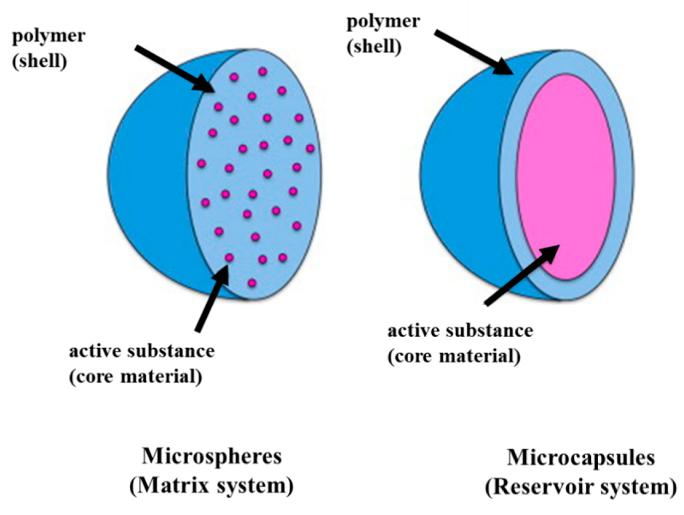
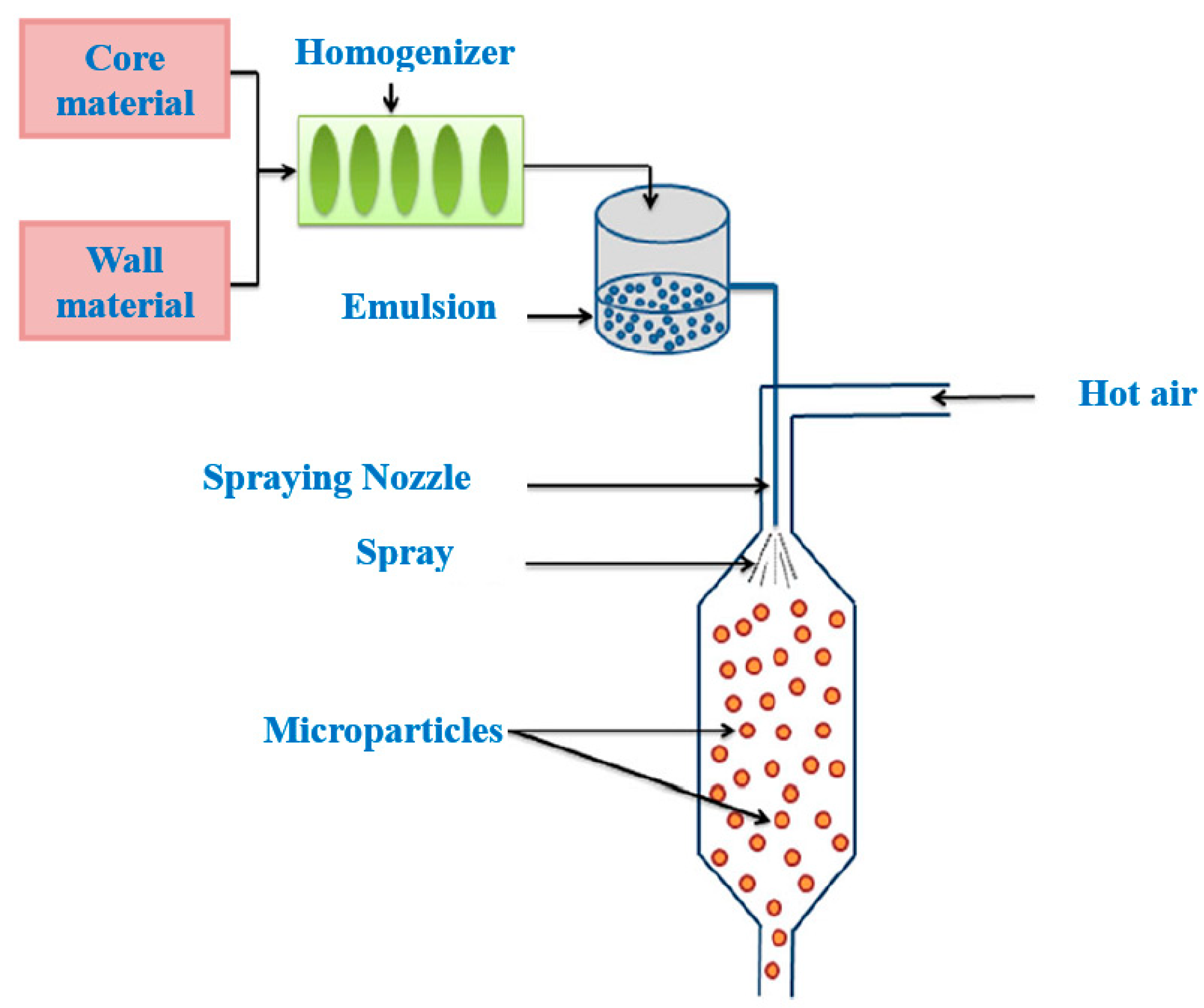
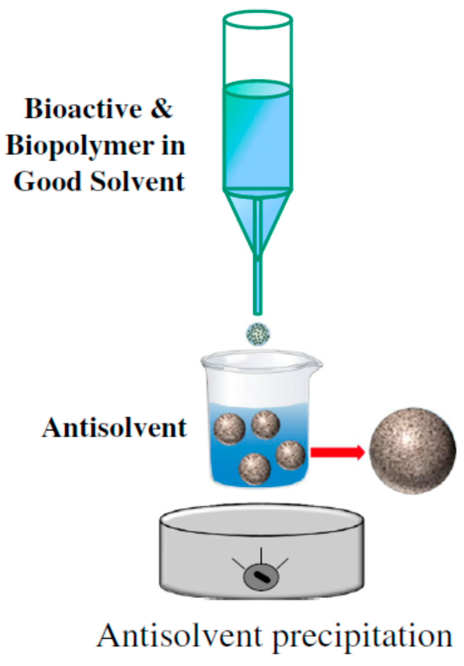
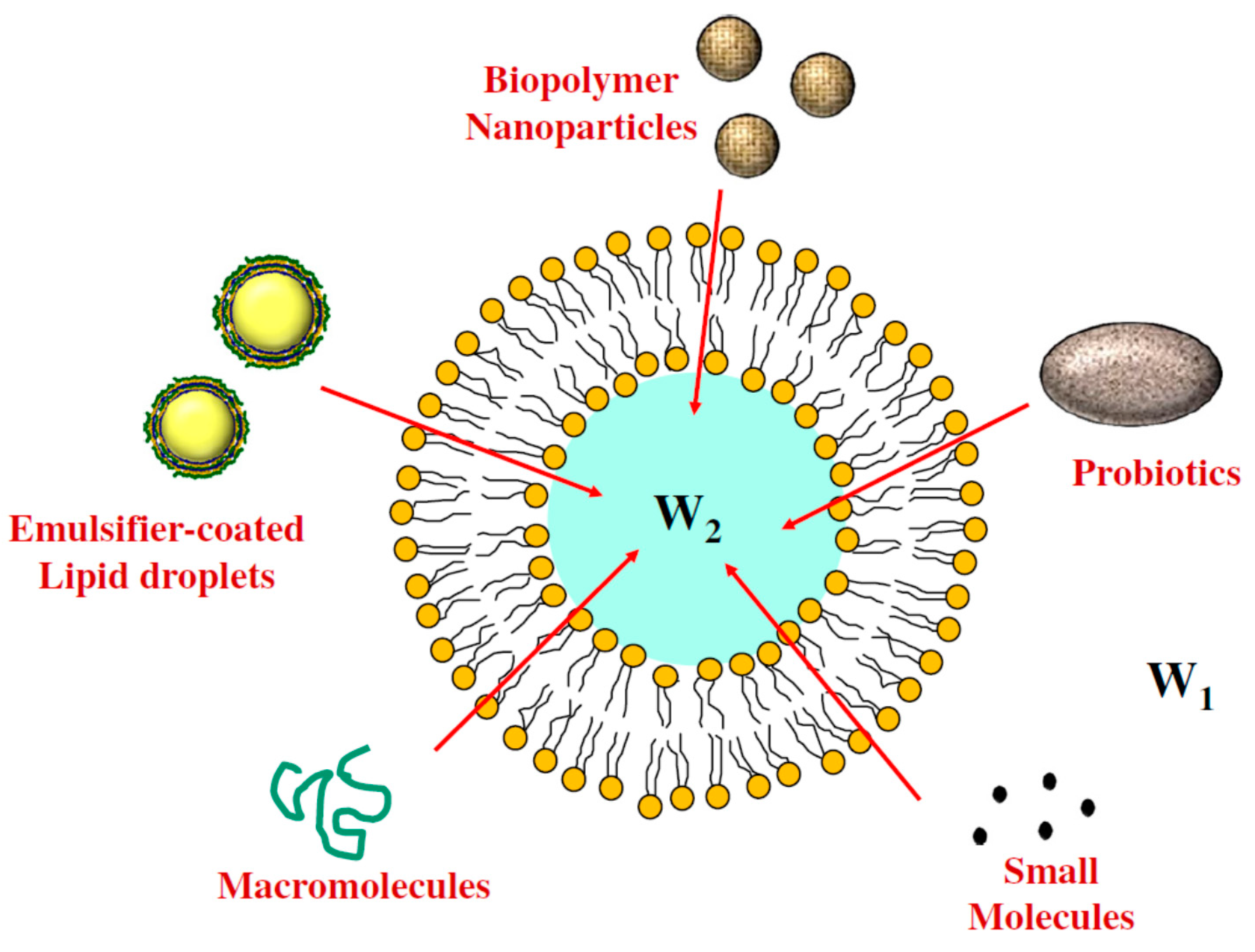
Table 1.
Different methods, wall materials, encapsulation efficiency and main results for microencapsulation of antimicrobial agents.
Table 1.
Different methods, wall materials, encapsulation efficiency and main results for microencapsulation of antimicrobial agents.
| Microencapsulation Method | Wall Material | Antimicrobial Agent | Encapsulation Efficiency | Main Results | References |
|---|---|---|---|---|---|
| Inclusion | β-cyclodextrin | Eugenol | - | The eugenol-βCD complexes showed enhanced antibactieral activity compared to free eugenol (a concentration of eugenol higher than 5.03 mmol L−1 inhibited the growth of Staphylococcus aureus and Escherichia coli) | [11] |
| Inclusion (KN-FD) | HPBCD | Carvacrol | 78.09% for KN, and 83.74% for FD | Encapsulated carvacrol showed a lower minimum concentration (300 μg/mL) than free carvacrol (1000 μg/mL) for both bacteria, Escherichia coli and Salmonella enterica | [12] |
| Inclusion (KN-FD) | β-cyclodextrin | Thymol and thyme oil | 71 to 83% | β- cyclodextrine inclusion complexes were able to inhibit Escherichia coli at lower concentrations (0.37 mg/mL) than free oils. | [13] |
| Inclusion (by spray drying and precipitation methods) | HPBCD, β-CD | Lemongrass volatile oil | 56–60% and 26–29% using β-CD and HP-β-CD, respectively | More effective inclusion of lemongrass oil with beta-CD | [14] |
| Inclusion | Β-cyclodextrin, HE-β-CD, HP-β-CD | Eugenol | - | The inclusion process was deduced to be an exothermic and enthalphy-driven process | [15] |
| Spray drying | Low methoxyl pectin | Lysozyme | - | Higher pectin concentrations (above 0.5 g/L) preserved lysozyme structure and activity | [16] |
| Spray drying | Gum arabic, starch, maltodextrin, inulin | Rosemary EO | - | The combination of modified starch and inulin was shown to be a viable substitute for gum arabic in foods | [17] |
| Spray drying | Modified starch, gum arabic, maltodextrin | Oregano EO | - | The inlet air temperature and the emulsion feed rate significantly affected the powder recovery, moisture content and the oil retention | [18] |
| Spray drying | Zein | Nisin | - | Encapsulated nisin was more effective than free antimicrobials in inhibiting the growth of L. monocytogenes | [19] |
| Simple coacervation | PVA crosslinked by glutaraldehyde | Lemongrass EO | - | When SDS at 0.03 wt.% was used, no agglomeration was observed | [20] |
| Simple coacervation | Gelatin microparticles crosslinked with glutaraldehyde | Holy basil EO (HBEO) | 95.41% | Extended shelf-life of microencapsulated HBEO up to 18 months at 25 °C | [21] |
| Complex coacervation | Gelatin–gum arabic crosslinked with genipin | Mustard seed EO | - | Genipin-hardened microcapsules exhibited strong chemical stability with a particle size of mainly 5–10 μm | [22] |
| Complex coacervation | Soy protein–pectin | Propolis extract | 72.01% and 66.12% for formulations with 2.5 and 5.0 g/100 mL of colloids, respectively | This process preserves the phenolic and flavonoids compounds with antioxidant activity and inhibitory activity of S. aureus | [23] |
| Complex coacervation | Whey protein isolate–chitosan | Garlic extract | 51% to 61% | The CH/WPI complexes are revealed to be good alternatives for use as wall systems | [24] |
| Single emulsion diffusion method | Poly lactic acid (PLA) | Nisin | 12 to 16% | The encapsulation efficiency was enhanced with the increase in nisin loading in the aqueous solution | [25] |
| Emulsion diffusion | Polycaprolacton (PCL) | Eugenol | 100%, 90.9% and 89.1% for PCL, β-CD eugenol and 2-HP-β-CD eugenol, respectively | The emulsion–diffusion method was more effective for eugenol encapsulation to protect against light oxidation during storage time | [26] |
| s/o/w emulsion–evaporation extraction | Poly (lactic-co-glycolic) (PLGA) | Lysozyme | 73% | More hydrophilic polymers were less prone to protein adsorption | [27] |
| Alginate microbeads | Calcium alginate–cellulose nanocrystals (CNC) | Nisin | - | The beads containing nisin significantly reduced the L. monocytogenes counts after 28 days of storage compared with free nisin | [28] |
| Alginate microbeads | Calcium–Alginate | Clove, thyme and cinnamon EOs | 90 to 94% | Encapsulation in Ca-alginate microspheres could effectively reduce the evaporation rate of EOs | [29] |
| Alginate microbeads | Sodium alginate–guar gum | Nisin | 36.35% | The encapsulation efficiency of nisin under optimal conditions was 36.65% | [30] |
| Ionic gelation | Sodium alginate | Propolis extract | 99.3% | Na-alginate encapsulation increased the bioavailability of propolis extract | [31] |
| SAS | PLGA and CaCo3 | Lysozyme | ~60% | The supercritical CO2 process offers optimal conditions for protein stability and integrity and permitted the retention of 90% of the biological activity of lysozyme | [32] |
| Liposome | PC, PC/PG | Nisin | 89–91, 78–83 and 72–78% for PC/PG 6:4, PC/PG 8:2 and PC, respectively | Liposomes formulated with PC and PG appeared to be relatively stable to pasteurization protocols | [33] |
| Liposome and emulsification method | Lecithin–chol Alginate, chitosan or starch | Plant herbs, spices and lyzozyme | - | Particles with co-encapsulated herbs and lysozyme are more active against different types of bacteria | [34] |
| Vibrating technology | alginate | Nisin | 75% | Microcapsules efficiently protected nisin from protease activity and retarded nisin release | [35] |
Table 2.
Applications of encapsulated antimicrobials for food preservation.
| Antimicrobial Agents | Wall Material | Encapsulation Method | Food Products | Target Microorganism | Main Results | References |
|---|---|---|---|---|---|---|
| Nisin | Gum arabic | Spray drying | Milk | L. monocytogenes, B. cereus | Spray-dried commercial nisin had an antimicrobial effect under refrigeration (90 days) | [109] |
| Clove oil | Β-CD–porous structure | Spray drying | Meat products | Mold spores | Encapsulated clove oil had a strong heat resistance and a high antiseptic effect on meat products | [110] |
| Grape seed extract/carvacrol | Chitosan | Ionic gelation | Salmon | Psycrophilic, mesophilic bacteria, Pseudomonas spp. | Prepapred microcapsules increased the shelf-life of refrigerated salmon to 4–7 days of storage | [111] |
| Coconut shell liquid smoke | Dextrin | Spray-drying | Tilapia meat | TPC | Formulated microcapsules could reduce quality deterioration on fresh fish meat | [112] |
| Curcumin | Gelatin–porous starch | Spray-drying | Tofu/bread/cooked pork | Mold spores | Compared with free curcumin, microcapsules reduced mold spores from (34.4 ± 2.5) to (52.3 ± 4.1)% | [113] |
| Nisin | Zein | Spray drying | milk | L. monocytogenes | Capsules were more effective than free antimicrobials in inhibiting the growth of L. monocytogenes in 2% reduced fat milk at 25 °C | [19] |
| Rosemary EO | Modified starch–maltodextrin | Spray drying | Fresh dough | Fungi and yeast | The encapsulated rosemary essential oil provided long-term antimicrobial activity when applied to fresh dough | [114] |
| Nisin | Alginate–cellulose nanocrystals (CNC) microbeads | Alginate microbead | RTE meat | L. monocytogenes | Microencapsulation of nisin (63 μg/mL) increased the lag phase of bacterial growth up to 28 days | [28] |
| Nisin Z | Proliposome H | Liposome | Cheddar cheese | L. innocua Lactobacillus spp. L. casei subsp. casei | Nisin-containing liposomes could provide a powerful tool to improve nisin stability and availability in the cheese matrix | [115] |
| Lactoferrin | Corn oil–butter fat–polyglycerol polyricinoleate | Emulsification | Bologna slices | Carnobacterium viridans | Microencapsulated lactoferrin had greater antimicrobial activity against Carnobacterium viridans than the free one | [116] |
Publisher’s Note: MDPI stays neutral with regard to jurisdictional claims in published maps and institutional affiliations. |
© 2022 by the authors. Licensee MDPI, Basel, Switzerland. This article is an open access article distributed under the terms and conditions of the Creative Commons Attribution (CC BY) license (https://creativecommons.org/licenses/by/4.0/).
Share and Cite
MDPI and ACS Style
Eghbal, N.; Liao, W.; Dumas, E.; Azabou, S.; Dantigny, P.; Gharsallaoui, A. Microencapsulation of Natural Food Antimicrobials: Methods and Applications. Appl. Sci. 2022, 12, 3837. https://doi.org/10.3390/app12083837
AMA Style
Eghbal N, Liao W, Dumas E, Azabou S, Dantigny P, Gharsallaoui A. Microencapsulation of Natural Food Antimicrobials: Methods and Applications. Applied Sciences. 2022; 12(8):3837. https://doi.org/10.3390/app12083837
Chicago/Turabian StyleEghbal, Noushin, Wei Liao, Emilie Dumas, Samia Azabou, Philippe Dantigny, and Adem Gharsallaoui. 2022. "Microencapsulation of Natural Food Antimicrobials: Methods and Applications" Applied Sciences 12, no. 8: 3837. https://doi.org/10.3390/app12083837
Note that from the first issue of 2016, this journal uses article numbers instead of page numbers. See further details here.