Article

The Importance of Atmospheric Microbial Contamination Control in Dental Offices: Raised Awareness Caused by the SARS-CoV-2 Pandemic

1
Department of Cranio-Maxillofacial Surgery and Radiology, Faculty of Dental Medicine, Iuliu Hatieganu University of Medicine and Pharmacy, 400012 Cluj-Napoca, Romania
2
Clinic of Dermatology and Allergic Diseases, University Clinic Ulm, 89075 Ulm, Germany
3
Department of Morphological Sciences, Faculty of Medicine, Iuliu Hatieganu University of Medicine and Pharmacy, 400012 Cluj-Napoca, Romania
4
Department of Medical Education, Faculty of Medicine, Iuliu Hatieganu University of Medicine and Pharmacy, 400012 Cluj-Napoca, Romania
5
Department of Molecular Sciences, Faculty of Medicine, Iuliu Hatieganu University of Medicine and Pharmacy, 400012 Cluj-Napoca, Romania
6
Faculty of Food Science and Technology, University of Agricultural Sciences and Veterinary Medicine of Cluj-Napoca, 400374 Cluj-Napoca, Romania
7
Department of Dental Propaedeutics and Esthetics, Faculty of Dental Medicine, Iuliu Hatieganu University of Medicine and Pharmacy, 400012 Cluj-Napoca, Romania
8
Department of Oral Health, Faculty of Dental Medicine, Iuliu Hatieganu University of Medicine and Pharmacy, 400012 Cluj-Napoca, Romania
*
Author to whom correspondence should be addressed.
Avram Manea and Diana Crisan contributed equally to this article.
Appl. Sci. 2021, 11(5), 2359; https://doi.org/10.3390/app11052359
Submission received: 11 February 2021 / Revised: 28 February 2021 / Accepted: 4 March 2021 / Published: 7 March 2021

Abstract

:

Featured Application

Our research provides valuable information for dentists and other medical professionals in their fight against the new Coronavirus by offering solutions to protect themselves and their patients during their work in medical offices.

Abstract

Background: In the context of the Sars-CoV-2 pandemic that started in 2020, more attention is being paid to the air quality in medical offices. The medical, economic and social implications of this crisis are unprecedented. Dental offices, particularly, were significantly affected by this pandemic due to the high exposure of dental workers, limited availability of personal protective equipment (PPE) and serious financial problems. Methods: Four different procedures were compared regarding their effectiveness in air contamination reduction, both from a biological point of view and from a management point of view: Fogging, Ultraviolet C (UVC) lamps, UVC air circulation units and natural ventilation. A total of 56 Petri dishes were used to evaluate air contamination. Results: All four procedures offered good results but the decontamination time and overall effect varied depending on the chosen method. Fogging was the only method that managed to remove all the identifiable pathogens. Conclusions: Fogging proved to be superior from a medical point of view, while the UVC air circulation unit proved to be more efficient from a management point of view.

1. Introduction

The pandemic caused by SARS-CoV-2 (Severe Acute Respiratory Syndrome Coronavirus 2) started in the first months of 2020 and raised huge global health issues (the first coronavirus cases had been already reported before the end of 2019) [1,2]. In humans, coronaviruses induce respiratory diseases such as the Middle Eastern respiratory syndrome (MERS) or the severe acute respiratory syndrome (SARS). Coronaviruses were first identified in the 1960s. They are spherical in form and covered with a helically symmetrical capsid and a peri-capsid crossed by glycoprotein structures. This layout gives it the typical appearance (crown). The entire viral genome is composed by a single strain of RNA. The virus mainly affects the respiratory and gastrointestinal tract. Although there are more types of coronaviruses that can affect humans, the SARS-CoV-2 is by far the best-known variant since it caused a global health crisis that affects each of us. The incubation period is between 2 and 29 days (most frequently 2–14 days). The main transmission form for this virus is through respiratory droplets (sneeze, cough or direct personal contact). Although many uncertainties still exist regarding the different transmission routes, the interest towards air disinfection protocols and ventilation, especially in medical environments, is increasing [2].
COVID-19, the disease caused by this virus, was an unknown pathology, raising diagnostic and treatment challenges. Treatment protocols have been elaborated, based on the ongoing quest for understanding this deadly threat [3,4]. Despite the many uncertainties, one thing was sure from the very beginning: the fact that prevention is better than any treatment method, a well-known and widely spread rule in medicine. During this pandemic, fields such as telemedicine gained lots of attention amongst health workers and patients as well [5]. However, since the personal interaction between doctors and patients cannot always be done remotely, new and strict protection protocols had to be developed. Amongst the health workers who are most exposed to the viral contamination, dentists and related professions, together with ophthalmologists, are at the top of the list [6,7,8]. All other medical branches were affected as well, directly or indirectly. For instance, otolaryngologists had to deal with many COVID-19 patients due to their experience in airway management, making them very exposed to contamination risks [9]. Anesthesiologists and emergency personnel were also at high risk. Recently published papers show an increased frequency of Burnout syndrome in relation with the COVID-19 crisis amongst medical professionals [10]. For dentists, this is mostly due to their physical proximity to the patient’s open oral cavity during treatments that include multiple aerosols generating procedures. Such aerosols contain a high concentration of bacteria, fungi and viruses [11,12,13]. Although aerosols are the main culprit in bacterial dissemination throughout the dental office, other procedures like the basic examination or manual scaling can easily spread pathogens, despite the fact that they are not considered aerosol generating [14]. The impact of this pandemic on dental offices was very important, most of them being closed for several months or having the procedure list narrowed-down drastically, typically to emergency treatments. At this point, dental practices all over the world are slowly re-opening for a complete range of treatments, however, with increased safety measures and personal protective equipment (PPE) that was never considered for dental usage in the past. It is very likely that dental office disinfection and sterilization protocols will be forever changed after this pandemic, with an obvious impact on both practitioners and patients.
The air quality in dental offices has been a concern for a long time. Studies on this topic have been published as early as the 1970s [15,16]. In several studies between 1970 and 1975, air filtration systems (Electrostatic filters) were tested and proved to reduce microorganism concentration in the dental office air [17,18]. A study conducted by Luksamijarulkul in 2009 in a hospital dental clinic showed that only 36.7% of the dental personnel rigorously respected the decontamination, disinfection and sterilization protocols, leading to an increased occupational risk [19]. More recent studies (2018) also focused on the sterilization protocols and their efficiency (surfaces, instruments, air etc.), showing less than optimal outcomes. The authors of the article suggest the implementation of biologic monitoring protocols [20]. Several other studies that were conducted in the past few decades tend to show the high level of air contamination in dental offices, especially during dental procedures and the importance of biologic monitoring and strict disinfection and sterilization protocols [21,22].
The bacterial contamination of the dental office is a valuable indicator in the evaluation of the infective risk, both for dental personnel and patients entering the office [23]. Although, during the past few months, the focus was mostly shifted towards viral infections, evaluation of the viral load is more difficult and presents a higher contamination risk. The prevalence of Hepatitis C virus (HCV) was measured by Piazza and his team on different surfaces in the dental office by harvesting samples in order to identify HCV-RNA strands and analyze them by polymerase chain reaction (PCR) [24]. Positive HCV patients took part in that study. Such a testing protocol would have been unethical to conduct with SARS-CoV-2 patients because of the extremely high risks involved.
National and international guides were elaborated during the pandemic in order to reduce the risk of cross contamination and medical personnel infection. Each country outlined their own set of indications, but most of them were common throughout the world. One of the most important recommendations was related to oral cavity rinsing with antiseptic solutions before aerosol inducing procedures. The efficiency of this measure in reducing the bacterial load of oral cavities has been well-documented for a long time. Some studies suggest a decrease of the bacterial load in aerosols by up to 92%.
Scrubs, coats or other medical uniforms are also a reservoir of bacteria resulted from aerosol inducing procedures; therefore, changing scrubs between patients is considered a good way to prevent cross contamination [14]. During this period, impermeable, single use overalls were recommended to avoid such risks. PPE also included FFP2/3 face masks (N95 or above). Their efficiency has been determined to be very high, if used correctly: 99.7% for influenza A virus, rhinovirus 14 and S. Aureus, and 99.3% for surrogate particulates (paraffin oil and sodium chloride) [25]. Face shields and protective spectacles were also included in the mandatory PPE list, since their efficiency has also been proven for a long time [8]. Although it is not part of the PPE per se, the dental dam was one of the most effective ways to control the spread of pathogens through aerosols. Intraoral radiographs were forbidden for several months, using panoramic scans and CT (CBCT) scans as an alternative.
The recommendations for air disinfections included natural or forced ventilation, Ultraviolet C (UVC) light and dry aerosols. A study from 1994 conducted in Italy by Signoretto recommended the use of a dry aerosol producing machine that vaporizes phenols or chlorhexidine for environmental disinfection. This method seemed to remove all salivary microorganisms from the dental office. The authors suggested the use of this device at the end of every workday [26]. Other studies recommended fumigation of the office at least once a month [27]. Little information, though, has been offered regarding the used substances, concentrations, particle sizes etc. Sources of UVC light and air recirculating devices using UVC modules also seem to have an increased efficiency in reducing the airborne bacterial and viral load [28,29,30].
Several methods of sampling the air were used in previous studies, such as customized air-sampling devices [12], Casella Slit Sampler for Airborne Bacteria [17], sedimentation plates [21], portable RCS PLUS Air Sampler [31] etc. All of them proved efficient in measuring the air contamination.
Medical offices, especially dental practices, include several devices that are necessary for the medical act. All these complex machines present a variety of surfaces which are more or less suited and accessible for classical disinfection methods. Since aerosol size is less than 5 μm, droplets containing dangerous pathogens may easily reach these surfaces and not be inactivated by the previously used disinfection protocols [2]. This is why the study of more efficient air and surface decontamination methods is so important during this period. Even the materials used for PPE were considerably modified. For instance, new materials containing Zinc pyrithione were used to produce overalls since it presents proven antiviral, antibacterial and antifungal properties [32,33]. In Romania, such materials were approved by the Center for Medico-Military Scientific Research.
Since the issue of air quality in dental offices was once again raised by the SARS-CoV-2 pandemic, we decided to compare the efficiency of several air decontamination techniques. We aimed to verify whether plain natural ventilation is a sufficient method for ensuring air decontamination in the dental office, or whether more elaborated decontamination methods are required. The results of the present study could, therefore, guide the choice of air disinfection protocols for dental practices.

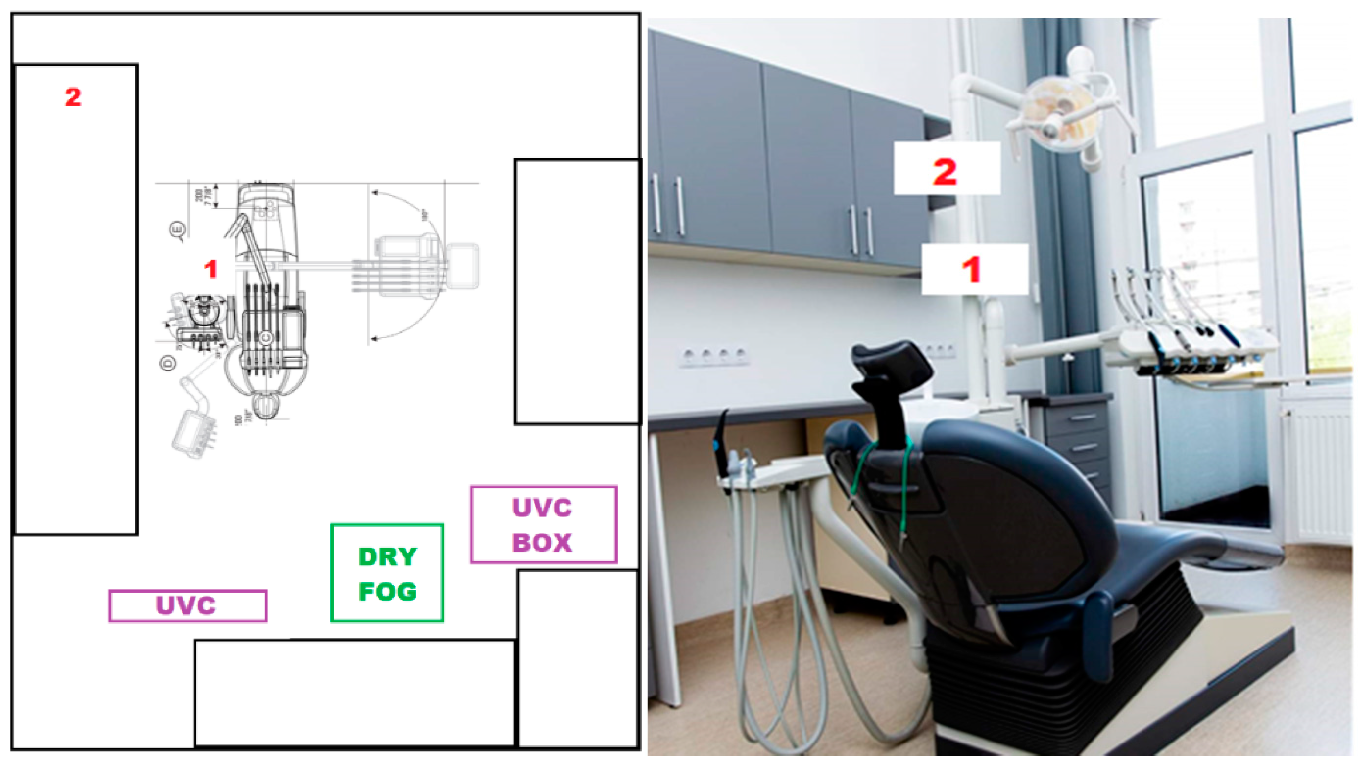
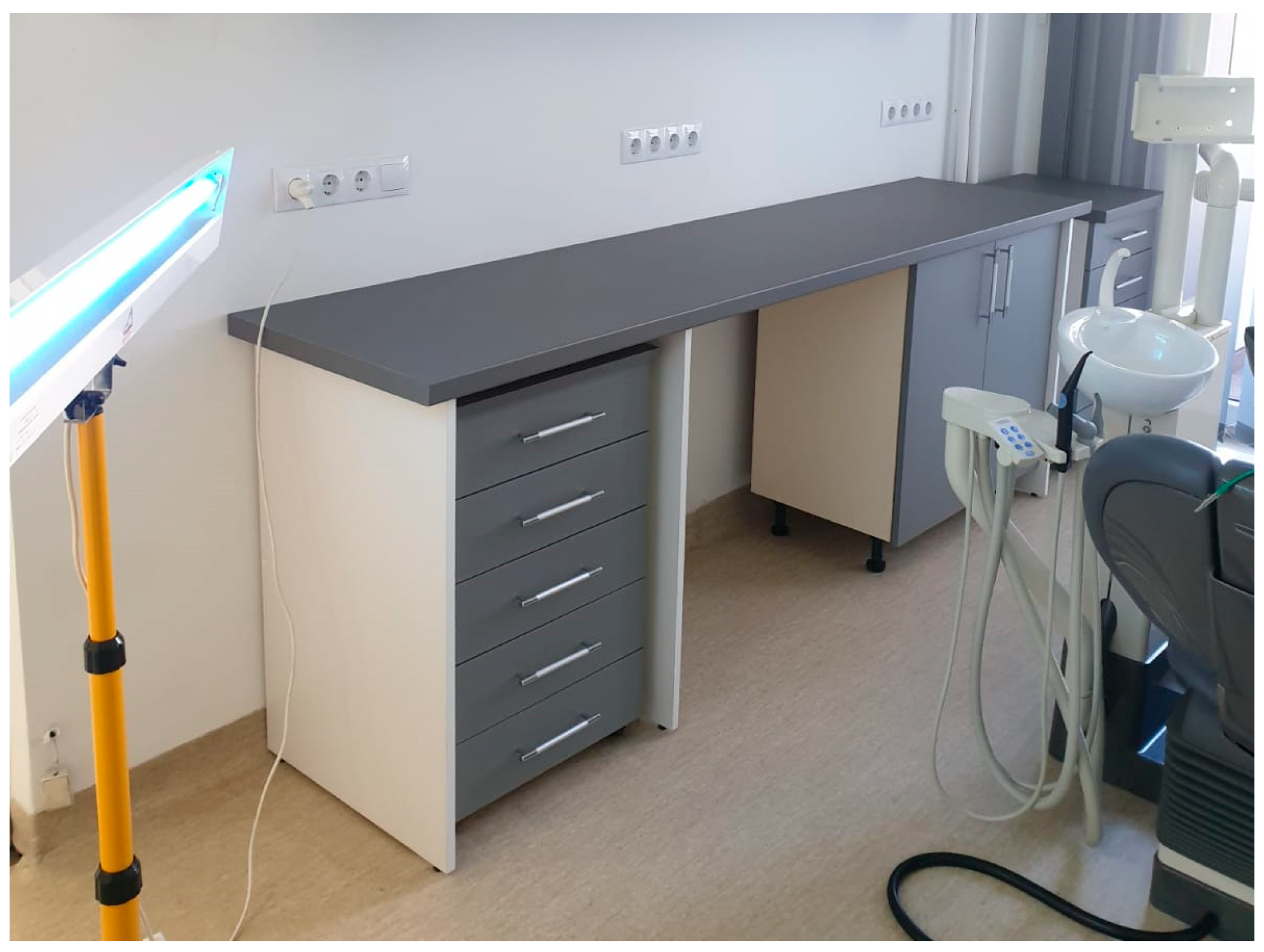
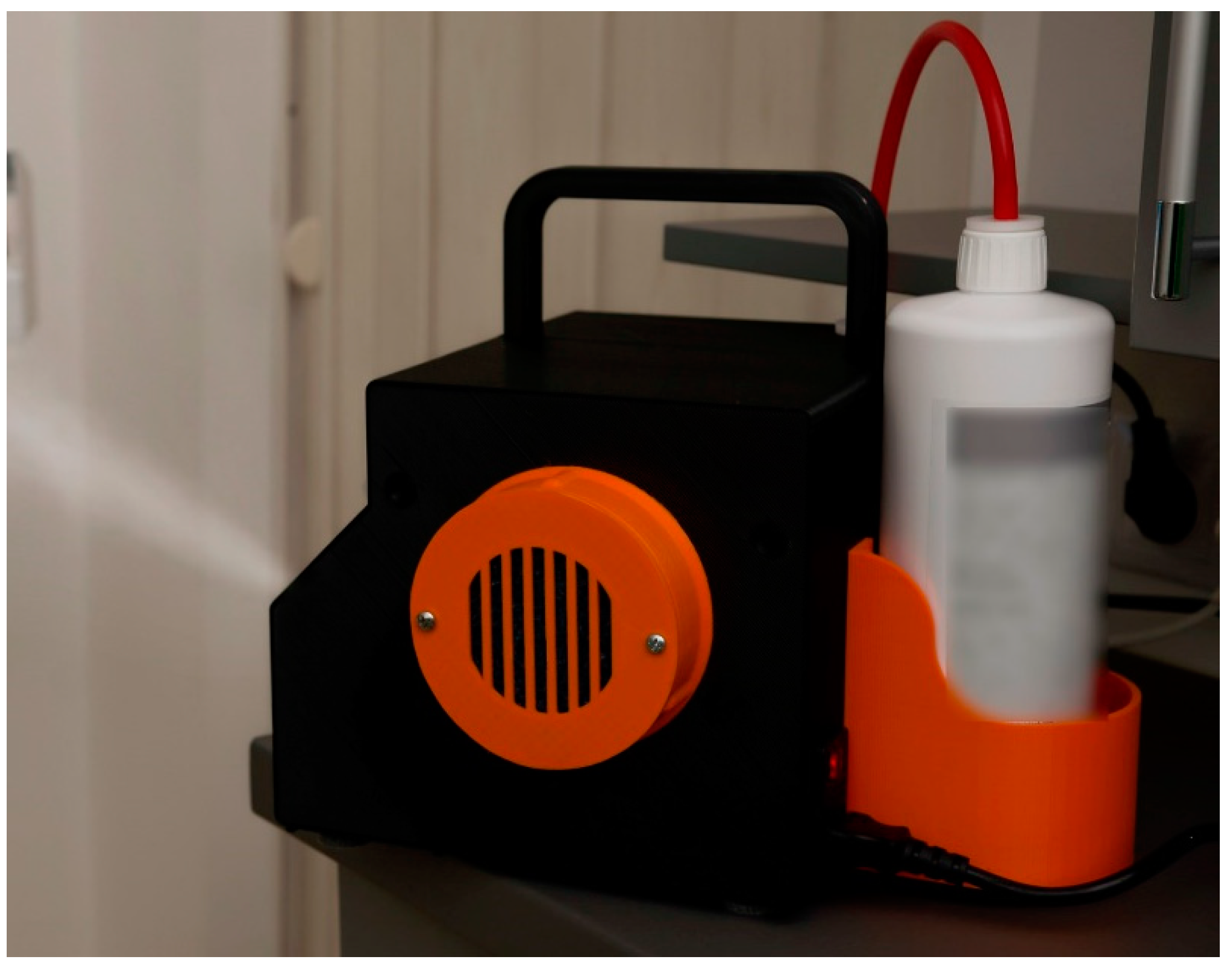
2. Materials and Methods

The present study was conducted in a private dental office in Cluj-Napoca, Romania. The treatment room in which the aerial bacterial load was measured had a surface of 18 m2 (193 ft2) and a volume of 72 m3 (2542 ft3). It hosted a single dental unit and was connected, by separate access points, to a sterilization area and to a waiting room. The UVC projector, UVC air recirculating unit and nebulizer (fogging machine) were placed as depicted in Figure 1. The window that was used for ventilation was 220 cm by 80 cm.
The UVC projector (Biocomp, Iași, Romania) was a 55 W, single fluorescent tube germicidal lamp, with the dominant wavelength of 253 nm. It was placed at 1.2 m above ground level, on a pivotal support (Figure 2).
The fogging machine (KOX, Bucharest, Romania) had a 650 W turbine, producing particles of less than 5 µm, projecting them at a 220 km/h speed. For this study, we used Sanosil (Sanosil Ltd. International, Hombrechtikon, Switzerland), a substance containing 5 g/100 g H2O2 and 0.005 g/100g Ag+ (silver ions) that is supposed to effectively inactivate bacterial spores, bacteria, yeasts, fungi and viruses (all types), including enveloped viruses (Influenza, Corona). Hydrogen peroxide is an effective yet environmentally friendly substance and the traces of silver that remain on various surfaces are supposed to have an extended antibacterial effect, even after the fogging procedure has finished (Figure 3).
The UVC air recirculation unit (UVC box)—Sterybox (TISSIMedica, S. Giuliano Milanese, Italy) is a device with a total power of 350 W, with five 25 W lamps and a minimum air flow of at least 50 m3/h. The device was designed to cover a room floor size of 20 m2 or a total room volume of 50 m3.
A total of 56 Petri dishes were used for the present test as follows: 8 during the treatment and 8 after the disinfection method application for ventilation, fogging and UVC, respectively. While using the Sterybox, 8 dishes were used, since there was no separate decontamination phase for this method.
The experiments were conducted in 4 stages: Stage 1 entailed the collection of airborne bacteria during the 30 min of patient treatment by using sedimentation plates (Petri dishes), followed by 2 h of natural ventilation (open window, no forced ventilation, door of cabinet remained closed), after which a new set of bacterial sedimentation on Petri dishes was collected for 30 min. Stage 2 was similar to stage 1, but the decontamination of the air was performed by using the UVC for 30 min. Stage 3 included the same steps, but the air was disinfected by using the fogging machine (6 mL/m3), followed by 40 min of settlement and action time for the airborne substance. Stage 4 was different, since we used the UVC box for 15 min before patient admission, during patient treatment and for 15 min after the patient had left the treatment room. Before every patient entered the office, current national guidelines (UVC, ventilation and fogging, with a total of 5 h gap between patients) for disinfection were applied, to ensure maximum patient safety. This also provides a constant disinfection guideline.
For every measurement performed during patient treatments, we used a total of 8 Petri dishes: 2xNA-Nutrient-Agar, 2xYPD - YPD-Agar, 2xGPY-Glucose-Peptone-Yeast-Agar and 2x MHA-Mueller-Hinton-Agar (Table 1).
Four Petri dishes, one for each culture medium, were placed in position 1 of the dental office and a second such set of 4 Petri dishes was placed in position 2 of the dental office (Figure 1). Position 1 was situated at a distance of 1 m from the patient’s oral cavity, with no obstacles in between, while position 2 was located at a distance of 3.5 m from the patient’s oral cavity, in an area where it was not directly exposed to treatment-related airflow. A total of 56 dishes were used (Figure 4).
The dishes were labeled “N” for the fogging samples (nebulization), “UVC” for the UVC light samples, “A” for the natural ventilation samples and “B” for the UVC air recirculation unit (UVC box). Two digits followed: the first referred to the position (1 or 2) and the second referred to the time period (1—during treatment; 2—after applying the air decontamination method).
The dental personnel wore all the required PPE and the treatment protocol was in no way altered by the ongoing of the bacterial recordings. None of the treatments were aerosol-generating. There were only emergency treatments done without rotary instruments (root canal drainage). All patients rinsed their mouth (Chlorhexidine 2% solution) and received rubber dams.
All the samples were then incubated for 48 h, at 35 ± 2 ℃ and the microbial load was measured in colony-forming units (CFU).

3. Results

Several Petri dishes exhibited bacterial colonies after incubation. Most of these positive Petri dishes had been collected during patient treatment, both in the samples closest to the patient (position 1), but also in the ones situated farther away (position 2). A drastic reduction in the number of positive cultures (Table 2), and also in the number of CFU (Table 3), was observed after applying any of the air decontamination methods.
Dry fogging was the only air decontamination method that was followed by completely negative cultures, despite the high number of positive cultures obtained from the preceding samples that had been collected during patient treatment.

4. Discussion

A high contamination risk amongst dentists appears to be suggested by these results, even though the treatment procedures performed as part of this study were not aerosol-generating. As an initial investigation, this study was conducted using a relatively small number of Petri dishes (56); nevertheless, its results may already suggest a number of partial conclusions which yield useful clinical implications. Naturally, further experimental studies, using larger numbers of Petri dishes and possibly other air decontamination methods, should follow, in order to elaborate a complete guide for medical offices and particularly dental offices. Other limitations of the present study, besides the number of Petri dishes, are the number of recordings, the dish placement and the investigation of only bacterial and not also viral contamination. For obvious ethical reasons, while aiming to reduce contamination risks in dental offices and thereby also limit the spread of SARS-CoV-2, the present study could not be conducted by experimentally spreading this virus in a dental office, given the immense risks such an approach would have posed to both patients and healthcare personnel. Nevertheless, strong similarities in the airborne transmission of bacteria and viruses do exist and the decontamination/disinfection methods that were employed in this study were also proven to be effective against SARS-CoV-2 [28,30,34]. These arguments open up the possibility of also applying the insights yielded by the current study in the prevention of SARS-CoV-2 transmission.
Reducing transmission rates inside healthcare environments represents an important aim, by finding efficient methods for decontamination, thereby saving the lives of medical workers, patients and easing the pressure on the entire medical system. Other studies have also focused on SARS-CoV-2 transmission rates and on ways of decreasing the risk of its transmission [35,36,37].
The most relevant factors considered in further analyzing the results of our study were the efficiency of the microbial air load decrease (due to obvious medical reasons) and the time factor (mostly due to financial reasons) [38,39]. Long pauses between patients and expensive PPE lead to a decrease in the profitability of dental practices and even to the bankruptcy of some of them. For this reason, finding a quick and economically feasible yet highly effective decontamination method is vital, especially for small, privately owned healthcare facilities.
Dry fogging or nebulization has been proven to be efficient in air and surface decontamination because of its high penetrability and spread ability. It has been used in operating rooms, commercial setups, industry, etc., but it was never a common practice in dental offices. Peracetic acid and hydrogen peroxide are some of the most commonly used substances for this procedure. Their efficiency is comparable, while hydrogen peroxide is an environmentally safer agent compared to peracetic acid [40]. Dry fogging has been recommended for use in healthcare facilities and even in severely contaminated spaces like intensive care units treating severely ill SARS-CoV-2 patients [41,42]. Sanosil, the substance we used for this study, was proven to have the required antiviral and bactericidal effect [43]. Dry fogging, in our study, was the only decontamination technique providing zero CFU on all the employed culture media. The disadvantage of this technique is that the fogging machine presents the highest acquisition costs and it also requires a high quantity of the used substance for each run, which also adds considerable costs. The action and settlement time for this method was 40 min, but national and international guidelines require another 2 to 4 h of natural ventilation to avoid any unwanted effects, which would unfeasibly modify the expected patient flow of a dental practice.
The UVC air recirculation unit also proved to be extremely efficient. Among all corresponding Petri dishes, only two CFU were formed throughout the entire duration of the patient’s visit and treatment. Confirming our findings, other studies also showed good results for these devices, although these studied other working environments [28,30]. The initial costs of the device used in this study were high, but not as high as those of the fogging device. In addition, except for electricity, no other consumables were needed in this case. The important advantage of this technique is that it drastically reduces the time gap needed between patients and, most of all, that it prevents patient to doctor or doctor to patient contamination, by an important decrease in air contamination. Another major advantage is the fact that it can also be used with no harmful effects in the direct proximity of humans, thereby ensuring the necessary air decontamination both between and during treatment sessions. Hence, we can recommend the use of such a device in all dental offices.
Use of the UVC projector is a validated air decontamination method for bacteria, viruses and fungi. Its effect on Sars-CoV-2 is proven by several older and, especially, more recent studies [44,45]. A decontamination time of 30 min between patients is not difficult to manage and this method is also efficient from an economic point of view. The air decontamination obtained by using it is not as good as other methods, most likely because of the relevance of the distance between the UVC source and the different parts of a dental office, and even more so because of its inactivity in areas that are positioned in closed spaces or in the shadow of other objects. Another drawback would be the fact that UVC projectors must not be used in the presence of humans, given the health hazards of direct exposure to ultraviolet radiation.
Natural ventilation of the office for two hours also showed a drastic decrease in air contamination. The major disadvantage of this method is the fact that it requires long breaks between patients, which is not beneficial from a financial point of view. However, still from a financial point of view, this method is obviously the cheapest to enact, since it requires no financial investment. Reaching a balance between these two economic aspects should be considered by each dental practice individually, in order to evaluate the real economic impact of natural ventilation as a stand-alone decontamination method.
Further studies on this subject should be conducted, given that we evaluated bacterial and fungal air-loading by using only 56 dishes and four interventions. Other air decontamination methods are also under consideration as potential candidates for our future studies. Although this has only been a preliminary study, its results may already suggest certain guidelines that could be valuable in reducing the risk of biological contamination and in assisting with the management of dental offices and other medical facilities which host significant daily flows of patients.

5. Conclusions

In the context of the Sars-CoV-2 pandemic, more attention has been paid to the air quality in medical and especially dental offices. Within the limitations of this study, an objective comparison between several air decontamination techniques has been performed. Fogging appears to be the most effective decontamination method, but it requires a prolonged time gap between patients. UVC air recirculation units show very good decontamination results and remarkable economic performance, since the time between patients is significantly reduced. Another aspect worth mentioning regarding UVC recirculation units is the fact that, by reducing the air contamination both between and during treatments, they can make the office a safer place for both patients and medical professionals. UVC lamps and natural ventilation are also effective methods against microbial contamination, but their efficacy appears to be lower compared to the former two methods. Like fogging, UVC lamps and natural ventilation are also time-consuming, but they are considerably cheaper. Still, the effectiveness from both a biological and, especially, a financial point of view will more accurately be quantified in future studies for all the methods that were described in the present study.
Healthcare workers should become aware of the advantages and limitations presented by various atmospheric microbial contamination control measures, so that they can choose and implement optimal ways for protecting themselves and their patients from Sars-CoV-2 and other airborne pathogens.

Author Contributions

Conceptualization, A.M. and S.B.; methodology, A.M. and D.C.; software, H.C. and M.B.; validation, D.V., A.A. and O.A.; formal analysis, M.B. and G.B.; investigation, A.M. and D.C.; resources, M.C. and I.C.; data curation, G.B. and G.A.; writing—original draft preparation, A.M. and D.C.; writing—review and editing, S.B., I.C. and H.C.; visualization, S.B. and C.D.; supervision, F.O., C.D. and M.C.; project administration, A.A. and O.A.; funding acquisition, G.A., F.O. and D.V. All authors have read and agreed to the published version of the manuscript.

Funding

This research received no external funding.

Institutional Review Board Statement

Not applicable.

Informed Consent Statement

Not applicable.

Acknowledgments

The authors Avram Manea and Diana Crisan contributed equally to this paper.

Conflicts of Interest

The authors declare no conflict of interest.

References

  1. Valencia, D.N. Brief Review on COVID-19: The 2020 Pandemic Caused by SARS-CoV-2. Cureus 2020, 12, e7386. [Google Scholar] [CrossRef] [Green Version]
  2. Cervino, G.; Fiorillo, L.; Surace, G.; Paduano, V.; Fiorillo, M.T.; De Stefano, R.; Laudicella, R.; Baldari, S.; Gaeta, M.; Cicciù, M. SARS-CoV-2 Persistence: Data Summary up to Q2 2020. Data 2020, 5, 81. [Google Scholar] [CrossRef]
  3. D’Errico, S.; Zanon, M.; Montanaro, M.; Radaelli, D.; Sessa, F.; Di Mizio, G.; Montana, A.; Corrao, S.; Salerno, M.; Pomara, C. More than Pneumonia: Distinctive Features of SARS-Cov-2 Infection. From Autopsy Findings to Clinical Implications: A Systematic Review. Microorganisms 2020, 8, 1642. [Google Scholar] [CrossRef] [PubMed]
  4. Contini, C.; Caselli, E.; Martini, F.; Maritati, M.; Torreggiani, E.; Seraceni, S.; Vesce, F.; Perri, P.; Rizzo, L.; Tognon, M. COVID-19 Is a Multifaceted Challenging Pandemic Which Needs Urgent Public Health Interventions. Microorganisms 2020, 8, 1228. [Google Scholar] [CrossRef]
  5. Cervino, G.; Oteri, G. COVID-19 Pandemic and Telephone Triage before Attending Medical Office: Problem or Opportunity? Medicina 2020, 56, 250. [Google Scholar] [CrossRef] [PubMed]
  6. Day, C.J.; Sandy, J.R.; Ireland, A.J. Aerosols and splatter in dentistry–A neglected menace? Dent. Update 2006, 33, 601–606. [Google Scholar] [CrossRef]
  7. Fini, M.B. What dentists need to know about COVID-19. Oral Oncol. 2020, 105, 104741. [Google Scholar] [CrossRef] [PubMed]
  8. Bordea, I.R.; Xhajanka, E.; Candrea, S.; Bran, S.; Onișor, F.; Inchingolo, A.D.; Malcangi, G.; Pham, V.H.; Inchingolo, A.M.; Scarano, A.; et al. Coronavirus (SARS-CoV-2) Pandemic: Future Challenges for Dental Practitioners. Microorganisms 2020, 8, 1704. [Google Scholar] [CrossRef] [PubMed]
  9. Balakrishnan, K.; Schechtman, S.; Hogikyan, N.D.; Teoh, A.Y.B.; McGrath, B.; Brenner, M.J. COVID-19 Pandemic: What Every Otolaryngologist–Head and Neck Surgeon Needs to Know for Safe Airway Management. Otolaryngol. Head Neck Surg. 2020, 162, 804–808. [Google Scholar] [CrossRef] [Green Version]
  10. Dimitriu, M.C.T.; Pantea-Stoian, A.; Smaranda, A.C.; Nica, A.A.; Carap, A.C.; Constantin, V.D.; Davitoiu, A.M.; Cirstoveanu, C.; Bacalbasa, N.; Bratu, O.G.; et al. Burnout syndrome in Romanian medical residents in time of the COVID-19 pandemic. Med. Hypotheses 2020, 144, 109972. [Google Scholar] [CrossRef]
  11. Szymańska, J. Endotoxin level as a potential marker of concentration of Gram-negative bacteria in water effluent from dental units and in dental aerosols. Ann. Agric. Environ. Med. 2005, 12, 229–232. [Google Scholar]
  12. Fine, D.H.; Furgang, D.; Korik, I.; Olshan, A.; Barnett, M.L.; Vincent, J.W. Reduction of viable bacteria in dental aerosols by preprocedural rinsing with an antiseptic mouthrinse. Am. J. Dent. 1993, 6, 219–221. [Google Scholar] [PubMed]
  13. Al Maghlouth, A.; Al Yousef, Y.; Al Bagieh, N. Qualitative and quantitative analysis of bacterial aerosols. J. Contemp. Dent. Pract. 2004, 5, 91–100. [Google Scholar] [CrossRef]
  14. Huntley, D.E.; Campbell, J. Bacterial contamination of scrub jackets during dental hygiene procedures. J. Dent. Hyg. JDH 1998, 72, 19–23. [Google Scholar]
  15. Miller, R.L.; Micik, R.E. Air pollution and its control in the dental office. Dent. Clin. North Am. 1978, 22, 453–476. [Google Scholar]
  16. Tudose, P.; Wittemberg, T.; Gavrişiu, F.; Luştrea, V. Some imperfections in dental practice. Stomatologia 1972, 19, 109–116. [Google Scholar] [PubMed]
  17. Iversen, D.B.; Tolo, K. Electrostatic air filters for dental practice. Nor Tann. Tid. 1975, 85, 446–448. [Google Scholar]
  18. Shreve, W.B.; Wachtel, L.W.; Pelleu, G.B., Jr. Air cleaning devices for reduction in number of airborne bacteria. J. Dent. Res. 1970, 49, 1078–1082. [Google Scholar] [CrossRef]
  19. Luksamijarulkul, P.; Panya, N.; Sujirarat, D.; Thaweboon, S. Microbial air quality and standard precaution practice in a hos-pital dental clinic. J. Med. Assoc. Thai. 2009, 92 (Suppl. S7), S148–S155. [Google Scholar] [PubMed]
  20. Dagher, J.; Sfeir, C.; Abdallah, A.; Majzoub, Z. Sterilization and Biologic Monitoring in Private Dental Clinics in Lebanon. J. Contemp. Dent. Pract. 2018, 19, 853–861. [Google Scholar]
  21. Harrel, S.K.; Molinari, J. Aerosols and splatter in dentistry: A brief review of the literature and infection control implications. J. Am. Dent. Assoc. 2004, 135, 429–437. [Google Scholar] [CrossRef]
  22. Yamada, H.; Ishihama, K.; Yasuda, K.; Hasumi-Nakayama, Y.; Shimoji, S.; Furusawa, K. Aerial dispersal of blood-contaminated aerosols during dental procedures. Quintessence Int. 2011, 42, 399–405. [Google Scholar] [PubMed]
  23. Monarca, S.; Grottolo, M.; Feretti, D.; Gigola, P.; Zerbini, I.; Alberti, A.; Zani, C.; Sapelli, P.L. Environmental monitoring of infective risks in Italian dental offices. Minerva Stomatol. 2002, 51, 319–326. [Google Scholar] [PubMed]
  24. Piazza, M.; Borgia, G.; Picciotto, L.; Nappa, S.; Cicciarello, S.; Orlando, R. Detection of hepatitis C virus-RNA by polymerase chain reaction in dental surgeries. J. Med. Virol. 1995, 45, 40–42. [Google Scholar] [CrossRef] [PubMed]
  25. Zhou, S.S.; Lukula, S.; Chiossone, C.; Nims, R.W.; Suchmann, D.B.; Ijaz, M.K. Assessment of a respiratory face mask for capturing air pollutants and pathogens including human influenza and rhinoviruses. J. Thorac. Dis. 2018, 10, 2059–2069. [Google Scholar] [CrossRef] [PubMed] [Green Version]
  26. Signoretto, C.; Canepari, P.; Urbani, G. Microbiological contamination in the dental office and its possible decrease. Minerva Stomatol. 1994, 43, 263–272. [Google Scholar]
  27. Shivakumar, K.M.; Prashant, G.M.; Madhu Shankari, G.S.; Subba Reddy, V.V.; Chandu, G.N. Assessment of atmospheric microbial contamination in a mobile dental unit. Indian J. Dent. Res. 2007, 18, 177–180. [Google Scholar] [CrossRef]
  28. Eisenlöffel, L.; Reutter, T.; Horn, M.; Schlegel, S.; Truyen, U.; Speck, S. Impact of UVC-sustained recirculating air filtration on airborne bacteria and dust in a pig facility. PLoS ONE 2019, 14, e0225047. [Google Scholar] [CrossRef] [Green Version]
  29. Curtis, G.L.; Faour, M.; Jawad, M.; Klika, A.K.; Barsoum, W.K.; Higuera, C.A. Reduction of Particles in the Operating Room Using Ultraviolet Air Disinfection and Recirculation Units. J. Arthroplast. 2018, 33, S196–S200. [Google Scholar] [CrossRef]
  30. Kim, D.-K.; Kang, D.-H. UVC LED Irradiation Effectively Inactivates Aerosolized Viruses, Bacteria, and Fungi in a Chamber-Type Air Disinfection System. Appl. Environ. Microbiol. 2018, 84, e00944–18. [Google Scholar] [CrossRef] [Green Version]
  31. Szymańska, J. Exposure to airborne fungi during conservative dental treatment. Ann. Agric. Environ. Med. 2006, 13, 177–179. [Google Scholar]
  32. Te Velthuis, A.J.W.; van den Worm, S.H.E.; Sims, A.C.; Baric, R.S.; Snijder, E.J.; Van Hemert, M.J. Zn(2+) inhibits coronavirus and arterivirus RNA polymerase activity in vitro and zinc ionophores block the replication of these viruses in cell culture. PLoS Pathog. 2010, 6, e1001176. [Google Scholar] [CrossRef]
  33. Schwartz, J.R. Zinc Pyrithione: A Topical Antimicrobial With Complex Pharmaceutics. J. Drugs Dermatol. JDD 2016, 15, 140–144. [Google Scholar]
  34. Tang, S.; Mao, Y.; Jones, R.M.; Tan, Q.; Ji, J.S.; Li, N.; Shen, J.; Lv, Y.; Pan, L.; Ding, P.; et al. Aerosol transmission of SARS-CoV-2? Evidence, prevention and control. Environ. Int. 2020, 144, 106039. [Google Scholar] [CrossRef]
  35. Correa-Martínez, C.L.; Schwierzeck, V.; Mellmann, A.; Hennies, M.; Kampmeier, S. Healthcare-Associated SARS-CoV-2 Transmission—Experiences from a German University Hospital. Microorganisms 2020, 8, 1378. [Google Scholar] [CrossRef] [PubMed]
  36. Dar Odeh, N.; Babkair, H.; Abu-Hammad, S.; Borzangy, S.; Abu-Hammad, A.; Abu-Hammad, O. COVID-19: Present and Future Challenges for Dental Practice. Int. J. Environ. Res. Public Health 2020, 17, 3151. [Google Scholar] [CrossRef] [PubMed]
  37. Mick, P.; Murphy, R. Aerosol-generating otolaryngology procedures and the need for enhanced PPE during the COVID-19 pandemic: A literature review. J. Otolaryngol. Head Neck Surg. 2020, 49, 1–10. [Google Scholar] [CrossRef]
  38. Ali, S.; Farooq, I.; Abdelsalam, M.; AlHumaid, J. Current Clinical Dental Practice Guidelines and the Financial Impact of COVID-19 on Dental Care Providers. Eur. J. Dent. 2020, 14, S140–S145. [Google Scholar] [CrossRef]
  39. Schwendicke, F.; Krois, J.; Gomez, J. Impact of SARS-CoV2 (Covid-19) on dental practices: Economic analysis. J. Dent. 2020, 99, 103387. [Google Scholar] [CrossRef] [PubMed]
  40. Bounoure, F.; Fiquet, H.; Arnaud, P. Comparison of hydrogen peroxide and peracetic acid as isolator sterilization agents in a hospital pharmacy. Am. J. Health Syst. Pharm. 2006, 63, 451–455. [Google Scholar] [CrossRef] [PubMed]
  41. Cutts, T.; Kasloff, S.; Safronetz, D.; Krishnan, J. Decontamination of common healthcare facility surfaces contaminated with SARS-CoV-2 using peracetic acid dry fogging. J. Hosp. Infect. 2021, 109, 82–87. [Google Scholar] [CrossRef]
  42. Schinköthe, J.; Scheinemann, H.A.; Diederich, S.; Freese, H.; Eschbaumer, M.; Teifke, J.P.; Reiche, S. Airborne Disinfection by Dry Fogging Efficiently Inactivates Severe Acute Respiratory Syndrome Coronavirus 2 (SARS-CoV-2), Mycobacteria, and Bacterial Spores and Shows Limitations of Commercial Spore Carriers. Appl. Environ. Microbiol. 2021, 87, e02019-20. [Google Scholar] [CrossRef] [PubMed]
  43. Yousefshahi, F.; Khajavi, M.R.; Anbarafshan, M.; Khashayar, P.; Najafi, A. Sanosil, a more effective agent for preventing the hospital-acquired ventilator associated pneumonia. Int. J. Health Care Qual. Assur. 2010, 23, 583–590. [Google Scholar] [CrossRef]
  44. Heilingloh, C.S.; Aufderhorst, U.W.; Schipper, L.; Dittmer, U.; Witzke, O.; Yang, D.; Zheng, X.; Sutter, K.; Trilling, M.; Alt, M.; et al. Susceptibility of SARS-CoV-2 to UV irradiation. Am. J. Infect. Control 2020, 48, 1273–1275. [Google Scholar] [CrossRef] [PubMed]
  45. Kitagawa, H.; Nomura, T.; Nazmul, T.; Omori, K.; Shigemoto, N.; Sakaguchi, T.; Ohge, H. Effectiveness of 222-nm ultraviolet light on disinfecting SARS-CoV-2 surface contamination. Am. J. Infect. Control 2021, 49, 299–301. [Google Scholar] [CrossRef] [PubMed]
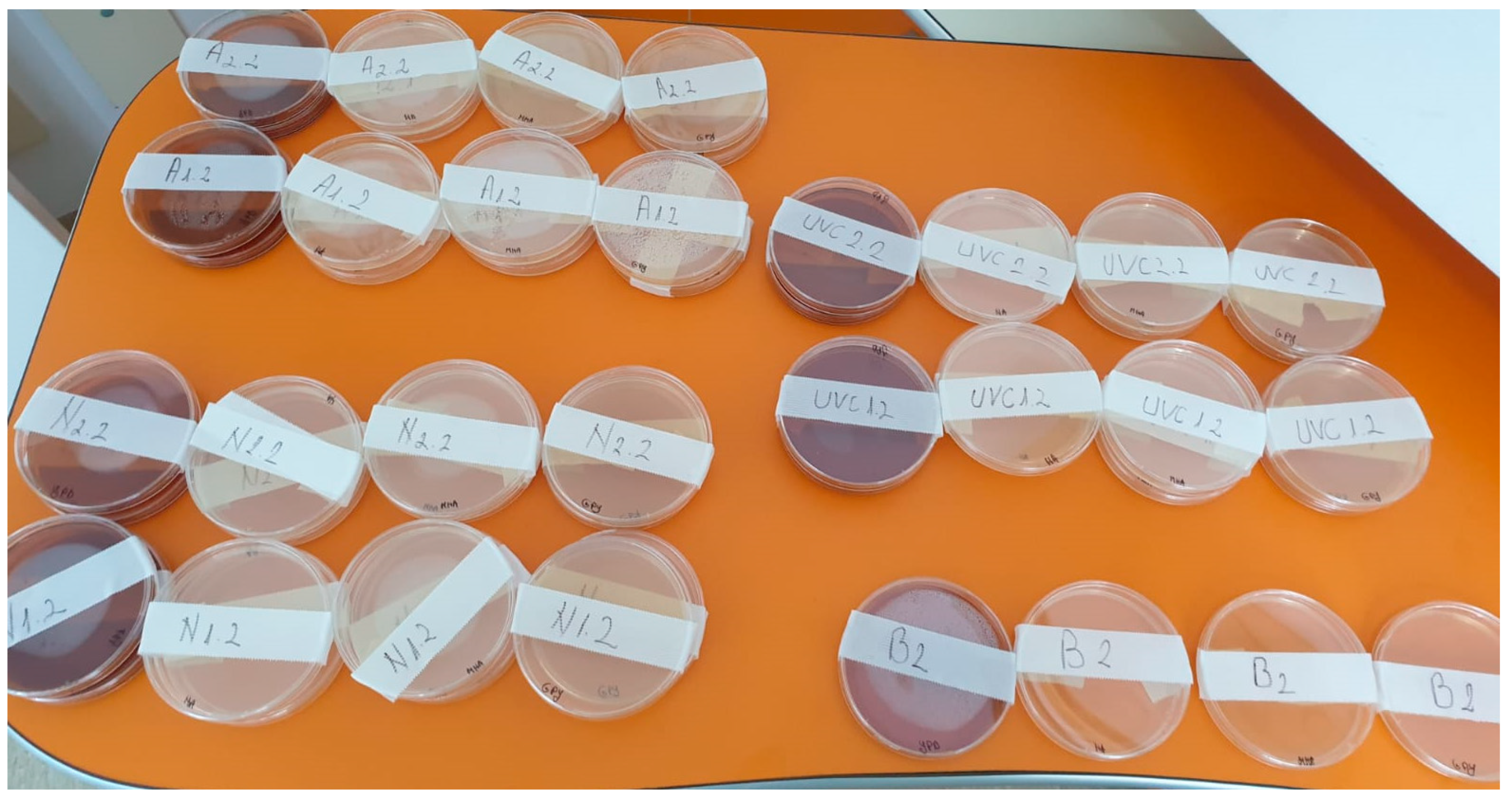
Figure 1. Treatment room layout.
Applsci 11 02359 g001
Figure 2. The Ultraviolet C (UVC) lamp.
Applsci 11 02359 g002
Figure 3. Fogging machine.
Applsci 11 02359 g003
Figure 4. All the Petri dishes used for the test, labeled accordingly.
Applsci 11 02359 g004
Table 1. Culture media and specific properties.
MHANon-selective matching for Haemophilus spp., Enterococcus spp., Escherichia coli, Pseudomonas spp., Staphylococcus spp. and Streptococcus spp. for bacteria (General Media) and for coliforms.
NANon-selective matching for Enterococcus spp., Escherichia coli, Salmonella spp., Shigella spp. and Staphylococcus spp. for bacteria (General Media) and for coliforms.
GPYRecommended for yeast isolation.
YPDMedia for yeast development. Non-selective matching for Candida spp., Pichia spp., Saccharomyces spp. and Zygosaccharomyces spp.
Table 2. Culture media that showed bacterial growth.
VentilationFoggingUVCUVC Box
P1P2P1P2P1P2P2
During treatmentMHA+++++++
NA++++++
GPY +++
YPD ++
After air decontaminationMHA + +
NA
GPY
YPD
Table 3. Culture media that showed bacterial growth—number of culture-forming unites (CFU) in each Petri dish.
MHANAGPYYPD
UVC 1.111--
UVC 1.21---
UVC 2.121--
A 1.113--
A 2.1 213-
A 2.21---
B22---
N 1.1141331
N 2.13821
Publisher’s Note: MDPI stays neutral with regard to jurisdictional claims in published maps and institutional affiliations.

