Nanostructured Biosilica of Diatoms: From Water World to Biomedical Applications
by
 , , and
Chiara Tramontano
1,2, 
Giovanna Chianese
2,
Monica Terracciano
1,
Luca de Stefano
2,* and
and
Ilaria Rea
2
1
Department of Pharmacy, University of Naples Federico II, via Domenico Montesano, 49, 80131 Napoli, Italy
2
National Research Council, Institute of Applied Sciences and Intelligent Systems, Unit of Naples, 80131 Naples, Italy
*
Author to whom correspondence should be addressed.
Appl. Sci. 2020, 10(19), 6811; https://doi.org/10.3390/app10196811
Submission received: 8 July 2020
/
Revised: 10 September 2020
/
Accepted: 24 September 2020
/
Published: 28 September 2020
(This article belongs to the Special Issue New Frontiers in Diatom Nanotechnology)
Abstract
:Diatoms—unicellular photosynthetic algae—are promising natural sources of nanostructured silica. These microorganisms produce in their membrane approximately a highly ordered porous cell wall called a frustule as protection from environmental stress. Diatom frustules consist of hydrated silica that show peculiar properties including biocompatibility, tailorable surface chemistry, chemical inertness, and thermal stability. Frustules harvested from aquatic ecosystems or diatomaceous fossil sediments represent an excellent cost-effective source of biosilica for a broad range of biomedical applications. The porous ultrastructure of the frustules displays a large surface area available for coating with various biomolecules through different functionalization methods. In this review article, we highlight the main features of diatom biosilica and present some of the most advantageous properties that support the employment of frustules in the field of drug delivery, biosensing, and regenerative medicine. In particular, it is offered an insight into the most common functionalization strategies through which diatom physicochemical properties can be modified and tailored according to the described field of application.
1. Introduction
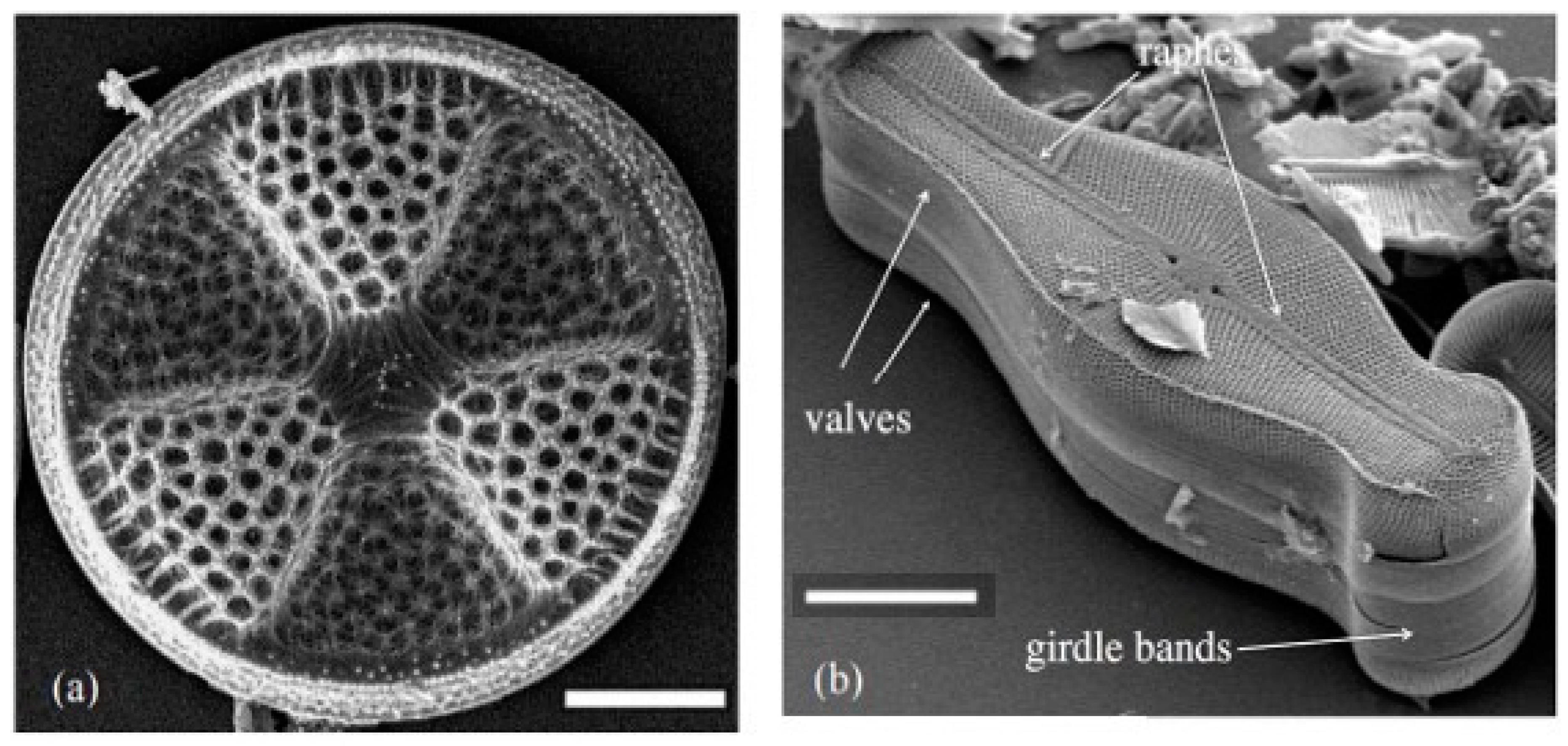
Research interest in the utilization of diatom biosilica for biomedical applications has grown over the last decade [1]. The unique properties of this material, conventionally studied by taxonomists and skilled amateur scientists, have conquered bio/nanotechnology fields emerging into a rich and dynamic research area with hundreds of articles to date. Diatom silica is a valuable alternative to synthetic porous materials thanks to its high surface area (200 m2/g), hierarchical structure, porosity, chemical inertness, biocompatibility, thermal and mechanical stability, as well as optical and photonic features. There are more than 200,000 estimated species classified by their unique morphology and size (from 2 µm to 2 mm) [2,3]. The most distinctive feature of the diatom is the unique 3D structure of its silica shell, the so-called frustule. The diatom cell wall is constructed in a Petri-dish-like fashion consisting of a top half (epitheca) that overlaps the slightly smaller bottom half (hypotheca). Each theca is formed by a valve, displaying an ornate structure with elaborate pore patterns, and a series of linking siliceous bands (girdle bands), exhibiting generally uniform pore patterns (Figure 1) [4,5]. The valve pore pattern is genome-encoded, species-specific, and forms an array called the areola, which propagates into the frustule depth and can be round, polygonal, or elongate. The pores extend from a tenth to hundreds of nanometers and can be partially obstructed by flat or domed cribra, which are silica flaps occluding areolae [6]. Diatoms can be classified according to the shape of their frustule in centric diatoms, which show radial symmetry, and pennate ones, which are bilateral symmetric [7]. All motile diatoms are pennates, with two slits in each valve known as raphes.
Diatoms are the principal organisms involved in the biosilicification, the process by which living organisms incorporate inorganic silicon in the form of silica [9]. In aqueous solution near the neutral pH, the main form of silicon is the orthosilicic (Si(OH)4). Beyond its solubility limit (2 mM), the orthosilicic acid turns into silica (SiO2) consisting of individual particles formed due to the repulsion between negatively charged particle surfaces [10,11]. The orthosilicic acid is carried from the aquatic environment by specific silicic acid transporter proteins (SITs) and spilled into specialized vacuoles in the diatom, called silica deposition vesicles (SDVs). Within the SDVs, orthosilicic acid turns into hydrated amorphous silica thanks to the cooperation of long-chain polyamines (LCPAs), silaffin proteins, and polyanionic silacidin peptide. Long-chain polyamines modulate silica morphology by changing nanoparticles size, silaffin proteins stimulate silica formation, and the polyanionic silacidin peptide supports self-association of the positively charged molecules. The presence of different silaffins or different ratios of silaffins to LCPAs confers to each diatom species the unique morphology [12].
The variability of frustule morphology offers to scientists from various fields the possibility to choose the diatom species matching with the desired application. The species Coscinodiscus has interesting radial symmetry with large and flat surfaces featuring well-organized multilevel pores [13]. Within this species, the diatom Coscinodiscus granii has gained attention as slab waveguide photonic crystals thanks to the ordered periodic pattern of high and low refractive index [14]; the diatom Coscinodiscus wailesii has been widely studied due to the fact of its ability to focus light [15]. Diatom species with a nanoporous pattern, such as Coscinodiscus concinnus, Thalassiosira weissflogii, Thalassiosira pseudonana, and Nitzschia, have intrigued scientists for their capacity to deliver drugs, while other diatom species with nanometer-sized structures, such as Odontella and Phaeodactylum tricornutum, are considered suitable for semiconductor nanolithography and photoluminescence spectroscopy [16,17,18,19,20]. Diatoms are an environmentally friendly source of silica and can be intensively cultivated in laboratories and photobioreactors on large scale [21,22].
Diatoms have settled and fossilized on the ocean and freshwater floors over millions of years in the form of diatomaceous earth minerals [23,24,25,26]. The amorphous silica surface shows high levels of free reactive hydroxyl groups (–OH) that can be exploited to modify the frustule with chemical groups (–NH2, –COOH, –SH, and –CHO). These groups are suitable to bind different biomolecules to diatoms (i.e., enzymes, proteins, antibodies, peptides, DNA, aptamers) [7,27]. Therefore, diatom frustules have been increasingly recognized as a promising material in biomedical applications. The large surface-area-to-volume-ratio and the nanoporous structure, together with the tunable surface, enables drug loading levels exceeding those of other common drug delivery carriers [28]. Indeed, although drugs are generally physisorbed or chemisorbed onto surfaces, the porous structure allows sustained drug release overcoming the constraints of conventional delivery and, according to the surface modification, stimuli-responsive therapy [29]. Moreover, the optical properties of diatom nanostructured biosilica have made a significant contribution to the fabrication of cost-effective surface-enhanced Raman scattering (SERS) platforms for label-free diagnosis. The ability of frustule pore patterns to enhance the Raman scattering of biomolecules attached to the surface promoted the fabrication of diatom-based SERS platforms for label-free diagnosis. Throughout this review, different applications of diatom biosilica are summarized to outline the potentiality of this natural material. The enormous versatility of natural biosilica is highlighted from many observation points: chemical, by pointing out the numerous modifications on the biosilica surface; therapeutic, by investigating the advantages of diatoms as drug carriers; diagnostics, as SERS platforms for label-free sensing; biological, by illustrating the efficiency of diatoms as a scaffold for cell growth and proliferation. In vivo application of the material is also presented to endorse the employment of diatoms in biomedicine.
2. Chemical Modification of the Frustule Surface
Chemical modification of porous drug carriers is known to be an essential factor controlling diffusion and drug delivery rate [30]. Many other properties, such as biocompatibility and cellular uptake, can be improved by binding proteins or bio-materials on the surface of drug carriers [31]. Surface modifications not only affect molecular interactions but also control the identification of biomolecules offering interesting perspectives for targeted-drug delivery.
The ability to be easily modified with different molecules makes diatom frustules useful in biomedicine. Biomedical applications necessitate intense purification treatments of diatom frustules to obtain a safe and biocompatible material. Before purification, the features of diatom frustules are completely masked by the presence of many impurities on their surface, such as organic and inorganic compounds from the environment. Rea et al. [32] described an accurate purification procedure for diatomite, a sedimentary rock white to yellow, composed of fossilized skeletons of diatoms. The siliceous skeletons contain silicon dioxide (SiO2) and small amounts of associated compounds, such as inorganic components and traces of iron, alkali metals, and other constituents [33]. Diatomite powder was suspended in a Piranha solution (2 M H2SO4, 10% H2O2) and intensively washed to remove traces of sulfuric acid. Subsequently, a solution 5.0 M of hydrochloric acid (HCl) was added to diatomite dispersion to remove the inorganic compounds (Al2O3, FeO3, CaCO3, CaO, K2O, Na2O, MgO) without altering the siliceous structure. The removal of impurities from the frustules modified their composition and exposed the reactive hydroxyl groups (Si–OH) on the surface, which facilitates the binding of biomolecules on diatom biosilica walls [34,35,36]. As a result of this attractive property, the number of reports including diatom functionalization strategies has enormously increased.
A significant drawback of immunocomplex-based biosensors is that chemical labeling techniques are generally required for analyte sensing. Current label-free biosensors for immune-complex detection depend on surface plasmon resonance (SPR), electrochemical or impedance measurements, all of which involve technical and costly instruments [37,38]. To face this problem, Zhen et al. [39] modified photoluminescent (PL)-active frustules from Pinnularia sp. with an anti-trinitrotoluene (TNT) single-chain variable fragment (scFv) to make a TNT-biosensor based on PL-detection. For this purpose, surface silanol groups on diatom frustules were aminated by reaction with 3-aminopropyl-trimethoxysilane (APTMS), and the scFv was covalently attached to aminated biosilica through the crosslinking-agent disuccinimidyl suberate (DSS). The authors assumed that the estimated scFv coverage on diatom frustules was approximately 2.5 × 10⁴ molecules per µm2, or 40% of the total surface area, which is more than enough for the fabrication of a selective antibody-based biosensor for TNT label-free detection.
Silane-based chemistry is one of the most exploited modifications of the diatom surface since it offers a facile way to attach a biological or chemical molecule to the surface. Sasirekha et al. [40] exploited the advantages of silanized diatom frustules using 3-aminopropyltriethoxysilane (APTES) to bind biomolecules on the diatom frustule likewise. They proposed a chitosan (Chi) grafting on Amphora frustules (Chi@AF) to encapsulate the chemotherapeutic anthracycline antibiotic Doxorubicin (DOX) in the frustule ultrastructure. The Chi-grafting on the AF surface was obtained by linking the amino groups of the polysaccharide to APTES-functionalized diatom frustules in the presence of glutaraldehyde as a cross-linker. The resulting biodegradable and biocompatible DOX-loaded AF (Chi@AF-DOX) exhibited discernible hemolytic effects compared to free DOX, which, instead, is known to cause many side effects including cardiotoxicity. This study offers an interesting reflection on the ability of polymer-functionalized drug carriers to reduce the toxicity of the chemotherapeutic drug and outlines their promising applications for cancer therapy. Losic et al. [41] were the first to outline the great potentiality of diatoms for drug delivery and many efforts have been done to investigate and improve diatom frustule applications to date.
The use of nanoporous silica-based materials as drug-delivery vehicles has recently proven successful, but their production often involves costly and toxic chemicals. Uthappa et al. proposed a cost-effective porous drug carrier represented by polydopamine (PDA)-coated diatom frustules modified with folic acid (FA) ligands for controlled and stimuli–responsive curcumin (CUR) release [42]. In this study, purified frustules loaded with curcumin (DE-CUR) were coated with PDA, which acted as a gatekeeper by controlling the curcumin release and simplified the conjugation of the ligand FA on the surface of diatoms (DE-CUR-PDA). The release of curcumin from the modified carrier was slightly slower than the unmodified one (DE-CUR), thanks to the interactions between the thin PDA layer and the drug, which delayed its release from the carrier. The drug release was more rapidly when the medium pH decreased from 7.4 to 1.2, due to the rupture and disassembly of PDA coating. Since abnormal pH has been universally recognized as a diagnostic hallmark of cancer, pH-responsive carriers that can change their chemical or physical properties are promising candidates for cancer treatments [43].
The challenging treatment of cancer is mainly attributed to its multifactorial nature. In the past decades, the alteration of redox balance has been illustrated as one of the most important factors underlying tumor development, its progression and invasion into neighboring tissues. Oxidative stress can damage cells, proteins, and DNA, playing a considerable role not only in neoplastic disease but also in diabetes, cancer, and neurodegenerative pathologies [44]. In some cases, reactive oxygen species (ROS) production can be induced by drug intermediates generated during metabolism and can increase drug toxicity. Chemically modified diatom frustules with both drug delivery and scavenger properties were investigated by Cicco et al. to reduce the drug-induced oxidative stress caused by Ciprofloxacin (CPFX) administration [16]. To this aim, nanostructured biosilica produced by Thalassiosira weissflogii frustules was covalently bound via a silanization strategy to the cyclic nitroxide 2,6,6-tetramethylpiperidine-N-oxyl (TEMPO) in consideration of its ability to scavenge superoxide, peroxide, and alkyl radicals. Alongside this, the antimicrobial drug CPFX was encapsulated into diatom frustules. The intention was to deliver the CPFX to cells and simultaneously decrease the drug-induced oxidative. The authors confirmed the efficacy of the silanization strategy for covalent attachment of molecules on the surface of diatom frustules and outlined the therapeutic potential of this material to reduce oxidative stress.
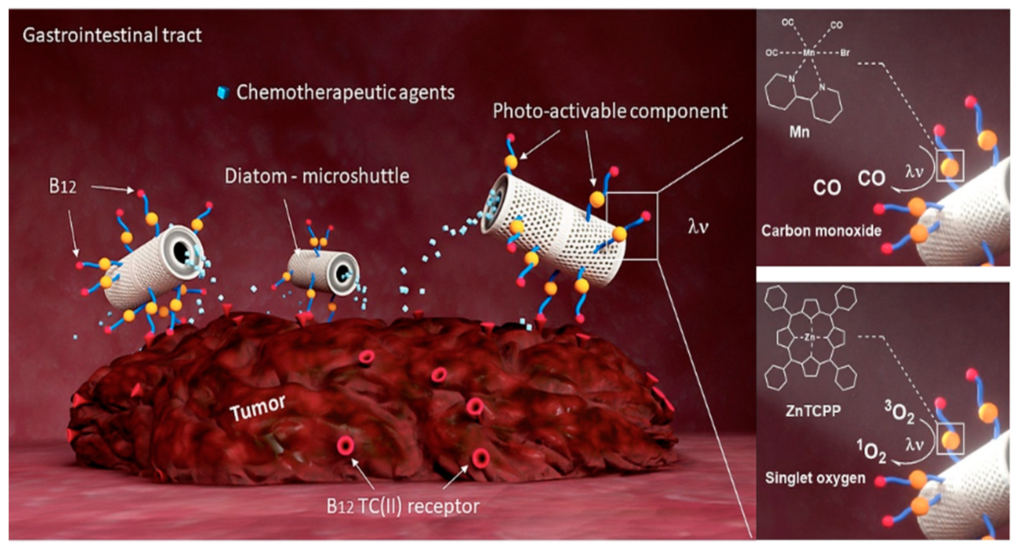
The ability of carriers to reduce the drug release rate and control drug diffusion to cells is a critical point that can be addressed by engineering the surface of the carriers. The porous ultrastructure of the frustule can be coated with polymers preventing the burst release of the therapeutic molecule. These coated frustule elements can then find applications in site-specific delivery of drugs. Recently, drug selectivity and targeted drug delivery have been reported to decrease undesirable side effects to healthy cells and reduce patient suffering. Delasoie et al. [45] described an innovative colon-focused drug delivery system (CFDD) based on the functionalization of diatom frustules (DEM) with vitamin B12 (DEM-B12), which binds the transcobalamin (II) receptor (TC II) overexpressed on cancer cells. Additionally, the DEM-B12 surface was modified with either porphyrin as photosensitizer elements or the 2,2′-bipyridine-3,4-dicarboxyilic acid manganese tricarbonyl compound as a photo-triggered CO-releasing molecule (Figure 2).
Through the multi-step functionalization process, the authors developed a photo-therapeutic platform able to produce highly toxic ROS or carbon monoxide (CO) and deliver rhenium(I)-tricarbonyl complexes with anticancer effects. ROS and CO produced by light-activation are known to exhibit antiproliferative effects and induce tumors cells apoptosis, potentiating the cytotoxic effect of the delivered drug. The so assembled photo-therapeutic platform showed enhanced adherence to colorectal cancer (CRC) cells via TC (II) receptors and a gradual drug release for a few days. Moreover, cytotoxicity tests on the human colorectal cell line (HCT-116) showed a 2 fold increase of cytotoxicity upon light-activation of both modified frustules, lowering the drug dose needed for equal therapy effectiveness. Also, MTT assay carried out on HCT-116 cell lines under dark/light conditions confirmed the efficacy of light-activable compounds on the surface. Hence, the synergistic chemo-photodynamic therapy could be adopted for lowering the chemotherapeutic needed dose and its side effects for effective cancer therapy.
The ability of surface-engineered systems to identify and target cancer cells was exploited by Esfandyari et al. [46], who reported a novel magnetic antibody-labeled biosilica structure with either biosensing and targeted drug delivery potentialities. In this work, the frustules of Chaetoceros sp. diatoms were magnetized through iron oxide nanoparticles and labeled with the antiHER-2 monoclonal antibody Trastuzumab. Paramagnetic antibody-labeled frustules were able to discriminate between HER-2 positive or negative cells through to the specific antibody-antigen interactions. This specific binding ability was detected by fluorescence microscopy thanks to the optical properties of the silica frustule. Moreover, the ability of magnetic particles to move in the presence of a neodymium magnet was outlined to show that the paramagnetic antibody-labeled system can be useful to discriminate between targeted cells from untargeted ones.
Terracciano et al. [47] also investigated a surface bioengineering approach to diatomite nanoparticles (DNPs) based on polyethylene glycol (PEG) and cell-penetrating peptide (CPP) conjugation to improve nanoparticles physicochemical and biological properties. In this study, DNPs obtained by mechanical crushing, sonication, filtering, and purification of natural diatomaceous earth (DE) were amino-modified by APTES and chemically conjugated to a heterobifunctional PEG polymer ending up with CPP at the N-terminal extremity. The biocompatible platform was engineered for delivering the poorly-water soluble drug Sorafenib to cancer cells. Cellular uptake analysis evaluated by confocal fluorescence microscopy indicated that CPP-modified DNPs were internalized into MCF-7 cells with a homogeneous distribution in the cytoplasm and close to the nucleus, whereas no cellular uptake was observed for the unmodified-DNPs. Moreover, morphological studies of red blood cells (RBCs) and cell viability assay on two different cancer lines outlined the safety of the formulation up to a concentration of 200 µg/mL for 24 h. This manipulation of matter opens up the possibility to create functional systems for a diverse range of applications that take advantage of the much larger surface of nanomaterials compared to conventional medicinal systems. The advantages of diatom frustules in biomedicine are more emphasized when the great tunability of their surface is taken into account for therapeutic purposes. Therefore, the various functionalization strategies which have been proposed in the last years reflect the urgency to achieve efficient drug carriers with simultaneous functions for site-specific therapy.
3. Diatom Biosilica for Drug Delivery
Targeted carriers offer the opportunity to improve the selective accumulation of active agents in diseased tissue and cells. By adding targeting agents to drug carriers, we could control the destination of the drug and increase the likelihood of response. To deliver chemotherapeutic drugs to CD20+ B-cells specifically, Delalat et al. [17] realized a nanoporous biosilica platform from naturally engineered microalgae. The diatom Thalassiosira pseudonana was genetically modified via recombinant DNA technology: a synthetic sequence that encodes the IgG-binding domain of protein G (GB1) was fused to the amino-terminal peptides S and T8, which are naturally expressed on the frustules. The GB1-biosilica was further labeled with Rituximab, an antibody specific for the CD20 molecule expressed on B-cells. The specificity of the antibody-labeled-frustules was demonstrated by in vitro and in vivo studies. Antibody-labeled frustules bound to B-lymphocytes release the drug more efficiently than untargeted ones. Indeed, 54% of B-cells interacted with modified-frustules, whereas none of the T-cells exhibited interactions with unmodified-frustules. Positively charged lipid-based colloids loaded with camptothecin (CPT) and 7-ethyl-10-hydroxy-CPT (SN38) were electrostatically adsorbed on the surface of modified microalgae. The dual-step strategy for drug encapsulation provided effective CPT and SN38 loading efficiencie is 61.2% and 85.8%, respectively. Cytotoxicity tests assessed on SH-SY5Y neuroblastoma and baby hamster fibroblast (BSR) cell lines confirmed the possible application of genetically modified frustules for targeted cancer therapy (for in vivo research, see the dedicated Section 6).
Delasoie et al. have recently functionalized diatom frustules belonging to the genera Aulacoseira granulata with vitamin B12 for Cisplatin and 5-fluorouracil (5-FU) delivery to colorectal cancer cells [48]. Colorectal cancer was selected as a disease model since it is one of the most recurrent tumors in humans, and its pathogenesis strongly correlates with diet and lifestyle factors [49]. The B12 functionalization strategy was based on the idea that cancer cells need more vitamin B12 and show a higher expression of the transcobalamin II receptor (TCII) on the surface. Vitamin-B12-modified frustules (DEMs-B12) provided a high drug concentration at the site of action while minimizing off-target exposure. Cisplatin and 5-FU were encapsulated into DEMs-B12, by Vasani method [50] with a loading efficiency of 5.9% and 9.9%, respectively. Frustule drug encapsulation is a suitable strategy to deliver different drugs, but the burst release of the drug in aqueous media (generally within 1 h) hinders drug delivery applications of frustules. In this study, 90% of the drug was released from modified frustules within 4 h. Hence, carrier functionalization can positively affect the carrier release properties and offer suitable prospects for controlled therapy.
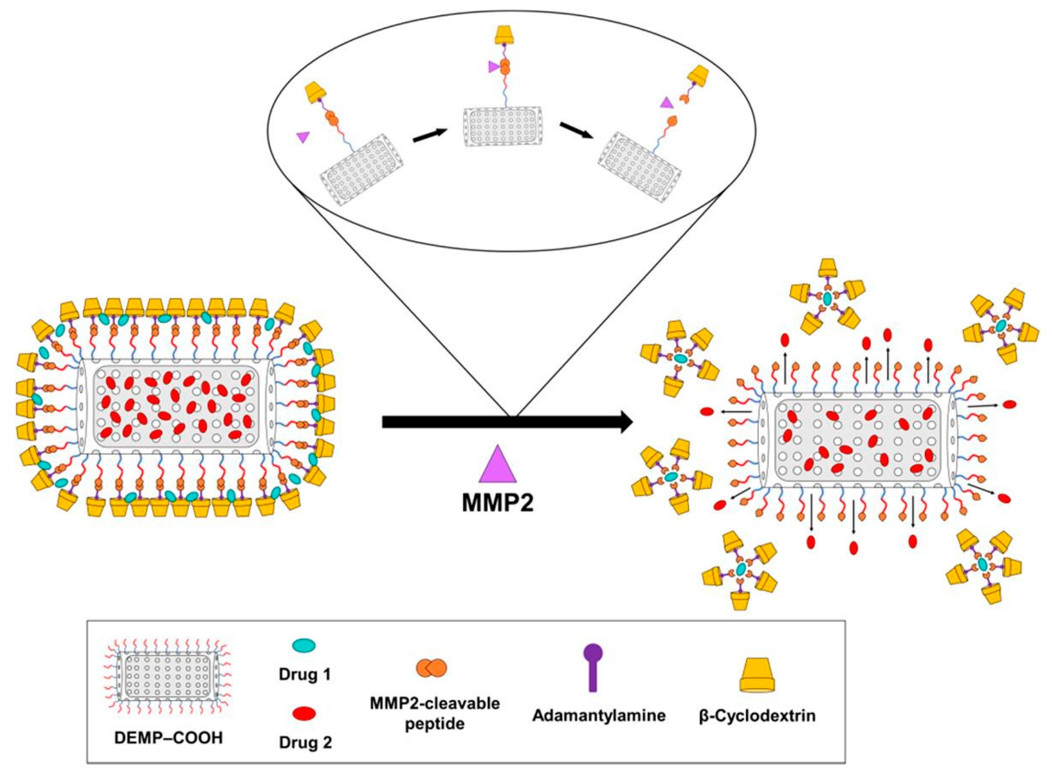
Combined therapy is the cornerstone of all cancer treatments and, in most cases, the cause of therapy failure due to multidrug resistance (MDR) phenomena [51,52]. Kabir et al. [53] employed self-assembled microparticles obtained from diatomaceous earth (DEMP) for the dual delivery of chemotherapeutic agents. They overcame the antagonist effect between the drugs by using a different molar ratio of chemotherapeutics. (Figure 3).
The self-assembled platform was obtained by tethering diatom microparticles to the homodifunctionalized-linker 1-adamantylamine (Ad-DEMPs) as a guest molecule to get a host–guest interaction with β-cyclodextrin (β-CD) (Figure 3). Doxorubicin (DOX) and paclitaxel (PTX) were encapsulated in two different compartments: the siliceous core and the self-assembled β-CD with ad-thixotropic gel corona on the surface of microparticles, respectively. This strategy was successful to control the release rate of both drugs and maintain a constant in situ molar ratio. DOX and PTX are often combined in therapy and require high molar ratio DOX/PTX to guarantee a synergistic effect. In this work, the siliceous core and the outer gel corona released a high amount of drug to the tumor site. The chemotherapeutic activity on Hela and HT1080 cancer cells was improved and, as a consequence, the IC50 value was lower than the free drug. Taking into consideration that the β-CD-capping lessens the release of encapsulated drugs from DEMP core, the shell structure was further optimized by incorporating a matrix metalloproteinases-2 (MMP-2) peptide as a linker between the core and β-CD: Ad shell. Since tumor-associated enzymes can degrade the linker, the strategy also resulted in a stimuli–responsive therapy. Both PTX and DOX showed enhanced and synergistic cytotoxic effects that outlined a valuable strategy for the encapsulation of different molar ratios of chemotherapeutics in diatom frustules.
It is well known that the efficiency of a drug delivery system strongly depends on the strategy of encapsulation and the amount of drug in the carrier. Therefore, the improvement of drug encapsulation properties may lead to a stronger therapeutic approach and fewer side effects. Traditional techniques for drug encapsulation involve adsorption processes and electrostatic interactions between the drug and the outer carrier surface. In the last years, new strategies have been exploited to enhance the drug encapsulation efficiency of porous carriers. Mancera et al. [54] developed an interesting silicon microfluidic device to encapsulate Isorhamnetin into in-house developed biogenic silica from Cyclotella sp. frustules. The authors reported a loading capacity of 1.63%, which is an encouraging result for a novel methodology that reduces handling errors and time-consuming drug loading reactions. Frustules showed a typical release profile with an initial burst release of 48% of the drug in the first hour and the remaining in 3 h.
Diatom biosilica has found application even in gene-therapy as nucleic-acid delivery systems [55]. Martucci et al. [56] proposed engineered diatomite nanoparticles (DNPs) for short-interfering RNA (siRNA) delivery to lymphoma cancer cells. Diatomite nanoparticles were modified with amino-silanization and covalently conjugated to the Id-peptide (pA2036), a negatively charged peptide specifically recognized by B-cell receptors (BCR) on the A20 cell line. Poly-Arginine-small interference RNA was bound to DNPs through electrostatic interactions between siRNA positive charges and the negative surface of nanoparticles. Cells treated with the engineered DNPs-siRNA showed a decrease of 40% ± 4.5% of the targeted mRNA expressions, whereas only a 10% of decreasing expression occurred in cells treated with non-conveyed siRNA, resulting in a specific delivery of the siRNA only to target cells. Rea et al. investigated the ability of modified DNPs to deliver siRNA to epidermoid cancer cells (H1355) as well [32].
In this work, siRNA conjugation to DNPs enhanced its half-life by improving serum stability and protecting siRNA from RNase degradation. The complex showed a burst release of siRNA within 12 h, followed by a slow-release phase due to the weakening electrostatic interactions between the peptide and siRNA. The internalization kinetics of DNPs studied by Managò et al. [57] through Raman imaging revealed the cytoplasmatic localization of DNPs conjugated to RNA interference (RNAi). The author assumed that the internalization process took place through endocytosis and that nanoparticles were co-localized in lipidic vesicles.
Although targeted nanoplatforms can overcome the limitations of traditional drugs, advancements in the field of engineered nanoplatforms are still a demanding scope. The reason is that the rational design of targeted delivery systems relies on the simultaneous improvement of the drug loading efficiency and the choice of the type of ligand and receptor–ligand pair. Finding the right balance between these factors can be an arduous matter in hand. Despite the progress in the realization of modified frustules for site-specific therapy, there are still aspects to be improved. The burst release of drugs in aqueous media and the poor degradability of frustules in biological fluids are the main limitations to their medical applications. For this reason, the development of modified frustules for drug release is still in the early stage.
4. Diatoms as Surface-Enhanced Raman Scattering (SERS) Platforms for Label-Free Diagnosis
Surface-enhanced Raman scattering (SERS) is a powerful vibrational spectroscopy technique suitable in biomedical label-free sensing thanks to high sensitivity, low detection limit (10−8 M), and selective detection of analytes. Surface-enhanced Raman scattering analysis relies on the amplification of localized electromagnetic fields generated by the excitation of localized surface plasmons [58,59,60]. The choice of plasmonic material and its preparation are the main requirements for successful SERS analysis. The poor reproducibility and instability of conventional SERS substrates (e.g., silver, copper, gold) led scientists to study new classes of artificial optical structures known as “photonic crystals” [61]. The production of these materials requires highly specialized design, lithography techniques, advanced facilities, and experienced staff, resulting in expensive and time-consuming processes [62]. Surprisingly, diatoms are a cheaper and more accessible alternative material, whose ordered pore distribution bears a resemblance to a photonic crystal. A photonic crystal is a spatial, periodic distribution of refractive index, through which light cannot propagate in specific ranges [63]. The use of diatoms as plasmonic substrates and their combination with metal plasmonic structures resulted in an alternative to photolithography fabrication of cost-effective SERS substrates [1,61,64,65,66]. Ren et al. [67] combined the photonic-crystal-like structure of Pinnularia sp. frustules with the plasmonic resonance properties of silver nanoparticles (AgNPs) to enhance the localized surface plasmon resonance. The synergy between the frustule photonic structure and the localized plasmon resonance of AgNPs led to a 4-fold improvement of nanoparticle sensitivity. Pannico et al. [68] proposed the electroless deposition of gold (Au) on diatom frustules for sensing applications. A three-step chemical method was adopted to obtain a uniform, continuous, thin layer of AuNPs on the surface of Aulacoseria frustules. The gold coverage on diatom frustules provided an enhancement factor (EF) of 1.0 × 10⁵ for p-mercaptoaniline (pMA) probe molecules adsorbed on frustules. Nonetheless, the intensity of single peaks changed from point to point, meaning that the SERS response was not completely homogeneous. The spectra enhancement reported by the authors demonstrated that diatom- based substrates could be considered good candidates for SERS analysis, but at the same time, their response is not completely reproducible in terms of intensity.
A noteworthy strategy for the fabrication of a diatom-based SERS platform was proposed by Terracciano et al. [69]. Diatomite nanoparticles were modified with amino-silanization and the amino-groups were covalently bound to the carboxyl groups of heterobifunctional PEG (PEG–DNPs). Positive charges of PEG–DNPs complex were involved in electrostatic interactions with AuCl4- precursor ions resulting in an in-situ synthesis of AuNPs.
Recently, Kaminska et al. presented an ultrasensitive SERS immunoassay based on AuNPs and frustules from Pseudostaurosira trainorii to detect interleukin-8 (IL-8) in human blood plasma [70]. The biosensor was a sandwich-type immunoassay composed of IL-8 antibodies covalently bound to the amino-silanized biosilica and anti-IL-8 antibodies conjugated to AuNPs and labeled with dinitrothiocyanobenzene (DNTB) as a Raman reporter. The SERS immunoassay developed on diatom frustules exhibited a limit of detection six orders of magnitude higher than a SERS immune assay on flat glass.
Wang et al. proposed an alternative method to analyze complex human plasma which employed thin layer chromatography (TLC) plates made from diatomite for SERS analysis for phenethylamine (PEA) detection [71]. The high molecular weight of diatomite plates offered separation performance outstanding standard TLC plates and a significant improvement of sensitivity. The same group integrated AuNPs on diatom-based TLC for detecting trace levels of tetrahydrocannabinol (THC) in saliva and urine samples via a portable Raman spectrometer [72]. Rapid and sensitive detection of THC analyte was performed in concentrations ranging from 1000 to 10 ppm in complex samples. The group also developed a microfluidic diatomite analytical device (µDAD) for cocaine sensing [73]. In this work, the nanoporous photonic channels showed high-resolution separation of complex biofluids and high-sensitivity detection of cocaine down to parts-per-billion. The use of TLC-SERS plates made from diatom was also investigated by the group for the detection and quantification of seafood allergen in situ to enhance food safety [74].
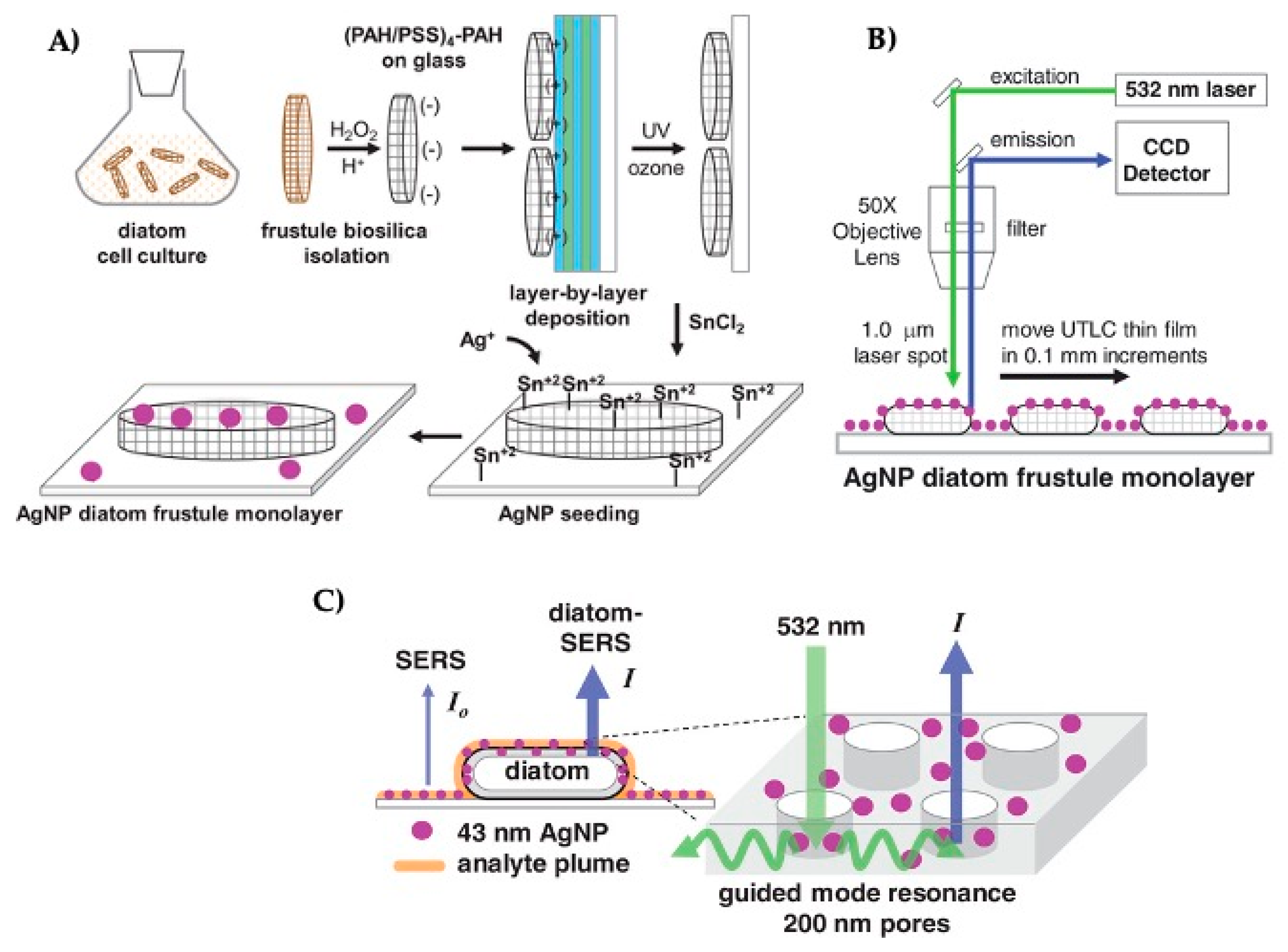
A detailed investigation of the utilization of diatom frustules as substrates for the detection of different analytes has been recently shown by Kraai et al. [75]. The group investigated the growth of silver nanoparticles (AgNPs) on the porous surface of Pinnularia sp. frustules to develop ultrathin layer chromatography (UTLC)-SERS plates. Frustules were deposited as a monolayer on a glass film through polyelectrolyte layer-by-layer dip-coating technique, using four bilayers of positively charged poly (allylamine hydrochloride) (PAH) and negatively charged poly (sodium 4-styrene sulfonate) (PSS) (Figure 4A).
The SEM images revealed that coverage of diatom frustules on the thin film was approximately 10 µg·cm−2 and that most of the frustules adhered face down to the surface valve, creating an ideal layer of 5 µm for UTLC. AgNPs with a size of 43 nm were deposited by in situ Ag+ nucleation process to form a single layer on the outer surface and within the pores. The non-polar Nile red and the weakly cationic malachite green were examined as model analytes to evaluate the separation performance of the diatom-based UTLC plate by micro-Raman spectroscopy (Figure 4B,C). When the excitation light source (532 nm Raman laser) coupled by the guided mode resonances within the diatom frustule pore arrays, the electromagnetic field generated by AgNPs improved the local enhancement of SERS signals associated to the analyte molecule on the frustule.
Flexible, gold-coated frustules guarantee favorable chemical detection levels towards single-molecule detection. Flexibility has been considered a crucial parameter for the development of SERS substrates, since it allows the frustules to trap the molecules in the hot spot sites generating a strong enhancement.
Korkmaz et al. used strips filled with diatom frustules to design simple, inexpensive, and flexible adhesive tapes [76]. A transparent adhesive tape was applied to the template strip and peeled off to obtain a tape strip embedding diatoms. Tape strips embedded with frustules and decorated with AgNPs showed an enhancement factor nine times higher than the control. In this way, the great potentiality of diatom-based SERS strips for global health applications was outlined.
Diatom biosilica and diatom frustules have not only the unique property of enhancing SERS sensitivity thanks to the 2D periodic pores, but they also display a high Langmuir adsorption capacity. This adsorption isotherm model is commonly used for molecular adsorption at interfaces, providing an insight into the surface coverage via physisorption or chemisorption phenomenon [77]. The high Langmuir adsorption properties of diatom frustules can effectively enhance analyte mass transport into the sensing area [78]. Sivashanmugan et al. [79] demonstrated the efficiency of a microfluidic channel with photonic crystal-enhanced plasmonic mesocapsules. Mesocapsules were produced by isolating diatom frustules from Pinnularia sp and synthesizing AgNPs in situ. The authors presented ultrasensitive optofluidic sensing of rhodamine-6G (R6G) with a detection limit of 10−13 M inside a microfluidic channel. Porous diatom frustules decorated with AgNPs generated periodically distributed hotspots and, at the same time, created a large surface-to-volume ratio for effective analyte capture. For the detection of analytes, the fluids containing various concentrations of target molecules were injected together with the mesocapsules through the microfluidic channel. Photonic crystal-enhanced plasmonic mesocapsules showed an enhancement factor 100-fold higher than regular colloidal AgNPs and a 1000-fold reduced detection limit.
The reported results show how the combination of diatoms frustule and metal nanoparticles can be a valid strategy for the fabrication of new SERS substrates. However, it is still unclear how the species-specific variability of diatoms (in particular their morphology and size) can influence the properties of the substrates, such as the enhancement factor or sensitivity. Therefore, to promote the employment of diatoms in the fabrication of SERS platforms, it could be useful to investigate how the diatom species-specific pattern affects the sensing surface.
5. Diatom as a Promising Scaffold for Cell Proliferation and Differentiation
Bioactive mesoporous silica materials (MSNs) have been clinically investigated as scaffolds for cell attachment, proliferation, and differentiation, on account of their specific surface area, porous structure, and tunable pore size [80,81]. Nevertheless, the synthesis of the MSNs is often expensive, since it involves toxic reagents, time-consuming and complicated processes. On the other hand, diatoms are an alternative source of silica with a naturally self-assembled 3D porous structure. This multifunctional material with unique mechanical and molecular transport properties is a promising material for regenerative medicine [82]. It is important to remember that surface chemistry plays a crucial role in the fabrication of platforms for biomedical applications. Hence, special consideration should be given to the possibility of attaching biomolecules to the scaffold surface. Diatom frustules offer a large surface area that could be modified with osteoinductive agents (e.g., peptides or growth factors), which represent attractive signals for bone cells and should promote the bone regeneration process.
Chemical modifications of diatom frustules for bone cell adhesion and proliferation were proposed by Cicco et al., who studied the adhesion and growth of normal human dermal fibroblasts (NHDF) and human osteosarcoma cell line (Saos-2) on bare and silane-coated frustules [83]. In this study, nanoporous microcapsules 10–15 µm in size were obtained from the pelagic centric diatoms T. weissflogii and functionalized with APTES, a popular modification we have previously mentioned on account of its ability to facilitate the attachment of organic compounds to silica substrates. The authors also carried out the grafting of silanes containing mercapto (SH) groups by self-assembling monolayers of 3-mercaptopropyl-trimethoxysilane (MPTMS) on the surface of the microcapsules. The cytocompatibility of bare and modified microcapsules was tested by studying NHDF and Saos-2 cell viability, spreading, and proliferation. These cell lines are generally used for testing the biocompatibility of silicate-derived scaffolds [84]. Both cell lines adhered to the mesostructured bare frustules showing improved vitality and morphological shapes compared to glass substrates. Bare diatoms stimulated cells to emit long filopodia and large lamellipodia, which extended connections to the substrate and played fundamental roles in cell migration. Cells showed higher vitality on bare and mercapto-modified frustules between 24 and 96 h. The SEM investigations of single frustule-cell interactions showed that cells reached maximal stretching on bare and mercapto-modified frustules during the adhesion process. On the contrary, frustules modified with amino groups hindered cell adhesion and proliferation. Amino groups have been reported to reduce cell adhesion and proliferation due to a supposed massive albumin subtraction from the cell media [85]. These results highlight the need to find the right functionalization strategy to promote cell growth on diatom-derived substrates and encourages the study of this material for further in vivo applications.
Fused diatom frustules (FDF) from Cyclostephanos sp. were tested by Amoda et al. for their ability to support the colony growth of a variety of cell types including osteoblasts, HEK cells, and breast cancer cells [86]. The interconnected pore network of FDF provided topographical irregularity that improved osteogenic differentiation and mineralization of pre-osteoblasts (MC3T3) cells presumably through mechanical interlocking [87]. Fused diatom frustules resulted to be non-toxic to cells and showed in all cases a cell adherence stronger than NUNC surface culture plates, enabling a high-density cell growth over 21 days. The high porous structure allowed cells to exchange gases and turn nutrients into usable energy through the 3D-structure of the biomaterial. Contrary to many artificial bone graft materials in which porosity is formed through techniques that could be a source of potential contamination [39,40], frustules show naturally designed porous architectures. Additionally, diatom frustules can be reused subsequently to a series of washings with 96% ethanol and PBS. Afterward, a sterilization process is compulsory to seed new cells on the scaffold without conditioning their survival rate.
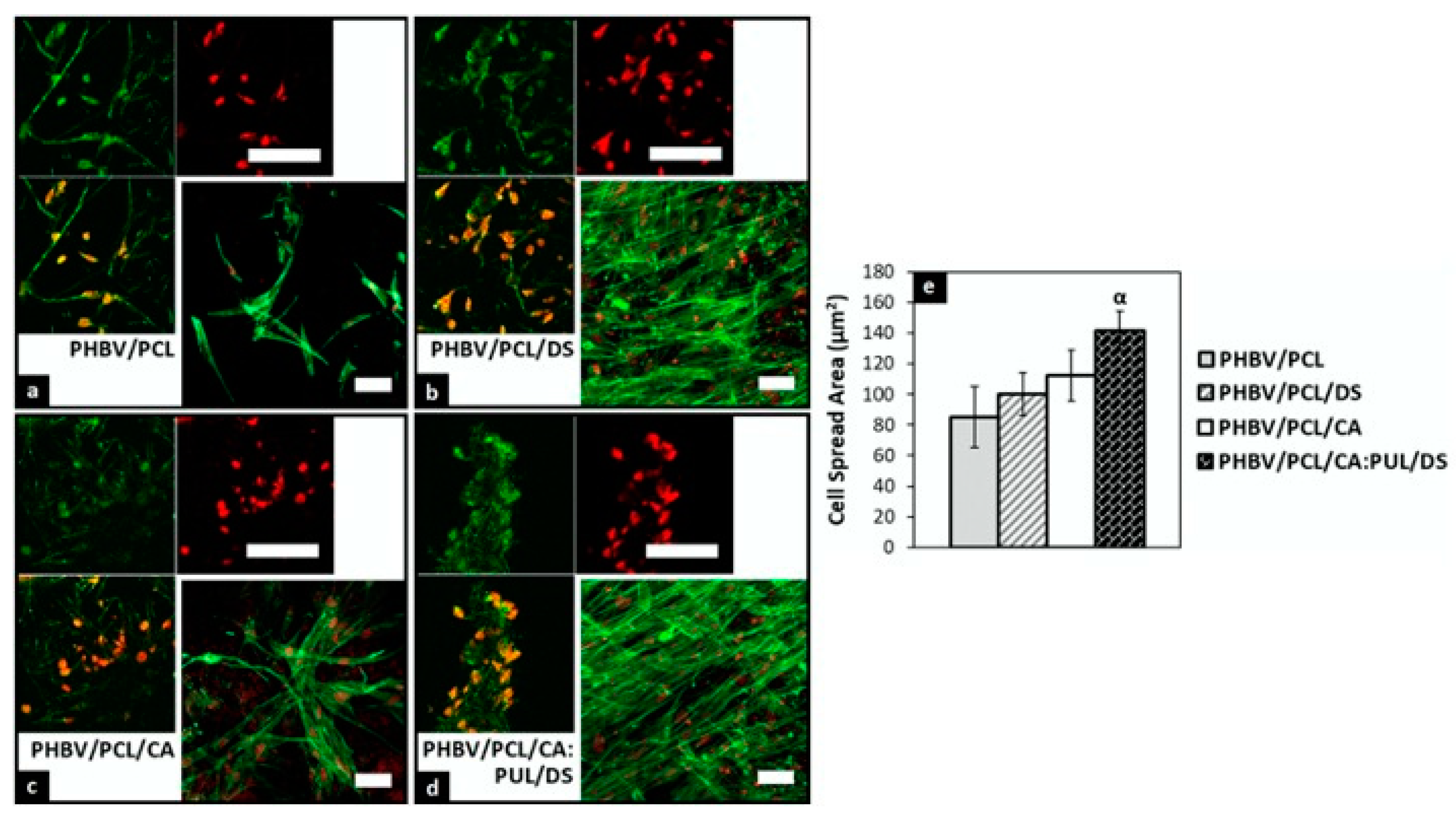
Silicon plays a crucial role in the bone regeneration process and influences bone mineral density. Silicon induces calcium phosphate precipitation in the early stage of bone mineralization and improves cell proliferation. Scaffolds incorporating amorphous silica, among which silica–collagen scaffolds [88,89], and silk–silica composites [90], enhance osteoconductivity, alkaline phosphatase activity (ALP), matrix deposition levels, and osteocalcin synthesis. The fabrication of synthetic silica scaffolds involves the use of chemicals and surfactants whose residues might have toxic effects on cells. For this reason, there is a great interest in diatom frustules as a novel, safe source of biogenic silicon. Le et al. [91] have recently demonstrated that diatom particles obtained from acid-purified raw diatomite powder can undergo degradation in an aqueous environment and release silicon ions in the medium. Later, Dalgic et al. [92] reported the osteoinductive potentiality of micro-nano frustules incorporated into a multifunctional 3D fibrous scaffold. Micro-nano frustules obtained from broken frustules were incorporated into a matrix of poly (hydroxybutyrate-co-hydro-xyvalerate)/poly(ε-caprolactone) (PHBV/PCL) fibers and pullulan (PUL), which prevented any possible toxic effect on cells. The co-electrospinning method was successful to create a random mesh of nano and microfibers, which created a high surface for cell interaction and enabled mechanical support. Osteocompatibility tests, performed by incubating Saos-2 cells with the silica-doped scaffold, revealed improved cell viability after 4 and 7 days of incubation. The entrapment of micro-nano particles into the fibrous scaffold increased calcium deposition up to 20% compared to the undoped PHBV/PCL control scaffold after 7 days, which outlined the positive effect of silica-enriched substrate on Saos-2 ALP activity. The developed silica-doped scaffold improved cell distribution and spreading thanks to the synergistic effect of biosilica and polymers; silica structures promoted cell bioactivity, whereas PUL fibers improved cell spreading by increasing overall hydrophilicity. Moreover, Saos-2 cells seeded on the doped scaffold showed a remarkable elongated morphology compared to the undoped substrate, in which cells had a round morphology which indicates low interaction with fibers (Figure 5).
One of the disadvantages of diatom frustules is that silica has low biodegradability in biological fluids. That leads to its toxic gathering in the human body, particularly in the liver. Accumulation of silica particles in the liver has been reported in previous studies as a result of particle uptake by macrophages in the reticuloendothelial system [93,94]. To overcome these limitations, Maher et al. converted diatom silica into degradable silicon replicas for intraocular therapy [95]. Silica (SiO2) turned into silicon replicas through a three-steps magnesiothermic process in which the magnesium source was melted and caused the reduction of silica to silicon replicas. Silicon replicas showed a better degradation rate compared to silica in vitro. The 20% of silicon dissolved within 30 days, whereas less than 1% of diatom silica degraded the same timeframe. Comparative SEM imaging revealed significant changes in silicon replicas structure, showing that the silicon core was completely dissolved after 30 days. Therefore, the conversion from diatom silica to silicon can be considered a valid strategy to prevent particle accumulation in the body fluids. This assumption is based on the fact that silicon degrades into orthosilicic acid, which is absorbed by humans and naturally found in numerous body tissues [88].
6. In Vivo Evaluation of Biodistribution and Toxicity of Diatom Biosilica
Animal research is indispensable for medical devices and novel therapies. Although there is considerable research on the effectiveness of biosilica in vitro, research about its safety and efficacy in vivo is still scarce.
To investigate the possible side effects of injected frustules, Delalat et al. [17] administered genetically-engineered frustules in BALB/c mice via intravenous injection and studied their biodistribution for eight days. Frustules were mainly trapped by the kidney due to the particle accumulation in the mononuclear phagocyte system (MPS) and liver. Biosilica administration did not cause structural alterations of the main organs, including the brain, kidney, heart, lung, liver, and tail. However, the accumulation of the silica particles could be particularly dangerous in the case of repeated administration in organs with limited blood supply (e.g., vitreous body).
Therefore, considering alternative routes of administration (e.g., topical, transdermal) may be useful to enable new therapeutic platforms that have unfavorable pharmacokinetic properties. That has led scientists to explore new strategies outside of the traditional oral or intravenous routes and create new opportunities for diatom applications. Feng′s group [96] found that the topical application of diatoms and diatomite on injuries can help to manage bleeding from surgical surfaces. Topical hemostatic agents are generally required when surgical hemostasis is inadequate or impractical. Feng’s group [96] compared the hemostatic property of chitosan-modified diatoms, diatomite, zeolite, and traditional gauze on amputated rat tail. Diatoms frustule provided efficient hemorrhage control, thanks to the ability of silanol groups to stimulate the coagulation factors. Biosilica concentrated platelets and coagulation factors in the local injury, thereby reducing the thrombin generation time and inducing activated platelets stick to form the blood clot. Therefore, the blood clot was formed by diatoms more rapidly than all other hemostatic agents.
Lee et al. found that the synergy between silanol anions and nano/microstructures on the frustule surface confers to diatoms hydrophilic and hemophilic properties [88]. Frustules from Melosira nummuloides exhibited interesting wettability and contact angles towards blood samples. Frustules applied to wounds formed a “clay-like” mixture able to respond as a viscoelastic material under shear stress, and to control hemorrhage in a model of liver bleeding by incision. Anionic charges on the frustule surface promoted the activation of factor XII to FXIIa and provoked plasma clotting. Contrary to traditional hemostatic agents, which have low efficiency and undesirable collateral effects, silica from diatom is a biocompatible and safe material. Hence, the topical administration of biosilica could represent a fascinating approach to treat skin diseases -without unfavorable side effects- and to avoid silica accumulation in the kidney.
The choice of the appropriate animal model is crucial to evaluate in vivo biosilica effects. The complexity of diatoms and their frustules places higher demands on animal models. Recently, many researchers turned to less studied and more unusual systems, such as Hydra polyps, for their studies. These systems can make it easier to answer questions that have remained unanswered to date. Terracciano et al. [97] confirmed the safety of biosilica administration on the cnidarian freshwater polyp Hydra Vulgaris. Hydra is a small polyp from 1 to 20 mm in body length found attached to aquatic vegetation, submerged wood, or stones in freshwater habitats. Hydra polyps are highly sensitive to both organic and inorganic pollutants in their environment, leading in most cases to delayed growth, morphological changes, induction of apoptosis, and ever alteration of gene expression [98]. Terracciano et al. [97] monitored the effect of diatomite nanoparticles on Hydra morphology, population growth rate, and cell apoptosis. Diatomite nanoparticles did not alter polyp morphology or reproduction capabilities up to a concentration of 3.5 mg/mL, excluding any long-term effect of biosilica. Confocal fluorescence studies on DAPI dye stained nuclei did not reveal any significant cell apoptosis induced by internalized biosilica, confirming its safeness towards living organisms.
Unfortunately, there is still little evidence of the applicability of diatoms in biomedical fields. The reported results confirmed the biocompatibility of the material and its safeness towards living organisms. Nevertheless, several issues hinder their application, such as the hostile pharmacokinetic profile. Future research should include the development of effective evaluation strategies for a good appreciation of biosilica toxicology, pharmacokinetics, and pharmacodynamics in vivo.
7. Conclusions
Diatoms pose a novel example of a natural source of nanostructured silica with suitable properties for the generation of cost-effective biomaterials. The high surface area available and easily tunable surface chemistry makes the porous ultrastructure of diatom frustules an alternative cheap scaffold for the development of innovative silica systems. In this work, the authors have reviewed recent uses of diatom skeletons for different biomedical applications. By proper handling/processing, chemical modification, and biofunctionalization of the frustules, it is possible to obtain different hybrid silica nanostructures with applications in drug delivery, biosensing as well as cell adhesion and proliferation. The results outline the valuable success in utilizing these intricate natural 3D porous structures instead of conventional expensive synthetic ones, on account of their peculiar physicochemical properties.
While reported results are astonishing and exhibit valuable potentiality of biogenic silica of diatoms for future applications, breakthroughs are still needed for biomedical purposes. For example, most of the results on potential medical applications of diatom frustules (e.g., drug delivery, regenerative medicine) missing of in vivo comparative studies such as information regarding circulation properties in the blood, circulation half-time, possible immunogenicity, tissues or organs accumulation, long-term effect, inflammatory processes, genotoxicity.
Indeed, the Environmental Protection Agency (EPA), United States Department of Agriculture, USDA), and Food and Drug Administration (FDA) approved the use of diatomaceous earth as an anti-caking agent in livestock feed, chemical pesticides, and additive for pharmaceutical preparations. However, the official regulatory approval as a bioactive scaffold for drug delivery or other biomedical applications has not yet been released.
Whereas there are very few biomedical applications that require synthetic silica-based nanostructures with specific pore arrays or dimension, where possible, the expensive nanofabrication effort could be without any doubts overcoming by using low cost and broad availability biogenic silica. The biosilica of diatoms may play an important role in biomedical devices design for clinical applications, so the turning point is an intensive research effort to dispel doubts and better know this attractive raw material.
Author Contributions
C.T. and G.C. writing—draft preparation; M.T. writing—review and editing; I.R. and L.d.S. supervision. All authors have read and agreed to the published version of the manuscript.
Funding
This research received no external founding.
Acknowledgments
M.T. acknowledges financial support from PON-AIM RTDA_L1 (AIM 1873131-2).
Conflicts of Interest
The authors declare no conflict of interest.
References
- Panwar, V.; Dutta, T. Diatom Biogenic Silica as a Felicitous Platform for Biochemical Engineering: Expanding Frontiers. ACS Appl. Bio Mater. 2019, 2, 2295–2316. [Google Scholar] [CrossRef]
- Mann, D.G.; Droop, S.J.M. Biodiversity, biogeography and conservation of diatoms. Hydrobiologia 1996, 336, 19–32. [Google Scholar] [CrossRef]
- MGrachev, A.; Annenkov, V.V.; Likhoshway, Y.V. Silicon nanotechnologies of pigmented heterokonts. BioEssays 2008, 30, 328–337. [Google Scholar] [CrossRef] [PubMed]
- Round, D.G.; Crawford, F.E.; Mann, R.M. The Diatoms. Biology and Morphology of the Genera; Cambridge University Press: Cambridge, UK, 1990. [Google Scholar]
- Medarević, D.P.; Lošić, D.; Ibrić, S.R. Diatoms—Nature materials with great potential for bioapplications. Hem. Ind. 2016, 70, 613–627. [Google Scholar] [CrossRef] [Green Version]
- De Stefano, M.; de Stefano, L. Nanostructures in diatom frustules: Functional morphology of valvocopulae in Cocconeidacean monoraphid taxa. J. Nanosci. Nanotechnol. 2005, 5, 15–34. [Google Scholar] [CrossRef] [PubMed]
- Rea, I.; Terracciano, M.; de Stefano, L. Synthetic vs Natural: Diatoms Bioderived Porous Materials for the Next Generation of Healthcare Nanodevices. Adv. Healthc. Mater. 2017, 6. [Google Scholar] [CrossRef]
- Gutiérrez, A.; Guney, M.G.; Fedder, G.K.; Dávila, L.P. The role of hierarchical design and morphology in the mechanical response of diatom-inspired structures: Via simulation. Biomater. Sci. 2018, 6, 146–153. [Google Scholar] [CrossRef] [PubMed]
- Poulsen, N.; Sumper, M.; Kröger, N. Biosilica formation in diatoms: Characterization of native silaffin-2 and its role in silica morphogenesis. Proc. Natl. Acad. Sci. USA 2003, 100, 12075–12080. [Google Scholar] [CrossRef] [Green Version]
- Iler, V.R.K. The Chemistry of Silica. Solubility, Polymerization, Colloid and Surface Properties, and Biochemistry; John Wiley and Sons: Hoboken, NJ, USA, 1979. [Google Scholar]
- Hildebrand, M.; Lerch, S.J.L.; Shrestha, R.P. Understanding diatom cell wall silicification-moving forward. Front. Mar. Sci. 2018, 5, 125. [Google Scholar] [CrossRef] [Green Version]
- Hildebrand, M. Diatoms, biomineralization processes, and genomics. Chem. Rev. 2008, 108, 4855–4874. [Google Scholar] [CrossRef]
- Fuhrmann, T.; Landwehr, S.; el Rharbi-Kucki, M.; Sumper, M. Diatoms as living photonic crystals. Appl. Phys. B 2004, 78, 257–260. [Google Scholar] [CrossRef]
- Ragni, R.; Scotognella, F.; Vona, D.; Moretti, L.; Altamura, E.; Ceccone, G.; Mehn, D.; Cicco, S.R.; Palumbo, F.; Lanzani, G.; et al. Hybrid Photonic Nanostructures by In Vivo Incorporation of an Organic Fluorophore into Diatom Algae. Adv. Funct. Mater. 2018, 28, 1706214. [Google Scholar] [CrossRef]
- De Stefano, L.; Arcari, P.; Lamberti, A.; Sanges, C.; Rotiroti, L.; Rea, I.; Rendina, I. DNA Optical Detection Based on Porous Silicon Technology: From Biosensors to Biochips. Sensors 2007, 7, 214–221. [Google Scholar] [CrossRef] [Green Version]
- Cicco, S.R.; Vona, D.; De Giglio, E.; Cometa, S.; Mattioli-Belmonte, M.; Palumbo, F.; Ragni, R.; Farinola, G.M. Chemically Modified Diatoms Biosilica for Bone Cell Growth with Combined Drug-Delivery and Antioxidant Properties. ChemPlusChem 2015, 80, 1104–1112. [Google Scholar] [CrossRef] [PubMed]
- Delalat, B.; Sheppard, V.; Ghaemi, S.R.; Rao, S.; Prestidge, C.A.; McPhee, G.; Rogers, M.L.; Donoghue, J.F.; Pillay, V.; Johns, T.G.; et al. Targeted drug delivery using genetically engineered diatom biosilica. Nat. Commun. 2015, 6, 8791. [Google Scholar] [CrossRef] [Green Version]
- Javalkote, V.S.; Pandey, A.P.; Puranik, P.R.; Deshmukh, P.K. Magnetically responsive siliceous frustules for efficient chemotherapy. Mater. Sci. Eng. C 2015, 50, 107–116. [Google Scholar] [CrossRef]
- Gnanamoorthy, P.; Anandhan, S.; Prabu, V.A. Natural nanoporous silica frustules from marine diatom as a biocarrier for drug delivery. J. Porous Mater. 2014, 21, 789–796. [Google Scholar] [CrossRef]
- Ikehata, K.; Zhao, Y.; Maleky, N.; Komor, A.T.; Anderson, M.A. Aqueous silica removal from agricultural drainage water and reverse osmosis concentrate by brackish water diatoms in semi-batch photobioreactors. J. Appl. Phycol. 2017, 29, 223–233. [Google Scholar] [CrossRef]
- Bowler, C.; de Martino, A.; Falciatore, A. Diatom cell division in an environmental context. Curr. Opin. Plant Biol. 2010. [Google Scholar] [CrossRef]
- Lebeau, T.; Robert, J.M. Diatom cultivation and biotechnologically relevant products. Part II: Current and putative products. Appl. Microbiol. Biotechnol. 2003. [Google Scholar] [CrossRef]
- Kumar, K.; Mishra, S.K.; Shrivastav, A.; Park, M.S.; Yang, J.W. Recent trends in the mass cultivation of algae in raceway ponds. Renew. Sustain. Energy Rev. 2015, 51, 875–885. [Google Scholar] [CrossRef]
- Fischer, C.; Adam, M.; Mueller, A.C.; Sperling, E.; Wustmann, M.; van Pée, K.-H.; Kaskel, S.; Brunner, E. Gold Nanoparticle-Decorated Diatom Biosilica: A Favorable Catalyst for the Oxidation of d -Glucose. ACS Omega 2016, 1, 1253–1261. [Google Scholar] [CrossRef] [PubMed] [Green Version]
- Terracciano, M.; de Stefano, L.; Rea, I. Diatoms green nanotechnology for biosilica-based drug delivery systems. Pharmaceutics 2018, 10, 242. [Google Scholar] [CrossRef] [Green Version]
- Aitken, Z.H.; Luo, S.; Reynolds, S.N.; Thaulow, C.; Greer, J.R. Microstructure provides insights into evolutionary design and resilience of Coscinodiscus sp. Frustule. Proc. Natl. Acad. Sci. USA 2016, 113, 2017–2022. [Google Scholar] [CrossRef] [Green Version]
- Townley, H.E.; Parker, A.R.; White-Cooper, H. Exploitation of diatom frustules for nanotechnology: Tethering active biomolecules. Adv. Funct. Mater. 2008, 18, 369–374. [Google Scholar] [CrossRef]
- Tarn, D.; Ashley, C.E.; Xue, M.; Carnes, E.; Zink, J.I.; Brinker, J. Mesoporous Silica Nanoparticle Nanocarriers. Acc. Chem. Res. 2013, 46, 792. [Google Scholar] [CrossRef] [PubMed] [Green Version]
- Delasoie, J.; Zobi, F. Natural diatom biosilica as microshuttles in drug delivery systems. Pharmaceutics 2019, 11, 537. [Google Scholar] [CrossRef] [Green Version]
- Bariana, M.; Aw, M.S.; Losic, D. Tailoring morphological and interfacial properties of diatom silica microparticles for drug delivery applications. Adv. Powder Technol. 2013, 24, 757–763. [Google Scholar] [CrossRef]
- Patel, P.; Hanini, A.; Shah, A.; Patel, D.; Patel, S.; Bhatt, P.; Pathak, Y.V. Surface modification of nanoparticles for targeted drug delivery. In Surface Modification of Nanoparticles Targeted Drug Delivery; Pathak, Y.V., Ed.; Springer International Publishing: Berlin/Heidelberg, Germany, 2019; pp. 19–31. [Google Scholar] [CrossRef]
- Rea, I.; Martucci, N.M.; De Stefano, L.; Ruggiero, I.; Terracciano, M.; Dardano, P.; Migliaccio, N.; Arcari, P.; Taté, R.; Rendina, I.; et al. Diatomite biosilica nanocarriers for siRNA transport inside cancer cells. Biochim. Biophys. Acta Gen. Subj. 2014, 1840, 3393–3403. [Google Scholar] [CrossRef]
- Cummins, B. Diatomite. In Industrial Minerals and Rocks, 3rd ed.; American Institute of Mining, Metallurgical, and Petroleum Engineers: New York, NY, USA, 1960; pp. 303–319. [Google Scholar]
- Pavitra, E.; Dariya, B.; Srivani, G.; Kang, S.M.; Alam, A.; Sudhir, P.R.; Kamal, M.A.; Raju, G.S.R.; Han, Y.K.; Lakkakula, B.V.K.S.; et al. Engineered nanoparticles for imaging and drug delivery in colorectal cancer. Semin. Cancer Biol. 2019, 1–14. [Google Scholar] [CrossRef]
- Gale, D.K.; Cutu, T.; Jiao, J.; Chang, C.H.; Rorrer, G.L. Photoluminescence detection of biomolecules by antibody-functionalized diatom biosilica. Adv. Funct. Mater. 2009, 926–933. [Google Scholar] [CrossRef]
- De Stefano, L.; Lamberti, A.; Rotiroti, L.; de Stefano, M. Interfacing the nanostructured biosilica microshells of the marine diatom Coscinodiscus wailesii with biological matter. Acta Biomater. 2008, 4, 126–130. [Google Scholar] [CrossRef] [PubMed]
- Mitchell, J. Small molecule immunosensing using surface plasmon resonance. Sensors 2010, 10, 7323–7346. [Google Scholar] [CrossRef] [PubMed] [Green Version]
- Florea, A.; Melinte, G.; Simon, I.; Cristea, C. Electrochemical biosensors as potential diagnostic devices for autoimmune diseases. Biosensors 2019, 9, 38. [Google Scholar] [CrossRef] [PubMed] [Green Version]
- Zhen, L.; Ford, N.; Gale, D.K.; Roesijadi, G.; Rorrer, G.L. Photoluminescence detection of 2,4,6-trinitrotoluene (TNT) binding on diatom frustule biosilica functionalized with an anti-TNT monoclonal antibody fragment. Biosens. Bioelectron. 2016, 79, 742–748. [Google Scholar] [CrossRef] [Green Version]
- Sasirekha, R.; Sheena, T.S.; Sathiya Deepika, M.; Santhanam, P.; Townley, H.E.; Jeganathan, K.; Dinesh Kumar, S.; Premkumar, K. Surface engineered Amphora subtropica frustules using chitosan as a drug delivery platform for anticancer therapy. Mater. Sci. Eng. C 2019, 94, 56–64. [Google Scholar] [CrossRef] [PubMed]
- Losic, D.; Yu, Y.; Aw, M.S.; Simovic, S.; Thierry, B.; Addai-Mensah, J. Surface functionalisation of diatoms with dopamine modified iron-oxide nanoparticles: Toward magnetically guided drug microcarriers with biologically derived morphologies. Chem. Commun. 2010, 46, 6323–6325. [Google Scholar] [CrossRef]
- Uthappa, U.T.; Kigga, M.; Sriram, G.; Ajeya, K.V.; Jung, H.Y.; Neelgund, G.M.; Kurkuri, M.D. Facile green synthetic approach of bio inspired polydopamine coated diatoms as a drug vehicle for controlled drug release and active catalyst for dye degradation. Microporous Mesoporous Mater. 2019, 288. [Google Scholar] [CrossRef]
- Tang, H.; Zhao, W.; Yu, J.; Li, Y.; Zhao, C. Recent development of pH-responsive polymers for cancer nanomedicine. Molecules 2019, 24, 4. [Google Scholar] [CrossRef] [Green Version]
- Pizzino, G.; Irrera, N.; Cucinotta, M.; Pallio, G.; Mannino, F.; Arcoraci, V.; Squadrito, F.; Altavilla, D.; Bitto, A. Oxidative Stress: Harms and Benefits for Human Health. Oxid. Med. Cell. Longev. 2017, 2017. [Google Scholar] [CrossRef]
- Delasoie, J.; Schiel, P.; Vojnovic, S.; Nikodinovic-runic, J.; Zobi, F.; Delasoie, C. Photoactivatable Surface-Functionalized Diatom Microalgae for Colorectal Cancer Targeted Delivery and Enhanced Cytotoxicity of Anticancer Complexes. Pharmaceutics 2020, 12, 480. [Google Scholar] [CrossRef] [PubMed]
- Esfandyari, J.; Shojaedin-Givi, B.; Hashemzadeh, H.; Mozafari-Nia, M.; Vaezi, Z.; Naderi-Manesh, H. Capture and detection of rare cancer cells in blood by intrinsic fluorescence of a novel functionalized diatom. Photodiagnosis Photodyn. Ther. 2020, 101753. [Google Scholar] [CrossRef] [PubMed]
- Terracciano, M.; Shahbazi, M.A.; Correia, A.; Rea, I.; Lamberti, A.; De Stefano, L.; Santos, H.A. Surface bioengineering of diatomite based nanovectors for efficient intracellular uptake and drug delivery. Nanoscale 2015, 7, 20063–20074. [Google Scholar] [CrossRef] [PubMed] [Green Version]
- Delasoie, J.; Rossier, J.; Haeni, L.; Rothen-Rutishauser, B.; Zobi, F. Slow-targeted release of a ruthenium anticancer agent from vitamin B12 functionalized marine diatom microalgae. Dalt. Trans. 2018, 47, 17221–17232. [Google Scholar] [CrossRef] [PubMed]
- Perera, P.S.; Thompson, R.L.; Wiseman, M.J. Recent Evidence for Colorectal Cancer Prevention Through Healthy Food, Nutrition, and Physical Activity: Implications for Recommendations. Curr. Nutr. Rep. 2012, 1, 44–54. [Google Scholar] [CrossRef]
- Vasani, R.B.; Losic, D.; Cavallaro, A.; Voelcker, N.H. Fabrication of stimulus-responsive diatom biosilica microcapsules for antibiotic drug delivery. J. Mater. Chem. B 2015, 3, 4325–4329. [Google Scholar] [CrossRef]
- Yi, X.; Zhao, D.; Zhang, Q.; Xu, J.; Yuan, G.; Zhuo, R.; Li, F. A co-delivery system based on a reduction-sensitive polymeric prodrug capable of loading hydrophilic and hydrophobic drugs for combination chemotherapy. Polym. Chem. 2016, 7, 5966–5977. [Google Scholar] [CrossRef]
- Zhu, Z.; Li, Y.; Yang, X.; Pan, W.; Pan, H. The reversion of anti-cancer drug antagonism of tamoxifen and docetaxel by the hyaluronic acid-decorated polymeric nanoparticles. Pharmacol. Res. 2017, 126, 84–96. [Google Scholar] [CrossRef]
- Kabir, A.; Nazeer, N.; Bissessur, R.; Ahmed, M. Diatoms embedded, self-assembled carriers for dual delivery of chemotherapeutics in cancer cell lines. Int. J. Pharm. 2020, 573, 118887. [Google Scholar] [CrossRef]
- Mancera-Andrade, E.I.; Parsaeimehr, A.; Ruiz-Ruiz, F.; Rorrer, G.L.; González-Valdez, J.; Iqbal, H.M.N.; Parra-Saldivar, R. Isorhamnetin encapsulation into biogenic silica from Cyclotella sp. using a microfluidic device for drug delivery applications. Biocatal. Agric. Biotechnol. 2019, 19. [Google Scholar] [CrossRef]
- Jin, S.; Ye, K. Nanoparticle-mediated drug delivery and gene therapy. Biotechnol. Prog. 2007, 23, 32–41. [Google Scholar] [CrossRef] [PubMed]
- Martucci, N.M.; Migliaccio, N.; Ruggiero, I.; Albano, F.; Calì, G.; Romano, S.; Terracciano, M.; Rea, I.; Arcari, P.; Lamberti, A. Nanoparticle-based strategy for personalized B-cell lymphoma therapy. Int. J. Nanomed. 2016, 11, 6089–6101. [Google Scholar] [CrossRef] [PubMed] [Green Version]
- Managò, S.; Migliaccio, N.; Terracciano, M.; Napolitano, M.; Martucci, N.M.; De Stefano, L.; Rendina, I.; De Luca, A.C.; Lamberti, A.; Rea, I. Internalization kinetics and cytoplasmic localization of functionalized diatomite nanoparticles in cancer cells by Raman imaging. J. Biophotonics 2018, 11, e201700207. [Google Scholar] [CrossRef]
- Campion, A.; Kambhampati, P. Surface-enhanced Raman scattering. Chem. Soc. Rev. 1998, 27, 241–250. [Google Scholar] [CrossRef]
- Shafer-Peltier, K.E.; Haynes, C.L.; Glucksberg, M.R.; van Duyne, R.P. Toward a Glucose Biosensor Based on Surface-Enhanced Raman Scattering. J. Am. Chem. Soc. 2003, 125, 588–593. [Google Scholar] [CrossRef] [PubMed]
- Managò, S.; Zito, G.; Rogato, A.; Casalino, M.; Esposito, E.; De Luca, A.C.; De Tommasi, E. Bioderived Three-Dimensional Hierarchical Nanostructures as Efficient Surface-Enhanced Raman Scattering Substrates for Cell Membrane Probing. ACS Appl. Mater. Interfaces 2018, 10, 12406–12416. [Google Scholar] [CrossRef]
- Chamuah, N.; Chetia, L.; Zahan, N.; Dutta, S.; Ahmed, G.A.; Nath, P. A naturally occurring diatom frustule as a SERS substrate for the detection and quantification of chemicals. J. Phys. D Appl. Phys. 2017, 50. [Google Scholar] [CrossRef]
- De Tommasi, E.; Gielis, J.; Rogato, A. Diatom Frustule Morphogenesis and Function: A Multidisciplinary Survey. Mar. Genomics 2017, 35, 1–18. [Google Scholar] [CrossRef]
- Rea, I.; de Stefano, L. Recent advances on diatom-based biosensors. Sensors 2019, 19, 5208. [Google Scholar] [CrossRef] [Green Version]
- Rea, I.; Dardano, P.; Ferrara, A.; de Stefano, L. Micro- and nano-optical devices from diatom nanostructures: Light control by mother nature. In Diatom Nanotechnology: Progress and Emerging Applications; Royal Society of Chemistry: London, UK, 2018; pp. 111–125. [Google Scholar]
- Bismuto, A.; Setaro, A.; Maddalena, P.; de Stefano, L.; de Stefano, M. Marine diatoms as optical chemical sensors: A time-resolved study. Sens. Actuators B Chem. 2008, 130, 396–399. [Google Scholar] [CrossRef]
- De Tommasi, E.; De Luca, A.C.; Lavanga, L.; Dardano, P.; De Stefano, M.; De Stefano, L.; Langella, C.; Rendina, I.; Dholakia, K.; Mazilu, M. Biologically enabled sub-diffractive focusing. Opt. Express 2014, 22, 27214. [Google Scholar] [CrossRef] [PubMed]
- Ren, F.; Campbell, J.; Wang, X.; Rorrer, G.L.; Wang, A.X. Enhancing surface plasmon resonances of metallic nanoparticles by diatom biosilica. Opt. Express 2013, 21, 15308–15313. [Google Scholar] [CrossRef] [PubMed]
- Pannico, M.; Rea, I.; Chandrasekaran, S.; Musto, P.; Voelcker, N.H.; de Stefano, L. Electroless Gold-Modified Diatoms as Surface-Enhanced Raman Scattering Supports. Nanoscale Res. Lett. 2016, 11. [Google Scholar] [CrossRef] [Green Version]
- Terracciano, M.; Napolitano, M.; de Stefano, L.; de Luca, A.C.; Rea, I. Gold decorated porous biosilica nanodevices for advanced medicine. Nanotechnology 2018, 29. [Google Scholar] [CrossRef] [PubMed]
- Kamińska, A.; Sprynskyy, M.; Winkler, K.; Szymborski, T. Ultrasensitive SERS immunoassay based on diatom biosilica for detection of interleukins in blood plasma. Anal. Bioanal. Chem. 2017, 409, 6337–6347. [Google Scholar] [CrossRef] [PubMed] [Green Version]
- Kong, X.; Wang, A.X. Facile detection of biogenic amines in plasma using photonic crystal biosilica combining surface-enhanced Raman spectroscopy and thin layer chromatography. In Proceedings of the 2016 IEEE Photonics Conference IPC, Waikoloa, HI, USA, 2–6 October 2017; pp. 364–365. [Google Scholar] [CrossRef]
- Sivashanmugan, K.; Zhao, Y.; Wang, A.X. Tetrahydrocannabinol sensing in complex biofluid with portable raman spectrometer using diatomaceous SERS substrates. Biosensors 2019, 9, 125. [Google Scholar] [CrossRef] [Green Version]
- Kong, X.; Chong, X.; Squire, K.; Wang, A.X. Microfluidic diatomite analytical devices for illicit drug sensing with ppb-Level sensitivity. Sens. Actuators B Chem. 2018, 259, 587–595. [Google Scholar] [CrossRef]
- Tan, A.; Zhao, Y.; Sivashanmugan, K.; Squire, K.; Wang, A.X. Quantitative TLC-SERS detection of histamine in seafood with support vector machine analysis. Food Control 2019, 103, 111–118. [Google Scholar] [CrossRef] [Green Version]
- Kraai, J.A.; Wang, A.X.; Rorrer, G.L. Photonic Crystal Enhanced SERS Detection of Analytes Separated by Ultrathin Layer Chromatography Using a Diatom Frustule Monolayer. Adv. Mater. Interfaces 2020, 7, 2000191. [Google Scholar] [CrossRef]
- Korkmaz, A.; Kenton, M.; Aksin, G.; Kahraman, M.; Wachsmann-Hogiu, S. Inexpensive and Flexible SERS Substrates on Adhesive Tape Based on Biosilica Plasmonic Nanocomposites. ACS Appl. Nano Mater. 2018, 1, 5316–5326. [Google Scholar] [CrossRef]
- Yu, Y.; Addai-Mensah, J.; Losic, D. Functionalized diatom silica microparticles for removal of mercury ions. Sci. Technol. Adv. Mater. 2012, 13. [Google Scholar] [CrossRef] [PubMed] [Green Version]
- Ostuni, E.; Grzybowski, B.A.; Mrksich, M.; Roberts, C.S.; Whitesides, G.M. Adsorption of proteins to hydrophobic sites on mixed self-assembled monolayers. Langmuir 2003, 19, 1861–1872. [Google Scholar] [CrossRef]
- Sivashanmugan, K.; Squire, K.; Kraai, J.A.; Tan, A.; Zhao, Y.; Rorrer, G.L.; Wang, A.X. Biological Photonic Crystal-Enhanced Plasmonic Mesocapsules: Approaching Single-Molecule Optofluidic-SERS Sensing. Adv. Opt. Mater. 2019, 7, 1900415. [Google Scholar] [CrossRef] [PubMed]
- Moroni, L.; de Wijn, J.R.; van Blitterswijk, C.A. Integrating novel technologies to fabricate smart scaffolds. J. Biomater. Sci. Polym. Ed. 2008, 19. [Google Scholar] [CrossRef] [PubMed]
- Shadjou, N.; Hasanzadeh, M. Bone tissue engineering using silica-based mesoporous nanobiomaterials: Recent progress. Mater. Sci. Eng. C 2015, 55, 401–409. [Google Scholar] [CrossRef]
- Gordon, R.; Losic, D.; Tiffany, M.A.; Nagy, S.S.; Sterrenburg, F.A.S. The Glass Menagerie: Diatoms for novel applications in nanotechnology. Trends Biotechnol. 2009, 27, 116–127. [Google Scholar] [CrossRef]
- Cicco, S.R.; Vona, D.; Gristina, R.; Sardella, E.; Ragni, R.; Lo Presti, M.; Farinola, G.M. Biosilica from living diatoms: Investigations on biocompatibility of bare and chemically modified Thalassiosira weissflogii silica shells. Bioengineering 2016, 3, 35. [Google Scholar] [CrossRef] [Green Version]
- Wang, X.; Tolba, E.; Schröder, H.C.; Neufurth, M.; Feng, Q.; Diehl-Seifert, B.; Müller, W.E.G. Effect of bioglass on growth and biomineralization of Saos-2 cells in hydrogel after 3d cell bioprinting. PLoS ONE 2014, 9, e112497. [Google Scholar] [CrossRef]
- Witecka, A.; Yamamoto, A.; Dybiec, H.; Swieszkowski, W. Surface characterization and cytocompatibility evaluation of silanized magnesium alloy AZ91 for biomedical applications. Sci. Technol. Adv. Mater. 2012, 13. [Google Scholar] [CrossRef]
- Amoda, A.; Borkiewicz, L.; Rivero-Müller, A.; Alam, P. Sintered nanoporous biosilica diatom frustules as high efficiency cell-growth and bone-mineralisation platforms. Mater. Today Commun. 2020, 24, 100923. [Google Scholar] [CrossRef]
- Huang, H.H.; Ho, C.T.; Lee, T.H.; Lee, T.L.; Liao, K.K.; Chen, F.L. Effect of surface roughness of ground titanium on initial cell adhesion. Biomol. Eng. 2004, 21, 93–97. [Google Scholar] [CrossRef] [PubMed]
- Jugdaohsingh, R. Silicon and bone health. J. Nutr. Health Aging 2007, 11, 99–110. [Google Scholar] [PubMed]
- Desimone, M.F.; Hélary, C.; Rietveld, I.B.; Bataille, I.; Mosser, G.; Giraud-Guille, M.; Livage, J.; Coradin, T. Silica-collagen bionanocomposites as three-dimensional scaffolds for fibroblast immobilization. Acta Biomater. 2010, 6, 3998–4004. [Google Scholar] [CrossRef] [PubMed]
- Mieszawska, A.J.; Fourligas, N.; Georgakoudi, I.; Ouhib, N.M.; Belton, D.J.; Perry, C.C.; Kaplan, D.L. Osteoinductive silk-silica composite biomaterials for bone regeneration. Biomaterials 2010, 31, 8902–8910. [Google Scholar] [CrossRef] [PubMed] [Green Version]
- Le, T.D.H.; Bonani, W.; Speranza, G.; Sglavo, V.; Ceccato, R.; Maniglio, D.; Motta, A.; Migliaresi, C. Processing and characterization of diatom nanoparticles and microparticles as potential source of silicon for bone tissue engineering. Mater. Sci. Eng. C. 2016, 59, 471–479. [Google Scholar] [CrossRef]
- ADalgic, D.; Atila, D.; Karatas, A.; Tezcaner, A.; Keskin, D. Diatom shell incorporated PHBV/PCL-pullulan co-electrospun scaffold for bone tissue engineering. Mater. Sci. Eng. C 2019, 100, 735–746. [Google Scholar] [CrossRef]
- Cuellar, T.L.; Barnes, D.; Nelson, C.; Tanguay, J.; Yu, S.F.; Wen, X.; Scales, S.J.; Gesch, J.; Davis, D.; van Brabant Smith, A.; et al. Systematic evaluation of antibody-mediated siRNA delivery using an industrial platform of THIOMAB-siRNA conjugates. Nucleic Acids Res. 2015, 43, 1189–1203. [Google Scholar] [CrossRef]
- Ivanov, S.; Zhuravsky, S.; Yukina, G.; Tomson, V.; Korolev, D.; Galagudza, M. In vivo toxicity of intravenously administered silica and silicon nanoparticles. Materials 2012, 5, 1873–1889. [Google Scholar] [CrossRef] [Green Version]
- Maher, S.; Alsawat, M.; Kumeria, T.; Fathalla, D.; Fetih, G.; Santos, A.; Habib, F.; Losic, D. Luminescent Silicon Diatom Replicas: Self-Reporting and Degradable Drug Carriers with Biologically Derived Shape for Sustained Delivery of Therapeutics. Adv. Funct. Mater. 2015, 25, 5107–5116. [Google Scholar] [CrossRef]
- Feng, C.; Li, J.; Wu, G.S.; Mu, Y.Z.; Kong, M.; Jiang, C.Q.; Cheng, X.J.; Liu, Y.; Chen, X.G. Chitosan-Coated Diatom Silica as Hemostatic Agent for Hemorrhage Control. ACS Appl. Mater. Interfaces 2016, 8, 34234–34243. [Google Scholar] [CrossRef] [PubMed]
- Terracciano, M.; de Stefano, L.; Tortiglione, C.; Tino, A.; Rea, I. In Vivo Toxicity Assessment of Hybrid Diatomite Nanovectors Using Hydra vulgaris as a Model System. Adv. Biosyst. 2019, 3, 1800247. [Google Scholar] [CrossRef] [PubMed]
- Wilby, O.K.; Tesh, J.M. The Hydra assay as an early screen for teratogenic potential. Toxicol. Vitr. 1990, 4, 582–583. [Google Scholar] [CrossRef]
Figure 1.
Scanning electron micrograph (SEM) of marine centric Actinoptychus senarius (a) and pennate Didymosphenia geminate (b) diatom frustules (skeletons). The image shows the typical structure of the frustules. The centric diatom (scale bar 10 μm) shows a radial symmetry with circular valves, whereas the pennate diatom (scale bar 25 μm) exhibits bilaterally symmetry. The pore pattern array (areola), the girdle bands between the valves, and the raphes are visible. Reproduced from Reference [8] with permission from The Royal Society of Chemistry.

Figure 2.
Left: Concept showing the bio-inspired hybrid drug delivery system described by Delasoie et al. [45]. Diatom frustule surface was functionalized with photoactivable molecules (orange spheres) linked to vitamin B-12 (red sphere) acting as a tumor-targeting tag. The system can be loaded with chemotherapeutic drugs (light blue spheres), which can be selectively delivered to colorectal cancer cells. Besides, diatomite microparticles can be photoactivated to generate carbon monoxide or free radicals inducing tumor cell apoptosis. Right: Photo-induced chemical reactions at the surface DEM. Reproduced from [45].

Figure 3.
Schematic proof-of-concept of the self-assembled carrier for dual-delivery: the drug release from the core and corona of the functionalized diatom microparticles starts after the cleaving of matrix metalloproteinases-2 (MMP-2) specific peptides in the presence of MMP-2 enzyme. Reproduced from Reference [53], copyright (2020), with permission from Elsevier.

Figure 4.
(A) Fabrication of the AgNPs–diatom frustule biosilica monolayer on glass. (B) Configuration of micro-Raman laser for scanning ultra-thin layer chromatography film embedded with diatom for surface enhancement Raman scattering (SERS). (C) Schematic representation of SERS signal intensity of separated analyte plume around AgNPs–diatom frustule monolayer versus AgNPs on the glass, illustrating the guided-mode resonance effect. Reproduced and adapted from Reference [75], copyright (2020), with permission from John Wiley and Sons.

Figure 5.
Laser scanning confocal microscopy images of Saos-2 cells on PHBV/PCL (a), PHBV/PCL/DS (b), PHBV/PCL/CA (c), and PHBV/PCL/CA: PUL/DS (d) scaffold groups after 7 days of incubation, (e) Cells spread area. Green and red colors are representing Alexa Fluor 4. Reproduced from Reference [92], copyright (2019), with permission from Elsevier.

© 2020 by the authors. Licensee MDPI, Basel, Switzerland. This article is an open access article distributed under the terms and conditions of the Creative Commons Attribution (CC BY) license (http://creativecommons.org/licenses/by/4.0/).
Share and Cite
MDPI and ACS Style
Tramontano, C.; Chianese, G.; Terracciano, M.; de Stefano, L.; Rea, I. Nanostructured Biosilica of Diatoms: From Water World to Biomedical Applications. Appl. Sci. 2020, 10, 6811. https://doi.org/10.3390/app10196811
AMA Style
Tramontano C, Chianese G, Terracciano M, de Stefano L, Rea I. Nanostructured Biosilica of Diatoms: From Water World to Biomedical Applications. Applied Sciences. 2020; 10(19):6811. https://doi.org/10.3390/app10196811
Chicago/Turabian StyleTramontano, Chiara, Giovanna Chianese, Monica Terracciano, Luca de Stefano, and Ilaria Rea. 2020. "Nanostructured Biosilica of Diatoms: From Water World to Biomedical Applications" Applied Sciences 10, no. 19: 6811. https://doi.org/10.3390/app10196811
Note that from the first issue of 2016, this journal uses article numbers instead of page numbers. See further details here.