Article

Physiological Performance, Antioxidant and Immune Status, Columnaris Resistance, and Growth of Nile Tilapia That Received Alchemilla vulgaris-Supplemented Diets

by
Abdallah Tageldein Mansour
1,2,*,
Heba H. Mahboub
3,
Gehad E. Elshopakey
4,
Enas K. Aziz
5,
Adnan H. M. Alhajji
1,
Gamal Rayan
1,
Hesham S. Ghazzawy
6 and
Walaa El-Houseiny
3,*
1
Aquaculture and Animal Production Department, Agricultural & Food Sciences College, King Faisal University, P.O. Box 400, Al-Ahsa 31982, Saudi Arabia
2
Fish and Animal Production Department, Faculty of Agriculture (Saba Basha), Alexandria University, Alexandria 21531, Alexandria, Egypt
3
Department of Fish Diseases and Management, Faculty of Veterinary Medicine, Zagazig University, Zagazig 44511, Sharkia, Egypt
4
Department of Clinical Pathology, Faculty of Veterinary Medicine, Mansoura University, Mansoura 35516, Dakahlia, Egypt
5
Department of Husbandry and Animal Wealth Development, Faculty of Veterinary Medicine, University of Sadat City, El Sadat City 23897, Monufia, Egypt
6
Date Palm Research Center of Excellence, King Faisal University, Hofuf 31982, Saudi Arabia
*
Authors to whom correspondence should be addressed.
Antioxidants 2022, 11(8), 1494; https://doi.org/10.3390/antiox11081494
Submission received: 1 June 2022 / Revised: 26 July 2022 / Accepted: 27 July 2022 / Published: 29 July 2022
(This article belongs to the Special Issue Antioxidants in Husbandry Animal Production)

Abstract

:
The current perspective is a pioneering trial to assess the efficacy of the dietary supplementation of Alchemilla vulgaris powder (AVP) in the diet of Nile tilapia (Oreochromis niloticus) on growth performance, blood picture, hepatic and renal biomarkers, immune status, and serum and tissue antioxidant capacity and to investigate the resistance against Flavobacterium columnare challenge. Fish (n = 360) were distributed into six groups (three replicates each) and received increasing AVP supplementation levels (0, 2, 4, 6, 8, and 10 g kg−1) for 60 days. Furthermore, fish were exposed to the bacterial challenge of a virulent F. columnare strain and maintained under observation for 12 days. During the observation period, clinical signs and the cumulative mortality percentage were recorded. The results demonstrated that the growth performance, feed conversion ratio, and hematological profile were noticeably enhanced in the AVP-supplemented groups compared to the control. The most promising results of weight gain and feed conversion ratio were recorded in the groups with 6, 8, and 10 g AVP kg−1 diets in a linear regression trend. The levels of hepatorenal function indicators were maintained in a healthy range in the different dietary AVP-supplemented groups. In a dose-dependent manner, fish fed AVP dietary supplements displayed significant augmented serum levels of innate immune indicators (lysozyme, nitric oxide, and complement 3) and antioxidant biomarkers (Catalase (CAT), superoxide dismutase (SOD), total antioxidant (TAC), and reduced glutathione (GSH) with a marked decrease in myeloperoxidase (MPO) and malondialdehyde (MDA) levels). Likewise, hepatic CAT and SOD activities were significantly improved, and the opposite trend was recorded with hepatic MDA. The highest AVP-supplemented dose (10 g/kg) recorded the highest immune-antioxidant status. Based on the study findings, we highlight the efficacy of AVP as a nutraceutical dietary supplementation for aquaculture to enhance growth, physiological performance, and immune-antioxidant status and as a natural economic antibacterial agent in O. niloticus for sustaining aquaculture. It could be concluded that the dietary supplementation of 10 g AVP/kg enhanced O. niloticus growth, physiological performance, immune-antioxidant status, and resistance against F. columnare.

1. Introduction

Fish play a significant role in improving human nutrition by providing easily digested animal protein and are considered the main source of long-chain polyunsaturated n-3 fatty acids, which are involved in various metabolic functions [1,2]. Nile tilapia (Oreochromis niloticus) is the most common cultured species in Egypt and the third most common in the world because of its rapid growth rate, high feed efficiency, natural spawning, and tasty edible parts [3].
The World Health Organization has declared the spreading of multi-resistant bacterial strains. To avoid the resistance of the pathogen to antibiotics [4], plants are a non-antibiotic, safe, and cheap source that exhibits potent antibacterial activity in aquaculture [5,6,7,8]. Another significant reason for utilizing plant extracts is their natural multi-constituents that are well-documented and verified for promising fish growth and immune-antioxidant traits [9,10,11]. In addition, they protect farmed fish exposed to biotic and abiotic stressors [12], which negatively affect the performance of fish [13].
Among plant extracts, Alchemilla vulgaris, commonly known as lion’s foot, bear’s foot, or lady’s mantle, is extensively utilized in public medicine and belongs to the rose family (Rosaceae) [14]. It has been characterized by strong antioxidant activity due to its phenolic constituents [15] and because of the presence of large quantities of tannins (mainly agrimoniin), flavonoids (kaempferol, quercetin, and their glycosides), and phenolcarboxylic acids (caffeic acid, gallic, and ellagic) documented in A. vulgaris [16]. A. vulgaris could be considered one of the main sources of vegetable tannin, which could be up to 1900 kg/ha [17].
Recently, it has been utilized as a diuretic and in the therapy of hepatic inflammation, asthma, and diabetes as well as renal, gastric, and intestinal disorders [18]. In addition, A. vulgaris is used for the treatment of anemia [19]. The potential antibacterial activity of A. vulgaris was verified in vitro [20,21] against Salmonella typhimurium, Enterococcus faecalis, Micrococcus lysodeikticus, and Bacillus mycoides [22].
Columnaris disease affects all stages of life in farmed Nile tilapia fish [23]. It has global economic implications and has resulted in catastrophic mortality up to 100% [24]. Infections are spread either directly or indirectly through contact with unhealthy fish or through the aquatic environment and coexistence with diseased fish that shed bacteria into the water [25]. F. columnare has a variety of virulence characteristics, including a strong ability to adhere to fish tissues, the generation of extracellular proteases, and the existence of numerous virulence genes, all of which contribute to its pathogenicity in fish [26]. Consequently, researchers are looking forward to the establishment of efficient control strategies for columnaris disease by creating nutritional techniques that would provide dual benefits as prophylactics for bacterial diseases while improving the health and performance of cultured fish species [27,28].
To the best of our knowledge, there are no reports investigating the efficacy of Alchemilla vulgaris powder (AVP) as a dietary supplementation in aquaculture. Therefore, the current study aimed to assess the administration of different concentrations of AVP in O. niloticus diets. We wanted to address its impacts on growth performance; hematological, immunological, and antioxidant biomarkers; hepatorenal function; and fish resistance to F. columnare infection.

2. Materials and Methods

2.1. Tested Compounds and Chemicals

Whole Alchemilla vulgaris plants (aerial parts and roots) were collected during the full flowering period in midsummer 2020 at Zagazig city, Sharkia province, Egypt. Taxonomic identification was provided by the Botany Department of the Faculty of Agriculture of the University of Zagazig. The collected plant material was air-dried, ground using a laboratory blender to obtain a fine powder, packaged, and kept at 4 °C until it was used. The phytochemical composition of A. vulgaris was previously reported according to ref. [16,22], including tannins, flavonoids, and phenolcarboxylic acids. All other analytical-grade chemicals were purchased from Sigma-Aldrich, St. Louis, MO, USA.

2.2. Experimental Fish

Apparently healthy Nile tilapia, Oreochromis niloticus, fingerlings (average body weight: 22.66 ± 0.75 g/fish) were acquired from a commercial fish farm in Egypt’s Kafr El-Sheik governorate. The fish were diagnosed to be free from ecto-parasitic infestation and flavobacterium infection. Fish were stocked and acclimated for two weeks in 75 L glass aquaria (80 × 40 × 30 cm) with dechlorinated tap water and continuous aeration delivered by an air stone from a central air compressor before the trials, during which they were nourished on a basal diet. After the acclimation period, Nile tilapia fingerlings were distributed into 18 glass aquaria (three replicates/treatment with initial stocking densities of 20 fish/aquarium). Three times a week, around 30% of the water was drained to remove excreta and replaced with dechlorinated tap water. Fish were subjected to a photoperiod of 12 h of light and 12 h of darkness. The physicochemical parameters of the water used in the aquaria were assessed. The temperature was kept at 25 ± 0.36 °C, and the pH and dissolved oxygen levels were kept at 7.25 ± 0.4 and 7.2 ± 0.25 mg/L. The averages of nitrite, ammonia, and nitrate were estimated to be 0.12 ± 0.03, 0.11 ± 0.05, and 2.40 ± 0.04 mg/L.
The study was applied at the Faculty of Veterinary Medicine, Zagazig University, Egypt, in the Fish Diseases and Management Department. Our institution’s Animal Use in Research Committee (IACUC) authorized the experiment. The experiments were completed in compliance with the National Institutes of Health’s (NIH) Ethical Guidelines for the Use and Care of Laboratory Animals in Scientific Investigations.

2.3. Diet Formulation

Six experimental diets were used. The control group (AVP0) was fed a basal diet without additives. The second, third, fourth, fifth, and sixth groups were supplemented with AVP diets with various concentrations [2 (AVP2), 4 (AVP4), 6 (AVP6), 8 (AVP8), and 10 g kg−1 (AVPP10)].
After thoroughly mixing the ingredients of the experimental diet, they were mechanically pelleted, air-dried at room temperature for 24 h, and then stored at 4 °C until use. The basal diet was designed to meet the optimal nutrient requirements of fish, as recommended by the Nutrient Requirements of Fish [29]. Each experimental diet was tested for moisture in a drying oven, crude protein in a macro-Kjeldahl, crude fat with an ether extraction method, total ash in a muffle furnace, and crude fiber following AOAC [30] guidelines. The ingredients and proximate chemical analysis of the experimental diets are shown in Table 1. Fish were fed at the rate of 5% of their live body weight three times daily (9:00, 12:00, and 16:00). The trial lasted 60 days, and the total fish weight in each aquarium was measured every two weeks to monitor fish growth.

2.4. Growth Performance Assessment

At the outset of the experiment and every two weeks thereafter, the fish in each replicate were counted and weighed to determine growth performance. The final body weight (FBW), weight gain (WG), weight gain percentage, specific growth rate (SGR), feed intake (FI), feed conversion ratio (FCR), and condition factor (K) were calculated as follows:
FBW = Total weight of fish/the number of fish in each replicate
WG = Total body weight − initial body weight
weight gain percentage = [(Final average body weight-Initial average body weight)/Initial average body weight] × 100
SGR = [(log final body weight-log initial body weight)/time (days)] × 100
FI = feed consumed/Number of survival fish.
FCR = feed fed (g)/weight gain (g).
Condition factor (K) = (Weight/length3) × 100.
where weight = total weight of fish (g) and L = length of fish (cm), which is measured from the tip of the snout to the end of the middle caudal fin.

2.5. Blood and Tissue Sampling

At the end of the 60 day feeding period, blood was collected from the caudal veins of 10 anesthetized fish (50 mg/L clove oil) and divided into two halves. For the first part, EDTA tubes were used to acquire a complete blood cell count. The second sample was taken without anticoagulant to develop the clot, and the tubes were kept at 4 °C overnight and centrifuged at 150 g for 5 min and then 350 g for 15 min. The immunological function, serum protein electrophoretic pattern, oxidant/antioxidant status, and liver and kidney functions were all assessed in the serum. The sampled fish were killed by neck incision and dissected to retrieve the liver. In ten volumes of phosphate-buffered saline, the tissues were homogenized (pH 7.4). The homogenates were then centrifuged at 664 g at 4 °C for 30 min, and the supernatants were stored at −80 °C until further investigation. The liver homogenates were utilized to perform oxidant/antioxidant status tests.

2.6. Estimation of Hematological Parameters

The red blood cell counts (RBCs), packed cell volume (PCV), hemoglobin concentration (Hb), mean corpuscular volume (MCV), mean corpuscular hemoglobin (MCH), and mean corpuscular hemoglobin concentration (MCHC) were determined using a Hema Screen 18 automatic hematology analyzer (Hospitex Diagnostics, Sesto Fiorentino, Italy), according to the method described by Feldman, et al. [31]. The total leukocytes (WBCs) and differential leukocyte counts (neutrophils, lymphocytes, and monocytes) were all counted manually using Dacie and Lewis’s approach [32].

2.7. Biochemical Analysis

2.7.1. Hepatorenal Function Indicators

Aspartate aminotransferase (AST), alkaline phosphatase (ALP), alanine aminotransferase (ALT), creatinine, and urea were assessed in serum with Spinreact kits (Esteve De Bas, Girona, Spain) in accordance with the protocols proven by Burtis and Ashwood [33], Wenger, et al. [34], Murray and Kaplan [35], Neely and Phillipson [36], and Martinen [37], respectively. The electrophoretic pattern of serum proteins, including total proteins, albumin, and total globulins, was evaluated as described by [38].

2.7.2. Immune System Response

Selective immunological parameters, including complement 3, were evaluated using a Fish Complement 3, C3 ELISA Kit (Code: CSB-E09727s) following the manufacturer’s instructions. Meanwhile, serum lysozyme activity was determined by a turbidimetric assay using spectrophotometry [39]. The method of Montgomery and Dymock [40] was used to determine the concentration of nitric oxide (NO).

2.7.3. Oxidant/Antioxidant Stress Biomarkers

Indicators of stress were measured using colorimetric commercial kits purchased from Bio-diagnostic Co. (Cairo, Egypt). CAT was evaluated according to the method of Aebi [41]. The method is based on the decomposition of H2O2 by catalase. A known concentration of H2O2 was present during the incubation of the sample containing catalase. Sodium azide was used to quench the reaction after exactly one minute of incubation. The oxidative coupling reaction of 4-aminophenazone and 3,5-dichloro-2-hydroxybenzenesulfonic acid in the presence of H2O2, which was catalyzed by horseradish peroxidase, was then used to quantify the amount of H2O2 still present in the reaction mixture. The resultant quinoneimine dye was measured at 510 nm. The CAT activity was expressed as μmol/mg protein.
The activity of superoxide dismutase (SOD) was measured using the method of Nishikimi, et al. [42]. The technique is based on the SOD enzyme’s capacity to prevent the nitroblue tetrazolium dye from being reduced by phenazine methosulphate. A pH 8.5 buffer, 0.1 mL of nitroblue tetrazolium (NBT), and 0.1 mL of NADH were combined with 0.05 mL of sample. The reaction was started by adding 0.01 mL of phenazine methosulphate (PMs), and the absorbance was then raised and measured for 5 min at 560 nm. SOD activity was expressed as units/mg protein. In order to estimate the level of reduced glutathione (GSH), 0.2 mL of sample homogenate and 3 mL of the precipitating agent, sulfosalicylic acid, were combined with 1.8 mL of distilled water. This mixture underwent 3000× g centrifugation for 4 min. The supernatant was then mixed with 4.5 mL of Ellman’s reagent in a volume of 0.5 mL. Then, 0.5 mL of the diluted precipitating agent, 4 mL of phosphate buffer, and 0.5 mL of Ellman’s reagent were combined to create a blank. Within 30 min of color development, the reaction mixture’s absorbance at 412 nm was measured against a control sample. The GSH standard curve was used to extrapolate the GSH concentration [43]. The serum total antioxidant capacity (TAC) was estimated with colorimetric commercial kits (Bio diagnostic Co., Cairo, Egypt). The determination of the antioxidative capacity was performed by the reaction of antioxidants in the sample with a defined amount of exogenously provided H2O2. A portion of the hydrogen peroxide supplied was removed by the antioxidants in the sample. By means of an enzymatic reaction, including the creation of a colored product from 3,5, dichloro-2-hydroxy benzensulphonate, the residual H2O2 was measured colorimetrically.
Malondialdehyde (MDA) was measured using the method by Uchiyama and Mihara [44]. By employing a spectrophotometer and the thiobarbituric acid method, the MDA assay in serum was examined. Trichloroacetic acid (TCA) in the amount of 200 microliters was added to the sample, which was then centrifuged at 5000× g for 10 min. The pellet was then discarded, and 0.4 mL of thiobarbituric acid (TBA) reagent was added. For 10 min, the solution was incubated in a bath of hot water to produce a pink tint. Using a spectrophotometer, materials were scanned at 532 nm after cooling to ambient temperature. The myeloperoxidase (MPO) activity in the serum of fish was determined using the method of Kumari and Sahoo [45]. First, 30 µL of serum was diluted with 370 mL of HBSS devoid of Ca2+ or Mg2+ in Eppendorf tubes. A mixture of 0.006 percent fresh hydrogen peroxide and 100 µL of 3,3′,5,5′-tetramethylbenzidine dihydrochloride at 0.1 mg/mL each was added. By monitoring the increase in absorbance, the process was followed kinetically. The amount of enzyme needed to produce a 0.001 rise in absorbance per minute in 0.5 mL of the reaction mixture (A 450/min/mL) was used to calculate the reaction velocities.

2.8. Pathogenic Bacteria and Immersion Challenge Test

Flavobacterium columnare was previously isolated from diseased O. niloticus and was previously identified and validated as pathogenic. The isolate was presumptively defined by the biochemical approach of Griffin [46] using both cultural characteristics and conventional biochemical assays. The isolate was withdrawn out of the freezer at −80 °C and streaked on Anacker and Ordal’s medium. Using a sterile cotton swab, the isolate was dislodged from the agar and transferred to F. columnare growth medium broth (FCGM) [47]. The inoculated broth was incubated at 28 °C for 24 h with continuous shaking at 100 rpm.
At the end of the feeding trial (60 days), 10 fish from each treatment were randomly selected and were challenged by the immersion method in a bucket (10 L of continuously aerated water) containing the bacterial broth for 2 h with a final concentration of 1.5 × 10⁸ CFU/mL (a sub-lethal concentration). The lethal dose (LD50) of F. columnare was first estimated to determine the sub-lethal dose suitable for the challenge by bathing fish in a 10 L water bucket of five different concentrations of live bacteria for 24 h. The actual concentration of flavobacterium inoculum was determined by the standard plate count method on specific agar. The mortality of infected fish was recorded seven days after the challenge, and the LD50 was calculated as 5.7 × 10⁸ CFU/mL. The two-hour exposure time was based on preliminary experiments indicating that 2 h was necessary to produce an acute infection. After the challenge, fish were removed from the challenged suspensions, returned to their respective tanks, and maintained under normal husbandry conditions. The challenged fish were monitored for twelve days to observe the signs of columnaris disease. The daily and accumulative mortality were tracked to preliminarily evaluate the effects of the six diets. The isolation of F. columnare from moribund and dead fish to confirm columnaris as a cause of death was performed.

2.9. Statistical Analysis

The data were statistically investigated using a one-way analysis of variance (ANOVA) (SPSS version 16.0, SPSS Inc., Chicago, IL, USA). Tukey’s multiple comparisons post hoc test was used to compare the differences between groups, with statistical significance set at p < 0.05. The results of the analysis are presented as means ± SE (standard error). In addition, a fit regression model was created between the increasing dietary levels of AVP, and weight gain and FCR were estimated using SPSS version 16, SPSS Inc., Chicago, IL, USA.

3. Results

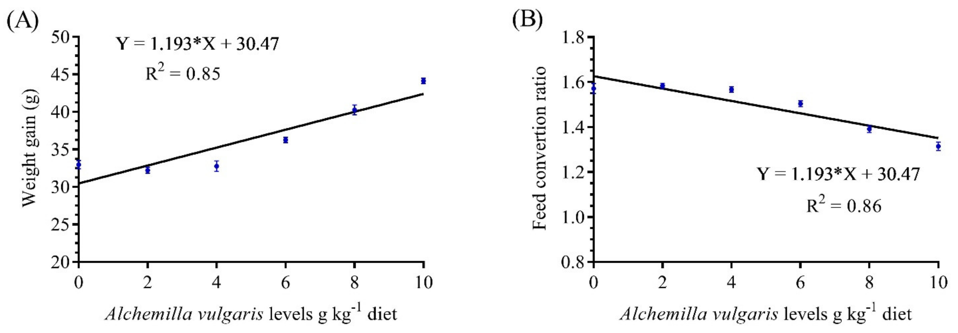
3.1. Growth Performance and Feed Utilization

The final weight (FW), weight gain (WG), weight gain % (WG %), and specific growth rate (%) (SGR) were meaningfully (p < 0.05) enhanced by supplementing AVP at 6–10 g/kg (Table 2). Feed intake was significantly increased with increasing AVP supplementation levels in the fish diet. Further, the condition factor (K) was markedly increased in the AVP8 and AVP10 groups compared to the other groups.
Fish fed AVP-supplemented rations expressed significantly better FCR values. Additionally, the lowest FCR values were achieved by those fish fed 10 g/kg, while the highest FCR values were obtained in AVP2 group. The maximum growth performance was obtained in the group fed 10 g/kg, while the lowest one was obtained in the control group without AVP supplementation (Table 2). The dose–response study showed a linear increasing trend of WG and FCR in response to the increasing dietary supplementation of AVP, with a strong correlation (R2 = 0.85) (Figure 1).

3.2. Hematological Indices

The effects of AVP supplementation in the diets of O. niloticus fish on hematological indices are presented in Table 3. The erythrogram revealed that RBCs, Hb, and PCV were significantly higher in the AVP6, AVP8, and AVP10 groups compared to the other groups. On the other hand, there were no significant differences between the treatments for MCV and MCH. Marked increases in the total WBCs, lymphocytes, neutrophils, eosinophils, and monocyte levels were observed as a result of increasing the concentration of AVP from 6 to 10 g/kg. Furthermore, fish treated with 10 g/kg had higher leukogram indices.

3.3. Hepatorenal Function Indicators

The effects of the dietary supplementation of AVP on Nile tilapia serum biochemical parameters are shown in Table 4. The serum total protein of fish was significantly influenced by the dietary treatments. The serum total protein and albumin were increased in a level-dependent manner with the increasing dietary supplementation of AVP. Fish fed 8 and 10 g/kg AVP experienced significant drops in ALT, AST, ALP, urea, and creatinine levels. Fish fed diets of 0-6 g AVP/kg displayed the highest levels of the same parameters.

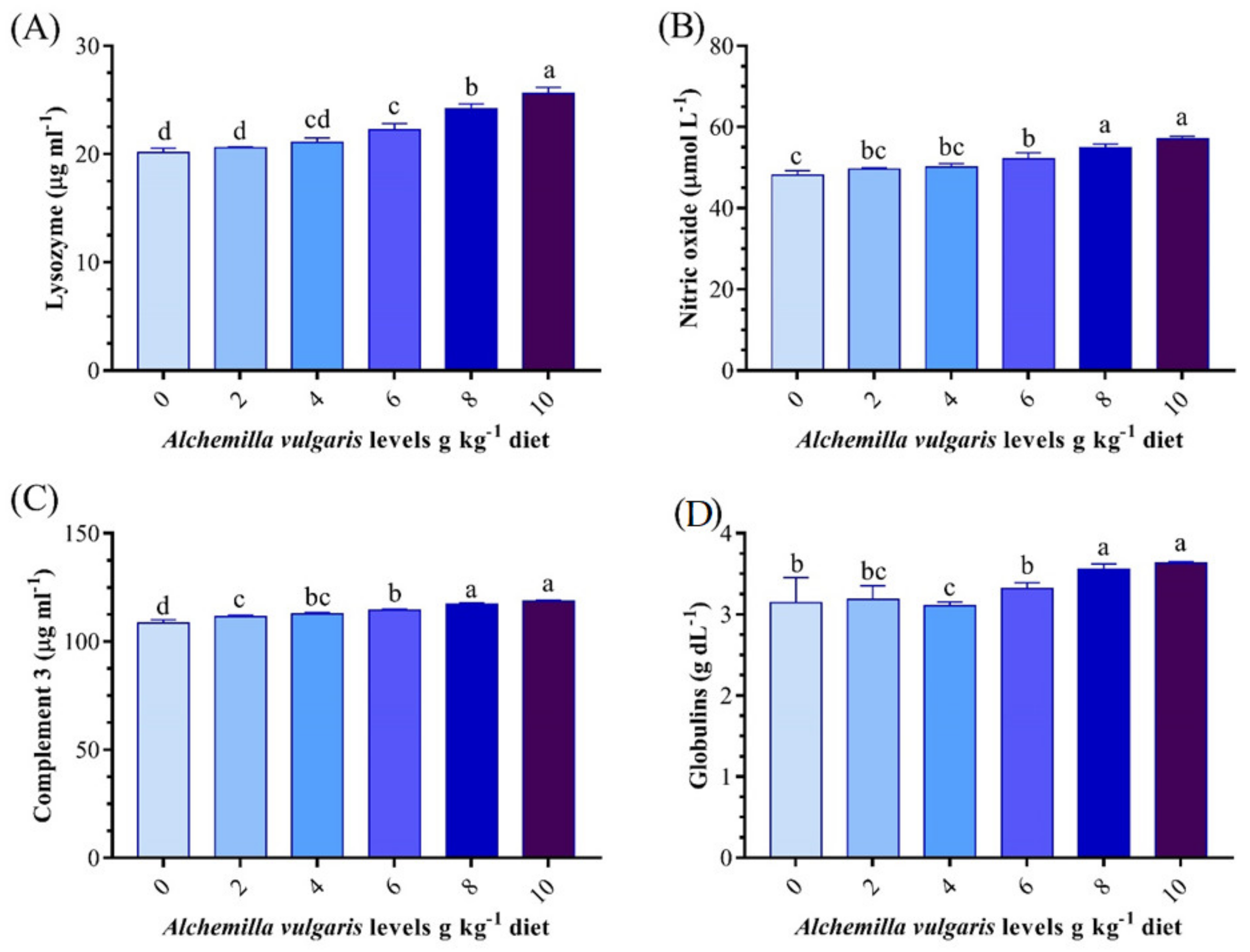
3.4. Innate Immune Response

The innate immunity parameters (lysozyme, C3, and NO) were significantly influenced by the experimental treatments (Figure 2). The lysozyme activity, C3, and NO levels were increased significantly with AVP doses over a 4 g/kg diet. The highest immune status was recorded in the group fed the 10 g AVP/kg diet. The globulin level was increased significantly in groups fed 8–10 g AVP/kg1 diets compared to the other treatments.

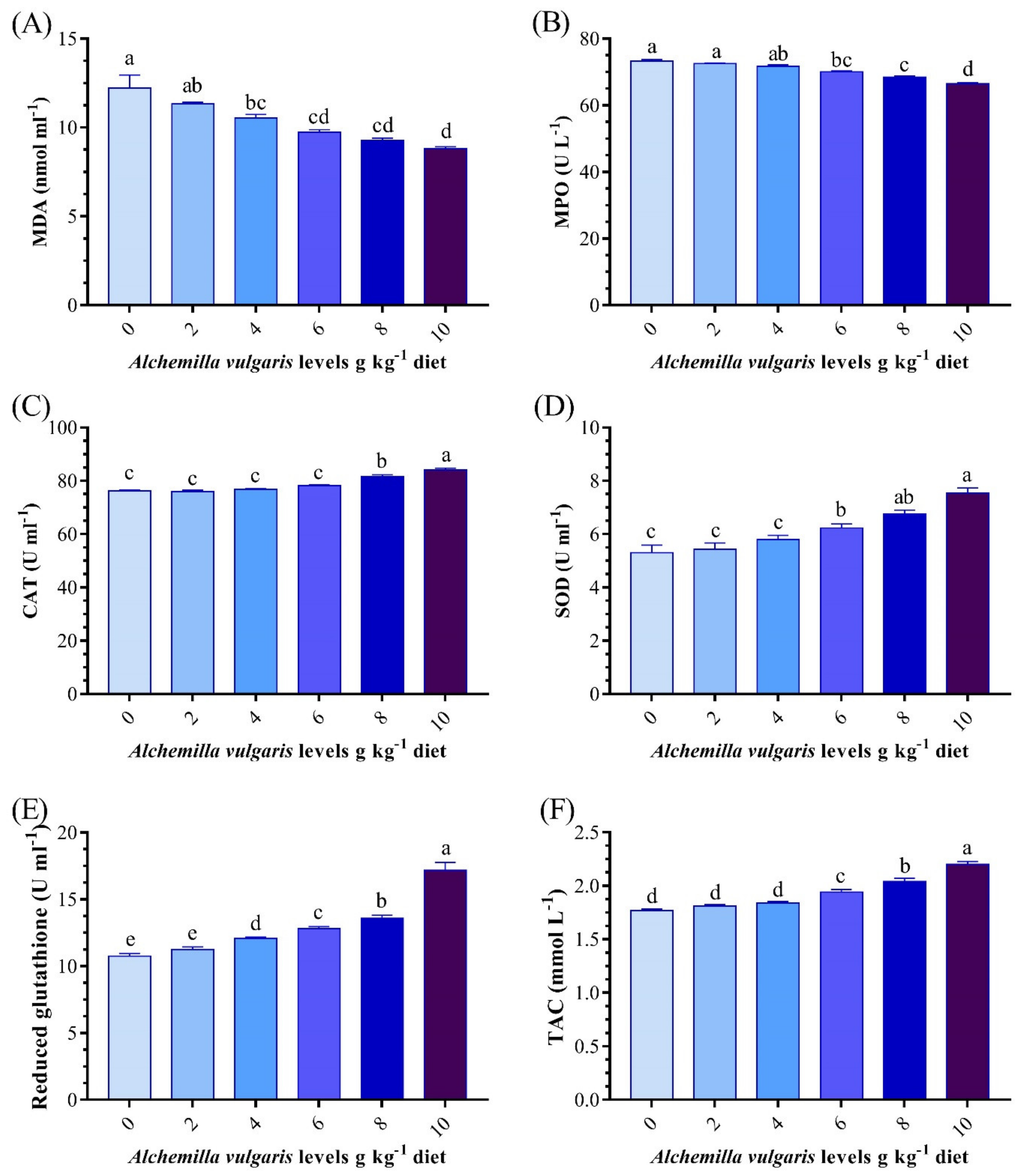
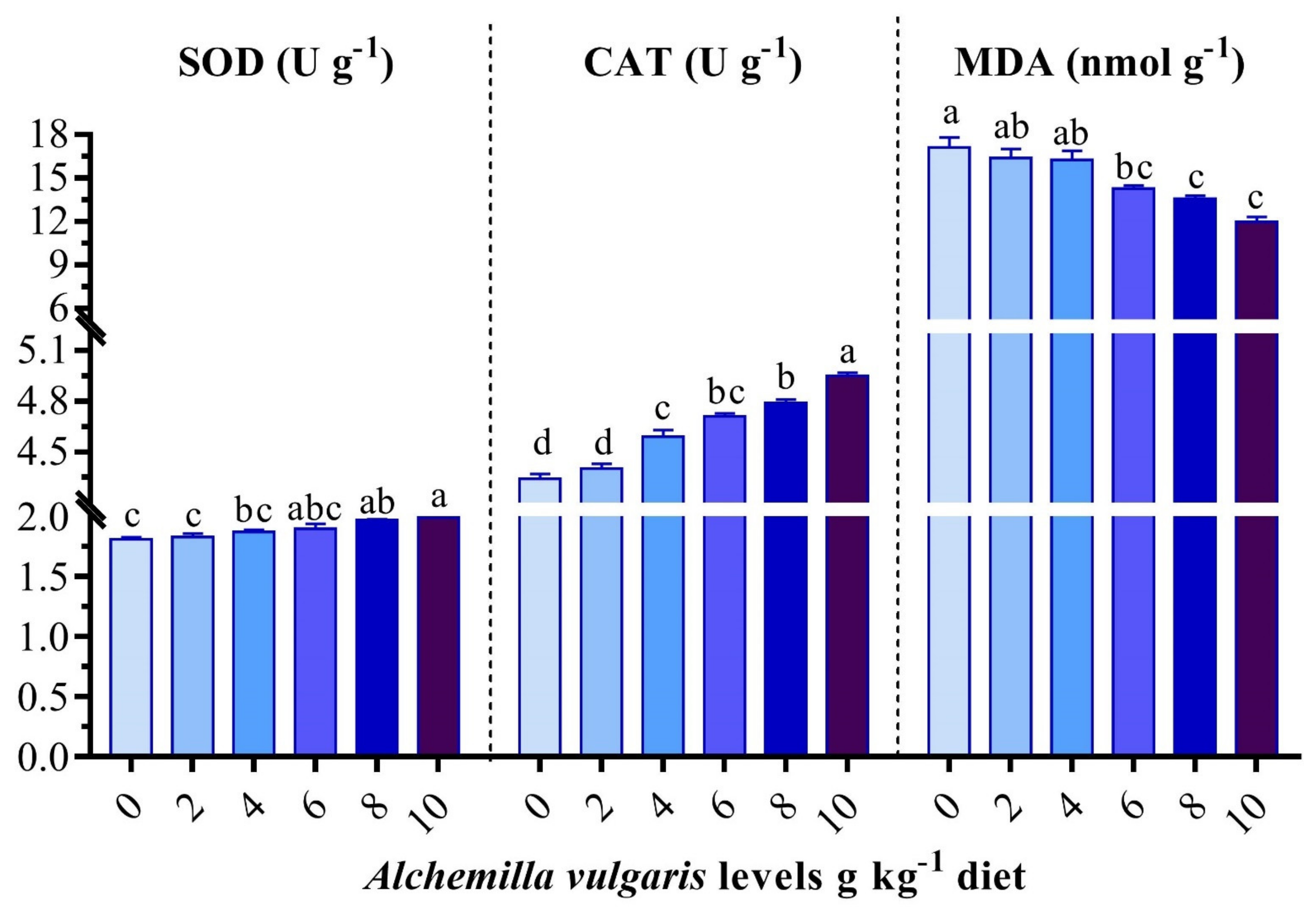
3.5. Antioxidant and Oxidative Stress Biomarkers

The SOD, CAT, and MPO activities and GSH, TAC, and MDA levels were significantly changed from the controls in the groups receiving the experimental diets (Figure 3). The groups of fish that received dietary AVP at rates of 8 and 10 g/kg showed pronounced increases (p < 0.05) in the serum SOD, CAT, GSH, and TAC activities (Figure 3). Nevertheless, the AVP-supplemented diets decreased stress, as conveyed by the gradual drop in MDA and MPO. The best results were obtained in the AVP10 group. Likewise, the hepatic CAT as well as SOD activities in AVP6, AVP8, and AVP10 were significantly higher than those of the other groups. Meanwhile, the opposite trend was observed for the MDA content (Figure 4).

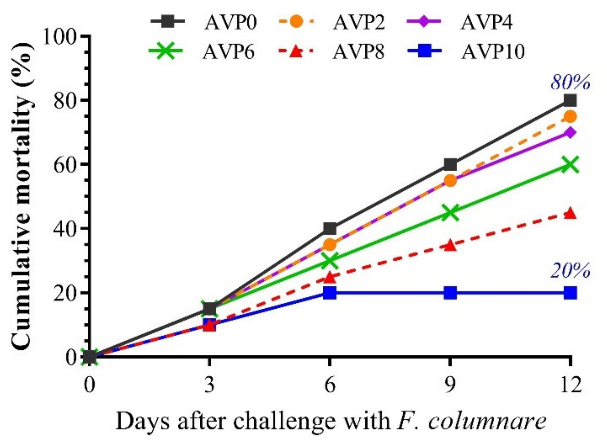
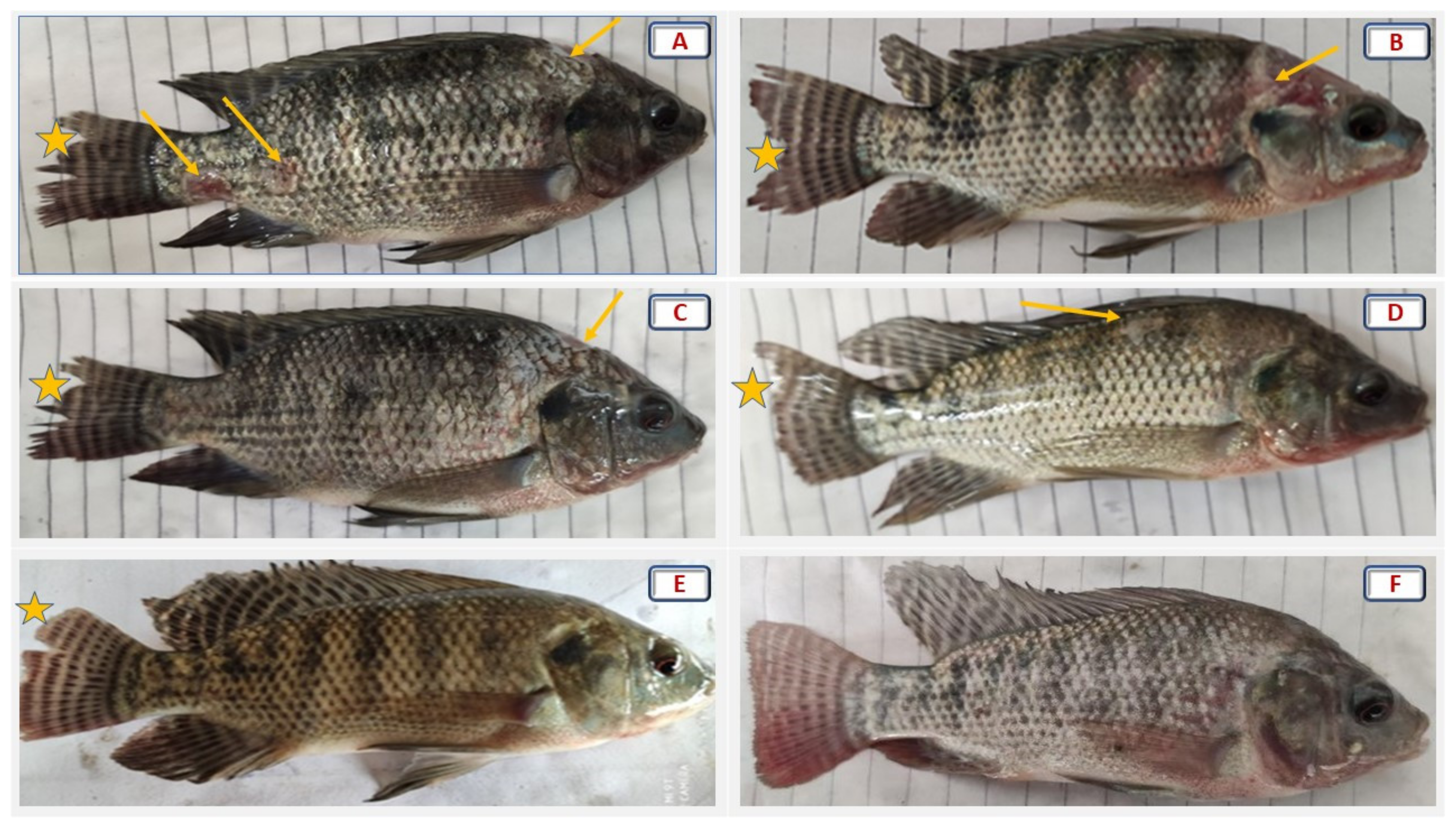
3.6. Fish Resistance against Challenge with Flavobacterium columnare

To further assess the impact of AVP supplementation on the efficiency of the immune system of O. niloticus, fish were challenged with F. columnare (Figure 5). The cumulative mortality percent in the AVP0 group reached 80% after 12 days of the challenge. Meanwhile, the dietary supplementation with AVP decreased the cumulative mortalities in a dose-dependent manner. The lowest cumulative mortality (20%) was recorded in the group that was fed a 10 g AVP/kg diet.
As shown in Figure 6, different degrees of fin rot, hemorrhage, and pale ulcers with hemorrhagic boundaries, especially on the upper head (saddleback lesion) were evident in the challenged groups fed 0–6 g AVP/kg. Meanwhile, the groups fed 8 and 10 g/kg revealed clear improvements in fish resistance to F. columnare infection. Notably, the AVP10 group showed the highest recovery and the lowest cumulative mortality percentage after the bacterial challenge.

4. Discussion

In the last decade, great changes have occurred in classic medicine, which have had a great influence on the studies of natural extracts as potential remedies for several animal and human diseases [48]. The Alchemilla vulgaris powder, as a promising source of feed supplement, contains high levels of polyphenolic components (395.65 mg/100 g) and flavonoid content (183.10 mg/100 g), including benzoic acid (1084.63 ppm), ellagic acid (614.16 ppm), catechol (580.54 ppm), catechin (566.53 ppm), salicylic acid (479.71 ppm), and protocatechuic acid (444.43 ppm) [16]. Accordingly, the present work was established to evaluate AVP as an available and cheap plant with significant nutritional importance to sustain aquaculture by improving fish performance and health and strengthening the resistance against F. columnare. There are no reports elucidating AVP usage in aquaculture. Accordingly, this study highlights the effect of AVP on growth performance, blood picture, immune-antioxidant-associated indices, hepatorenal functions, and F. columnare resistance in O. niloticus in response to different AVP-incorporated diets.
In aquaculture, medicinal plants and their extracts display a significant role in modulating growth performance [38]. Herein, the results reveal better growth performance and feed utilization in all AVP-supplemented groups, and the best results were recorded at higher doses of supplementation. Accordingly, the dietary supplementation of AVP improved the weight gain and FCR of heat-stressed broilers [49]. In addition, AVP at a dose of 3% improved quail production and the feed conversion ratio and mitigated the negative effects of high temperature [50]. This could be dominated by the potent influence of AVP on digestive activity, which thereby has a positive effect on growth and feed utilization. Our findings are supported by Samah et al. [51], who stated that AVP has a direct impact on digestive enzyme activities, which enhance digestion and consequently reflect a better growth rate. In addition, tannin, as the main active component of AVP, has a positive influence on fish growth performance. In the same line, the dietary Elephantopus scaber extract, a rich source of flavonoids and phenols, significantly improved the WG, SGR, and FCR of Nile tilapia at the level of 5 g/kg [52]. The dietary supplementation of condensed tannin (1 g/kg) improved growth performance and decreased the serum glucose concentration of Lateolabrax japonicus [53], which could be due to the regulation effect of tannin on the activity and gene expression of hepatic glycolysis and gluconeogenesis enzymes [53].
Meanwhile, the growth performance and intestinal histomorphology of L. japonicus fed low doses of tannin (lower than 400 mg/kg diet) were not affected [54], and using extremely high levels of tannin in the grass carp (Ctenopharyngodon idella) diet (over 20 g/kg diet) caused severe growth suppression and enteritis morbidity [55]. Therefore, the present findings revealed that dietary AVP up to 10 g/kg diet could provide the optimal tannin level for the growth performance and feed utilization of Nile tilapia.
An assessment of fish blood profile is a substantial tool to reflect the efficacy of plant extracts on fish physiological performance [56,57,58]. The present perspective demonstrated elevated levels of hematological parameters, particularly monocytes, reflecting a strong hematological response in association with the AVP dietary supplementation. Similarly, a recent study reported that monocyte levels were notably augmented by increasing the dose of AVP in broiler diets [49]. Furthermore, the MCV, MCH, and platelet distribution width increased significantly in quails fed 1% AVP compared to the control [59]. Such plant extracts could be used to treat anemic cases and stimulate hematopoiesis organs [57]. The phenols, among the other phytochemicals in AVP, could significantly stimulate the synthesis of blood cells. Accordingly, the leukocytes counts were increased significantly in common carp, Cyprinus carpio, fed 1.5, 4.5, or 9 g pistachio, Pistacia vera/kg diets, as a rich source of phenols, compared to the control [60].
In addition, the hepatorenal functions, as a center of metabolism and excretion processes in the fish body, could be also considered in evaluating any new feed supplement to reveal its safety and/or efficiency for animals. The present results showed that the administration of AVP modulated the levels of liver function enzymes (AST, ALP, and ALT) and kidney function biomarkers (urea and creatinine) and increased the total protein and albumin levels in the serum of O. niloticus compared to the control group. In the same line, the dietary supplementation of 100 ppm ethanol leaves extract or 2% A. vulgaris powder ameliorated the effect of CCl4 toxicity on liver and kidney functions and reduced the liver histopathological and biochemical changes in the intoxicated rats [16]. In addition, Ozbek et al. [61] found that the extracts of A. mollis had a hepatoprotective activity via modulating the level of aminotransferases in mouse serum. The improving effect of AVP on liver and kidney functions could be attributed to its phytochemical contents of phenolics and flavonoids that are well-known as strong antioxidants, which could protect the hepatocytes and glomerulus from oxidative damage [62,63]. Dietary flavonoids significantly decreased serum AST and ALT activities in the intoxicated C. Idella [64].
Alchemilla vulgaris is characterized by potent antioxidant activity due to its richness in phenolics, especially tannins, which are responsible for its antioxidant activity [59,65,66]. The current investigation implied a strong antioxidant response in fish fed AVP-enriched diets, as evidenced by the marked increase in the antioxidant biomarkers (CAT, SOD, TAC, and GSH). In addition, the MDA level and MPO activity were decreased with increasing AVP dietary supplementation. The abovementioned antioxidant effects of AVP could be attributed to its natural bioactive compounds, mostly phenolic compounds, including tannins, phenolcarboxylic acids, and flavonoids [67]. Whereas, Vlaisavljević et al. [66] quantified twenty-six phenolic compounds in AVP-enriched diets and detected its richness in quercetin, catechin, hexoside, apigenin, luteolin, gallic, and caffeic acids, which are proven to play major roles in protecting against oxidative stress. A. vulgaris dried leaves and different leaf extracts have high free radical scavenging activities in terms of DPPH [16,68]. In the same line, broilers fed AVP-supplemented diets experienced low serum MDA and suppressed lipid peroxidation in a dose-dependent manner [49].
Increasing the supplementation levels of flavonoids in the diet of snakehead fish (Channa argus) significantly enhanced the gene expression and activities of liver antioxidant enzymes (SOD, CAT, and glutathione peroxidase) and reduced the MDA levels in the liver [62]. MDA and protein carbonyl in the livers, spleens, and gills of chromium-intoxicated grass carp, Ctenopharyngodon idella, decreased significantly with dietary flavonoids of Allium mongolicum Regel [64]. In addition, natural phenolic compound (derived from mango peel) supplementation into the zebrafish diet significantly decreased muscle and increased CAT activity. However, glutathione peroxidase and SOD activities did not affect phenolic supplementation [69]. The dietary condensed tannin upregulated the hepatic mRNA levels of nuclear factor erythroid 2-related factor 2, heme oxygenase 1, CAT, and SOD in [53]. Several studies support our outcomes and elucidate that plant phytochemicals exhibit strong scavenging activity and are antioxidant system stimulators [22,70,71,72,73].
The non-specific immunity status in fish in response to a dietary supplement represents the first line of defense against all infections and assists the development of a specific immune response [74,75]. In the current study, the activity of lysozyme, nitric oxide, and complement 3 and the globulin level experienced higher values in AVP-incorporated diets, suggesting an immune-modulating impact of AVP and an increased tolerance of the bacterial infection. The promising influence of AVP on the fish immune system could be attributed to its richness in phyto-components such as flavonoids, phenolics, tannins, and ellagic acids [22,76]. Dietary flavonoids significantly increased lysozyme activity and the C3 level in the kidney and intestine in C. Idella [64]. Moreover, dietary phenolic extracts of licorice root increased the respiratory burst and lysozyme activities of Nile tilapia and improved fish bacterial resistance [77].
The challenge trial is a validating tool to investigate the fish’s immune-antioxidant function [69]. Fish that received the dietary intervention of AVP for 8 weeks before being challenged with F. columnaris exhibited remarkable improvements in the clinical signs, especially at higher supplemented doses. A lower mortality rate was also reported in AVP-enriched diets. This could be dominated by the effect of AVP on elevating monocytes, which are considered blood phagocytic cells that support the immune system by killing pathogenic bacterium and consequently enhance fish resistance, as reported by [78]. A. vulgaris showed dose-dependent antiviral and antibacterial properties [20,21]. Moreover, several feed additives revealed significant improvements in fish against F. columnaris infection [79,80].
In addition, the noticeable improvement in skin ulcers could be dominated by the impact of the phenolic constituents of AVP on curing ulcers, implying an anti-inflammatory effect. Our findings were supported by [81] who described that this plant is active against inflammation and is used for wound healing due to its potent and anti-inflammatory and antimicrobial impacts. The application of an ointment containing an Alchemilla vulgaris and Mimosa tenuiflora herbal mixture promoted the migration of keratinocytes, fibroblasts, and endothelial cells and the proliferation of macrophages and lymphatic vessels and induced rapid wound healing [82]. Based on the outcomes, the current study implied that the administration of AVP in O. niloticus diets enhanced growth, the hematological response, hepatorenal function, and immunity-antioxidant status. Moreover, it augmented the tolerance of fish against infection with F. columnaris.

5. Conclusions

Until now, the current study represents a pioneering trial in aquaculture to investigate the efficacy of increasing levels of dietary Alchemilla vulgaris powder (AVP) (0, 2, 4, 6, 8, and 10 g/kg diet) on growth, blood picture, immune-antioxidant response, and hepatorenal function and against bacterial infection with Flavobacterium columnare (F. columnare) in O. niloticus. The results revealed that dietary supplements with AVP promoted fish performance, hematological profile, immunity, antioxidant capacity, and liver and kidney functions in a dose-dependent manner. In addition, the AVP-incorporated diets strengthened the resistance against F. columnare by alleviating the clinical columnaris symptoms and reducing the mortality rate. The AVP dietary intervention is highly motivated as an environmentally friendly growth stimulator and immune-antioxidant promoter and for the treatment of anemic cases. It can also be utilized as an alternative antibacterial treatment to avoid the resistance of antibiotics, with less hazardous impacts to the environment. Further studies are needed to assess other AVP antimicrobial activities and to test the efficacy of AVP antimicrobial activities in in vitro conditions.

Author Contributions

Conceptualization, H.H.M., G.E.E. and W.E.-H.; methodology, H.H.M., G.E.E. and W.E.-H.; software, A.T.M., H.H.M., G.E.E., E.K.A. and W.E.-H.; validation, H.H.M., G.E.E., E.K.A. and W.E.-H.; formal analysis, A.T.M., H.H.M., G.E.E. and W.E.-H.; investigation, H.H.M., G.E.E. and W.E.-H.; resources, A.T.M., A.H.M.A., G.R. and H.S.G.; data curation, A.T.M., H.H.M., G.E.E. and W.E.-H.; writing—original draft preparation, A.T.M., H.H.M. and W.E.-H.; writing—review and editing, A.T.M.; visualization, A.T.M., H.H.M., G.E.E. and W.E.-H.; supervision, H.H.M., G.E.E., E.K.A. and W.E.-H. All authors have read and agreed to the published version of the manuscript.

Funding

This work was supported by the Deanship of Scientific Research, Vice Presidency for Graduate Studies and Scientific Research, King Faisal University, Saudi Arabia (Project No. GRANT554).

Institutional Review Board Statement

This study was conducted in accordance with the Declaration of Helsinki and was approved by the Ethics Committee of Animal Use in Research Committee (IACUC), Faculty of Veterinary Medicine, Zagazig University. The experiments were completed in compliance with the National Institutes of Health’s (NIH) Ethical Guidelines for the Use and Care of Laboratory Animals in Scientific Investigations.

Informed Consent Statement

Not applicable.

Data Availability Statement

The data that support the findings of this study are contained within the article.

Acknowledgments

The authors would like to acknowledge the Deanship of Scientific Research, Vice Presidency for Graduate Studies and Scientific Research, King Faisal University, Saudi Arabia (Project No. GRANT554).

Conflicts of Interest

No potential conflict of interest is reported by the authors.

References

  1. Tilami, S.K.; Sampels, S. Nutritional value of fish: Lipids, proteins, vitamins, and minerals. Rev. Fish. Sci. Aquac. 2018, 26, 243–253. [Google Scholar] [CrossRef]
  2. Mansour, A.T.; Ashour, M.; Alprol, A.E.; Alsaqufi, A.S. Aquatic Plants and Aquatic Animals in the Context of Sustainability: Cultivation Techniques, Integration, and Blue Revolution. Sustainability 2022, 14, 3257. [Google Scholar] [CrossRef]
  3. El-Sayed, A.-F.M. Tilapia Culture, 2nd ed.; Academic Press: Cambridge, MA, USA; Elsevier: Amsterdam, The Netherlands, 2019. [Google Scholar]
  4. Tartor, Y.; Taha, M.; Mahboub, H.; El Ghamery, M. Yeast species associated with diseased fish: Occurrence, identification, experimental challenges and antifungal susceptibility testing. Aquaculture 2018, 488, 134–144. [Google Scholar] [CrossRef]
  5. Rahman, A.N.A.; Mansour, D.A.; Abd El-Rahman, G.I.; Elseddawy, N.M.; Zaglool, A.W.; Khamis, T.; Mahmoud, S.F.; Mahboub, H.H. Imidacloprid toxicity in Clarias gariepinus: Protective role of dietary Hyphaene thebaica against biochemical and histopathological disruption, oxidative stress, immune genes expressions, and Aeromonas sobria infection. Aquaculture 2022, 555, 738170. [Google Scholar] [CrossRef]
  6. Mahboub, H.H.; Faggio, C.; Hendam, B.M.; Algharib, S.A.; Alkafafy, M.; Hashem, M.A.; Mahmoud, Y.K.; Khamis, T.; Abdel-Ghany, H.M.; Masoud, S.R. Immune-antioxidant trait, Aeromonas veronii resistance, growth, intestinal architecture, and splenic cytokines expression of Cyprinus carpio fed Prunus armeniaca kernel-enriched diets. Fish Shellfish Immunol. 2022, 124, 182–191. [Google Scholar] [CrossRef]
  7. Mahboub, H.H.; Elsheshtawy, H.M.; Sheraiba, N.I.; Fahmy, E.M.; Mohamed, E.A.; Abdelnaeim, N.S.; Mohamed, D.I.; Ismail, T.A.; Ahmed, S.A. Dietary black cumin (Nigella sativa) improved hemato-biochemical, oxidative stress, gene expression, and immunological response of Nile tilapia (Oreochromis niloticus) infected by Burkholderia cepacia. Aquacult. Rep. 2022, 22, 100943. [Google Scholar] [CrossRef]
  8. Mahboub, H.H.; Nada, H.S.; Abdel-Ghany, H.M.; Ghanem, R.; Ahmed Ismail, T.; Abdel Rahman, A.N. Detection, diagnosis, Koch’s postulate, hepatorenal and antioxidant indicators for some systemic pathogenic fungi invading the liver and kidneys of African catfish (Clarias gariepinus) in Egypt with a histopathological approach. Aquacult. Res. 2022, 53, 2670–2685. [Google Scholar] [CrossRef]
  9. El-Bouhy, Z.M.; Reda, R.M.; Mahboub, H.H.; Gomaa, F.N. Bioremediation effect of pomegranate peel on subchronic mercury immunotoxicity on African catfish (Clarias gariepinus). Environ. Sci. Pollut. Res. 2021, 28, 2219–2235. [Google Scholar] [CrossRef]
  10. Rashidian, G.; Mahboub, H.H.; Hoseinifar, S.H.; Ghafarifarsani, H.; Zare, M.; Punyatong, M.; Van Doan, H. Allium hirtifolium protects Cyprinus carpio against the detrimental responses mediated by foodborne zinc oxide nanoparticle. Aquaculture 2022, 555, 738252. [Google Scholar] [CrossRef]
  11. Rashidian, G.; Mahboub, H.H.; Fahim, A.; Hefny, A.A.; Prokić, M.D.; Rainis, S.; Boldaji, J.T.; Faggio, C. Mooseer (Allium hirtifolium) boosts growth, general health status, and resistance of rainbow trout (Oncorhynchus mykiss) against Streptococcus iniae infection. Fish Shellfish Immunol. 2022, 120, 360–368. [Google Scholar] [CrossRef]
  12. Chen, J.; Chen, D.; Li, J.; Liu, Y.; Gu, X.; Teng, X. Cadmium-induced oxidative stress and immunosuppression mediated mitochondrial apoptosis via JNK-FoxO3a-PUMA pathway in common carp (Cyprinus carpio L.) gills. Aquat. Toxicol. 2021, 233, 105775. [Google Scholar] [CrossRef]
  13. Sun, Q.; Liu, Y.; Teng, X.; Luan, P.; Teng, X.; Yin, X. Immunosuppression participated in complement activation-mediated inflammatory injury caused by 4-octylphenol via TLR7/IκBα/NF-κB pathway in common carp (Cyprinus carpio) gills. Aquat. Toxicol. 2022, 249, 106211. [Google Scholar] [CrossRef]
  14. Al-osaj, S.L. Cytotoxic Evaluation of Alchemilla Vulgaris Favonoids in Normal Blood Lymphocytes; Al-Nahrain University: Baghdad, Iraq, 2016. [Google Scholar]
  15. Duckstein, S.M.; Lotter, E.M.; Meyer, U.; Lindequist, U.; Stintzing, F.C. Phenolic constituents from Alchemilla vulgaris L. and Alchemilla mollis (Buser) Rothm. at different dates of harvest. Z. Für Nat. C 2012, 67, 529–540. [Google Scholar] [CrossRef] [Green Version]
  16. El-Hadidy, E.M.; Refat, O.G.; Halaby, M.S.; Elmetwaly, E.M.; Omar, A.A. Effect of Lion’s Foot (Alchemilla vulgaris) on Liver and Renal Functions in Rats Induced by CCl4. Food Nutr. Sci. 2018, 9, 46–63. [Google Scholar]
  17. Maier, M.; Oelbermann, A.-L.; Renner, M.; Weidner, E. Screening of European medicinal herbs on their tannin content-New potential tanning agents for the leather industry. Ind. Crops Prod. 2017, 99, 19–26. [Google Scholar] [CrossRef]
  18. Özbilgin, S.; Özbek, H.; Kirmizi, N.İ.; Öz, B.E.; Kurtul, E.; Özrenk, B.C.; Işcan, G.S.; Acikara, Ö.B. Evaluation of the antidiabetic activity of Alchemilla persica Rothm. In mice with diabetes induced by alloxan. Turk. J. Pharm. Sci. 2019, 16, 261. [Google Scholar] [CrossRef]
  19. Jarić, S.; Mačukanović-Jocić, M.; Djurdjević, L.; Mitrović, M.; Kostić, O.; Karadžić, B.; Pavlović, P. An ethnobotanical survey of traditionally used plants on Suva planina mountain (south-eastern Serbia). J. Ethnopharmacol. 2015, 175, 93–108. [Google Scholar] [CrossRef]
  20. Kuete, V. Potential of Cameroonian plants and derived products against microbial infections: A review. Planta Med. 2010, 76, 1479–1491. [Google Scholar] [CrossRef] [Green Version]
  21. Mazurkova, N.; Protsenko, M.; Filippova, E.; Kukushkina, T.; Vysochina, G.; Lobanova, I.; Mazurkov, O.Y.; Shishkina, L.; Agafonov, A. Investigation of the antiviral activity of experimental samples obtained from the grass and roots of Alchemilla vulgaris L. against vaccinia virus and ectromelia virus. Drug Dev. Regist. 2019, 8, 9–15. [Google Scholar] [CrossRef]
  22. Boroja, T.; Mihailović, V.; Katanić, J.; Pan, S.-P.; Nikles, S.; Imbimbo, P.; Monti, D.; Stanković, N.; Stanković, M.; Bauer, R. The biological activities of roots and aerial parts of Alchemilla vulgaris L. S. Afr. J. Bot. 2018, 116, 175–184. [Google Scholar] [CrossRef]
  23. Figueiredo, H.; Klesius, P.; Arias, C.; Evans, J.; Shoemaker, C.; Pereira, D., Jr.; Peixoto, M. Isolation and characterization of strains of Flavobacterium columnare from Brazil. J. Fish. Dis. 2005, 28, 199–204. [Google Scholar] [CrossRef]
  24. Dong, H.; Senapin, S.; LaFrentz, B.; Rodkhum, C. Virulence assay of rhizoid and non-rhizoid morphotypes of Flavobacterium columnare in red tilapia, Oreochromis sp., fry. J. Fish. Dis. 2016, 39, 649–655. [Google Scholar] [CrossRef]
  25. Groff, J.M.; Lapatra, S.E. Infectious diseases impacting the commercial culture of salmonids. J. Appl. Aquacult. 2000, 10, 17–90. [Google Scholar] [CrossRef]
  26. Elgendy, M.Y.; Abdelsalam, M.; Mohamed, S.A.; Ali, S.E. Molecular characterization, virulence profiling, antibiotic susceptibility, and scanning electron microscopy of Flavobacterium columnare isolates retrieved from Nile tilapia (Oreochromis niloticus). Aquacult. Int. 2022, 30, 845–862. [Google Scholar] [CrossRef]
  27. El-Houseiny, W.; Khalil, A.A.; Abd-Elhakim, Y.M.; Badr, H.A. The potential role of turmeric and black pepper powder diet supplements in reversing cadmium-induced growth retardation, ATP depletion, hepatorenal damage, and testicular toxicity in Clarias gariepinus. Aquaculture 2019, 510, 109–121. [Google Scholar] [CrossRef]
  28. Khalil, A.A.; Abd-Elhakim, Y.M.; Said, E.N.; Moselhy, A.A.; Abu-Elsaoud, A.M.; El-Houseiny, W. Milk thistle and co-enzyme Q10 fortified diets lessen the nickel chloride-induced neurotoxic and neurobehavioral impairments in Oreochromis niloticus via regulating the oxidative stress response, acetylcholinesterase activity, and brain nickel content. Aquaculture 2022, 553, 738102. [Google Scholar] [CrossRef]
  29. NRC. Nutrient Requirements of Fish and Shrimp; National Academies Press: Washington, DC, USA, 2011. [Google Scholar]
  30. AOAC. Official Methods of Analysis, 15th ed.; Association of Official Analysis of Chemist: Washington, DC, USA, 2000. [Google Scholar]
  31. Feldman, B.V.; Zinkl, J.G.; Jain, N.C.; Schalm, O.W. Schalm’s Veterinary Hematology, 5th ed.; Lippincott Williams & Wilkins: Philadelphia, PA, USA, 2000. [Google Scholar]
  32. Bain, B.J.; Bates, I.; Laffan, M.A. Dacie and Lewis Practical Haematology E-Book; Elsevier Health Sciences: Amsterdam, The Netherlands, 2016. [Google Scholar]
  33. Burtis, C.A.; Ashwood, E.R. Tietz Textbook of Clinical Chemistry; American Assosiation for Clinical Chemistry: Washington, DC, USA, 1994. [Google Scholar]
  34. Wenger, C.; Kaplan, A.; Rubaltelli, F.; Hammerman, C. Alkaline phosphatase. Clin. Chem. 1984, 62, 1094–1098. [Google Scholar]
  35. Murray, R.; Kaplan, A. Aspartate Aminotransferase; CV Mosby Company: Maryland Heights, MO, USA, 1984; pp. 1105–1108. [Google Scholar]
  36. Neely, W.; Phillipson, J. Automated enzymatic method for determining ammonia in plasma with 14 day stability. Clin. Chem. 1988, 34, 1868–1869. [Google Scholar] [CrossRef]
  37. Martinen, K. Improved micro-method for determination of serum bilirubin. Clin. Chim. Acta 1966, 13, 161–170. [Google Scholar] [CrossRef]
  38. Rashidian, G.; Shahin, K.; Elshopakey, G.E.; Mahboub, H.H.; Fahim, A.; Elabd, H.; Prokić, M.D.; Faggio, C. The Dietary Effects of Nutmeg (Myristica fragrans) Extract on Growth, Hematological Parameters, Immunity, Antioxidant Status, and Disease Resistance of Common Carp (Cyprinus carpio) against Aeromonas hydrophila. J. Mar. Sci. Eng. 2022, 10, 325. [Google Scholar] [CrossRef]
  39. Ellis, A.E. Lysozyme assays. Tech. Fish Immunol. 1990, 1, 101–103. [Google Scholar]
  40. Montgomery, H.; Dymock, J.F. The determination of nitrite in water. Analyst 1961, 86, 414–416. [Google Scholar]
  41. Aebi, H. Catalase in vitro. Methods Enzymology. 1984, 105, 121–126. [Google Scholar]
  42. Nishikimi, M.; Rao, N.A.; Yagi, K. The occurrence of superoxide anion in the reaction of reduced phenazine methosulfate and molecular oxygen. Biochem. Biophys. Res. Commun. 1972, 46, 849–854. [Google Scholar] [CrossRef]
  43. Beutler, E. Improved method for the determination of blood glutathione. J. Lab. Clin. Med. 1963, 61, 882–888. [Google Scholar]
  44. Uchiyama, M.; Mihara, M. Determination of malonaldehyde precursor in tissues by thiobarbituric acid test. Anal. Biochem. 1978, 86, 271–278. [Google Scholar] [CrossRef]
  45. Kumari, J.; Sahoo, P. Effects of cyclophosphamide on the immune system and disease resistance of Asian catfish Clarias batrachus. Fish Shellfish Immunol. 2005, 19, 307–316. [Google Scholar] [CrossRef]
  46. Griffin, B. A simple procedure for identification of Cytophaga columnaris. J. Aquat. Anim. Health 1992, 4, 63–66. [Google Scholar] [CrossRef]
  47. Farmer, B.D. Improved Methods for the Isolation and Characterization of Flavobacterium Columnare; Louisiana State University: Baton Rouge, LA, USA, 2004. [Google Scholar]
  48. García-Beltrán, J.M.; Mansour, A.T.; Alsaqufi, A.S.; Ali, H.M.; Esteban, M.Á. Effects of aqueous and ethanolic leaf extracts from drumstick tree (Moringa oleifera) on gilthead seabream (Sparus aurata L.) leucocytes, and their cytotoxic, antitumor, bactericidal and antioxidant activities. Fish Shellfish Immunol. 2020, 106, 44–55. [Google Scholar] [CrossRef]
  49. Köseman, A.; Akdemir, F.; Üremiş, N.; Şeker, İ.; Türköz, Y. Effects of Alchemilla vulgaris on growth performance, carcass characteristics and some biochemical parameters of heat stressed broilers. J. Hell. Vet. Med. Soc. 2020, 71, 2491–2498. [Google Scholar] [CrossRef]
  50. Akdemir, F.; Köseman, A.; Şeker, I. Alchemilla vulgaris effects on egg production and quality expressed by heatstressed quail during the late laying period. S. Afr. J. Anim. Sci. 2019, 49, 857–868. [Google Scholar] [CrossRef]
  51. Samah, S.; Abdullah, K.; Ream, N. Phytochemical screening of Alchemilla vulgaris, Sophora Japonica, Crataegus azarolus, and their inhibitory activity on lipase and α-amylase. Int. J. Appl. Sci. Res. 2018, 6, 1–21. [Google Scholar]
  52. Van Doan, H.; Hoseinifar, S.H.; Sringarm, K.; Jaturasitha, S.; Khamlor, T.; Dawood, M.A.; Esteban, M.Á.; Soltani, M.; Musthafa, M.S. Effects of elephant’s foot (Elephantopus scaber) extract on growth performance, immune response, and disease resistance of nile tilapia (Oreochromis niloticus) fingerlings. Fish Shellfish Immunol. 2019, 93, 328–335. [Google Scholar] [CrossRef]
  53. Peng, K.; Wang, G.; Zhao, H.; Wang, Y.; Mo, W.; Wu, H.; Huang, Y. Effect of high level of carbohydrate and supplementation of condensed tannins on growth performance, serum metabolites, antioxidant and immune response, and hepatic glycometabolism gene expression of Lateolabrax japonicus. Aquacult. Rep. 2020, 18, 100515. [Google Scholar] [CrossRef]
  54. Peng, K.; Zhao, H.; Wang, G.; Chen, B.; Mo, W.; Huang, Y. Effect of condensed tannins on growth performance, intestinal immune capacity and bacterial microbiomes of Lateolabrax japonicus. Aquacult. Res. 2021, 52, 5321–5331. [Google Scholar] [CrossRef]
  55. Li, M.; Feng, L.; Jiang, W.-D.; Wu, P.; Liu, Y.; Jiang, J.; Kuang, S.-Y.; Tang, L.; Zhou, X.-Q. Condensed tannins decreased the growth performance and impaired intestinal immune function in on-growing grass carp (Ctenopharyngodon idella). Br. J. Nutr. 2020, 123, 737–755. [Google Scholar] [CrossRef]
  56. El-Houseiny, W.; Abd El-Hakim, Y.M.; Metwally, M.M.; Ghfar, S.S.A.; Khalil, A.A. The single or combined Silybum marianum and co-enzyme Q10 role in alleviating fluoride-induced impaired growth, immune suppression, oxidative stress, histological alterations, and reduced resistance to Aeromonas sobria in African catfish (Clarias gariepinus). Aquaculture 2022, 548, 737693. [Google Scholar]
  57. Mansour, A.T.; Hamed, H.S.; El-Beltagi, H.S.; Mohamed, W.F. Modulatory Effect of Papaya Extract against Chlorpyrifos-Induced Oxidative Stress, Immune Suppression, Endocrine Disruption, and DNA Damage in Female Clarias gariepinus. Int. J. Environ. Res. Public 2022, 19, 4640. [Google Scholar] [CrossRef]
  58. Sallam, A.E.; Mansour, A.T.; Srour, T.M.; Goda, A.M.A. Effects of different carotenoid supplementation sources with or without sodium taurocholate on growth, feed utilization, carotenoid content and antioxidant status in fry of the European seabass, Dicentrarchus Labrax. Aquacult. Res. 2017, 48, 3848–3858. [Google Scholar] [CrossRef]
  59. Köseman, A.; Akdemir, F.; Üremiş, N.; Şeker, İ.; Türköz, Y. Effects of Alchemilla vulgaris on haematology and antioxidant status of heatstressed quails during the late laying period. J. Hell. Vet. Med. Soc. 2021, 72, 3463–3472. [Google Scholar] [CrossRef]
  60. Motamedi-Tehrani, J.; Ebrahimi-Dorcheh, E.; Malekpouri, P.; Goli, S. Liver alteration and hematological and serum biochemical responses of common carp, Cyprinus carpio Linnaeus, 1758, following long-term feeding of pistachio (Pistacia vera) green hull extract as a source of natural phenol. J. Appl. Ichthyol. 2016, 32, 906–912. [Google Scholar] [CrossRef]
  61. Ozbek, H.; Acikara, O.B.; Keskin, I.; Kirmizi, N.I.; Ozbilgin, S.; Oz, B.E.; Kurtul, E.; Ozrenk, B.C.; Tekin, M.; Saltan, G. Evaluation of hepatoprotective and antidiabetic activity of Alchemilla mollis. Biomed. Pharmacother. 2017, 86, 172–176. [Google Scholar] [CrossRef]
  62. Li, M.; Zhu, X.; Tian, J.; Liu, M.; Wang, G. Dietary flavonoids from Allium mongolicum Regel promotes growth, improves immune, antioxidant status, immune-related signaling molecules and disease resistance in juvenile northern snakehead fish (Channa argus). Aquaculture 2019, 501, 473–481. [Google Scholar] [CrossRef]
  63. Emeish, W.F.; Al-Amgad, Z.; Ahmed, H. Antioxidant, immunostimulant and renal protective activities of tri-herbal combination in African Sharptooth Catfish, Clarias gariepinus. J. Vet. Med. Res. 2018, 25, 213–229. [Google Scholar] [CrossRef]
  64. Zhao, L.; Yuan, B.-D.; Zhao, J.-L.; Jiang, N.; Zhang, A.-Z.; Wang, G.-Q.; Li, M.-Y. Amelioration of hexavalent chromium-induced bioaccumulation, oxidative stress, tight junction proteins and immune-related signaling factors by Allium mongolicum Regel flavonoids in Ctenopharyngodon idella. Fish Shellfish Immunol. 2020, 106, 993–1003. [Google Scholar] [CrossRef]
  65. Shilova, I.; Suslov, N.; Samylina, I.; Baeva, V.; Lazareva, N.; Mazin, E. Neuroprotective properties of common lady’s mantle infusion. Pharm. Chem. J. 2020, 53, 1059–1062. [Google Scholar] [CrossRef]
  66. Vlaisavljević, S.; Jelača, S.; Zengin, G.; Mimica-Dukić, N.; Berežni, S.; Miljić, M.; Stevanović, Z.D. Alchemilla vulgaris agg. (Lady’s mantle) from central Balkan: Antioxidant, anticancer and enzyme inhibition properties. RSC Adv. 2019, 9, 37474–37483. [Google Scholar] [CrossRef] [Green Version]
  67. Tadić, V.M.; Krgović, N.; Žugić, A. Lady’s mantle (Alchemilla vulgaris L., Rosaceae): A review of traditional uses, phytochemical profile, and biological properties. Lek. Sirovine 2020, 40, 66–74. [Google Scholar] [CrossRef]
  68. Jain, S.; Yadav, A.; Gothalwal, R. Assessment of total phenolic, flavonoid content and in vitro antioxidant properties of Alchemillia vulgaris (lady’s mantle). J. Adv. Sci. Res. 2021, 12, 205–209. [Google Scholar] [CrossRef]
  69. Lizárraga-Velázquez, C.E.; Hernández, C.; González-Aguilar, G.A.; Heredia, J.B. Effect of dietary intake of phenolic compounds from mango peel extract on growth, lipid peroxidation and antioxidant enzyme activities in zebrafish (Danio rerio). Lat. Am. J. Aquat. Res. 2019, 47, 602–611. [Google Scholar] [CrossRef] [Green Version]
  70. Mansour, A.T.; Espinosa, C.; García-Beltrán, J.M.; Miao, L.; Francisco, D.C.C.; Alsaqufi, A.S.; Esteban, M.Á. Dietary supplementation of drumstick tree, Moringa oleifera, improves mucosal immune response in skin and gills of seabream, Sparus aurata, and attenuates the effect of hydrogen peroxide exposure. Fish Physiol. Bioch. 2020, 46, 981–996. [Google Scholar] [CrossRef] [PubMed]
  71. Mansour, A.T.; El-Feky, M.M.; El-Beltagi, H.S.; Sallam, A.E. Synergism of Dietary Co-Supplementation with Lutein and Bile Salts Improved the Growth Performance, Carotenoid Content, Antioxidant Capacity, Lipid Metabolism, and Lipase Activity of the Marbled Spinefoot Rabbitfish, Siganus rivulatus. Animals 2020, 10, 1643. [Google Scholar] [CrossRef] [PubMed]
  72. Mansour, A.T.; Miao, L.; Espinosa, C.; García-Beltrán, J.M.; Francisco, D.C.C.; Esteban, M.Á. Effects of dietary inclusion of Moringa oleifera leaves on growth and some systemic and mucosal immune parameters of seabream. Fish Physiol. Bioch. 2018, 44, 1223–1240. [Google Scholar] [CrossRef] [PubMed]
  73. Kiselova, Y.; Ivanova, D.; Chervenkov, T.; Gerova, D.; Galunska, B.; Yankova, T. Correlation between the in vitro antioxidant activity and polyphenol content of aqueous extracts from Bulgarian herbs. Phytother. Res. 2006, 4, 961–965. [Google Scholar] [CrossRef]
  74. Ahmed, S.A.; Nada, H.S.; Elsheshtawy, H.M.; Ibrahim, S.M.; Fahmy, E.M.; Khedr, M.H.; Moustafa, S.M.; Ismail, T.A.; Gesriha, S.; Assayed, M.E. Comparative antitoxic potency of honey and natamycin-supplemented diets against aflatoxicosis and their influences on growth, serum biochemistry, immunohistochemistry, and residual deposition in Nile tilapia (Oreochromis niloticus). Aquaculture 2022, 551, 737934. [Google Scholar] [CrossRef]
  75. Mansour, A.T.; Esteban, M.Á. Effects of carbon sources and plant protein levels in a biofloc system on growth performance, and the immune and antioxidant status of Nile tilapia (Oreochromis niloticus). Fish Shellfish Immunol. 2017, 64, 202–209. [Google Scholar] [CrossRef]
  76. Kostadinović, L.; Popović, S.; Pelić, D.L.; Čabarkapa, I.; Đuragić, O.; Lević, J. Medicinal plants as natural alternative to coccidial synthetic drugs in broiler chicken production. J. Agron. 2019, 5, 325–334. [Google Scholar]
  77. El Mesallamy, A.M.; El-Marakby, H.I.; Souleman, A.M.; Abd El-Naby, F.S. Evaluation of phenolic extract of licorice roots in diets of Nile tilapia (Oreochromis niloticus). Egypt. Pharm. J. 2015, 14, 117. [Google Scholar]
  78. Secombes, C.; Wang, T. The Innate and Adaptive Immune System of Fish. In Infectious Disease in Aquaculture; Elsevier: Amsterdam, The Netherlands, 2012; pp. 3–68. [Google Scholar]
  79. Tie, H.-M.; Feng, L.; Jiang, W.-D.; Wu, P.; Liu, Y.; Jiang, J.; Kuang, S.-Y.; Tang, L.; Zhou, X.-Q. Dietary exogenous supplementation of nucleotides strengthens the disease resistance, antioxidant capacity and immunity in the gill of on-growing grass carp (Ctenopharyngodon idella) following a challenge with Flavobacterium columnare. Aquaculture 2021, 540, 736729. [Google Scholar] [CrossRef]
  80. Harikrishnan, R.; Devi, G.; Van Doan, H.; Balasundaram, C.; Esteban, M.Á.; Abdel-Tawwab, M. Impact of grape pomace flour (GPF) on immunity and immune-antioxidant-anti-inflammatory genes expression in Labeo rohita against Flavobacterium columnaris. Fish Shellfish Immunol. 2021, 111, 69–82. [Google Scholar] [CrossRef]
  81. Tasić, S. Ethnobotany in SEE-WB countries; traditional uses of indigenous plants. Lek. Sirovine 2012, 32, 71–81. [Google Scholar]
  82. Choi, J.; Park, Y.-G.; Yun, M.-S.; Seol, J.-W. Effect of herbal mixture composed of Alchemilla vulgaris and Mimosa on wound healing process. Biomed. Pharmacother. 2018, 106, 326–332. [Google Scholar] [CrossRef]
Figure 1. Fit linear regression models of weight gain (A) and feed conversion ratio (B) in response to increasing dietary supplementation of Alchemilla vulgaris (g/kg).
Antioxidants 11 01494 g001
Figure 2. Effect of Alchemilla vulgaris dietary supplementation on innate immune parameters of O. niloticus for 60 days. (A) lysozyme, (B) nitric oxide, (C) complement 3, (D) globulin. Columns bearing different letters are significantly different at p < 0.05.
Antioxidants 11 01494 g002
Figure 3. Effect of Alchemilla vulgaris dietary supplementation on antioxidant biomarkers in the serum of O. niloticus. (A) Malondialdehyde, (B) myeloperoxidase, (C) catalase, (D) superoxide dismutase, (E) reduced glutathione, (F) total antioxidant capacity. Columns bearing different letters are significantly different at p < 0.05.
Antioxidants 11 01494 g003
Figure 4. Effect of Alchemilla vulgaris dietary supplementation on antioxidant biomarkers in the liver homogenate of O. niloticus. SOD: superoxide dismutase, CAT: catalase, MDA: malondialdehyde. Columns bearing different letters are significantly different at p < 0.05.
Antioxidants 11 01494 g004
Figure 5. Effect of increasing dietary levels of Alchemilla vulgaris on cumulative mortality percent of Oreochromis niloticus challenged with pathogenic Flavobacterium columnare for 12 days.
Antioxidants 11 01494 g005
Figure 6. Effect of increasing dietary levels of Alchemilla vulgaris on columnaris clinical symptoms of Oreochromis niloticus challenged with pathogenic Flavobacterium columnare for 12 days. Stars identify fin rots, and arrows identify pale ulcers with hemorrhagic boundaries, especially on the upper head (saddleback lesion) and caudal peduncle. (A) 0 g AVP/kg diet, (B) 2 g AVP/kg diet, (C) 4 g AVP/kg diet, (D) 6 g AVP/kg diet, (E) 8 g AVP/kg diet, (F) 10 g AVP/kg diet.
Antioxidants 11 01494 g006
Table 1. Ingredients and proximate chemical analysis of the experimental diets (g/kg).
Ingredients (g/kg)Dietary Alchemilla vulgaris Levels (g/kg Diet)
0246810
Fishmeal110110110110110110
Corn flour330330330330330330
Soybean meal 44%290290290290290290
Corn gluten meal 60%120120120120120120
Wheat bran807876747270
Soybean oil202020202020
Fish oil202020202020
Alchemilla vulgaris powder (AVP)0246810
Vitamin premix 1151515151515
Mineral premix 2151515151515
Total100010001000100010001000
Chemical analysis (g/kg)
Crude protein (n × 6.25)308.8309.5309.7310.0310.3310.6
Crude lipids74.975.175.776.578.479.1
Crude fiber52.353.453.854.154.554.9
Ash52.253.754.556.957.958.4
Nitrogen-free extract 3511.8508.3506.3502.5498.9497
Gross energy (kcal/kg) 4455645474546453945444545
1 Vitamin premix (per kg of premix): vitamin A, 8,000,000 IU; vitamin E, 7000 mg; vitamin D3, 2,000,000 IU; vitamin K3, 1500 mg; biotin, 50 mg; folic acid, 700 mg; nicotinic acid, 20,000 mg; pantothenic acid, 7000 mg; vitamin B1, 700 mg; vitamin B2, 3500 mg; vitamin B6, 1000 mg; vitamin B12, 7 mg. 2 Mineral premix (per kg of premix): zinc sulfate, 4.0 g; iron sulfate, 20 g; manganese sulfate, 5.3 g; copper sulfate, 2.7 g; calcium iodine, 0.34 g; sodium selenite, 70 mg; cobalt sulfate, 70 mg; and CaHPO4·2H2O up to 1 kg. 3 Calculated by difference (100 − protein% + lipids% + ash% + crude fiber %). 4 Gross energy (GE) was calculated as 5.65, 9.45, and 4.11 kcal/g for protein, lipid, and NFE, respectively (NRC, 1993).
Table 2. Effect of Alchemilla vulgaris dietary supplementation on growth performance and feed utilization of O. niloticus.
ItemsDietary Alchemilla vulgaris Levels (g/kg Diet)
0246810
Final body weight (g)55.63 d ± 0.4655.60 d ± 0.5655.50 d ± 0.5559.16 c ± 0.4463.70 b ± 0.7067.0 a ± 0.57
Weight gain (g)32.96 d ± 0.3132.20 d ± 0.2332.76 d ± 40.036.26 c ± 0.2040.26 b ± 0.3744.13 a ± 0.20
Daily weight gain (g)0.54 d ± 0.0050.53 d ± 0.0030.54 d ± 0.0060.60 c ± 0.0030.67 b ± 0.0060.73 a ± 0.002
Specific growth rate (%)0.65 c ± 0.0180.62 c ± 0.0070.64 c ± 0.0170.68 bc ± 0.0140.72 ab ± 0.0030.77 a ± 0.015
Feed intake (g)52.00 c ± 0.5751.00 c ± 0.5751.33 c ± 0.4454.50 b ± 0.2856.00 ab ± 0.2858.00 a ± 0.57
Feed conversion ratio 1.57 a ± 0.0121.58 a ± 0.0061.56 a ± 0.0071.50 b ± 0.0071.39 c ± 0.0081.31 d ± 0.010
Condition factor (K)2.02 c ± 0.0082.04 c ± 0.0172.05 c ± 0.0202.065 c ± 0.0102.22 b ± 0.0042.38 a ± 0.026
Values are presented as means ± SE, and n = 3. The means within the same row carrying different superscripts letters are significant at p < 0.05.
Table 3. Effect of Alchemilla vulgaris dietary supplementation on hematological indices of O. niloticus.
ItemsDietary Alchemilla vulgaris Levels (g/kg Diet)
0246810
RBCs (106/mm3)3.78 d ± 0.0083.81 cd ± 0.0083.84 c ± 0.0083.89 b ± 0.0083.93 ab ± 0.0053.96 a ± 0.017
Hb (gm/dL)11.20 d ± 0.0511.30 cd ± 0.0211.34 cd ± 0.0111.44 bc ± 0.0211.52 b ± 0.0111.80 a ± 0.04
PCV (%)33.60 d ± 0.1733.92 cd ± 0.0834.03 cd ± 0.0434.33 bc ± 0.0734.57 b ± 0.0435.42 a ± 0.14
MCV(fl)88.73 ab ± 0.6688.87 ab ± 0.1388.46 ab ± 0.1088.10 b ± 0.0587.96 b ± 0.0289.44 a ± 0.06
MCH (%)29.57 ab ± 0.2229.62 ab ± 0.0429.48 ab ± 0.0329.36 b ± 0.0129.32 b ± 0.00729.81 a ± 0.02
WBCs (103/mm3)5.25 d ± 0.025.28 d ± 0.015.32 cd ± 0.015.37 bc ± 0.0085.42 b ± 0.015.51 a ± 0.02
Lymphocytes (103/mm3)2.90 c ± 0.0052.90 c ± 0.0082.90 c ± 0.0052.92 bc ± 0.0052.94 b ± 0.0082.99 a ± 0.011
Neutrophils (103/mm3)1.38 c ± 0.0061.40 bc ± 0.0051.41 bc ± 0.0081.43 ab ± 0.0121.43 ab ± 0.0051.46 a ± 0.008
Eosinophils (103/mm3)0.33 b ± 0.0050.33 b ± 0.0050.35 ab ± 0.0080.38 a ± 0.0050.38 a ± 0.0050.37 a ± 0.005
Monocytes (103/mm3)0.64 b ± 0.0050.65 b ± 0.0050.65 b ± 0.0110.64 b ± 0.0050.67 ab ± 0.0050.69 a ± 0.005
Values are presented as means ± SE, and n = 10. The means within the same row carrying different superscripts letters are significant at p < 0.05.
Table 4. Effect of Alchemilla vulgaris dietary supplementation on serum biochemical parameters of O. niloticus.
ItemsDietary Alchemilla vulgaris Levels (g/kg Diet)
0246810
Total proteins (g/dL)5.46 d ± 0.106.10 c ± 0.0696.13 bc ± 0.046.44 b ± 0.0866.81 a ± 0.0606.94 a ± 0.026
Albumin (g/dL)2.31 c ± 0.1012.92 b ± 0.1363.03 ab ± 0.023.12 ab ± 0.063.26 ab ± 0.023.31 a ± 0.014
ALT (U/L)13.24 a ± 0.4313.26 a ± 0.4812.45 ab ± 0.29 11.96 ab ± 0.1211.73 b ± 0.0611.58 b ± 0.04
AST (U/L)27.80 a ± 0.3227.70 a ± 0.3227.02 ab ± 0.0726.85 ab ± 0.1326.71 b ± 0.1226.15 b ± 0.09
ALP (IU/L)24.30 a ± 0.1524.21 a ± 0.0624.08 a ± 0.0623.90 ab ± 0.0923.60 b ± 0.0823.09 c ± 0.06
Urea (mg/dL)2.76 a ± 0.0372.76 a ± 0.0232.68 a ± 0.022.59 a ± 0.032.18 b ± 0.0442.14 b ± 0.049
Creatinine (mg/dL)0.436 a ± 0.010.44 a ± 0.0050.41 ab ± 0.010.356 bc ± 0.010.323 c ± 0.010.246 d ± 0.01
Values are presented as means ± SE, and n = 10. The means within the same row carrying different superscripts letters are significant at p < 0.05.
Publisher’s Note: MDPI stays neutral with regard to jurisdictional claims in published maps and institutional affiliations.

